distal respiratory tract viral infections in young ...distal respiratory tract viral infections in...
TRANSCRIPT

Distal respiratory tract viral infections inyoung children trigger a marked increasein alveolar mast cells
Cecilia K. Andersson1,2, Medya Shikhagaie2, Michiko Mori2, Amal Al-Garawi3,Jennifer L. Reed4, Alison A. Humbles5, Robert Welliver6, Thais Mauad7,Leif Bjermer1, Manel Jordana3 and Jonas S. Erjefält2
Affiliations: 1Dept of Respiratory Medicine and Allergology, Lund University, Lund, Sweden. 2Unit of AirwayInflammation, Lund University, Lund, Sweden. 3McMaster Immunology Research Centre, McMaster University,Hamilton, ON, Canada. 4Laboratory of Plasma Derivatives, Center for Biologics Evaluation and Research, Foodand Drug Administration, Rockville, MD, USA. 5Dept of Respiratory, Inflammation, and Autoimmunity,MedImmune LLC, Gaithersburg, MD, USA. 6Dept of Pediatrics, University of Oklahoma University HealthSciences Center, Oklahoma City, OK, USA. 7Dept of Pathology, São Paulo University, São Paulo, Brazil.
Correspondence: Cecilia K. Andersson, Unit of Airway Inflammation, Respiratory Medicine and Allergology,Dept of Clinical Sciences, BMC D12, Lund University, SE-22184, Lund, Sweden.E-mail: [email protected]
ABSTRACT Viral infections predispose to the development of childhood asthma, a disease associatedwith increased lung mast cells (MCs). This study investigated whether viral lower respiratory tractinfections (LRTIs) can already evoke a MC response during childhood.
Lung tissue from young children who died following LRTIs were processed for immunohistochemicalidentification of MCs. Children who died from nonrespiratory causes served as controls. MCs wereexamined in relation to sensitisation in infant mice exposed to allergen during influenza A infection.
Increased numbers of MCs were observed in the alveolar parenchyma of children infected with LRTIs(median (range) 12.5 (0–78) MCs per mm2) compared to controls (0.63 (0–4) MCs per mm2, p=0.0005).The alveolar MC expansion was associated with a higher proportion of CD34+ tryptase+ progenitors(controls: 0% (0–1%); LRTIs: 0.9% (0–3%) CD34+ MCs (p=0.01)) and an increased expression of thevascular cell adhesion molecule (VCAM)-1 (controls: 0.2 (0.07–0.3); LRTIs: 0.3 (0.02–2) VCAM-1 permm2 (p=0.04)). Similarly, infant mice infected with H1N1 alone or together with house dust mite (HDM)developed an increase in alveolar MCs (saline: 0.4 (0.3–0.5); HDM: 0.6 (0.4–0.9); H1N1: 1.4 (0.4–2.0);HDM+H1N1: 2.2 (1.2–4.4) MCs per mm2 (p<0.0001)). Alveolar MCs continued to increase and remainedsignificantly higher into adulthood when exposed to H1N1+HDM (day 36: 2.2 (1.2–4.4); day 57: 4.6(1.6–15) MCs per mm2 (p=0.01)) but not when infected with H1N1 alone.
Our data demonstrate that distal viral infections in young children evoke a rapid accumulation ofalveolar MCs. Apart from revealing a novel immune response to distal infections, our data may haveimportant implications for the link between viral infections during early childhood and subsequent asthmadevelopment.
@ERSpublicationsViral infections in children evokes a rapid recruitment and accumulation of mast cells in thealveolar parenchyma http://ow.ly/i9eN30meNM7
Cite this article as: Andersson CK, Shikhagaie M, Mori M, et al. Distal respiratory tract viralinfections in young children trigger a marked increase in alveolar mast cells. ERJ Open Res 2018; 4:00038-2018 [https://doi.org/10.1183/23120541.00038-2018].
The content of this work is copyright of the authors or their employers. Design and branding are copyright ©ERS 2018.This version is distributed under the terms of the Creative Commons Attribution Non-Commercial Licence 4.0.
This article has supplementary material available from openres.ersjournals.com
Received: March 07 2018 | Accepted after revision: Oct 05 2018
https://doi.org/10.1183/23120541.00038-2018 ERJ Open Res 2018; 4: 00038-2018
ORIGINAL ARTICLERESPIRATORY INFECTIONS
IntroductionLower respiratory tract infections (LRTIs) are associated with a substantial disease burden in youngchildren. Influenza A, respiratory syncytial virus (RSV) and adenovirus (ADV) are major causes of LRTIsincluding bronchiolitis, croup and pneumonia in infants [1]. Studies on the frequency of viral infections indeveloped countries have shown that infants and preschool children experience 6–10 viral infectionsannually [2]. The risk of developing asthma is increased with frequent viral-induced bronchiolitic episodesin combination with allergic sensitisation to airborne allergens [3, 4]. Albeit that there is strong evidencefrom longitudinal studies, it is not known how distal viral infections during early childhood are connectedto the development of asthma [5, 6].
Previous studies have revealed a surprisingly high density of alveolar mast cells (MCs) in healthy adultlungs [7]. The presence of large numbers of MCs at mucosal surfaces such as the lung suggests that thesecells may play a crucial role in the innate immune defence against pathogens, toxins and tissue damage[8]. Indeed, studies in animal models have shown increased mortality after infections in MC-deficientmice [9, 10]. In addition, increased numbers of bronchiolar MCs have been reported in an animal modelof viral bronchiolitis and this increase was associated with hyperresponsiveness to methacholine [11].However, almost nothing is known regarding the MC responses to viral infection in human lungs,especially in the alveolar parenchyma, where both human and animal data are lacking.
Although asthma has classically been considered a disease of the central airways, new data suggest thatboth bronchioles and the alveolar parenchyma are subjected to inflammation [12–18]. Interestingly,among the most marked alveolar changes in asthma is an expanded and activated pool of alveolar MCs[18, 19]. Whether a similar MC response occurs during LRTIs as well as the potential link between LRTIsand distal MC responses in asthma, remains unknown.
Here, we have studied the recruitment and heterogeneity of MCs in a unique lung tissue material fromchildren who suffered from fatal LRTIs. Furthermore, the connection to allergic sensitisation and thedevelopment of an asthmatic phenotype was studied in a mouse model of influenza A infection and housedust mite (HDM) exposure in infant mice.
Material and methodsModel of influenza A infection and allergic inflammation in infant mice15-day pregnant (female) BALB/c mice were purchased from Charles River Laboratories (Ottawa, ON,Canada), housed under specific pathogen-free conditions, and maintained on a 12-h light–dark cycle withfood and water ad libitum. Upon birth, mothers were housed with their litters in light-protected cagesuntil completion of the study (or weaning at 4 weeks of age). 8-day-old mice were infected with influenzaA and, 7 days later, exposed to HDM 5 days a week for a total of 3 weeks. To investigate whether changespersisted, the protocol was recapitulated and mice were allowed to rest for a period of 3 weeks. Animalswere monitored for signs of illness twice daily for a period of 10 days following infection. To evaluate thetotal number of MCs, 3-μm sections were prepared and the sections were stained with a 1% solution oftoluidine blue. Thereafter, MCs per section were counted and related to tissue area using ImageScope(Aperio, Vista, CA, USA). All experiments described in this study were approved by the AnimalResearch Ethics Board of McMaster University (Hamilton, ON, Canada). For details, see thesupplementary material.
SubjectsLung samples from post mortem lung tissue from young children (median age (range): 4 (5–16) months)who died from fatal LRTIs was collected (RSV, n=5; ADV, n=10; and influenza A, n=6). Control lungtissues were obtained from 10 age-matched children (3 (0.4–24) months) who died from nonrespiratorycauses. All tissue samples were collected with informed parental consent. This study was approved by theinstitutional medical ethical committee, Sao Paulo, Brazil, and at the Institute of Legal Medicine of theMedical School of the Hanover University, Hanover, Germany. For details, see the supplementary material.
ImmunohistochemistryDouble immunohistochemical staining of MCTC and MCTA double staining protocol was used for simultaneous visualisation of MCTC and MCT cells [7, 18, 20].The staining was performed on paraformaldehyde-fixed, paraffin-embedded tissue by an automatedimmunohistochemistry robot. For details, see table 1 and the supplementary material.
https://doi.org/10.1183/23120541.00038-2018 2
RESPIRATORY INFECTIONS | C.K. ANDERSSON ET AL.

Immunohistochemical identification of VCAM-1 and CD34, ITGB1/A4 and Ki-67+ MCsImmunofluorescence double staining was used to visualise expression of CD34, integrin β1 (ITGB1),integrin α4 (ITGA4) or Ki-67 on tryptase-positive MCs. A single staining protocol was used forvisualisation of VCAM-1. For details, see table 1 and the supplementary material.
Tissue analysisQuantification of density of MC subtypesHigh-resolution digital images of sections double-stained for MCTC and MCT were generated through a20× microscope lens by an automated digital slide-scanning robot (Scanscope CS; Aperio). All MCs werequantified manually on blinded sections and related to the perimeter of the basal membrane (smallairways and pulmonary vessels) or tissue area (alveolar parenchyma) using Visiomorph (Visiopharm,Hoersholm, Denmark) [18]. For details, see the supplementary material.
Quantification of VCAM-1 and CD34, ITGB1, ITGA4 and Ki-67+ MCsThe immunoreactivity per square millimetre of tissue area of VCAM-1 was calculated using Visiomorph.Tryptase-positive MCs and expression of CD34, ITGB1, ITGA4 and Ki-67 was analysed and theproportion (%) of positive MCs was calculated. For details, see the supplementary material.
Statistical analysisData were analysed using Kruskal–Wallis test with Dunn’s multiple comparisons test for comparisonamong three groups or more and Mann–Whitney rank sum test was used for comparison between twogroups using GraphPad Prism version 5 (GraphPad Software, Inc., La Jolla, CA, USA). The Spearman test(two-tailed) was used to study the correlations. Correlation analysis was made within pooled LRTI-infectedchildren. Results were considered significant at p⩽0.05.
ResultsIncreased number of MCs in a model of allergic inflammation during the course of an influenza Ainfection in infant miceTo address whether LRTIs directly contribute to increased pulmonary abundance of MCs in the context ofallergic asthma, we investigated MCs in mice infected post-natally (8 days old) with influenza A (H1N1)and or challenged with HDM (figure 1a and b). The number of MCs, defined as purple toluidineblue-positive granulated cells [21], were quantified in the alveolar parenchyma of control mice and miceinfected with influenza A, exposed to HDM, or both infected with influenza A and exposed to HDM, andnormalised for tissue area. Only low numbers of MCs were detected in the lungs of saline- orHDM-treated infant mice (figure 1c, f and g). At day 36, MCs were increased in infant mice infected withinfluenza A and, even more so, in mice infected with influenza A with concomitant HDM exposure
TABLE 1 Antibodies used for immunohistochemistry
Antibody Species Dilution Clone Origin Secondary antibody
Tryptase Mouse 1:12000 G3 Chemicon, (Temecula,CA, USA)
EnVision G|2 DoublestainSystem directly labelled with Alexa
Fluor 488Chymase Mouse 1:100 CC1 Novocastra (Newcastle
upon Tyne, UK)EnVision G|2 Doublestain System
(Dako, Glostrup, Denmark)CD34 Mouse 1:300 QBEnd/
10Dako, (Glostrup,
Denmark)Invitrogen (Eugene, OR, USA)
VCAM-1 Mouse 1:100 1.4C3 Invitrogen MolecularProbes (Eugene, OR,
USA)
EnVision Detection system, Dako
Ki-67 Rabbit 1:100 Biocare (Pacheco, CA,USA)
Invitrogen (Eugene, OR, USA)
ITGA4 Rabbit 1:50 LifeSpan Biosciences(Seattle, WA, USA)
Invitrogen (Eugene, OR, USA)
ITGB1 Rabbit 1:50 LifeSpan Biosciences(Seattle, WA, USA)
Invitrogen (Eugene, OR, USA)
Heat-induced antigen retrieval was performed in PT link (Dako, Glostrup, Denmark) with EnVision FLEXTarget retrieval solution high pH (Dako). VCAM: vascular cell adhesion molecule; ITGA4: integrin α4;ITGB1: integrin β1.
https://doi.org/10.1183/23120541.00038-2018 3
RESPIRATORY INFECTIONS | C.K. ANDERSSON ET AL.
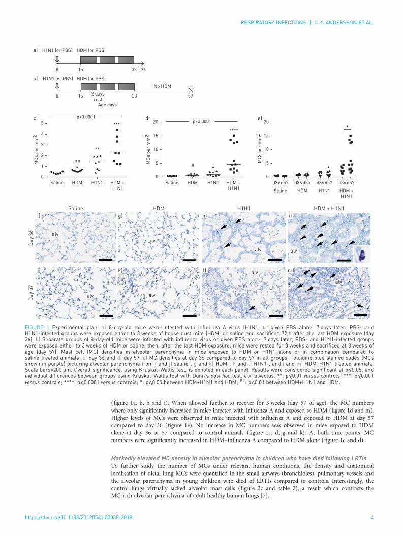
(figure 1a, b, h and i). When allowed further to recover for 3 weeks (day 57 of age), the MC numberswhere only significantly increased in mice infected with influenza A and exposed to HDM (figure 1d and m).Higher levels of MCs were observed in mice infected with influenza A and exposed to HDM at day 57compared to day 36 (figure 1e). No increase in MC numbers was observed in mice exposed to HDMalone at day 36 or 57 compared to control animals (figure 1c, d, g and k). At both time points, MCnumbers were significantly increased in HDM+influenza A compared to HDM alone (figure 1c and d).
Markedly elevated MC density in alveolar parenchyma in children who have died following LRTIsTo further study the number of MCs under relevant human conditions, the density and anatomicallocalisation of distal lung MCs were quantified in the small airways (bronchioles), pulmonary vessels andthe alveolar parenchyma in young children who died of LRTIs compared to controls. Interestingly, thecontrol lungs virtually lacked alveolar mast cells (figure 2c and table 2), a result which contrasts theMC-rich alveolar parenchyma of adult healthy human lungs [7].
H1N1 (or PBS) HDM (or PBS)
H1N1 (or PBS)
8 15 33 36
8
5
MC
s p
er
mm
2 4
3
2
1
0
15 33
Age days
2 days
rest57
HDM (or PBS)
No HDM
HDM
HDM
p<0.0001
Saline
Saline
H1N1 HDM +
H1N1
HDMSaline H1N1
H1H1
HDM +
H1N1 HDMSaline
d36 d57 d36
Da
y 3
6D
ay
57
d57 d36 d57 d36 d57
H1N1 HDM +
H1N1
HDM + H1N1
a)
b)
c)
f)
alv
alv
g)
alv
h)
alv
i)
alv
j)
alv
k) alvl)
alv
m)
d)***
**
## MC
s p
er
mm
2 15
10
5
0
20 p<0.0001
****
#
MC
s p
er
mm
2
20
15
10
5
0
e)
*
FIGURE 1 Experimental plan. a) 8-day-old mice were infected with influenza A virus (H1N1) or given PBS alone. 7 days later, PBS- andH1N1-infected groups were exposed either to 3 weeks of house dust mite (HDM) or saline and sacrificed 72 h after the last HDM exposure (day36). b) Separate groups of 8-day-old mice were infected with influenza virus or given PBS alone. 7 days later, PBS- and H1N1-infected groupswere exposed either to 3 weeks of HDM or saline, then, after the last HDM exposure, mice were rested for 3 weeks and sacrificed at 8 weeks ofage (day 57). Mast cell (MC) densities in alveolar parenchyma in mice exposed to HDM or H1N1 alone or in combination compared tosaline-treated animals: c) day 36 and d) day 57. e) MC densities at day 36 compared to day 57 in all groups. Toluidine blue stained slides (MCsshown in purple) picturing alveolar parenchyma from f and j) saline-, g and k) HDM-, h and l) H1N1-, and i and m) HDM+H1N1-treated animals.Scale bars=200 µm. Overall significance, using Kruskal–Wallis test, is denoted in each panel. Results were considered significant at p⩽0.05, andindividual differences between groups using Kruskal–Wallis test with Dunn’s post hoc test. alv: alveolus. **: p⩽0.01 versus controls; ***: p⩽0.001versus controls; ****: p⩽0.0001 versus controls; #: p⩽0.05 between HDM+H1N1 and HDM; ##: p⩽0.01 between HDM+H1N1 and HDM.
https://doi.org/10.1183/23120541.00038-2018 4
RESPIRATORY INFECTIONS | C.K. ANDERSSON ET AL.

The density of MCs in small airways or the pulmonary vessels was not statistically altered in LRTI-affectedlungs compared to controls (figure 2a, b, d, f, j and k). However, there was a marked and significantincrease in MC numbers in the alveolar parenchyma in LRTIs (figure 2c, e, g–i and m). The increase was
d)
SA
V
alv
alv
alv
alv
lu
ep
ep
ep
lu lu
alv SA
g)
j) k)
h)
l) m)
i)
e) f)
MC
s p
er
mm
pe
rim
ete
r
a)60 NS
40
20
0Controls LRTIs
b)
NS
MC
s p
er
mm
pe
rim
ete
r
60
40
20
0Controls LRTIs
ControlsInfluenza AADVRSV
c)
***100
80
60
40
20
0
Controls LRTIs
MC
s p
er
mm
2
FIGURE 2 Mast cell (MC) densities in a) small airways, b) pulmonary vessels and c) alveolar parenchyma. Double immunohistochemical stainingfor tryptase (red) and chymase (brown). Representative photograph of d) a small airway and pulmonary vessel in a control lung from a child ande) alveolar parenchyma. f) Small airway in a lung from a child infected with influenza A. Alveolar parenchyma from young children who have diedfollowing g) respiratory syncytial virus (RSV), h) adenovirus (ADV) and i) influenza infections. Immunohistochemical staining for influenza A (red) inthe epithelium with j and k) fluorescence and l) 3,3′-diaminobenzidine. k) Tryptase-positive MCs are shown in the subepithelium (green). m) RSVpositivity in the alveolar parenchyma. Scale bars: d, f and g) 500 µm; e) 200 µm; h and k) 100 µm; i and m) 70 µm; j and l) 50 µm. Results wereconsidered significant at p⩽0.05. NS: not significant; LRTI: lower respiratory tract infection; SA: small airway; V: vessel; alv: alveolus; lu: lumen;ep: epithelium. ***: p⩽0.001 using Mann–Whitney rank sum test, in comparison to controls.
https://doi.org/10.1183/23120541.00038-2018 5
RESPIRATORY INFECTIONS | C.K. ANDERSSON ET AL.
significant for both MCT and MCTC, subpopulations and present in RSV, ADV and influenza A infection.For details regarding different LRTIs and MC populations, see table 2.
Increased frequency of MC progenitors in the alveolar parenchyma after viral infectionsThe median time with symptoms before death in the infected children were only 4 days (range 1–8 days).Since the LRTI-induced expansion of the alveolar MC pool thus took place rapidly after infection, wepostulated that this would be linked with elevated levels of parenchymal MC progenitors, defined here astryptase+ MCs double positive for the hematopoietic marker CD34. In small airways and pulmonaryvessels, the frequency of MC progenitors was not significantly altered in children with LRTIs (figure 3aand b). The alveolar parenchyma, the only pulmonary structure where an accumulation of MCs wasobserved, contained a significantly higher frequency of MC progenitors (p<0.01) (figure 3a). Next, it wasconfirmed that mast cells in LRTI-affected lungs did express adhesion molecules integrin α4β1 that isthought to be critical for MC progenitor homing into tissue [22]. Although constituting a smallproportion of all MCs, tryptase+ integrin α4β1+ MCs, phenotypically characterised as small and round,were clearly observed in the alveolar parenchyma of children who died following LRTIs (figure 3f–k).
MCs do not proliferate locally in the lung of young children with LRTIWe next analysed the proliferation of MCs, defined as tryptase+ cells double positive for the proliferationmarker Ki67. Scattered Ki-67+ cells were found throughout the lungs (figure 3c and d). Ki-67immunoreactivity was not detected within tryptase+ cells, suggesting that the increased MC density wasnot dependent on local proliferation. In agreement with this, proliferative MCs were not found in adultlungs from patients with chronic obstructive pulmonary disease (COPD) or cystic fibrosis (data notshown). However, ∼2% of the total MCs (tryptase+) were identified as proliferating by Ki-67immunostaining in bronchial lymph nodes from patients with very severe COPD (data not shown).
Our group has previously shown that FcεRI+ alveolar MCs and IgE bound to alveolar MCs is a hallmarkof atopic asthma in adults [18, 19]. Immunofluorescence double staining revealed that lungs from bothnoninfected and LRTI-affected young children lacked MCs with surface-bound IgE (figure 3e). However,IgE+ CD138+ plasma cells were found in close proximity to MCs in the lungs of children with LRTI(inset in figure 3e).
LRTI-affected lungs express elevated levels of the MC migration-facilitating molecule VCAM-1Tissue recruitment and homing of MCs occurs through a process of endothelial VCAM-1 binding of MCintegrins [23]. To investigate whether MC accumulation among children with fatal LRTI is associated withan induction of endothelial VCAM-1, the immunoreactivity of VCAM-1 was measured and related tosequential slides where the MCs were quantified. No difference in VCAM-1 expression was observed in
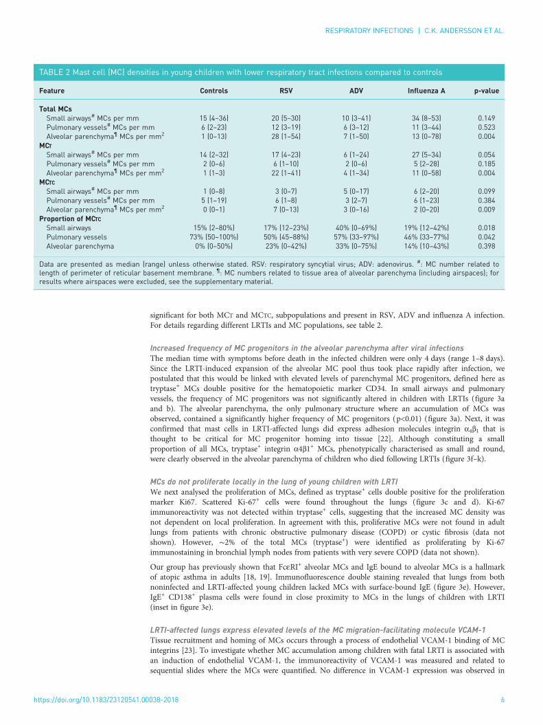
TABLE 2 Mast cell (MC) densities in young children with lower respiratory tract infections compared to controls
Feature Controls RSV ADV Influenza A p-value
Total MCsSmall airways# MCs per mm 15 (4–36) 20 (5–30) 10 (3–41) 34 (8–53) 0.149Pulmonary vessels# MCs per mm 6 (2–23) 12 (3–19) 6 (3–12) 11 (3–44) 0.523Alveolar parenchyma¶ MCs per mm2 1 (0–13) 28 (1–54) 7 (1–50) 13 (0–78) 0.004
MCTSmall airways# MCs per mm 14 (2–32) 17 (4–23) 6 (1–24) 27 (5–34) 0.054Pulmonary vessels# MCs per mm 2 (0–6) 6 (1–10) 2 (0–6) 5 (2–28) 0.185Alveolar parenchyma¶ MCs per mm2 1 (1–3) 22 (1–41) 4 (1–34) 11 (0–58) 0.004
MCTCSmall airways# MCs per mm 1 (0–8) 3 (0–7) 5 (0–17) 6 (2–20) 0.099Pulmonary vessels# MCs per mm 5 (1–19) 6 (1–8) 3 (2–7) 6 (1–23) 0.384Alveolar parenchyma¶ MCs per mm2 0 (0–1) 7 (0–13) 3 (0–16) 2 (0–20) 0.009
Proportion of MCTCSmall airways 15% (2–80%) 17% (12–23%) 40% (0–69%) 19% (12–42%) 0.018Pulmonary vessels 73% (50–100%) 50% (45–88%) 57% (33–97%) 46% (33–77%) 0.042Alveolar parenchyma 0% (0–50%) 23% (0–42%) 33% (0–75%) 14% (10–43%) 0.398
Data are presented as median (range) unless otherwise stated. RSV: respiratory syncytial virus; ADV: adenovirus. #: MC number related tolength of perimeter of reticular basement membrane. ¶: MC numbers related to tissue area of alveolar parenchyma (including airspaces); forresults where airspaces were excluded, see the supplementary material.
https://doi.org/10.1183/23120541.00038-2018 6
RESPIRATORY INFECTIONS | C.K. ANDERSSON ET AL.

small airway walls (figure 4a). In pulmonary vessels, the VCAM-1 density decreased compared to controls(figure 4b). In contrast, an increased VCAM-1 expression was observed in the alveolar parenchyma inLRTI-affected lungs compared to controls (figure 4c).
DiscussionThe results from our study demonstrate that a viral infection affecting the peripheral lung evokes a rapidaccumulation of MCs in the alveolar parenchyma in both young children and mice. Apart from revealinga novel type of immune response in LRTI-affected human lungs, our experimental data suggest thatvirus-induced alveolar establishment of MCs in combination with allergen exposure in the distal lung mayevoke a long-lasting immune alteration since MCs are long lived and are likely to remain in the tissue wellafter the infection has been cleared. Speculatively, LRTI-induced alveolar MC expansion early in life mighttherefore affect sensitisation and susceptibility towards allergens and asthma development later in life.
CD
34
+ M
Cs %
a)1.5
**
1.0
0.5
0
Controls LRTIsControls
Small
airways
Pulmonary
vessels
Alveolar
parenchyma
LRTIs Controls LRTIs
b)
c)*
Tryptase
Ki-67
Tryptase
Ki-67
Tryptase
ITGB1
Tryptase
ITGA4
Tryptase
IgE
**
*
* **
d)
e)
f)
g)
h)
i)
j)
k)
lu
lu
FIGURE 3 a) Proportion of CD34+ mast cells (MCs) in small airways, pulmonary vessels and alveolar parenchyma. b) CD34+ (red) MCs (green) in apulmonary vessel wall. Scale bar=30 µm). c and d) Influenza A-infected lung stained for tryptase (green) and proliferation marker Ki-67 (red).Arrows show Ki-67+ cells and * denotes tryptase+ cells. e) Alveolar parenchyma stained for tryptase (green) and IgE (red). Inset showsneighbouring IgE+ cell and MC. f–h) Alveolar parenchyma stained for tryptase (green) and integrin β1 (ITGB1) (red). i–k) Tryptase (green) integrinα4 (ITGA4) (red). Integrin
+ MCs are denoted with arrows. Results were considered significant at p⩽0.05. lu: lumen. **: p⩽0.01 using Mann–Whitneyrank sum test, in comparison to controls.
https://doi.org/10.1183/23120541.00038-2018 7
RESPIRATORY INFECTIONS | C.K. ANDERSSON ET AL.

Many studies have suggested a connection between early-in-life viral infections, especially in the lowerairways, and subsequent development of asthma. Children who are affected by viral LRTI duringsensitisation by aeroallergens have up to a 30-fold increased risk of developing persistent or severe asthma,particularly when the events take place during postnatal lung growth [24]. The risk is affected by bothintensity and frequency of the viral infection as well as allergen-specific IgE levels [25, 26]. The potentiallink between LRTI and asthma is further underscored by the fact that robust alveolar MC changes seem tobe common features of adult asthma [18, 19, 27].
A main finding in our study is that a resident alveolar MC population was virtually absent in thenondiseased lungs from young children. This is in stark contrast with the healthy human adult lung wherethe highest density of MCs is found in the alveolar parenchyma [7]. Likewise, laboratory mice do not haveresident alveolar MCs. In rodents, it is believed that such a population does not emerge with age unlesssubjected to a viral insult to the lung [28]. Indeed, our study shows that a viral infection evokes a rapidestablishment of an alveolar MC population in mice and children. If this occurs during infancy, which is acritical stage of lung and immune response development, this could theoretically lead to increasedsusceptibility for sensitisation and asthma development later in life. Although our animal study showed anincrease in MC numbers with influenza A alone, the numbers further increased with a combination ofinfluenza A and HDM at both day 36 and 57. This might indicate that the inflammatory milieu caused bya combination of virus and allergen further enhances homing and/or survival of MC into the affectedtissue. Although the methods used in our study are unable to evaluate MC activation, our analysisimplicates that young children who suffered from LRTIs and survived, and who have susceptibilitytowards allergic sensitisation, may have a higher predisposition to react with an elevated MC responseupon activation.
Due to their strategic position at mucosal surfaces MCs are among the first cell types to encounter foreignagents. As a consequence of this, MCs have been proposed to have important roles in the defence againstpathogens [29–31]. Both in vivo and in vitro studies have demonstrated that MCs express several receptorsthat facilitate cellular responses to pathogens, including CD48, complement receptors, Fc receptors andToll-like receptors [32–35]. In a recent study, GRAHAM et al. [36] showed that cultured murine MCs are
2.5a)
d) e) f)
b) c)**
NS*2.0
1.5
VC
AM
-1 i
mm
un
ore
acti
vity
pe
r µ
m2
1.0
0.5
0.0Controls
V
alv
alv
LRTIs Controls LRTIs Controls
ControlsInfluenza AADVRSV
LRTIs
2.5
2.0
1.5
VC
AM
-1 i
mm
un
ore
acti
vity
pe
r µ
m2
1.0
0.5
0.0
2.5
2.0
1.5
VC
AM
-1 i
mm
un
ore
acti
vity
pe
r µ
m2
1.0
0.5
0.0
FIGURE 4 Vascular cell adhesion molecule (VCAM)-1 expression in a) small airway and b) pulmonary vessel walls, and c) alveolar parenchyma.d) Immunohistochemical staining of VCAM-1 in a pulmonary vessel from a child with lower respiratory tract infection (LRTI). VCAM-1 expressionin alveolar parenchyma from e) a control child and f) a child with LRTI, respectively. Scale bars: d) 50 µm; e) 200 µm; f ) 100 µm. Results wereconsidered significant at p⩽0.05. NS: not significant; ADV: adenovirus; RSV: respiratory syncytial virus; V: vessel; alv: alveolus. *: p⩽0.05 usingMann–Whitney rank sum test, in comparison to controls; **: p⩽0.01 using Mann–Whitney rank sum test, in comparison to controls.
https://doi.org/10.1183/23120541.00038-2018 8
RESPIRATORY INFECTIONS | C.K. ANDERSSON ET AL.

activated during influenza infection. Whereas activation and de novo production of cytokines is regulatedby RIG-I/MAVS-dependent mechanisms, their study showed that degranulation is independent of thissignalling pathway, indicating highly controlled MC activation during viral infections in mice. It was alsoshown that mice lacking MCs develop less severe inflammatory response and lung damage to influenza Acompared to their wild-type counterparts [36]. Higher RSV-specific IgE titres have been shown innasopharyngeal secretions of wheezing young children compared to young children who do not wheezeupon RSV infection [37–39]. Thus, virus-specific IgE might activate MCs and other FcεRI-expressing cellsthat results in the release of various pro-inflammatory mediators. Indeed, the nasopharyngealconcentration of the bronchoconstrictive MC mediator leukotriene C4 correlated with IgE titres in youngchildren with RSV bronchiolitis [40]. Another evidence of infection-induced MC activation is thathistamine levels are increased in patients during a virally induced asthma exacerbation [41]. An elevatedMC response might therefore be part of the normal immune defence but the time point of infection, typeof virus, or whether or not there is a simultaneous allergic sensitisation might predispose for asthmadevelopment. An expanded alveolar MC population in childhood, during a time-point of lung growth, islikely to have an impact on the development and remodelling of lung tissue as well as the immuneresponses and sensitisation towards harmful agents and allergens. Thus, establishment of an alveolar MCpopulation early in life is likely to contribute to an aggravated inflammatory response against allergens orother pro-inflammatory agents that reach the distal lung [42, 43].
Migration of MCs to the airways has previously been shown in mouse models of allergic airwayinflammation [44] and the increase in pulmonary MCs is preceded by a recruitment of MC progenitorsfrom the blood into the lung [45]. For this recruitment to occur, MC progenitors need to express integrinα4β1 and α4β7, which interact with VCAM-1 on the lung endothelium [46]. In our study, we investigatedwhether the increase in lung MCs upon LRTI could result from recruitment of MC progenitors from theblood. We used CD34 in combination with tryptase to identify MC progenitors in the lung since tryptaseis a MC specific marker and mature human MCs do not express CD34 [47, 48]. In young childreninfected with virus, this population of MC progenitors was increased within the alveolar parenchyma. It islikely that the frequency of MC progenitors is significantly underestimated using this combination ofsurface markers, as any MC progenitors that still have not developed tryptase-laden (i.e. CD34+ tryptase−
cells) will not be identified with this staining panel. Although few in numbers, the MC progenitors werefound to be significantly increased in the alveolar parenchyma of young children affected by viralinfections, which together with the increased VCAM-1 expression may well have resulted in the highertotal MC density in the peripheral lung. This is supported by animal studies of influenza A infection inmice [28]. Importantly, BRIGHTLING et al. [49] showed that the number of tryptase+ mast cells increased inthe airway smooth muscle (ASM) in subjects with asthma compared with subjects with eosinophilicbronchitis and to that in normal controls. This group also showed that MCs within the ASM layer arelinked to airway hyperresponsiveness [50]. Very few studies have investigated MCs in childhood asthmaand some have found no difference in MC numbers in comparison to controls [51, 52]. An importantstudy by LEZMI et al. [53] showed that the number of submucosal MCs was higher in symptomaticchildhood asthma than in the paucisymptomatic group. They concluded that MCs are associated withsevere exacerbations and submucosal eosinophilic inflammation in children with severe asthma, whichsuggests an important role for MCs in the development of asthmatic symptoms.
A study by AL-GARAWI et al. [54] showed that infant mice are, unlike adult mice, hyporesponsive to HDMexposure. The hyporesponsiveness is, however, overcome in mice previously infected with influenza Ainfection. Furthermore, the pathological tissue changes persist into adulthood and are associated withreduced lung function. These findings correlate well to the alternations in MCs that we describe here. Inthe present study, we used tissue from the same experimental study [54] and found an increased numberof alveolar MCs in influenza A and influenza A+HDM challenged mice, but not in mice exposed to HDMalone. The increase was significant in infected infant mice after 36 days but further elevated whencombined with an allergic inflammation caused by HDM. The increase was still present after 57 days eventhough mice had been allowed to recover for 3 weeks and acute inflammation had subsided. At day 57,there were still patchy areas of remodelled lung architecture with MCs remaining in the influenza A+HDMmice. Our data indicate that the increased number of MCs persists well after the infection has cleared.This might give a predisposition to react to allergens, viruses and other MC stimuli in the future, and leadto asthma development later in life. However, it should be noted that the murine model used may not beequivalent to processes happening in the human lung, and that it is difficult to make this link withoutmore human data. Although not in the scope of this study, it would be of great interest to explore thelong-term presence of alveolar MCs even after the alveolar architecture has returned to the pre-infectedstate. Based on our result that only influenza A+HDM-treated mice had an increased MC population, onemight speculate that the combination of HDM and influenza A causes the requirement for a maintainedaltered parenchymal architecture, which thereby supports MC survival and localisation.
https://doi.org/10.1183/23120541.00038-2018 9
RESPIRATORY INFECTIONS | C.K. ANDERSSON ET AL.
In conclusion, by studying a unique human tissue material from young children who have died fromLRTIs, we show here that distal viral infections cause a rapid de novo increase in MC populations in thealveolar parenchyma. The recruited MCs are likely to have important roles in the host defence againstviruses. Our animal data further indicate that infection with influenza A and concomitant allergenexposure lead to abnormally elevated accumulation of MCs that persists into adulthood. Hence, LRTIsseem to represent an important mechanism of establishing a long-lasting presence of alveolar MCs, apopulation that is dramatically altered in adult asthma [12, 13]. Thus, children who suffer from a distalrespiratory viral infection and survive are likely to have an increased resident alveolar MC populationalready in younger years, which speculatively affect their susceptibility to allergen sensitisation anddevelopment of asthma. Further research regarding the role of alveolar MCs in viral infection and theconnection to asthma development under human in vivo conditions is warranted.
Acknowledgements: Special thanks to Anette Debertin and Thomas Tschernig (Hannover Medical School, Hannover,Germany) who performed the autopsies of the control children. We thank Karin Jansner and Britt-Marie Nilsson(Unit of Airway Inflammation, Lund University, Lund, Sweden) for skilful technical assistance with tissue processingand immunohistochemical staining. We also thank Göran Eriksson (Dept of Respiratory Medicine and Allergology,Lund University, Lund, Sweden) for reading and providing helpful comments on the manuscript.
Conflict of interest: C.K. Andersson has nothing to disclose. M. Shikhagaie has nothing to disclose. M. Mori hasnothing to disclose. A. Al-Garawi has nothing to disclose. J.L. Reed has nothing to disclose. A.A. Humbles is anemployee of and holds shares in MedImmune LLC. R. Welliver has nothing to disclose. T. Mauad has nothing todisclose. L. Bjermer has nothing to disclose. M. Jordana has nothing to disclose. J.S. Erjefält has nothing to disclose.
Support statement: This study was supported by the Heart and Lung Foundation, Sweden; the Swedish MedicalResearch Council; the Swedish Asthma and Allergy Association Research Foundation; and The Crafoord Foundation.Funding information for this article has been deposited with the Crossref Funder Registry.
References1 Martinez FD. Viral infections and the development of asthma. Am J Respir Crit Care Med 1995; 151: 1644–1647.2 Pavia AT. Viral infections of the lower respiratory tract: old viruses, new viruses, and the role of diagnosis.
Clin Infect Dis 2011; 52: Suppl. 4, S284–S289.3 Holt PG, Sly PD. Interaction between adaptive and innate immune pathways in the pathogenesis of atopic asthma:
operation of a lung/bone marrow axis. Chest 2011; 139: 1165–1171.4 Mackenzie KJ, Anderton SM, Schwarze J. Viral respiratory tract infections and asthma in early life: cause and
effect? Clin Exp Allergy 2014; 44: 9–19.5 Jackson DJ, Lemanske RF Jr. The role of respiratory virus infections in childhood asthma inception. Immunol
Allergy Clin North Am 2010; 30: 513–522.6 Sigurs N, Gustafsson PM, Bjarnason R, et al. Severe respiratory syncytial virus bronchiolitis in infancy and asthma
and allergy at age 13. Am J Respir Crit Care Med 2005; 171: 137–141.7 Andersson CK, Mori M, Bjermer L, et al. Novel site-specific mast cell subpopulations in the human lung. Thorax
2009; 64: 297–305.8 Dawicki W, Marshall JS. New and emerging roles for mast cells in host defence. Curr Opin Immunol 2007; 19:
31–38.9 Malaviya R, Ikeda T, Ross E, et al. Mast cell modulation of neutrophil influx and bacterial clearance at sites of
infection through TNF-alpha. Nature 1996; 381: 77–80.10 Echtenacher B, Mannel DN, Hultner L. Critical protective role of mast cells in a model of acute septic peritonitis.
Nature 1996; 381: 75–77.11 Castleman WL, Sorkness RL, Lemanske RF Jr, et al. Viral bronchiolitis during early life induces increased
numbers of bronchiolar mast cells and airway hyperresponsiveness. Am J Pathol 1990; 137: 821–831.12 Gelfand EW, Kraft M. The importance and features of the distal airways in children and adults. J Allergy Clin
Immunol 2009; 124: Suppl., S84–S87.13 Sutherland ER, Martin RJ, Bowler RP, et al. Physiologic correlates of distal lung inflammation in asthma. J Allergy
Clin Immunol 2004; 113: 1046–1050.14 Bergqvist A, Andersson CK, Mori M, et al. Alveolar T-helper type-2 immunity in atopic asthma is associated with
poor clinical control. Clin Sci 2015; 128: 47–56.15 Balzar S, Chu HW, Strand M, et al. Relationship of small airway chymase-positive mast cells and lung function in
severe asthma. Am J Respir Crit Care Med 2005; 171: 431–439.16 Kraft M, Djukanovic R, Wilson S, et al. Alveolar tissue inflammation in asthma. Am J Respir Crit Care Med 1996;
154: 1505–1510.17 Wenzel SE. Asthma: defining of the persistent adult phenotypes. Lancet 2006; 368: 804–813.18 Andersson CK, Bergqvist A, Mori M, et al. Mast cell-associated alveolar inflammation in patients with atopic
uncontrolled asthma. J Allergy Clin Immunol 2011; 127: 905–912.19 Andersson CK, Tufvesson E, Aronsson D, et al. Alveolar mast cells shift to an FcεRI-expressing phenotype in
mild atopic asthma: a novel feature in allergic asthma pathology. Allergy 2011; 66: 1590–1597.20 Andersson CK, Mori M, Bjermer L, et al. Alterations in lung mast cell populations in patients with chronic
obstructive pulmonary disease. Am J Respir Crit Care Med 2010; 181: 206–217.21 Forssell J, Sideras P, Eriksson C, et al. Interleukin-2-inducible T cell kinase regulates mast cell degranulation and
acute allergic responses. Am J Respir Cell Mol Biol 2005; 32: 511–520.22 Hallgren J, Gurish MF. Pathways of murine mast cell development and trafficking: tracking the roots and routes of
the mast cell. Immunol Rev 2007; 217: 8–18.
https://doi.org/10.1183/23120541.00038-2018 10
RESPIRATORY INFECTIONS | C.K. ANDERSSON ET AL.
23 Gurish MF, Boyce JA. Mast cells: ontogeny, homing, and recruitment of a unique innate effector cell. J AllergyClin Immunol 2006; 117: 1285–1291.
24 Jackson DJ. The role of rhinovirus infections in the development of early childhood asthma. Curr Opin AllergyClin Immunol 2010; 10: 133–138.
25 Holt PG, Rowe J, Kusel M, et al. Toward improved prediction of risk for atopy and asthma among preschoolers: aprospective cohort study. J Allergy Clin Immunol 2010; 125: 653–659.
26 Schwarze J, Hamelmann E, Bradley KL, et al. Respiratory syncytial virus infection results in airwayhyperresponsiveness and enhanced airway sensitization to allergen. J Clin Invest 1997; 100: 226–233.
27 Balzar S, Strand M, Rhodes D, et al. IgE expression pattern in lung: relation to systemic IgE and asthmaphenotypes. J Allergy Clin Immunol 2007; 119: 855–862.
28 Zarnegar B, Mendez-Enriquez E, Westin A, et al. Influenza infection in mice induces accumulation of lung mastcells through the recruitment and maturation of mast cell progenitors. Front Immunol 2017; 8: 310.
29 Joulia R, Gaudenzio N, Rodrigues M, et al. Mast cells form antibody-dependent degranulatory synapse fordedicated secretion and defence. Nat Commun 2015; 6: 6174.
30 Cruse G, Fernandes VE, de Salort J, et al. Human lung mast cells mediate pneumococcal cell death in response toactivation by pneumolysin. J Immunol 2010; 184: 7108–7115.
31 Dakhama A, Lee YM, Ohnishi H, et al. Virus-specific IgE enhances airway responsiveness on reinfection withrespiratory syncytial virus in newborn mice. J Allergy Clin Immunol 2009; 123: 138–145.
32 Shelburne CP, Abraham SN. The mast cell in innate and adaptive immunity. Adv Exp Med Biol 2011; 716:162–185.
33 Dietrich N, Rohde M, Geffers R, et al. Mast cells elicit proinflammatory but not type I interferon responses uponactivation of TLRs by bacteria. Proc Natl Acad Sci USA 2010; 107: 8748–8753.
34 Guhl S, Franke R, Schielke A, et al. Infection of in vivo differentiated human mast cells with hantaviruses. J GenVirol 2010; 91: 1256–1261.
35 Burke SM, Issekutz TB, Mohan K, et al. Human mast cell activation with virus-associated stimuli leads to theselective chemotaxis of natural killer cells by a CXCL8-dependent mechanism. Blood 2008; 111: 5467–5476.
36 Graham AC, Hilmer KM, Zickovich JM, et al. Inflammatory response of mast cells during influenza A virusinfection is mediated by active infection and RIG-I signaling. J Immunol 2013; 190: 4676–4684.
37 Sung RY, Hui SH, Wong CK, et al. A comparison of cytokine responses in respiratory syncytial virus andinfluenza A infections in infants. Eur J Pediatr 2001; 160: 117–122.
38 Welliver RC, Duffy L. The relationship of RSV-specific immunoglobulin E antibody responses in infancy,recurrent wheezing, and pulmonary function at age 7–8 years. Pediatr Pulmonol 1993; 15: 19–27.
39 Welliver RC, Sun M, Rinaldo D, et al. Respiratory syncytial virus-specific IgE responses following infection:evidence for a predominantly mucosal response. Pediatr Res 1985; 19: 420–424.
40 Volovitz B, Welliver RC, De Castro G, et al. The release of leukotrienes in the respiratory tract during infectionwith respiratory syncytial virus: role in obstructive airway disease. Pediatr Res 1988; 24: 504–507.
41 Yamaya M. Virus infection-induced bronchial asthma exacerbation. Pulm Med 2012; 2012: 834826.42 Custovic A, Woodcock H, Craven M, et al. Dust mite allergens are carried on not only large particles. Pediatr
Allergy Immunol 1999; 10: 258–260.43 Taylor PE, Flagan RC, Miguel AG, et al. Birch pollen rupture and the release of aerosols of respirable allergens.
Clin Exp Allergy 2004; 34: 1591–1596.44 Schmit D, Le DD, Heck S, et al. Allergic airway inflammation induces migration of mast cell populations into the
mouse airway. Cell Tissue Res 2017; 369: 331–340.45 Hallgren J, Jones TG, Abonia JP, et al. Pulmonary CXCR2 regulates VCAM-1 and antigen-induced recruitment of
mast cell progenitors. Proc Natl Acad Sci USA 2007; 104: 20478–20483.46 Abonia JP, Hallgren J, Jones T, et al. Alpha-4 integrins and VCAM-1, but not MAdCAM-1, are essential for
recruitment of mast cell progenitors to the inflamed lung. Blood 2006; 108: 1588–1594.47 Drew E, Huettner CS, Tenen DG, et al. CD34 expression by mast cells: of mice and men. Blood 2005; 106:
1885–1887.48 Welker P, Grabbe J, Zuberbier T, et al. Mast cell and myeloid marker expression during early in vitro mast cell
differentiation from human peripheral blood mononuclear cells. J Invest Dermatol 2000; 114: 44–50.49 Brightling CE, Bradding P, Symon FA, et al. Mast-cell infiltration of airway smooth muscle in asthma. N Engl J
Med 2002; 346: 1699–1705.50 Siddiqui S, Mistry V, Doe C, et al. Airway hyperresponsiveness is dissociated from airway wall structural
remodeling. J Allergy Clin Immunol 2008; 122: 335–341.51 Bossley CJ, Fleming L, Gupta A, et al. Pediatric severe asthma is characterized by eosinophilia and remodeling
without TH2 cytokines. J Allergy Clin Immunol 2012; 129: 974–982.52 O’Reilly R, Ullmann N, Irving S, et al. Increased airway smooth muscle in preschool wheezers who have asthma at
school age. J Allergy Clin Immunol 2013; 131: 1024–1032.53 Lezmi G, Galmiche-Rolland L, Rioux S, et al. Mast cells are associated with exacerbations and eosinophilia in
children with severe asthma. Eur Respir J 2016; 48: 1320–1328.54 Al-Garawi A, Fattouh R, Botelho F, et al. Influenza A facilitates sensitization to house dust mite in infant mice
leading to an asthma phenotype in adulthood. Mucosal Immunol 2011; 4: 682–694.
https://doi.org/10.1183/23120541.00038-2018 11
RESPIRATORY INFECTIONS | C.K. ANDERSSON ET AL.