s. mansonibolsters anti-viral immunity in the murine respiratory … · s. mansonibolsters...
TRANSCRIPT
S. mansoni Bolsters Anti-Viral Immunity in the MurineRespiratory TractSebastian Scheer1,2,3¤, Christine Krempl4, Carsten Kallfass5, Stefanie Frey6, Thilo Jakob6,
Gabriel Mouahid7, Helene Mone8, Annette Schmitt-Graff9, Peter Staeheli5, Marinus C. Lamers1*
1 Max Planck Institute of Immunobiology and Epigenetics, Freiburg, Germany, 2 International Max Planck Research School of Molecular and Cellular Biology, Freiburg,
Germany, 3 University of Freiburg, Freiburg, Germany, 4 Institute of Virology and Immunology, University of Wuerzburg, Wuerzburg, Germany, 5 Institute for Virology,
Department of Medical Microbiology and Hygiene, University Medical Center Freiburg, Freiburg, Germany, 6 Allergy Research Group, Department of Dermatology,
University Medical Center Freiburg, Freiburg, Germany, 7 Univ. Perpignan Via Domitia, Ecologie et Evolution des Interactions, UMR 5244, F-66860, Perpignan, France,
8 CNRS, Ecologie et Evolution des Interactions, UMR 5244, F-66860, Perpignan, France, 9 Institute of Pathology, University of Freiburg, Freiburg, Germany
Abstract
The human intestinal parasite Schistosoma mansoni causes a chronic disease, schistosomiasis or bilharzia. According to thecurrent literature, the parasite induces vigorous immune responses that are controlled by Th2 helper cells at the expense ofTh1 helper cells. The latter cell type is, however, indispensable for anti-viral immune responses. Remarkably, there is noreliable literature among 230 million patients worldwide describing defective anti-viral immune responses in the upperrespiratory tract, for instance against influenza A virus or against respiratory syncitial virus (RSV). We therefore re-examinedthe immune response to a human isolate of S. mansoni and challenged mice in the chronic phase of schistosomiasis withinfluenza A virus, or with pneumonia virus of mice (PVM), a mouse virus to model RSV infections. We found that mice withchronic schistosomiasis had significant, systemic immune responses induced by Th1, Th2, and Th17 helper cells. High serumlevels of TNF-a, IFN-c, IL-5, IL-13, IL-2, IL-17, and GM-CSF were found after mating and oviposition. The lungs of diseasedmice showed low-grade inflammation, with goblet cell hyperplasia and excessive mucus secretion, which was alleviated bytreatment with an anti-TNF-a agent (Etanercept). Mice with chronic schistosomiasis were to a relative, but significant extentprotected from a secondary viral respiratory challenge. The protection correlated with the onset of oviposition and TNF-a-mediated goblet cell hyperplasia and mucus secretion, suggesting that these mechanisms are involved in enhancedimmune protection to respiratory viruses during chronic murine schistosomiasis. Indeed, also in a model of allergic airwayinflammation mice were protected from a viral respiratory challenge with PVM.
Citation: Scheer S, Krempl C, Kallfass C, Frey S, Jakob T, et al. (2014) S. mansoni Bolsters Anti-Viral Immunity in the Murine Respiratory Tract. PLoS ONE 9(11):e112469. doi:10.1371/journal.pone.0112469
Editor: Jie Sun, Indiana University, United States of America
Received July 29, 2014; Accepted October 3, 2014; Published November 14, 2014
Copyright: � 2014 Scheer et al. This is an open-access article distributed under the terms of the Creative Commons Attribution License, which permitsunrestricted use, distribution, and reproduction in any medium, provided the original author and source are credited.
Data Availability: The authors confirm that all data underlying the findings are fully available without restriction. All relevant data are within the paper and itsSupporting Information files.
Funding: SS was supported by the Fonds National de la Recherche, Luxembourg (http://www.fnr.lu) (PHD-08-045-RE). The funders had no role in study design,data collection and analysis, decision to publish, or preparation of the manuscript.
Competing Interests: The authors have declared that no competing interests exist.
* Email: [email protected]
¤ Current address: The Biomedical Research Centre, University of British Columbia, Vancouver, Canada
Introduction
Immunity to intracellular pathogens like viruses and mycobac-
teria is ensured by type I immune responses. These responses are
dependent on and characterised by IFN-c [1]. In contrast,
infestation with helminths, including that by the parasitic
trematode worms of the genus Schistosoma, typically elicits host
type-II immune responses that are characterised by a massive
production of the cytokines IL-4, IL-5 and IL-13 [2,3]. These
cytokines are thought to inhibit the induction of type I immune
responses [1,3,4]. Helminthic infections are moreover often
brought in context with a generalised immune regulation or
suppression. Improved hygienic conditions, in which helminth
infections have become rare, are now often implied in diseases that
are caused by unbalanced immune system activity, like autoim-
mune diseases and allergy. This is subsumised in the hygiene
hypothesis [5]. In this passed-on framework of mutually exclusive
immune response types and generalised immunosuppression in
worm infestation, anti-viral and anti-mycobacterial responses
should be severely hampered in areas with a high spread of
Schistosoma parasites. However, an unambiguous disease pattern
was not found [6,7].
Schistosomiasis or bilharzia is a chronic and, when untreated,
finally lethal parasitic disease; approximately 230 million people
worldwide require treatment yearly [8]. Murine schistosomiasis
caused by S. mansoni resembles human schistosomiasis in many,
but not all respects [9]. The pathogenesis involves a passage of the
helminth through several organ systems and elicits several types of
immune defence mechanisms. An initial Th1-biased host immune
response is induced by the invasion of the infectious cercariae. The
immature worms (schistosomula) travel through the lungs and liver
to their final destination in the portal veins, where they mate.
Around 6 weeks after entry, egg deposition by the worm pairs
starts. The Th1-biased immune response converts into a Th2-
PLOS ONE | www.plosone.org 1 November 2014 | Volume 9 | Issue 11 | e112469
biased response, and is accompanied by a regulatory (suppressive)
response with the production of the prototypical cytokine IL-10
[10]. Eggs must travel through the walls of vessel and gut to reach
the gut lumen and to leave the host to continue their life cycle. The
passage through the gut wall affects its integrity, requiring repair
mechanisms and an increased anti-bacterial response, and is
accompanied by extensive inflammation. Repair mechanisms are
induced by IL-4 and IL-13 [11], whereas defence to extracellular
bacteria is dependent on Th17 responses with the signature
cytokine IL-17 [12]. Not all eggs make it to the gut lumen. Many
are flushed into the liver or are stuck in the gut wall, where they
are then encapsulated by a fibrotic granulomatous response.
Coinfection studies in animal models which could have shed
light on the anomalous comorbidity situation were also not
conclusive and often difficult to interpret: hepatotropic viruses like
HCV and LCMV target an organ, which is stressed by the
granulomatous reaction and which is extremely sensitive to
cytopathologic effects of TNF-a and IL-12 [13–15].
The lack of expected comorbidities and the ambiguous animal
coinfection studies are puzzling in the light of the predominant
immunological dogmas. More study is required, with emphasis on
systemic effects. The observations also raise the question, whether,
from an evolutionary point of view, the parasitic relationship does
not only have an unfavourable, but also a certain beneficial effect
on the host [16]. Further, recent evidence points to the (gut)
microbiome as a modulator of immune responses, also in distant
locations like the airways, to respiratory viruses [17,18].
In the present study we infected mice, without antibiotic
treatment, with a human isolate of S. mansoni from Oman. We
tested the immune response in the chronic phase of the disease, to
viruses with tropism for tissues that are not directly targeted in
schistosomiasis. Hereto we have chosen the respiratory tract
viruses pneumonia virus of mice (PVM) [19] and influenza A [20].
We show that a primary, chronic infection with the Omani isolate
induces strong, coexistent, and systemic cytokine secretion of the
Th1, Th2, and Th17 type. In particular, high serum levels of
TNF-a were found that induced a low-grade inflammatory
response and a marked hyperplasia of mucus-producing cells in
the lung. Under these conditions, mice were relatively well
protected from a secondary infectious challenge with the
pneumotropic viruses influenza A and PVM. This suggests that
chronic schistosomiasis can provide a beneficial effect on the host
by reducing the susceptibility to respiratory viral infections.
Materials and Methods
Ethics StatementAnimal experimentation was approved by the local animal
welfare authority (Regierungsprasidium Freiburg, Abt. 3, Referat
35; reference: AZ.: 35/9185.81/G-08/104), based on local law
and animal ethics regulations (Tierschutzgesetz: http://www.
gesetze-im-internet.de/bundesrecht/tierschg/gesamt.pdf). The
health status of the mice was scored with some modifications
according to Morton and Griffiths [21]. Criteria used for
termination were: appearance (grooming, starey coat, posture,
dehydration, crusty eyes or nose); body weight (slight (,5%),
moderate (5–10%), moderate with decreased appetite (10–20%),
severe (.20%); clinical symptoms (breathing rate, heart rate, body
temperature, diarhoea, anaemia); handling (alertness, agressive-
ness, irritability); and behaviour (self-mutilation). Animal condition
was monitored on a daily basis, if necessary, at shorter intervals.
Mice were sacrificed by asphyxia in carbondioxide.
MiceMice were bred and kept in the SPF animal facility of the Max-
Planck-Institute of Immunobiology and Epigenetics or of the
Virology Department of the University of Freiburg, without
antibiotic treatment. B6.A2G-Mx1 mice carrying functional Mx1
alleles were previously described [20].
InfectionsS. mansoni. An Omani human isolate [22] was maintained
using Biomphalaria species as intermediate hosts. Mice were
anaesthetized with Ketamine/Xylazine (0.1 mg resp. 0.5 mg/
mouse) and exposed to 80–90 cercariae via the shaved belly
method [23]. Infection efficiency was ,75% (number of cercariae
left after 1 h of exposure/number of input cercariae). Mice were
infected at 10–11 weeks of age.
Influenza. Mice were anaesthetized with Ketamine/Xylazine
and infected intranasally (i.n.) with either influenza A strain hvPR8
or strain PR8. For influenza A strain hvPR8 (a highly virulent
variant of A/PR/8/34 [20]) Mx1-competent mice received 60 or
200 pfu in 50 ml MEM. Mx1-competent control mice received
56105 U hybrid human IFN-aB/D one day before virus
challenge. For influenza A strain PR8 C57BL/6J mice (Mx1-
deficient) received 2000 pfu i.n. in 50 ml MEM. Challenge was at
10–12 weeks after primary infection with S. mansoni. Control
mice were left untreated until the infection with influenza virus.
PVM. Mice were anaesthetized with isoflurane (Baxter) and
infected i.n. with either 200 or 2000 pfu of PVM clone 15 (ATCC)
in 80 ml MEM medium. Virus titers were determined as described
previously [24]. Viral challenge was at 12 weeks after primary
infection with S. mansoni, unless stated otherwise. Control mice
were infected at 12–15 weeks of age.
After infection, mice were observed daily and weighed at
indicated time points at the same time of day.
Treatment with EtanerceptMice received Etanercept (a kind gift of Pfizer Germany) at
4 mg/kg body weight intraperitoneally at the indicated time
points.
Immunisation with OVAMice were immunised i.p. with 20 mg chicken egg ovalbumin
(OVA; grade V; Sigma-Aldrich) in 200 ml Alum (4 mg/ml in PBS;
Imject Alum Adjuvant; ThermoScientific), at day 0, 7 and 14.
Two weeks later, the mice were treated 3 times at daily intervals
with aerosolised OVA (1% w/v OVA in PBS) for 20 min [25].
24 h after the last treatment, the mice were either sacrificed and
analysed, or infected with 200 pfu/mouse of PVM.
Broncho-alveolar lavage (BAL)Lungs were lavaged with 46800 ml PBS. The BAL fluid (BALF)
of the first lavage was used for quantification of cytokines. The
cells of the lavages from one mouse were pooled, counted, and
used for analysis or ex vivo stimulation.
OrgansAfter BAL, the right half of the lung was dissected, chopped in
pieces (,1 mm3) and digested for 1 h with 25 mg/ml Liberase DL
and 10 mg/ml DNase I (both Roche) in HBSS medium. The cell
suspension was treated with Gey’s solution to deplete erythrocytes,
washed, filtered through a 100 mm mesh, and used for flow
cytometry. The left half of the lung was snap-frozen and stored at
280uC until analysis.
Bilharzia Bolsters Immunity to Respiratory Viruses
PLOS ONE | www.plosone.org 2 November 2014 | Volume 9 | Issue 11 | e112469

For the assessment of virus entry, lungs were instilled with
800 ml 4% formalin and kept in 4% formalin for 2 h at room
temperature. Afterwards, the lungs were washed with PBS,
tumbled for 24 h in 30% sucrose at 4uC, washed with PBS,
embedded in OCT, and kept at 4uC for another 24 h before
storage at 280uC.
Muc5ac and Muc5b expressionLungs were snap frozen in liquid nitrogen and stored at 280uC.
For RNA extraction, the tissue was extracted with TriReagent
(Sigma), and the RNA was immediately reverse-transcribed with
SuperScript III (Invitrogen) using random hexamers as primers.
Quantitative PCR analysis was performed with a Lightcycler
(Roche), 18S RNA was used as internal standard. Primers used
were described earlier [26].
Cytokine analysisSerum and BALF were stored at 280uC and analysed for IL-2,
IL-4, IL-5, IL-6, IL-10, IL-12p70 (IL-12), IL-13, IL-17, GM-CSF,
MIP-1a (CCL3), TNF-a, and IFN-c applying the Pro Cytokine
Assay with the Bio-Plex 200 System (both Bio-Rad). IFN-a was
determined with the Premium ELISA kit (eBioscience). To gauge
expression of IFN-b, Mx-1-competent mice harbouring a reporter
for IFN-b [27] were infected with S. mansoni. 12 weeks after
infection mice were sacrificed, and luciferase activity in lung tissue
was determined as described [27].
Endotoxin determinationEndotoxin levels in the serum of mice was determined by LAL
assay (QCL-1000, LONZA).
Flow cytometrySingle-cell suspensions of organs were treated with Gey’s
solution where appropriate, and washed with PBS/3% FCS.
The cells were preincubated with 20 mg anti-CD16/32 antibody
(clone 2.4G2), washed and stained with a combination of
fluorochrome-labelled monoclonal antibodies (all steps at 4uC).
Antibodies were from BD Biosciences {name [alternative name]
clone)} {TCRb (H57-597); CD4 (RM4-5 or GK1.5), CD5 ([Ly-1]
53–7.3), CD8 (53–6.7), CD11b (M1/70); CD11c (HL3), CD11c
(IM7), CD19 (1D3), Siglec F (E50-2440), NK1.1 (PK136), Ly6G
(1A8)}, eBioscience {I-Ab [MHC class II] AF6-120.1), H-2Kb
[MHC class I] AF6-88.5.5.3), CD3e (eBio500A2 or 145-2C11),
CD25 (PC61.5), CD27 (LG.7F9), CD45 (30-F11), CD90.2 (53–
2.1), F4/80 (BM8), Ly6C (HK1.4)}, and MD Bioproducts {T1-
ST2 (DJ8)}. Samples were processed on a LSR II flow cytometer
equipped for 10-parameter acquisition, and analysed with
TreeStar FlowJo software (version 9.5).
Characterization of ILCs and lung interstitial leukocytesLungs were digested as above and ILCs were prepared and
characterised as described [28] using the lineage markers CD11c,
CD3, B220, TCRb, CD5, CD27, NK1.1 and CD11b. Lineageneg
cells were considered ILCs when they were positive for the
markers CD45, CD90, CD25 and T1-ST2. Data shown are from
10, 8, 8, and 14 animals for control mice and from 10, 5, 6, and 4
animals for S. mansoni-infected mice at 0, 4, 7, and 10 days post
infection with PVM, respectively.
HistologySections of paraffin-embedded lungs were stained with H&E or
PAS and analysed for columnar cell hyperplasia, goblet cell
hyperplasia, atypica, degenerative changes, epithelial necrosis (for
the assessment of involvement of the bronchial mucosa), fibrosis,
and inflammation (for the assessment of interstitial disease).
Sections were scored blindly by one experienced observer. Scoring
was as follows: 0: normal; 1: minor; 2: intermediate; 3: markedly
increased pathology. Sections of OCT-embedded lungs were fixed
with 75% acetone/25% methanol, rehydrated with PBS, blocked
with anti-CD16/32 antibody (clone 2.4G2) and stained with rabbit
anti-PVM-antibody specific for the PVM-G protein [24]. They
were then washed, blocked with 5% goat serum and counter-
stained with goat Alexa555-conjugated anti-rabbit IgG (Invitro-
gen). The sections were embedded with ProLong Gold Antifade
containing DAPI (Invitrogen) and analysed on a Zeiss Imager Z1
at 106magnification (Zeiss, Plan-Apochromat, 0.17) with a Zeiss
AxioCam MRc. Data was acquired and processed with Zeiss
Axiovision software (release 4.8.2).
StatisticsSignificant differences between two groups were estimated using
the unpaired t test with Welch correction for data with Gaussian
distribution (passed assumption method of Kolmogorov and
Smirnov), and the Mann-Whitney test for data with non-Gaussian
distribution. Significant differences between three groups were
estimated using two-way ANOVA with Bonferroni post-test.
Statistical relevance was indicated in the plots when significant
(p#0.05).
Results
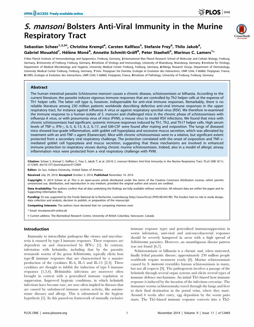
Systemic cytokine response in C57BL/6 mice infectedwith Schistosoma mansoni
In our studies we used a recent human isolate of S. mansoni,which originated from Oman [22,29]. We first established the
optimal number of cercariae that caused chronic schistosomiasis in
C57Bl/6 mice (Fig. S1). 70–75 cercariae per mouse, infected at 9–
10 weeks of age, was considered optimal. Herewith a moderate,
chronic course of the disease was established that resembled
Henderson’s moderate splenomegaly syndrome (MSS) of mouse
schistosomiasis [30], and that allowed the study of secondary
infections in the chronic phase of the disease. Survival at 16 weeks
of age was more than 95% in a large group of animals that were
infected at different time points during this study. Serum cytokine
profiles were also established. As expected, a slight increase in
IFN-c levels was seen immediately after infection, indicative of
Th1-type immune responsiveness. Significant Th2-type respon-
siveness was seen from week 7 of infection onwards, after the onset
of oviposition, with IL-4 and IL-5 rising ahead of IL-13. Also, IL-
17 and IL-2 levels rose significantly after week 7. We found very
high systemic levels of the proinflammatory cytokines IFN-c, TNF-
a and GM-CSF after week 7, which challenges the idea of
mutually exclusive immune responses (Fig. 1). IL-10 showed small
peaks of activity at week 1, 4, and 8, which most likely reflected a
reaction to inflammatory processes after skin and lung passage of
the schistosomula, and egg passage into the gut [31].
These findings were not dependent on the Th1-prone mouse
strain C57BL/6J; in Th2-prone BALB/c mice we obtained similar
data, with the exception of GM-CSF, which did not increase (Fig.
S2).
We also analysed the ability of T cells from spleen and
mesenteric and pulmonary (mediastinal) lymph nodes to secrete
cytokines after polyclonal stimulation. The T cells produced high
levels of the Th1-type cytokines in vitro, also later in the infection
(Table S1). However, the increase of Th1 cytokines was less
profound than those of Th2 cytokines, in agreement with previous
reports [3].
Bilharzia Bolsters Immunity to Respiratory Viruses
PLOS ONE | www.plosone.org 3 November 2014 | Volume 9 | Issue 11 | e112469
The high levels of serum IFN-c, but not those for TNF-a during
an infection with S. mansoni is dependent on ligands for TLR2
and/or TLR4, because in mice deficient for both TLRs serum
IFN-c was low throughout the infection (Fig. S3). In IL-12Rb2-
deficient mice neither a systemic IFN-c, nor a TNF-a response was
found (Fig. S3). We could not detect increased systemic levels of
endotoxin in mice that were infected with S. mansoni (Fig. S4).
We conclude that the immune response in C57BL/6 mice to a
chronic infection with S. mansoni is not polarised systemically.
High levels of the signature Th1 cytokine IFN-c are dependent on
TLR2 or 4 ligands, both TNF-a and IFN-c are dependent on
IL12Rb2.
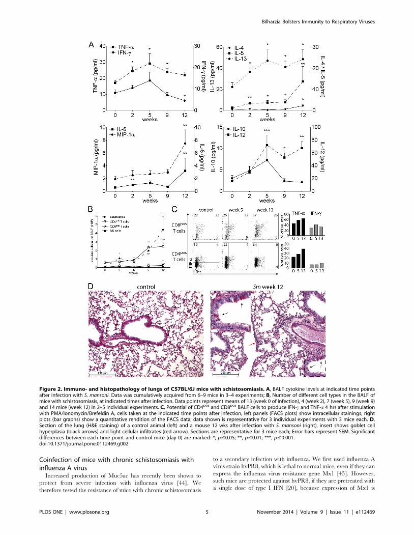
Influence of chronic schistosomiasis on the lungenvironment
The epithelial barrier layer of the lung reacts to environmental
stimuli with the production of alarmins like IL-33 and GM-CSF
[32–34], which attract dendritic cells. These cytokines also
stimulate innate lymphoid cells in the lung (ILCs [35,36]) to
produce IL-5 and IL-13, which create an intensely Th2-prone
environment, and to produce amphiregulin, which is important for
the integrity of the barrier layer [37]. We studied the steady state
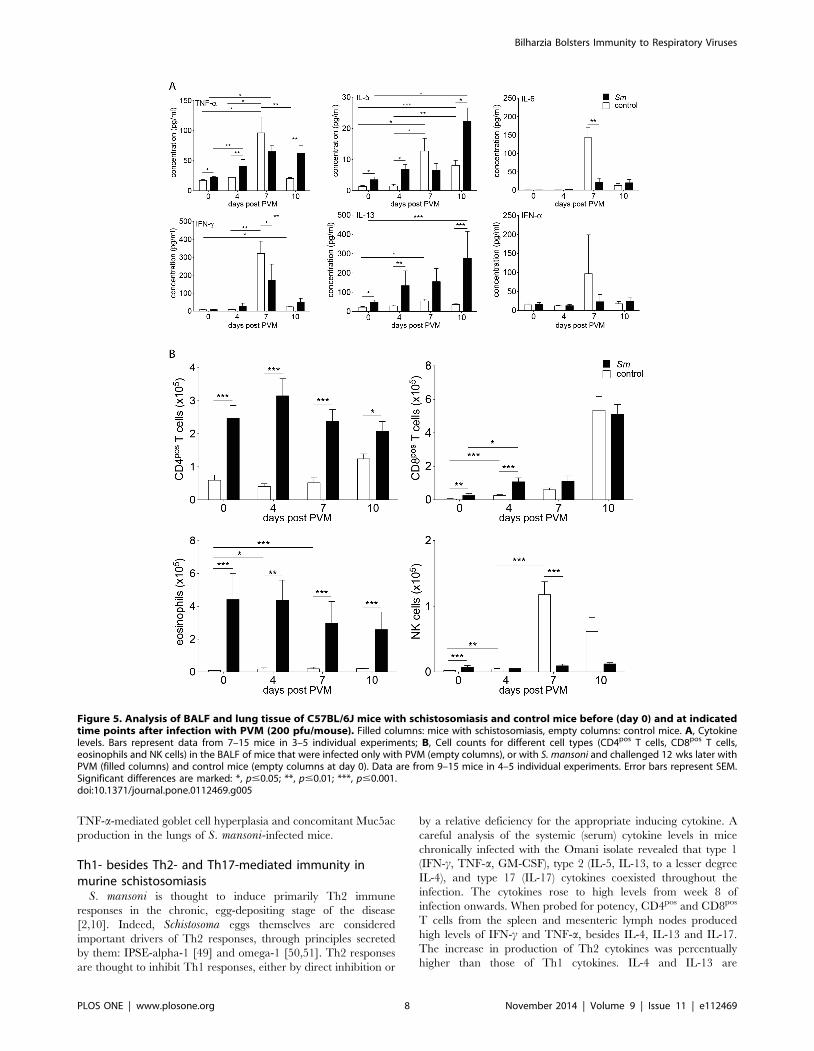
conditions in the lung during the infection with S. mansoni(Fig. 2). Analysis of the broncho-alveolar lavage fluid (BALF)
showed a transient increase in the Th1 cytokines IFN-c, TNF-aand IL-12 at week 5 post infection. At this time point
schistosomula have passed through the lungs and establish in the
portal system. Thereafter, IFN-c fell below normal levels, while
TNF-a and IL-12 remained significantly increased throughout the
measurement period. The Th2 cytokines IL-5 and IL-13 increased
steadily from week 2 onwards; IL-4 was only increased late in
infection. IL-10 was increased around week 5 post infection, while
the stress cytokines IL-6 and MIP-1a were significantly increased
late in infection (Fig. 2A). Thus, also in the BALF we registered
concomitant, not mutually exclusive Th1 and Th2 immune
responses during the infection with S. mansoni.We also analysed the cellular composition of the BALF
(Fig. 2B). Total cell numbers of eosinophils were increased
throughout the infection, with an early peak at week 2 of
infection. The number of CD4pos and CD8pos T cells and NK cells
were increased late in infection. In vitro TNF-asecretion by
CD4pos and CD8pos T cells was doubled late in infection (Fig. 2C).
The epithelial cell layer of the airway showed discrete signs of an
increased barrier function with mucus-producing goblet cell
hyperplasia, columnar cell hyperplasia, and light perivascular
and peribronchial infiltrates (Fig. 2D and Fig. S5A). Furthermore,
as signs of increased immunological alertness we saw a marked
increase in the expression of MHC class I and II on CD45pos
CD11bpos SiglecFneg interstitial lung cells (Fig. 3B, left panels). We
did not find an expansion of ILCs, rather we observed a relative
contraction of this cell population (Fig. S6A).
We conclude that in murine schistosomiasis a low-grade
inflammation is found in the lung with strong signs of an increased
barrier function.
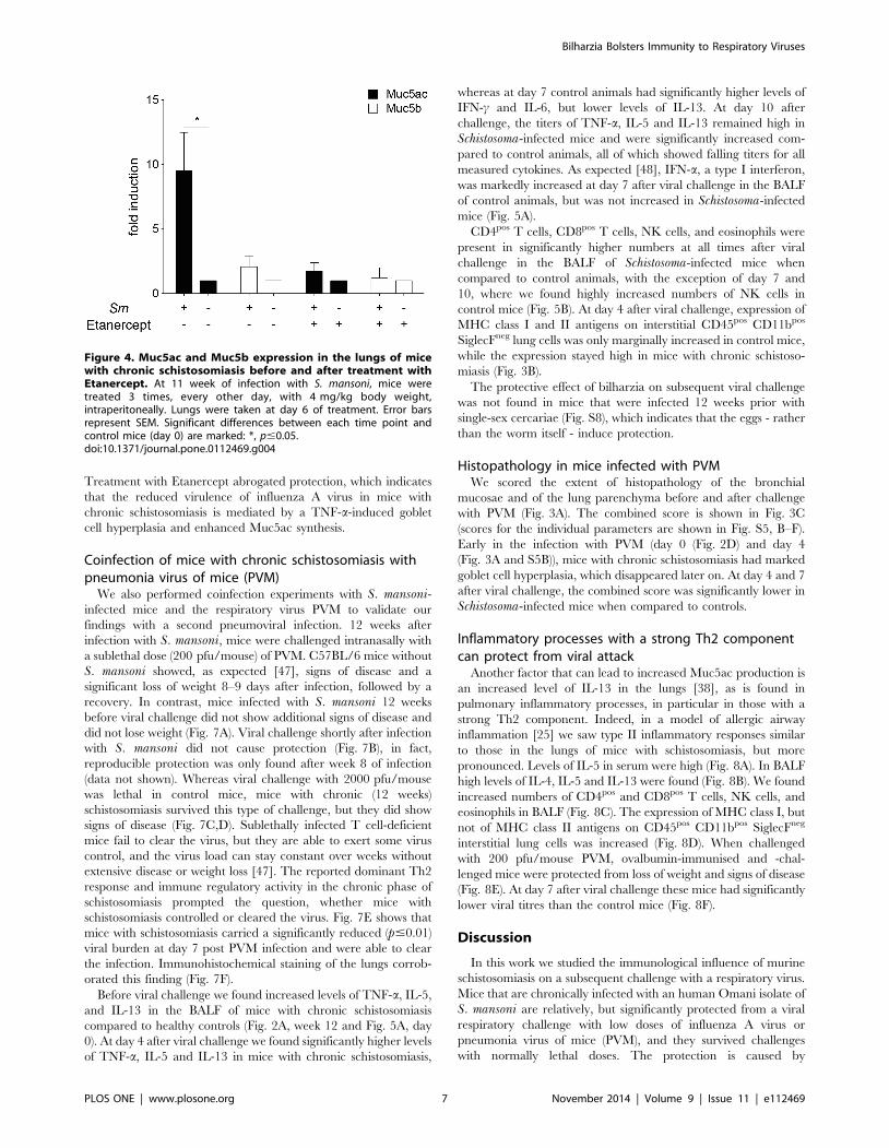
Muc5a expression is increased in mice with chronicschistosomiasis and is inhibited by depletion of TNF-a
Goblet cell hyperplasia can be induced, among others, by IL-13
[38] and TNF-a [39,40]. A hallmark of goblet cell hyperplasia is
the increased production of the gel-forming mucin Muc5ac
[41,42]. We determined Muc5ac mRNA levels in lungs of mice
infected with S. mansoni and in control mice. Muc5ac mRNA
levels were 106 times higher in infected mice compared to control
mice. As expected [42], only a moderate induction of the levels of
Muc5b was found in infected animals. We treated mice with
soluble TNF-receptor (Etanercept) [43] to explore the role of
TNF-a in the increased mucus production. After treatment of the
mice (starting at week 11 of infection), mRNA for Muc5ac
returned to baseline levels (Fig. 4). Thus, we conclude that TNF-ais essential for pulmonary goblet cell hyperplasia in chronic
schistosomiasis.
Figure 1. Serum cytokine levels of C57BL/6J mice infected withS. mansoni. Cytokine levels were determined at indicated time pointsafter infection. The data points represent means from 4–10 mice. Errorbars represent SEM. Significant differences between each time pointand control mice (day 0) are marked: *, p#0.05; **, p#0.01; ***, p#0.001.doi:10.1371/journal.pone.0112469.g001
Bilharzia Bolsters Immunity to Respiratory Viruses
PLOS ONE | www.plosone.org 4 November 2014 | Volume 9 | Issue 11 | e112469
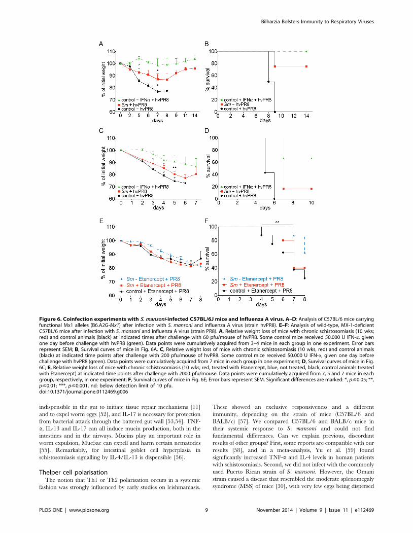
Coinfection of mice with chronic schistosomiasis withinfluenza A virus
Increased production of Muc5ac has recently been shown to
protect from severe infection with influenza virus [44]. We
therefore tested the resistance of mice with chronic schistosomiasis
to a secondary infection with influenza. We first used influenza A
virus strain hvPR8, which is lethal to normal mice, even if they can
express the influenza virus resistance gene Mx1 [45]. However,
such mice are protected against hvPR8, if they are pretreated with
a single dose of type I IFN [20], because expression of Mx1 is
Figure 2. Immuno- and histopathology of lungs of C57BL/6J mice with schistosomiasis. A, BALF cytokine levels at indicated time pointsafter infection with S. mansoni. Data was cumulatively acquired from 6–9 mice in 3–4 experiments; B, Number of different cell types in the BALF ofmice with schistosomiasis, at indicated times after infection. Data points represent means of 13 (week 0 of infection), 4 (week 2), 7 (week 5), 9 (week 9)and 14 mice (week 12) in 2–5 individual experiments. C, Potential of CD4pos and CD8pos BALF cells to produce IFN-c and TNF-a 4 hrs after stimulationwith PMA/Ionomycin/Brefeldin A, cells taken at the indicated time points after infection, left panels (FACS plots) show intracellular stainings, rightplots (bar graphs) show a quantitative rendition of the FACS data; data shown is representative for 3 individual experiments with 3 mice each. D,Section of the lung (H&E staining) of a control animal (left) and a mouse 12 wks after infection with S. mansoni (right), insert shows goblet cellhyperplasia (black arrows) and light cellular infiltrates (red arrow). Sections are representative for 3 mice each; Error bars represent SEM. Significantdifferences between each time point and control mice (day 0) are marked: *, p#0.05; **, p#0.01; ***, p#0.001.doi:10.1371/journal.pone.0112469.g002
Bilharzia Bolsters Immunity to Respiratory Viruses
PLOS ONE | www.plosone.org 5 November 2014 | Volume 9 | Issue 11 | e112469
regulated by type I and type III interferons [46]. We found no
evidence for the presence of IFN-ain the BALF of mice infected
with S. mansoni (Fig. 5A). Further, IFN-b-reporter mice [27] that
were chronically infected with S. mansoni showed no evidence for
enhanced production of IFN-b in the lungs. However, IFN-b was
significantly enhanced in liver and to a lesser extent in the kidney
(Fig. S7). We infected Mx1-competent mice in the chronic phase
of schistosomiasis with 60 pfu of influenza A strain hvPR8. Mice
infected with S. mansoni, without pretreatment with IFN, were
significantly protected from a challenge with hvPR8, and they
survived longer when compared to non-infected (S. mansoni-free)
controls (Fig. 6A,B). When the challenge dose of influenza virus
was increased to 200 pfu per animal, animals with chronic
schistosomiasis were still relatively protected and survived longer
than control animals (Fig. 6C,D). From these experiments we
conclude that infection with S. mansoni offers a relative, but
significant protection from influenza virus, which is independent of
IFN production.
To gauge the role of Muc5ac, we treated Mx1-deficient mice
that were infected S. mansoni 10 weeks previously with Etanercept
or PBS 3 and 1 day before, and also 1 and 3 days after infection
with influenza A virus strain PR8 (2000 pfu/mouse). Control mice
which were not infected with S. mansoni were treated similarly. As
expected, control mice were not protected from infection
(Fig. 6E,F). However, mice with chronic schistosomiasis were
significantly protected with less weight loss and enhanced survival.
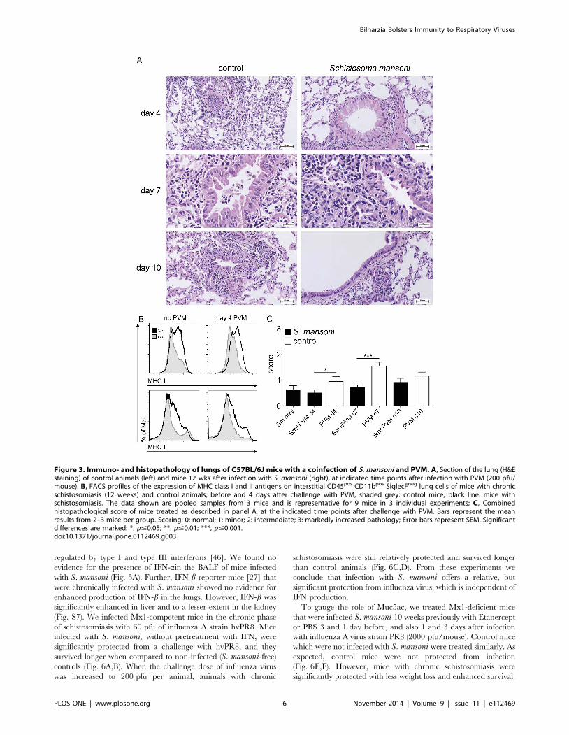
Figure 3. Immuno- and histopathology of lungs of C57BL/6J mice with a coinfection of S. mansoni and PVM. A, Section of the lung (H&Estaining) of control animals (left) and mice 12 wks after infection with S. mansoni (right), at indicated time points after infection with PVM (200 pfu/mouse). B, FACS profiles of the expression of MHC class I and II antigens on interstitial CD45pos CD11bpos SiglecFneg lung cells of mice with chronicschistosomiasis (12 weeks) and control animals, before and 4 days after challenge with PVM, shaded grey: control mice, black line: mice withschistosomiasis. The data shown are pooled samples from 3 mice and is representative for 9 mice in 3 individual experiments; C, Combinedhistopathological score of mice treated as described in panel A, at the indicated time points after challenge with PVM. Bars represent the meanresults from 2–3 mice per group. Scoring: 0: normal; 1: minor; 2: intermediate; 3: markedly increased pathology; Error bars represent SEM. Significantdifferences are marked: *, p#0.05; **, p#0.01; ***, p#0.001.doi:10.1371/journal.pone.0112469.g003
Bilharzia Bolsters Immunity to Respiratory Viruses
PLOS ONE | www.plosone.org 6 November 2014 | Volume 9 | Issue 11 | e112469
Treatment with Etanercept abrogated protection, which indicates
that the reduced virulence of influenza A virus in mice with
chronic schistosomiasis is mediated by a TNF-a-induced goblet
cell hyperplasia and enhanced Muc5ac synthesis.
Coinfection of mice with chronic schistosomiasis withpneumonia virus of mice (PVM)
We also performed coinfection experiments with S. mansoni-infected mice and the respiratory virus PVM to validate our
findings with a second pneumoviral infection. 12 weeks after
infection with S. mansoni, mice were challenged intranasally with
a sublethal dose (200 pfu/mouse) of PVM. C57BL/6 mice without
S. mansoni showed, as expected [47], signs of disease and a
significant loss of weight 8–9 days after infection, followed by a
recovery. In contrast, mice infected with S. mansoni 12 weeks
before viral challenge did not show additional signs of disease and
did not lose weight (Fig. 7A). Viral challenge shortly after infection
with S. mansoni did not cause protection (Fig. 7B), in fact,
reproducible protection was only found after week 8 of infection
(data not shown). Whereas viral challenge with 2000 pfu/mouse
was lethal in control mice, mice with chronic (12 weeks)
schistosomiasis survived this type of challenge, but they did show
signs of disease (Fig. 7C,D). Sublethally infected T cell-deficient
mice fail to clear the virus, but they are able to exert some virus
control, and the virus load can stay constant over weeks without
extensive disease or weight loss [47]. The reported dominant Th2
response and immune regulatory activity in the chronic phase of
schistosomiasis prompted the question, whether mice with
schistosomiasis controlled or cleared the virus. Fig. 7E shows that
mice with schistosomiasis carried a significantly reduced (p#0.01)
viral burden at day 7 post PVM infection and were able to clear
the infection. Immunohistochemical staining of the lungs corrob-
orated this finding (Fig. 7F).
Before viral challenge we found increased levels of TNF-a, IL-5,
and IL-13 in the BALF of mice with chronic schistosomiasis
compared to healthy controls (Fig. 2A, week 12 and Fig. 5A, day
0). At day 4 after viral challenge we found significantly higher levels
of TNF-a, IL-5 and IL-13 in mice with chronic schistosomiasis,
whereas at day 7 control animals had significantly higher levels of
IFN-c and IL-6, but lower levels of IL-13. At day 10 after
challenge, the titers of TNF-a, IL-5 and IL-13 remained high in
Schistosoma-infected mice and were significantly increased com-
pared to control animals, all of which showed falling titers for all
measured cytokines. As expected [48], IFN-a, a type I interferon,
was markedly increased at day 7 after viral challenge in the BALF
of control animals, but was not increased in Schistosoma-infected
mice (Fig. 5A).
CD4pos T cells, CD8pos T cells, NK cells, and eosinophils were
present in significantly higher numbers at all times after viral
challenge in the BALF of Schistosoma-infected mice when
compared to control animals, with the exception of day 7 and
10, where we found highly increased numbers of NK cells in
control mice (Fig. 5B). At day 4 after viral challenge, expression of
MHC class I and II antigens on interstitial CD45pos CD11bpos
SiglecFneg lung cells was only marginally increased in control mice,
while the expression stayed high in mice with chronic schistoso-
miasis (Fig. 3B).
The protective effect of bilharzia on subsequent viral challenge
was not found in mice that were infected 12 weeks prior with
single-sex cercariae (Fig. S8), which indicates that the eggs - rather
than the worm itself - induce protection.
Histopathology in mice infected with PVMWe scored the extent of histopathology of the bronchial
mucosae and of the lung parenchyma before and after challenge
with PVM (Fig. 3A). The combined score is shown in Fig. 3C
(scores for the individual parameters are shown in Fig. S5, B–F).
Early in the infection with PVM (day 0 (Fig. 2D) and day 4
(Fig. 3A and S5B)), mice with chronic schistosomiasis had marked
goblet cell hyperplasia, which disappeared later on. At day 4 and 7
after viral challenge, the combined score was significantly lower in
Schistosoma-infected mice when compared to controls.
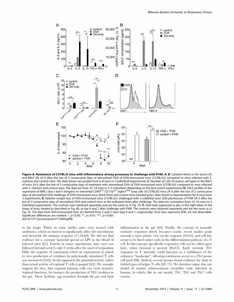
Inflammatory processes with a strong Th2 componentcan protect from viral attack
Another factor that can lead to increased Muc5ac production is
an increased level of IL-13 in the lungs [38], as is found in
pulmonary inflammatory processes, in particular in those with a
strong Th2 component. Indeed, in a model of allergic airway
inflammation [25] we saw type II inflammatory responses similar
to those in the lungs of mice with schistosomiasis, but more
pronounced. Levels of IL-5 in serum were high (Fig. 8A). In BALF
high levels of IL-4, IL-5 and IL-13 were found (Fig. 8B). We found
increased numbers of CD4pos and CD8pos T cells, NK cells, and
eosinophils in BALF (Fig. 8C). The expression of MHC class I, but
not of MHC class II antigens on CD45pos CD11bpos SiglecFneg
interstitial lung cells was increased (Fig. 8D). When challenged
with 200 pfu/mouse PVM, ovalbumin-immunised and -chal-
lenged mice were protected from loss of weight and signs of disease
(Fig. 8E). At day 7 after viral challenge these mice had significantly
lower viral titres than the control mice (Fig. 8F).
Discussion
In this work we studied the immunological influence of murine
schistosomiasis on a subsequent challenge with a respiratory virus.
Mice that are chronically infected with an human Omani isolate of
S. mansoni are relatively, but significantly protected from a viral
respiratory challenge with low doses of influenza A virus or
pneumonia virus of mice (PVM), and they survived challenges
with normally lethal doses. The protection is caused by
Figure 4. Muc5ac and Muc5b expression in the lungs of micewith chronic schistosomiasis before and after treatment withEtanercept. At 11 week of infection with S. mansoni, mice weretreated 3 times, every other day, with 4 mg/kg body weight,intraperitoneally. Lungs were taken at day 6 of treatment. Error barsrepresent SEM. Significant differences between each time point andcontrol mice (day 0) are marked: *, p#0.05.doi:10.1371/journal.pone.0112469.g004
Bilharzia Bolsters Immunity to Respiratory Viruses
PLOS ONE | www.plosone.org 7 November 2014 | Volume 9 | Issue 11 | e112469
TNF-a-mediated goblet cell hyperplasia and concomitant Muc5ac
production in the lungs of S. mansoni-infected mice.
Th1- besides Th2- and Th17-mediated immunity inmurine schistosomiasis
S. mansoni is thought to induce primarily Th2 immune
responses in the chronic, egg-depositing stage of the disease
[2,10]. Indeed, Schistosoma eggs themselves are considered
important drivers of Th2 responses, through principles secreted
by them: IPSE-alpha-1 [49] and omega-1 [50,51]. Th2 responses
are thought to inhibit Th1 responses, either by direct inhibition or
by a relative deficiency for the appropriate inducing cytokine. A
careful analysis of the systemic (serum) cytokine levels in mice
chronically infected with the Omani isolate revealed that type 1
(IFN-c, TNF-a, GM-CSF), type 2 (IL-5, IL-13, to a lesser degree
IL-4), and type 17 (IL-17) cytokines coexisted throughout the
infection. The cytokines rose to high levels from week 8 of
infection onwards. When probed for potency, CD4pos and CD8pos
T cells from the spleen and mesenteric lymph nodes produced
high levels of IFN-c and TNF-a, besides IL-4, IL-13 and IL-17.
The increase in production of Th2 cytokines was percentually
higher than those of Th1 cytokines. IL-4 and IL-13 are
Figure 5. Analysis of BALF and lung tissue of C57BL/6J mice with schistosomiasis and control mice before (day 0) and at indicatedtime points after infection with PVM (200 pfu/mouse). Filled columns: mice with schistosomiasis, empty columns: control mice. A, Cytokinelevels. Bars represent data from 7–15 mice in 3–5 individual experiments; B, Cell counts for different cell types (CD4pos T cells, CD8pos T cells,eosinophils and NK cells) in the BALF of mice that were infected only with PVM (empty columns), or with S. mansoni and challenged 12 wks later withPVM (filled columns) and control mice (empty columns at day 0). Data are from 9–15 mice in 4–5 individual experiments. Error bars represent SEM.Significant differences are marked: *, p#0.05; **, p#0.01; ***, p#0.001.doi:10.1371/journal.pone.0112469.g005
Bilharzia Bolsters Immunity to Respiratory Viruses
PLOS ONE | www.plosone.org 8 November 2014 | Volume 9 | Issue 11 | e112469
indispensible in the gut to initiate tissue repair mechanisms [11]
and to expel worm eggs [52], and IL-17 is necessary for protection
from bacterial attack through the battered gut wall [53,54]. TNF-
a, IL-13 and IL-17 can all induce mucin production, both in the
intestines and in the airways. Mucins play an important role in
worm expulsion, Muc5ac can expell and harm certain nematodes
[55]. Remarkably, for intestinal goblet cell hyperplasia in
schistosomiasis signalling by IL-4/IL-13 is dispensible [56].
Thelper cell polarisationThe notion that Th1 or Th2 polarisation occurs in a systemic
fashion was strongly influenced by early studies on leishmaniasis.
These showed an exclusive responsiveness and a different
immunity, depending on the strain of mice (C57BL/6 and
BALB/c) [57]. We compared C57BL/6 and BALB/c mice in
their systemic response to S. mansoni and could not find
fundamental differences. Can we explain previous, discordant
results of other groups? First, some reports are compatible with our
results [58], and in a meta-analysis, Yu et al. [59] found
significantly increased TNF-a and IL-4 levels in human patients
with schistosomiasis. Second, we did not infect with the commonly
used Puerto Rican strain of S. mansoni. However, the Omani
strain caused a disease that resembled the moderate splenomegaly
syndrome (MSS) of mice [30], with very few eggs being dispersed
Figure 6. Coinfection experiments with S. mansoni-infected C57BL/6J mice and Influenza A virus. A–D: Analysis of C57BL/6 mice carryingfunctional Mx1 alleles (B6.A2G-Mx1) after infection with S. mansoni and influenza A virus (strain hvPR8). E–F: Analysis of wild-type, MX-1-deficientC57BL/6 mice after infection with S. mansoni and influenza A virus (strain PR8). A, Relative weight loss of mice with chronic schistosomiasis (10 wks;red) and control animals (black) at indicated times after challenge with 60 pfu/mouse of hvPR8. Some control mice received 50.000 U IFN-a, givenone day before challenge with hvPR8 (green). Data points were cumulatively acquired from 3–4 mice in each group in one experiment. Error barsrepresent SEM; B, Survival curves of mice in Fig. 6A. C, Relative weight loss of mice with chronic schistosomiasis (10 wks, red) and control animals(black) at indicated time points after challenge with 200 pfu/mouse of hvPR8. Some control mice received 50.000 U IFN-a, given one day beforechallenge with hvPR8 (green). Data points were cumulatively acquired from 7 mice in each group in one experiment; D, Survival curves of mice in Fig.6C; E, Relative weight loss of mice with chronic schistosomiasis (10 wks; red, treated with Etanercept, blue, not treated, black, control animals treatedwith Etanercept) at indicated time points after challenge with 2000 pfu/mouse. Data points were cumulatively acquired from 7, 5 and 7 mice in eachgroup, respectively, in one experiment; F, Survival curves of mice in Fig. 6E; Error bars represent SEM. Significant differences are marked: *, p#0.05; **,p#0.01; ***, p#0.001, nd: below detection limit of 10 pfu.doi:10.1371/journal.pone.0112469.g006
Bilharzia Bolsters Immunity to Respiratory Viruses
PLOS ONE | www.plosone.org 9 November 2014 | Volume 9 | Issue 11 | e112469
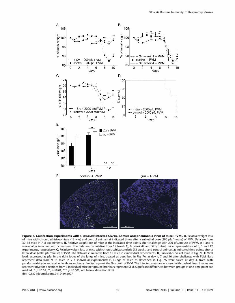
Figure 7. Coinfection experiments with S. mansoni-infected C57BL/6J mice and pneumonia virus of mice (PVM). A, Relative weight lossof mice with chronic schistosomiasis (12 wks) and control animals at indicated times after a sublethal dose (200 pfu/mouse) of PVM. Data are from30–38 mice in 7–8 experiments. B, Relative weight loss of mice at the indicated time points after challenge with 200 pfu/mouse of PVM, at 1 and 4weeks after infection with S. mansoni. The data are cumulative from 15 (week 1), 6 (week 4), and 52 (control) mice representative of 3, 1 and 12experiments, respectively; C, Relative weight loss of mice with chronic schistosomiasis (12 weeks) and control animals at indicated time points after alethal dose (2000 pfu/mouse) of PVM. The data are cumulative from 10 mice in 2 individual experiments; D, Survival curves of mice in Fig. 7C; E, Viralload, expressed as pfu, in the right lobes of the lungs of mice, treated as described in Fig. 7A, at day 4, 7 and 10 after challenge with PVM. Barsrepresent data from 9–15 mice in 2–4 individual experiments; F, Lungs of mice as described in Fig. 7A were taken at day 4, fixed withparaformaldehyde and stained with an antibody directed against the G-protein of PVM. The infected areas are enclosed with dashed lines. Images arerepresentative for 6 sections from 3 individual mice per group; Error bars represent SEM. Significant differences between groups at one time point aremarked: *, p#0.05; **, p#0.01; ***, p#0.001, nd: below detection limit.doi:10.1371/journal.pone.0112469.g007
Bilharzia Bolsters Immunity to Respiratory Viruses
PLOS ONE | www.plosone.org 10 November 2014 | Volume 9 | Issue 11 | e112469
to the lungs. Third, in some studies mice were treated with
antibiotics, which are known to significantly affect the microbiome
and therewith the immune response [17,18,60]. We did not find
evidence for a systemic bacterial spread or LPS in the blood of
infected mice [61]. Fourth, in many experiments, mice were not
followed beyond week 8, only 2 weeks after the onset of oviposition
Fifth, the majority of conclusions relied on experiments in which
ex vivo production of cytokines by polyclonally stimulated T cells
was measured [3,62]. In this approach the potential activity rather
than actual activity of regional T cells is gauged [63]. We strongly
support the idea, that regional immune cells can exert exclusive
regional functions, for instance the production of Th2 cytokines in
the gut. These facilitate egg transition through the gut and limit
inflammation in the gut [60]. Finally, the concept of mutually
exclusive responses slowly becomes cracks: recent studies point
towards a more plastic view on the response [64,65], and cell fate
seems to be fixed rather early in the differentiation pathway of a T
cell. In this concept, specifically responsive cells can be called upon
later, when demand is present [66,67]. Early systemic Th1
responses in S. mansoni could function as a stabilisator of the
enhancer ‘‘landscape’’, allowing continuous access to a Th1-prone
cell pool [68]. Indeed, several groups found evidence for dual or
hybrid types of helper T cells [69–71]. We therefore argue that our
model of murine schistosomiasis resembles early infection in
human, in which, like in our model, Th1, Th2 and Th17 cells
coexist.
Figure 8. Resistance of C57BL/6 mice with inflammatory airway processes to challenge with PVM. A, B, Cytokine levels in the serum (A)and BALF (B) 24 h after the last of 3 consecutive days of aerosolised OVA of OVA-immunised mice (C57BL/6J) compared to mice infected with S.mansoni and control mice. The data shown are pooled from 4–8 mice in 3 individual experiments; C, Number of cells of various cell types in the BALFof mice 24 h after the last of 3 consecutive days of treatment with aerosolised OVA of OVA-immunised mice (C57BL/6J) compared to mice infectedwith S. mansoni and control mice. The data are from 10–14 mice in 3–5 individual (depending on the time point) experiments; D, FACS profiles of theexpression of MHC class I and II antigens on interstitial CD45pos CD11bpos SiglecFneg lung cells of C57BL/6J mice 24 h after the last of 3 consecutivedays of aerosolised OVA challenge of OVA-immunised mice (thick lines) and control mice (shaded grey). Data shown is representative for 6 mice from2 experiments; E, Relative weight loss of OVA-immunised mice (C57BL/6J) challenged with a sublethal dose (200 pfu/mouse) of PVM 24 h after thelast of 3 consecutive days of aerosolised OVA and control mice at the indicated times after challenge. The data are cumulative from 10–16 mice in 3individual experiments. The controls were obtained separately and are the same as in Fig. 7A. F, Viral load, expressed as pfu, in the right lobes of thelungs of mice, treated as described in Fig. 8E, at day 4 and 7 after challenge with PVM. The controls were obtained separately and are the same as inFig. 7E. The data from OVA-immunised mice are derived from 2 and 3 mice (day 4 and 7, respectively). Error bars represent SEM. nd: not detectable.Significant differences are marked: *, p#0.05; **, p#0.01; ***, p#0.001.doi:10.1371/journal.pone.0112469.g008
Bilharzia Bolsters Immunity to Respiratory Viruses
PLOS ONE | www.plosone.org 11 November 2014 | Volume 9 | Issue 11 | e112469
The role of TNF-a in schistosomiasisHigh levels of TNF-a and IFN-c are often implied in the
pathogenesis of a severe form of human schistosomiasis, the
hepato-splenic form [72], which could resemble Henderson’s
hypersplenomegaly syndrome (HSS). As outlined, we do not find
evidence for HSS in our model. Also in the mild form, hepatic
involvement in mice is more pronounced than in benign cases of
human schistosomiasis. Also in MSS increased levels of TNF-a are
found in liver tissue [73]. In mice both protective and deleterious
effects have been described for TNF-a, together with evidence that
granuloma-induced TNF production is necessary for the matura-
tion of the adult worms [74,75].
Pulmonary milieu in murine schistosomiasisIn chronic murine schistosomiasis the lungs show low-grade
inflammatory processes, with goblet cell hyperplasia and increased
production of the gel-forming mucin Muc5ac (see Fig. S9 for a
graphical summary of our data). Early in the infection Th1
cytokines were found in BALF, they stayed high with the exception
of IFN-c. Th2 cytokines also rose early, with the exception of IL-4.
Pathology in our model was less pronounced than in that
described by Crosby et al. [58], which could well be explained
by the lower number of parasite eggs found in the lungs of our
mice. The sustained high levels of IL-13 and IL-5 without a joined
increase of IL-4 suggests that they were not derived from Th2-type
helper cells, but rather from ILC2-type cells [76,77]. However, we
saw a relative decrease in number of ILCs during the course of
schistosomiasis, likely arguing against an activated state of the
ILCs [37]. Instead, our results indicate that sustained high levels of
systemic TNF-a were primarily responsible for the goblet cell
hyperplasia.
Coinfection studies with S. mansoni and influenza virusor PVM
In this Schistosoma-moulded milieu respiratory viruses do not
spread as easily as in a healthy environment. PMV did not expand
as rapidly and was cleared with a lower expenditure of CD8pos
cytotoxic T cells, and therefore with less airway inflammation and
involvement. As a consequence, a lower combined histopathology
score was found. The virulent influenza virus strain hvPR8 is lethal
to normal (Mx-competent) mice, unless they are pretreated with
type-I or type III interferons, which induce the indispensible Mx
protein. Mice with chronic schistosomiasis were to a certain extent
resistant to strain hvPR8, indicating that the infection with S.mansoni had either induced the production of type I or type III
interferons or that other protective mechanisms, like the produc-
tion of Muc5ac [44], were active. We were unable to detect
significant amounts of IFN-a by ELISA in mice with schistoso-
miasis, and reporter mice for IFN-b did not show any significant
involvement of IFN-b. Therefore, we conclude that type I
interferons did not play a significant role in the observed
protection, although we cannot completely exclude the involve-
ment of type III interferons [78]. High levels of isolated production
of Muc5ac in the lung, as found in Muc5ac-transgenic mice,
protects from infection with influenza A virus [44]. In these studies
Muc5ac production was increased by a factor of 20, in our studies,
mice with chronic schistosomiasis had a ten-fold induction of
Muc5ac. Treatment of the mice with Etanercept revealed that
upstream TNF-a was responsible for this induction. Indeed, Mx1-
deficient mice with chronic schistosomiasis were relatively, but not
absolutely protected from an infection with influenza A virus, and
the protection was lost after treatment with Etanercept.
TNF-a does not seem to affect viral replication in primary
infections [47,79,80]. However, TNF has a significant effect on
immunopathology: it negatively regulates the extent of pathology
[79], or increases pathology [80], depending on virus strain and
study design. Therefore we favour the notion that was put forward
by Ehre et al. [44]: mucus alone is sufficient to sequester virus and
reduce viral entry into the lung cells. Less cytotoxic T cells were
needed for viral control, which resulted in reduced tissue damage.
The role of TNF-a in Muc5ac expression and goblet cellhyperplasia
TNF-a has a direct influence on Muc5ac transcription via the
NF-kB pathway, and it induces expression of the EGFR [40]. NF-
kB further modulates IL-13Ra1-signalling [81]. Both the IL-
13Ra1 and the EGFR are essential in the development of goblet
cell hyperplasia. It is debated, whether these factors work in
parallel or whether engagement of the EGFR is making the goblet
cell receptive for consecutive IL-13 signalling [82]. We saw that
short-time treatment of mice with Etanercept, i.e. neutralising
TNF-a in the continuing presence of IL-13, inhibited Muc5ac
expression and interfered with protection to viral challenge. This
suggests a role for TNF-a that is independent of IL-13. Indeed, it
was clearly shown that the mere transgenic expression of Muc5ac,
without the involvement of (IL-13-driven) inflammatory responses,
is protective in influenza infections [44].
Factors influencing protectionThe increased resistance to viral attack and the increased
immunological ‘‘alertness’’ in the lungs of mice with schistosomi-
asis is not dependent on worm passage through the lungs, because
infections with single-sex cercariae did not protect from viral
attack. Protection is reliably found after week eight of infection,
thus after the onset of oviposition. This coincides with the first
deposition of eggs in the liver and the subsequent granulomatous
reaction, but more importantly, also the passage of the eggs
through the intestinal wall. This causes considerable damage, leads
to inflammation, the secretion of high amounts of IL-17, and a
noticeable change in the stool constitution with first some, later
more - and also bloody - mucoid stool. Conditions in the gut of
mice with schistosomiasis are therefore very different from normal.
Already under normal conditions, bacterial products can enter the
body, enhance systemic innate immunity [36], and exert
considerable influence also in distant compartments like the lung.
Effective immunity to influenza virus is dependent on the existing
microbiome [17,18], with bacterial products of gram-positive
bacteria and certain TLR-ligands (TLR2,-3,-4 or -9) as active
principle, even when rectally applied [17]. Abt et al. showed that
interferon-induced genes were responsible for this kind of
protection [18]. We could show, that systemic IFN-c responses
were absent in TLR2,4-double-deficient mice with schistosomiasis.
In IL-12Rb2-deficient mice both IFN-c and TNF-awere absent,
pointing to an IL-12/IFN-c axis in protection in mice with
schistosomiasis.
Allergic inflammation and viral infectionsWe then asked whether pulmonary inflammatory processes in
general could lead to protection. We found that in a mouse model
of asthma mice were protected from viral attack by PVM, to a
similar degree as found in mice with schistosomiasis. Indeed,
conditions in the lung were to a large degree comparable, with a
strong Th2 component in the inflammation. These results were
not unexpected, Th2 cytokines, in particular IL-13, are potent
inducers of goblet cell hyperplasia and Muc5ac production and
Bilharzia Bolsters Immunity to Respiratory Viruses
PLOS ONE | www.plosone.org 12 November 2014 | Volume 9 | Issue 11 | e112469
thus protective in viral attacks. Remarkably, in the recent
pandemic influenza (H1N1) infection, asthma was independently
associated with a lower risk of dying during hospitalisation [83].
We interpret these results as a reflection of an increased immune
alertness, with goblet cell hyperplasia and Muc5ac production.
This response will initially protect the host from pneumotropic
viruses, but in chronic conditions it may be detrimental for the
host because of ensuing, debilitating chronic inflammatory
processes like COPD [84].
Other helminths will exert other effectsWe do not think that our findings can be transferred to all
helminth infections. In infections in which the parasite deploys
systemic immunosuppression, as does for instance Heligmoso-moides polygyrus [85,86], or induces only a weak Th1 response,
the outcome can be different. Indeed, two recent articles
supported this notion. In a murine model, infection with H.polygyrus and S. mansoni eggs (a complete infectious cycle was not
tested) could reactivate the mouse c-herpes virus MHV68 in vivo,
via an IL-4- and Stat6-dependent pathway[87]. In a second study,
Trichinella spiralis and H polygyrus both could inhibit specific T-
cell responses to murine norovirus, or to influenza virus (only
tested with T. spiralis). Immune modulation was shown to be
dependent on a Stat6-dependent alternative activation of macro-
phages and their secreted product Ym1 [88]. In both cases, Th2-
type immune responses are induced, without previous Th1-type
immunity. Unfortunately, no data were provided for the lung
environment.
In schistosomiasis, we did not find evidence for a generalised
immune suppression. The similarity of S. mansoni-induced and
allergen-induced pulmonary inflammation rather predicts a more
severe course, when allergy and schistosomiasis coexist. The
immune regulatory activity of T regulatory cells and IL-10 is
therefore most likely secondary to inflammation and antigen-
specific [54]. Remarkably, in schistosomiasis, a disease with at least
200 million people a year needing treatment, little tenable
information is available regarding comorbidities. Of course, many
variables can influence the outcome of coinfections, as discussed
extensively by Abruzzi and Fried [6].
In conclusion, our results shed a new light both on the biology of
schistosomiasis and the immune response to the parasite. Contrary
to previous reports, we do not find the immune responses to be
polarised systemically, but they reflect local need for defence.
From an evolutionary point of view, the parasite would initially
benefit from an increased barrier function towards common
pathogens like respiratory tract viruses.
Supporting Information
Figure S1 Determination of the optimal cercarial doseof S. mansoni that leads to a chronic disease. A, five
C57BL/6J mice were exposed to different number of cercariae
and analysed 12 weeks after infection for worm burden (D), worm
pairs (X), infection rate (#) and weight of liver (&) and spleen
(%). B, total number of eggs/liver 12 weeks after infection, relative
to the cercarial dose. Data points represent mean data from 3–7
mice in one experiment. Error bars represent SEM. Grey zones
indicate the optimal cercarial dose we have used in further
experiments to obtain a chronic course of the disease.
(TIF)
Figure S2 Serum cytokine levels in BALB/cJ mice atindicated time points after infection with S. mansoni. A,
signature Th1 cytokines IFN-c and TNF-a and B, D, signature
Th2 cytokines IL-5, IL-13 and IL-4 were measured, as well as C,
D, IL-6, IL-17 and IL-2. The data points represent means from 2–
7 mice. Error bars represent SEM. Significant differences between
each time point and control mice (day 0) are marked: *, p#0.05;
**, p#0.01; ***, p#0.001.
(TIF)
Figure S3 Serum IFN-c(A) and TNF-a(B) levels inC57BL/6J (black line), TLR2–/–/4–/– (green line) andIL-12Rb2–/– (blue dotted line) mice at indicated timepoints after infection with S. mansoni. Error bars represent
SEM. Significant differences between each time point and control
mice (day 0) are marked: *, p#0.05; **, p#0.01; ***, p#0.001.
(TIF)
Figure S4 Serum endotoxin levels in C57BL/6 mice atindicated time points after infection with S. mansoni.Serum endotoxin levels were determined by LAL assay. The data
points represent means from 11, 4 and 12 mice at week 0, 5, and
12, respectively. Error bars represent SEM. ANOVA analysis
(Kruskall-Wallis): p = 0.0488.
(TIF)
Figure S5 Histo-pathological scores of the lungs ofC57BL/6J mice with chronic schistosomiasis. A, basal values
at 12 weeks after infection with S. mansoni; B–F, scores of mice with
schistosomiasis (12 wks; black) and control animals (white) at
indicated times after a sublethal dose (200 pfu/mouse) of PVM.
Columns represent mean results from 2–3 mice per group. Error bars
represent SEM. Scoring: 0: normal; 1: minor; 2: intermediate; 3:
markedly increased. Absence of bars represents normal (0) scoring.
(TIF)
Figure S6 Analysis of ILCs in the lungs of control miceand at indicated time points after infection with S.mansoni. ILCs were prepared from the right lobes of the lungs.
A, gating strategy. Lin: Lineage markers. The plots are
representative for n = 10 (control), 4 (week 2), 2 (week 5), 4 (week
9) and 10 (week 12) mice. B, total number of cells that are Lin-
negative and positive for the markers CD25, CD90 and T1-ST2.
Significant differences between groups are marked: *, p#0.05; **,
p#0.01; ***, p#0.001.
(TIF)
Figure S7 Type I interferon levels in mice infected withS. mansoni at week 12 of infection. A, IFN-a levels in BALF;
B, IFN-b levels in indicated organs (RLU: relative luciferase units)
Error bars represent SEM. Significant differences between each
time point and control mice (day 0) are marked: *, p#0.05; ***,
p#0.001.
(TIF)
Figure S8 Coinfection experiments with S. mansoni-infected mice and pneumonia virus of mice (PVM). Mice
were i.n. challenged at day 0 with 200 pfu of virus. Relative weight
loss of C57BL/6J mice infected with 100 cercariae of a single sex
(cercariae obtained from snails infected with only 1 miracidium)
(12 wks) and control animals at indicated times. Data are pooled
from 13 mice in 2 experiments. Error bars represent SEM.
Significant differences between data points from normal and single
sex infection with S. mansoni are marked: *, p#0.05; **, p#0.01.
(TIF)
Figure S9 Graphical summary. Representation of pulmo-
nary conditions. Upper left: normal condition. Upper right:
chronic schistosomiasis. Lower left: infection with a respiratory
virus. Lower right: secondary infection with a respiratory virus in
chronic schistosomiasis.
(TIF)
Bilharzia Bolsters Immunity to Respiratory Viruses
PLOS ONE | www.plosone.org 13 November 2014 | Volume 9 | Issue 11 | e112469
Table S1 Ex vivo stimulation of cells. Pulmonary lymph
nodes (pLN), spleens and mesenteric lymph nodes (mLN) were
taken from mice on the C57BL/6J background and single cell
suspensions were prepared. At week 0 not enough cells could be
prepared from the pLN to perform in vitro stimulation. Cells were
then incubated for 4 h in complete medium containing phorbol
12-myristate 13-acetate (PMA, 20 ng/ml), Ionomycin (500 ng/ml)
and Brefeldin A (1 mg/ml). Cells were stained extracellularly and,
after fixation, stained intracellularly with fluorochrome-labelled
anti-TNF-a (MP6-XT22), anti-IFN-c (XMG1.2), anti-IL-4
(11B11) and anti IL-13 (eBio13A), using the IntraSure fixation
and permeabilisation kit (all from BD Biosciences, except for anti
IL-13, which was from eBioscience). The percentage of cells
positive for the indicated cytokine is given.
(TIF)
Acknowledgments
We thank Drs. Markus Simon, Stefan Martin and Peter Nielsen for
critically reading the manuscript and for fruitful discussions; Sandra Groß
(MPI-IE), Kristin Hauck and Theresa Kreuzahler (University of Wuerz-
burg) for excellent technical assistance.
Author Contributions
Conceived and designed the experiments: SS HM PS MCL. Performed the
experiments: SS C. Krempl C. Kallfass SF GM HM AS-G PS MCL.
Analyzed the data: SS C. Krempl TJ GM HM AS-G PS MCL.
Contributed reagents/materials/analysis tools: C. Krempl GM HM AS-
G PS. Wrote the paper: SS TJ PS MCL.
References
1. Zhu J, Paul WE (2008) CD4 T cells: fates, functions, and faults. Blood 112:
1557–1569. doi: 10.1182/blood-2008-05-078154.
2. Dunne DW, Cooke A (2005) A worm’s eye view of the immune system:
consequences for evolution of human autoimmune disease. Nat Rev Immunol 5:
420–426. doi: 10.1038/nri1601.
3. Pearce EJ, Caspar P, Grzych J-M, Lewis FA, Sher A (1991) Downregulation of
Th1 cytokine production accompanies induction of Th2 responses by a parasitic
helminth, Schistosoma mansoni. J Exp Med 173: 159–166. doi: 10.1084/
jem.173.1.159.
4. Mosmann TR, Coffman RL (1989) TH1 and TH2 cells: different patterns of
lymphokine secretion lead to different functional properties. Annu Rev Immunol
7: 145–173. doi: 10.1146/annurev.iy.07.040189.001045.
5. Strachan DP (1989) Hay fever, hygiene, and household size. BMJ 299: 1259.
6. Abruzzi A, Fried B (2011) Coinfection of Schistosoma (Trematoda) with
Bacteria, Protozoa and Helminths. Adv Parasitol 77: 1–85. doi: 10.1016/B978-
0-12-391429-3.00005-8.
7. Salgame P, Yap GS, Gause WC (2013) Effect of helminth-induced immunity on
infections with microbial pathogens. Nat Immunol 14: 1118–1126. doi:
10.1038/ni.2736.
8. WHO | Schistosomiasis Fact sheet # 115 (2012) WHO | Schistosomiasis Fact
sheet # 115. WHO.
9. Fallon PG (2000) Immunopathology of schistosomiasis: a cautionary tale of mice
and men. Immunology Today 21: 29–35.
10. Fallon PG, Mangan NE (2007) Suppression of TH2-type allergic reactions by
helminth infection. Nat Rev Immunol 7: 220–230. doi: 10.1038/nri2039.
11. Brunet LR, Finkelman FD, Cheever AW, Kopf MA, Pearce EJ (1997) IL-4
protects against TNF-alpha-mediated cachexia and death during acute
schistosomiasis. J Immunol 159: 777–785.
12. Herbert DR, Orekov T, Roloson A, Ilies M, Perkins C, et al. (2010) Arginase I
suppresses IL-12/IL-23p40-driven intestinal inflammation during acute schisto-
somiasis. J Immunol 184: 6438–6446. doi: 10.4049/jimmunol.0902009.
13. Actor JK, Shirai M, Kullberg MC, Buller RM, Sher A, et al. (1993) Helminth
infection results in decreased virus-specific CD8+ cytotoxic T-cell and Th1
cytokine responses as well as delayed virus clearance. Proc Natl Acad Sci USA
90: 948–952.
14. Araujo MI, Bliss SK, Suzuki Y, Alcaraz A, Denkers EY, et al. (2001) Interleukin-
12 promotes pathologic liver changes and death in mice coinfected with
Schistosoma mansoni and Toxoplasma gondii. Infect Immun 69: 1454–1462.
doi: 10.1128/IAI.69.3.1454-1462.2001.
15. Edwards MJ, Buchatska O, Ashton M, Montoya M, Bickle QD, et al. (2005)
Reciprocal immunomodulation in a schistosome and hepatotropic virus
coinfection model. J Immunol 175: 6275–6285.
16. Van Valen L (1973) A new Evolutionary law. Evolutionary Theory 1: 1–30.
17. Ichinohe T, Pang IK, Kumamoto Y, Peaper DR, Ho JH, et al. (2011)
Microbiota regulates immune defense against respiratory tract influenza A virus
infection. Proc Natl Acad Sci USA 108: 5354–5359. doi: 10.1073/
pnas.1019378108.
18. Abt MC, Osborne LC, Monticelli LA, Doering TA, Alenghat T, et al. (2012)
Commensal bacteria calibrate the activation threshold of innate antiviral
immunity. Immunity 37: 158–170. doi: 10.1016/j.immuni.2012.04.011.
19. Krempl CD, Collins PL (2004) Reevaluation of the virulence of prototypic strain
15 of pneumonia virus of mice. J Virol 78: 13362–13365. doi: 10.1128/
JVI.78.23.13362-13365.2004.
20. Grimm D, Staeheli P, Hufbauer M, Koerner I, Martınez-Sobrido L, et al. (2007)
Replication fitness determines high virulence of influenza A virus in mice
carrying functional Mx1 resistance gene. Proc Natl Acad Sci USA 104: 6806–
6811. doi: 10.1073/pnas.0701849104.
21. Morton D, Griffiths P (1985) Guidelines on the recognition of pain, distress and
discomfort in experimental animals and an hypothesis for assessment. Veterinary
Record 116: 431–436. doi: 10.1136/vr.116.16.431.
22. Mouahid G, Idris MA, Verneau O, Theron A, Shaban MMA, et al. (2012) A
new chronotype of Schistosoma mansoni: adaptive significance. Trop Med IntHealth 17: 727–732. doi: 10.1111/j.1365-3156.2012.02988.x.
23. Standen OD (1953) The penetration of the cercariae of Schistosoma mansoni
into the skin and lymphatics of the mouse. Trans R Soc Trop Med Hyg 47:292–298.
24. Krempl CD, Wnekowicz A, Lamirande EW, Nayebagha G, Collins PL, et al.
(2007) Identification of a novel virulence factor in recombinant pneumonia virusof mice. J Virol 81: 9490–9501. doi: 10.1128/JVI.00364-07.
25. Kool M, Soullie T, van Nimwegen M, Willart MAM, Muskens F, et al. (2008)
Alum adjuvant boosts adaptive immunity by inducing uric acid and activating
inflammatory dendritic cells. J Exp Med 205: 869–882. doi: 10.1084/jem.20071087.
26. Wu CA, Peluso JJ, Shanley JD, Puddington L, Thrall RS (2008) Murine
Cytomegalovirus Influences Foxj1 Expression, Ciliogenesis, and Mucus Pluggingin Mice with Allergic Airway Disease. Am J Pathol 172: 714–724. doi: 10.2353/
ajpath.2008.070462.
27. Lienenklaus S, Cornitescu M, Zietara N, Łyszkiewicz M, Gekara N, et al. (2009)Novel reporter mouse reveals constitutive and inflammatory expression of IFN-
beta in vivo. J Immunol 183: 3229–3236. doi: 10.4049/jimmunol.0804277.
28. Chang Y-J, Kim HY, Albacker LA, Baumgarth N, McKenzie ANJ, et al. (2011)Innate lymphoid cells mediate influenza-induced airway hyper-reactivity
independently of adaptive immunity. Nat Immunol 12: 631–638. doi:
10.1038/ni.2045.
29. Morgan JAT, Dejong RJ, Adeoye GO, Ansa EDO, Barbosa CS, et al. (2005)
Origin and diversification of the human parasite Schistosoma mansoni. Mol Ecol
14: 3889–3902. doi: 10.1111/j.1365-294X.2005.02709.x.
30. Henderson GS, Nix NA, Montesano MA, Gold D, Freeman GL, et al. (1993)
Two distinct pathological syndromes in male CBA/J inbred mice with chronic
Schistosoma mansoni infections. Am J Pathol 142: 703–714.
31. Scheer S, Gross S, Mouahid G, Mone H, Lamers MC (2014) A novel tool toidentify the relative contribution of lymphoid cell types that contribute to IL-10
production during the infection with Schistosoma mansoni: The TIGER index.J Immunol Meth 406: 66–73. doi: 10.1016/j.jim.2014.03.008.
32. Willart MAM, Deswarte K, Pouliot P, Braun H, Beyaert R, et al. (2012)
Interleukin-1a controls allergic sensitization to inhaled house dust mite via theepithelial release of GM-CSF and IL-33. J Exp Med 209: 1505–1517. doi:
10.1084/jem.20112691.
33. Lambrecht BN, Hammad H (2009) Biology of lung dendritic cells at the origin ofasthma. Immunity 31: 412–424. doi: 10.1016/j.immuni.2009.08.008.
34. Pichery M, Mirey E, Mercier P, Lefrancais E, Dujardin A, et al. (2012)
Endogenous IL-33 is highly expressed in mouse epithelial barrier tissues,lymphoid organs, brain, embryos, and inflamed tissues: in situ analysis using a
novel Il-33-LacZ gene trap reporter strain. J Immunol 188: 3488–3495. doi:
10.4049/jimmunol.1101977.
35. Neill DR, Wong SH, Bellosi A, Flynn RJ, Daly M, et al. (2010) Nuocytesrepresent a new innate effector leukocyte that mediates type-2 immunity. Nature
464: 1367–1370. doi: 10.1038/nature08900.
36. Mjosberg JM, Trifari S, Crellin NK, Peters CP, van Drunen CM, et al. (2011)Human IL-25- and IL-33-responsive type 2 innate lymphoid cells are defined by
expression of CRTH2 and CD161. Nat Immunol 12: 1055–1062. doi: 10.1038/ni.2104.
37. Monticelli LA, Sonnenberg GF, Abt MC, Alenghat T, Ziegler CGK, et al.
(2011) Innate lymphoid cells promote lung-tissue homeostasis after infection withinfluenza virus. Nat Immunol 12: 1045–1054. doi: 10.1038/ni.2131.
38. Zhen G, Park S-W, Nguyenvu LT, Rodriguez MW, Barbeau R, et al. (2007) IL-
13 and epidermal growth factor receptor have critical but distinct roles inepithelial cell mucin production. Am J Resp Cell Mol Biol 36: 244–253. doi:
10.1165/rcmb.2006-0180OC.
39. Lora JM, Zhang DM, Liao SM, Burwell T, King AM, et al. (2005) Tumor
necrosis factor-alpha triggers mucus production in airway epithelium through an
Bilharzia Bolsters Immunity to Respiratory Viruses
PLOS ONE | www.plosone.org 14 November 2014 | Volume 9 | Issue 11 | e112469
IkappaB kinase beta-dependent mechanism. J Biol Chem 280: 36510–36517.
doi: 10.1074/jbc.M507977200.40. Thai P, Loukoianov A, Wachi S, Wu R (2008) Regulation of airway mucin gene
expression. Annu Rev Physiol 70: 405–429. doi: 10.1146/annurev.phy-
siol.70.113006.100441.41. Young HWJ, Williams OW, Chandra D, Bellinghausen LK, Perez G, et al.
(2007) Central role of Muc5ac expression in mucous metaplasia and itsregulation by conserved 59 elements. Am J Resp Cell Mol Biol 37: 273–290. doi:
10.1165/rcmb.2005-0460OC.
42. Evans CM, Kim K, Tuvim MJ, Dickey BF (2009) Mucus hypersecretion inasthma: causes and effects. Curr Opin Pulm Med 15: 4–11. doi: 10.1097/
MCP.0b013e32831da8d3.43. Peppel K, Crawford D, Beutler B (1991) A tumor necrosis factor (TNF) receptor-
IgG heavy chain chimeric protein as a bivalent antagonist of TNF activity. J ExpMed 174: 1483–1489.
44. Ehre C, Worthington EN, Liesman RM, Grubb BR, Barbier D, et al. (2012)
Overexpressing mouse model demonstrates the protective role of Muc5ac in thelungs. Proc Natl Acad Sci USA 109: 16528–16533. doi: 10.1073/
pnas.1206552109.45. Horisberger MA, Staeheli P, Haller O (1983) Interferon induces a unique
protein in mouse cells bearing a gene for resistance to influenza virus. Proc Natl
Acad Sci USA 80: 1910–1914.46. Holzinger D, Jorns C, Stertz S, Boisson-Dupuis S, Thimme R, et al. (2007)
Induction of MxA gene expression by influenza A virus requires type I or typeIII interferon signaling. J Virol 81: 7776–7785. doi: 10.1128/JVI.00546-06.
47. Frey S, Krempl CD, Schmitt-Graff A, Ehl S (2008) Role of T cells in viruscontrol and disease after infection with pneumonia virus of mice. J Virol 82:
11619–11627. doi: 10.1128/JVI.00375-08.
48. Buchholz UJ, Ward JM, Lamirande EW, Heinze B, Krempl CD, et al. (2009)Deletion of nonstructural proteins NS1 and NS2 from pneumonia virus of mice
attenuates viral replication and reduces pulmonary cytokine expression anddisease. J Virol 83: 1969–1980. doi: 10.1128/JVI.02041-08.
49. Schramm G, Mohrs K, Wodrich M, Doenhoff MJ, Pearce EJ, et al. (2007)
Cutting edge: IPSE/alpha-1, a glycoprotein from Schistosoma mansoni eggs,induces IgE-dependent, antigen-independent IL-4 production by murine
basophils in vivo. J Immunol 178: 6023–6027.50. Steinfelder S, Andersen JF, Cannons JL, Feng CG, Joshi M, et al. (2009) The
major component in schistosome eggs responsible for conditioning dendritic cellsfor Th2 polarization is a T2 ribonuclease (omega-1). J Exp Med 206: 1681–
1690. doi: 10.1084/jem.20082462.
51. Everts B, Perona-Wright G, Smits HH, Hokke CH, van der Ham AJ, et al.(2009) Omega-1, a glycoprotein secreted by Schistosoma mansoni eggs, drives
Th2 responses. J Exp Med 206: 1673–1680. doi: 10.1084/jem.20082460.52. Marillier RG, Brombacher TM, Dewals B, Leeto M, Barkhuizen M, et al. (2010)
IL-4Ra-responsive smooth muscle cells increase intestinal hypercontractility and
contribute to resistance during acute Schistosomiasis. Am J Physiol GastrointestLiver Physiol 298: G943–G951.
53. Korn T, Bettelli E, Oukka M, Kuchroo VK (2009) IL-17 and Th17 Cells. AnnuRev Immunol 27: 485–517. doi: 10.1146/annurev.immunol.021908.132710.
54. Perona-Wright G, Lundie RJ, Jenkins SJ, Webb LM, Grencis RK, et al. (2012)Concurrent bacterial stimulation alters the function of helminth-activated
dendritic cells, resulting in IL-17 induction. J Immunol 188: 2350–2358. doi:
10.4049/jimmunol.1101642.55. Hasnain SZ, Evans CM, Roy M, Gallagher AL, Kindrachuk KN, et al. (2011)
Muc5ac: a critical component mediating the rejection of enteric nematodes.J Exp Med 208: 893–900. doi: 10.1084/jem.20102057.
56. Marillier RG, Michels C, Smith EM, Fick LC, Leeto M, et al. (2008) IL-4/IL-13
independent goblet cell hyperplasia in experimental helminth infections. BMCImmunol 9: 11. doi: 10.1186/1471-2172-9-11.
57. Heinzel FP, Sadick MD, Holaday BJ, Coffman RL, Locksley RM (1989)Reciprocal expression of interferon gamma or interleukin 4 during the resolution
or progression of murine leishmaniasis. Evidence for expansion of distinct helper
T cell subsets. J Exp Med 169: 59–72. doi: 10.1084/jem.169.1.59.58. Crosby A, Jones FM, Southwood M, Stewart S, Schermuly R, et al. (2010)
Pulmonary vascular remodeling correlates with lung eggs and cytokines inmurine schistosomiasis. Am J Respir Crit Care Med 181: 279–288. doi:
10.1164/rccm.200903-0355OC.59. Yu L, Sun X, Yang F, Yang J, Shen J, et al. (2012) Inflammatory cytokines IFN-
c, IL-4, IL-13 and TNF-a alterations in schistosomiasis: a meta-analysis.
Parasitol Res 110: 1547–1552. doi: 10.1007/s00436-011-2661-4.60. Hooper LV, Littman DR, Macpherson AJ (2012) Interactions between the
microbiota and the immune system. Science 336: 1268–1273. doi: 10.1126/science.1223490.
61. Khosravi A, Yanez A, Price JG, Chow A, Merad M, et al. (2014) Gut microbiota
promote hematopoiesis to control bacterial infection. Cell Host Microbe 15:374–381. doi: 10.1016/j.chom.2014.02.006.
62. McKee A, Pearce E (2004) CD25(+)CD4(+) cells contribute to Th2 polarizationduring helminth infection by suppressing Th1 response development. J Immunol
173: 1224–1231.63. Liu F, Whitton JL (2005) Cutting edge: re-evaluating the in vivo cytokine
responses of CD8+ T cells during primary and secondary viral infections.
J Immunol 174: 5936–5940.
64. Wei G, Wei L, Zhu J, Zang C, Hu-Li J, et al. (2009) Global mapping of
H3K4me3 and H3K27me3 reveals specificity and plasticity in lineage fatedetermination of differentiating CD4+ T cells. Immunity 30: 155–167. doi:
10.1016/j.immuni.2008.12.009.
65. O’Shea JJ, Paul WE (2010) Mechanisms underlying lineage commitment and
plasticity of helper CD4+ T cells. Science 327: 1098–1102. doi: 10.1126/science.1178334.
66. Vahedi G, Takahashi H, Nakayamada S, Sun H-W, Sartorelli V, et al. (2012)STATs Shape the Active Enhancer Landscape of T Cell Populations. Cell 151:
981–993. doi: 10.1016/j.cell.2012.09.044.
67. Kanno Y, Vahedi G, Hirahara K, Singleton K, O’Shea JJ (2012) Transcrip-
tional and epigenetic control of T helper cell specification: molecularmechanisms underlying commitment and plasticity. Annu Rev Immunol 30:
707–731. doi: 10.1146/annurev-immunol-020711-075058.
68. Xu J, Smale ST (2012) Designing an enhancer landscape. Cell 151: 929–931.
doi: 10.1016/j.cell.2012.11.007.
69. Gallo E, Katzman S, Villarino AV (2012) IL-13-producing Th1 and Th17 cells
characterize adaptive responses to both self and foreign antigens. Eur J Immu-nol 42: 2322–2328. doi: 10.1002/eji.201142227.
70. Cosmi L, Liotta F, Maggi E, Romagnani S, Annunziato F (2011) Th17 cells: newplayers in asthma pathogenesis. Allergy 66: 989–998. doi: 10.1111/j.1398-
9995.2011.02576.x.
71. Peine M, Rausch S, Helmstetter C, Frohlich A, Hegazy AN, et al. (2013) Stable
T-bet(+)GATA-3(+) Th1/Th2 hybrid cells arise in vivo, can develop directlyfrom naive precursors, and limit immunopathologic inflammation. PLoS Biol
11: e1001633. doi: 10.1371/journal.pbio.1001633.
72. Mwatha JK, Kimani G, Kamau T, Mbugua GG, Ouma JH, et al. (1998) High
levels of TNF, soluble TNF receptors, soluble ICAM-1, and IFN-gamma, butlow levels of IL-5, are associated with hepatosplenic disease in human
schistosomiasis mansoni. J Immunol 160: 1992–1999.
73. Adewusi OI, Nix NA, Lu X, Colley DG, Secor WE (1996) Schistosoma
mansoni: relationship of tumor necrosis factor-alpha to morbidity and collagendeposition in chronic experimental infection. Exp Parasitol 84: 115–123. doi:
10.1006/expr.1996.0097.
74. Davies SJ, Grogan JL, Blank RB, Lim KC, Locksley RM, et al. (2001)
Modulation of blood fluke development in the liver by hepatic CD4+lymphocytes. Science 294: 1358–1361. doi: 10.1126/science.1064462.
75. Davies SJ, Lim KC, Blank RB, Kim J-H, Lucas KD, et al. (2004) Involvement ofTNF in limiting liver pathology and promoting parasite survival during
schistosome infection. Int J Parasitol 34: 27–36. doi: 10.1016/j.ijpara.
2003.10.010.
76. Spits H, Cupedo T (2012) Innate lymphoid cells: emerging insights indevelopment, lineage relationships, and function. Annu Rev Immunol 30:
647–675. doi: 10.1146/annurev-immunol-020711-075053.
77. Price AE, Liang H-E, Sullivan BM, Reinhardt RL, Eisley CJ, et al. (2010)
Systemically dispersed innate IL-13-expressing cells in type 2 immunity. Proc
Natl Acad Sci USA 107: 11489–11494. doi: 10.1073/pnas.1003988107.
78. Mordstein M, Michiels T, Staeheli P (2010) What have we learned from theIL28 receptor knockout mouse? J Interferon Cytokine Res 30: 579–584. doi:
10.1089/jir.2010.0061.
79. Damjanovic D, Divangahi M, Kugathasan K, Small C-L, Zganiacz A, et al.
(2011) Negative regulation of lung inflammation and immunopathology byTNF-a during acute influenza infection. Am J Pathol 179: 2963–2976. doi:
10.1016/j.ajpath.2011.09.003.
80. Szretter KJ, Gangappa S, Lu X, Smith C, Shieh W-J, et al. (2007) Role of host
cytokine responses in the pathogenesis of avian H5N1 influenza viruses in mice.
J Virol 81: 2736–2744. doi: 10.1128/JVI.02336-06.
81. Chapoval SP, Al-Garawi A, Lora JM, Strickland I, Ma B, et al. (2007) Inhibitionof NF-kappaB activation reduces the tissue effects of transgenic IL-13.
J Immunol 179: 7030–7041.
82. Curran DR, Cohn L (2010) Advances in mucous cell metaplasia: a plug for
mucus as a therapeutic focus in chronic airway disease. Am J Resp Cell Mol Biol
42: 268–275. doi: 10.1165/rcmb.2009-0151TR.
83. de Miguel-Diez J, Carrasco-Garrido P, Hernandez-Barrera V, Rodrıguez-Rodrıguez P, Puente-Maestu L, et al. (2012) Hospitalizations from pandemic
influenza (pH1N1) infections among patients with asthma or COPD in Spain.
J Infect 65: 95–98. doi: 10.1016/j.jinf.2012.03.003.
84. Decramer M, Janssens W, Miravitlles M (2012) Chronic obstructive pulmonarydisease. Lancet 379: 1341–1351. doi: 10.1016/S0140-6736(11)60968-9.
85. Allen JE, Maizels RM (2011) Diversity and dialogue in immunity to helminths.Nat Rev Immunol 11: 375–388. doi: 10.1038/nri2992.
86. Grainger JR, Smith KA, Hewitson JP, McSorley HJ, Harcus Y, et al. (2010)Helminth secretions induce de novo T cell Foxp3 expression and regulatory
function through the TGF-b pathway. J Exp Med 207: 2331–2341. doi:10.1084/jem.20101074.
87. Reese TA, Wakeman BS, Choi HS, Hufford MM, Huang SC, et al. (2014)Helminth infection reactivates latent -herpesvirus via cytokine competition at a
viral promoter. Science 345: 573–577. doi: 10.1126/science.1254517.
88. Osborne LC, Monticelli LA, Nice TJ, Sutherland TE, Siracusa MC, et al. (2014)
Virus-helminth coinfection reveals a microbiota-independent mechanism ofimmunomodulation. Science 345: 578–582. doi: 10.1126/science.1256942.
Bilharzia Bolsters Immunity to Respiratory Viruses
PLOS ONE | www.plosone.org 15 November 2014 | Volume 9 | Issue 11 | e112469