development of a system and method for automated … development of a system and method for...
TRANSCRIPT
1
Development of a system and method for automated isolation of stromal
vascular fraction from adipose tissue lipoaspirate
Swathi SundarRaj*, Abhijeet Deshmukh
#, Nancy Priya
#, Vidya S Krishnan, Murali Cherat and
Anish Sen Majumdar
Stempeutics Research Pvt. Ltd., Akshay Tech Park, #72 & 73, 2nd Floor, EPIP Zone, Phase 1,
Whitefield, Bangalore – 560066, INDIA.
#Abhijeet Deshmukh and Nancy Priya contributed equally to this work.
*Corresponding author: Swathi SundarRaj, PhD. ; Stempeutics Research Pvt. Ltd., Akshay
Tech Park, #72 & 73, 2nd Floor, EPIP Zone, Phase 1, Whitefield, Bangalore – 560066, INDIA.
Email: [email protected], [email protected]

2
Abstract:
Autologous fat grafting for soft tissue reconstruction is challenged by unpredictable long-term
graft survival. Fat derived stromal vascular fraction (SVF) is gaining popularity in tissue
reconstruction as SVF-enriched fat grafts demonstrate improved engraftment. SVF also has
potential in regenerative medicine for repair and regeneration of ischemic tissues by promoting
angiogenesis. Since SVF cells do not require culture expansion, attempts are being made to
develop automated devices to isolate SVF at the point-of-care. We report development of an
automated system to process upto 500ml lipoaspirate within a closed environment using cell
size-dependent filtration technology, which obviates the need for centrifugation. The yield of
SVF obtained by automated tissue digestion and filtration (1.17±0.5x10⁵ cells/gram) was
equivalent to that obtained by manual isolation (1.15±0.3x10⁵; p=0.8); and the viability of the
cells isolated by both methods was greater than 90%. Cell composition included CD34+CD31-
adipose stromal cell, CD34+CD31+ endothelial progenitor cell and CD34-CD31+ endothelial
cell populations, and their relative percentages was equivalent to SVF isolated by the manual
method. CFU-F capacity and expression of angiogenic factors was also comparable with the
manual method, establishing proof-of-concept for fully automated isolation of SVF, suitable for
use in reconstructive surgeries and regenerative medicine applications.
Keywords: adipose derived stem cell, ADSC, ASC, autologous fat transfer, automated system,
fat graft, lipofilling, mesenchymal stromal cell, Stromal Vascular Fraction, SVF

3
Abbreviations:
7-AAD, 7-aminoactinomycin D; ASC, adipose derived stem/stromal cell; BMI, body mass
index; CFU-F, colony-forming unit-fibroblasts; cGMP, current good manufacturing practices;
DMEM, Dulbecco’s modified Eagle’s medium; EC, endothelial cell; EPC, endothelial progenitor
cell; FBS, fetal bovine serum; FSC, forward scatter; HGF, hepatocyte growth factor; IGF, Insulin
like growth factor; PCTE, polycarbonate track etch; RBC, red blood cell; RT-PCR, reverse
transcriptase polymerase chain reaction; SSC, side scatter; SVF, stromal vascular fraction; VE-
cadherin, vascular endothelial cadherin; VEGF, vascular endothelial growth factor; vWF, von
Willebrand factor

4
Introduction:
Adipose tissue represents a cell source with a distinct advantage for autologous therapies and
stem cell banking. Fat tissue can be easily harvested by liposuction and the lipoaspirate, an
otherwise discarded by-product of cosmetic surgery, can be processed by enzymatic or
mechanical dissociation to obtain the component cellular fractions. The stromal vascular fraction
(SVF) obtained by this process is a rich source of different types of stem and progenitor cells and
has become central to an increasing array of applications in regenerative medicine [1]. SVF
contains multipotent stromal cells called adipose-derived mesenchymal stromal cells (ASC),
endothelial cells (EC), endothelial progenitor cells (EPC), pericytes, preadipocytes, as well as
hematopoietic cells [2,3]. This cocktail of progenitor cells in the SVF, particularly the ASC and
the EPC polulations, is well documented to have angiogenic and neo-vasculogenic properties [4]
which are being exploited in several clinical trials to bring about therapeutic angiogenesis [1].
The SVF also harbors mature cells such as fibroblasts, vascular smooth mucle cells, endothelial
cells, lymphocytes, monocytes, red blood cells (RBC) and a small fraction of adipocytes [2,3].
Many clinical trials have demonstrated safety [5-7] and efficacy of autologous SVF use in
regenerative cell therapy for wound healing, skeletal regeneration, cardiovascular and peripheral
vascular diseases and tissue engineering [1, 8].
In addition, SVF has demonstrated significant potential in plastic and reconstructive surgery for
soft tissue repair, reconstruction and augmentation for therapeutic or aesthetic purposes [9, 10].
Clinical use of SVF in this field has developed at a particularly rapid pace over the past few
years owing to the ease of access to subcutaneous adipose tissue for plastic surgeons. Common
procedures where SVF has been used are post mastectomy breast reconstruction [11, 12],
cosmetic breast augmentation [11, 13], facial restructuring [11, 14, 15] , scar and deformity

5
correction and lipoatrophy treatment [16] . SVF-enriched fat grafting performed for such
procedures has demonstrated improved engraftment of autologous fat grafts leading to several
clinical practicioners adopting this as the preferred method for treating large volume soft- tissue
defects. The activity of the ASC and EPC populations in the SVF has been postulated to bring
about rapid angiogenesis and promote survival of the ischemic fat graft, while the preadipocyte
population is believed to contribute to adipocyte turnover and maintain graft volume in the long
run [17].
One of the main challenges for the entry of SVF based therapeutics to the clinic is the generation
of a clinically acceptable grade of SVF cells with minimal manipulation. It is important to
identify and control all possible factors that may affect the safety and quality of the SVF cell
preparation, which should be performed in accordance with current good manufacturing
practices (cGMP) guidelines. Isolation of SVF requires the fat to be harvested by liposuction and
transportation to a cGMP-compliant laboratory for further processing. The various steps towards
SVF isolation broadly include washing of the fat tissue to remove blood and tumescent fluids,
enzymatic digestion and centrifugation to recover the cells. Manual processing therefore requires
expensive infrastructure and skilled technicians which are not available with most clinics. Many
clinics procure SVF by transferring the fat to an external facility for cell isolation, which in turn
entails storage, handling and transportation of fat and cells, and multiple patient visits.
Consistency of handling of the tissue sample and safety of the bench process in an open system
is also a concern. These challenges are being largely overcome by attempts to develop fully
automated, point-of-care devices that can separate SVF from fat in a highly quality controlled
and consistent manner.

6
In this report, we describe the design and development of an automated system for processing
lipoaspirate to obtain SVF cells, intended for point-of-care use. Towards automation, we have
developed a novel process for isolation of SVF by partitioning the SVF into the aqueous phase of
the digest and subsequent recovery by membrane filtration. This process was first standardized
by manual processing. A prototype device was then developed in accordance with the process
requirements. The method was subsequently automated in the prototype device and the
characteristics of the SVF obtained was determined. The SVF isolated using the system was
validated for yield, viability, composition, clonal expansion capacity and expression of
angiogenic growth factors, in comparison against the manual process using centrifugation. The
data presented here demonstrate that the SVF isolated by this automated process fulfils the
expected criteria for potential therapeutic applications.
Materials and Methods:
Human Adipose tissue samples: The study was conducted in accordance with the ethics
committee and the committee for stem cell research and therapy of Manipal hospital, Bangalore,
India (Study No. MIRM/002/08). Human adipose tissue samples were obtained with written
informed consent from individuals undergoing elective cosmetic surgery at the department of
plastic surgery, Manipal hospital. Lipoaspirate tissue was obtained from abdomen, thigh or hip
regions of both male and female donors (n=11). Mean age of donors was 30.86 years (range 17-
47 years), and average body mass index (BMI) 29.4 (range 26-35). All donors were of Indian
ethnicity. Each tissue sample was simultaneously processed by both the manual and automated

7
methods for all comparative studies of yield, viability, cell composition and functional
parameters.
Manual isolation of SVF by centrifugation of whole tissue digest: SVF was isolated
enzymatically from lipoaspirate tissue by digestion with collagenase [18]. Briefly, the aspirate
was washed three or four times with lactated Ringer’s solution and digested with collagenase
NB-4 (SERVA Electrophoresis GmbH) in lactated Ringer’s solution in standard tissue culture
flasks (BD Falcon). Digestion was performed at 37 oC with 5% humidified CO2 and continuous
agitation for 60 min. The digest was then centrifuged for 20 min at 400 x g. The supernatant
containing adipocytes was discarded and the pellet containing the SVF was washed twice and
filtered through a 100 µm cell strainer (BD Falcon). The SVF cells were counted manually using
a Neubauer chamber. Viability was determined by staining with 7-aminoactinomycin D (7-AAD)
(BD Biosciences) and flow cytometry analysis (BD LSR II, BD Biosciences). Counting and
viability analyses were performed in two replicates for each sample.
Verification of the accuracy of manual counting of SVF was performed by direct comparison of
the manual counting method against counts obtained using an automated image based cell
counter (Tali Image Cytometer, Life Technologies). Difference between the two methods was
found to be not significant (Supplementary table 1).
Manual isolation of SVF by phase separation and filtration: Lipoaspirate tissue was washed
three or four times with lactated Ringer’s solution and digested with collagenase NB-4 (SERVA
Electrophoresis GmbH) as above with continuous agitation. At the end of tissue digestion the
digest was allowed to rest for a period of 10 min at room temperature by placing the tissue
culture flasks in vertical position, to allow clear separation of the upper fatty and lower aqueous

8
phases. The lower aqueous phase was then transferred to a fresh tube and centrifuged to obtain
the SVF cell pellet in the initial experiments, to standardize the phase separation process. In
subsequent experiments, the phase separation time was reduced to 2 min as the fatty and aqueous
phases were found to partition efficiently within that time. To standardize the filtration process,
the aqueous phase of the digest was serially passed through membrane filters of different
porosity. Nylon filters (Millipore) of 100 µm and 35 µm pore size were used for pre-filtration,
and polycarbonate track etch (PCTE, Sterlitech corporation) filters of pore size 10 µm, 8 µm, 5
µm, 3 µm or 2 µm were used to for final retention and recovery of SVF. The filter retentate was
recovered and the SVF cells were counted manually using a Neubauer chamber. Viability of
SVF in the retentate was determined by staining with 7-AAD (BD Biosciences) and flow
cytometry analysis (BD LSR II, BD Biosciences). Counting and viability analysis was performed
in two replicates for each sample. Composition of the SVF was determined by
immunophenotyping and flow cytometry analysis.
Automated isolation of SVF: The main operational modules of the automated system (patent
pending) comprised of a tissue digestion chamber, heating and agitation mechanism, and a three-
stage filtration system. The tissue digestion chamber was designed to provide a maximum
surface area to volume ratio to enable maximal contact between the fat and enzyme layers.
Architectural elements in the chamber along with an orbital agitation mechanism were designed
to enable efficient mixing of the fat and the enzyme. The agitation mechanism was equipped to
lift the digestion chamber to a vertical position to allow phase separation. The geometry of the
digestion chamber was also ideal to provide maximal height for the efficient partitioning,
separation and drainage of the aqueous and fatty phases of the contents as required. The filtration
system comprised of multiple filtration units, each with a filtration capacity of 100 ml. A single
9
filtration unit comprised of a serial arrangement of three filter membranes of 100 µm, 35 µm and
5 µm porosity. The filtration unit was fitted with a vibration mechanism to facilitate filtration.
The entire system was programmed to be controlled through a digital user interface. Flow of
tissue and liquids was controlled using peristaltic pumps and pinch valves.
The lipoaspirate tissue samples were transferred into the tissue digestion chamber, and the
various steps of washing, digestion and filtration were carried out in an automated fashion. The
collected SVF was recovered from the filter chambers using a syringe. The SVF cells were
counted manually using a Neubauer chamber. Viability of SVF was determined by staining with
7-AAD (BD Biosciences) and flow cytometry analysis (BD LSR II, BD Biosciences). Counting
and viability analysis was performed in two replicates for each sample. Composition of the SVF
was determined by immunophenotyping and flow cytometry analysis.
Immunophenotyping: The following fluorochrome conjugated antibodies were used to label
cell surface antigens: CD34 PE-Cy7, CD31 APC, CD73 PE, CD146 PE, CD45 PE, HLA DR
FITC and Glycophorin A PE (BD Pharmingen). The relevant mouse isotypes (BD Pharmingen)
were used as control. Cells were stained with labelled antibodies in the dark at 4 oC for 45 min.
A minimum of 30 000 events were acquired on a BDTM
LSR II flow cytometer and the results
were analyzed using BD FACSDivaTM
software.
CFU-F Assay: Freshly isolated SVF cells were seeded in Dulbecco’s modified Eagle’s medium
(DMEM; Invitrogen) supplemented with 10% fetal bovine serum (FBS; Hyclone), 2 mM
glutamine and antibiotics (Invitrogen) in six-well tissue culture plates (BD Falcon) at plating
densities of 2000, 1000 and 500 cells/cm2. The SVF were plated in duplicates for each plating
density. After 9 days of incubation, the cells were washed with PBS, fixed in 1%

10
paraformaldehyde for 20 min, stained with 0.1% toluidine blue (in 1% formaldehyde solution)
for 1 h on a shaker and then rinsed with water. The numbers of colony-forming unit-fibroblasts
(CFU-F) were then counted where aggregates of 50 cells or more were defined as CFU-Fs.
RT-PCR validation: Total RNA was extracted using an RNeasy mini kit (Qiagen), treated with
DNA free DNase I (Ambion), reverse transcribed using a Superscript III first strand kit
(Invitrogen) and the cDNA used for PCR. 18s ribosomal RNA expression was used to normalize
the cDNA concentrations for all sample sets. The primer sequences used and amplicon size are
listed in Table 1. Gene expression analysis was carried out in duplicate sets for each sample.
Statistical analyses: The differences between corresponding pairs of data sets for SVF isolated
by manual and automated methods were examined for statistical significance using the paired
Student’s t-test; p≤0.05 was considered significant.

11
Results:
Freshly obtained lipoaspirate tissues were divided into two halves for further processing. As
described in the Methods section, one half of the tissue was manually washed and digested
following which the separated aqueous phase of the digest was centrifuged to obtain the SVF. In
parallel, the other half of the tissue was processed by the conventional manual method wherein
the whole tissue digest comprising fatty and aqueous components was centrifuged to obtain the
SVF cell pellet.
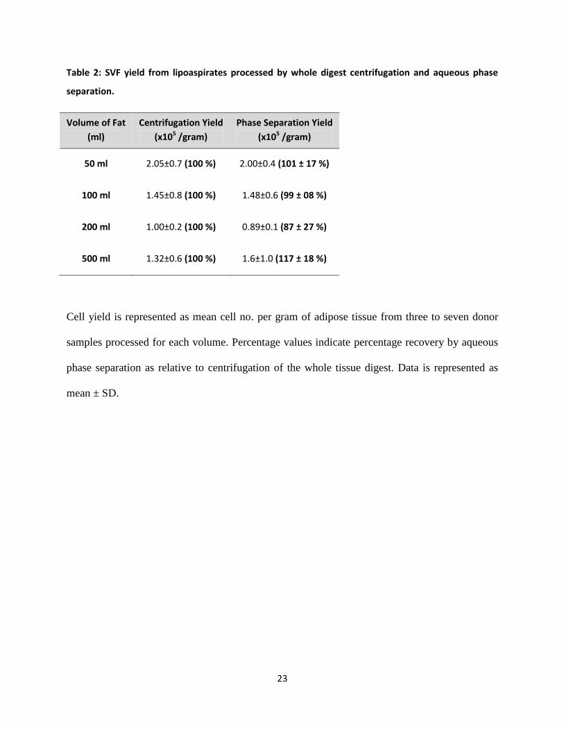
Recovery of SVF into aqueous phase of digest and scale-up: No statistically significant
differences were observed between the yields of SVF obtained from the separated aqueous phase
alone, as compared to centrifugation of the whole tissue digest (Table 2). Recovery of SVF from
the aqueous phase was tested to be efficient for volumes ranging from 50-500 ml of tissue
processed. The mean SVF yield obtained from the aqueous phase of the digest ranged from 0.89-
2.00 x10⁵ cells per gram of adipose tissue for all volumes of fat processed, and this yield ranges
between 87-117% of the concomitant yield obtained by centrifugation of the whole tissue digest.
The SVF obtained by both the methods was also evaluated for the number of colony forming
progenitors by CFU-F assay (Table 3). The number of CFU-F derived from the SVF isolated
from the aqueous phase alone (mean CFU-F per 100 cells=0.45) was found to be comparable
with that obtained by centrifugation of the whole tissue digest (mean CFU-F per 100 cells=0.41)
(Table 3). The results clearly show that the clonal expansion capacity in the SVF isolated using
the two methods was equivalent. Therefore, recovery of SVF by sedimentation into the aqueous
phase of the digest was determined to be an efficient method for further automation, and was
scalable from a volume of 50 ml to 500 ml lipoaspirate.

12
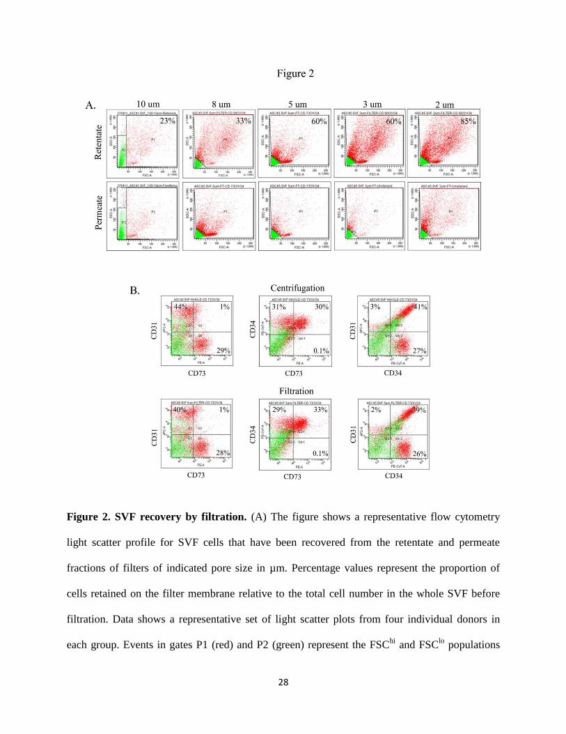
Recovery of SVF by retention on membrane filters: Relative differences in cell size between
the different cell types in the SVF was detected by measuring their light scattering properties by
flow cytometry. The major cell types known to be present in the SVF were further distinguished
by their surface marker profile as follows: ASC (CD31- CD34+), EPC (CD31+ CD34+) and EC
(CD31+ CD34-). From the light scatter profile (Figure 1) the ASC, EPC and EC populations
were found to have high forward scatter (FSChi
), and could be distinguished from the debris
which showed low forward and side scatter properties (FSClo
and SSClo
). Some EPC were also
found in the FSClo
region indicating that the EPC are smaller than the ASC and EC. These
results suggest that all the regenerative cell populations in the SVF, namely the ASC, EPC and
EC could be separated from cellular debris by retention on a membrane filter.
In order to recover SVF cells without centrifugation, the separated aqueous phase of the digest
was passed through filters of various biocompatible material and porosity to select the filter
specifications for retention of the desired cell populations from the SVF. The aqueous fraction
was first pre-filtered sequentially through woven nylon filters of 100 µm and 35 µm pore size to
remove coarse material such as undigested tissue, collagen fibers and cell aggregates. The filtrate
was then loaded onto polycarbonate track etch (PCTE) filters of different pore size ranging from
10 µm to 2 µm. The fractionation of the different SVF cell components between the retentate
(collected on the filter) and the permeate (flow-through) was then detected by flow cytometry.
Greater than 60% of the SVF cells were recovered in the retentate on PCTE filters of pore size 5
µm and below (Figure 2A), while considerable loss of cells was observed where 10 µm and 8 µm
filters were used. A specification of 5 µm was therefore selected as the optimal pore size for
maximal recovery of SVF cells by filtration from the aqueous phase of the tissue digest.

13
Retention of cells on the 5 um filter was visualized by staining with Toluidene blue.
Representative images for the stained filters is provided in Supplementary figure 1.
The composition of SVF recovered by aqueous phase filtration through the 5 µm PCTE
membranes was compared against SVF recovered by centrifugation of the whole tissue digest
and the results were found to be highly similar (Figure 2B). The average proportions of the
different SVF components from centrifugation and filtration methods was determined to be
22±7% and 26±3% respectively for CD31- CD34+ ASC, 36±10% and 31±10% respectively for
CD31+ CD34+ EPC and 3±1% and 3±2% respectively for CD31+ CD34- EC populations. These
results led us to conclude that separation of the aqueous phase containing SVF from the fat
fraction, followed by recovery of the cells on membrane filters is feasible and warrants further
development towards automation.
Process automation: A tissue digestion chamber, heating and agitation mechanism, and
multistage filtration units were developed towards automation of the various steps involved in
SVF isolation namely (Figure 3), washing and digestion of the lipoaspirate tissue, aqueous phase
separation, cell concentration by sequential filtration through nylon filters of 100 µm and 35 µm
pore size and ultimate recovery of the SVF on 5 µm PCTE filters. The device was programmed
to process 500 ml of lipoaspirate tissue. Effect of peristaltic pump was initially studied to ensure
that the pump action does not cause mechanical damage to the tissue and cells. The yield and
viability of SVF isolated from the lipoaspirate tissue before and after passing through the
peristaltic pump was found to be comparable (Supplementary table 2, p=0.99 by paired t test,
n=3).

14
Individual subsystems developed for tissue digestion and filtration were tested independently to
determine the efficiency of the automated process in comparion with manual SVF isolation by
centrifugation. The time taken for each of the process steps is shown in the accompanying table
in figure 3. The total process time from initiation of the run to recovery of the SVF was found to
be 133 min. The mean volume of SVF recovered from a single filter unit was found to be 10.8
ml (range 4-20 ml). The yield and viability of the SVF cells obtained by automated tissue
digestion and filtration using the device was found to be equivalent (p=0.8, n=5, paired t test) to
that obtained by the manual process (Figure 4A & B; Table 4). The average yield from the
automated isolation was found to be 94±28% for the digestion and 98±21% for the filtration
systems respectively, when compared to the baseline (100%) yields obtained with the manual
process; and the viability of the cells isolated by both methods was consistently greater than
90%. The composition of the SVF cells recovered from the automated process was determined
by flow cytometric analysis, and was found to be comparable with the conventional manual
process (Table 5). In particular, the mean percentage of CD31- CD34+ cells from the automated
process was 25±9% as compared to 20±5% from the manual process, and the percentage of
CD31+ CD34+ cells obtained was 24±6% and 30±10% respectively for the automated and
manual methods. Adipose tissue is also reported to contain a population of pericytes that are
CD31-CD146+ [19]. For detection of pericytes, the CD31-ve cells in the SVF were gated (Figure
5A, upper and lower left panels) and analyzed for the expression of CD34 and CD146 (Figure
5A, upper and lower right panels). As shown in Figure 5A, the percentage of CD34-CD146+
cells within the CD31-ve population of the SVF was found to be comparable between cells
isolated by the manual and automated methods. The percentage of CD31-CD146+ cells in the
total SVF was also found to be equivalent between the manual process (1.9±1.2%) and the

15
automated process (2.0 ±1.4%). These observations confirm that the SVF isolated using the
automated device contains the relevant proportions of ASC, EPC and pericyte populations.
Gene expression analysis by semi-quantitative RT-PCR revealed that the expression levels of
vascular endothelial growth factor (VEGF-A), hepatocyte growth factor (HGF), Insulin like
growth factor-1 (IGF-1), CD31, CD34, von Willebrand factor (vWF) and vascular endothelial
cadherin (VE-Cadherin) in the SVF cells isolated by the automated process were comparable
with the manually isolated SVF (Figure 5B). Although minor differences in the expression levels
of certain genes, for example HGF, were observed between different donors, there were no
remarkable differences between SVF isolated using manual or automated methods. The number
of CFU-F derived from SVF isolated by the automated system was also found to be comparable
with the SVF obtained by manual processing (Figure 5C).
Taken together, these data suggest that the yield and viability of the SVF cells, as well as their
phenotypic composition, CFU-F potential and angiogenic gene expression profile are equivalent
between the manual and automated processes.
Discussion:
SVF harbors enormous therapeutic potential for a multitude of regenerative and reconstructive
applications. However, its use can be limited by the numerous challenges associated with
obtaining clinical grade SVF cells. These include lack of c-GMP compliant infrastructure,
physician know-how and skilled technical staff; time taken for isolation as well as logistic
hurdles, all of which can impact the sterility, viability, functionality and consistency of the cell
preparation. These challenges can be overcome by the availability of an automated, closed

16
system for cell isolation that can be used at the clinic to deliver SVF cells in a highly quality
controlled and consistent manner.
Following enzymatic digestion of the fat tissue, centrifugation of the whole digest is the
conventional and most widely used process for recovery of SVF, both by manual processing [18]
and in existing automated systems [11, 20]. We aimed at developing a system and process for
isolating clinical grade SVF cells without the use of a centrifuge in order to decrease the device
footprint and develop a gentle cell isolation process. Towards this aim, we have developed a
novel process for SVF isolation wherein the aqueous phase of digested adipose tissue was
separated from the digested fat and the SVF cells were recovered from the aqueous phase by
retention on a membrane filter. Device development was then initiated by design and prototyping
of the main operational modules comprising of the tissue digestion chamber, heating and
agitation mechanism, and a three-stage filtration unit. The entire system was programmed to be
controlled through a digital user interface. The system was then used to digest and recover SVF
from input lipoaspirate tissue. The SVF isolated using the system was validated for yield,
viability, composition, clonal expansion capacity and expression of angiogenic growth factors, in
comparison against the manual process using centrifugation.
The partitioning of the SVF into the aqueous phase of the tissue digest was found to be efficient,
as the yield, viability, cell composition and CFU-F capacity of SVF recovered by such
fractionation was observed to be equivalent to the conventional centrifugation method. The yield
of SVF per gram of adipose tissue from our process (1-2 x10⁵) also compares well with those
reported for the Celution (2-2.5 x10⁵) and Multi-station (1 x10
5) systems [20], and is far superior
to yields reported for the Cha-station (< 2.5 x10⁴) and Lipokit (< 5 x10
4) [20], all of which are
systems developed for point-of-care isolation of SVF.

17
Lipofilling procedures for soft-tissue reconstruction can require a range of volumes of fat tissue
for grafting depending on the volume of the defect or the desired extent of augmentation. While
facial lipoatrophy can be addressed by grafting as low as 10-20 ml of fat tissue, breast
augmentation procedures might require upto 200-300 ml of the fat graft for a single breast [11-
13]. Although the ratio of SVF to be supplemented to a given volume of fat graft has still not
been systematically studied in humans, supplementation of SVF recovered from a corresponding
volume of fat tissue has been widely reported to be safe, and provide favorable clinical outcomes
[11-16]. Currently available automated systems for SVF isolation are limited by the volume of
fat that can be used. The maximal processing capacity for the fully automated Celution [20] and
Tissue Genesis Cell Isolation Systems [11] have been reported to be 360 ml and 60 ml fat tissue
respectively. Although the Multi-station has a higher capacity of 400 ml fat, it is a manually
operated system. The mean processing time reported for Celution, Multi-station, Cha-station and
Lipokit ranges from 88 to 115 minutes [20], while the Tissue Genesis Cell Isolation System has
been reported to have a shorter processing time of 65 min, its processing capacity is also
considerably lower [11]. Based on the literature cited above, the device reported in this study can
process a larger volume of fat than other isolation systems within a reasonable time frame of 133
min. These factors, combined with the advantage of complete automation, makes the device
extremely viable for use in the operating room, and potentially address a wide range of clinical
applications.
Selection of the filter specifications was optimized to obtain an SVF yield, viability and
composition equivalent to that obtained using the conventional centrifugation method. A three-
stage continuous filtration system fitted with a mechanical agitation unit was developed, wherein
the aqueous phase is pre-filtered through 100 µm and 35 µm nylon filters and the SVF is

18
ultimately recovered on a 5 µm PCTE filter. Undigested tissue, cell and tissue aggregates and
collagen fibers are separated from the SVF during 1st and 2
nd stage filtration. Our results
demonstrate that the cellular debris is also separated from the SVF which is retained on the 3rd
filter. Centrifugation, on the other hand, would result in sedimentation of all dense material
including aggregates and fibrous tissue along with the SVF cells. Therefore, we conclude that the
filtration system employed has an advantage over centrifugation-based methods in obtaining
SVF cells that are free from tissue aggregates and collagen fibers, which could pose
complications in injection of SVF during surgery.
Composition of SVF obtained by the automated process was found to include CD34+CD31-
ASC, CD34+CD31+ endothelial progenitor cells, CD34-CD31+ mature endothelial cells and
CD31-CD146+ pericytic cells. The relative percentages of the different cell types was
comparable to SVF isolated by the manual centrifugation-based method. The functionality of the
SVF isolated by the automated process was demonstrated by the ability to form colony forming
units (CFU-F) representing self-renewal capacity of the ASC contained in the SVF. Gene
expression analysis confirmed the presence of endothelial and progenitor cells from the
expression of CD31, CD34, VE-cadherin and Von Willebrand factor. Production of angiogenic
and anti-apoptotic growth factors was also confirmed from expression of VEGF and IGF in the
SVF.
Conclusions:
In conclusion, we have successfully demonstrated proof-of-concept for automated isolation of
SVF using a prototype device and a xeno-free isolation process. We are currently working

19
towards development of a beta unit that will be validated for efficacy, sterility and safety
requirements prior to pilot human trials. Concentration of clinical grade SVF cells without the
use of a centrifuge would deliver a compact system with a small footprint that can be easily
accommodated in a clinical setting. In a country like India and other developing countries where
lack of infrastructure and physician awareness pose huge hurdles for the adoption and
penetration of SVF based interventions in the clinic, such a point-of-care device can provide
considerable benefit and access to SVF.
Acknowledgements: We sincerely thank Dr. Anantheshwar YN, Dr. Ashok BC, Dr. Gunasekar
Vuppalapati and Dr. Prashantha Kesari for their expert opinion and consultancy. We
acknowledge and thank Dr. Venkatesh Gopal for his contribution to device design, as well as Mr.
Manjunath Bylappa, Mr. Prajode Thiruvambattil and Mr. Vasanth Kumar who were the
engineering team behind the device. We acknowledge and thank Dr. Nutan and Mr. Sudip
Biswas for invaluable assistance provided during the study. This study was fully funded by
Stempeutics Research Pvt. Ltd. All authors declare no potential conflict of interest.
20
References:
[1] Mizuno H, Tobita M, Uysal AC. Concise review: Adipose-derived stem cells as a novel tool
for future regenerative medicine. 1
Stem Cells 2012 May;30 (5):804-10.
[2] Tallone T, Realini C, Böhmler A et al. Adult human adipose tissue contains several types of
multipotent cells. J Cardiovasc Transl Res. 2011 Apr;4 (2):200-10.
[3] Bourin P, Bunnell BA, Casteilla L et al. Stromal cells from the adipose tissue-derived
stromal vascular fraction and culture expanded adipose tissue-derived stromal/stem cells: a
joint statement of the International Federation for Adipose Therapeutics and Science
(IFATS) and the International Society for Cellular Therapy (ISCT). Cytotherapy. 2013
Jun;15(6):641-8.
[4] Szöke K, Brinchmann JE. Concise review: therapeutic potential of adipose tissue-derived
angiogenic cells. Stem Cells Transl Med. 2012 Sep; 1(9):658-67.
[5] Ra JC, Shin IS, Kim SH et al. Safety of intravenous infusion of human adipose tissue-derived
mesenchymal stem cells in animals and humans Stem Cells Dev. 2011 Aug;20(8):1297-308.
[6] Pak J, Chang JJ, Lee JH, Lee SH. Safety reporting on implantation of autologous adipose
tissue-derived stem cells with platelet-rich plasma into human articular joints. BMC
Musculoskelet Disord. 2013 Dec 1;14:337.
[7] Tzouvelekis A, Paspaliaris V, Koliakos G et al. A prospective, non-randomized, no placebo-
controlled, phase Ib clinical trial to study the safety of the adipose derived stromal cells-
stromal vascular fraction in idiopathic pulmonary fibrosis. J Transl Med. 2013 Jul 15;11:171.
[8] Gimble JM, Bunnell BA, Guilak F. Human adipose-derived cells: an update on the transition
to clinical translation. Regen Med. 2012 Mar;7(2):225-35.
[9] Yoshimura K, Suga H, Eto H. Adipose-derived stem/progenitor cells: roles in adipose tissue
remodeling and potential use for soft tissue augmentation. Regen Med. 2009 Mar;4(2):265-
73.
[10] Hsu VM, Stransky CA, Bucky LP, Percec I. Fat grafting's past, present, and future: why
adipose tissue is emerging as a critical link to the advancement of regenerative medicine.
Aesthet Surg J. 2012 Sep;32(7):892-9.
[11] Doi K, Tanaka S, Iida H et al. Stromal vascular fraction isolated from lipo-aspirates using an
automated processing system: bench and bed analysis. J Tissue Eng Regen Med. 2013
Nov;7(11):864-70.
[12] Pérez-Cano R, Vranckx JJ, Lasso JM et al. Prospective trial of adipose-derived regenerative
cell (ADRC)-enriched fat grafting for partial mastectomy defects: the RESTORE-2 trial. Eur
J Surg Oncol. 2012 May;38(5):382-9.
[13] Yoshimura K, Sato K, Aoi N, Kurita M, Hirohi T, Harii K. Cell-assisted lipotransfer for
cosmetic breast augmentation: supportive use of adipose-derived stem/stromal cells.

21
Aesthetic Plast Surg. 2008 Jan;32(1):48-55; discussion 56-7 Aesthetic Plast Surg. 32(1), 48-
55; discussion 56-57.
[14] Sterodimas A, de Faria J, Nicaretta B, Boriani F. Autologous fat transplantation versus
adipose-derived stem cell-enriched lipografts: a study. Aesthet Surg J. 2011 Aug;31(6):682-
93.
[15] Tanikawa DY, Aguena M, Bueno DF, Passos-Bueno MR, Alonso N. Fat grafts supplemented
with adipose-derived stromal cells in the rehabilitation of patients with craniofacial
microsomia. Plast Reconstr Surg. 2013 Jul;132(1):141-52.
[16] Chang Q, Li J, Dong Z, Liu L, Lu F. Quantitative volumetric analysis of progressive
hemifacial atrophy corrected using stromal vascular fraction-supplemented autologous fat
grafts. Dermatol Surg. 2013 Oct;39(10):1465-73.
[17] Yoshimura K, Eto H, Kato H, Doi K, Aoi N. In vivo manipulation of stem cells for adipose
tissue repair/reconstruction. Regen Med. 2011 Nov;6(6 Suppl):33-41.
[18] Yu G, Floyd ZE, Wu X, Halvorsen YD, Gimble JM. Isolation of human adipose-derived
stem cells from lipoaspirates. Methods Mol Biol. 2011;702:17-27.
[19] Zimmerlin L, Donnenberg VS, Pfeifer ME, Meyer EM, Péault B, Rubin JP, Donnenberg AD.
Stromal vascular progenitors in adult human adipose tissue. Cytometry A. 2010
Jan;77(1):22-30.
[20] Aronowitz JA, Ellenhorn JD. Adipose stromal vascular fraction isolation: a head-to-head
comparison of four commercial cell separation systems. Plast Reconstr Surg. 2013
Dec;132(6):932e-9e.

22
Table 1: Primer Sequence used for semi quantitative RT-PCR analysis
Ta, annealing temperature; bp, base pair.
Gene
Forward Primer
Reverse primer
Ta °C
Size (bp)
18S 5`-CGGCTACCACATCCAAGGAA-3` 5`-GCTGGAATTACCGCGGCT-3` 56 186
CD31 5`-CAGGGTGACACTGGACAAGA-3` 5`-GGAGCAGGACAGGTTCAGTC-3` 59 650
CD34 5`-AATGAGGCCACAACAAACATCACA-3` 5`CTGTCCTTCTTAACCTCCGCACAGC-3` 57 400
VEGF-A 5`-CGAAGTGGTGAAGTTCATGGATG-3` 5`TTCTGTATCAGTCTTTCCTGGTGAG-3` 60 476
IGF-1 5`-GACATGCCCAAGACCCAGAAGGA-3` 5`-CGGTGGCATGTCACTCTTCACTC-3` 63 118
HGF 5`-ATGCATCCAAGGTCAAGGAG-3` 5`-TTCCATGTTCTTGTCCCACA-3` 61 349
v-WF 5`-TAAGTCTGAAGTAGAGGTGG-3` 5`-AGAGCAGCAGGAGCACTGGT-3` 59 100
VE-Cad 5`-GTGGAAGCGCGAGATGCCCA-3` 5`-AGCGTCCTGGTAGTCGCCCC-3` 59 237
23
Table 2: SVF yield from lipoaspirates processed by whole digest centrifugation and aqueous phase
separation.
Volume of Fat
(ml)
Centrifugation Yield
(x105 /gram)
Phase Separation Yield
(x105 /gram)
50 ml 2.05±0.7 (100 %) 2.00±0.4 (101 ± 17 %)
100 ml 1.45±0.8 (100 %) 1.48±0.6 (99 ± 08 %)
200 ml 1.00±0.2 (100 %) 0.89±0.1 (87 ± 27 %)
500 ml 1.32±0.6 (100 %) 1.6±1.0 (117 ± 18 %)
Cell yield is represented as mean cell no. per gram of adipose tissue from three to seven donor
samples processed for each volume. Percentage values indicate percentage recovery by aqueous
phase separation as relative to centrifugation of the whole tissue digest. Data is represented as
mean ± SD.

24
Table 3: Fibroblastic colony forming unit (CFU-F) assay for SVF recovered by aqueous phase separation as compared to whole digest centrifugation.
Plating Density (SVF Cells /10 cm2)
Centrifugation Phase Separation
20000 72±16 101±24
10000 48±14 42±16
5000 20±6 21±5
Data represents mean number of CFU-F obtained for each plating density from three individual
donors, each assayed in duplicate. Errors represent SD from three individual donor samples.

25
Table 4: Yield and viability of SVF obtained by the automated process using filtration technology as compared to the manual process of cell isolation using centrifugation
Digestion Filtration
Manual Automated Manual Automated
SVF Yield (x105 /gram) 0.90 ± 0.5 0.82 ± 0.6 1.15 ± 0.3 1.17 ± 0.5
Percentage Yield 100 % 94 ± 28 % 100 % 98 ± 21 %
Percentage Viability 96± 3.0 % 96± 2.1 % 97.3 ± 1.5 % 97.5 ± 2.8 %
Data is represented as mean ± SD for five individual samples tested. SVF Yield is represented as
cell no. x105 / gram of adipose tissue. Percentage yield indicates percentage recovery by
automated processing relative to the yield from manual processing taken as the baseline
comparator. Difference in cell yield between manual centrifugation method and automated
filtration is not significant (p=0.8, paired t-test).

26
Table 5: Composition of SVF isolated by the automated process as compared to manual isolation.
Cell Type Marker Profile Manual Automated
ASC CD34+CD31- 20 ±5 25 ±9
EPC CD34+CD31+ 30 ±10 24 ±6
Mature EC CD34-CD31+ 4 ±3 5 ±1
Pericyte CD31--CD146+ 1.9 ±1.2 2.0 ±1.4
Lymphocytes HLA DR+ 24 ±10 27 ±10
RBC GpA+ 12 ±5 15 ±11
Table represents mean percentage positive cells with standard error, from four different data sets.
ASC= adipose derived stem/stromal cells; EPC= endothelial progenitor cells; RBC= red blood
cells.

27
Figure 1. Detection of ASC, EPC and EC populations in SVF with respect to cell size. The
figure shows a representative flow cytometry scatter profile for SVF cells that have been stained
with fluorescent tagged antibodies against the cell surface markers CD31 and CD34. (A)
Forward scatter (FSC) and side scatter (SSC). Events in gates P1 and P2 represent the FSChi
and
FSClo
populations respectively. (B) Detection of FSChi
cells expressing CD31 and CD34. (C)
Detection of FSClo
cells expressing CD31 and CD34.
28
Figure 2. SVF recovery by filtration. (A) The figure shows a representative flow cytometry
light scatter profile for SVF cells that have been recovered from the retentate and permeate
fractions of filters of indicated pore size in µm. Percentage values represent the proportion of
cells retained on the filter membrane relative to the total cell number in the whole SVF before
filtration. Data shows a representative set of light scatter plots from four individual donors in
each group. Events in gates P1 (red) and P2 (green) represent the FSChi
and FSClo
populations

29
respectively. (B) Antibody labelling and flow cytometric detection of cell surface markers
expressed by SVF cells recovered by centrifugation or filtration using 5 µm PCTE filter. The
percentage positive cells for each marker combination is calculated after subtraction of the non-
specific fluorescence obtained with the isotype controls. Data shows a representative set of plots
from four individual donors in each group. Plots represent events from both FSChi
(red) and
FSClo
(green) populations.

30
Figure 3. Schematic representation of the automated process for isolation of SVF from
lipoaspirate tissue. The schematic represents the various steps involved in SVF isolation. (1)
The automated run is initiated by the user via a graphical user interphase; (2) Lipoaspirate tissue
is transferred into the tissue processing unit with the help of pump; (3) Wash sequence comprises
of (3a) transfer of buffer into the tissue processing unit with the help of pump, (3b) washing of
tissue by agitation and (3c) phase separation into upper fat and lower aqueous phases, followed
by removal of the lower aqueous phase. The wash sequence is repeated thrice for complete
removal of blood and tumescent fluids from the fat tissue; (4) the tissue is enzymatically
digested. A digestion sequence comprises of (4a) transfer of enzyme into the tissue processing
unit with the help of pump, (4b) the tissue is digested under controlled temperature and constant

31
agitation, (4c) phase separation into upper fat and lower aqueous phases; (5) the aqueous fraction
of the digest is concentrated by filtration to deliver the SVF. The time taken for each of the
process steps is shown in the accompanying table. Total process time is 133 min.

32
Figure 4. Comparison of SVF yield from manual and automated process. Yield of SVF
obtained by automated digestion (A) and filtration (B) as compared to the manual process of cell
isolation using centrifugation technology. Data is represented as mean ± SD for four individual
samples tested. Difference in cell yield between manual centrifugation method and automated
filtration is not significant (p=0.8, paired t-test).

33
Figure 5. Phenotypic and functional characterization of SVF isolated by the manual and
automated process. (A) Detection of pericytes in SVF isolated by the manual and automated
process. The CD31-ve cells in the SVF were gated (depicted in blue) and analyzed for expression
of CD34 and CD146. Values represent percentage of CD31-ve cells expressing CD34 or CD146.
Data shows a representative set of plots from four individual donors in each group. (B) Semi-
quantitative RT–PCR analysis for expression of angiogenic markers in SVF obtained from
manual and automated process. 18s ribosomal RNA expression was used to normalize cDNA

34
concentration for each sample set. Data depicts gene expression in two pairs of samples out of
four pairs of individual donors analysed. Man= Manual, Auto= Automated. (C) Clonal expansion
potential of SVF obtained by manual and automated process. Data represents mean number of
CFU-F obtained for each plating density from four individual donors, each assayed in duplicate.
Errors represent SD from four individual donors.