detecting diabetic retinopathy from retinal fundus images
TRANSCRIPT
Detecting Diabetic Retinopathy fromRetinal fundus images using DC-CNN
MSc Research Project
Data Analytics
Ananya Pratap Singh ChandelStudent ID: x19237529
School of Computing
National College of Ireland
Supervisor: Prof. Noel Cosgrave
www.ncirl.ie

National College of IrelandProject Submission Sheet
School of Computing
Student Name: Ananya Pratap Singh Chandel
Student ID: x19237529
Programme: Data Analytics
Year: 2021
Module: MSc Research Project
Supervisor: Prof. Noel Cosgrave
Submission Due Date: 16-08-2021
Project Title: Detecting Diabetic Retinopathy from Retinal fundus imagesusing DC-CNN
Word Count: 6763
Page Count: 20
I hereby certify that the information contained in this (my submission) is informationpertaining to research I conducted for this project. All information other than my owncontribution will be fully referenced and listed in the relevant bibliography section at therear of the project.
ALL internet material must be referenced in the bibliography section. Students arerequired to use the Referencing Standard specified in the report template. To use otherauthor’s written or electronic work is illegal (plagiarism) and may result in disciplinaryaction.
Signature:
Date:
PLEASE READ THE FOLLOWING INSTRUCTIONS AND CHECKLIST:
Attach a completed copy of this sheet to each project (including multiple copies). �Attach a Moodle submission receipt of the online project submission, toeach project (including multiple copies).
�
You must ensure that you retain a HARD COPY of the project, both foryour own reference and in case a project is lost or mislaid. It is not sufficient to keepa copy on computer.
�
Assignments that are submitted to the Programme Coordinator office must be placedinto the assignment box located outside the office.
Office Use Only
Signature:
Date:
Penalty Applied (if applicable):
Detecting Diabetic Retinopathy from Retinal fundusimages using DC-CNN
Ananya Pratap Singh Chandelx19237529
Abstract
Today one of the major causes of blindness in adults is diabetic retinopathy(DR). It is a progressive disease and can be categorized into four stages accord-ing to its severity. Early detection of DR can save individuals from developingpermanent blindness. Hence, the governments run several screening programs toprevent DR. DR detection remains a problem due to the limited number of trainedclinicians who can perform the diagnosis. Hence need to develop an automated DRdetection system is evident. This research aims at developing a novel dual-channelconvolutional network (DC-CNN) for the detection of DR using retinal fundus im-ages. DC-CNN performs a binary classification under two labels DR and No DR.The designed DC-CNN utilizes two channels for deeper feature extraction, designedusing the VGG16 transfer learning model and customized tuned CNN model. Theperformance of the designed DC-CNN has been evaluated using evaluation metricslike accuracy, sensitivity, specificity, and F1 score. ResNet50 and Inception v3 aretrained on the same retinal fundus image dataset to perform a comparative analysisof the DC-CNN model. The Designed DC-CNN model outperforms all the modelsto produce an accuracy of 95.23% and sensitivity of 96.94%.
1 Introduction
Diabetic retinopathy has turned out to be a leading cause of loss of vision. It is estimatedthat by 2040, around 600 million individuals are expected to have diabetes, and one-thirdof them may have diabetic retinopathy (DR) (Luo et al.; 2021). DR is a disease that af-fects both the eyes and is most prevalent in patients whose blood sugar or blood pressureis too high. The blood vessels that supply blood to the retina are affected by DR, whichaffects the light signals sent to the brain. Proliferative DR (PDR) is the advanced stageof DR and may lead to permanent blindness. Mild non-proliferative DR (NPDR) is anearly stage of DR that can be corrected if detected on time. Due to the seriousness of thedisease, Iceland and the United Kingdom conduct systematic retina screening nationwideto prevent blindness among working-age adults. All professionals have suggested routineDR screening is a mandate, yet there is a bottleneck in screening programs due to limitedhuman assessors.
The advancement in computational capabilities and the use of deep learning algorithmswhich learn from large available datasets can surpass human assessor capabilities. Sev-eral deep learning algorithms have been applied to detect DR with high sensitivity and
1
specificity to minimize the vision-threatening DR. There are several challenges involvedwhile integrating deep learning techniques to detect DR using retinal fundus images.Using deep learning techniques for DR detection is to develop models that can deliverscreening at the community level. This research discusses the design and implementationof a novel dual-channel convolutional neural network (DC-CNN). The DC-CNN aims atachieving higher sensitivity and specificity for detecting DR using retinal fundus images.
1.1 Background & Motivation
The majority of the working-age population is a risk of developing diabetic retinopathy(DR). DR is expected to affect over 93 million people across the world. Progression toblindness can be prevented if DR is detected in its early stage, although it’s a challengingtask to detect as DR shows few symptoms in the early stages. At present, there areseveral bottlenecks in detecting DR. DR detection can be a time-consuming process as itis detected manually, where clinicians are trained to examine retinal fundus images and,based on the fundus photograph of the retina, examine the disease. The results are oftendelayed due to the reviewers’ late submission of results, which leads to delay in treatmentdue to lost follow-ups. An affected retina shows vascular abnormalities, which are causeddue to the disease. These abnormalities lead to the formation of lesions that the trainedClinicians can examine towards detection of DR.
Since the number of people having diabetes is increasing day by day, there is a strong needfor and infrastructure and skilled clinicians who can detect DR to prevent blindness. Butthe equipment and expertise required are not enough as the number continues to grow,and the rate of diabetes in the local population continues to increase. DR detection is ofutmost importance. Hence, the need for a comprehensive automatic method for detectingDR has been recognized long back. Several efforts have been made towards detecting DRby using deep learning algorithms, image classification, machine learning, and patternrecognition. The aim is to achieve a DR detection system that can be used at the com-munity level and detect DR with clinical standards. This motivates the development ofthe DCCNN model that aims at improving the specificity and sensitivity towards thedetection of Diabetic retinopathy.
1.2 Research Question & Objective
1.2.1 Research Question
RQ: To what extent can the designed dual-channel convolution neural network (DC-CNN)provide better sensitivity and specificity in detecting diabetic retinopathy using retinalfundus images?
1.2.2 Research Objective
The research objectives can be seen in the Table 1 below.
2
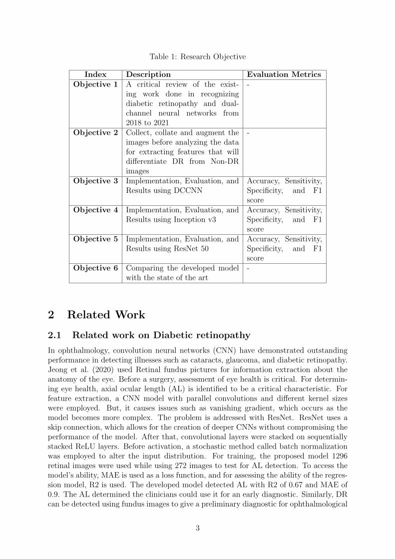
Table 1: Research Objective
Index Description Evaluation MetricsObjective 1 A critical review of the exist-
ing work done in recognizingdiabetic retinopathy and dual-channel neural networks from2018 to 2021
-
Objective 2 Collect, collate and augment theimages before analyzing the datafor extracting features that willdifferentiate DR from Non-DRimages
-
Objective 3 Implementation, Evaluation, andResults using DCCNN
Accuracy, Sensitivity,Specificity, and F1score
Objective 4 Implementation, Evaluation, andResults using Inception v3
Accuracy, Sensitivity,Specificity, and F1score
Objective 5 Implementation, Evaluation, andResults using ResNet 50
Accuracy, Sensitivity,Specificity, and F1score
Objective 6 Comparing the developed modelwith the state of the art
-
2 Related Work
2.1 Related work on Diabetic retinopathy
In ophthalmology, convolution neural networks (CNN) have demonstrated outstandingperformance in detecting illnesses such as cataracts, glaucoma, and diabetic retinopathy.Jeong et al. (2020) used Retinal fundus pictures for information extraction about theanatomy of the eye. Before a surgery, assessment of eye health is critical. For determin-ing eye health, axial ocular length (AL) is identified to be a critical characteristic. Forfeature extraction, a CNN model with parallel convolutions and different kernel sizeswere employed. But, it causes issues such as vanishing gradient, which occurs as themodel becomes more complex. The problem is addressed with ResNet. ResNet uses askip connection, which allows for the creation of deeper CNNs without compromising theperformance of the model. After that, convolutional layers were stacked on sequentiallystacked ReLU layers. Before activation, a stochastic method called batch normalizationwas employed to alter the input distribution. For training, the proposed model 1296retinal images were used while using 272 images to test for AL detection. To access themodel’s ability, MAE is used as a loss function, and for assessing the ability of the regres-sion model, R2 is used. The developed model detected AL with R2 of 0.67 and MAE of0.9. The AL determined the clinicians could use it for an early diagnostic. Similarly, DRcan be detected using fundus images to give a preliminary diagnostic for ophthalmological
3
examination.
Fundus pictures might be used to diagnose diabetic retinopathy thanks to advances indeep learning technology (DR). The majority of systems rely on standard fundus im-ages. Ultra-wide fundus (UWF) embodiments were utilized by Oh et al. (2021), as theretinal surface covered by UWF was 82 percent. Using ultra-widefield fundus imagesand applying deep learning, they presented a diabetic retinopathy diagnosis method byutilizing a picture segmentation software ETDRS 7S. ResNet-34 was utilized as a resid-ual network with deep learning architecture that comprised 34 layers for classificationand DR detection. Because the UWF fundus pictures were so large, pixels with a lotof intensity were disregarded. Adam optimizer was used for optimization while keepingthe learning rate to be 0.0001. Based on the UWF photographs, a DR detection systemwas implemented, which resulted in an AUC of 91.50 and 83.3 percent accuracy in DRdetection. The technique had a flaw in that UWF photos needed to be aligned to avoidobstructions like eyelids and eyelashes. Such stumbling blocks could be avoided by usingnormal retinal fundus images. This motivates using retinal fundus images for diabeticretinopathy detection.
Artificial neural networks are usually not used for image classification purposes due to thedevelopment of various other new deep learning architectures. Harun et al. (2019) focusedon detecting Diabetic retinopathy by classifying images as DR or non-DR using artificialneural networks. They used Bayesian Regularization (BR) and Levenberg-Marquardt(LM) to train their Multi-Layered Perceptron (MLP) for performing the classification ofdata. For classification, the network utilized nineteen features that were extracted fromfundus images as inputs. The model was evaluated by varying the number of hidden nodesfor analysis. It was found that the use of LM led to poor performance when comparedwith MLP, which was trained using BR with classification performance of 67.47% testingand 72.11% training. This study showed potential for using BR in other artificial neuralnetworks. Poor classification efficiency was observed on blurry and low contrast images,which could be used by utilizing new neural model architectures like a Dual ChannelConvolutional Neural Network (DC-CNN).
A clinical technician usually diagnoses fundus images by looking at it, which makes ithard for them to recognize the presence of lesions, hence making the detection of the dis-ease difficult. Gayathri et al. (2020) realized that automated detection of DR could be achallenging task in which feature extraction can play a crucial role. Compared with olderhand-crafted methodologies, Convolutional neural networks have a superior performancein image classification efficiency. Their work used a novel CNN architecture that was usedto extract features from retinal images and later used as input for machine learning clas-sifiers. The model was evaluated using various classifiers like J48, Random Forest, SVM,Naıve Bias on image datasets like MESSIDOR and from Kaggle. Classification efficiencywas calculated by comparing precision, specificity, and accuracy for every classifier. Thestudy showed that the J48 classifier performed the best classification on MESSIDOR andother datasets with an accuracy of 99.89% for binary classification. This study showedpotential for combining CNN with other models to improve the classification accuracyfor DR detection.
Deperlıoglu and Kose (2018) utilized deep learning and image processing to diagnose
4
Diabetic retinopathy by using retinal fundus images. They used enhancement techniqueslike histogram equalization, V transform algorithm, and HSV for enhancing retinal fundusimages. Lastly, the retinal fundus images were passed through a Gaussian low pass filter.Once the images were pre-processed, a Convolutional Neural network was used to performclassification. Four hundred images were used to assess the performance of the proposedmodel from the Diabetic retinopathy detection database of Kaggle. The classificationwas performed for every stage of image pre-processing. To find the average of values,twenty experiments were performed at each stage. Experiments resulted in an accuracyof 97% and specificity of 93.33%, and sensitivity of 96.67%. Results showed high effi-ciency towards detection of Diabetic retinopathy using retinal fundus images. The imagepre-processing techniques used in this research can be used with other Hybrid modelsthat might help in enhancing the results further.
A technique called Microaneurysm Retinal vein Haemorrhage Exudate (MRHE) for fea-ture extraction and hybrid pre-processing was proposed by Zubair et al. (2020). MRHEused Edge detection (FEED) and feature enhancement to extract image features involvingvery little complexity. An efficient deep convolutional neural network called the D-CNNmodel was used to classify DR. Salient features like MA’s, haemorrhages and retinal veinswere used to train the D-CNN model that was extracted using image pre-processing tech-niques on raw images. The proposed novel model was able to classify DR on a Structuredanalysis of the retina (STARE) database that comprised of retinal fundus images. Theproposed model was compared with existing DR classification models like ANN, SVM,etc. This study suggests new models can be developed enhancing the performance ofexisting CNN models that may outperform the existing architectures to attain betterresults towards the classification of Diabetic retinopathy using retinal fundus images.
Amalia et al. (2021) utilized a mix of two deep learning architectures to identify Dia-betic Retinopathy using retinal fundus images: Long Short-Term Memory (LSTM) andConvolution Neural Network (CNN). GoogleNet was the CNN model utilized in theirpaper. A summary of the features in retinal fundus pictures was included in the output.The picture characteristics were fed into LSTM as a vector with a sentence description.Two deep learning architectures, CNN and LSTM, were used to describe and identifyDR, with the model achieving 90% accuracy. The model’s output, a descriptive phrase,would aid radiologists in their diagnosis. The study makes no mention of the disease’sseverity. The research shows potential for combining deep learning models with retinalfundus pictures to detecting diabetic retinopathy by fundus imaging.
2.2 Use of Dual Channel Convolutional Neural networks
For Image classification, deep neural networks are capable of giving deep extracted fea-tures. Yang et al. (2018) performed image classification of hyperspectral images (HIS)by designing a dual-channel convolutional neural network (CNN). The first channel wasdesigned to extract hierarchical spectral features to gain maximum advantage from HSIimages, while the second channel is used to extract the hierarchical spatial-related fea-ture. The two channels were designed using two customized DenseNets. The designedmodel was trained experimentally by dense growth rates and several widen factors to at-tain optimal performance and tuning hyperparameters. The designed dual-channel neuralarchitecture, when compared with baseline models, resulted in higher classification ac-
5
curacies. Although the performance achieved by the proposed network was highest, theproposed network took longer training time. This study motivates the idea of designinga dual-channel convolutional neural network to extract extra features from retinal fundusimages and classify it to detect Diabetic retinopathy.
Poliyapram et al. (2019) proposed a new dual-channel CNN for the classification of Po-larimetric synthetic aperture radar (PolSAR) images. A dual-channel CNN is proposedto extract abundant spatial information from a PolSAR image and improve classificationresults. Both the channels are designed using two separate CNN architectures. Thesechannels can extract two sets of features that are later concatenated to achieve the finalclassification result. The research showed improved classification results, but further byadding more labeled samples for training could have been improved. Proposed dual-channel CNN showed promising improvements over other single-channel models. Thisdesign can be improved by using transfer learning in one of the channels for extractingmore features.
With the advancement in computer vision applications, it can also be applied in thedomain of civil infrastructure. Nowadays, inspection and monitoring of concrete struc-tures is assisted by computer vision techniques. Kumar and Ghosh (2020) proposed acrack detection system based on CNN for detecting cracks in concrete structures. Thecrack detection system was designed using the proposed Dual Channel ConvolutionalNetwork (DuCCNet) model that works on two channels working parallelly. The modelwas optimized further to deal with the vanishing gradient problem and increase stability.The first channel was implemented with 21 hidden layers, while the second channel isdesigned using seven layers. The proposed dual-channel model resulted in high validationaccuracy of 92.25 towards the classification of concrete defect data.
Identifying smoke is important for fire prevention and safety warning systems used inthe industries. Due to the complicated color, texture, and shape, it remains a challengeto detect smoke from an image. Gu et al. (2019) addressed this problem by designinga dual-channel CNN. The first channel of the network is composed of multiple convolu-tional layers, and max-pooling layers selective batch normalization is applied to preventoverfitting and accelerate training. The first channel is utilized for extracting detailedfeatures like the texture of smoke. In the second channel, along with convolutional andmax-pooling layers, skip connection, and global average pooling are used to avoid over-fitting and vanishing gradient problems. The second channel captures basic informationfrom a smoke image like contour. Finally, both the layers are concatenated to comple-ment each other’s performances. The results obtained from the designed dual-channelCNN beat the state-of-the-art performance by resulting in an accuracy of 99.5% over apublic dataset. This research motivates the idea of using two channels for extractingfeatures from retinal fundus images and then concatenating them to complement eachother’s performance for the detection of diabetic retinopathy (DR).
3 Methodology
An enormous amount of data is being generated from multiple sources. Extracting usefulinformation from the data that would help in decision-making is crucial. Currently, there
6
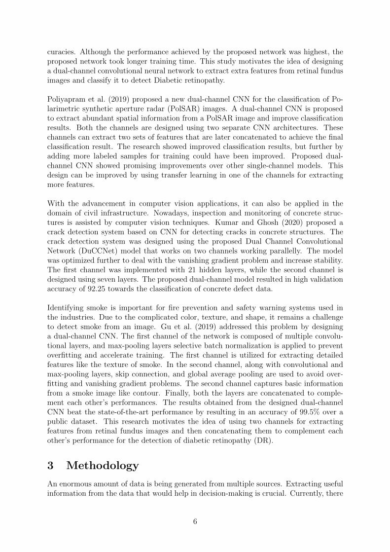
is an abundance of datasets for diabetic retinopathy detection. This research is carried outusing Knowledge Discovery in Databases (KDD) with few modifications. KDD helps find,transform, and extract meaning from raw retinal fundus data towards detecting diabeticretinopathy by designing a new dual-channel convolutional neural network. Figure 1represents the modified KDD Methodology used in the research.
Figure 1: KDD Methodology
3.1 Data Collection
To detect diabetic retinopathy (DR) from retinal fundus images, the dataset is collectedfrom Kaggle, a public repository. The images are obtained from the Diabetic RetinopathyGaussian filtered dataset, a subset of the dataset named APTOS Blindness Detection1
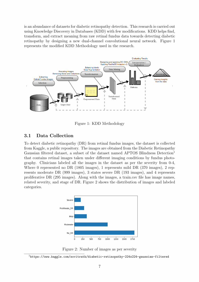
that contains retinal images taken under different imaging conditions by fundus photo-graphy. Clinicians labeled all the images in the dataset as per the severity from 0-4,Where 0 represented no DR (1805 images), 1 represents mild DR (370 images), 2 rep-resents moderate DR (999 images), 3 states severe DR (193 images), and 4 representsproliferative DR (295 images). Along with the images, a train.csv file has image names,related severity, and stage of DR. Figure 2 shows the distribution of images and labeledcategories.
Figure 2: Number of images as per severity
1https://www.kaggle.com/sovitrath/diabetic-retinopathy-224x224-gaussian-filtered
7
3.2 Data Preprocessing
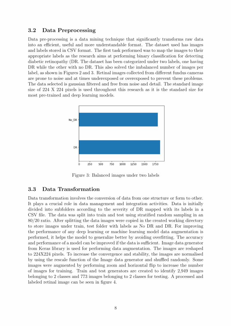
Data pre-processing is a data mining technique that significantly transforms raw datainto an efficient, useful and more understandable format. The dataset used has imagesand labels stored in CSV format. The first task performed was to map the images to theirappropriate labels as the research aims at performing binary classification for detectingdiabetic retinopathy (DR. The dataset has been categorized under two labels, one havingDR while the other with no DR. This also solved the imbalanced number of images perlabel, as shown in Figures 2 and 3. Retinal images collected from different fundus camerasare prone to noise and at times underexposed or overexposed to prevent these problems.The data selected is gaussian filtered and free from noise and detail. The standard imagesize of 224 X 224 pixels is used throughout this research as it is the standard size formost pre-trained and deep learning models.
Figure 3: Balanced images under two labels
3.3 Data Transformation
Data transformation involves the conversion of data from one structure or form to other.It plays a crucial role in data management and integration activities. Data is initiallydivided into subfolders according to the severity of DR mapped with its labels in aCSV file. The data was split into train and test using stratified random sampling in an80/20 ratio. After splitting the data images were copied in the created working directoryto store images under train, test folder with labels as No DR and DR. For improvingthe performance of any deep learning or machine learning model data augmentation isperformed, it helps the model to generalize better by avoiding overfitting. The accuracyand performance of a model can be improved if the data is sufficient. Image data generatorfrom Keras library is used for performing data augmentation. The images are reshapedto 224X224 pixels. To increase the convergence and stability, the images are normalisedby using the rescale function of the Image data generator and shuffled randomly. Someimages were augmented by performing zoom and horizontal flip to increase the numberof images for training. Train and test generators are created to identify 2,949 imagesbelonging to 2 classes and 773 images belonging to 2 classes for testing. A processed andlabeled retinal image can be seen in figure 4.
8
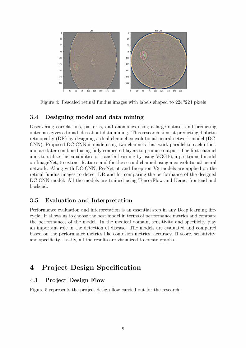
Figure 4: Rescaled retinal fundus images with labels shaped to 224*224 pixels
3.4 Designing model and data mining
Discovering correlations, patterns, and anomalies using a large dataset and predictingoutcomes gives a broad idea about data mining. This research aims at predicting diabeticretinopathy (DR) by designing a dual-channel convolutional neural network model (DC-CNN). Proposed DC-CNN is made using two channels that work parallel to each other,and are later combined using fully connected layers to produce output. The first channelaims to utilize the capabilities of transfer learning by using VGG16, a pre-trained modelon ImageNet, to extract features and for the second channel using a convolutional neuralnetwork. Along with DC-CNN, ResNet 50 and Inception V3 models are applied on theretinal fundus images to detect DR and for comparing the performance of the designedDC-CNN model. All the models are trained using TensorFlow and Keras, frontend andbackend.
3.5 Evaluation and Interpretation
Performance evaluation and interpretation is an essential step in any Deep learning life-cycle. It allows us to choose the best model in terms of performance metrics and comparethe performances of the model. In the medical domain, sensitivity and specificity playan important role in the detection of disease. The models are evaluated and comparedbased on the performance metrics like confusion metrics, accuracy, f1 score, sensitivity,and specificity. Lastly, all the results are visualized to create graphs.
4 Project Design Specification
4.1 Project Design Flow
Figure 5 represents the project design flow carried out for the research.
9
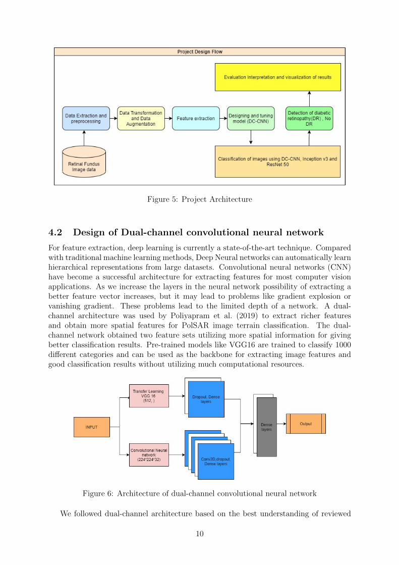
Figure 5: Project Architecture
4.2 Design of Dual-channel convolutional neural network
For feature extraction, deep learning is currently a state-of-the-art technique. Comparedwith traditional machine learning methods, Deep Neural networks can automatically learnhierarchical representations from large datasets. Convolutional neural networks (CNN)have become a successful architecture for extracting features for most computer visionapplications. As we increase the layers in the neural network possibility of extracting abetter feature vector increases, but it may lead to problems like gradient explosion orvanishing gradient. These problems lead to the limited depth of a network. A dual-channel architecture was used by Poliyapram et al. (2019) to extract richer featuresand obtain more spatial features for PolSAR image terrain classification. The dual-channel network obtained two feature sets utilizing more spatial information for givingbetter classification results. Pre-trained models like VGG16 are trained to classify 1000different categories and can be used as the backbone for extracting image features andgood classification results without utilizing much computational resources.
Figure 6: Architecture of dual-channel convolutional neural network
We followed dual-channel architecture based on the best understanding of reviewed
10
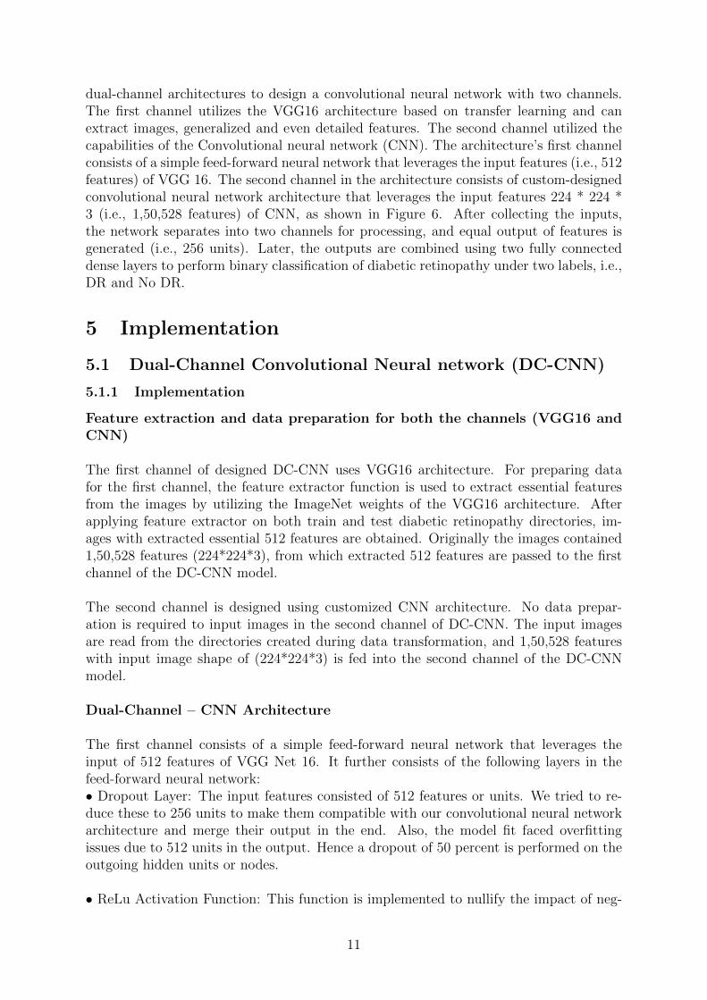
dual-channel architectures to design a convolutional neural network with two channels.The first channel utilizes the VGG16 architecture based on transfer learning and canextract images, generalized and even detailed features. The second channel utilized thecapabilities of the Convolutional neural network (CNN). The architecture’s first channelconsists of a simple feed-forward neural network that leverages the input features (i.e., 512features) of VGG 16. The second channel in the architecture consists of custom-designedconvolutional neural network architecture that leverages the input features 224 * 224 *3 (i.e., 1,50,528 features) of CNN, as shown in Figure 6. After collecting the inputs,the network separates into two channels for processing, and equal output of features isgenerated (i.e., 256 units). Later, the outputs are combined using two fully connecteddense layers to perform binary classification of diabetic retinopathy under two labels, i.e.,DR and No DR.
5 Implementation
5.1 Dual-Channel Convolutional Neural network (DC-CNN)
5.1.1 Implementation
Feature extraction and data preparation for both the channels (VGG16 andCNN)
The first channel of designed DC-CNN uses VGG16 architecture. For preparing datafor the first channel, the feature extractor function is used to extract essential featuresfrom the images by utilizing the ImageNet weights of the VGG16 architecture. Afterapplying feature extractor on both train and test diabetic retinopathy directories, im-ages with extracted essential 512 features are obtained. Originally the images contained1,50,528 features (224*224*3), from which extracted 512 features are passed to the firstchannel of the DC-CNN model.
The second channel is designed using customized CNN architecture. No data prepar-ation is required to input images in the second channel of DC-CNN. The input imagesare read from the directories created during data transformation, and 1,50,528 featureswith input image shape of (224*224*3) is fed into the second channel of the DC-CNNmodel.
Dual-Channel – CNN Architecture
The first channel consists of a simple feed-forward neural network that leverages theinput of 512 features of VGG Net 16. It further consists of the following layers in thefeed-forward neural network:• Dropout Layer: The input features consisted of 512 features or units. We tried to re-duce these to 256 units to make them compatible with our convolutional neural networkarchitecture and merge their output in the end. Also, the model fit faced overfittingissues due to 512 units in the output. Hence a dropout of 50 percent is performed on theoutgoing hidden units or nodes.
• ReLu Activation Function: This function is implemented to nullify the impact of neg-
11
ative features by replacing them with 0 and considering only positive impacting features.Another functionality provided by ReLu is maintaining the original non-linearity of theimages.
• Dense Layer: Finally, after disabling the 50 percent of incoming hidden units to DenseLayer, the final hidden units in this layer were initialized to 256 later to merge it withthe output from the second channel.
The second channel in the architecture is designed using a customized CNN that uses150,528 input features from retinal images. Hyperparameter optimization is performedusing Keras Tuner to determine the number of layers and units utilized for the designedCNN architecture. Different configurations with different layers and filters are used forcreating a model. Later Randomised search from Keras tuner is used to determine thebest model by considering validation loss as an objective function across different mod-els. Keras tuner is trained using original DR train and test images to determine the bestmodel with the least validation loss. The CNN used in the second channel is implementedby taking into consideration the best model produced by Keras tuners randomized search.Along with tuned parameters, few additional parameters are tweaked based on acquiredknowledge and test runs to get optimal performance from the network and making itcompatible with respect to the first channel. It further consists of the following layers inthe Convolutional Neural Network architecture:
• Input Data: Consisted of all the input training retinal fundus images of the shape(224*224*3)
• Convolutional Layer: The model consisted of 4 convolutional layers with hidden unitsobtained by the best model of Keras Tuner. These layers were utilized to develop accuratefeature maps by leveraging appropriate feature detectors.
• The first convolutional layer uses the minimum number of hidden units, i.e., 32,and feature detector or filter size of (3,3), which is efficient in working with theinput shape of images.
• In the second convolutional layer, hidden units are gradually increased to 64, andthe feature detector or size of the filter increases to (5,5).
• Finally, Layer 3 and 4 were set with maximum hidden units to capture 128 mostessential features to be passed to the final output layer.
• Pooling Layer: Max Pooling is implemented on the feature maps obtained from theConvolutional Layer to focus on the important features or extract features containing themaximum value.
• The filter is set to (2,2) throughout the convolutional layers, thereby ensuring thatwe capture almost all the important features in the images.
• Dropout Layer: The dropout Layer is added to overcome the final model’s overfitting.Instead of dropping the maximum percentage of hidden units from the single-layer, 25percent of hidden units from each layer are dropped to maintain uniformity in the modeland improve the fit.
12
• Flatten Layer: After all the convolutional, pooling, and dropout layer implementa-tion. The final 15,488 features were flattened into a 1D array to create input for the nextlayer.
• ReLu Activation Function: This function was implemented to nullify the impact ofnegative features by replacing them with 0 and considering only positive impacting fea-tures. Another functionality provided by ReLu is maintaining the original non-linearityof the images and preventing vanishing gradient problems.
• Dense Layer: For obtaining the same number of features as that of the first inputchannel, Fifty percent of features are randomly dropped out. And the initialized units inthe last layers are set to be 256.
Once the architecture of both the channels is finalized, and equal output of 256 unitsis obtained for both channels, it needs to be combined for performing classification ofretinal fundus images. The output is combined using fully connected dense layers with adropout of 50 percent to disable hidden units from the combined dual-channel networkand propagate only relevant features to the fully connected network. Lastly, a dense layerwith one hidden unit generates the output using the sigmoid activation function. Thesigmoid activation function is used as the network will perform binary classification ofretinal fundus images. The images are classified under two labels diabetic retinopathy(DR) or no diabetic retinopathy (No DR). The final DC-CNN model is compiled usingAdam and RMSProp Optimizer, in which Adam optimizer resulted in the best results.The loss function used for the model is binary cross-entropy since the model is performingbinary classification.
Learning curves (accuracy and loss on train test data) and accuracy are used to evaluatethe performance of the model. Finally, after running multiple instances of the modelfrom 50 to 100 epochs and batch sizes of 16,32,64 with different hyperparameters andconfigurations, the model is trained on 50epochs and batch size of 64 as it provides thebest optimal results in classifying retinal images to categories DR and No DR.
5.1.2 Evaluation and Interpretation
The model is trained multiple times by changing the network’s type, size, and the numberof layers. Best results are obtained by performing hyperparameter optimization on onechannel, consisting of a convolutional neural network using Keras tuner. Along withtuning, the model parameters like activation functions, loss functions, and the number ofepochs is varied to attain the best optimum performance by the designed model. Figure7 represents the loss curve of the model and the accuracy of the model. These Learningcurves are used to determine and optimize the performance of the designed DC-CNNmodel by diagnosing underfitting or the overfitting problem on the training and validationdata. The loss curve seen in Figure 7 shows a generalization gap as training loss is almostlower than validation loss, and the training loss gradually decreases to stability point.The learning accuracy curve shows a sudden spike and then increases gradually for bothtraining and validation. Overall, the DC-CNN model illustrates a decent fit when trainedfor 50 epochs with a batch size of 64. DC-CNN model resulted in an accuracy of 95.22%
13
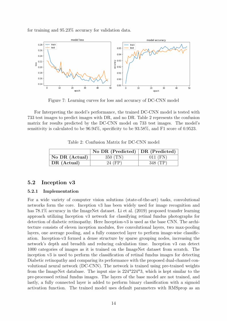
for training and 95.23% accuracy for validation data.
Figure 7: Learning curves for loss and accuracy of DC-CNN model
For Interpreting the model’s performance, the trained DC-CNN model is tested with733 test images to predict images with DR, and no DR. Table 2 represents the confusionmatrix for results predicted by the DC-CNN model on 733 test images. The model’ssensitivity is calculated to be 96.94%, specificity to be 93.58%, and F1 score of 0.9523.
Table 2: Confusion Matrix for DC-CNN model
No DR (Predicted) DR (Predicted)No DR (Actual) 350 (TN) 011 (FN)DR (Actual) 24 (FP) 348 (TP)
5.2 Inception v3
5.2.1 Implementation
For a wide variety of computer vision solutions (state-of-the-art) tasks, convolutionalnetworks form the core. Inception v3 has been widely used for image recognition andhas 78.1% accuracy in the ImageNet dataset. Li et al. (2019) proposed transfer learningapproach utilizing Inception v3 network for classifying retinal fundus photographs fordetection of diabetic retinopathy. Here Inception-v3 is used as the base CNN. The archi-tecture consists of eleven inception modules, five convolutional layers, two max-poolinglayers, one average pooling, and a fully connected layer to perform image-wise classific-ation. Inception-v3 formed a dense structure by sparse grouping nodes, increasing thenetwork’s depth and breadth and reducing calculation time. Inception v3 can detect1000 categories of images as it is trained on the ImageNet dataset from scratch. Theinception v3 is used to perform the classification of retinal fundus images for detectingDiabetic retinopathy and comparing its performance with the proposed dual-channel con-volutional neural network (DC-CNN). The network is trained using pre-trained weightsfrom the ImageNet database. The input size is 224*224*3, which is kept similar to thepre-processed retinal fundus images. The layers of the base model are not trained, andlastly, a fully connected layer is added to perform binary classification with a sigmoidactivation function. The trained model uses default parameters with RMSprop as an
14
optimizer, learning rate as 0.0001, and loss function as binary cross-entropy as it is abinary classification.
5.2.2 Evaluation and Interpretation
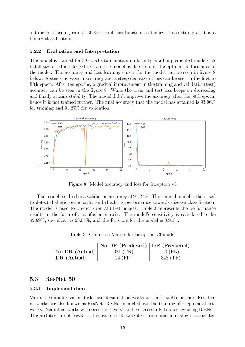
The model is trained for 50 epochs to maintain uniformity in all implemented models. Abatch size of 64 is selected to train the model as it results in the optimal performance ofthe model. The accuracy and loss learning curves for the model can be seen in figure 8below. A steep increase in accuracy and a steep decrease in loss can be seen in the first tofifth epoch. After ten epochs, a gradual improvement in the training and validation(test)accuracy can be seen in the figure 8. While the train and test loss keeps on decreasingand finally attains stability. The model didn’t improve the accuracy after the 50th epoch;hence it is not trained further. The final accuracy that the model has attained is 93.96%for training and 91.27% for validation.
Figure 8: Model accuracy and loss for Inception v3
The model resulted in a validation accuracy of 91.27%. The trained model is then usedto detect diabetic retinopathy and check its performance towards disease classification.The model is used to predict over 733 test images. Table 3 represents the performanceresults in the form of a confusion matrix. The model’s sensitivity is calculated to be89.69%, specificity is 93.04%, and the F1 score for the model is 0.9134.
Table 3: Confusion Matrix for Inception v3 model
No DR (Predicted) DR (Predicted)No DR (Actual) 321 (TN) 40 (FN)DR (Actual) 24 (FP) 348 (TP)
5.3 ResNet 50
5.3.1 Implementation
Various computer vision tasks use Residual networks as their backbone, and Residualnetworks are also known as ResNet. ResNet model allows the training of deep neural net-works. Neural networks with over 150 layers can be successfully trained by using ResNet.The architecture of ResNet 50 consists of 50 weighted layers and four stages associated
15
with it. Every stage has a total of 3 convolutional layers and is replicated. A shortcutconnection enables skipping these blocks of convolutional layers. This feature helps Res-Net in learning global features specific to data. For implementing transfer learning, theparameters for the convolutional layers are kept intact. ResNet is imported using tensor-flow.keras library, the data used as an input is already in the standard size of 224*224*3.After defining the input, the model uses pre-trained weights from the ImageNet database.Hence training all layers is not required. The Last layer is replaced with a fully connectedlayer. For performing binary classification of retinal fundus images under two labels i.e.,DR and No DR. network is then trained using Default settings like max-pooling, a learn-ing rate of 0.0001, sigmoid activation function, and binary crossentropy loss function. Asimilar architecture was used by Elswah et al. (2020) for detecting DR and determine itsgrade (e.g., Proliferative Diabetic Retinopathy (PDR), moderate, severe, or mild). Thewas trained using Indian Diabetic Retinopathy Image Dataset (IDRiD) dataset. Highestclassification accuracy achieved by their proposed model is 86.67%.
5.3.2 Evaluation and Interpretation
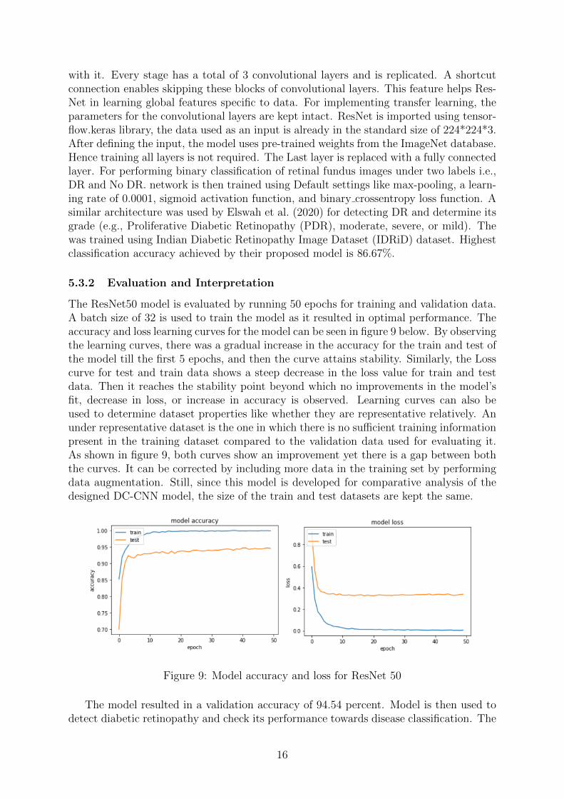
The ResNet50 model is evaluated by running 50 epochs for training and validation data.A batch size of 32 is used to train the model as it resulted in optimal performance. Theaccuracy and loss learning curves for the model can be seen in figure 9 below. By observingthe learning curves, there was a gradual increase in the accuracy for the train and test ofthe model till the first 5 epochs, and then the curve attains stability. Similarly, the Losscurve for test and train data shows a steep decrease in the loss value for train and testdata. Then it reaches the stability point beyond which no improvements in the model’sfit, decrease in loss, or increase in accuracy is observed. Learning curves can also beused to determine dataset properties like whether they are representative relatively. Anunder representative dataset is the one in which there is no sufficient training informationpresent in the training dataset compared to the validation data used for evaluating it.As shown in figure 9, both curves show an improvement yet there is a gap between boththe curves. It can be corrected by including more data in the training set by performingdata augmentation. Still, since this model is developed for comparative analysis of thedesigned DC-CNN model, the size of the train and test datasets are kept the same.
Figure 9: Model accuracy and loss for ResNet 50
The model resulted in a validation accuracy of 94.54 percent. Model is then used todetect diabetic retinopathy and check its performance towards disease classification. The
16
model is used to predict over 733 test images. Table 4 represents the performance resultsin the form of a confusion matrix. The model’s sensitivity is calculated to be 96.11%,specificity is 93.03%, and the F1 score for the model is 0.94.
Table 4: Confusion Matrix for ResNet 50 model
No DR (Predicted) DR (Predicted)No DR (Actual) 347 (TN) 14 (FN)DR (Actual) 26 (FP) 346 (TP)
6 Comparison of Results and Discussions
This section compares the three applied deep learning models, i.e., newly design dual-channel convolutional neural network (DC-CNN), ResNet, and Inception v3. The mainobjective of the research is to compare the performance of the newly designed DC-CNNwith other state-of-the-art models that have been previously applied for detecting dia-betic retinopathy (DR) using retinal fundus images. ResNet and Inception v3 architec-tures have been used previously for the detection of diabetic retinopathy by Elswah et al.(2020) and Li et al. (2019). They have achieved good classification accuracies, whichmakes them a suitable choice for performing comparative analysis. Figure 10 representsthe comparative analysis of all the three applied models. All the models are comparedon the basis of accuracy, sensitivity, specificity, and f1 score. As shown from Figure10, The newly designed DC-CNN model results in the best classification performance of(DR) with an accuracy of 95.23 percent. ResNet50 achieves the second-highest accuracyof 94.54 percent, and Inception v3 achieves the lowest accuracy of 91.27 percent. Thesensitivity of a model plays a very important role in developing a solution in the medicaldomain, the higher the sensitivity better is the model. DC-CNN model achieved thehighest sensitivity when compared with other models. The DC-CNN model achieved asensitivity of 96.95 percent, specificity of 93.58 percent, and f1 score of 95.23 percent.
In contrast, the second-highest performance is achieved by the ResNet50 model witha sensitivity of 96.11 percent, specificity of 93.03 percent, and f1 score of 94.55 percent.Lastly, the Inception v3 model achieved the lowest performance with 89.69% sensitivity,specificity of 93.04%, and f1 score of 91.34%. Overall, on observing the loss and accuracylearning curves on the train and test data, the DC-CNN model was found to fit the datavery well, while the other two models just showed decent enough fits with few errors.Hence, it can be concluded from the overall results that the newly designed DC-CNNmodel outperforms the ResNet and Inception v3 models when trained on the same datawith default settings towards predicting diabetic retinopathy from retinal fundus images.
17
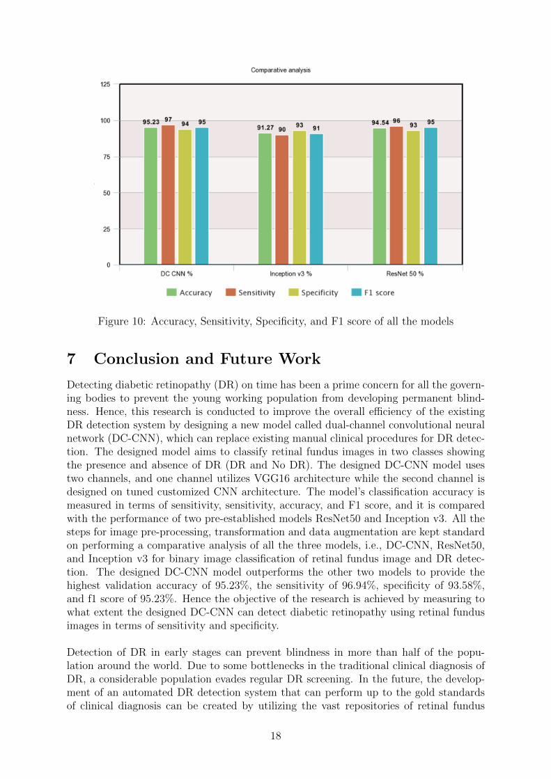
Figure 10: Accuracy, Sensitivity, Specificity, and F1 score of all the models
7 Conclusion and Future Work
Detecting diabetic retinopathy (DR) on time has been a prime concern for all the govern-ing bodies to prevent the young working population from developing permanent blind-ness. Hence, this research is conducted to improve the overall efficiency of the existingDR detection system by designing a new model called dual-channel convolutional neuralnetwork (DC-CNN), which can replace existing manual clinical procedures for DR detec-tion. The designed model aims to classify retinal fundus images in two classes showingthe presence and absence of DR (DR and No DR). The designed DC-CNN model usestwo channels, and one channel utilizes VGG16 architecture while the second channel isdesigned on tuned customized CNN architecture. The model’s classification accuracy ismeasured in terms of sensitivity, sensitivity, accuracy, and F1 score, and it is comparedwith the performance of two pre-established models ResNet50 and Inception v3. All thesteps for image pre-processing, transformation and data augmentation are kept standardon performing a comparative analysis of all the three models, i.e., DC-CNN, ResNet50,and Inception v3 for binary image classification of retinal fundus image and DR detec-tion. The designed DC-CNN model outperforms the other two models to provide thehighest validation accuracy of 95.23%, the sensitivity of 96.94%, specificity of 93.58%,and f1 score of 95.23%. Hence the objective of the research is achieved by measuring towhat extent the designed DC-CNN can detect diabetic retinopathy using retinal fundusimages in terms of sensitivity and specificity.
Detection of DR in early stages can prevent blindness in more than half of the popu-lation around the world. Due to some bottlenecks in the traditional clinical diagnosis ofDR, a considerable population evades regular DR screening. In the future, the develop-ment of an automated DR detection system that can perform up to the gold standardsof clinical diagnosis can be created by utilizing the vast repositories of retinal fundus
18
images available. Due to limitations in computation power, this research performs abinary classification by designing a new DC-CNN architecture. With enough computa-tional power, DC-CNN can be used to classify the severity of DR in retinal images, and ahigh-performing model can be deployed at the community level. The designed DC-CNNarchitecture can also be used for other similar computer vision applications.
Acknowledgement
I want to thank Prof. Noel Cosgrave for clarifying all the doubts and guiding me through-out the supervision to be on the right track. Without which it would have been tough topursue this research.
References
Amalia, R., Bustamam, A. and Sarwinda, D. (2021). Detection and description gener-ation of diabetic retinopathy using convolutional neural network and long short-termmemory, journal of physics: conference series, Vol. 1722, IOP Publishing, p. 012010.
Deperlıoglu, O. and Kose, U. (2018). Diagnosis of diabetic retinopathy by using imageprocessing and convolutional neural network, 2018 2nd International Symposium onMultidisciplinary Studies and Innovative Technologies (ISMSIT), IEEE, pp. 1–5.
Elswah, D. K., Elnakib, A. A. and Moustafa, H. E.-d. (2020). Automated diabetic ret-inopathy grading using resnet, 2020 37th National Radio Science Conference (NRSC),IEEE, pp. 248–254.
Gayathri, S., Gopi, V. P. and Palanisamy, P. (2020). A lightweight cnn for diabetic ret-inopathy classification from fundus images, Biomedical Signal Processing and Control62: 102115.
Gu, K., Xia, Z., Qiao, J. and Lin, W. (2019). Deep dual-channel neural network forimage-based smoke detection, IEEE Transactions on Multimedia 22(2): 311–323.
Harun, N. H., Yusof, Y., Hassan, F. and Embong, Z. (2019). Classification of fundusimages for diabetic retinopathy using artificial neural network, 2019 IEEE JordanInternational Joint Conference on Electrical Engineering and Information Technology(JEEIT), IEEE, pp. 498–501.
Jeong, Y., Lee, B., Han, J.-H. and Oh, J. (2020). Ocular axial length prediction based onvisual interpretation of retinal fundus images via deep neural network, IEEE Journalof Selected Topics in Quantum Electronics 27(4): 1–7.
Kumar, B. and Ghosh, S. (2020). Detection of concrete cracks using dual-channel deepconvolutional network, 2020 11th International Conference on Computing, Communic-ation and Networking Technologies (ICCCNT), IEEE, pp. 1–7.
Li, F., Liu, Z., Chen, H., Jiang, M., Zhang, X. and Wu, Z. (2019). Automatic detection ofdiabetic retinopathy in retinal fundus photographs based on deep learning algorithm,Translational vision science & technology 8(6): 4–4.
19
Luo, X., Pu, Z., Xu, Y., Wong, W. K., Su, J., Dou, X., Ye, B., Hu, J. and Mou, L.(2021). Mvdrnet: Multi-view diabetic retinopathy detection by combining dcnns andattention mechanisms, Pattern Recognition 120: 108104.
Oh, K., Kang, H. M., Leem, D., Lee, H., Seo, K. Y. and Yoon, S. (2021). Early detectionof diabetic retinopathy based on deep learning and ultra-wide-field fundus images,Scientific Reports 11(1): 1–9.
Poliyapram, V., Imamoglu, N. and Nakamura, R. (2019). Deep learning model for wa-ter/ice/land classification using large-scale medium resolution satellite images, IGARSS2019-2019 IEEE International Geoscience and Remote Sensing Symposium, IEEE,pp. 3884–3887.
Yang, G., Gewali, U. B., Ientilucci, E., Gartley, M. and Monteiro, S. T. (2018). Dual-channel densenet for hyperspectral image classification, IGARSS 2018-2018 IEEE In-ternational Geoscience and Remote Sensing Symposium, IEEE, pp. 2595–2598.
Zubair, M., Naik, M. U. K. and Mouli, G. N. C. (2020). Facile diabetic retinopathy detec-tion using mrhe-feed and classification using deep convolutional neural network, 2020IEEE 15th International Conference on Industrial and Information Systems (ICIIS),IEEE, pp. 247–252.
20