descriptions oi aphodiusdistinctus mull, and a. scybalarus ... · descriptions of aphodius...
TRANSCRIPT
Entomological Review, Vol. 76, No. 5, 1996, pp. 619-626. Translated from Entomologicheskoe Obozrenie, Vol. 75, No. 3, 1996, pp. 577-586.
Original Russian Text Copyright © 1996 by Frolov.
Descriptions oi Aphodius distinctus Mull, and A. scybalarus F. (Coleoptera, Scarabeidae) Larvae
A. V. FrolovBelarussian State University, Minsk, Belarus
Received February 1, 1995
Abstract—Larvae of Aphodius distinctus Mull, and A. scybalarius F. are described for the first time.
From materials collected in Belarus a description of Aphodius distinctus Mull, and A. scybalarius F. larvae is given.
Aphodius distinctus Mull.
The description of Aphodius distinctus larva is the first for larvae of the Chilothorax Motsch. (=Volinus Muls. et Rey) group.
Larvae used for morphological studies were collected in October 1993 in the environs of Ravnopol’e Village (Belorus). Beetles identified as A. distinctus Mull, were reared from part of larvae indistinguishable in external characters.
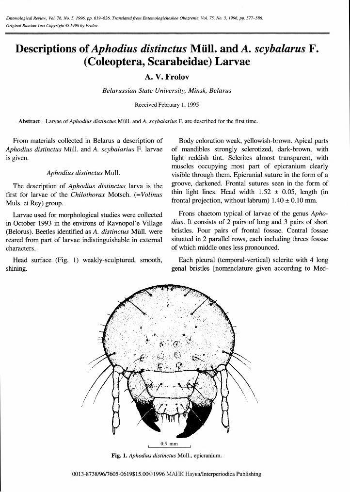
Flead surface (Fig. 1) weakly-sculptured, smooth, shining.
Body coloration weak, yellowish-brown. Apical parts of mandibles strongly sclerotized, dark-brown, with light reddish tint. Sclerites almost transparent, with muscles occupying most part of epicranium clearly visible through them. Epicranial suture in the form of a groove, darkened. Frontal sutures seen in the form of thin light lines. Head width 1.52 ± 0.05, length (in frontal projection, without labrum) 1.40 ±0.10 mm.
Frons chaetom typical of larvae of the genus Aphodius. It consists of 2 pairs of long and 3 pairs of short bristles. Four pairs of frontal fossae. Central fossae situated in 2 parallel rows, each including threes fossae of which middle ones less pronounced.
Each pleural (temporal-vertical) sclerite with 4 long genal bristles [nomenclature given according to Med-
0.5 mm
Fig. 1. Aphodius distinctus Mull., epicranium.
0013-8738/96/7605-0619$ 15.00© 1996 MAHK HavKa/Interperiodica Publishing
620 FROLOV
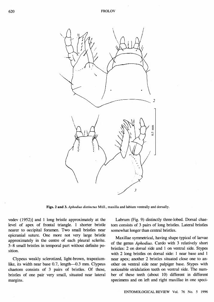
Figs. 2 and 3. Aphodius distinctus Mull., maxilla and labium ventrally and dorsally.
vedev (1952)] and 1 long bristle approximately at the level of apex of frontal triangle. 1 shorter bristle nearer to occipital foramen. Two small bristles near epicranial suture. One more not very large bristle approximately in the centre of each pleural sclerite. 5-8 small bristles in temporal part without definite position.
Clypeus weakly sclerotized, light-brown, trapeziumlike, its width near base 0.7, length—0.3 mm. Clypeus chaetom consists of 3 pairs of bristles. Of these, bristles of one pair very small, situated near lateral margins.
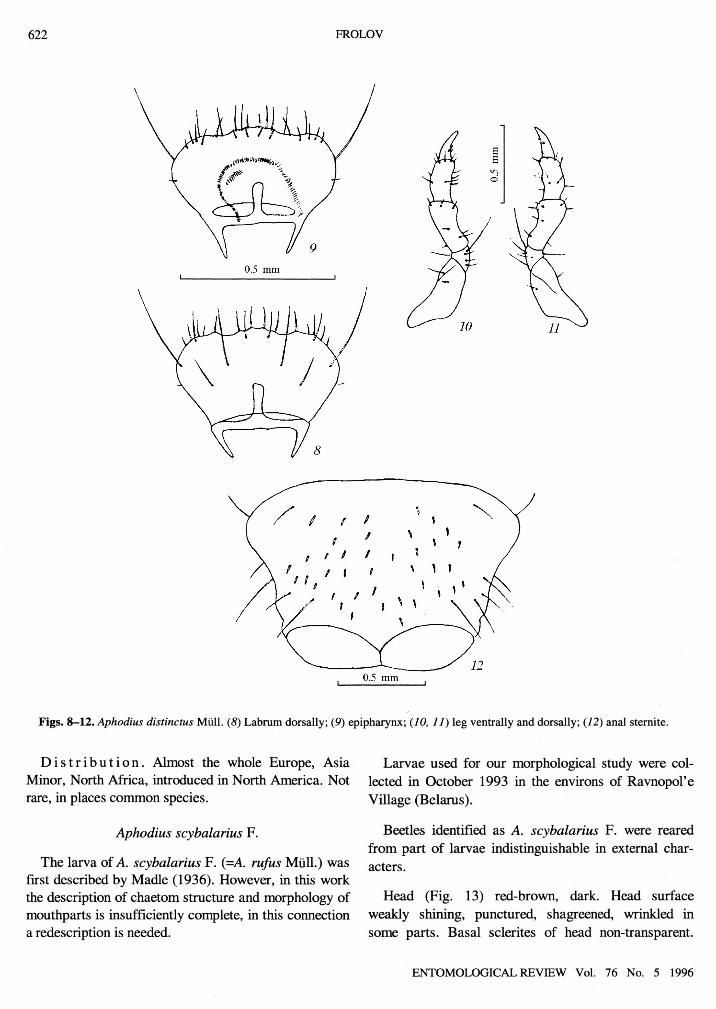
Labrum (Fig. 9) distinctly three-lobed. Dorsal chaetom consists of 3 pairs of long bristles. Lateral bristles somewhat longer than central bristles.
Maxillae symmetrical, having shape typical of larvae of the genus Aphodius. Cardo with 3 relatively short bristles: 2 on dorsal side and 1 on ventral side. Stypes with 2 long bristles on dorsal side: 1 near base and 1 near apex; another 2 bristles situated close one to another on ventral side near palpiger base. Stypes with noticeable stridulation teeth on ventral side. The number of these teeth (about 10) different in different specimens and on left and right maxillae in one speci
ENTOMOLOGICAL REVIEW Vol. 76 No. 5 1996

DESCRIPTIONS OF APHODIUS DISTINCTUS MULL. AND A. SCYBALARUS F. 621
Figs. 4-7. Aphodius distinctus Mull. (4, 5) Left mandible; (6, 7) right mandible, (4, 6) dorsally; (5, 7) ventrally.
men. Maxillary palpi with 4 bristles: 1 on segments 1 and 4; each 2 on segment 3. Apex of lacinia bifurcate; with 7 bristles along lateral margin. Galea with 11 bristles: 5 short bristles along dorsal surface, 4 long bristles closer to apex, 2 short bristles on ventral side. Maxilla and labium shown in Figs. 2 and 3.
Mandibles (Figs. 4-7) asymmetrical. Epipharynx shown in Fig. 8. Legs (Figs. 10 and 11) of the same length, with all parts developed, tarsus 1-segmented. Chaetom of typical pattern consists of 35 bristles. Coxa with 3, femur with 11, tibia with 12 bristles, tarsus with 2 short bristles. One of 7 bristles on trochanter noticea
bly longer than others. Legs of different pairs the same in length and chaetom.
Anal sternite (Fig. 12) with about 40 spines, arranged asymmetrically, without definite order and indistinctly separated into two groups. Anal sternite also with about 10 relatively long bristles. Two of these near anterior margin of sternite.
Second instar larva differs in lesser extent of epicra- nium sclerotization. Head width 1.04 ± 0.05, head length (without labrum) 0.74 ± 0.02 mm. First instar larva: head width 0.67 ± 0.02, head length0.57 ± 0.02 mm.
ENTOMOLOGICAL REVIEW Vol. 76 No. 5 1996
622 FROLOV
Figs. 8-12. Aphodius distinctus Mull. (8) Labrum dorsally; (9) epipharynx; (10, 11) leg ventrally and dorsally; (12) anal sternite.
D i s t r i b u t i o n . Almost the whole Europe, Asia Minor, North Africa, introduced in North America. Not rare, in places common species.
Aphodius scybalarius F.
The larva of A. scybalarius F. (=A. rufus Mull.) was first described by Madle (1936). However, in this work the description of chaetom structure and morphology of mouthparts is insufficiently complete, in this connection a redescription is needed.
Larvae used for our morphological study were collected in October 1993 in the environs of Ravnopol’e Village (Belarus).
Beetles identified as A. scybalarius F. were reared from part of larvae indistinguishable in external characters.
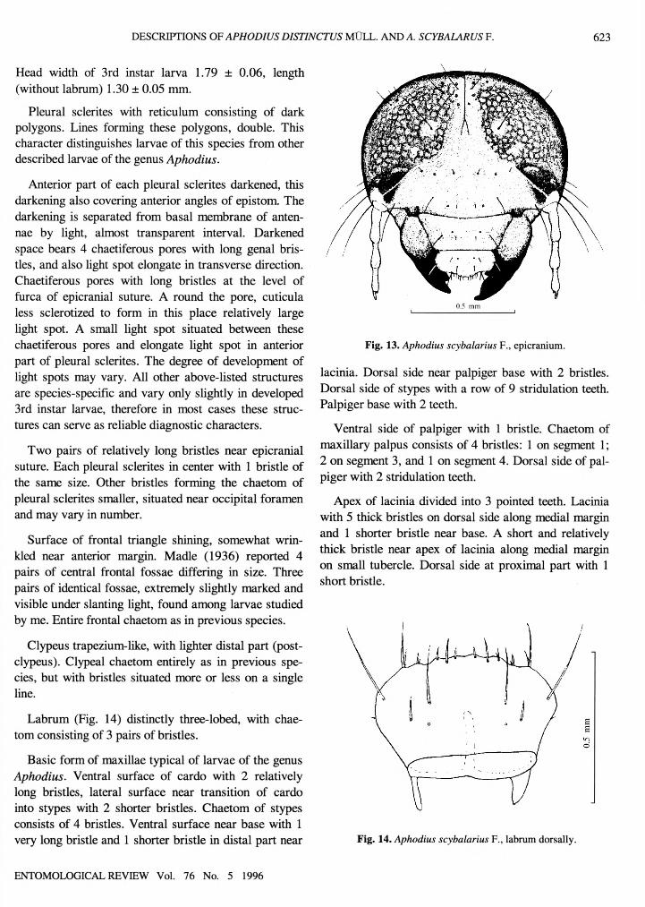
Head (Fig. 13) red-brown, dark. Head surface weakly shining, punctured, shagreened, wrinkled in some parts. Basal sclerites of head non-transparent.
ENTOMOLOGICAL REVIEW Vol. 76 No. 5 1996
DESCRIPTIONS OF APHODIUS DISTINCTUS MULL. AND A. SCYBALARUS F. 623
Head width of 3rd instar larva 1.79 ± 0.06, length (without labrum) 1.30 ± 0.05 mm.
Pleural sclerites with reticulum consisting of dark polygons. Lines forming these polygons, double. This character distinguishes larvae of this species from other described larvae of the genus Aphodius.
Anterior part of each pleural sclerites darkened, this darkening also covering anterior angles of epistom. The darkening is separated from basal membrane of antennae by light, almost transparent interval. Darkened space bears 4 chaetiferous pores with long genal bristles, and also light spot elongate in transverse direction. Chaetiferous pores with long bristles at the level of furca of epicranial suture. A round the pore, cuticula less sclerotized to form in this place relatively large light spot. A small light spot situated between these chaetiferous pores and elongate light spot in anterior part of pleural sclerites. The degree of development of light spots may vary. All other above-listed structures are species-specific and vary only slightly in developed 3rd instar larvae, therefore in most cases these structures can serve as reliable diagnostic characters.
Two pairs of relatively long bristles near epicranial suture. Each pleural sclerites in center with 1 bristle of the same size. Other bristles forming the chaetom of pleural sclerites smaller, situated near occipital foramen and may vary in number.
Surface of frontal triangle shining, somewhat wrinkled near anterior margin. Madle (1936) reported 4 pairs of central frontal fossae differing in size. Three pairs of identical fossae, extremely slightly marked and visible under slanting light, found among larvae studied by me. Entire frontal chaetom as in previous species.
Clypeus trapezium-like, with lighter distal part (post- clypeus). Clypeal chaetom entirely as in previous species, but with bristles situated more or less on a single line.
Labrum (Fig. 14) distinctly three-lobed, with chaetom consisting of 3 pairs of bristles.
Basic form of maxillae typical of larvae of the genus Aphodius. Ventral surface of cardo with 2 relatively long bristles, lateral surface near transition of cardo into stypes with 2 shorter bristles. Chaetom of stypes consists of 4 bristles. Ventral surface near base with 1 very long bristle and 1 shorter bristle in distal part near
0.5 mm
Fig. 13. Aphodius scybalarius F., epicranium.
lacinia. Dorsal side near palpiger base with 2 bristles. Dorsal side of stypes with a row of 9 stridulation teeth. Palpiger base with 2 teeth.
Ventral side of palpiger with 1 bristle. Chaetom of maxillary palpus consists of 4 bristles: 1 on segment 1; 2 on segment 3, and 1 on segment 4. Dorsal side of palpiger with 2 stridulation teeth.
Apex of lacinia divided into 3 pointed teeth. Lacinia with 5 thick bristles on dorsal side along medial margin and 1 shorter bristle near base. A short and relatively thick bristle near apex of lacinia along medial margin on small tubercle. Dorsal side at proximal part with 1 short bristle.
Fig. 14. Aphodius scybalarius F., labrum dorsally.
ENTOMOLOGICAL REVIEW Vol. 76 No. 5 1996
624 FROLOV
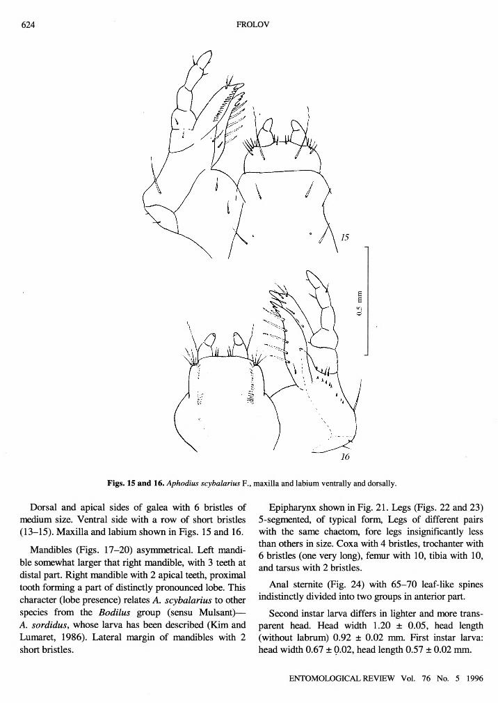
Figs. 15 and 16. Aphodius scybalarius F., maxilla and labium ventrally and dorsally.
Dorsal and apical sides of galea with 6 bristles of medium size. Ventral side with a row of short bristles (13-15). Maxilla and labium shown in Figs. 15 and 16.
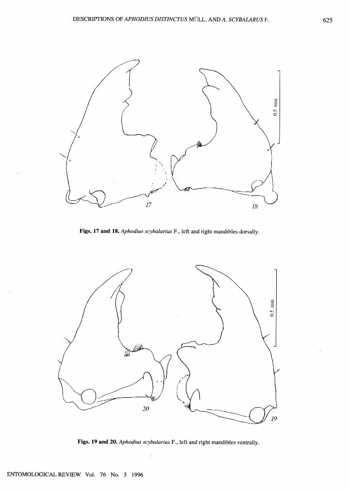
Mandibles (Figs. 17-20) asymmetrical. Left mandible somewhat larger that right mandible, with 3 teeth at distal part. Right mandible with 2 apical teeth, proximal tooth forming a part of distinctly pronounced lobe. This character (lobe presence) relates A. scybalarius to other species from the Bodilus group (sensu Mulsant)— A. sordidus, whose larva has been described (Kim and Lumaret, 1986). Lateral margin of mandibles with 2 short bristles.
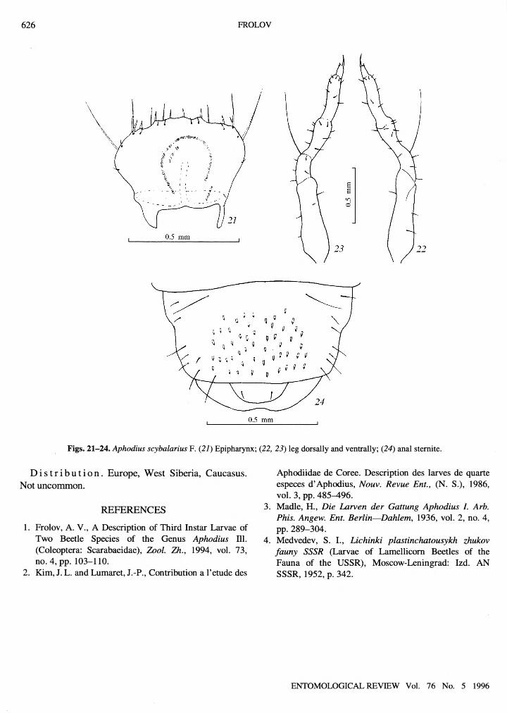
Epipharynx shown in Fig. 21. Legs (Figs. 22 and 23) 5-segmented, of typical form, Legs of different pairs with the same chaetom, fore legs insignificantly less than others in size. Coxa with 4 bristles, trochanter with 6 bristles (one very long), femur with 10, tibia with 10, and tarsus with 2 bristles.
Anal sternite (Fig. 24) with 65-70 leaf-like spines indistinctly divided into two groups in anterior part.
Second instar larva differs in lighter and more transparent head. Head width 1.20 ± 0.05, head length (without labrum) 0.92 ± 0.02 mm. First instar larva: head width 0.67 ± 0.02, head length 0.57 ± 0.02 mm.
ENTOMOLOGICAL REVIEW Vol. 76 No. 5 1996
DESCRIPTIONS OF APHODIUS DISTINCTUS MULL. AND A. SCYBALARUS F. 625
Figs. 17 and 18. Aphodius scybalarius F., left and right mandibles dorsally.
Figs. 19 and 20. Aphodius scybalarius F., left and right mandibles ventrally.
ENTOMOLOGICAL REVIEW Vol. 76 No. 5 1996
626 FROLOV
Figs. 21-24. Aphodius scybalarius F. (21) Epipharynx; (22, 23) leg dorsally and ventrally; (24) anal stemite.
D i s t r i b u t i o n . Europe, West Siberia, Caucasus. Not uncommon.
REFERENCES
1. Frolov, A. V., A Description of Third Instar Larvae of Two Beetle Species of the Genus Aphodius 111. (Coleoptera: Scarabaeidae), Zool. Zh., 1994, vol. 73, no. 4, pp. 103-110.
2. Kim, J. L. and Lumaret, J.-P., Contribution a l’etude des
Aphodiidae de Coree. Description des larves de quarte especes d’Aphodius, Nouv. Revue Ent., (N. S.), 1986, vol. 3, pp. 485-496.
3. Madle, H., Die Larven der Gattung Aphodius I. Arb. Phis. Angew. Ent. Berlin—Dahlem, 1936, vol. 2, no. 4, pp. 289-304.
4. Medvedev, S. I., Lichinki plastinchatousykh zhukov fauny SSSR (Larvae of Lamellicorn Beetles of the Fauna of the USSR), Moscow-Leningrad: Izd. AN SSSR, 1952, p. 342.
ENTOMOLOGICAL REVIEW Vol. 76 No. 5 1996