d -v p n xenopus e edward m. de robertis and … · molecularly) (de robertis & sasai 1996,...
TRANSCRIPT
10 Sep 2004 12:47 AR AR226-CB20-10.tex AR226-CB20-10.SGM LaTeX2e(2002/01/18) P1: GCE10.1146/annurev.cellbio.20.011403.154124
Annu. Rev. Cell Dev. Biol. 2004. 20:285–308doi: 10.1146/annurev.cellbio.20.011403.154124
Copyright c© 2004 by Annual Reviews. All rights reservedFirst published online as a Review in Advance on June 18, 2004
DORSAL-VENTRAL PATTERNING AND NEURAL
INDUCTION IN XENOPUS EMBRYOS
Edward M. De Robertis and Hiroki KurodaHoward Hughes Medical Institute, Department of Biological Chemistry, University ofCalifornia, Los Angeles, California 90095-1662, email: [email protected],[email protected]
Key Words beta-Catenin, Chordin, Noggin, Xnr3, Cerberus, sFRP, Frzb,Crescent, Dickkopf, Crossveinless-2, Tsg, Xolloid-related, Bambi, Sizzled, FGF,IGF, Urbilateria
■ Abstract We review the current status of research in dorsal-ventral (D-V) pat-terning in vertebrates. Emphasis is placed on recent work on Xenopus, which provides aparadigm for vertebrate development based on a rich heritage of experimental embryol-ogy. D-V patterning starts much earlier than previously thought, under the influence ofa dorsal nuclear β-Catenin signal. At mid-blastula two signaling centers are present onthe dorsal side: The prospective neuroectoderm expresses bone morphogenetic protein(BMP) antagonists, and the future dorsal endoderm secretes Nodal-related mesoderm-inducing factors. When dorsal mesoderm is formed at gastrula, a cocktail of growthfactor antagonists is secreted by the Spemann organizer and further patterns the em-bryo. A ventral gastrula signaling center opposes the actions of the dorsal organizer, andanother set of secreted antagonists is produced ventrally under the control of BMP4.The early dorsal β-Catenin signal inhibits BMP expression at the transcriptional leveland promotes expression of secreted BMP antagonists in the prospective central ner-vous system (CNS). In the absence of mesoderm, expression of Chordin and Nogginin ectoderm is required for anterior CNS formation. FGF (fibroblast growth factor) andIGF (insulin-like growth factor) signals are also potent neural inducers. Neural induc-tion by anti-BMPs such as Chordin requires mitogen-activated protein kinase (MAPK)activation mediated by FGF and IGF. These multiple signals can be integrated at thelevel of Smad1. Phosphorylation by BMP receptor stimulates Smad1 transcriptionalactivity, whereas phosphorylation by MAPK has the opposite effect. Neural tissue isformed only at very low levels of activity of BMP-transducing Smads, which requirethe combination of both low BMP levels and high MAPK signals. Many of the molec-ular players that regulate D-V patterning via regulation of BMP signaling have beenconserved between Drosophila and the vertebrates.
CONTENTS
INTRODUCTION . . . . . . . . . . . . . . . . . . . . . . . . . . . . . . . . . . . . . . . . . . . . . . . . . . . . . 286Inductive Properties of Dorsal Mesoderm . . . . . . . . . . . . . . . . . . . . . . . . . . . . . . . . . 286D-V Patterning Mutations Affect the BMP Pathway . . . . . . . . . . . . . . . . . . . . . . . . 287
1081-0706/04/1115-0285$14.00 285
Ann
u. R
ev. C
ell.
Dev
. Bio
l. 20
04.2
0:28
5-30
8. D
ownl
oade
d fr
om a
rjou
rnal
s.an
nual
revi
ews.
org
by "
UN
IV. O
F C
AL
IF.,
LO
S A
NG
E"
on 0
2/15
/05.
For
per
sona
l use
onl
y.

10 Sep 2004 12:47 AR AR226-CB20-10.tex AR226-CB20-10.SGM LaTeX2e(2002/01/18) P1: GCE
286 DE ROBERTIS � KURODA
THE EARLY β-CATENIN SIGNAL . . . . . . . . . . . . . . . . . . . . . . . . . . . . . . . . . . . . . . 288The Cortical Rotation . . . . . . . . . . . . . . . . . . . . . . . . . . . . . . . . . . . . . . . . . . . . . . . . 288Two Dorsal Signaling Centers at Blastula . . . . . . . . . . . . . . . . . . . . . . . . . . . . . . . . . 288
THE DORSAL GASTRULA CENTER . . . . . . . . . . . . . . . . . . . . . . . . . . . . . . . . . . . . 290The Spemann Organizer Is a Source of Secreted Antagonists . . . . . . . . . . . . . . . . . 290Chordin, Noggin, and Xnr3 . . . . . . . . . . . . . . . . . . . . . . . . . . . . . . . . . . . . . . . . . . . . 290The Wnt Antagonists: Frzb-1, Crescent, sFRP-2, and Dickkopf . . . . . . . . . . . . . . . 292Cerberus . . . . . . . . . . . . . . . . . . . . . . . . . . . . . . . . . . . . . . . . . . . . . . . . . . . . . . . . . . . 293
THE VENTRAL GASTRULA CENTER . . . . . . . . . . . . . . . . . . . . . . . . . . . . . . . . . . . 293The BMP4 Synexpression Group . . . . . . . . . . . . . . . . . . . . . . . . . . . . . . . . . . . . . . . 293Crossveinless-2, Twisted Gastrulation, Xolloid-Related, and Bambi . . . . . . . . . . . . 294Sizzled . . . . . . . . . . . . . . . . . . . . . . . . . . . . . . . . . . . . . . . . . . . . . . . . . . . . . . . . . . . . 296
THE ROLE OF NEUROECTODERM IN CNS FORMATION . . . . . . . . . . . . . . . . . . 296The Neural Induction Default Model . . . . . . . . . . . . . . . . . . . . . . . . . . . . . . . . . . . . 297Integrating Signals at the Level of Smads . . . . . . . . . . . . . . . . . . . . . . . . . . . . . . . . . 298β-Catenin, Chordin, and Noggin Are Required in Blastula Ectoderm . . . . . . . . . . . 300
CONSERVED MOLECULAR MECHANISMS OF BMPREGULATION . . . . . . . . . . . . . . . . . . . . . . . . . . . . . . . . . . . . . . . . . . . . . . . . . . . . . . 301
Inversion of the D-V Axis in Evolution . . . . . . . . . . . . . . . . . . . . . . . . . . . . . . . . . . . 301Communicating Dorsal and Ventral Signals . . . . . . . . . . . . . . . . . . . . . . . . . . . . . . . 303
INTRODUCTION
All vertebrates share a conserved body plan. In the anterior-posterior (A-P) axisthey form head, trunk, and tail, and in the dorsal-ventral (D-V) axis the backboneand belly of the animal. During development, dorsal ectoderm gives rise to neu-ral plate and eventually to the entire central nervous system (CNS), and ventralectoderm gives rise to epidermis and its derivatives. The mesoderm differentiates–from dorsal to ventral— into prechordal plate and notochord, somite (which formsskeletal muscle, vertebral column, and dermis), kidney, lateral plate mesoderm,and ventral blood islands. The molecular nature of the signals that ensure thesestereotypical choices of cell and tissue differentiations in the embryo is beginningto emerge.
Inductive Properties of Dorsal Mesoderm
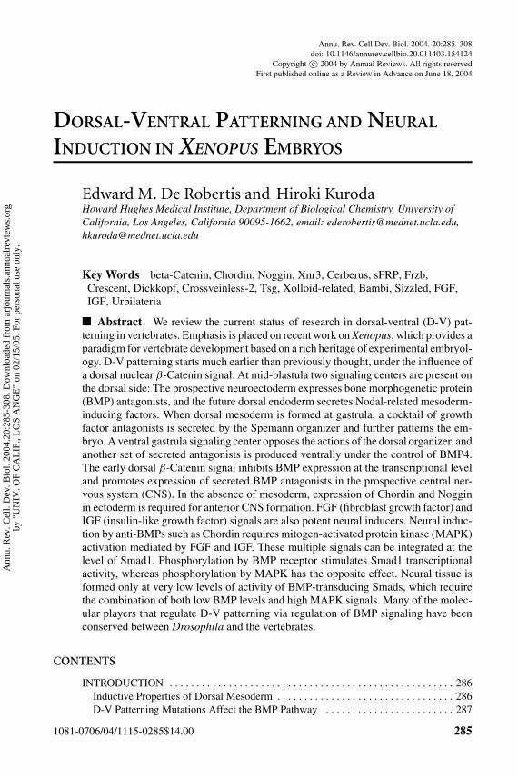
The starting point was provided by an experiment carried out by Hans Spemannand Hilde Mangold in 1924 in which they transplanted a small fragment of dorsalmesoderm into ventral mesoderm of the salamander gastrula (Spemann & Mangold1924) and found it could induce a twinned embryo. They grafted a mesodermfragment of different pigmentation, as in the experimental reproduction shown inFigure 1. The transplanted cells changed the differentiation of neighboring ventralcells in the host, inducing CNS, somite, and other axial components. In other words,both the ectoderm and the mesoderm became dorsalized. Research stimulatedby this experiment has continued to this day, in particular with the molecular
Ann
u. R
ev. C
ell.
Dev
. Bio
l. 20
04.2
0:28
5-30
8. D
ownl
oade
d fr
om a
rjou
rnal
s.an
nual
revi
ews.
org
by "
UN
IV. O
F C
AL
IF.,
LO
S A
NG
E"
on 0
2/15
/05.
For
per
sona
l use
onl
y.
10 Sep 2004 12:47 AR AR226-CB20-10.tex AR226-CB20-10.SGM LaTeX2e(2002/01/18) P1: GCE
D-V PATTERNING AND NEURAL INDUCTION 287
Figure 1 The Spemann-Mangold organizer experiment repeated in Xenopus laevis.(Top) control swimming tadpole; (bottom-right) Spemann organizer graft at the samestage. In the bottom-left embryo the size of the graft can be visualized as a white patch(Spemann organizer from an albino donor embryo) at early gastrula (vegetal view).The dorsal lip of the blastopore may be seen as a thin crescent opposite the graft.
exploration of Spemann’s organizer phenomenon (De Robertis & Arechaga 2001,Grunz 2004, Stern 2004).
D-V Patterning Mutations Affect the BMP Pathway
Numerous mutations that affect D-V embryonic pattern have been isolated inzebrafish and Drosophila. Most, if not all, of these mutations affect the bone mor-phogenetic protein (BMP) signaling pathway. In zebrafish, seven genes affectingD-V patterning were isolated in large-scale mutant screens (Hammerschmidt &Mullins 2002). swirl encodes BMP2b, and snailhouse BMP7. chordino encodesthe homologue of Xenopus Chordin, a BMP antagonist secreted by Spemann’s or-ganizer. mini fin encodes Tolloid, an extracellular zinc metalloprotease that cleavesand inactivates Chordin. lost-a-fin encodes a BMP receptor. somitabun and pig-gytail are mutations of the BMP-regulated transcription factor Smad5. Finally,ogon/mercedes is caused by mutations in Sizzled, a secreted Frizzled-related pro-tein that displays anti-BMP phenotypic effects (Yabe et al. 2003, Martyn & Schulte-Merker 2003). In Drosophila, seven genes that affect D-V patterning were isolatedin the classical zygotic mutant screens of Nusslein-Volhard and Wieschaus. Thesewere: Decapentaplegic and screw (Dpp and Scw, which are BMP growth factors),short gastrulation (Sog, a BMP antagonist homologous to vertebrate Chordin),twisted gastrulation (dTsg, a BMP-binding protein that functions as a cofactor of
Ann
u. R
ev. C
ell.
Dev
. Bio
l. 20
04.2
0:28
5-30
8. D
ownl
oade
d fr
om a
rjou
rnal
s.an
nual
revi
ews.
org
by "
UN
IV. O
F C
AL
IF.,
LO
S A
NG
E"
on 0
2/15
/05.
For
per
sona
l use
onl
y.
10 Sep 2004 12:47 AR AR226-CB20-10.tex AR226-CB20-10.SGM LaTeX2e(2002/01/18) P1: GCE
288 DE ROBERTIS � KURODA
Sog), tolloid (Tld, a zinc metalloprotease that cleaves and inactivates Sog), zen (ahomeobox gene activated by BMP), and shrew (which has not yet been identifiedmolecularly) (De Robertis & Sasai 1996, Lall & Patel 2001). From these geneticstudies one can conclude that the BMP signaling pathway plays a major role inD-V axis formation in animal development.
In this review, we examine the current status of research on D-V patterningand the induction of neural tissue. We place emphasis on recent work on Xenopusembryos because this organism provides a paradigm for vertebrate development.We build on previous reviews on this topic (Harland & Gerhart 1997, Weinstein& Hemmati-Brivanlou 1999, De Robertis et al. 2000, Harland 2000).
THE EARLY β-CATENIN SIGNAL
The Cortical Rotation
Sperm entry initiates a rotation of the cortex of the egg with respect to the internal,more yolky, cytoplasm. The rotation is driven by parallel arrays of microtubulesthat are nucleated by the centriole, which in all animals is contributed by thesperm (reviewed by De Robertis et al. 2000). These egg cortical microtubulesmay correspond to the astral cortical microtubules present in most mitotic somaticcells. Organelles that can be labeled with hydrophobic membrane dyes are trans-ported from the vegetal pole toward the dorsal side along these microtubule tracks.The dorsal lip is formed later on, at gastrula, opposite to the sperm entry point.Some of the molecular components of the dorsal determinants that move fromthe vegetal pole to the dorsal side are beginning to emerge and include Dishev-elled and the GSK3-binding protein GBP (Miller et al. 1999, Weaver et al. 2003).Transport of dorsal determinants results in the stabilization and nuclear translo-cation of β-Catenin protein on the dorsal side of the Xenopus blastula (Schneideret al. 1996, Schohl & Fagotto 2002), providing the earliest molecular D-Vasymmetry.
Two Dorsal Signaling Centers at Blastula
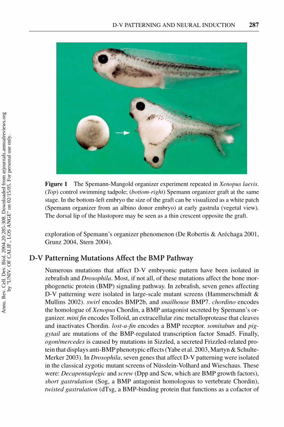
The nuclear localization of β-Catenin on the dorsal side extends from the bottom(vegetal) to the top (animal) pole of the blastula. The egg cytoplasm is heteroge-neous, and when zygotic gene transcription starts, after the midblastula transition,the Nieuwkoop center is formed in dorsal-vegetal cells (Figure 2). Nieuwkoopcenter cells express Xenopus Nodal-related factors (Xnr1, 2, 4, 5, and 6) that arepotent mesoderm inducers (Agius et al. 2000, Takahashi et al. 2000). High levels ofnodals induce dorsal mesoderm (Spemann organizer) in overlying cells, whereasthe Nieuwkoop center cells themselves go on to form anterior endoderm. In cellslocated above the Nieuwkoop center, in the dorsal animal cap and marginal zone,the β-Catenin signal induces the expression of BMP antagonists such as Chordinand Noggin (Wessely et al. 2001). This region of the animal cap was initially
Ann
u. R
ev. C
ell.
Dev
. Bio
l. 20
04.2
0:28
5-30
8. D
ownl
oade
d fr
om a
rjou
rnal
s.an
nual
revi
ews.
org
by "
UN
IV. O
F C
AL
IF.,
LO
S A
NG
E"
on 0
2/15
/05.
For
per
sona
l use
onl
y.
10 Sep 2004 12:47 AR AR226-CB20-10.tex AR226-CB20-10.SGM LaTeX2e(2002/01/18) P1: GCE
D-V PATTERNING AND NEURAL INDUCTION 289
Figure 2 Organizer formation in Xenopus laevis. At blastula stages, two signalingcenters, the BCNE center in the animal region and the Nieuwkoop center in the vegetalregion, pattern the embryo. Both are dependent on nuclear localization of β-Cateninon the dorsal side of the embryo. The Nieuwkoop center is formed in vegetal cells atthe intersection of the VegT, Vg1, and β-Catenin gene products. The BCNE center isinvolved in the formation of anterior neural tissue and expresses chordin, noggin, andXnr3. The Nieuwkoop center releases Nodal-related signals that induce Spemann’sorganizer in dorsal mesoderm at gastrula.
designated as the preorganizer region and has more recently been renamed theBCNE center, for blastula Chordin and Noggin expression center (Kuroda et al.2004). Molecular studies show that there is limited overlap between the Nieuwkoopand BCNE centers. Additional genes are expressed in a localized fashion in theblastula: The Nieuwkoop center expresses the secreted antagonist cerberus, andthe BCNE center expresses the homeobox gene Siamois, the winged-helix genepintallavis/FoxA4a/HNF3β, and Xnr3 (Kuroda et al. 2004, Wessely et al. 2004).Both blastula centers are formed simultaneously, as soon as zygotic transcriptionstarts. Both require the β-Catenin signal on the dorsal side of the embryo, butthe Nieuwkoop center also requires mRNAs located in the vegetal pole of theunfertilized egg (Figure 2).
In later development, BCNE cells give rise to all of the forebrain, most of mid-and hind-brain, floor plate, and notochord (Kuroda et al. 2004). Thus chordin andnoggin are transiently expressed in prospective neuroectoderm at blastula. Lateron, at gastrula stages, the same genes are expressed in the Spemann organizermesoderm under the control of Nodal-related signals (Agius et al. 2000, Wesselyet al. 2001). When BCNE explants are cultured in saline they form CNS tis-sue, indicating that neural specification takes place very early, at the blastulastage, in Xenopus ectoderm. When the BCNE center is excised, brain forma-tion fails in the resulting embryos. This requirement of BCNE cells for brainformation can be rescued by transplantation of dorsal, but not ventral, animalcap tissue (Kuroda et al. 2004). Thus the early β-Catenin signal triggers the for-mation of two signaling centers at blastula, one involved in dorsal endodermdevelopment (Nieuwkoop center) and the other in neural specification (BCNEcenter).
Ann
u. R
ev. C
ell.
Dev
. Bio
l. 20
04.2
0:28
5-30
8. D
ownl
oade
d fr
om a
rjou
rnal
s.an
nual
revi
ews.
org
by "
UN
IV. O
F C
AL
IF.,
LO
S A
NG
E"
on 0
2/15
/05.
For
per
sona
l use
onl
y.
10 Sep 2004 12:47 AR AR226-CB20-10.tex AR226-CB20-10.SGM LaTeX2e(2002/01/18) P1: GCE
290 DE ROBERTIS � KURODA
THE DORSAL GASTRULA CENTER
The Spemann Organizer Is a Source of Secreted Antagonists
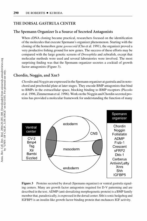
When cDNA cloning became practical, researchers focused on the identificationof the molecules that execute Spemann’s organizer phenomenon. Starting with thecloning of the homeobox gene goosecoid (Cho et al. 1991), the organizer proved avery productive fishing ground for new genes. The success of these efforts may becompared with the large genetic screens of Drosophila and zebrafish, except thatmolecular methods were used and several laboratories were involved. The mostsurprising finding was that the Spemann organizer secretes a cocktail of growthfactor antagonists (Figure 3).
Chordin, Noggin, and Xnr3
Chordin and Noggin are expressed in the Spemann organizer at gastrula and in noto-chord and prechordal plate at later stages. They encode BMP antagonists that bindto BMPs in the extracellular space, blocking binding to BMP receptors (Piccoloet al. 1996, Zimmerman et al. 1996). Work on the Noggin and Chordin secreted pro-teins has provided a molecular framework for understanding the function of many
Figure 3 Proteins secreted by dorsal (Spemann organizer) or ventral gastrula signal-ing centers. Many are growth factor antagonists required for D-V patterning and aredescribed in the text. ADMP (anti-dorsalizing morphogenetic protein) is a BMP familymember that, paradoxically, is expressed in the dorsal center. Shh is sonic hedgehog andIGFBP5 is an insulin-like growth factor-binding protein that enchances IGF activity.
Ann
u. R
ev. C
ell.
Dev
. Bio
l. 20
04.2
0:28
5-30
8. D
ownl
oade
d fr
om a
rjou
rnal
s.an
nual
revi
ews.
org
by "
UN
IV. O
F C
AL
IF.,
LO
S A
NG
E"
on 0
2/15
/05.
For
per
sona
l use
onl
y.
10 Sep 2004 12:47 AR AR226-CB20-10.tex AR226-CB20-10.SGM LaTeX2e(2002/01/18) P1: GCE
D-V PATTERNING AND NEURAL INDUCTION 291
extracellular proteins. The three-dimensional structure of Noggin bound to BMP7revealed that this BMP antagonist contains a cystine knot (Groppe et al. 2002).The cystine knot structural motif is found in many extracellular proteins, such asthe TGF-β superfamily, luteinizing hormone (LH), follicle stimulating hormone(FSH), platelet-derived growth factor (PDGF), nerve growth factor (NGF), andothers (Avsian-Kretchmer & Hsueh 2004). Because BMP and Noggin share cys-tine knots and conserved protein folds, it has been proposed that the ligand and itsantagonist may have evolved from ancestrally related proteins (Groppe et al. 2002).Chordin is a large protein of about 1000 amino acids containing four cysteine-richdomains of about 70 amino acids each (called CR1 to CR4), which constituteBMP binding modules (Larraın et al. 2000). The structures of Chordin or CR do-mains have not been solved, but some predictions indicate a possible cystine knot(Avsian-Kretchmer & Hsueh 2004). CR repeats, also called von Willebrand factorC domains (vWF-C), are present in a large number of extracellular proteins, manyof which have been found to regulate BMP or TGF-β signaling. Proteins contain-ing CR domains include CTGF (connective tissue growth factor), Procollagens,Amnionless, Chordin-like/Ventroptin/Neuralin-1 and -2, CRIM-1 (cysteine-richmotor neuron protein), Nel (neural tissue protein containing Egf-Like domains),Nel-like 1 and 2, Keilin, and Crossveinless-2 (Garcia-Abreu et al. 2002).
On the dorsal side of the Xenopus embryo, Chordin protein is present at con-centrations of 6 to 12 nM in the extracellular space (Piccolo et al. 1996). BecauseBMPs are expressed in the picomolar range in the embryo, Chordin alone shouldsuffice to block BMP signaling on the dorsal side. However, the knockout of theChordin gene in mouse causes only a small percentage of embryos to becomeventralized at the gastrula stage (Bachiller et al. 2003). These infrequent embryoshave a small neural plate and embryonic region, and an enlarged allantois (ventralmesoderm). Most chd−/− embryos have a normal CNS and die at birth, mimickinga human malformation called DiGeorge syndrome, which is caused by the lack ofChordin in pharyngeal endoderm at a later developmental stage (Bachiller et al.2003). The mouse Noggin knockout has normal gastrulation and neural plate for-mation, and a strong skeletal phenotype later on (McMahon et al. 1998). Mouseembryos mutant for HNF3β, in which Hensen’s node–the equivalent of Spemann’sorganizer–does not develop, do not express Chordin or Noggin at gastrula but stillform a neural plate (Klingensmith et al. 1999). Because these two BMP antagonistsare required for neural plate development, they must also function at earlier stagesbefore HNF3β is expressed. Indeed, chordin;noggin double mutant embryos dis-play a loss of the prosencephalic vesicle, lack of anterior notochord (ventralizationof the mesoderm), and randomization of heart left-right asymmetry (Bachiller et al.2000). Thus the Chordin and Noggin BMP antagonists have redundant functionsand are required for the patterning of the three embryonic axes of the mouse.
In Xenopus, knockdown of Chordin expression is achieved using chordin anti-sense morpholino oligos (Chd-MO) (Oelgeschlager et al. 2003a). The phenotypeobtained is very similar to that of the chordino zebrafish mutant (Schulte-Merkeret al. 1997), displaying a reduction of the size of the neural plate and eventually
Ann
u. R
ev. C
ell.
Dev
. Bio
l. 20
04.2
0:28
5-30
8. D
ownl
oade
d fr
om a
rjou
rnal
s.an
nual
revi
ews.
org
by "
UN
IV. O
F C
AL
IF.,
LO
S A
NG
E"
on 0
2/15
/05.
For
per
sona
l use
onl
y.
10 Sep 2004 12:47 AR AR226-CB20-10.tex AR226-CB20-10.SGM LaTeX2e(2002/01/18) P1: GCE
292 DE ROBERTIS � KURODA
CNS tissue, and an expansion of ventral mesoderm. When the Xenopus embryois experimentally manipulated, strong requirements for Chordin are observed. Forexample, the dorsalizing effects of lithium chloride (LiCl), a treatment that sta-bilizes β-Catenin, can be completely blocked by Chd-MO (Oelgeschlager et al.2003a). When the Spemann organizer transplantation experiment is repeated usingdorsal lip explants injected with Chd-MO, the grafts completely lose their induc-ing activity (Oelgeschlager et al. 2003a). Organizer grafts probably require a fullcomplement of BMP antagonists, and even the loss of a single one, Chordin, hasprofound effects.
Xnr-3 encodes a Nodal-related protein that lacks mesoderm-inducing activity,presumably because it is mutated in a critical cysteine residue of the cystine knot.Xnr3 is able to induce neural differentiation when overexpressed in animal caps(Hansen et al. 1997) and is able to antagonize BMP signaling through its amino-terminal proregion (Haramoto et al. 2004). Xnr3 homologues have not been foundin any other vertebrates, but in Xenopus it is, after Chordin, the gene most stronglyinduced by the early β-Catenin signal in genome-wide studies (Wessely et al.2004).
The Wnt Antagonists: Frzb-1, Crescent, sFRP-2, and Dickkopf
Secreted Frizzled–related proteins (sFRPs) constitute a large family of Wnt an-tagonists that encode secreted forms of the amino-terminal, cysteine-rich domainof the Wnt receptor Frizzled (Kawano & Kypta 2003). They bind Wnt proteins inthe extracellular space and prevent them from signaling (Leyns et al. 1997). TheXenopus gastrula expresses high levels of sFRPs; a screen for cDNAs encodingsecreted proteins resulted in a surprising 24% of isolates encoding sFRPs (Pera& De Robertis 2000). The Spemann organizer expresses Frzb-1/sFRP3, Crescentand sFRP2 (Figure 3).
Xenopus Dickkopf-1 (Dkk-1) is a secreted inhibitor of Wnt signaling thatfunctions through a different and interesting molecular mechanism. It encodesa cysteine-rich secreted protein expressed in dorsal endomesoderm that defines anew protein family (Glinka et al. 1998). Dkk-1 binds to a Wnt coreceptor calledLDL receptor-related protein-5/6 (LRP5/6) (Mao et al. 2001). Wnt binds to bothFrizzled and LRP6, forming a ternary receptor complex on the cell surface (Tamaiet al. 2000). This triggers phosphorylation of the intracellular domain of LRP5/6 atconserved PPPSP sites, causing the recruitment of the β-Catenin destruction com-plex protein Axin to the cell membrane and inhibition of β-Catenin degradation(Tamai et al. 2004). Thus LRP5/6 specifically links Wnt signaling to β-Cateninstabilization. Dkk-1 binds not only to LRP5/6 but also to a second transmembraneprotein called Kremen. The resulting trimolecular complex of LRP5/6, Dkk, andKremen is endocytosed, resulting in the depletion of LRP5/6 coreceptor from theplasma membrane (Mao et al. 2002). This provides an elegant molecular explana-tion for how Dkk selectively inhibits the action of Wnt on the canonical β-CateninWnt pathway without affecting other aspects of Wnt signaling. In Xenopus, Dkk1
Ann
u. R
ev. C
ell.
Dev
. Bio
l. 20
04.2
0:28
5-30
8. D
ownl
oade
d fr
om a
rjou
rnal
s.an
nual
revi
ews.
org
by "
UN
IV. O
F C
AL
IF.,
LO
S A
NG
E"
on 0
2/15
/05.
For
per
sona
l use
onl
y.
10 Sep 2004 12:47 AR AR226-CB20-10.tex AR226-CB20-10.SGM LaTeX2e(2002/01/18) P1: GCE
D-V PATTERNING AND NEURAL INDUCTION 293
neutralizing antibodies inhibit head and prechordal plate formation (Glinka et al.1998, Kazanskaya et al. 2000). In the mouse, Dkk-1 homozygous mutants lackCNS structures anterior to the midbrain (Mukhopadhyay et al. 2001), and het-erozygotes show strong cooperation with noggin in head formation (del Barcoet al. 2003).
Cerberus
Cerberus, a secreted protein expressed at high levels in the anterior dorsal endodermof gastrula, has the remarkable property of inducing ectopic head structures in theabsence of trunk formation (Bouwmeester et al. 1996). Its discovery identifiedthe first head-inductive signal from endoderm, a finding that was later supportedby studies in the mouse on the role of the anterior visceral endoderm (AVE) inhead development (Beddington & Robertson 1999). Cerberus protein binds to, andprevents signaling by, Nodal, BMP, and Wnt-8 (Piccolo et al. 1999). A fragmentof Cerberus consisting of its carboxy-terminal cystine knot has only the Nodal-inhibiting activity. This artificial construct, called Cerberus-short (Cer-S), providesa useful reagent to block Nodal signaling in embryos. For example, the use of Cer-S allowed Agius et al. (2000) to demonstrate that the induction of both dorsal andventral mesoderm is mediated by a gradient of Nodal-related signals emanatingfrom endoderm at the late-blastula stage (Figure 2).
In Xenopus, Cerberus is required for head induction; a Cer-MO inhibits headbut not trunk-tail development (Kuroda et al. 2004). In the mouse, knockout of acerberus-like gene expressed in the AVE lacks gastrulation phenotypes. Mutationsin another nodal antagonist expressed in AVE cells, Lefty-1, also lacks gastrulationphenotypes. However, when cer-l−/−; lefty-1−/− double mutants are generated, de-velopment of the anterior embryo is greatly impaired owing to excessive Nodalsignaling in the anterior region of the embryo (Perea-Gomez et al. 2002, Yamamotoet al. 2004). In chick, a cerberus homologue expressed in the hypoblast (the equiv-alent of mouse AVE) prevents formation of trunk mesoderm in prospective headneuroectoderm via its anti-Nodal activity (Bertocchini & Stern 2002). In summary,these studies in vertebrate embryos support the view that secreted antagonists ofNodal, BMP, and Wnt signals play a fundamental role in promoting head devel-opment and repressing trunk-tail development (Piccolo et al. 1999, Niehrs 2001,Agathon et al. 2003).
THE VENTRAL GASTRULA CENTER
The BMP4 Synexpression Group
Evidence indicating that a ventral signaling center exists in the gastrula has beensteadily accumulating. Several genes encoding secreted or cell surface proteinsare expressed in ventral mesoderm and ectoderm 180◦ from Spemann’s organizer(Figure 3). Their expression patterns indicate they are members of the BMP4
Ann
u. R
ev. C
ell.
Dev
. Bio
l. 20
04.2
0:28
5-30
8. D
ownl
oade
d fr
om a
rjou
rnal
s.an
nual
revi
ews.
org
by "
UN
IV. O
F C
AL
IF.,
LO
S A
NG
E"
on 0
2/15
/05.
For
per
sona
l use
onl
y.
10 Sep 2004 12:47 AR AR226-CB20-10.tex AR226-CB20-10.SGM LaTeX2e(2002/01/18) P1: GCE
294 DE ROBERTIS � KURODA
synexpression group. Synexpression groups consist of genes that are coordinatelyexpressed in the embryo and frequently function in a common signaling pathway(Niehrs & Pollet 1999). Early indications of the existence of a ventral center camefrom studies on the expression of homebox genes such as Vent/Vox/Vega and Eve1that are induced by BMP4 in the ventral region of the gastrula (Gawantka et al.1995, Kawahara et al. 2000, Joly et al. 1993). Interestingly, many of the proteinssecreted by the ventral gastrula center have biochemical activities similar to thoseof the Spemann organizer but are under the opposite transcriptional regulation (seebelow).
Crossveinless-2, Twisted Gastrulation, Xolloid-Related,and Bambi
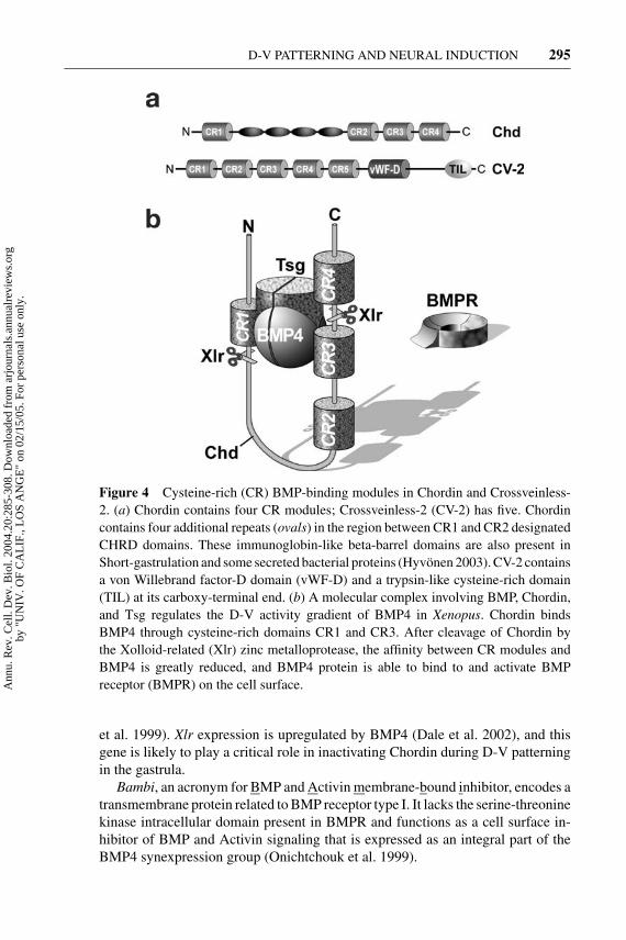
Drosophila Crossveinless-2 (CV-2) is a gene required for the formation of the wingcrossveins, structures that require Dpp signaling during development. Drosophilaand vertebrate CV-2 contain five CR domains of the types present in the BMP-binding modules of Chordin, as well as a von Willebrand factor-D domain(Figure 4A) (Conley et al. 2000, Coffinier et al. 2002). Mouse and Xenopus CV-2are expressed in ventral mesoderm and ectoderm and when overexpressed behaveas BMP antagonists (Coffinier et al. 2002, Moser et al. 2003, Binnerts et al. 2004;C. Coffinier & E.M. De Robertis, unpublished observations). Whereas Chordinexpression is repressed by BMP4, CV-2 expression is upregulated by BMP4.
Twisted gastrulation (Tsg) encodes a protein that binds to both BMP andChordin (Oelgeschlager et al. 2000, Chang et al. 2001, Scott et al. 2001, Rosset al. 2001, Blitz et al. 2003). Chordin, BMP, and Tsg form ternary complexesthat prevent binding of BMP to its receptor (Figure 4B). Chordin is cleaved by theTolloid/Xolloid protease at two specific cleavage sites. The resulting proteolyticfragments have lower affinity, and BMP signaling through its receptors is restored(Figure 4B). Tsg facilitates this proteolytic cleavage and promotes BMP signals(Larraın et al. 2001, Oelgeschlager et al. 2003b). The key switch is provided bythe levels of Xolloid protease. The effects of Tsg overexpression depend on theamount of Tolloid present; at high Xolloid levels Tsg promotes BMP signalingand at low levels it inhibits it (Larraın et al. 2001, Oelgeschlager et al. 2003b).In mouse knockouts, Tsg mutants have phenotypes and genetic interactions thatreflect both its anti- and pro-BMP4 activities (Nosaka et al. 2003, Petryk et al.2004, Zakin & De Robertis 2004). In Drosophila, dTsg is required for maximalDpp signaling levels in the dorsal-most region (called the amnioserosa) and forthe diffusion of Dpp in the early embryo (Eldar et al. 2002). In vertebrates, itappears likely that Tsg may interact with other CR-containing proteins in additionto Chordin (Oelgeschlager et al. 2003b).
Xolloid-related (Xlr) is a recently identified Tolloid metalloprotease that isspecifically expressed in the ventral gastrula center of Xenopus (Dale et al. 2002).There are multiple Tolloid-like genes in vertebrates, and Xlr is most similar tozebrafish mini-fin/Tolloid (Connors et al. 1999) and mouse Tolloid-like-1 (Scott
Ann
u. R
ev. C
ell.
Dev
. Bio
l. 20
04.2
0:28
5-30
8. D
ownl
oade
d fr
om a
rjou
rnal
s.an
nual
revi
ews.
org
by "
UN
IV. O
F C
AL
IF.,
LO
S A
NG
E"
on 0
2/15
/05.
For
per
sona
l use
onl
y.
10 Sep 2004 12:47 AR AR226-CB20-10.tex AR226-CB20-10.SGM LaTeX2e(2002/01/18) P1: GCE
D-V PATTERNING AND NEURAL INDUCTION 295
Figure 4 Cysteine-rich (CR) BMP-binding modules in Chordin and Crossveinless-2. (a) Chordin contains four CR modules; Crossveinless-2 (CV-2) has five. Chordincontains four additional repeats (ovals) in the region between CR1 and CR2 designatedCHRD domains. These immunoglobin-like beta-barrel domains are also present inShort-gastrulation and some secreted bacterial proteins (Hyvonen 2003). CV-2 containsa von Willebrand factor-D domain (vWF-D) and a trypsin-like cysteine-rich domain(TIL) at its carboxy-terminal end. (b) A molecular complex involving BMP, Chordin,and Tsg regulates the D-V activity gradient of BMP4 in Xenopus. Chordin bindsBMP4 through cysteine-rich domains CR1 and CR3. After cleavage of Chordin bythe Xolloid-related (Xlr) zinc metalloprotease, the affinity between CR modules andBMP4 is greatly reduced, and BMP4 protein is able to bind to and activate BMPreceptor (BMPR) on the cell surface.
et al. 1999). Xlr expression is upregulated by BMP4 (Dale et al. 2002), and thisgene is likely to play a critical role in inactivating Chordin during D-V patterningin the gastrula.
Bambi, an acronym for BMP and Activin membrane-bound inhibitor, encodes atransmembrane protein related to BMP receptor type I. It lacks the serine-threoninekinase intracellular domain present in BMPR and functions as a cell surface in-hibitor of BMP and Activin signaling that is expressed as an integral part of theBMP4 synexpression group (Onichtchouk et al. 1999).
Ann
u. R
ev. C
ell.
Dev
. Bio
l. 20
04.2
0:28
5-30
8. D
ownl
oade
d fr
om a
rjou
rnal
s.an
nual
revi
ews.
org
by "
UN
IV. O
F C
AL
IF.,
LO
S A
NG
E"
on 0
2/15
/05.
For
per
sona
l use
onl
y.
10 Sep 2004 12:47 AR AR226-CB20-10.tex AR226-CB20-10.SGM LaTeX2e(2002/01/18) P1: GCE
296 DE ROBERTIS � KURODA
Sizzled
Sizzled is a ventral center gene of particular interest. It encodes an sFRP-likemolecule (Salic et al. 1997). Microinjection of sizzled antisense morpholino oli-gos causes ventralization of the Xenopus embryo, including an increase of ventralmesoderm and reduction of the neural plate (Collavin & Kirshner 2003). The mostsurprising aspect of this phenotype is that it is indistinguishable from that of theloss-of-function of Chordin in Xenopus (Oelgeschlager et al. 2003a). Knockdownof Chordin expands the expression of sizzled in the ventral gastrula center, pre-sumably through increased BMP4 signaling on the ventral side. Understandinghow these two secreted proteins expressed at opposite poles of the embryo cangenerate similar phenotypes when inhibited may hold the key to understandingD-V regulation in Xenopus.
In zebrafish, only two ventralized mutants have been isolated, chordino andogon/mercedes. Both result in reduced CNS and expanded ventral tail structuresconsistent with increased BMP signaling. The ogon (meaning tail in Polish) geneencodes sizzled (Yabe et al. 2003, Martyn & Schulte-Merker 2003). This gene,which has the molecular structure of a Wnt inhibitor, must have important interac-tions with the BMP pathway because ogon/sizzled mRNA injection dorsalizes thewild-type zebrafish embryo. Interestingly, microinjected sizzled mRNA is inactivein chordino mutants (Yabe et al. 2003). Thus Sizzled must work through Chordin.The molecular mechanism of Sizzled action remains a puzzle. An initial studyindicated that it functioned as an xWnt-8 antagonist (Salic et al. 1997), but subse-quent studies suggested that Sizzled behaved as a BMP4 antagonist (Collavin &Kirschner 2003, Yabe et al. 2003). One of the challenges for the near future is de-termining whether the Sizzled protein regulates the activity of a Wnt signal, whichin turn affects BMP expression, or whether it acts directly on BMP or Chordinactivity. Sizzled is most similar to Crescent, an sFRP that functions as a Wnt an-tagonist on the dorsal side of the embryo (Pera & De Robertis 2000, Bradley et al.2000). Thus Chordin and Crescent are expressed in the Spemann organizer, andtwo other genes of related structures, Crossveinless-2 and Sizzled, respectively,are expressed in the ventral center. Dorsal and ventral center genes have similaractivities when overexpressed in embryos, but whereas the expression of organizergenes is inhibited by BMPs, expression of Crossveinless-2 and Sizzled/Ogon isincreased by BMP4 signaling. The importance of the ventral signaling center wasunrecognized for a long time, perhaps because it lacks inductive activity upontransplantation. With the availability of morpholino loss-of-function reagents, thegastrula ventral center is likely to become the focus of much research.
THE ROLE OF NEUROECTODERM IN CNS FORMATION
Traditionally, research in amphibian neural induction centered on the role ofthe mesodermal Spemann organizer signals at gastrula, also known as the pri-mary embryonic induction (Spemann 1938). A possible role for the prospective
Ann
u. R
ev. C
ell.
Dev
. Bio
l. 20
04.2
0:28
5-30
8. D
ownl
oade
d fr
om a
rjou
rnal
s.an
nual
revi
ews.
org
by "
UN
IV. O
F C
AL
IF.,
LO
S A
NG
E"
on 0
2/15
/05.
For
per
sona
l use
onl
y.
10 Sep 2004 12:47 AR AR226-CB20-10.tex AR226-CB20-10.SGM LaTeX2e(2002/01/18) P1: GCE
D-V PATTERNING AND NEURAL INDUCTION 297
neuroectoderm itself in CNS formation has long been debated (Goerttler 1925,Holtfreter 1933, Spemann 1938). In the very influential exogastrulation exper-iment, Holtfreter removed the egg membranes and placed axolotl embryos inhypertonic saline, preventing the involution of endomesoderm. In the absence ofits mesodermal substratum, the entire ectodermal layer became epidermis, and noCNS developed (Holtfreter 1933). Since then, traditional embryological thinkinghas been that the Spemann organizer mesoderm secretes the neural inducers andthat the ectoderm itself has no role (Spemann 1938). One way of understandingHoltfreter’s intriguing result in modern terms is that perhaps in exogastrulatedectoderm BMPs reach very high levels, reversing any labile bias present in the ec-toderm toward neural formation. Below, we discuss recent evidence indicating thatthe ectoderm of the BCNE center does indeed play a crucial role in neural induction.
The Neural Induction Default Model
Early efforts to identify neural inducers used ectodermal explants exposed to manysubstances, such as dead organizers, methylene blue, sterols, fatty acids, and evensand particles. All were found to neuralize embryonic ectoderm. Gradually, thesearch for the Spemann organizer neural inducer became a funeral march for thefield (reviewed by Holtfreter & Hamburger 1955). Eventually it was realized thatneuralization of axolotl ectoderm could be obtained in the complete absence of anyinducer simply by culturing animal caps in an inadequate saline solution (Barth1941). This effect could be mimicked in other amphibians by partial cell dissocia-tion with citrate, oxalate, or low pH treatments that received the unfortunate nameof “sublethal cytolysis” (Holtfreter & Hamburger 1955). Xenopus ectoderm is rel-atively resistant to neuralization, but neural differentiation can be elicited by celldissociation and culture for several hours (reviewed by Weinstein & Hemmati-Brivanlou 1999, Munoz-Sanjuan & Brivanlou 2002). This neuralization can bereversed by adding BMP4 to the culture medium, which led to the proposal thatduring dissociation BMP4 protein is diluted by diffusion into the culture medium(Wilson & Hemmati-Brivanlou 1995). BMP acts within the ectoderm to induceepidermis, and it was proposed that when it diffuses away in dissociated cells, neu-ral differentiation by a default pathway would ensue. However, given the recentrealization that activation of MAPK signaling can downregulate the BMP signalingpathway at the level of Smad1 phosphorylation (see below), alternative interpre-tations for why cell dissociation and abnormal substances have neural-inducingactivities in ectodermal cells will have to be explored.
Although the BMP default neural induction model has generally received sup-port in Xenopus (Harland 2000), work in other model systems has highlightedthe role of FGF and Wnt signals in neural induction and de-emphasized a rolefor BMP (reviewed in Wilson & Edlund 2001, Streit & Stern 1999, Stern 2002,Lemaire et al. 2002). In chick, FGF can initiate ectopic expression of neural mark-ers but Chordin and Noggin cannot. However, Chordin can stabilize expression oftransiently induced neural markers, expand an already formed neural plate, and
Ann
u. R
ev. C
ell.
Dev
. Bio
l. 20
04.2
0:28
5-30
8. D
ownl
oade
d fr
om a
rjou
rnal
s.an
nual
revi
ews.
org
by "
UN
IV. O
F C
AL
IF.,
LO
S A
NG
E"
on 0
2/15
/05.
For
per
sona
l use
onl
y.
10 Sep 2004 12:47 AR AR226-CB20-10.tex AR226-CB20-10.SGM LaTeX2e(2002/01/18) P1: GCE
298 DE ROBERTIS � KURODA
induce ectopic primitive streaks (Streit & Stern 1999). Explants of the medialchick epiblast differentiate into neural tissue, but when BMP4 is added developinto epidermis instead. Lateral chick epiblast explants develop into epidermis anddo not respond to FGF or BMP antagonists; however, if Wnt signaling levels arelowered by treatment with Wnt antagonists, then both FGFs and BMP antagonistscan induce neural differentiation in lateral epiblast cells (Wilson & Edlund 2001).Additional lines of evidence indicate that Wnt signals are involved in the choicebetween epidermal and neural differentiation. A functional screening of cDNA forgenes able to cause differentiation of mouse embryonic stem cells into neuronsresulted in the isolation of the sFRP-2 Wnt antagonist (Aubert et al. 2002). InXenopus animal cap explants, Wnt antagonists are able to transiently induce neu-ral markers (Glinka et al. 1997), and FGFs function as neural inducers (Harland2000). Taken together, the available evidence suggests that multiple signaling path-ways are involved in neural induction in all vertebrates. Rather than emphasizingthe differences between organisms, the field now needs ways of integrating thesediverse neural-inducing signaling pathways. One such integration of cell-cell sig-nals may occur at the level of Smad1 phosphorylation.
Integrating Signals at the Level of Smads
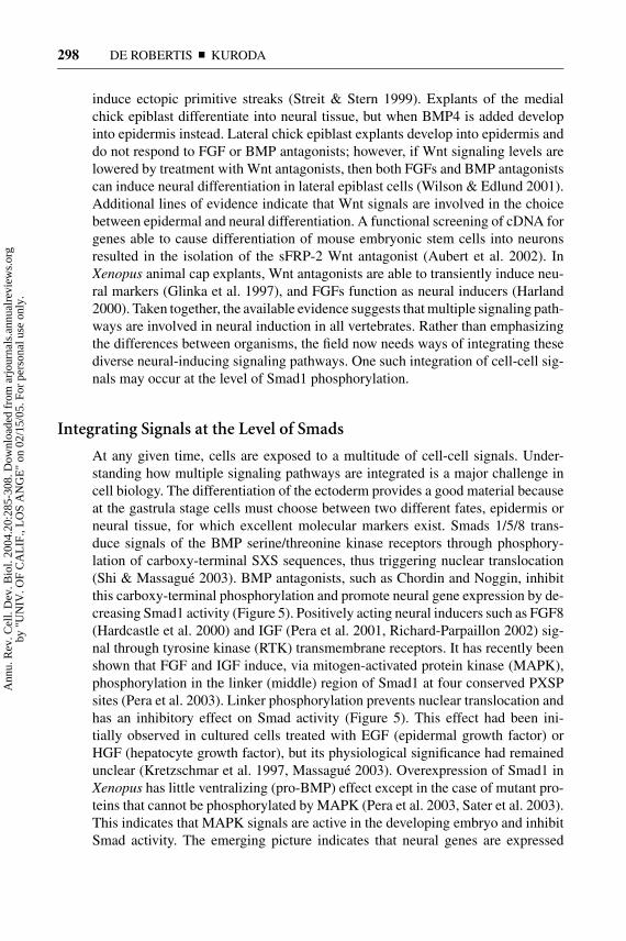
At any given time, cells are exposed to a multitude of cell-cell signals. Under-standing how multiple signaling pathways are integrated is a major challenge incell biology. The differentiation of the ectoderm provides a good material becauseat the gastrula stage cells must choose between two different fates, epidermis orneural tissue, for which excellent molecular markers exist. Smads 1/5/8 trans-duce signals of the BMP serine/threonine kinase receptors through phosphory-lation of carboxy-terminal SXS sequences, thus triggering nuclear translocation(Shi & Massague 2003). BMP antagonists, such as Chordin and Noggin, inhibitthis carboxy-terminal phosphorylation and promote neural gene expression by de-creasing Smad1 activity (Figure 5). Positively acting neural inducers such as FGF8(Hardcastle et al. 2000) and IGF (Pera et al. 2001, Richard-Parpaillon 2002) sig-nal through tyrosine kinase (RTK) transmembrane receptors. It has recently beenshown that FGF and IGF induce, via mitogen-activated protein kinase (MAPK),phosphorylation in the linker (middle) region of Smad1 at four conserved PXSPsites (Pera et al. 2003). Linker phosphorylation prevents nuclear translocation andhas an inhibitory effect on Smad activity (Figure 5). This effect had been ini-tially observed in cultured cells treated with EGF (epidermal growth factor) orHGF (hepatocyte growth factor), but its physiological significance had remainedunclear (Kretzschmar et al. 1997, Massague 2003). Overexpression of Smad1 inXenopus has little ventralizing (pro-BMP) effect except in the case of mutant pro-teins that cannot be phosphorylated by MAPK (Pera et al. 2003, Sater et al. 2003).This indicates that MAPK signals are active in the developing embryo and inhibitSmad activity. The emerging picture indicates that neural genes are expressed
Ann
u. R
ev. C
ell.
Dev
. Bio
l. 20
04.2
0:28
5-30
8. D
ownl
oade
d fr
om a
rjou
rnal
s.an
nual
revi
ews.
org
by "
UN
IV. O
F C
AL
IF.,
LO
S A
NG
E"
on 0
2/15
/05.
For
per
sona
l use
onl
y.
10 Sep 2004 12:47 AR AR226-CB20-10.tex AR226-CB20-10.SGM LaTeX2e(2002/01/18) P1: GCE
D-V PATTERNING AND NEURAL INDUCTION 299
Figure 5 Integration of multiple signaling pathways at the level of Smad1 phospho-rylation during neural induction. Neural induction requires extremely low levels ofSmad1 activity that are reached through the combination of two signaling systems.One is inhibition of binding between BMP4 and its serine/threonine kinase (RS/TK)receptor by anti-BMP molecules such as Chordin and Noggin. The other is inhibitoryphosphorylation of Smad1 in the linker region by receptor tyrosine kinase (RTK) sig-nals such as FGF, IGF, HGF, and EGF mediated by activation of MAPK. MH1 andMH2 are evolutionarily conserved globular Mad-homology domains; MH1 containsthe DNA-binding domain and MH2 multiple protein interaction sites.
only at very low levels of Smad1 activity, which requires both low BMP levels andhigh MAPK signals (Figure 5). In agreement with this view, neural induction byChordin can be blocked by agents that inhibit FGF or IGF signaling (Pera et al.2003).
In the mouse, knock-in of Smad1 forms that are insensitive to MAPK phos-phorylation in the linker region exhibit phenotypes in gastrointestinal epitheliumand germ cells (Aubin et al. 2004). These animals express normal levels of mu-tant Smad1 (as well as of wild-type Smad5 and Smad8), supporting the view thatintegrating BMP and MAPK signals at the level of Smad1 is required in vivo.
The finding that FGF signaling can cause inhibition of signaling by BMP Smadsvia the hard-wired mechanism shown in Figure 5 may help explain other situationsin which FGF and BMP signals oppose each other during development. A clas-sical example is the antagonism between FGF4 and BMP2 in limb development(Niswander & Martin 1993). Similarly, opposing effects of FGFs and BMPs have
Ann
u. R
ev. C
ell.
Dev
. Bio
l. 20
04.2
0:28
5-30
8. D
ownl
oade
d fr
om a
rjou
rnal
s.an
nual
revi
ews.
org
by "
UN
IV. O
F C
AL
IF.,
LO
S A
NG
E"
on 0
2/15
/05.
For
per
sona
l use
onl
y.
10 Sep 2004 12:47 AR AR226-CB20-10.tex AR226-CB20-10.SGM LaTeX2e(2002/01/18) P1: GCE
300 DE ROBERTIS � KURODA
been reported in lung morphogenesis, cranial suture fusion, and tooth develop-ment (Weaver et al. 2000, Warren et al. 2003, Thesleff & Mikkola 2002). In future,one aspect of the neural induction default model that should be reinvestigated iswhether animal cap dissociation, in addition to lowering BMP levels, causes theactivation of other signaling pathways such as MAPK.
β-Catenin, Chordin, and Noggin Are Requiredin Blastula Ectoderm
It has long been known that the Xenopus gastrula dorsal animal cap has a predispo-sition for neural induction by dorsal endomesoderm (Sharpe et al. 1987, Londonet al. 1988) and that it has lower levels of BMP4 expression (Fainsod et al. 1994).In the course of a functional cDNA screen, Baker et al. (1999) made the importantdiscovery that an activated form of mouse β-Catenin was able to induce neuraltissue in animal caps. This neural induction was accompanied by the extinctionof BMP4 expression in animal cap explants and could be inhibited by constitu-tively activated BMP receptor (Baker et al. 1999). In zebrafish, a homeobox genecalled bozozok/dahrma is expressed on the dorsal side in response to the earlyβ-Catenin signal and directly represses BMP2b gene transcription (Leung et al.2003). Because bozozok and chordino double mutants show synergistic losses ofneural tissue and dorsal structures (Gonzalez et al. 2000), it appears that duringdevelopment dorsal BMP levels are inhibited both by transcriptional regulatorsand by BMP antagonists.
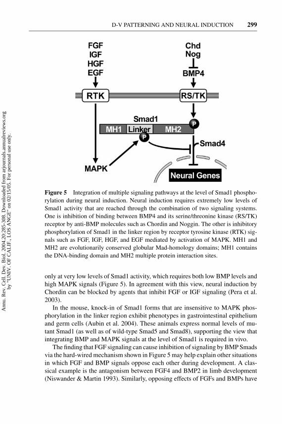
In Xenopus, the early β-Catenin signal induces the early expression of Chordinand Noggin in the BCNE center (Wessely et al. 2001, Kuroda et al. 2004). Inlater development, the BCNE becomes the brain and floor plate. When mesodermformation is blocked (by inhibiting Nodal signals with Cer-S), brain structuresstill develop, even though these embryos lack expression of chordin, noggin, andfollistatin at gastrula. However, transcription of BMP antagonists can be tran-siently detected at blastula in the BCNE center that forms under the influenceof the β-Catenin signal (Wessely et al. 2001). Anterior CNS differentiation inthe absence of mesoderm is entirely dependent on the early β-Catenin signaland can be blocked by Chd-MO or Noggin-MO; posterior neural markers arenot affected and require FGF signals (Kuroda et al. 2004). The labile neural de-termination of the BCNE region is reinforced by signals from the underlyingendomesoderm. The Nieuwkoop center, which expresses cerberus, involutes andcomes into intimate contact with the future brain (Figure 6), providing a “doubleassurance” mechanism for brain formation. The requirement for signals from twodifferent cell layers during anterior CNS formation can be revealed by injectingChd-MO into prospective neuroectoderm and Cer-MO into the future endome-soderm (Kuroda et al. 2004). A crucial role is played in the ectodermal layeritself by the early dorsal β-Catenin signal that activates expression of the BMPantagonists Chordin and Noggin in the future CNS of the embryo at the blastulastage.
Ann
u. R
ev. C
ell.
Dev
. Bio
l. 20
04.2
0:28
5-30
8. D
ownl
oade
d fr
om a
rjou
rnal
s.an
nual
revi
ews.
org
by "
UN
IV. O
F C
AL
IF.,
LO
S A
NG
E"
on 0
2/15
/05.
For
per
sona
l use
onl
y.
10 Sep 2004 12:47 AR AR226-CB20-10.tex AR226-CB20-10.SGM LaTeX2e(2002/01/18) P1: GCE
D-V PATTERNING AND NEURAL INDUCTION 301
Figure 6 Signaling centers at blastula and gastrula that have critical roles for bodyplan formation in Xenopus. The BCNE center is located in the dorsal animal cap region(left) and gives rise to prospective brain and floor plate, as well as the notochord regionof the Spemann organizer at gastrula (right). Nieuwkoop center cells become anteriorendoderm at gastrula, coming into close apposition with prospective anterior CNS.Both signaling centers are required for brain formation. At gastrula, a ventral signalingis formed opposite the organizer.
CONSERVED MOLECULAR MECHANISMSOF BMP REGULATION
There is general agreement that Urbilateria, the last common ancestor of the ver-tebrate and invertebrate lineages, had a conserved A-P patterning system regu-lated by Hox genes. Many D-V patterning genes have also been conserved be-tween Xenopus and Drosophila, except that their expression patterns have beeninverted with respect to each other (De Robertis & Sasai 1996, Carroll et al.2001).
Inversion of the D-V Axis in Evolution
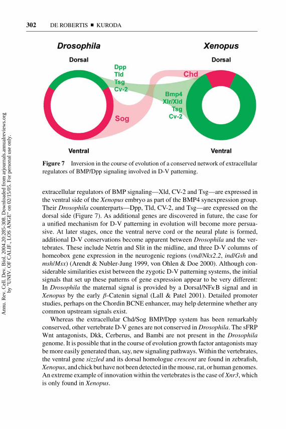
As shown in Figure 7, Sog is expressed ventrally in Drosophila embryos (first inthe ventral two thirds of the blastoderm, then in neurogenic ventral ectoderm, andfinally in ventral midline cells), whereas Chordin is expressed dorsally in the ver-tebrates. An argument against homologous roles for Chordin and Sog was that onewas expressed in mesoderm and the other in neuroectoderm. The realization thatChordin is expressed in the BCNE center, which gives rise to the brain and floorplate (Kuroda et al. 2004), now removes this objection because in both animalsthe initial expression is found in neuroectoderm. A number of recently identified
Ann
u. R
ev. C
ell.
Dev
. Bio
l. 20
04.2
0:28
5-30
8. D
ownl
oade
d fr
om a
rjou
rnal
s.an
nual
revi
ews.
org
by "
UN
IV. O
F C
AL
IF.,
LO
S A
NG
E"
on 0
2/15
/05.
For
per
sona
l use
onl
y.
10 Sep 2004 12:47 AR AR226-CB20-10.tex AR226-CB20-10.SGM LaTeX2e(2002/01/18) P1: GCE
302 DE ROBERTIS � KURODA
Figure 7 Inversion in the course of evolution of a conserved network of extracellularregulators of BMP/Dpp signaling involved in D-V patterning.
extracellular regulators of BMP signaling—Xld, CV-2 and Tsg—are expressed inthe ventral side of the Xenopus embryo as part of the BMP4 synexpression group.Their Drosophila counterparts—Dpp, Tld, CV-2, and Tsg—are expressed on thedorsal side (Figure 7). As additional genes are discovered in future, the case fora unified mechanism for D-V patterning in evolution will become more persua-sive. At later stages, once the ventral nerve cord or the neural plate is formed,additional D-V conservations become apparent between Drosophila and the ver-tebrates. These include Netrin and Slit in the midline, and three D-V columns ofhomeobox gene expression in the neurogenic regions (vnd/Nkx2.2, ind/Gsh andmsh/Msx) (Arendt & Nubler-Jung 1999, von Ohlen & Doe 2000). Although con-siderable similarities exist between the zygotic D-V patterning systems, the initialsignals that set up these patterns of gene expression appear to be very different:In Drosophila the maternal signal is provided by a Dorsal/NFκB signal and inXenopus by the early β-Catenin signal (Lall & Patel 2001). Detailed promoterstudies, perhaps on the Chordin BCNE enhancer, may help determine whether anycommon upstream signals exist.
Whereas the extracellular Chd/Sog BMP/Dpp system has been remarkablyconserved, other vertebrate D-V genes are not conserved in Drosophila. The sFRPWnt antagonists, Dkk, Cerberus, and Bambi are not present in the Drosophilagenome. It is possible that in the course of evolution growth factor antagonists maybe more easily generated than, say, new signaling pathways. Within the vertebrates,the ventral gene sizzled and its dorsal homologue crescent are found in zebrafish,Xenopus, and chick but have not been detected in the mouse, rat, or human genomes.An extreme example of innovation within the vertebrates is the case of Xnr3, whichis only found in Xenopus.
Ann
u. R
ev. C
ell.
Dev
. Bio
l. 20
04.2
0:28
5-30
8. D
ownl
oade
d fr
om a
rjou
rnal
s.an
nual
revi
ews.
org
by "
UN
IV. O
F C
AL
IF.,
LO
S A
NG
E"
on 0
2/15
/05.
For
per
sona
l use
onl
y.

10 Sep 2004 12:47 AR AR226-CB20-10.tex AR226-CB20-10.SGM LaTeX2e(2002/01/18) P1: GCE
D-V PATTERNING AND NEURAL INDUCTION 303
Communicating Dorsal and Ventral Signals
During development the dorsal and ventral sides of the embryo must be able to talkto each other over many cell diameters. As seen in Figure 1, when transplanted atgastrula (about 10,000-cell stage), a small organizer graft will induce a perfectlypatterned second embryo. Similarly, if a gastrula is divided into two halves with ahair loop, two smaller but well-proportioned embryos can be obtained (Spemann1938). This problem of regulation in a developing field of cells that tend to re-formnormal structures after experimental perturbations constitutes one of the unsolvedmysteries of developmental biology. The realization that the Xenopus gastrula hasdorsal and ventral signaling centers that secrete related growth factor antagonists(such as Chd/CV-2 and crescent/sizzled) under opposite transcriptional control byBMP signals, now sets the stage for investigating the molecular nature of thesecell-cell communications between the dorsal and ventral poles of the embryo. Theuse of loss-of-function morpholino reagents in combination with the cut-and-pasteembryological experiments that are possible in Xenopus, holds promise for furtheradvances in our understanding of D-V patterning regulation, morphogenetic fields,and neural differentiation.
ACKNOWLEDGMENTS
We thank B. Reversade, H. Lee, A. Ikeda, L. Fuentealba, A. Mays, J. Kim, L.Zakin, and D. Geissert for comments on the manuscript. Our work is supported bythe NIH and the HHMI. The authors are Investigator and Associate, respectively,of the Howard Hughes Medical Institute.
The Annual Review of Cell and Developmental Biology is online athttp://cellbio.annualreviews.org
LITERATURE CITED
Agathon A, Thisse C, Thisse B. 2003. Themolecular nature of the zebrafish tail orga-nizer. Nature 424:448–52
Agius E, Oelgeschlager M, Wessely O, KempC, De Robertis EM. 2000. EndodermalNodal-related signals and mesoderm induc-tion in Xenopus. Development 127:1173–83
Arendt D, Nubler-Jung K. 1999. Comparison ofearly nerve cord development in insects andvertebrates. Development 126:2309–25
Aubert J, Dunstan H, Chambers I, Smith A.2002. Functional gene screening in embry-onic stem cells implicates Wnt antagonism
in neural differentiation. Nat. Biotechnol. 20:1240–45
Aubin J, Davy A, Soriano P. 2004. In vivoconvergence of BMP and MAPK signal-ing pathways: impact of differential Smad1phosphorylation on development and home-ostasis. Genes Dev. In press
Avsian-Kretchmer O, Hsueh AJ. 2004. Com-parative genomic analysis of the eight-membered ring cystine knot-containing bonemorphogenetic protein antagonists. Mol. En-docrinol. 18:1–12
Bachiller D, Klingensmith J, Shneyder N, An-derson R, Tran U, et al. 2000. The organizer
Ann
u. R
ev. C
ell.
Dev
. Bio
l. 20
04.2
0:28
5-30
8. D
ownl
oade
d fr
om a
rjou
rnal
s.an
nual
revi
ews.
org
by "
UN
IV. O
F C
AL
IF.,
LO
S A
NG
E"
on 0
2/15
/05.
For
per
sona
l use
onl
y.
10 Sep 2004 12:47 AR AR226-CB20-10.tex AR226-CB20-10.SGM LaTeX2e(2002/01/18) P1: GCE
304 DE ROBERTIS � KURODA
factors Chordin and Noggin are required formouse forebrain development. Nature 403658–61
Bachiller D, Klingensmith J, Shneyder N, TranU, Anderson R, et al. 2003. The role ofchordin/BMP signals in mammalian pharyn-geal development and DiGeorge syndrome.Development 130:3567–78
Baker JC, Beddington RS, Harland RM. 1999.Wnt signaling in Xenopus embryos inhibitsbmp4 expression and activates neural devel-opment. Genes Dev. 13:3149–59
Barth LG. Neural diffferentiation without orga-nizer. 1941. J. Exp. Zool. 87:371–84
Beddington RS, Robertson EJ. 1999. Axis de-velopment and early asymmetry in mam-mals. Cell 96:195–209
Bertocchini F, Stern CD. 2002. The hypoblastof the chick embryo positions the primitivestreak by antagonizing nodal signaling. Dev.Cell 3:735–44
Binnerts ME, Wen X, Cant-Barrett K, Bright J,Chen HT, et al. 2004. Human Crossveinless-2 is a novel inhibitor of bone morphogeneticproteins. Biochem. Biophys. Res. Commun.315:272–80
Blitz IL, Cho KW, Chang C. 2003. Twisted gas-trulation loss-of-function analyses support itsrole as a BMP inhibitor during early Xenopusembryogenesis. Development 130:4975–88
Bouwmeester T, Kim S, Sasai Y, Lu B, DeRobertis EM. 1996. Cerberus is a head-inducing secreted factor expressed in theanterior endoderm of Spemann’s organizer.Nature 382:595–601
Bradley L, Sun B, Collins-Racie L, LaVallieE, McCoy J, Sive H. 2000. Different activi-ties of the frizzled-related proteins frzb2 andsizzled2 during Xenopus anteroposterior pat-terning. Dev. Biol. 227:118–32
Carroll SB, Grenier JK, Weathebee SD. 2001.From DNA to Diversity: Molecular Geneticsand the Evolution of Animal Design. Malden,MA: Blackwell Sci.
Chang C, Holtzman DA, Chau S, Chickering T,Woolf EA, et al. 2001. Twisted gastrulationcan function as a BMP antagonist. Nature410:483–87
Cho KW, Blumberg B, Steinbeisser H, DeRobertis EM. 1991. Molecular nature of Spe-mann’s organizer: the role of the Xenopushomeobox gene goosecoid. Cell 67:1111–20
Coffinier C, Ketpura N, Tran U, Geissert D, DeRobertis EM. 2002. Mouse Crossveinless-2is the vertebrate homolog of a Drosphila ex-tracellular regulator of BMP signaling. Mech.Dev. 119:179–84
Collavin L, Kirschner MW. 2003. The secretedFrizzled-related protein Sizzled functions asa negative feedback regulator of extremeventral mesoderm. Development 130:805–16
Conley CA, Silburn R, Singer MA, Ralston A,Rohw-Nutter D, et al. 2000. Crossveinless2 contains cysteine-rich domains and is re-quired for high levels of BMP-like activityduring the formation of the cross veins inDrosophila. Development 127:3947–59
Connors SA, Trout J, Ekker M, MullinsMC. 1999. The role of tolloid/mini fin indorsoventral pattern formation of the ze-brafish embryo. Development 126:3119–30
Dale L, Evans W, Goodman SA. 2002. Xolloid-related: a novel BMP1/Tolloid-related metal-loprotease is expressed during early Xenopusdevelopment. Mech. Dev. 119:177–90
del Barco-Barrantes I, Davidson G, Grone HJ,Westphal H, Niehrs C. 2003. Dkk1 and nog-gin cooperate in mammalian head induction.Genes Dev. 17:2239–44
De Robertis EM, Sasai Y. 1996. A common planfor dorso-ventral patterning in Bilateria. Na-ture 380:37–40
De Robertis EM, Larraın J, Oelgeschlager M,Wessely O. 2000. The establishment of Spe-mann’s organizer and patterning of the ver-tebrate embryo. Nat. Rev. Genet. 1:2053–62
De Robertis EM, Arechaga J, eds. 2001. TheSpemann Organizer 75 Years. Vol. 45. Bil-bao, Spain: Univ. Basque Country Press
Eldar A, Dorfman R, Weiss D, Ashe H, ShiloBZ. 2002. Robustness of the BMP mor-phogen gradient in Drosophila embryonicpatterning. Nature 419:304–8
Fainsod A, Steinbeisser H, De Robertis EM.
Ann
u. R
ev. C
ell.
Dev
. Bio
l. 20
04.2
0:28
5-30
8. D
ownl
oade
d fr
om a
rjou
rnal
s.an
nual
revi
ews.
org
by "
UN
IV. O
F C
AL
IF.,
LO
S A
NG
E"
on 0
2/15
/05.
For
per
sona
l use
onl
y.
10 Sep 2004 12:47 AR AR226-CB20-10.tex AR226-CB20-10.SGM LaTeX2e(2002/01/18) P1: GCE
D-V PATTERNING AND NEURAL INDUCTION 305
1994. On the function of BMP-4 in pattern-ing the marginal zone of the Xenopus em-bryo. EMBO J. 13:5015–25
Garcia-Abreu J, Coffinier C, Larrain J, Oel-geschlager M, De Robertis EM. 2002.Chordin-like CR domains and the regulationof evolutionarily conserved extracellular sig-naling systems. Gene 287:39–47
Gawantka V, Delius H, Hirschfeld K, Blumen-stock C, Niehrs C. 1995. Antagonizing theSpemann organizer: role of the homeoboxgene Xvent-1. EMBO J. 14:6268–79
Glinka A, Wu W, Delius H, Monaghan AP,Blumenstock C, et al. 1998. Dickkopf-1 isa member of a new family of secreted pro-teins and functions in head induction. Nature391:357–62
Glinka A, Wu W, Onichtchouk D, BlumenstockC, Niehrs C. 1997. Head induction by si-multaneous repression of Bmp and Wnt sig-nalling in Xenopus. Nature 389:517–19
Goerttler K. 1925. Die Formbildung derMedullaranlage bei Urodelen. Roux’s Arch.Entw. Mech. 106:503–41
Gonzalez EM, Fekany-Lee K, Carmany-Rampey A, Erter C, Topczewski J, et al. 2000.Head and trunk in zebrafish arise via coin-hibition of BMP signaling by bozozok andchordino. Genes Dev. 14:3087–92
Groppe J, Greenwald J, Wiater E, Rodriguez-Leon J, Economides AN, et al. 2002. Struc-tural basis of BMP signalling inhibition bythe cystine knot protein Noggin. Nature 420:636–42
Grunz H, ed. 2004. The Vertebrate Organizer.Berlin: Springer-Verlag
Hammerschmidt M, Mullins MC. 2002. Dor-soventral patterning in the zebrafish: bonemorphogenetic proteins and beyond. Res.Probl. Cell Differ. 40:72–95
Hansen CS, Marion CD, Steele K, George S,Smith WC. 1997. Direct neural induction andselective inhibition of mesoderm and epi-dermis inducers by Xnr3. Development 124:483–92
Haramoto Y, Tanegashima K, Onuma Y, Taka-hashi S, Sekizaki H, et al. 2004. Xeno-pus tropicalis nodal-related gene 3 regulates
BMP signaling: an essential role for the pro-region. Dev. Biol. 265:155–68
Hardcastle Z, Chalmers AD, Papalopulu N.2000. FGF-8 stimulates neuronal differenti-ation through FGFR-4a and interferes withmesoderm induction in Xenopus embryos.Curr. Biol. 10:1511–14
Harland R. 2000. Neural induction. Curr. Opin.Genet. Dev. 19:357–62
Harland R, Gerhart J. 1997. Formation andfunction of Spemann’s organizer. Annu. Rev.Cell Dev. Biol. 13:611–67
Holtfreter J. 1933. Die totale Exogastrulation,eine Selbstablosung des Ektoderms vom En-tomesoderm. Entwicklung und funktionellesVerhalten nervenloser Organe. Roux’s Arch.Entw. Mech. 129:670–93
Holtfreter J, Hamburger V. 1955. Embryoge-nesis: progressive differentiation—amphibi-ans. In Analysis of Development. ed. BHWillier, PA Weiss, V Hamburger, pp. 230–96. New York: Haffner
Hyvonen M. 2003. CHRD, a novel domainin the BMP inhibitor chordin, is also foundin microbial proteins. Trends Biochem. Sci.28:470–73
Joly JS, Joly C, Schulte-Merker S, BoulekbacheH, Condamine H. 1993. The ventral and pos-terior expression of the zebrafish homeoboxgene eve1 is perturbed in dorsalized and mu-tant embryos. Development 119:1261–75
Kawahara A, Wilm T, Solnica-Krezel L, DawidIB. 2000. Functional interaction of vega2and goosecoid homeobox genes in zebrafish.Genesis 28:58–67
Kawano Y, Kypta R. 2003. Secreted antago-nists of the Wnt signalling pathway. J. CellSci. 116:2627–34
Kazanskaya O, Glinka A, Niehrs C. 2000. Therole of Xenopus dickkopf1 in prechordalplate specification and neural patterning. De-velopment 127:4981–92
Klingensmith J, Ang SL, Bachiller D, RossantJ. 1999. Neural induction and patterning inthe mouse in the absence of the node and itsderivatives. Dev. Biol. 216:535–49
Kretzschmar M, Doody J, Massague J.1997. Opposing BMP and EGF signalling
Ann
u. R
ev. C
ell.
Dev
. Bio
l. 20
04.2
0:28
5-30
8. D
ownl
oade
d fr
om a
rjou
rnal
s.an
nual
revi
ews.
org
by "
UN
IV. O
F C
AL
IF.,
LO
S A
NG
E"
on 0
2/15
/05.
For
per
sona
l use
onl
y.
10 Sep 2004 12:47 AR AR226-CB20-10.tex AR226-CB20-10.SGM LaTeX2e(2002/01/18) P1: GCE
306 DE ROBERTIS � KURODA
pathways converge on the TGF-β family me-diator Smad1. Nature 389:618–22
Kuroda H, Wessely O, De Robertis EM. 2004.Neural induction in Xenopus: requirementfor ectodermal and endomesodermal signalsvia Chordin, Noggin, β-Catenin and Cer-berus. PLoS Biol. 2:625–34
Lall S, Patel NH. 2001. Conservation and di-vergence in molecular mechanisms of axisformation. Annu. Rev. Genet. 35:407–37
Larraın J, Bachiller D, Lu B, Agius E, Pic-colo, et al. 2000. BMP-binding modules inChordin: a model for signalling regulationin the extracellular space. Development 127:821–30
Larraın J, Oelgeschlager M, Ketpura NI, Re-versade B, Zakin L, et al. 2001. Proteolyticcleavage of Chordin as a switch for the dualactivities of Twisted gastrulation in BMP sig-naling. Development 128:4439–47
Lemaire P, Bertrand V, Hudson C. 2002. Earlysteps in the formation of neural tissue in as-cidian embryos. Dev. Biol. 252:151–69
Leung TC, Bischof J, Soll I, Niessing D, ZhangD, et al. 2003. bozozok directly repressesbmp2b transcription and mediates the earliestdorsoventral asymmetry of bmp2b expres-sion in zebrafish. Development 130:3639–49
Leyns L, Bouwmeester T, Kim SH, Piccolo S,De Robertis EM. 1997. Frzb-1 is a secretedantagonist of Wnt signaling expressed in theSpemann organizer. Cell 88:747–56
London C, Akers T, Phillips CR. 1988. Expres-sion of Epi1, an epidermal specific marker,in Xenopus laevis embryos is specified priorto gastrulation. Dev. Biol. 129:380–89
Mao B, Wu W, Li Y, Hoppe D, Stannek P,et al. 2001. LDL-receptor-related protein 6is a receptor for Dickkopf proteins. Nature411:321–25
Mao B, Wu W, Davidson G, Marhold J, Li M,et al. 2002. Kremen proteins are Dickkopfreceptors that regulate Wnt/beta-catenin sig-nalling. Nature 417:664–67
Martyn U, Schulte-Merker S. 2003. The ven-tralized ogon mutant phenotype is caused bya mutation in the zebrafish homologue of Siz-
zled, a secreted Frizzled-related protein. Dev.Biol. 260:58–67
Massague J. 2003. Integration of Smad andMAPK pathways: a link and a linker revis-ited. Genes Dev. 17:2993–97
McMahon JA, Takada S, Zimmerman LB, FanCM, Harland RM, et al. 1998. Noggin-mediated antagonism of BMP signaling is re-quired for growth and patterning of the neuraltube and somite. Genes Dev. 12:1438–52
Miller JR, Rowning BA, Larabell CA, Yang-Snyder JA, Bates RL, Moon RT. 1999. Es-tablishment of the dorsal-ventral axis inXenopus embryos coincides with the dorsalenrichment of dishevelled that is dependenton cortical rotation. J. Cell Biol. 146:427–37
Moser M, Binder O, Wu Y, Aitsebaomo J,Ren R. 2003. BMPER, a novel endothelialcell precursor-derived protein, antagonizesbone morphogenetic protein signaling andendothelial cell differentiation. Mol. Cell.Biol. 23:5664–79
Mukhopadhyay M, Shtrom S, Rodriguez-Esteban C, Chen L, Tsukui T, et al. 2001.Dickkopf1 is required for embryonic headinduction and limb morphogenesis in themouse. Dev. Cell 1:423–34
Munoz-Sanjuan I, Brivanlou AH. 2002. Neuralinduction, the default model and embryonicstem cells. Nat. Rev. Neurosci. 3:271–80
Nosaka T, Morita S, Kitamura H, Naka-jima H, Shibata F, et al. 2003. MammalianTwisted gastrulation is essential for skeleto-lymphogenesis. Mol. Cell Biol. 23:2969–80
Niehrs C. 2001. The Spemann organizer andembryonic head induction. EMBO J. 20:631–37
Niehrs C, Pollet N. 1999. Synexpression groupsin eukaryotes. Nature 402:483–87
Niswander L, Martin GR. 1993. FGF-4 andBMP-2 have opposite effects on limb growth.Nature 361:68–71
Oelgeschlager M, Larraın J, Geissert D, DeRobertis EM. 2000. The evolutionarily con-served BMP-binding protein Twisted gas-trulation promotes BMP signalling. Nature405:757–63
Oelgeschlager M, Kuroda H, Reversade B, De
Ann
u. R
ev. C
ell.
Dev
. Bio
l. 20
04.2
0:28
5-30
8. D
ownl
oade
d fr
om a
rjou
rnal
s.an
nual
revi
ews.
org
by "
UN
IV. O
F C
AL
IF.,
LO
S A
NG
E"
on 0
2/15
/05.
For
per
sona
l use
onl
y.
10 Sep 2004 12:47 AR AR226-CB20-10.tex AR226-CB20-10.SGM LaTeX2e(2002/01/18) P1: GCE
D-V PATTERNING AND NEURAL INDUCTION 307
Robertis EM. 2003a. Chordin is required forthe Spemann organizer transplantation phe-nomenon in Xenopus embryos. Dev. Cell 4:219–30
Oelgeschlager M, Reversade B, Larraın J, Lit-tle S, Mullins MC, et al. 2003b. The pro-BMP activity of Twisted gastrulation is inde-pendent of BMP binding. Development 130:4047–56
Onichtchouk D, Chen YG, Dosch R, Gawan-tka V, Delius H, et al. 1999. Silencing ofTGF-beta signalling by the pseudoreceptorBAMBI. Nature 401:480–85
Pera EM, De Robertis EM. 2000. A directscreen for secreted proteins in Xenopus em-bryos identifies distinct activities for the Wntantagonists Crescent and Frzb-1. Mech. Dev.96:183–95
Pera EM, Wessely O, Li S, De Robertis EM.2001. Neural and head induction by insulin-like growth factor signals. Dev. Cell 1:655–65
Pera EM, Ikeda A, Eivers E, De Robertis EM.2003. Integration of IGF, FGF and anti-BMPsignals via Smad1 phosphorylation in neuralinduction. Genes Dev. 17:3023–28
Perea-Gomez A, Vella FD, Shawlot W, Oulad-Abdelghani M, Chazaud C, et al. 2002. Nodalantagonists in the anterior visceral endodermprevent the formation of multiple primitivestreaks. Dev. Cell 3:745–56
Petryk A, Anderson RM, Jarcho MP, Leaf I,Carlson CS. 2004. The mammalian twistedgastrulation gene functions in foregut andcraniofacial development. Dev. Biol. 267:374–86
Piccolo S, Sasai Y, Lu B, De Robertis EM. 1996.Dorsoventral patterning in Xenopus: inhibi-tion of ventral signals by direct binding ofchordin to BMP-4. Cell 86:589–98
Piccolo S, Agius E, Leyns L, BhattacharyyaS, Grunz H, et al. 1999. The head in-ducer Cerberus is a multifunctional antago-nist of Nodal, BMP and Wnt signals. Nature397:707–10
Richard-Parpaillon L, Heligon C, Chesnel F,Boujard D, Philpott A. 2002. The IGF path-way regulates head formation by inhibiting
Wnt signaling in Xenopus. Dev. Biol. 244:407–17
Ross JJ, Shimmi O, Vilmos P, Petryk A, KimH, et al. 2001. Twisted gastrulation is a con-served extracellular BMP antagonist. Nature410:479–83
Salic AN, Kroll KL, Evans LM, Kirschner MW.1997. Sizzled: a secreted Xwnt8 antagonistexpressed in the ventral marginal zone ofXenopus embryos. Development 124:4739–34
Sater AK, El-Hodiri HM, Goswami M, Alexan-der TB, Al-Sheikh O, et al. 2003. Evidencefor antagonism of BMP-4 signals by MAP ki-nase during Xenopus axis determination andneural specification. Differentiation 71:434–44
Schneider S, Steinbeisser H, Warga RM,Hausen P. 1996. Beta-catenin translocationinto nuclei demarcates the dorsalizing cen-ters in frog and fish embryos. Mech. Dev. 57:191–98
Schohl A, Fagotto F. 2002. Beta-catenin,MAPK and Smad signaling during earlyXenopus development. Development 129:37–52
Schulte-Merker S, Lee KJ, McMahon AP, Ham-merschmidt M. 1997. The zebrafish orga-nizer requires chordino. Nature 387:862–63
Scott IC, Blitz IL, Pappano WN, ImamuraY, Clark TG, et al. 1999. MammalianBMP-1/Tolloid-related metalloproteinases,including novel family member mammalianTolloid-like 2, have differential enzymaticactivities and distributions of expression rel-evant to patterning and skeletogenesis. Dev.Biol. 213:283–300
Scott IC, Blitz IL, Pappano WN, Maas SA, ChoKW, et al. 2001. Homologues of Twisted gas-trulation are extracellular cofactors in antag-onism of BMP signalling. Nature 410:475–78
Sharpe CR, Fritz AF, De Robertis EM, GurdonJB. 1987. A homeobox-containing marker ofposterior neural differentiation shows the im-portance of predetermination in neural induc-tion. Cell 50:749–58
Ann
u. R
ev. C
ell.
Dev
. Bio
l. 20
04.2
0:28
5-30
8. D
ownl
oade
d fr
om a
rjou
rnal
s.an
nual
revi
ews.
org
by "
UN
IV. O
F C
AL
IF.,
LO
S A
NG
E"
on 0
2/15
/05.
For
per
sona
l use
onl
y.
10 Sep 2004 12:47 AR AR226-CB20-10.tex AR226-CB20-10.SGM LaTeX2e(2002/01/18) P1: GCE
308 DE ROBERTIS � KURODA
Shi Y, Massague J. 2003. Mechanisms of TGF-beta signaling from cell membrane to the nu-cleus. Cell 113:685–700
Spemann H. 1938. Embryonic Developmentand Induction. New Haven: Yale Univ. Press
Spemann H, Mangold H. 1924. Induction ofembryonic primordia by implantation of or-ganizers from a different species. Roux’sArch. Entw. Mech. 100:599–638. Reprintedand Transl. Int. J. Dev. Biol. 45:13–38
Stern CD. 2002. Induction and initial pattern-ing of the nervous system—the chick em-bryo enters the scene. Curr. Opin. Genet. Dev.12:447–51
Stern CD, ed. 2004. Gastrulation. Cold SpringHarbor, NY: Cold Spring Harbor Press
Streit AC, Stern CD. 1999. Neural induction. Abird’s eye view. Trends Genet. 15:20–24
Takahashi S, Yokota C, Takano K, TanegashimaK, Onuma Y, et al. 2000 Two novel nodal-related genes initiate early inductive eventsin Xenopus Nieuwkoop center. Development127:5319–29
Tamai K, Semenov M, Kato Y, Spokony R, LiuC, et al. 2000. LDL-receptor-related proteinsin Wnt signal transduction. Nature 407:530–35
Tamai K, Zeng X, Liu C, Zhang X, Harada Y,et al. 2004. A mechanism for Wnt coreceptoractivation. Mol. Cell 13:149–56
Thesleff I, Mikkola M. 2002. The role of growthfactors in tooth development. Int. Rev. Cytol.217:93–135
von Ohlen T, Doe CQ. 2000. Convergenceof dorsal, dpp, and egfr signaling pathwayssubdivides the Drosophila neuroectoderminto three dorsal-ventral columns. Dev. Biol.224:362–72
Warren SM, Brunet LJ, Harland RM, Econo-mides AN, Longaker MT. 2003. The BMPantagonist noggin regulates cranial suture fu-sion. Nature 422:625–29
Weaver M, Dunn NR, Hogan BL. 2000. Bmp4
and Fgf10 play opposing roles duringlung bud morphogenesis. Development 127:2695–704
Weaver C, Farr GH, Pan W, Rowning BA, WangJ, et al. 2003. GBP binds kinesin light chainand translocates during cortical rotation inXenopus eggs. Development 148:691–702
Weinstein DC, Hemmati-Brivanlou A. 1999.Neural induction. Annu. Rev. Cell Dev. Biol.15:411–33
Wessely O, Agius E, Oelgeschlager M, PeraEM, De Robertis EM. 2001. Neural inductionin the absence of mesoderm: beta-catenin-dependent expression of secreted BMP an-tagonists at the blastula stage in Xenopus.Dev. Biol. 234:161–73
Wessely O, Kim JI, Geissert D, Tran U, DeRobertis, EM. 2004. Analysis of Spemannorganizer formation in Xenopus embryos bycDNA macroarrays. Dev. Biol. 269:552–66
Wilson PA, Hemmati-Brivanlou A. 1995. In-duction of epidermis and inhibition of neuralfate by Bmp-4. Nature 376:331–33
Wilson SI, Edlund T. 2001. Neural induction:toward a unifying mechanism. Nat. Neurosci.4:1161–68
Yabe T, Shimizu T, Muraoka O, Bae YK, Hi-rata T, et al. 2003. Ogon/Secreted Frizzledfunctions as a negative feedback regulator ofBmp signaling. Development 130:2705–16
Zakin L, De Robertis EM. 2004. Inactivationof mouse Twisted gastrulation reveals its rolein promoting Bmp4 activity during forebraindevelopment. Development 131:413–24
Zimmerman LB, De Jesus-Escobar JM, Har-land RM. 1996. The Spemann organizer sig-nal noggin binds and inactivates bone mor-phogenetic protein 4. Cell 86:599–606
Yamamoto M, Saijoh Y, Perea-Gomez A,Shawlot W, Behringer RR, et al. 2004. Nodalantagonists regulate formation of the antero-posterior axis of the mouse embryo. Nature428:387–92
Ann
u. R
ev. C
ell.
Dev
. Bio
l. 20
04.2
0:28
5-30
8. D
ownl
oade
d fr
om a
rjou
rnal
s.an
nual
revi
ews.
org
by "
UN
IV. O
F C
AL
IF.,
LO
S A
NG
E"
on 0
2/15
/05.
For
per
sona
l use
onl
y.