comparative studies on ferredoxin-nadp+ … · 435 be dependent upon the type of cells, the age,...
TRANSCRIPT
Plant Physiol. (1993) 103: 435-440
Comparative Studies on Ferredoxin-NADP+ Oxidoreductase lsoenzymes Derived from Different Organs by Antibodies
Specific for the Radish Root- and Leaf-Enzymes’
Susumu Morigasaki2, Tie Jin, and Keishiro Wada*
Department of Biology, Faculty of Science, Kanazawa University, Kakuma, Kanazawa, Ishikawa 920-1 1 Japan
Determination of the prosthetic group and titration of sulfhydryl group of ferredoxin-NADP+ oxidoreductase (FNR) from roots of radish (Raphanus sativus var acanthiformis cv Miyashige) con- firmed its similarity to leaf-FNR. Antisera directed against radish root-FNR and leaf-FNR distinguished the enzyme forms from roots and leaves of radish as well as other flowering plants. The FNR isoenzymes showed organ-specific distributions. In horsetail (Equisetum arvense 1.) and cultured liverwort cells (Marchantia polymorpha), at least two FNR isoenzymes were distinguished by the antisera. FNR from Chlorella vulgaris reacted only with the anti-root-FNR antiserum. FNR from a cyanobacterium, Spirulina spp., failed to react with either antiserum.
In photosynthetic tissues, two enzymes functional in nitro- gen metabolism, Fd-glutamate synthase and Fd-nitrite reduc- tase, are located in chloroplasts. These enzymes require reduced Fd, which is generated by PSI. Early studies on nitrogen metabolism in roots were summarized by Oaks and Hirel(l985). Fd-dependent enzymes of nitrogen assimilation have also been demonstrated in heterotrophic tissues by using an in vitro system supplemented with leaf Fd or viologen dyes. It has been concluded that the Fd-dependent enzymes are present in plastids of roots and nongreen cultured tobacco cells (Washitani and Sato, 1977a, 1977b; Oaks and Hirel, 1985). In roots, the Fd required by the Fd-linked enzymes is reduced via FNR by NADPH, which appears to be generated by the pentose-phosphate pathway (Oji et al., 1985; Suzuki et al., 1985; Wada et al., 1986; Bowsher et al., 1989; Mori- gasaki et al., 1990b).
Root Fds have been purified and characterized from rad- ish; spinach, maize, and tomato (Wada et al., 1986; Kimata and Hase, 1989; Morigasaki et al., 1990a; Green et al., 1991). The primary sequences of radish and maize root Fds (the latter was deduced from the cDNA) were determined and compared with those of leaf counterparts (Wada et al., 1989; Hase et al., 1991). In these plants, the Fd isoproteins showed organ-specific distribution. Expression of Fd genes seems to
Supported in part by a Grant-in-Aid for Scientific Research (No. 02304005) to K.W. from the Ministry of Education, Science and Culture of Japan.
Present address: Department of Biochemistry, Toyama Medica1 and Pharmaceutical University, School of Medicine, Toyama 930-01 Japan.
* Corresponding author; fax 81-762-64-5745. 435
be dependent upon the type of cells, the age, and the envi- ronmental conditions.
FNR, showing the same distribution as Fd, was purified from radish (Raphanus sativus) and spinach roots and char- acterized (Morigasaki et al., 1990a, 1990b; Morigasaki and Wada, 1991). The root-FNRs resembled leaf-FNR in enzymic properties, absorption spectrum, and molecular mass, but differed in amino acid composition and amino-terminal se- quence. The results indicate that root- and leaf-FNRs are distinct isoenzymes that are encoded by different genes. In this study we further characterize FNR isoenzymes from the different range of plant groups by using antisera raised against root- and leaf-FNRs from radish.
MATERIALS AND METHODS
Plant Materials
Radish plants (Raphanus sativus var acanthiformis cv Mi- yashige) were purchased from a farmer or a local market. Pea (Pisum sativum L.), soybean (Glycine max L.), and maize (Zea mays L. cv Golden Cross Bantum T51) were grown in a greenhouse for about 1 month. Horsetail (Equisetum arvense L.) was collected on the campus of Kanazawa University in spring 1991. Green shoots and subterranean stems of horse- tail were used as the counterparts of leaves and roots, re- spectively. Cultured liverwort cells (Marchantia polymorpha) and Spirulina spp. cells were gifts from Professor K. Ohyama of Kyoto University and Professor H. Matsubara of Osaka University, respectively. An extract from Chlorella vulgaris cells that showed FNR activity was a gift of Dr. Y. Shiraiwa of Niigata University.
Large-Scale Preparation of FNR from Radish Roots
About 580 kg of lower, white roots of radish were ground in a large homogenizer (200 L) for 5 min in 100-kg lots each with 200 g of Tris powder. Two hundred liters of distilled water was added to 600 L of the combined homogenates in a 1000-L pot and the diluted homogenate was processed with a Yabuta’s squeezer. The crude extract (680 L) was adjusted to pH 7.9 with 520 g of Tris powder and diluted to 1300 L with distilled water. Wet DEAE-cellulose (10 L), equilibrated with 10 m~ Tris-HC1 buffer, pH 8.5, was added to the
Abbreviations: FAD, flavin adenine dinucleotide; FNR, ferredoxin- NADP+ oxidoreductase.
www.plantphysiol.orgon June 28, 2018 - Published by Downloaded from Copyright © 1993 American Society of Plant Biologists. All rights reserved.

436 Morigasaki et al. Plant Physiol. Vol. 103, 1993
resulting extract. The suspension was gently stirred for 10 min and allowed to remain for 30 min to allow the DEAE- cellulose to settle. The decanted supernatant solution was further diluted to 2000 L with distilled water. Another batch of DEAE-cellulose (15 L) was added to the solution and the suspension was decanted, as described above. The decanted solution was discarded and the combined DEAE-cellulose (25 L) was packed into columns (6.4 X 100 cm). Proteins were eluted with 0.7 M NaCl in 30 mM Tris-HC1 buffer, pH 8.5. The eluate, with 11,200 units of FNR activity, was further purified by the conventional method (Morigasaki et al., 1990b), except that the concentration of Tris-HC1 buffer was 30 mM. About 9 mg of FNR was obtained as the homogeneous state in SDS-PAGE showing a single band.
Preparation of Antisera against FNR lsoenzymes
Individual antisera against radish root-FNR and leaf-FNR were raised in rabbits. The homogenous root-FNR (0.35 mg) and leaf-FNR (0.29 mg) in 0.4 mL was thoroughly mixed with 0.4 mL of Freunds complete adjuvant and administered intradermally into rabbits four times (lst, 5th, 9th, and 16th d). The preimmune sera were taken from each rabbit before the immunization. On the 22nd d, rabbits were slaughtered and total blood was taken. The antisera were sedimented with 50% saturation of ammonium sulfate and the precipitate was dissolved in TBS (50 mM Tris-HC1 buffer, pH 7.5, con- taining 0.9% NaCl) and dialyzed against TBS. The dialysate was clarified by centrifugation and the supernatant fraction was stored at -8OOC until use.
lmmunotitration
Immunotitration was camed out essentially as described by Suzuki et al. (1982). Antisera were mixed with 0.1 unit of the radish FNR isoenzymes in TBS. The mixed solution was incubated for 1 h at 25OC and then for 12 h at 4OC. After centrifugation, the supematant was used to assay for FNR activity (Fd-dependent Cyt c reduction activity). The antisera had no FNR activity.
SDS-PACE
SDS-PAGE (12.5% gel concentration) was carried out ac- cording to Laemmli (1970). The gel was stained with Coo- massie brilliant blue R-250 or subjected to immunoblot analy- sis. Dalton Mark VII-L (Sigma) was used as a molecular mass marker.
lmmunoblot Analysis
The transfer of proteins from the polyacrylamide gel to Clear Blot Membrane-p (Atto, Tokyo, Japan) was carried out at 1.5 to 2.0 mA cm-’ for 2 h with Atto Holizeblot AE-6670 (semidry type), according to the supplier’s manual. After transfer, the Membrane-p was reacted with protein A-con- jugated horseradish peroxidase and then stained with 0.04% (w/v) 3,3’-diaminobenzidine tetrahydrochloride in TBS con- taining 0.012% H202 for 10 to 30 min, essentially according to Bumette (1981).
Preparation of FNR Samples for an lmmunoblot Analysis
FNRs were partially purified from cultured liverwort cells, Chlorella cell-free extracts, and the leaf and root tissues of higher plants (pea, soybean, maize, and horsetail) by am- monium sulfate fractionation and Blue-Cellulofine, acco rding to Morigasaki et al. (1990b) with the following modificaíions. The concentration of Tris-HC1 buffer was 30 mM and the Blue-Cellulofine column was equilibrated with 30 mM Tris- HCl buffer, pH 7.8, and 1 mM EDTA and eluted with 1.0 M
NaCl in 30 mM Tris-HC1 buffer, pH 7.8, and 0.1 mM EDTA. The active FNR fraction was concentrated and desalted by ultrafiltration (Advantec Ultrafilter, UK 10 or Amicon Cen- triprep-10) and used for immunoblot analysis as a partially purified FNR. Specific activities of partially purified FNR samples from radish roots and leaves were 4.0 and 21.3 units mg-’ of protein, respectively.
Before grinding, liverwort cells and horsetail tissues were pulverized in liquid nitrogen and silica sand with a wooden pestle in an earthenware mortar fitted with fine groovas. In the case of horsetail, the grinding buffer consisted of 100 mM Tris-borate buffer, pH 8.7, 1 mM EDTA, 0.03% (w/v) Triton X-100, 0.5 m~ PMSF, 20 mM diethyldithiocarbamic acid, and 1.5% (w/v) insoluble PVP; the buffer used for dissolvin;; the ammonium sulfate precipitate and for dialysis was Tris-borate buffer, pH 8.5, containing 1 mM EDTA. The horsetail dialy- sate was diluted with 2 vol of 1 mM EDTA before kieing applied to the Blue-Cellulofine column.
Dot-Blot Analysis
The amounts ranging from 0.2 to 0.003 units of the radish FNRs and FNR sample(s) used for immunoblot analysis were adsorbed in seven dots on a wet Clear Blot Membrarie-p. Two sets of the membrane were prepared and dried. The dots on the membrane reacted with each antiserum as de- scribed above.
Other Proteins
FNRs from radish leaves and Spirulina were purified by ammonium sulfate fractionation, anion-exchange chromatog- raphy, and two different affinity chromatographies to the homogeneous state in SDS-PAGE, as described by Moriga- saki et al. (1990b), except that the concentration of Tris-HC1 buffers was 30 m ~ . Conventional methods were usetl to purify Fd from Spirulina spp. (Matsubara and Wada, 1!)88) and Cyt c from bovine heart (Hagihara et al., 1958).
Assay for FNR Activity
FNR was assayed by the Fd-dependent Cyt c reduction assay as described by Morigasaki et al. (1990b), except that the buffer was 50 m~ Tris-HC1 buffer, pH 7.8. One unit of activity is defined as the amount of enzyme that can redluce 1 pmol of Cyt c per min.
Determination of Protein
Protein was determined by the dye binding methocl of Bradford (1976) using BSA as the standard.
www.plantphysiol.orgon June 28, 2018 - Published by Downloaded from Copyright © 1993 American Society of Plant Biologists. All rights reserved.

Fd-NADP+ Reductase lsoenzymes from Different Organs 43 7
Determination of Flavin
Dissociation of flavin from radish root-FNR was carried out by the CaCI2 extraction according to Bookjans et al. (1978). The absorption maxima at 264, 382, and 450 nm were typical of a flavin. Flavin was also dissociated with 2.5% cold TCA in the dark (Koziol, 1971). After centrifuga- tion, the supernatant was adjusted to pH 7 with 1 N NaOH. The absorption maxima were at 263, 375, and 450 nm. The extracted flavin was further extracted with the same volume of phenol. The phenol phase was mixed with 0.5 mL of distilled water and 8 mL of ether. The aqueous phase, con- taining the flavin, was subjected to paper chromatography as described by Solomonson et al. (1975) except for the use of Toyo filter paper 50. The content of flavin was calculated using a millimolar extinction coefficient of 11.3 at 450 nm (Koziol, 1971).
Measurement of Sulfhydryl Groups in Root-FNR
The measurement of sulfhydryl groups in root-FNR was carried out by the method of Yao et al. (1985). Radish root- FNR was denatured by the addition of 6 M guanidine-HC1 solution to give a final concentration of 3 M and was carbox- ymethylated in the presence or absence of 1 mM 2-mercap- toethanol (Crestfield et al., 1963). The carboxymethylated FNR was hydrolyzed at 110OC for 24 h in the evacuated and sealed tubes and then the S-carboxymethylated Cys was determined by an automatic amino acid analyzer (Hitachi L-8500).
Reagents
Biochemicals were purchased from Sigma, Nacalai Tesque, Inc. (Kyoto, Japan), Wako Pure Chemical Industries, Ltd. (Osaka, Japan), and Oriental Yeast Co., Ltd. (Osaka, Japan). DEAE-cellulose was obtained from Nacalai Tesque, Blue- Cellulofine was from Seikagaku-Kogyo Co., Ltd. (Tokyo, Japan), and Sephadex and cyanogen bromide-Sepharose 48 were from Pharmacia Fine Chemicals (Piscataway, NJ). Chemical reagents were obtained from commercial sources and were of the highest quality available.
RESULTS AND DISCUSSION
About 9 mg of FNR were obtained as a homogeneous state from the large-scale (580 kg) radish root preparation. The yield was about 1 % compared with that obtained from leaves. The flavin extracted from radish root-FNR co-migrated with FAD on the paper chromatography, and the spectral prop- erties of the extracted flavin were similar to those of authentic FAD (data not shown). The recovered FAD per mo1 of FNR was 0.82 and 0.86 mo1 by the cold TCA and the CaC12 extractions, respectively. This indicated that radish root-FNR had 1 mo1 of FAD per mol. This value is consistent with that of its leaf counterpart (Shin, 1971; Carrillo and Vallejos, 1987).
The number of sulfhydryl groups of root-FNR was deter- mined to be 4.02 -+ 0.02 and 4.52 f 0.20 mol/mol of FNR under nonreducing and reducing conditions, respectively. The figures suggest the absence of a disulfide bond in radish
root-FNR molecule as in leaf-FNR, as pointed out by Yao et al. (1985).
Root-FNRs have been purified and characterized from radish and spinach root (Morigasaki et al., 1990a, 1990b; Morigasaki and Wada, 1991). Their enzymic properties, ab- sorption spectra, and molecular masses did not distinguish them from their leaf counterparts. The results in this paper confirmed the similarity between root- and leaf-FNRs.
For easy detection and distinction of root-FNR and leaf- FNR, specific antisera were prepared against radish root- and leaf-FNR. The antigen specificity of each antiserum was confirmed by immunotitration (Fig. 1) and immunoblot analysis (Fig. 2) using the radish FNR isoenzymes. Figure 1 shows immunotitration of FNRs by each antisera. The anti- root-FNR antiserum effectively inhibited the activity of radish root-FNR, but not that of leaf-FNR. Conversely, the anti- leaf-FNR antiserum strongly inhibited leaf-FNR. Root-FNR was slightly inhibited by the anti-leaf-FNR antiserum but only when a large amount of the antiserum was added. For reasons that are unclear, both FNR activities were activated by preimmune sera.
The purified and partially purified radish root- and leaf- FNRs were subjected to immunoblot analysis (Fig. 2). Par- tially purified root- and leaf-FNRs were not recognized clearly (data not shown) on the protein staining gel, but were recognized with the anti-FNR antisera. The anti-root-FNR antiserum made a single clear band in both the purified and partially purified root-FNR samples (Fig. 2, Anti-Rt.-FNR, lanes 1 and 2). With the partially purified leaf-FNR sample, the anti-leaf-FNR antiserum showed two close and dense bands and a faint band (Fig. 2, Anti-Lf.-FNR, lane 4). These molecular heterogeneities may be caused by the limited pro- teolysis of FNR, as reported by some investigators (Hasumi
A Anti-Rt.-FNR 6 Anti-Lf.-FNR
O 10 20 30 i 0
"R o 10
4- 20 I 30
-A- 40
ANTISERUM (pL)
Figure 1. lmmunotitration of activities of radish FNRs for the anti- root-FNR antiserum (A) and the anti-leaf-FNR antiserum (B ) . Root- F N R and leaf-FNR activities are indicated by circles and triangles, respectively. F N R s were incubated with the antisera (open symbols) or the preimmune sera (closed symbols) for 1 h at 25°C and then for 12 h at 4°C. After centrifugation, the supernatant solution was used to assay for Fd-dependent Cyt c reduction activity. The relative activity units mean 0.103 & 0.008 pmol reduced Cyt c min-' mL-' and 0.1 13 & 0.003 pmol reduced Cyt c min-' mL-' for root-FNR and leaf-FNR, respectively.
www.plantphysiol.orgon June 28, 2018 - Published by Downloaded from Copyright © 1993 American Society of Plant Biologists. All rights reserved.
438 Morigasaki et al. Plant Physiol. Vol. 103, 1993
Anti-Rt-FNR Anti-Lf.-FNR1 2 3 4 1 2 3 4
kD-66--45-
-29- "••
S20.1xS14.2>
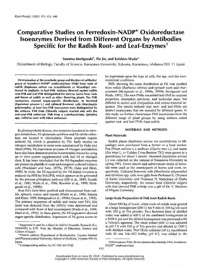
Figure 2. Immunoblot analysis with the anti-root-FNR antiserum(Anti-Rt.-FNR) and the anti-leaf-FNR antiserum (Anti-Lf.-FNR). Pu-rified root-FNR (0.25 ̂ g) and purified leaf-FNR (0.25 ̂ g) wereloaded in lanes 1 and 3, respectively. Partially purified FNR samples(30 ^g of protein) from roots and leaves were loaded in lanes 2 and4, respectively.
et al., 1983; Wada et al., 1983; Karplus et al., 1984). However,it is not clear whether proteolysis occurs during the FNRpreparation or even in intact cells. The two lower dense andfaint bands of the partially purified FNR from leaves wereremoved during subsequent purification.
Often, the anti-root-FNR antiserum reacted weakly withleaf-FNR (Fig. 2, Anti-Rt.-FNR, lane 4, and Fig. 3, Anti-Rt.-FNR, lane 2). By contrast, the anti-leaf-FNR antiserum en-tirely failed to react with root-FNR (Fig. 2, Anti-Lf.-FNR,lanes 1 and 2, and Fig. 3, Anti-Lf.-FNR, lane 3).
In the immunotitration, the anti-leaf-FNR antiserumslightly inhibited the root-FNR activity, as shown in Fig-ure IB. Nevertheless, the anti-leaf-FNR antiserum did notreact with the root-FNR in the immunoblot analysis. On theother hand, the anti-root-FNR antiserum had no effect onthe leaf-FNR activity, although it reacted weakly with theleaf-FNR in the immunoblot analysis. The discrepancy be-tween the immunotitration and the immunoblot analysisis probably the result of the differences between the twoimmunoreactions.
The immunoblot analysis for FNRs from other higherplants (pea, soybean, maize, and horsetail) was done and theresults from pea, soybean, and maize resembled that fromradish (data not shown). The immunoblot analysis confirmeda ubiquitous presence of root-type isoenzyme in the floweringplants tested. The root-type FNR and the leaf-type FNR wereprimarily present in roots and leaves, respectively. Althoughthe leaf type of FNR was detected only in leaves, it is notclear whether the root type of FNR is in leaves as well as inroots because the anti-root-FNR antiserum cross-reactedslightly with the purified radish leaf-FNR (Fig. 3, Anti-Rt.-FNR, lane 2).
Our radish anti-root-FNR antiserum reacted similarly withFNRs from roots of the other flowering plants tested. TheFNRs of roots seem to share common antigenicity with theirleaf counterparts. The weak reactivity of the anti-root-FNR
antiserum with FNRs of leaves indicates that there are, none-theless, common antigenic determinant sites between theFNR isoenzymes.
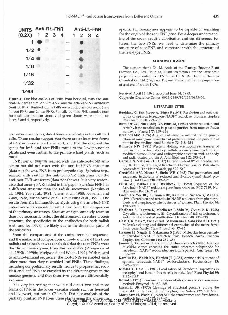
To obtain a prediction of the time when the FNR geneduplicates and differentiates into leaf- and root-FNRs, weinvestigated the reactions of FNRs from horsetail, liverwortcells, and eukaryotic and prokaryotic algae with two specificantisera. The FNR from horsetail subterranean stems couldnot be detected with either antisera, but the FNR fromhorsetail green shoots reacted with both antisera. Dot-blotanalysis found that the FNR from subterranean stems reactedweakly with both the anti-root- and the anti-leaf-FNR anti-sera (Fig. 4, lanes 3). Also, the FNR from green shoots reactedweakly with both the antisera (Fig. 4, lanes 4). The subter-ranean stem FNR was more reactive with the anti-root-FNRantiserum than with the anti-leaf-FNR antiserum and, con-versely, the green shoot FNR was more reactive with theanti-leaf-FNR antiserum than with the anti-root-FNR anti-serum (Fig. 4). This result from dot-blot analysis is caused bythe obscure distinction between the two forms of FNR byboth antisera and/or the presence of the two forms in eachorgan.
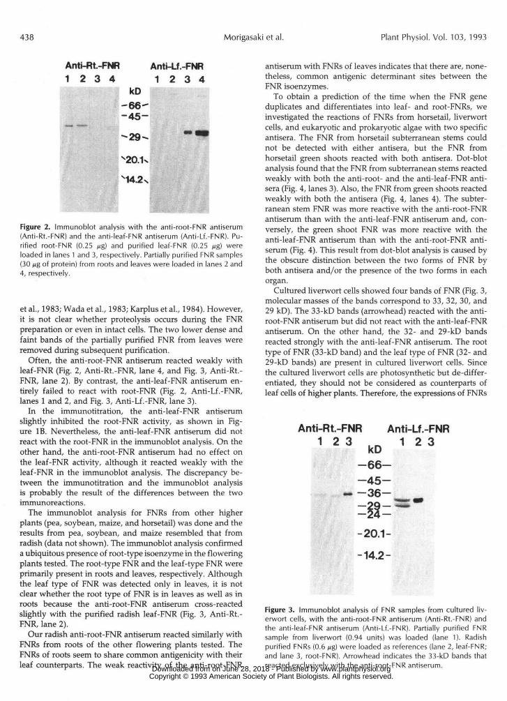
Cultured liverwort cells showed four bands of FNR (Fig. 3,molecular masses of the bands correspond to 33, 32, 30, and29 kD). The 33-kD bands (arrowhead) reacted with the anti-root-FNR antiserum but did not react with the anti-leaf-FNRantiserum. On the other hand, the 32- and 29-kD bandsreacted strongly with the anti-leaf-FNR antiserum. The roottype of FNR (33-kD band) and the leaf type of FNR (32- and29-kD bands) are present in cultured liverwort cells. Sincethe cultured liverwort cells are photosynthetic but de-differ-entiated, they should not be considered as counterparts ofleaf cells of higher plants. Therefore, the expressions of FNRs
Anti-Rt.-FNR1 2 3
Anti-Lf.-FNR1 2 3kD
-66--45-
- -36-
-20.1-
-14.2-
Figure 3. Immunoblot analysis of FNR samples from cultured liv-erwort cells, with the anti-root-FNR antiserum (Anti-Rt.-FNR) andthe anti-leaf-FNR antiserum (Anti-Lf.-FNR). Partially purified FNRsample from liverwort (0.94 units) was loaded (lane 1). Radishpurified FNRs (0.6 ^g) were loaded as references (lane 2, leaf-FNR;and lane 3, root-FNR). Arrowhead indicates the 33-kD bands thatreacted exclusively with the anti-root-FNR antiserum. www.plantphysiol.orgon June 28, 2018 - Published by Downloaded from
Copyright © 1993 American Society of Plant Biologists. All rights reserved.
Fd-NADP+ Reductase Isoenzymes from Different Organs 439
UNITS Anti-Rt.-FNR Anti-Lf.-FNR(0.2X) 1 2 3 4 1 2 3 4
1 * - •1/2 • - 01/4 • %1/8 "» - •
1/16 - a
1/321/64 — •>• • .i/imiaif
Figure 4. Dot-blot analysis of FNRs from horsetail, with the anti-root-FNR antiserum (Anti-Rt.-FNR) and the anti-leaf-FNR antiserum(Anti-Lf.-FNR). Purified radish FNRs were dotted as references (lane1, root-FNR; lane 2, leaf-FNR). Partially purified FNR samples fromhorsetail subterranean stems and green shoots were dotted onlanes 3 and 4, respectively.
are not necessarily regulated tissue specifically in the culturedcells. These results suggest that there are at least two formsof FNR in horsetail and liverwort, and that the origin of thegenes for leaf- and root-FNRs traces to the lower vascularplants and even further to the primitive land plants, such asmoss.
FNR from C. vulgaris reacted with the anti-root-FNR anti-serum but did not react with the anti-leaf-FNR antiserum(data not shown). FNR from prokaryoric alga, Spirulina spp.,reacted with neither the anti-leaf-FNR antiserum nor theanti-root-FNR antiserum (data not shown). It is very reason-able that among FNRs tested in this paper, Spirulina FNR hasa different structure than the radish isoenzymes (Karplus etal., 1984; Yao et al., 1984; Jansen et al., 1988; Newman andGray, 1988; Michalowski et al., 1989; Fillat et al., 1990). Theresults from the immunoblot analysis using the anti-leaf-FNRantiserum were consistent with those from the comparisonof the primary structures. Since an antigen-antibody reactiondoes not necessarily reflect the difference of an entire proteinstructure, the specificities of the antibodies for both radishroot- and leaf-FNRs are likely due to the dissimilar parts ofthe structures.
From the comparisons of the amino-terminal sequencesand the arnino acid compositions of root- and leaf-FNRs fromradish and spinach, it was concluded that the root-FNRs werethe distinct isoenzymes from the leaf-FNRs (Morigasaki etal., 1990a, 1990b; Morigasaki and Wada, 1991). With regardto amino-terminal sequence, the root-FNRs resembled eachother more than they resembled leaf-FNRs. These findings,including our preliminary results, led us to propose that root-FNR and leaf-FNR are encoded by the different genes in thenuclear genome, and that these two genes are differentiallyexpressed.
It is very interesting that we could detect two and moreforms of FNR in the lower vascular plants such as horsetailand liverwort, but not in Chlorella. Dot-blot analysis of thepartially purified FNR from these plants using the antiserum
specific for isoenzymes appears to be capable of searchingfor the origin of the root-FNR gene. For a deeper understand-ing of the organ-specific distribution and the difference be-tween the two FNRs, we need to determine the primarystructure of root-FNR and compare it with the structure ofthe leaf-type FNRs.
ACKNOWLEDGMENT
The authors thank Dr. M Ando of the Tsuruga Enzyme Plant(Toyobo Co., Ltd., Tsuruga, Fukui Prefecture) for the large-scalepreparation of radish root-FNR, and Dr. S. Murakami of ToyamaChemical Co. Ltd. (Toyama, Toyama Prefecture) for the preparationof antisera of radish FNRs.
Received April 14, 1993; accepted June 14, 1993.Copyright Clearance Center: 0032-0889/93/103/0435/06.
LITERATURE CITEDBookjans G, San Pietro A, Bbger P (1978) Resolution and reconsti-
tution of spinach ferredoxin-NADP+ reductase. Biochem BiophysRes Commun 80: 759-765
Bowsher CG, Hucklesby DP, Ernes MJ (1989) Nitrite reduction andcarbohydrate metabolism in plastids purified from roots of Pisumsativum L. Planta 177: 359-366
Bradford MM (1976) A rapid and sensitive method for the quanti-tation of microgram quantities of protein utilizing the principle ofprotein-dye binding. Anal Biochem 72: 248-254
Burnette NW (1981) Western blotting: electrophoretic transfer ofprotein from sodium dodecyl sulfate-polyacrylamide gels to un-modified nitrocellulose and radiographic detection with antibodyand radioiodated protein A. Anal Biochem 112: 195-203
Carrillo N, Vallejos RH (1987) Ferredoxin-NADP* oxidoreductase.In ] Barber, ed, The Light Reactions. Elsevier Science Publishers,Amsterdam, The Netherlands, pp 527-560
Crestfield AM, Moore S, Stein WH (1963) The preparation andenzymatic hydrolysis of reduced and S-carboxymethylated pro-teins. J Biol Chem 238: 622-627
Fillat MF, Bakker HAC, Weisbeek PJ (1990) Sequence of theferredoxin-NADP+ reductase gene from Anabaena PCC 7119. Nu-cleic Acids Res 18: 7161
Green LS, Yee BC, Buchanan BB, Kamide K, Sanada Y, Wada K(1991) Ferredoxin and ferredoxin-NADP reductase from photosyn-thetic and nonphotosynthetic tissues of tomato. Plant Physiol 96:1207-1213
Hagihara B, Tagawa K, Morikawa I, Shin M, Okunuki K (1958)Crystalline cytochrome c. III. Crystallization of fish cytochrome cand a third method of purification. J Biochem 45: 725-735
Hase T, Kimata Y, Yonekura K, Matsumura T, Sakakibara H (1991)Molecular cloning and differential expression of the maize ferre-doxin gene family. Plant Physiol 96: 77-83
Hasumi H, Nagata E, Nakamura S (1983) Molecular heterogeneityof ferredoxin-NADP+ reductase from spinach leaves. BiochemBiophys Res Commun 110: 280-286
Jansen T, Reilander H, Steppuhn I, Herrmann RG (1988) Analysisof cDNA clones encoding the entire precursor-polypeptide forferredoxin: NADP* oxidoreductase from spinach. Curr Genet 13:517-522
Karplus PA, Walsh KA, Herriott JR (1984) Amino acid sequence ofspinach ferredoxin:NADP+ oxidoreductase. Biochemistry 23:6576-6583
Kimata Y, Hase T (1989) Localization of ferredoxin isoproteins inmesophyll and bundle sheath cells in maize leaf. Plant Physiol 89:1193-1197
Koziol J (1971) Fluorometric analysis of riboflavin and its coenzymes.Methods Enzymol 18: 253-285
Laemmli UK (1970) Cleavage of structural proteins during theassembly of the head of bacteriophage T4. Nature 227: 680-685
Matsubara H, Wada K (1988) Soluble cytochromes and ferredoxins.Methods Enzymol 167: 387-410
www.plantphysiol.orgon June 28, 2018 - Published by Downloaded from Copyright © 1993 American Society of Plant Biologists. All rights reserved.
440 Morigasaki et al. Plant Physiol. Vol. 103, I993
Michalowski CB, Schmitt JM, Bohnert HJ (1989) Expression during salt stress and nucleotide sequence of cDNA for ferredoxin-NADP+ reductase from Mesembryanthemum crystallinum. Plant Physiol 8 9
Morigasaki S, Takata K, Sanada Y, Wada K, Yee BC, Shin S, Buchanan BB (1990a) Nove1 forms of ferredoxin and ferredoxin- NADP reductase from spinach roots. Arch Biochem Biophys 283
Morigasaki S, Takata K, Suzuki T, Wada K (1990b) Purification and characterization of a ferredoxin-NADE'+ oxidoreductase-like enzyme from radish root tissues. Plant Physiol 93: 896-901
Morigasaki S, Wada K (1991) Purification and characterization of ferredoxin-NADP+ oxidoreductase from non-photosynthetic tis- sues. In B Curti, S Ronchi, G Zanetti, eds, Flavins and Flavoproteins 1990. Walter de Gruyter, Berlin, pp 461-464
Newman BJ, Gray JC (1988) Characterization of a full-length cDNA clone for pea ferredoxin-NADP+ reductase. Plant Mo1 Biol 1 0
Oaks A, Hirel B (1985) Nitrogen metabolism in roots. Annu Rev Plant Physiol36 345-365
Oji Y, Watanabe M, Wakiuchi N, Okamoto S (1985) Nitrite reduc- tion in barley-root plastids: dependence on NADPH coupled with glucose-6-phosphate and 6-phosphogluconate dehydrogenases, and possible involvement of an electron camer and a diaphorase. Planta 165 85-90
Shin M (1971) Ferredoxin-NADP reductase from spinach. Methods Enzymol23: 440-447
Solomonson LP, Lorimer GH, Hall RL, Borchers R, Bailey JL (1975) Reduced nicotinamide adenine dinucleotide-nitrate reductase of Chlorella vulgaris. J Biol Chem 250: 4120-4127
817-822
75-80
511-520
Suzuki A, Oaks A, Jacquot J-l', Vidal J, Gadal P (1985) An e1e':tron transport system in maize roots for reactions of glutamate synthase and nitrite reductase. Physiological and immunochemical proper- ties of the electron carrier and pyridine nucleotide reductase. l'lant Physiol 78: 374-378
Suzuki A, Vidal J, Gadal P (1982) Glutamate synthase isoforrns in rice. Immunological studies of enzymes in green leaf, etiolated leaf, and root tissues. Plant Physiol70 827-832
Wada K, Onda M, Matsubara H (1986) Ferredoxin isolated from plant non-photosynthetic tissues: purification and characterization. Plant Cell Physiol27: 407-415
Wada K, Onda M, Matsubara H (1989) Amino acid sequenc1.s of ferredoxin isoproteins from radish roots. J Biochem 105: 619-625
Wada K, Tamura T, Matsubara H, Kodo K (1983) Spirulina ferre- doxin-NADP+ reductase: further characterization with an im- proved preparation. J Biochem 9 4 387-393
Washitani I, Sato S (1977a) Studies on the function of proplastids in the metabolism of in vitro cultured tobacco cells. I . Localiziition of nitrite reductase and NADP-dependent glutamate dehydrogen- ase. Plant Cell Physiol 18: 117-125
Washitani I, Sato S (1977b) Studies on the function of proplastids in the metabolism of in vitro cultured tobacco cells. 11. Glutamine synthetase/glutamate synthetase pathway. Plant Cell Physiol 18:
Yao Y, Tamura T, Wada K, Matsubara H, Kodo K (1984) Spiltirina ferredoxin-NADP+ reductase: the complete amino acid sequence. J Biochem 95: 1513-1516
Yao Y, Wada K, Takahashi Y, Katoh S, Matsubara H (1985) The sulfhydryl groups of ferredoxin-NADE'+ oxidoreductases: is a di- sulfide bond really present? J Biochem 98: 1079-1082
505-512
www.plantphysiol.orgon June 28, 2018 - Published by Downloaded from Copyright © 1993 American Society of Plant Biologists. All rights reserved.