clinicalreview - amazon web...
TRANSCRIPT

Clinical ReviewSpring Issue 2015
Elizabeth Yeu, MD Virginia Eye Consultants
Norfolk, VA, USA
Robert J. Weinstock, MDThe Weinstock Laser Eye Center
Largo, FL, USA
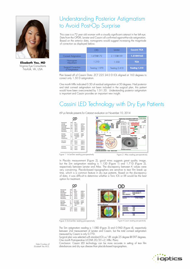
Michael Endl, MDFichte, Endl & ElmerAmherst, NY, USA
Farrell Toby Tyson, MDCape Coral Eye CenterCape Coral, FL, USA
A. John Kanellopoulos, MDLaservision Eye Institute
Athens, Greece
James Katz, MDThe Midwest Center for Sight
Chicago, IL, USA
Jonathan Solomon, MDSolomon Eye Associates
Bowie, MD, USA
Michael Manning, MDGulfcoast Eyecare
Palm Harbor, FL, USA
Dee Stephenson, MD Stephenson Eye Associates
Venice, FL, USA
James Schumer, MDRevision Eyes
Mansfield, OH, USA
Contributors to this issue
Cynthia Matossian, MDMatossian Eye Associates
Doylestown, PA, USA
Bradley Townend, MDCentral Coast Day Hospital
Erina, Australia
Studies have demonstrated that posterior corneal astigmatism could be a factor in generating unexpected postoperative outcomes. Research has shown that selecting toric IOLs based on anterior corneal measurements could lead to over-correction in eyes that have with-the-rule astigmatism (vertical steep axis) and under-correction in eyes that have against-the-rule astigmatism (horizontal steep axis). In addition, there seems to be a large variety in the relationship between anterior and posterior astigmatism in pre-cataract populations of patients. Keeping this challenge in mind, Cassini has worked closely with many key-opinion leading physicians to help develop a solution.
Cassini’s new Total Corneal Astigmatism functionality uses patented second Purkinje reflection-based analysis of the posterior cornea. Cassini posterior and anterior data is calculated to provide surgeons with the total corneal power, as well as steep axis and magnitude of astigmatism. This means that patients undergoing cataract surgery benefits from actual measurements of the Total Corneal Astigmatism (TCA) rather than using a generic nomogram. Cassini provides the data that enables cataract surgeons to create a unique, personalized surgical plan for each patient individually, without ignoring the posterior corneal astigmatism. Our leading surgeons provide interesting data and case examples including:
• Repeatability of Total Corneal Astigmatism Technology• Understanding Posterior Corneal Astigmatism to Avoid Post-Op Surprises• Using Total Corneal Astigmatism to Improve Planning in Patients with Lower Amounts of Cylinder• Capturing Reliable Data in Patients with Dry Eye
This Clinical Review provides us an insight into Cassini Total Corneal Astigmatism and the opportunities to improve surgical planning
Distributed by:
IQ Medical Pty Ltd2/86 Mary Street, Unley SA 5061 Phone (08) 8357 8022Email [email protected] www.iqmedical.com.au
Corneal Astigmatism
Expected Post-OpAstigmatism w/ SIA
Surgical Correctionof Astigmatism
OPDIII (Sim)
0.82D
1.21D
IOL Master(Sim)
1.16D
1.55D
Toric Lens - BL1UT 2.00(Treat 1.33D @ Corneal Plane)
Cassini TCA
0.30D
0.69D
Crystalens AT -52A0 with single LRI
Repeatable Total Corneal Analysis
Data Courtesy of Michael J. Endl M.D.
James. Katz M.D.James. Schumer M.D.
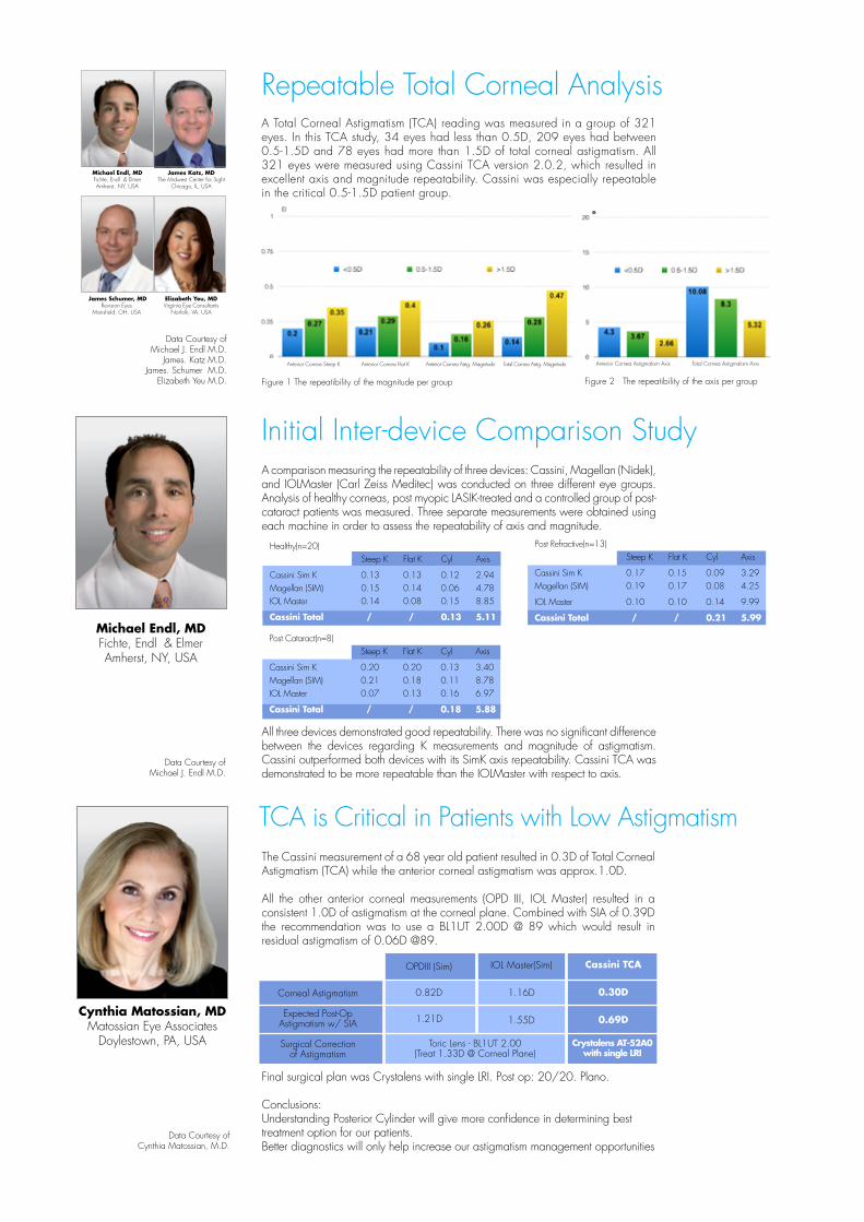
Elizabeth Yeu M.D. Figure 1 The repeatibility of the magnitude per group
The Cassini measurement of a 68 year old patient resulted in 0.3D of Total Corneal Astigmatism (TCA) while the anterior corneal astigmatism was approx.1.0D.
All the other anterior corneal measurements (OPD III, IOL Master) resulted in a consistent 1.0D of astigmatism at the corneal plane. Combined with SIA of 0.39D the recommendation was to use a BL1UT 2.00D @ 89 which would result in residual astigmatism of 0.06D @89.
TCA is Critical in Patients with Low Astigmatism
Data Courtesy of Cynthia Matossian, M.D.
Figure 2 The repeatibility of the axis per group
Final surgical plan was Crystalens with single LRI. Post op: 20/20. Plano.
Conclusions: Understanding Posterior Cylinder will give more confidence in determining best treatment option for our patients.Better diagnostics will only help increase our astigmatism management opportunities
Cynthia Matossian, MDMatossian Eye Associates
Doylestown, PA, USA
Data Courtesy of Michael J. Endl M.D.
Initial Inter-device Comparison Study
A Total Corneal Astigmatism (TCA) reading was measured in a group of 321 eyes. In this TCA study, 34 eyes had less than 0.5D, 209 eyes had between 0.5-1.5D and 78 eyes had more than 1.5D of total corneal astigmatism. All 321 eyes were measured using Cassini TCA version 2.0.2, which resulted in excellent axis and magnitude repeatability. Cassini was especially repeatable in the critical 0.5-1.5D patient group.
Elizabeth Yeu, MD Virginia Eye Consultants
Norfolk, VA, USA
Michael Endl, MDFichte, Endl & ElmerAmherst, NY, USA
James Katz, MDThe Midwest Center for Sight
Chicago, IL, USA
James Schumer, MDRevision Eyes
Mansfield, OH, USA
Michael Endl, MDFichte, Endl & ElmerAmherst, NY, USA
Superior accurate axis in Inter-device repeatibility study
A comparison measuring the repeatability of three devices: Cassini, Magellan (Nidek), and IOLMaster (Carl Zeiss Meditec) was conducted on three different eye groups. Analysis of healthy corneas, post myopic LASIK-treated and a controlled group of post-cataract patients was measured. Three separate measurements were obtained using each machine in order to assess the repeatability of axis and magnitude.
All three devices demonstrated good repeatability. There was no significant difference between the devices regarding K measurements and magnitude of astigmatism. Cassini outperformed both devices with its SimK axis repeatability. Cassini TCA was demonstrated to be more repeatable than the IOLMaster with respect to axis.
Healthy(n=20) Steep K Flat K Cyl Axis
Cassini Sim K 0.13 0.13 0.12 2.94Magellan (SIM) 0.15 0.14 0.06 4.78IOL Master 0.14 0.08 0.15 8.85
Cassini Total / / 0.13 5.11 Post Refractive(n=13) Steep K Flat K Cyl Axis
Cassini Sim K 0.17 0.15 0.09 3.29Magellan (SIM) 0.19 0.17 0.08 4.25
IOL Master 0.10 0.10 0.14 9.99
Cassini Total / / 0.21 5.99 Post Cataract(n=8) Steep K Flat K Cyl Axis
Cassini Sim K 0.20 0.20 0.13 3.40Magellan (SIM) 0.21 0.18 0.11 8.78IOL Master 0.07 0.13 0.16 6.97
Cassini Total / / 0.18 5.88
Anterior Cornea Astigmatism Axis Total Cornea Astigmatism Axis Anterior Cornea Steep K Anterior Cornea Flat K Anterior Cornea Astig. Magnitude Total Cornea Astig. Magnitude
Cassini LED Technology with Dry Eye Patients
In Placido measurement (Figure 2), good mires suggests great quality image, but the Sim astigmatism reading is 1.13D (Figure 1) and 1.71D (Figure 2), respectively between Lenstar and Atlas. The discrepancy between K values were very concerning. Placido-based topographers are sensitive to tear film break up time, which is a common feature in dry eye patients. Based on the discrepancy of data, it was difficult to determine whether a Toric IOL or LRI would be the best option for treatment.
69 yo female presents for Cataract evaluation on November 10, 2014
Figure 1 1st LenStar reading pre-operatively
Figure 3 2nd LenStar reading pre-operatively
The Sim astigmatism reading is 1.08D (Figure 3) and 0.96D (Figure 4), respectively between 2nd measurement of Lenstar and Cassini, but the total corneal astigmatism measured by Cassini is only 0.77D. Surgical plan was selected with standard IOL w/ LRI: single 25 degree @ 097 degrees. One month Post-operative UCDVA 20/20 +2; MRx: PlanoConclusion: Cassini LED technology can be more accurate in setting of tear film disturbances and dry eye disease than placido-based topographers.
Data Courtesy of Elizabeth Yeu M.D.
Figure 2 Atlas reading pre-operatively
Elizabeth Yeu, MDVirginia Eye Consultants
Norfolk, VA, USA
Figure 4 Cassini reading pre-operatively
Understanding Posterior Astigmatism to Avoid Post-Op Surprise
Corneal Astigmatism
Nomogram Adjustment
Surgical Correctionof Astigmatism
OPD
1.67D@172
1.21D
Lenstar
2.11D@159
1.55D
Treating 1.97D
Cassini TCA
1.51D@163
TCA
Treating 1.51DTreating 2.41D
This case is a 72 year old woman with a visually significant cataract in her left eye. Data from the OPDIII, Lenstar and Cassini all confirmed against-the-rule astigmatism. Based on the anterior data, nomograms would suggest increasing the magnitude of correction as displayed below.
Plan based off of Cassini Data: ZCT 225 24.0 D IOL aligned at 163 degrees to correct only 1.50 D astigmatism.
One month MRx indicated 0.5D of residual astigmatism at 50 degrees. Had posterior and total corneal astigmatism not been included in the surgical plan, this patient would have been overcorrected by 1.0-1.5D. Understanding posterior astigmatism is important and Cassini provides an important new insight.
For more information: i-Optics USA - [email protected] - +1 888 660 6965 i-Optics International - [email protected] - www.i-optics.com
Cassini SpecificationsTrue Axis• Multicolor LED imaging technology combined with 2nd Purkinje imaging technology • Anterior Axis repeatability within 3 degrees
True Magnitude• Diopter range 4.00D – 171.00D (Anterior)• Display K-values per zone 3/5/7/9mm (Anterior)• Keratometric indices display in D (diopters) or mm (millimeters)
True Capture• Auto Capture with joystick positioning• Measurement Quality Factor parameter• Auto pupil detection• Topographic indices - E (shape factor), e (eccentricity), Q (asphericity), p (form factor)• Keratoconus indices - SAI (Surface Asymmetry Index), SRI (Surface Regularity Index)
True Accuracy• Submicron accuracy due to color LED triangulation technology < 0.8μm (Anterior)
True Technology• External Ocular Photography• (Anterior)Topographic maps - Axial, Refractive, Tangential, Elevation, Corneal Aberrations, Recorded color HD external ocular photography• Multiple color spectrum options• Incorporated patient management program• USB, Direct print, PDF, JPG, 3rd party output connectivity• Mesopic and photopic pupillometry
Please refer to our Cassini publications:
1. Cornea, Accepted A. John Kanellopoulos, George Asimellis, Distribution and Repeatability of Corneal Astigmatism Measurements (Magnitude and Axis) Evaluated with Color LED Reflection Topography
2. Journal of Refractive Surgery, 2015 April in press. Stijn Klijn, Nicolaas J. Reus, Victor D. Sicam, Evaluation of Keratometry With a Novel Color-LED Corneal Topographer
3. Clinical Ophthalmology, 2015:9 245-252. A. John Kanellopoulos; George Asimellis Color light-emitting diode reflection topography: validation of keratometric repeatability in a large sample of wide cylindrical-range corneas
4. Case Rep Ophthalmology, 2014 Sep-Dec; 5(3): 311–317.A. John Kanellopoulos; George AsimellisClinical Correlation between Placido, Scheimpflug and LED Color Reflection Topographies in Imaging of a Scarred Cornea
5. Case Reports in Ophthalmology, 2013;4(3):199–209A. John Kanellopoulos; George AsimellisForme Fruste Keratoconus Imaging and Validation via Novel Multi-spot Reflection Topography
6. Opt Express, 2010 Aug 30;18(18):19324-38. Snellenburg JJ, Braaf B, Hermans EA, van der Heijde RG, Sicam VAForward ray tracing for image projection prediction and surface reconstruction in the evaluation of corneal topography systems.
Arthur Cummings, MDWellington Eye Clinic
Dublin, Ireland
William Trattler, MDCenter for Excellence
in Eye CareMiami, FL, USA
Douglas D. Koch, MDBaylor College of Medicine
Houston, TX, USA
Mitchell P. Weikert, MD, MSBaylor College of Medicine
Houston, TX, USA
Ronald Krueger, MDCleveland Clinic
Cleveland, OH, USA
Nic J. Reus, MDAmphia Hospital
Breda, Netherlands
Ming Wang, MDWang Vision Cataract
and Lasik CenterNashville, TN, USA
Burkhard Dick, MD Universitäts-Augenklinik Bochum
Bochum, Germany
Mitchell Jackson, MDJacksoneye
Chicago, IL, USA
Jose L. Güell MD Instituto Microcirugia Ocular
Autónoma University of Barcelona, Spain
Eric Donnenfeld, MDOphthalmic Consultants
of Long IslandGarden City, NY, USA
Ramón Ruiz Mesa, MDOFTALVIST Centers Andalucía, Spain
David Andreu, MD ICO Barcelona Innova
Ocular, Spain
Filomena Ribeiro, PHDHospital da LuzLisbon, Portugal