clinical review -...
TRANSCRIPT

Clinical ReviewFall Issue 2014
TCA is vital to calculating correct corneal refractive power for Toric IOL planningAccurate measurement of total corneal astigmatism in cataract patients is crucial for achieving optimum postoperative uncorrected visual acuity and patient satisfaction, especially with the implantation of Toric intraocular lenses (IOLs). Traditionally, the corneal power and astigmatism values have been calculated by assuming a fixed posterior:anterior curvature ratio based on the measurement of the anterior surface curvature only. Unfortunately, a number of studies consistently suggest that current methodology is inadequate for achieving optimum astigmatic outcomes1-3.
Cassini Total Corneal Astigmatism (TCA) uses specular reflection technology to reconstruct the shape of both the anterior (1st Purkinje image) and the posterior (2nd Purkinje image) surface of the cornea. Ray tracing of point images from the camera back to its source allows for excellent accuracy and repeatability of corneal shape measurements. Reliable Purkinje imaging technology and precision ray tracing technology is used to determine corneal shape and optical aberrations.
In this Clinical Review you will find a detailed explanation of the importance of posterior cornea reading in current clinical practice. Preliminary data showing the accuracy of Cassini TCA measurements allow us to present this innovation´s great potential to set a new standard for Toric IOL calculation.
1. Teus MA, Arruabarrena C, Hernandez-Verdejo JL, Sales-Sanz A, Sales-Sanz M. J Cataract Refract Surg 2010; 36:1671–16752. Sun X-Y, Vicary D, Montgomery P, Griffiths M. Ophthalmology 2000; 107:1776 –1781; discussion by RM Kershner, 1781–17823. Mendicute J, Irigoyen C, Aramberri J, Ondarra A, Montes- Mico R. J Cataract Refract Surg 2008; 34:601– 607
Find out why top surgeons choose Cassini for Total Corneal Astigmatism (TCA) diagnosis
Robert J. Weinstock, MDThe Weinstock Laser Eye Center
Largo, FL, USA
Michael Endl, MDFichte, Endl & ElmerAmherst, NY, USA
Arthur Cummings, MDWellington Eye Clinic
Dublin, Ireland
Farrell Toby Tyson, MDCape Coral Eye CenterCape Coral, FL, USA
William Trattler, MDCenter for Excellence
in Eye CareMiami, FL, USA
Douglas D. Koch, MDBaylor College of Medicine
Houston, TX, USA
A. John Kanellopoulos, MDLaservision Eye Institute
Athens, Greece
James Katz, MDThe Midwest Center for Sight
Chicago, IL, USA
Mitchell P. Weikert, MD, MSBaylor College of Medicine
Houston, TX, USA
Ronald Krueger, MDCleveland Clinic
Cleveland, OH, USA
Jonathan Solomon, MDSolomon Eye Associates
Bowie, MD, USA
Nic J. Reus, MDAmphia Hospital
Breda, Netherlands
Ming Wang, MDWang Vision Cataract
and Lasik CenterNashville, TN, USA
Ethan Sadri, MDAtlantis Eye
Huntington Beach, CA, USA
Tal Raviv, MDEye Center of New York,
New York, USA
Michael Manning, MDGulfcoast Eyecare
Palm Harbor, FL, USA
Dee Stephenson, MD Stephenson Eye Associates
Venice, FL, USA
Johny Gayton, MDEyesight Associates
Warner-Robins, GA, USA
Frank Bowden, MDBowden Eye and Associates
Jacksonville, FL, USA
Bradley Townend, MDCentral Coast Day Hospital
Erina, Australia
Burkhard Dick, MD Universitäts-Augenklinik Bochum
Bochum, Germany
UPDATED
AUGUST 2015
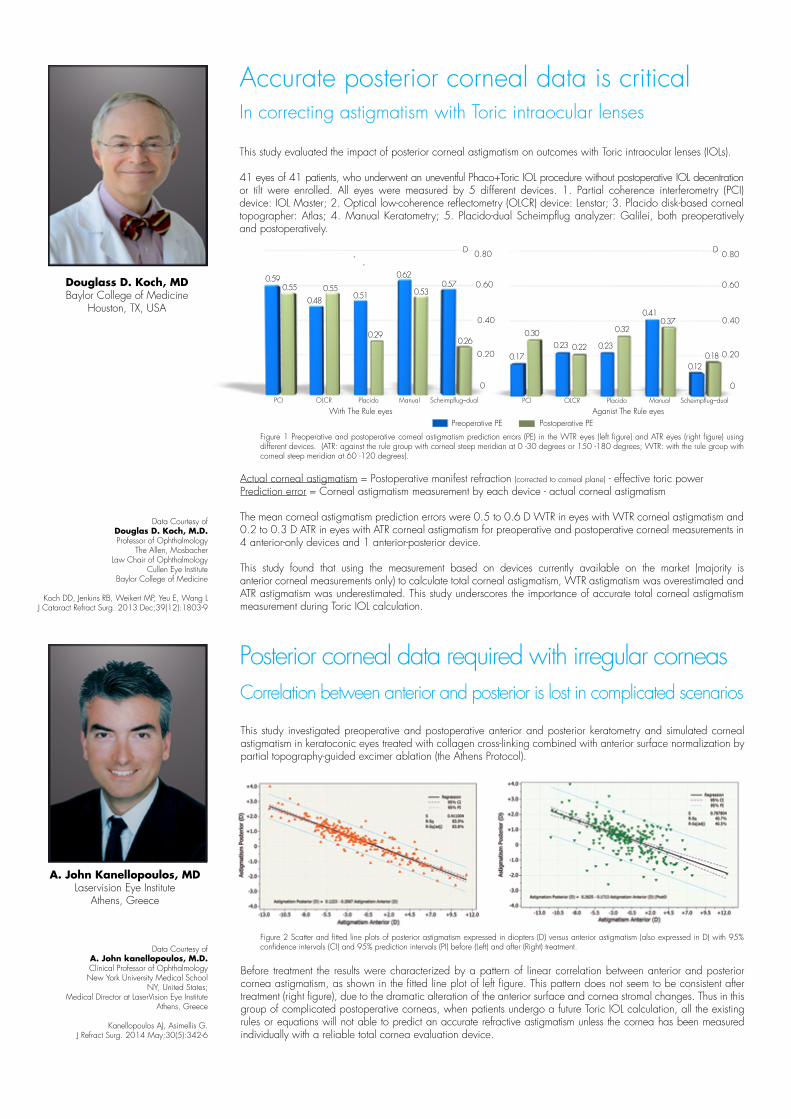
Accurate posterior corneal data is critical
A. John Kanellopoulos, MD Laservision Eye Institute
Athens, Greece
Data Courtesy of A. John kanellopoulos, M.D. Clinical Professor of Ophthalmology New York University Medical School
NY, United States; Medical Director at LaserVision Eye Institute
Athens, Greece
Kanellopoulos AJ, Asimellis G.J Refract Surg. 2014 May;30(5):342-6
Douglass D. Koch, MDBaylor College of Medicine
Houston, TX, USA
Figure 1 Preoperative and postoperative corneal astigmatism prediction errors (PE) in the WTR eyes (left figure) and ATR eyes (right figure) using different devices. (ATR: against the rule group with corneal steep meridian at 0 -30 degrees or 150 -180 degrees; WTR: with the rule group with corneal steep meridian at 60 -120 degrees).
In correcting astigmatism with Toric intraocular lenses
This study evaluated the impact of posterior corneal astigmatism on outcomes with Toric intraocular lenses (IOLs).
41 eyes of 41 patients, who underwent an uneventful Phaco+Toric IOL procedure without postoperative IOL decentration or tilt were enrolled. All eyes were measured by 5 different devices. 1. Partial coherence interferometry (PCI) device: IOL Master; 2. Optical low-coherence reflectometry (OLCR) device: Lenstar; 3. Placido disk-based corneal topographer: Atlas; 4. Manual Keratometry; 5. Placido-dual Scheimpflug analyzer: Galilei, both preoperatively and postoperatively.
Data Courtesy of Douglas D. Koch, M.D.Professor of Ophthalmology
The Allen, Mosbacher Law Chair of Ophthalmology
Cullen Eye Institute Baylor College of Medicine
Koch DD, Jenkins RB, Weikert MP, Yeu E, Wang L J Cataract Refract Surg. 2013 Dec;39(12):1803-9
Actual corneal astigmatism = Postoperative manifest refraction (corrected to corneal plane) - effective toric powerPrediction error = Corneal astigmatism measurement by each device - actual corneal astigmatism
The mean corneal astigmatism prediction errors were 0.5 to 0.6 D WTR in eyes with WTR corneal astigmatism and 0.2 to 0.3 D ATR in eyes with ATR corneal astigmatism for preoperative and postoperative corneal measurements in 4 anterior-only devices and 1 anterior-posterior device.
This study found that using the measurement based on devices currently available on the market (majority is anterior corneal measurements only) to calculate total corneal astigmatism, WTR astigmatism was overestimated and ATR astigmatism was underestimated. This study underscores the importance of accurate total corneal astigmatism measurement during Toric IOL calculation.
This study investigated preoperative and postoperative anterior and posterior keratometry and simulated corneal astigmatism in keratoconic eyes treated with collagen cross-linking combined with anterior surface normalization by partial topography-guided excimer ablation (the Athens Protocol).
Before treatment the results were characterized by a pattern of linear correlation between anterior and posterior cornea astigmatism, as shown in the fitted line plot of left figure. This pattern does not seem to be consistent after treatment (right figure), due to the dramatic alteration of the anterior surface and cornea stromal changes. Thus in this group of complicated postoperative corneas, when patients undergo a future Toric IOL calculation, all the existing rules or equations will not able to predict an accurate refractive astigmatism unless the cornea has been measured individually with a reliable total cornea evaluation device.
Figure 2 Scatter and fitted line plots of posterior astigmatism expressed in diopters (D) versus anterior astigmatism (also expressed in D) with 95% confidence intervals (CI) and 95% prediction intervals (PI) before (Left) and after (Right) treatment.
Correlation between anterior and posterior is lost in complicated scenarios
Posterior corneal data required with irregular corneas
0
0.20
0.40
0.60
0.80
0.590.55
0.480.55
0.51
0.29
0.62
0.530.57
0.26
0.17
0.300.23 0.22 0.23
0.32
0.410.37
0.120.18
With The Rule eyes Aganist The Rule eyesPCI OLCR Placido Manual Scheimpflug–dual
D D
Preoperative PE Postoperative PE
PCI OLCR Placido Manual Scheimpflug–dual
0
0.20
0.40
0.60
0.80

Nic. J. Reus, MDAmphia Ziekenhuis
Breda, the Netherlands
Data Courtesy of Nic. J. Reus, MD
Amphia Ziekenhuis
Cassini Panel Event, ESCRS 2014
This study indicated that the Cassini TCA measurement is closer to the objective auto refraction measurement compared to using Cassini Anterior. Given the previously published outstanding performance of Cassini Anterior measurements1-4, the new Cassini TCA function further improves the control of astigmatism error range when evaluating corneas for treatment.
1. Kanellopoulos A.J, Asimellis G, Friess D. The Clinical Impact of Color LED Topographic Variability Analysis. CRSToday, April 20142. Kanellopoulos A.J, Asimellis G, Clinical Correlation between Placido, Scheimpflug and LED Color Reflection Topographies in Imaging of
a Scarred Cornea. Case Rep Ophthalmol 2014;5:311-317 3. Kanellopoulos A.J, Asimellis G. Forme Fruste Keratoconus Imaging and Validation via Novel Multi-Spot Reflection Topography. Case Rep
Ophthalmol. 2013 Oct 25;4(3):199-209.4. Kanellopoulos A.J. Asimellis G, Cassini: Providing True Axis and Magnitude of Astigmatism. CRSToday Europe, September 2014
Figure 4 The magnitude difference of astigmatism of pseudophakic eyes (n=64) between Cassini anterior and Cassini TCA measurement
Cassini measures Total Corneal Astigmatism MagnitudeThis study was designed to evaluate the accuracy of Cassini Total Corneal Astigmatism (TCA) and determine the contribution of posterior corneal astigmatism to total corneal astigmatism using Cassini TCA.
Consecutive pesudophakic eyes were measured by Cassini Anterior, Cassini TCA and auto refraction (Nidek ARK 530A). The vector difference between corneal astigmatism and the cylinder measurement obtained from auto refraction were analyzed and showed in Figure 4.
All eyes With The Rule eyes Aganist The Rule eyes
0.50
0.33
0.25
0
D
Anterior Total
0.21
0.26
0.12
0.39
0.27
Cassini measures Total Corneal Asigmatism Axis°
0
5
10
2.7
7.0
CassiniAnterior
CassiniTCA
Anterior cornea measurement
Total cornea measurement
Figure 3 Good repeatability of determinination of axis both in anterior and Total Corneal Astigmatism

Data Courtesy of Nic. J. Reus, MD
Amphia Ziekenhuis
Cassini Panel Event, ESCRS 2014
Measurement Astigmatism
Cornea, Anterior 1.94D @ 69°
Cornea, Posterior 0.47D @163°
Cornea, TCA 1.52 D @ 68°
Refractive Cylinder 1.30 D @ 68°
Measurement Astigmatism
Cornea, Anterior 0.50 D @ 38°
Cornea, Posterior 0.05 D @ 0°
Cornea, TCA 0.52 D @ 36°
Refractive Cylinder 0.71 D @ 33°
With the rule astigmatism measurement case shows a higher anterior cornea measurement (A) than total corneal astigmatism measurement (B) as well as the auto refraction. Ignoring posterior cornea measurement may lead to overcorrection in Toric IOL calculations.
Against the rule astigmatism measurement case shows a lower anterior cornea measurement (C) than total corneal astigmatism measurement (D) as well as the auto refraction. Ignoring posterior cornea measurement may lead to undercorrection in Toric IOL calculations.
For more information: i-Optics USA - [email protected] - +1 888 660 6965 i-Optics International - [email protected] - www.i-optics.com
Nic. J. Reus, MDAmphia Ziekenhuis
Breda, the Netherlands
Cassini SpecificationsTrue Axis• Multicolor LED imaging technology combined with 2nd Purkinje imaging technology • Axis repeatability within 3 degrees
True Magnitude• Diopter range 4.00D – 171.00D• Display K-values per zone 3/5/7/9mm• Keratometric indices display in D (diopters) or mm (millimeters)
True Capture• Auto Capture with joystick positioning• Measurement Quality Factor parameter• Auto pupil detection• Topographic indices - E (shape factor), e (eccentricity), Q (asphericity), p (form factor)• Keratoconus indices - SAI (Surface Asymmetry Index), SRI (Surface Regularity Index) True Accuracy• Submicron accuracy due to color LED triangulation technology < 0.8µm
True Technology• External Ocular Photography• Topographic maps - Axial, Refractive, Tangential, Elevation, Corneal Aberrations, Recorded color HD external ocular photography• Multiple color spectrum options• Incorporated patient management program• USB, Direct print, PDF, JPG, 3rd party output connectivity• Mesopic and photopic pupillometry
Cassini TCA Case Examples
With the Rule Against the RuleA
B
C
D
Distributed by:
IQ Medical Pty Ltd2/86 Mary Street, Unley SA 5061 Phone (08) 8357 8022Email [email protected] www.iqmedical.com.au