clinical immunology laboratory handbook immunology... · clinical immunology laboratory but if...
TRANSCRIPT

Page 1 of 59
Clinical Immunology Laboratory Handbook, version 11.0
Reviewed 26/06/12
Clinical Immunology
Laboratory Handbook
Version 11
26/06/2012
Department of Biochemistry and
Immunology
Addenbrooke’s Hospital
Cambridge
The information contained within this document is subject to
regular review, therefore , please ensure that the latest version
of this handbook is referred to, when required.

Page 2 of 59
Clinical Immunology Laboratory Handbook, version 11.0
Reviewed 26/06/12
Version Number 11.0
Date of Issue 26.06.12
Review Interval Annual
Next Review Due 26.06.13
Document Location Addenbrooke’s Hospital
Intranet Pages( Connect)
Document Ownership Senior Executive, Dept of
Clinical Biochemistry and
Immunology
Document History
Version
Number
Issue Date Version
Number
Issue Date
2005 05/12/05 2011 v8 04/07/11
1.1 03/05/06 2011 v9 13/10/11
2008 v.1 15/03/08 2012 v10 05/01/12
2008 v.2 06/09/08 2012v11 26/06/12
2009 v.1 19/08/09
2009 v.2 02/10/09
2010 v.1 20/01/10
2010 v.2 17/03/10
2010 v.3 08/07/10
2010 v.4 09/07/10
2010 v.5 03/09/10
2011 v.6 11/01/11
2011 v7 01/05/11

Page 3 of 59
Clinical Immunology Laboratory Handbook, version 11.0
Reviewed 26/06/12
Contact Information
Telephone numbers – Addenbrooke’s extension number and
(number for outside callers)
Laboratory Service
Results 01223 257148
General enquiries 3215 (01223 217215)
Consultant Immunologist Dr D S Kumararatne
3166 (01223 217166) mobile 07590485799
Consultant Immunologist Dr H. Baxendale
3159(Monday & Friday) 01223 217159
Consultant Clinical Scientist Post Vacant
3159 (01223 217159)
Chief Biomedical Scientist Mr Graham Wood
3361 (01223 217361)
Specialist Registrars 2618 (01223 216618) bleep 154- 406
Duty Doctor 4140 (01223 274140) bleep 154-122
PA To Dr.Kumararatne (Caroline Gilbert) 6830 (01223 586830)
Safehaven Fax 3794 (01223 217794)
Allergy and Clinical Immunology Service
Director Dr P Ewan Special interest: Allergy & Clinical Immunology
3177 (01223 217777)
Consultant Immunologist, Dr D S Kumararatne Special interest: Immunodeficiency.
3166 (01223 217166) mobile 07740 762829
Consultant in Allergy and Asthma, Dr S Nasser Special interest: allergy, asthma and rhinitis
6978 (01223 586978)
Specialist Nurse Practitioners: 2431

Page 4 of 59
Clinical Immunology Laboratory Handbook, version 11.0
Reviewed 26/06/12
(01223 216431) bleep 152-829
Appointments 2646 (01223 216646)
Introduction The laboratory provides a comprehensive service to Addenbrooke’s hospital Cambridge University NHS Foundation Trust, and to hospitals within the Eastern Region and local GP’s. It specialises in the diagnosis and monitoring of patients with suspected or confirmed immunodeficiencies, autoimmune diseases, allergy and inflammatory states. It runs a wide range of assays including highly specialised cell function tests. We also carry out a wide range of specialised assays for hospitals outside the region. The medical staff, provide a clinical consultative service for patients with possible or confirmed immunodeficiencies and also provide a specialist care for the treatment of antibody deficient patients. Dr. P.Ewan and Dr.S.Nasser conduct clinics for patients with allergic diseases..
Normal Hours of Service The lab is open Monday to Friday 08.00-17.00 hr. Sunday 08.00 – 12.00hr
Urgent Requests Including Out of Hours Few tests performed by the laboratory are required clinically on an urgent out-of-hours basis. Exceptionally, when circumstances justify a more rapid result, the request should be made personally to the Consultant Immunologist, Consultant Clinical Scientist or Specialist Registrar. Outside normal working hours contact is via the hospital switchboard 01223 245151 Medical advice can be obtained from the Consultant Immunologist or Specialist Registrar. Results can be faxed urgently by arrangement with the laboratory.

Page 5 of 59
Clinical Immunology Laboratory Handbook, version 11.0
Reviewed 26/06/12
SAMPLE REQUIREMENTS
Serum Samples Most tests are performed on serum separated at room temperature. The exceptions are listed below. For serum, 10 ml blood should be collected in a plain brown or white top tube with no anticoagulant . In most cases samples should be sent to the pathology sample reception area in the Department of Clinical Biochemistry .
Lymphocyte Phenotyping Studies These are performed on a 2.7 ml EDTA (Monovette) sample
Neutrophil Function Studies NBT tests are performed on a a 2.7 ml EDTA (Monovette) sample taken directly to the laboratory. Neutrophil tests are time consuming, can only be done on Monday to Thursday and are only authorised after discussion with a medical immunologist.
Quantiferon TB Gold(Interferon gamma release assay-IGRA)
Adults and children: 1ml of blood must be drawn directly into each of the three QuantiFERON-GOLD tubes in order, which must be all labelled with appropriate patient identifying information. (1) Nil control (GREY TOP) (2) TB Antigen (RED TOP) (3) Mitogen Control (PURPLE CAP) (Collection tubes available from outpatients Phlebotomy and Clinical Immunology) Samples must be delivered directly to the laboratory and must arrive between 08.00 and 17.00 Monday-Friday. No weekend or out of hours sample collection. Samples must be incubated at 37º c within 16 hrs of collection, and must be kept at room temperature prior to this. Please contact the laboratory for further information if requesting test from non-Addenbrookes locations.
Cryoglobulins If cryoglobulins are suspected in autoimmune rheumatoid disorders, a 10 ml blood sample should be collected in a pre-warmed plain white Monovette tube and placed immediately into a vacuum flask containing water at about 37oC. The sample should be
immediately delivered to the Clinical Immunology Laboratory
during normal working hours (before 16.00 hr).

Page 6 of 59
Clinical Immunology Laboratory Handbook, version 11.0
Reviewed 26/06/12
Immunoglobulins Routine tests for total serum IgG, IgA and IgM are performed in the Clinical Immunology Laboratory but if other routine biochemical tests are also required, please send an additional sample to Clinical Biochemistry. Tests for IgG subclasses, IgE, very low immuno-globulins in hypogammaglobulinaemia and secretory IgA are performed in the Clinical Immunology laboratory.

Page 7 of 59
Clinical Immunology Laboratory Handbook, version 11.0
Reviewed 26/06/12
Functional Complement Studies including Functional C1
inhibitor Blood taken at Addenbrooke’s Hospital for these assays should reach the laboratory within 30 minutes of being taken and labelled as being urgent. (Do not rely on routine collections!) It should then be separated and frozen to a temperature of at least -40
oC within a
further 30 minutes. Distant laboratories must also ensure correct collection of the blood and arrange for it to reach this centre while still deep-frozen. If these procedures are not followed then abnormal low values could be due to in vitro degradation. Functional C1 inhibitor assay requires a citrated blood sample (green top tube) and should only be performed after initial screening for immunochemical C1 inhibitor concentration in cases where there is a documented normal value of inhibitor but the C4 is low.
Mast Cell Tryptase Blood taken for Mast Cell Tryptase levels should be centrifuged and the serum separated as soon as possible, preferably within 3 hours of venesection.(If the blood is collected into gel separator type tubes centrifugation alone is sufficient) The separated serum or spun tubes should be kept at 4
oC and sent to Addenbrookes
Immunology. If the samples will not reach Immunology at Addenbrookes within 5 days then separated serum should be frozen at -20
oC or below and
then dispatched in a frozen state. If the sample(s) have been taken as part of an anaphylactic/anaesthetic reaction investigation then it is vitally important that sample time and dates are accurately recorded on both sample and request
Urine Requests for Bence Jones protein require a 20ml aliquot of urine in a universal container (NO preservative).
Storage of Specimens Sera are stored at -40
○C and retained for about 1month before
disposal. If further tests are required on a patient in the light of earlier results, it may be possible to save the inconvenience of a repeat venesection for the patient by contacting the laboratory and arranging further tests on the stored serum. Specimens for cellular immunology tests cannot be retained and are useless after the time of the initial test.

Page 8 of 59
Clinical Immunology Laboratory Handbook, version 11.0
Reviewed 26/06/12
Reporting of Results Printed reports are issued when a group of related assays have been completed. Further reports may be issued if other, less frequently performed assays have also been requested. Unexpected or grossly abnormal results will be telephoned to the requesting physician whenever possible. Interpretation of the results of specialised assays will be added but if additional information is required please call the laboratory and a clinician or clinical scientist will be pleased to help.
Clinical Referrals Please contact the Consultant (Direct line 01223 217166 or ext. 3166. Secretary: 01223 586830) to arrange for a Clinical Immunologist to see a patient. Patients admitted to Addenbrooke’s Hospital under other consultants can be assessed after making a referral in writing or by telephoning. Out patient referrals may be made, by writing or by telephoning (indicating the degree of urgency) to the Consultant.

Page 9 of 59
Clinical Immunology Laboratory Handbook, version 11.0
Reviewed 26/06/12
Assays (in alphabetical order with their common
abbreviations), disease associations, normal ranges
and specimen requirements and turn around times .
Assay Specimen
Clinical indications for assay.
Normal range Turn around time
(including weekends)
for reporting of > 95 % of
tests
or method of reporting
Activation markers see Neutrophil studies
Acute Phase Proteins see CRP
Adhesion molecules see Neutrophil studies
Adrenal cortical antibodies Serum Anti-adrenal antibodies are found in 60% of patients with idiopathic hypoadrenalism where they are directed against the enzyme 21-hydroxylase, and in 90% of those with hypoadrenalism in association with ovarian failure (Autoimmune Polyglandular Syndrome-1, APS-1) where they react with 17-hydroxylase and the side chain cleaving enzyme complex involved in steroid bio-synthesis. They are found in <0.1% of the normal population. The titre of antibody is of no significance, and so is not measured. As the adrenal gland atrophies the antibodies may disappear so sequential follow-up is not useful.
Neg. / Pos. 10 days
Allergy antigen specific serum IgE assays see RAST
Alpha-1 anti-trypsin Serum This protein is a proteolytic inhibitor. Low serum concentrations (see below) are associated with centrilobular emphysema and also neonatal jaundice. Where levels are below the normal range, samples are analysed by isoelectric focusing. Low levels may be noticed on serum electrophoresis but this does not have adequate sensitivity to be a screening test.
0.9- 1.8g/l (adult) 4 days

Page 10 of 59
Clinical Immunology Laboratory Handbook, version 11.0
Reviewed 26/06/12
Alpha-1 anti-trypsin phenotyping Serum
Phenotyping is indicated where total levels of -1-antitrypsin are below the normal range. Ideally this requires a full pedigree, with names and clinical details to permit linkage to family records. The normal phenotype, found in about 85% of Caucasians in the UK, is MM. The commoner alleles that result in deficiency in the UK are S and Z. MS is found in about 10% of the population and MZ in 3%. Individuals who are of ZZ genotype have 10% of normal activity and SS individuals have 60%. Patients who are heterozygous with one normal and one deficient gene will have intermediate levels of
-1-antitrypsin (0.6-1.4g/l). A ZZ homozygous individual is predisposed to lung and liver disease. A heterozygous (MZ) individual is at a small risk of developing liver disease but has little or no increased risk of developing lung disease. Other common phenotypes (MS, MF, SS) are of no known clinical significance 28 days
Anti-Nuclear Antibodies (ANA) Serum These are a heterogeneous group of antibodies that bind to nuclear antigens. ANA negative SLE is extremely rare. However the test is not specific for SLE as anti nuclear antibodies can occur in other conditions e.g. rheumatoid arthritis, systemic sclerosis, chronic active hepatitis, juvenile arthritis, Sjögren’s syndrome, MCTD, fibrosing alveolitis, systemic infections and can be induced by drugs such as hydralazine. Low titre ANAs can also be found in the serum of many healthy elderly people. All positives will be further analysed for anti double stranded DNA(DNA) and Extractable Nuclear Antibodies(ENA) Our routine assay is a florescent immunoassay which detects antibodies to: U1RNP/RNP70/Sm/Ro/La/Scl-70/Jo-1,centromere,dsDNA, fibrillarin ,Pm-Scl, RNA Polymerase III, Mi-2, Ribosomal P, and PCNA(Proliferating Nuclear Antigen)
0 – 0.9 units 4 days ANA immunofluorescence on HEp-2 cells is available upon request.

Page 11 of 59
Clinical Immunology Laboratory Handbook, version 11.0
Reviewed 26/06/12
ANCA see Neutrophil antibodies
Avian precipitins Serum See Specific IgG
Basement Membrane Antibody see Glomerular BM Ab for Goodpasture’s Disease see Pemphigus Ab for Bullous Skin Diseases
2 microglobulin ( 2M) Serum
In multiple myeloma, 2-M concentration has been found to be the
single most effective prognostic indicator. Because 2M is metabolised in the renal tubules high levels are seen in patients with renal dysfunction. It may also be elevated in connective tissue disease and in granulomatous disease but measurement in these conditions is not generally clinically helpful
1.09 - 2.53 mg/l 4 days
B cell markers see Lymphocyte markers
Bence-Jones Protein see Urine electrophoresis
Beta Trace Protein Fluid Beta trace (also known as prostaglandin D synthetase) is a protein found at high concentration in csf (20mg/L) and low concentration in normal serum (0.5 mg/L). The concentration in serum may be elevated in renal failure. The measurement of beta trace is useful in identifying the presence of csf in otorrheoa or rhinorrhoea. It is untested in identifying the source of fluid originating from other sites. Greater than 2.0mg/L suggests presence of csf Greater than 6.0mg/L strongly suggests presence of csf 3 days
Brain antibodies see Neurological antibodies

Page 12 of 59
Clinical Immunology Laboratory Handbook, version 11.0
Reviewed 26/06/12
Caeruloplasmin Serum Caeruloplasmin is a copper-containing protein which shows a modest acute phase response. Serum concentrations are increased by oestrogens and may be decreased in severe liver disease. Low serum concentrations are seen in the majority of patients with Wilson's disease, an inherited defect of copper metabolism. It should be noted that urine copper can also be measured and is useful in monitoring the clearance of copper in response to treatment (contact clinical biochemistry to discuss sample requirements). To exclude Wilson's disease, all these tests and liver biopsy may be needed
0.2 - 0.5 g/L(adults)-pediatric ranges will be reported as appropriate 4 days
Cell Function Assays see Lymphocytes and Neutrophils
Cardiolipin antibodies see Phospholipid antibodies
Cyclic Citrullinated Peptide(CCP)antibodies Serum Anti-CCP antibodies have a greater specificity (96%) for rheumatoid arthritis than rheumatoid factor. They are rarely found in other conditions. Their sensitivity for detecting rheumatoid arthritis appears to be similar to that of rheumatoid factor. Please note- this assay is currently only available on a case by case basis.
0- 7 iu/ml 7 days
Centromere antibodies Serum Performed as part of ANA (see above)
These antibodies are characteristic of the CREST syndrome, a variant of systemic sclerosis with limited skin involvement but
associated with Calcinosis, Raynaud’s phenomenon oEsphageal
immobility, Sclerodactyly and Telangectasia. They are also found in about 10% of patients with primary biliary cirrhosis which often overlaps with systemic sclerosis. They are directed against centromere associated proteins. Patients with severe Raynaud's and other features of scleroderma, especially lung and other organ involvement, should also been screened for Scl-70 which is associated with diffuse systemic sclerosis.
Page 13 of 59
Clinical Immunology Laboratory Handbook, version 11.0
Reviewed 26/06/12
Neg. / Pos. 7 days
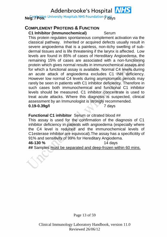
COMPLEMENT PROTEINS & FUNCTION C1 Inhibitor (Immunochemical) Serum This protein regulates spontaneous complement activation via the classical pathway. Inherited or acquired defects usually result in severe angioedema that is a painless, non-itchy swelling of sub-dermal tissues and is life threatening if the larynx is affected. Low levels are found in 85% of cases of Hereditary Angioedema, the remaining 15% of cases are associated with a non-functioning protein which gives normal results in immunochemical assays and for which a functional assay is available. Normal C4 levels during an acute attack of angioedema excludes C1 INH deficiency. However low normal C4 levels during asymptomatic periods may rarely be seen in patients with C1 inhibitor deficiency. Therefore in such cases both immunochemical and functional C1 inhibitor levels should be measured. C1 inhibitor concentrate is used to treat acute attacks. Where this diagnosis is suspected, clinical assessment by an Immunologist is strongly recommended.
0.19-0.39g/l 7 days
Functional C1 inhibitor Serum or citrated blood ## This assay is used for the confirmation of the diagnosis of C1 inhibitor deficiency in patients with angioedema (especially where the C4 level is reduced and the immunochemical levels of C1esterase inhibitor are equivocal).The assay has a specificitity of 91% and sensitivity of 99% for Hereditary Angiodema.
46-130 % 14 days
## Samples must be separated and deep-frozen within 60 mins.

Page 14 of 59
Clinical Immunology Laboratory Handbook, version 11.0
Reviewed 26/06/12
Complement C3 and C4 Serum C3 and C4 levels are useful in monitoring conditions associated with immune complexes, e.g. SLE, systemic vasculitis, SBE. A decrease, primarily of C3, can also be seen gram-negative bacteraemias and post-streptococcal GN. A profound decrease in C3 should alert the clinician to the possibility of a C3 nephritic factor (see below). In very rare cases low C4 levels can be found in individuals with C4 null alleles (these people have an increased risk of developing SLE) and in cases of active systemic rheumatic disease. An isolated decrease in C4, associated with angioedema, suggests C1 Inhibitor deficiency whereas a low C4 with renal disease and/or vasculitic rash suggests the presence of a cryoglobulin. Please discuss with an immunologist if the reason for hypocomplentaemia is not clinically apparent. Increased production can maintain normal levels even if consumption is rapid; .
C3 0.80-2.14 g/l 4 days
C4 0.13 - 0.60g/l
C3 Nephritic Factor Fresh serum This is an IgG autoantibody that stabilises C3bBb and therefore results in continuous C3 breakdown. The presence of this autoantibody is associated with type II membrano-proliferative glomerulonephritis, with or without partial lipo-dystrophy and results in greatly reduced C3 levels.
Please note that C3 Nephritic Factor levels will not be assayed in the presence of normal levels of C3. Fresh clotted blood
Neg./Pos. 21 days
Complement Alternative Pathway Fresh serum ## This assay tests the integrity of the Alternative Pathway of Complement. Low levels are found when any one component is absent. Assays for the individual complement components (Factors B, P & D) should be measured if Alternate Pathway activity is reduced.
% of Normal serum activity 21 days
(66 - 129%) ## sample must be separated and deep-frozen within 60 mins

Page 15 of 59
Clinical Immunology Laboratory Handbook, version 11.0
Reviewed 26/06/12
Complement Classical Pathway (CH50) Fresh serum ## This is a functional test of the Classical Complement Pathway (C1, C4, C2, C3, C5-9). Low levels occur if any component is absent. Any patient with more than one episode of meningococcal disease should be screened with a CH50 during convalescence. A CH50 test may also be indicated in patients with serious and persistent bacterial sepsis or when a qualitative deficiency of the Complement Pathway is suspected. The CH50
assay is not suitable for the routine monitoring of patients with SLE. A functional assay for the Alternative pathway CH50 is also available and it is recommended that both assays are carried out.
392 - 1019 U/ml 21 days ## Sample must be separated and deep-frozen within 60 mins
C-Reactive Protein (CRP) Serum Performed in Clinical biochemistry. The clinical value of the CRP assay is derived from the following observation. This acute phase protein is normally found in serum in very low concentrations (<5mg/l). Its level increases rapidly (within hours) and up to 100 fold, following bacterial infection or injury to tissues. Levels rapidly fall to baseline following resolution of the injury or infection. Specifically, CRP is raised in bacterial infections and inflammatory diseases e.g. rheumatoid arthritis and in other conditions associated with tissue necrosis such as vasculitides. The level is usually not raised in active SLE, MCTD or the scleroderma group of conditions unless there is associated serositis. CRP levels are therefore useful in distinguishing active SLE from infection (unless they co-exist), diagnosing infection in neutropaenic patients and for monitoring inflammatory conditions such as bacterial infections or systemic vasculitides. Steroids or immunosuppressive agents do not directly affect the level of CRP.
<6 mg/l is normal 1 day 10-49mg/l in autoimmune disease
50-100mg/l in bacterial infection or inflammatory arthritis.
>I00mg/l in a major bacterial infection (including mycobacteria)

Page 16 of 59
Clinical Immunology Laboratory Handbook, version 11.0
Reviewed 26/06/12
Cryoglobulins Serum@@
Monoclonal cryoglobulins are most commonly associated with myeloma and Waldenstrom’s Macroglobulinaemia whilst those of a polyclonal nature can be found in connective tissue diseases. Hepatitis C infection is now the commonest cause of mixed cryoglobulins. Cases of renal disease and low C4 should be tested for cryoglobulins, as should patients with an unexplained cutaneous vasculitis or Raynaud's.
Neg./Pos.(with Cryocrit %) 8 days @@
For this assay it is essential that the blood be taken into a pre-warmed syringe and tube and kept at 37
oC (using a 37
0 water
containing thermos) until the serum is removed from the clot. Please contact the laboratory for advice and always notify them when one is expected to arrive so that it can be dealt with promptly.
CSF IgG/Total Protein Ratio CSF & Serum CSF oligoclonal bands have superseded this test.
CSF Oligoclonal bands CSF & Serum Oligoclonal bands are the discrete populations of immunoglobulin detected in CSF by iso-electric focusing and which are NOT paralleled in serum from the same patient. Oligoclonal bands are seen in 85-95% of patients with clinically diagnosed multiple sclerosis. If there are more than 10 bands unique to the csf, it is highly specific for MS. Only 7 of 593 patients with neurological disease other than MS had this pattern [Bourahoui, A., et al., CSF isoelectrofocusing in a large cohort of MS and other neurological diseases. Eur J Neurol, 2004. 11(8): p. 525-9.] giving a specificity of 99%. However only 46% of patients with MS have this pattern. If there are fewer than 10 but more than 3 unique bands, this has a sensitivity of 85% and specificity of 92%. If there is a single unique band in csf, about a third or patients go on to develop typical oligoclonal bands, about a quarter revert to normal on follow up and the rest are associated with a variety of non-demyelinating conditions which may include cerebral lymphoma . Davies, G., et al., The clinical significance of an intrathecal monoclonal immunoglobulin band: a follow-up study. Neurology, 2003. 60(7): p. 1163-6. Csf oligoclonal bands can also occur in cerebrovascular accidents, in infections of the CNS e.g. neurosyphilis, SSPE and in

Page 17 of 59
Clinical Immunology Laboratory Handbook, version 11.0
Reviewed 26/06/12
pathological processes involving a local immune response within the CNS e.g. encephalitis, neurosarcoid and SLE.
NB the results of this assay can only be interpreted if serum and CSF can be are compared for the presence of matching bands. Sometimes similar oligoclonal bands are found in both serum and csf. This pattern is found in systemic inflammatory conditions
Neg./Pos. 14 days
dsDNA antibodies Serum A positive result for dsDNA antibodies supports the diagnosis of SLE. However only 60% of all patients with SLE have these antibodies in their serum and a negative test does not exclude the diagnosis. Occasionally dsDNA antibodies may be found in patients with autoimmune hepatitis type 1 .
0- 10 iu/ml 7 days
Endomysial IgA antibodies Serum This test has been superceded by Tissue Transglutaminase antibodies. (see later)
ENA and ANA Typing Serum ANA screen positives are followed up and typed for the following ENA(Extractable nuclear antigens),and other ANA/cytoplasmic specificities:
Neg. / Pos. 7 days
Sm Specific for SLE but found in only 20-30% of SLE patients with a higher incidence in non-Caucasians, especially those of Afro-Caribbean descent. There is no correlation with disease activity.
U1RNP A high titre positive result of U1RNP in the absence of other autoantibodies is diagnostic for undifferentiated (mixed) connective tissue disease(MCTD) but these antibodies are also found about 25% of SLE patients.

Page 18 of 59
Clinical Immunology Laboratory Handbook, version 11.0
Reviewed 26/06/12
RNP70 is a protein within the U1RNP complex. There are also two other proteins in U1RNP. These.are, RNP A and RNP C. Antibodies to RNP70 are more specific for undifferentiated connective tissue disease(MCTD) being found in only about 12% of patients with SLE.
Ro or SS-A The Ro (SS-A) antigen also occurs in the cell cytoplasm and very rarely a serum may be positive for Ro antibodies even in the absence of an ANA. These antibodies can cause congenital heart block and is recommended that all female patients suspected of SLE or Sjögren’s syndrome are screened for anti-SS-A (Ro) antibodies especially if they are considering pregnancy. These antibodies are associated with Sjögren’s syndrome (up to 75% in primary Sjögren’s), Sicca syndrome, and in many cases of Sjögren’s syndrome secondary to a variety of other autoimmune diseases. They are also found in variants of SLE including subacute cutaneous lupus and neonatal lupus with congenital heart block and also in SLE resulting from homozygous C2 or C4 deficiency.
La or SS-B Usually found with anti Ro in both primary and secondary Sjögren’s syndrome and SLE. Sjögren’s patients with anti-La are likely to have more extra-glandular disease.
Ro and La antibodies are often found together. La is a phosphoprotein and Ro a ribonucleoprotein and both can bind to the same molecule of a transfer RNA. SLE patients positive for Ro & La are likely to have lower DNA antibody titres and less renal disease.
Jo- 1(antibodies to aminoacyl-tRNA histidyl synthetase) Associated with inflammatory muscle disease, especially polymyositis (also called anti-synthetase syndrome). Patients with anti-synthetase syndrome have a characteristic clinical picture comprised of myositis and/or interstitial lung disease and/or chronic arthritis. Raynauds phenomenon is frequently observed in this condition.
Scl-7O (antibodies to Topoisomerase-I an enzyme catalysing the breaking and re-joining of ssDNA)

Page 19 of 59
Clinical Immunology Laboratory Handbook, version 11.0
Reviewed 26/06/12
Found in 20-40% of patients with systemic sclerosis, it is associated with facial skin, kidney and heart involvement, ischaemic fingertip ulcers and pulmonary fibrosis.
PM-Scl (antibodies to Pm/Scl proteins which function as exoriboncucleases during RNA Processing) Found almost exclusively in patients with idiopathic myositis(including overlap syndromes) or more rarely systemic sclerosis. Cardiac and renal involvement in these patients is very rare, so the prognosis is therefore relatively good.
Fibrillarin (fibrillarin is a 34 Kda protein and is the major component of the nucleolar U3-RNP complex, which is involved in pre-r RNA processing)The antibody is found in about 8% of systemic sclerosis patients overall, in 5% of those with diffuse disease and in 10% of those with limited forms. It is a prognostic marker for small intestine and skeletal muscle involvement, as well as pulmonary hypertension.
RNA Polymerase III (antibodies to RNA polymerase III are directed against 2 proteins(111A of 150Kda and 111B of 138 kda )located in the nucleoplasm. RNAPs are responsible for the transcription of genes that code for precursor molecules of r RNA ) The antibody is found in about 12-20 % of patients with systemic sclerosis and are thought to be highly specific. They are associated with diffuse or extensive skin manifestations.
Mi-2 (antibodies are thought to be directed against a 235-240 Kda antigen within a macromolecular nuclear complex) The antibody is found in 15-30% of patients with adult dermatomyositis, and in 10-15 % of juvenile dermatomyositis. They are rarely found in patients with polymyositis and are therefore highly specific for dermatomyositis.(~ 95%) In comparison to patients with aminoacyl-t RNA Synthetase antibodies( i.e. Jo-1), those with Mi-2 antibodies generally have a milder clinical course, rarely exhibit synovitis, lung manifestations or Raynauds phenomenon.
Ribosomal P(antibodies are mainly directed against the c- terminal region of the phosphoproteins P0(38kda),P1(19Kda) and P2(17 Kda) of the 60s subunit of the ribosomal complex) Ribosomal P antibodies are detected in between 10 – 20% of patients with SLE, and rarely in other autoimmune diseases. Although not confirmed by all studies Ribosomal P antibodies
Page 20 of 59
Clinical Immunology Laboratory Handbook, version 11.0
Reviewed 26/06/12
seem to be associated with severe depression and other neuropsychriatric manifestations of SLE. The antibody can also be seen in patients with Scleroderma and may be a sign of scleroderma/SLE overlap
PCNA(antibodies against Proliferating Cell Nuclear Antigen are directed against a 34kda auxiliary protein of DNA polymerase ) PCNA antibodies are found in about 3-7% of SLE patients but are not specific. In SLE, they are associated with renal involvement, CNS manifestations and thrombocytopenia.
Epidermal antibodies See Pemphigus and pemphigoid antibodies
Functional Antibody Assays see Specific Ab’s
Functional Complement Assays see C1 esterase inhibitor (functional) Complement Alternative Pathway Complement Classical Pathway CH50
Fungal precipitins Serum See Specific IgG.
GAD antibodies (see Neurological Antibodies) Serum (Glutamic Acid Decarboxylase) ab's NB their clinical utility in IDDM has not been proven.
1 – 5 u/ml 21 days
Ganglioside antibodies see Neurological antibodies
Gastric parietal cell ab's Serum These antibodies have a strong association with pernicious anaemia and autoimmune gastritis. Low titres are commonly found in normal elderly females. If positive the more specific assay for antibodies to intrinsic factor is carried out.
Neg./Pos. 4 days N.B. Clinical Biochemistry carry out assays for Vitamin B12

Page 21 of 59
Clinical Immunology Laboratory Handbook, version 11.0
Reviewed 26/06/12
Gliadin antibodies This test has been superceded by Tissue Transglutaminase antibodies. (see later).
Glomerular basement membrane antibodies Serum These antibodies are positive in Goodpasture's syndrome, which is a rapidly progressive glomerulonephritis. The antibody levels can also be of value in monitoring response to therapy of this disease. If the laboratory is contacted arrangements can be made to carry out a test with results ready in 2 hours during the working day. Direct immunofluorescence of a renal biopsy is the suggested method of diagnosing anti-GBM disease in patients with rapidly progressive glomerulonephritis. This can be arranged by contacting the histology department.
0 - 7.0 U/ml. 4 days (routine 3 hours (urgent)
Haptoglobin serum Haptoglobin binds free haemoglobin, the complex then being removed from the serum and metabolised. Lowering of the serum concentration is a sensitive indicator of intra-vascular haemolysis. Chronic liver disease also results in low values. The sensitivity of this protein as a marker of inflammation is generally poor due to the wide reference range in normals. The main indication for quantitation is in investigation of suspected haemolytic conditions. .
0.5 - 2.6 g/L 4 days
Hereditary Complement Deficiencies see C1 inhibitor deficiency. Complement Alternative Pathway Complement Classical Pathway
Histone Antibodies Serum In cases of suspected drug induced SLE the antibodies are more likely to be directed against the histone moiety of the nucleoprotein complex than to the dsDNA.
Neg. / Pos. 21 days

Page 22 of 59
Clinical Immunology Laboratory Handbook, version 11.0
Reviewed 26/06/12
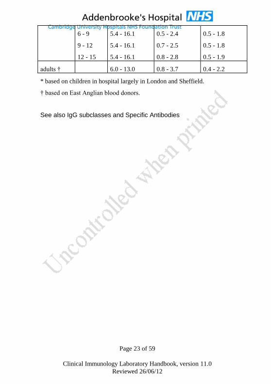
Immunoglobulins (IgG/A/M) Serum
for Myeloma investigations see Diagnosis of Myeloma Immunoglobulin levels are an essential investigation for 'failure to thrive', recurrent infections and lymphoproliferative diseases including myeloma. Reduced levels are found in many primary immunodeficiencies but secondary causes (e.g. lymphoproliferative disorders, nephrotic syndrome or protein losing enteropathy) are more common, especially in adults. In these conditions levels should be monitored as serious infective complications can occur. IgA deficiency occurs in 1 in 800 of the population and may not be associated with disease (but can lead to reactions to blood and blood products). All cases of suspected primary immunodeficiency should be discussed with a Clinical Immunologist so that comprehensive investigations can be arranged. Polyclonally raised IgG can be a feature of chronic infections (notably HIV, TB and trypanosomiasis), connective tissue disease or liver disease. Polyclonally raised IgA is also found in late stage HIV infection but more commonly associated with liver disease, especially alcoholic in origin. IgM is raised in Primary biliary cirrhosis.
: 4 days
Normal Ranges
Age IgG
(g/l)
IgA
(g/l)
IgM
(g/l)
cord * 5.2 - 18.0 < 0.02 0.02 - 0.20
weeks * 0 - 2
2 - 6
6 - 12
5.0 - 17.0
3.9 - 13.0
2.1 - 7.7
0.01 - 0.08
0.02 - 0.15
0.05 - 0.40
0.05 - 0.20
0.08 - 0.40
0.15 - 0.70
months * 3 - 6
6 - 9
9 - 12
2.4 - 8.8
3.0 - 9.0
3.0 - 10.9
0.10 - 0.5
0.15 - 0.7
0.20 - 0.7
0.2 - 1.0
0.4 - 1.6
0.6 - 2.1
years * 1 - 2
2 - 3
3 - 6
3.1 - 13.8
3.7 - 15.8
4.9 - 16.1
0.3 - 1.2
0.3 - 1.3
0.4 - 2.0
0.5 - 2.2
0.5 - 2.2
0.5 - 2.0
Page 23 of 59
Clinical Immunology Laboratory Handbook, version 11.0
Reviewed 26/06/12
6 - 9
9 - 12
12 - 15
5.4 - 16.1
5.4 - 16.1
5.4 - 16.1
0.5 - 2.4
0.7 - 2.5
0.8 - 2.8
0.5 - 1.8
0.5 - 1.8
0.5 - 1.9
adults † 6.0 - 13.0 0.8 - 3.7 0.4 - 2.2
* based on children in hospital largely in London and Sheffield.
† based on East Anglian blood donors.
See also IgG subclasses and Specific Antibodies

Page 24 of 59
Clinical Immunology Laboratory Handbook, version 11.0
Reviewed 26/06/12
IgG Subclasses Serum These are of doubtful clinical utility and have been superseded by specific antibacterial antibody measurements
Adult Normal Range 7 days IgG1: 3.20- 10.20g/l } IgG2: 1.20 - 6.60 g/l } IgG3: 0.20 - 1.90 g/l } IgG4: 0.00 - 1.30 g/l } Paediatric ranges will be given where appropriate.
IgE (total) Serum Serum lgE may be helpful in the confirmation of atopic diseases however the normal range is very wide and levels do not correlate well with symptoms. A high level of specific IgE to a single allergen may be seen with a normal level lgE. Very high levels of lgE are seen both in atopic eczema and in parasitic infestations and also in the rare hyper-IgE syndrome.
10 - 170 kU/L Adult Normal Range 7 days Paediatric ranges will be given where appropriate.
Interferon gamma release assay (IGRA)
See Quantiferon TB Gold
Intrinsic factor antibodies Serum Present in serum in the majority (60%) of patients with pernicious anaemia. Therefore a negative test for serum antibodies against intrinsic factor does not exclude pernicious anaemia. The combination of intrinsic factor antibody positivity and low vitamin B12 is diagnostic of Pernicious Anaemia.. This assay should be carried out on all patients who are positive for GPC antibodies. In rare cases it may be positive in the absence of GPC antibodies. 0 – 6 u/ml . 7 days N.B. Clinical Biochemistry carry out assays for Vitamin B12
Jo-l antibodies see ENA antibodies
LKM antibodies Serum
(Liver Kidney Microsomal)

Page 25 of 59
Clinical Immunology Laboratory Handbook, version 11.0
Reviewed 26/06/12
These are an uncommon but specific marker for a severe subset of patients with autoimmune hepatitis (formerly known as autoimmune chronic active hepatitis) and some drug induced hepatitis.
Neg. / Pos. 4 days
Lymphocyte cell markers EDTA Please contact the laboratory to discuss these requests. The principal indications for these tests are immunodeficiency, including HIV infection (and haematological malignancy but please note that requests for haematological phenotyping should be referred to the Department of Haematology).
Normal Range for the 4 days
relevant request is always
quoted with the results.
Lymphocyte Functional Assays Lithium heparin and EDTA These tests can only be performed by prior arrangement. Tests of lymphocyte function are available in cases of suspected primary immunodeficiency on discussion with Dr. D S Kumararatne or his deputy in the Immunology Department. These tests are particularly relevant in cases of recurrent viral, fungal or intracellular bacterial infection e.g. Mycobacterial, Salmonella, or Listeria infection that are not associated with an overt cause such as HIV infection. They are also used for the investigation of children with suspected Severe Combined Immunodeficiency. Report of findings 21 days
Mast cell tryptase Serum This enzyme is released from Mast cells when the patient suffers an anaphylactic reaction. Both alpha and beta tryptase are detected. Beta tryptase is a marker of mast cell degranulation. Alpha tryptase is elevated in mastocytosis.The enzymes can be detected in the blood up to 6 hours after anaphylaxis. (see Investigation of Anaesthetic Reactions at the end of this booklet.)
2 - 14 ng/ml 7 days
Microalbuminuria Serum A slightly increased urinary albumin (termed "microalbuminuria") has been shown to be predictive of nephropathy in insulin-dependent diabetes. Other causes of "microalbuminuria" include

Page 26 of 59
Clinical Immunology Laboratory Handbook, version 11.0
Reviewed 26/06/12
exercise, poor diabetic control and non-diabetic renal or systemic diseases including hypertension. Urine albumin in diabetics can be measured using a random urine or a 24 hr urine. Any patient showing an abnormally high result should repeat the test as there are other causes of a transiently raised level. In diabetes, incipient nephropathy is defined as albumin excretion in the range 20-200 mg/min and at this stage the renal disorder may be reversible by optimisation of diabetic control and management of hypertension.
Albumin creatinine ratio: 4 days
<2.5mg/mmol
Albumin excretion rate <20 mg/min
Mitochondrial antibodies Serum These are present in the vast majority of patients with Primary Biliary Cirrhosis (PBC) and are commonly found with a polyclonal elevation in IgM. The antigen associated with most cases PBC is the pyruvate dehydrogenase complex on the inner mitochondrial membrane.
Neg. / Pos. 4 days
Muscle antibodies see Smooth muscle antibodies (liver disorders) Striated muscle antibodies (myasthenia gravis)
Myeloperoxidase (MPO) see Neutrophil Antibodies
Neurological antibodies Serum These may due to an autoimmune process or may arise in patients with certain types of neoplastic diseases where the neoplasm bears antigens cross-reacting with those in the nervous system. With the exception of tests marked * the tests are not performed in this department but are sent to other laboratories. We cannot guarantee the turn around times stated as they are subject to change.
Acetylcholine receptor antibodies (ACR) * These antibodies are
positive in the large majority of patients with myasthenia gravis. About
10% of patients with myasthenia gravis may have negative results.(see
MUSK)
0.2 – 0.50 nmol/l 14 days

Page 27 of 59
Clinical Immunology Laboratory Handbook, version 11.0
Reviewed 26/06/12
Aquaporin(NMO)IgG antibodies to aquaproin 4 have been described in about 65% of patients with neuromyelitis
optica(NMO- also called Devic’s disease. Neg/Pos 28 days
Sent to Immunology Laboratory, Churchill Hospital,
Headington, Oxford
Basal Ganglia antibodies
ABGAs are associated with antecedent streptococcal infections(PANDAS-paediatric autoimmune neuropsychiatric disorders associated with steptococcal infection)Tourette's syndrome,Sydenham's chorea,some tic disorders and an Encephalitis lethargia-like sydrome. Neg/Pos 28 days Sent to Department of Neuroimmunology,National Hospital for Neurology and Neurosurgery,Queen Square,London
Ganglioside Antibodies* Antibodies to several
gangliosides (phospholipids) are associated with neurological diseases.
IgM anti-GM1 antibodies are associated with acquired motor neuropathies and are found in over 50% of cases.
IgG anti-GM1 antibodies -although these may be found in a small proportion (5 – 15%) of patients with Guillain-Barré syndrome, their measurement adds little to diagnosis or management
Neg. / Pos
IgG anti-GQ1b antibodies are found in over 90% of patients with Miller-Fisher syndrome.
IgM anti-GQ1b antibodies are associated with a minority of patients with of chronic ataxic sensory neuropathy.
Neg. / Pos 21 days
Myelin associated glycoprotein Antibodies(MAG)

Page 28 of 59
Clinical Immunology Laboratory Handbook, version 11.0
Reviewed 26/06/12
IgM anti MAG is associated with paraproteinaemic polyneuropathies where the paraprotein is IgM.
Neg. / Pos 28 days Sent to Immunology Laboratory, Churchill Hospital, Headington, Oxford
Muscle Specific receptor tyrosine kinase abs(MUSK) About 2/3 of patients with clinical evidence of myasthenia gravis without anti-AChR antibodies (traditionally called “seronegative myasthenia”) have serum antibodies to MUSK, a muscle kinase
Neg. / Pos 28 days Sent to Immunology Laboratory, Churchill Hospital, Headington, Oxford
NMDA Receptor Antibodies Positive in young women with teratomas and complex encephalopathies including psychiatric presentations,seizures,movement disorders and mutisms. Also positive in other unexplained, probably autoimmune or paraneoplastic cases.About 30% of males and 25% of women have tumours
Neg. / Pos 28 days Sent to Immunology Laboratory, Churchill Hospital, Headington, Oxford
Voltage Gated Potassium Channel abs (anti-VGKC)these are associated with 20-30% cases of acquired neuromyotonia and a proportion of cases of non-paraneoplastic limbic encephalitis.
Neg. / Pos 28 days Sent to Immunology Laboratory, Churchill Hospital, Headington, Oxford
Voltage Gated Calcium Channel abs (anti-VGCC) these are found in 85% of cases of Lambert-Eaton syndrome and rarely in autpimmune cerebrellar syndromes
Neg. / Pos 28 days Sent to Immunology Laboratory, Churchill Hospital, Headington, Oxford
Paraneoplastic antibodies: Serum

Page 29 of 59
Clinical Immunology Laboratory Handbook, version 11.0
Reviewed 26/06/12
Anti-Hu (ANNA-1) antibodies* are associated with
paraneoplastic encephalomyelitis, cerebellar degeneration and sensory neuropathy. They are associated mainly with small cell lung carcinomas.Hu proteins are regulators of mRNA that are needed for neural differentiation, proliferation & maintenance Neurological symptoms can precede presentation of the malignancy. These antigens are expressed in all neurones of the central & peripheral nervous systems and are concentrated in the cell nucleus but are also found in the cytoplasm.
Neg. / Pos
Anti-Ri (ANNA-2) antibodies* are paraneoplastic
autoantibodies associated with opsoclonus/myclonus ,paraneoplastic cerebellar degeneration and brainstem encephalomyelitis.The underlying neoplasm may be either small cell lung carcinoma or carcinoma of the breast. The Ri antigen is found in neurones of the CNS and has a similar distribution within the nucleus and cytoplasm as the Hu antigen.
Neg. / Pos
Purkinje cell antibodies* are found in subacute sensory
neuropathy and cerebellar degeneration, often paraneoplastic. Several autoantibodies have been further defined.
Anti-Yo antibodies* to antigens in the cytoplasm of
Purkinje cells and are associated with Paraneoplastic Cerebellar Degeneration. The most frequent underlying tumours are ovary and breast.
Neg. / Pos
Anti Amphiphysin antibodies * are found in Stiff Person
Syndrome and paraneoplastic encephalmomyelitis and are associated with breast cancer and small cell lung carcinoma. When found in breast cancer they are associated with the stiff person syndrome and when found in lung cancer with sensory neuropathy and paraneoplastic encephalitis. Non paraneoplastic stiff person syndrome is associated with anti GAD antibodies. Rarely anti GAD antibodies may be found in addition to anti amphiphysin in paraneoplastic stiff person syndrome

Page 30 of 59
Clinical Immunology Laboratory Handbook, version 11.0
Reviewed 26/06/12
Neg. / Pos
Anti CV2/CRMP5 antibodies * are found in peripheral
neuropathy, cerebellar ataxia and limbic encephalitis: They are associated with small cell lung carcinoma and thymoma
Neg. / Pos
Anti Ma1 antibodies *. are found in paraneoplastic
neurological disorder and brainstem encephalmomyelitis, and are associated with various tumours, including lung cancer but not testicular cancer.
Neg. / Pos
Ma 2/Ta antibodies * are found in brainstem and limbic
encephalmomyelitis, and are associated with testicular cancer.
Neg. / Pos
Anti Tr antibodies *.are found in paraneoplastic cerebellar
degeneration, and are associated with Hodgkin’s lymphoma.
Neg. / Pos 10 days
Anti-Glutamic Acid Decarboxylase antibodies(GAD) *
associated with ‘Stiff Man Syndrome’ Glutamic acid decarboxylase (GAD) is an enzyme concentrated in neurones, which control muscle tone and exteroreceptive spinal reflexes. Ab's to GAD are found in 60% of patients with Stiff man syndrome.
1- 5 u/ml 21 days
Neutrophil cytoplasmic ab's (ANCA) Serum This term encompasses antibodies to enzymes within the cytoplasmic granules of neutrophils. These are detected by indirect immunofluoresence (IIF) using human neutrophils. Antibodies directed against different enzymes are associated with different

Page 31 of 59
Clinical Immunology Laboratory Handbook, version 11.0
Reviewed 26/06/12
patterns of neutrophil cytoplasmic antibodies as detected by IIF. Please see below for clinical associations of ANCA. Enzyme Linked Immunosorbent Assays (ELISA) assays are recommended for the complete characterisation of ANCA. i.e. antibodies to Proteinase 3 (PR-3), and Myeloperoxidase (MPO) These assays allow more accurate quantitation of the antibody than titration by IIF. Diseases in which ANCA antibodies may be found. 1] Wegener’s granulomatosis Cytoplasmic ANCA (C-ANCA) is a found in only about 85% of patients with active generalised Wegener’s granulomatosis. Therefore the absence of these antibodies does not exclude the diagnosis. Antibody levels may fall with treatment. Patients with persisting elevations are more likely to relapse. Patients with limited Wegener’s granulomatosis are less likely to be positive for ANCA either by IIF or ELISA. Some patients with Wegener’s granulomatosis may have a Perinuclear ANCA (P-ANCA) pattern on IIF and be positive for MPO antibodies by ELISA. {% do not add up: need checking) 2] Microscopic polyangiitis PANCA, with MPO specificity is seen in 50-80% of patients with active microscopic polyangiitis (which may affect only the kidney). The titre of antibodies reflects disease activity. Patients with persisting elevations are more likely to relapse.. About 40% of patients with microscopic poyangiitis may be positive for PR3 antibodies. 3] Churg-Strauss syndrome Some patients may be positive for either P-ANCA or C-ANCA. 4] Rapidly Progressive Glomerulonephritis. Some patients may have C-ANCA or P-ANCA. 5] Drug-induced SLE or Vasculitis High levels of MPO-ANCA are found in patients with some forms of drug-induced SLE or vasculitis. These levels drop after the drug is withdrawn. 6] Other diseases Low titres of MPO-ANCA are occasionally found in RA, SLE, Chronic Hepatitis and Inflammatory Bowel Disease and Sclerosing

Page 32 of 59
Clinical Immunology Laboratory Handbook, version 11.0
Reviewed 26/06/12
Cholangitis. Such findings are of uncertain clinical significance. Low titre pANCA with specificities directed against antigens other than MPO also occurs commonly in the same group of diseases and again such findings are of uncertain clinical significance. Atypical ANCA refers to a variety of observed immunofluoresence patterns and such antibodies are directed against a range of antigens including bacterial permeability increasing protein, azurocidin, lactoferrin, elastase, cathepsin G and lysozyme. The clinical significance of atypical ANCA is uncertain. From the above the following points are clear 1) ANCA detected by IIF alone does not provide specific diagnostic information. It is essential to identify antibody specificity to MPO and PR3 by ELISA. (This is a recommendation of The International Consensus Group for Vasculitis.) 2) International multi-centre studies indicate that the presence of
ANCA detected by both IIF and ELISA (C-ANCA / PR3-ANCA & P-ANCA / MPO-ANCA) is very strongly linked to the presence of small vessel vasculitis.
Recommendations of the International Consensus Group 1)Savige J, Gillis D, Benson E, et al. International Consensus Statement on Testing and Reporting of Antineutrophil Cytoplasmic Antibodies (ANCA). Am J Clin Pathol. 1999;111:507-513.) 2)Savige, J. et al. Addendum to the International Consensus Statement on testing and reporting of antineutrophil cytoplasmic antibodies. Quality control guidelines, comments, and recommendations for testing in other autoimmune diseases. American Journal of Clinical Pathology 2003;120(3):312-8.
about the use of ANCA assays are as follows: 1) In patients presenting with acute renal failure associated pulmonary hemorrhage a rapid battery of tests for ANCA, GBM antibodies and ANA should ideally be performed. (If arrangements are made with the laboratory it should be possible to have the results available within 3 hours.) Positive results from these tests will then be confirmed by a quantitative assay at a later time. ANCA positive vasculitis is a commoner cause of the above syndrome than Goodpasture’s Disease, which is associated with GBM antibodies.

Page 33 of 59
Clinical Immunology Laboratory Handbook, version 11.0
Reviewed 26/06/12
2) For a new patient with suspected vasculitis and / or Rapidly Progressive Glomerulonephritis carry out an IIF screen for ANCA. If negative the result will be reported. If significantly positive the sample will be assayed for PR3, and MPO antibodies by ELISA. 3) Even if previously PR3 and MPO negative the most recent sample will have ELISA assays carried out as these antibodies may have appeared with disease progression.
Neg. / Pos. 4 day (screen)
MPO-ANCA 0- 3.4 iu/ml 4 days
PR3-ANCA 0- 1.9 iu/ml 3 hours (urgent)
ANCA antibodies to MPO and PR3 Serum See above for the clinical utility of these assays.
Neutrophil Function Assays EDTA These tests are useful for the diagnosis of rare primary neutrophil defects that usually present in childhood with recurrent deep-seated bacterial or fungal infections and poor wound healing. These tests can only be performed after prior discussion with the Consultant Immunologist. Flow cytometric test for Phagocyte Oxidase function is used to diagnose Chronic granulomatous disease (normal range: } Flow cytometric determination of cell-surface expression of Leucocyte Adhesins (CD11a,b,c,CD18 and CD15), is used to diagnose Leucocyte Adhesin deficiencies Type 1 and Type 2, respectively. CD62L shedding is used to diagnose innate immune defects increasing susceptibility to pyogenic infection caused by S.pneumoniae, S.aureus or gram negative bacteria. (IRAK4 or MyD88 deficiency.) 4 days
Ovarian Antibodies Serum These antibodies are found in 15-50% of patients with premature ovarian failure under the age of 40 years. These antibodies react with steroid producing cells and thus also stain the steroid producing Leydig cells of the testis, the placenta and often also in the adrenal cortex. They are often seen in Autoimmune Polyglandular Syndrome-1 (APS-1) where adrenal and ovarian failure may co-exist. Up to 70% of women

Page 34 of 59
Clinical Immunology Laboratory Handbook, version 11.0
Reviewed 26/06/12
may have transient anti-ovarian antibodies during IVF therapy.Patients with APS-1 have mutations in the Autoimmune Regulator (AIRE) gene. Mutation detection available:Please inquire. .
Neg./Pos. 10 days
Pancreatic islet cell ab's Serum These antibodies are present in up to 70% of newly presenting patients with type I diabetes mellitus but measurement is rarely clinically useful. They can be used to assess the risk of Type 1 diabetes in first degree relatives of affected patients but, currently, this is only of use in intervention studies
Neg./Pos. 10 days
Paraproteins see Serum Electrophoresis
Parathyroid Antibodies Serum These may be found in autoimmune hypoparathyroidism
Neg./Pos. 10 days

Page 35 of 59
Clinical Immunology Laboratory Handbook, version 11.0
Reviewed 26/06/12
Pemphigus and Pemphigoid Antibodies Serum Over 80-90% of patients with pemphigus have IgG antibodies to molecules expressed on the surface of keratinocytes intercellular antigens (located on the surfa giving a chicken wire type staining, detectable by indirect immunofloresence).These autoantibodies are directly pathogenic as they cause loss of cell-to-cell adhesion by the relevant adhesion structures (desmosomes) and thus blistering. In Pemphigus Vulgaris, the autoantibodies are directed against a desmosome protein called desmoglein 3.In Pemphigus foliaceus the autoantibodies are directed against desmoglein 1. Both types of antibodies are detected by our current test and give the same pattern of staining. Antibodies reactive with the dermal basement membrane are found in cases of pemphigoid, epidermolysis bullosa acquisita and a minority of cases of herpes gestationis. Bullous pemphigoid is blistering disease affecting skin and mucous membranes, while cicatrial pemphigoid primarily affects the mucous membranes. Blood of patients with bullous pemphigoid contain (IgG, Sometimes IgA) antibodies directed against molecules expressed on the surface of antigens expressed on the epidermal basement membrane (hemi-desmosomes). These antibodies cause activation of complement. Complement breakdown products attract phagocytic cells to the basement membrane zone. The combined action of the membrane attack complex of complement and enzymes liberated by the activated phagocytes results in damage to the basement membrane zone causing subepidermal blister formation. 80% of patients with bullous pemphigoid have IgG antibodies to the epidermal basement membrane zone. Only about 20% of patients with cicatrial pemphigoid have similar autoantibodies detectable with indirect immunofluoresence For patients with blistering skin diseases, an unfixed skin biopsy for the more sensitive test of direct immunofluorescence should also be sent to the histology department.
Neg. / Pos. 10 days

Page 36 of 59
Clinical Immunology Laboratory Handbook, version 11.0
Reviewed 26/06/12
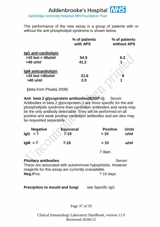
Phospholipid antibodies Serum
These are a family of antibodies (Cardiolipin, 2-glycoprotein-1 and the Lupus Anti-Coagulant) useful in the investigation of the anti phospholipid syndrome. This may be primary or secondary to SLE. Patients with the ‘anti-phospholipid syndrome’ may be positive for
both the lupus anti-coagulant and for the cardiolipin autoantibodies
or for only one of these assays. Therefore samples should be sent for both tests. (Please note that the lupus anticoagulant assay is performed in the Haematology Laboratory.) The diagnosis of the anti phospholipid syndrome requires the appropriate clinical setting (see below) together with persistently (longer than 12 weeks ) elevated anti phospholipid antibodies. The syndromes associated with anti-phospholipid antibodies are treatable and it is appropriate to seek its presence in the following groups of patients: a) Women with recurrent unexplained foetal loss. b) Young patients with stroke, myocardial infarction or transient
ischaemic attacks - without other predisposing factors. c) Young patients with recurrent venous or arterial thromboses. d) Patients with unexplained thrombocytopenia. e) Patients with chronic false positive VDRL. f) Patients with SLE as part of assessment of thrombotic risk in
pregnancy. The cardiolipin antibody assay (particularly IgM) may sometimes give false positive results in patients with infectious diseases (e.g.) syphilis) and in some individuals with antiDNA antibodies.
Cardiolipin antibodies
Negative Weak Positive Positive Units
IgG <10 10-40 > 40 gplu/ml
IgM <10 10-40 > 40 mplu/ml 7 days All positive and weak positive cardiolipin antibodies will be automatically tested for beta 2 glycoprotein 1 antibodies.
Page 37 of 59
Clinical Immunology Laboratory Handbook, version 11.0
Reviewed 26/06/12
The performance of the new assay in a group of patients with or without the anti phospholipid syndrome is shown below.
% of patients % of patients
with APS without APS
IgG anti-cardiolipin
>10 but < 40u/ml 54.9 6.3
>40 u/ml 41.2 1
IgM anticardiolipin
>10 but <40u/ml 21.6 8
>40 u/ml 2.0 1
(data from Phadia 2008)
Anti beta 2 glycoprotein antibodies(B2GP-1) Serum Antibodies to beta 2 glyocoprotein-1 are more specific for the anti phospholipids syndrome than cardiolipin antibodies and rarely may be the only antibody detectable. They will be performed on all positive and weak positive cardiolipin antibodies and are also may be requested separately.
Negative Equivocal Positive Units
IgG < 7 7-10 > 10 u/ml
IgM < 7 7-10 > 10 u/ml
7 days
Pituitary antibodies Serum These are assocated with autoimmune hypophisitis. However reagents for this assay are currently unavailable.
Neg./Pos. ? 10 days
Precipitins to mould and fungi see Specific IgG

Page 38 of 59
Clinical Immunology Laboratory Handbook, version 11.0
Reviewed 26/06/12
Pyruvate dehydrogenase antibodies(PDH-M2) Serum The subtypes of antimitochondrial antibodies (see above)associated with primary biliary cirrhosis are M2, M4, M8 and in early disease M9. Antibodies to pyruvate dehydrogenase subunits are M2 antibodies and are present in most patients with PBC.
Neg./Pos. 14 days
Quantiferon TB Gold Quantiferon Gold tubes
1. What is QUANTIFERON-TB Gold (QFTG)? This is a new NICE / US-FDA approved blood test for the detection of Mycobacterium tuberculosis infection, whether tuberculosis disease or latent tuberculosis infection (LTBI). It cannot distinguish between tuberculosis disease and LTBI and is intended for use in conjunction with clinical risk assessment, radiography, microbiology and other medical and diagnostic evaluations for the diagnosis of clinically significant mycobacterial infections. QFT-G is highly specific and a positive test is strongly predictive of true infection with M. tuberculosis complex (MTB). The test is approved as an aid for diagnosing both active TB disease and LTBI, but does not differentiate between the two.
2. How does QUANTIFERON - TB gold work? QFTG test is an indirect test for MTB infection. It measures a cell mediated immune response to defined mycobacterial antigens. The T lymphocytes of infected individuals are sensitised to MTB protein antigens. When whole blood is incubated with MTB specific antigens the T lymphocytes secrete a cytokine called Interferon gamma (IFN-G) which is measured by a sensitive enzyme linked immunosorbent assay (ELISA). QFTG specifically detects responses to three proteins early secondary antigentic target 6 (ESAT-6), culture filtrate protein 10 (CFP-10) and TB7.7 which are synthesised by MTB and are absent from all BCG vaccine preparations and most environmental (non-tuberculosis) mycobacteria with the exception of M. kansasii, M. marinum and M. szulgai. The QFTG test is a modern alternative to tuberculosis skin test and has many advantages over the latter.

Page 39 of 59
Clinical Immunology Laboratory Handbook, version 11.0
Reviewed 26/06/12
3. What are the current limitations of QFTG test?
Specimens for testing must be transferred to the laboratory for processing within 12 hours of venesection.
The test has not been extensively studied in many groups, such as those with immunodeficiency, those on immunosuppressive drugs and clinical conditions which may reduce immunocompetence including diabetes, silicosis, chronic renal failure, and haematological disorders, or malignancy.
The test has not been extensively evaluated in children or in pregnant women.
It has not been extensively tested in those who have been treated for latent TB infection or tuberculosis disease.
The ability of QFTG test to predict the risk of LTBI progression to tuberculosis disease has not been determined. The risk may be different in those positive for the QFTG test than in those with a positive tuberculous skin test.
4. Who is eligible for testing? QFTG can be used in patients who have been evaluated for possible M. tuberculosis – complex infection, whether tuberculosis disease or LTBI. In other words it can be used in all circumstances where the tuberculosis skin test is considered.
5. What are the advantages of QFTG test over the tuberculin
skin test (TST)?
QFT-G vs. TST
In vitro, controlled laboratory test with
Minimal inter-reader variability.
M.tb specific antigens used.
No boosting; 2 step testing not needed
1 patient visit possible
Unaffected by BCG and most environmental mycobacteria
Simple positive/negative
In vivo, subject to errors during implantation and interpretation
Less specific PPD antigen used
Boosting with repeated testing
2 patient visits minimum
False-positive results can occur after BCG and environmental mycobacterial exposure
Interpretation based on patient’s risk of TB exposure or development of disease

Page 40 of 59
Clinical Immunology Laboratory Handbook, version 11.0
Reviewed 26/06/12
result
6. How are the results reported and interpreted? The diagnosis or exclusion of tuberculosis disease, and assessing the probability of LTBI, requires a combination of epidemiological, historical, medical, radiological and microbiological findings that should be taken into account when interpreting test results. See general guidance on the diagnosis and treatment of tuberculosis disease and LTBI (http://www.cdc.gov/nchstp/tb/) or the NICE guidance on tuberculosis: http://www.nice.org.uk/guidance/index.jsp?action=byID&o=13422.. QUANTIFERON-TB Gold results are reported as positive, negative or indeterminate.
QUANTIFERON-TB Gold
result
Report/interpretation
Positive (EAST-6 and/or CFP-10 and/or TB7.7 responsiveness detected)
M. tuberculosis infection likely See text above
Negative (No ESAT-6 or CFP-10 and/or TB7.7responsiveness detected)
M. tuberculosis infection unlikely, but cannot be excluded especially when 1. any illness is consistent with tuberculosis disease 2. likelihood of progression to disease (e.g. because of immunosuppression) is increased See text above
Indeterminate Test not interpretable Unable to determine a result due to lack of stimulatory response to the positive control. This may be due to technical error in sample acquisition or an unspecified reason related to the clinical picture of the patient. Immune suppression or underlying immunodeficiency may be a
Page 41 of 59
Clinical Immunology Laboratory Handbook, version 11.0
Reviewed 26/06/12
contributing factor. Review patient in light of clinical findings.
7.Sample Collection
Adults and children: 1ml of blood must be drawn directly into each
of the three QuantiFERON-GOLD tubes in order, which must be all labeled with appropriate patient identifying information. (1) Nil control (GREY TOP) (2) TB Antigen (RED TOP) (3) Mitogen Control (PURPLE CAP)
(Collection tubes available from outpatients Phlebotomy, ATC
and Clinical Immunology- No other sample types can be used)
Samples must be delivered directly to the laboratory and must
arrive between 08.00 and 17.00 Monday-Friday. No weekend or
out of hours sample collection.
Turn around time: 7 days
RAST tests (allergen specific IgE) Serum RAST tests are available for a wide range of allergens but they are not generally as useful as history taking and skin (prick) testing. They are of use when the latter is contra-indicated because of extensive skin involvement (e.g. due to eczema) or a risk of
anaphylaxis. Clinical details and suspected allergens must be stated on the request. Requests for 'RAST Testing' without any antigens being listed will only receive a screen for the common allergens of cat, house dust mite and grass pollen. For the common food allergies of egg, milk, peanut and fish diagnostic levels of specific IgE have been identified which predict clinical reactivity (Sampson HA, Ho DG. Relationship between food-specific IgE concentrations and the risk of positive food challenges

Page 42 of 59
Clinical Immunology Laboratory Handbook, version 11.0
Reviewed 26/06/12
in children and adolescents. J Allergy Clin Immunol 1997;100:444-51.)
Egg (kUA/L)
Milk (kUA/L)
Peanut (kUA/L)
Fish (kUA/L)
Level above which clinical reactivity is >95%
6 32 15 20
Levels below which clinical reactivity is <10%
0.6 12.0 0.35 5
Many children ‘outgrow’ their childhood allergy to milk and egg and although the skin-prick tests remain positive for many years they are able to tolerate introduction of the food into their diet. Bee and Wasp Venom Specific IgE tests are available. However it is strongly recommended that these patients who are at risk of anaphylaxis after a sting should have a clinical evaluation by Dr Ewan in the Allergy Clinic as desensitisation is an effective therapy. In cases of drug sensitivity (e.g. antibiotic, anaesthetic agents) it is advisable to discuss the case with Dr P.Ewan or Dr.S.Nasser. RAST testing for specific IgE to penicillin is not completely reliable for diagnosing immediate type hypersensitivity to this drug.
KUa/l 7 days
Rheumatoid factor Serum In Rheumatoid Arthritis, the presence of a high titre RF at onset is of some predictive value as these patients have a worse prognosis than seronegative patients and are more likely to suffer from systemic manifestations of the disease than those who are RF negative. This test is of no value in monitoring RA; use CRP instead. A negative test for RF can be helpful in the differential diagnosis of rheumatic diseases as they are not usually detected in rheumatic fever, gout, Reiter's syndrome, ankylosing spondylitis, osteoarthritis, psoriatic arthritis and Juvenile Chronic Arthritis. Rheumatoid Factors are immunoglobulins which react with IgG and are found in a variety of conditions (viral infections, chronic bacterial infections, connective tissue diseases, lymphoproliferative disorders and low titres may be found in normal elderly people) and by themselves are of low diagnostic value.
<30 iu/ml. 4 days

Page 43 of 59
Clinical Immunology Laboratory Handbook, version 11.0
Reviewed 26/06/12
Salivary Duct Antibodies Serum Not available –see ENA
Scl-7O antibodies see ENA
Page 44 of 59
Clinical Immunology Laboratory Handbook, version 11.0
Reviewed 26/06/12
Serum electrophoresis Serum Sera are screened for qualitative abnormalities in proteins especially of the immunoglobulins. Paraproteins are detected on electrophoresis of serum and are found in most cases of myeloma and some cases of other B cell tumours. They can also arise in immunocompromised patients during severe intercurrent infection. Low levels of paraprotein are seen in up to 20% of patients over the age of 75 years. Scans demonstrating a monoclonal band should be followed up using immunofixation to determine both the isotype and the light chain of the monoclonal protein. Other typical patterns seen on electrophoresis may indicate evidence of acute phase response, immunodeficiency and absence of alpha-1-antitrypsin. Where myeloma is suspected urine and serum should be sent together. See Diagnosis of Myeloma. 4 days (screen)
Serum free light chains Serum Free immunoglobulin light chains are present at low levels in normal serum. Elevated levels of both free kappa and free lambda light chain can be found in renal failure (due to decreased loss in urine) or in inflammation (increased synthesis). In these conditions kappa and lambda are affected equally.Elevations of either kappa or lambda alone are found in myeloma and MGUS (monoclonal gammopathy of unknown significance).Measurement of serum free light chains is useful in monitoring patients with Bence Jones myeloma (myeloma in which only free light chain is produced) and has replaced the measurement of urine free light chain excretion. It is also useful in the investigation of Al amyloidosis.
3.30 – 19.4 mg/l(Serum kappa free light chain)
5.71 – 26.3 mg/l(serum lambda free light chain)
0.26 – 1.65 kappa:lambda ratio 7days
Skin antibodies see pemphigus and pemphigoid antibodies
Sm antibodies see ENA
Smooth muscle antibodies Serum Present in up to 75% of cases of autoimmune hepatitis. Low titre antibodies are found in a few patients with other liver diseases such as viral hepatitis or cholelithiasis.

Page 45 of 59
Clinical Immunology Laboratory Handbook, version 11.0
Reviewed 26/06/12
Neg. / Pos. 4 days
Specific Antibodies Serum
[Tetanus, , Pneumococcal, Haemophilus] These antibodies are protective and levels can be enhanced by immunization. The assays are of value in investigating: a) Patients, especially children, with recurrent bacterial sepsis;
particularly of the upper and lower respiratory tract. b) Patients with invasive disease caused by encapsulated
organisms. c) Patients with selective antibody deficiency states and in
monitoring immunoglobulin replacement therapy in such patients.
d) Immunological reconstitution following Bone Marrow Transplant. e) Patients who may need a booster Tetanus immunisation. f) Patients having haemoglobinopathies or who are due to
undergo or who have had a splenectomy should have their levels of antibodies to encapsulated bacteria (i.e. pneumoccocal & Hib) monitored.
If immunodeficiency is suspected then all three antibodies should be assayed as they test different arms of the antibody response. The results for Tetanus antibodies are reported in iu/ml of total IgG
antibody). Interpretation of levels is a follows:
Protective levels
Tetanus Hib
Minimum 0.1 iu/ml 0.15 g/ml
Optimum > 1.0 iu/ml
1.0ug/ml

Page 46 of 59
Clinical Immunology Laboratory Handbook, version 11.0
Reviewed 26/06/12
Pneumococcal Antibodies; We measure specific antibodies to
thirteen pneumococcal serotypes. Anti-pneumococcal antibodies are simultaneously measured using a flowcytometric bead array technology (Luminex).The antibody levels are calibrated using a WHO-certified reference serum (FDA pneumococcal reference serum 89SF).
All 13 serotypes,arecontained within the pnemococcal conjugate vaccine (Prevenar 13), used for routine childhood immunization. Measurement of these anti-bodies assesses the integrity of T-cell dependant antibody responses. All 13 serotypes are contained within the polysaccharide vaccine, Pneumovax. Measuring these antibody levels after immunization with Pneumovax allows us to test the integrity of thymus independent, anti-polysaccharide antibody responses.
Children under the age of 2 years do not normally mount an immune response to either Pneumovax or to natural exposure. In contrast, the conjugate pneumococcal vaccine (Prevenar), is fully immunogenic from birth .
Results are reported in g/ml . For each serotype, putative minimum
protective levels are around 0.35 g/ml. The report will give an interpretation of the results and suggested follow up action, e.g. immunization with a relevant vaccine.
Detailed clinical information and the patient’s age are essential
for the interpretation of results. Clinical consultation is strongly encouraged when first requesting these tests. Tetanus iu/ml 21 days
Hib g/ml
Pneumoccocal g/ml

Page 47 of 59
Clinical Immunology Laboratory Handbook, version 11.0
Reviewed 26/06/12
Specific IgG antibodies Serum
Allergic bronchopulmonary Aspergillosis.
This is a complex inflammatory response to inhaled spores and is seen in about 1% of asthma sufferers and up to 15% of patients with cystic fibrosis. Patients have a characteristic X ray appearance and elevated total IgE, and both specific IgG and specific IgE to aspergillus fumigatus. Levels of specific IgG have been defined which make the diagnosis likely. This is >90 mg/L in patients with cystic fibrosis and >35 mg/L in other patients.
Aspergillus IgG 0- 35 mg/l 7 days
Hypersensitivity pneumonitis (formally called extrinsic allergic alveolitis). Serological tests are available for farmer’s lung and bird fanciers lung. The presence of these antibodies in a symptomatic individual exposed to the antigen support the diagnosis. However not all patients with the disease have the antibody and exposed individuals without symptoms may have antibodies. Hypersensitivity pneumonitis may also be caused by other organic antigens for which tests are not available. For suspected bird fanciers lung, the test for IgG antibodies to pigeon serum is available. This will detect antibodies to all commonly kept birds including pigeon, budgerigar ,finch, cockatiel, parrot and canary. Levels above 10 mg/L indicate significant exposure to the antigen.
Pigeon Serum IgG 0- 10 mg/l 7 days For suspected farmer’s lung, tests are available for IgG antibodies to Saccheropolyspora reactivirgula (formally Micropolyspora faeni) and to Thermoactinomyces vulgaris
Micropolyspora Faeni IgG 0 – 22 mg/l 7 days
Thermoactinomyces vulgaris IgG
0- 36 mg/l 7 days

Page 48 of 59
Clinical Immunology Laboratory Handbook, version 11.0
Reviewed 26/06/12
Sperm Antibodies Serum These are used in the investigation of infertility where anatomical and endocrinological causes have been excluded. They may also be used as part of screening prior to vasectomy reversal. They are not recommended for the investigation of male fertility(NHS NICE guidance 2004 CG11-Ferility)The test is not performed in this department so samples are forwarded to a specialist laboratory. We cannot guarantee the turn around times stated as they are subject to change. NEG/POS(titre) 28 days
SS-A or Ro antibodies see ENA
SSB or La antibodies see ENA
Striated muscle ab's Serum Present in some patients with myasthenia gravis, particularly those with thymoma. Acetylcholine receptor antibodies are a more specific and sensitive test for myasthenia gravis.
Neg. / Pos. 10days
Tau protein see Beta Trace Protein
Testicular Antibodies Serum Autoimmune testicular failure
Neg. / Pos. 10days
Thyroid peroxidase antibodies Serum Anti-thyroid peroxidase antibodies are present in patients with autoimmune thyroid disease: Grave’s disease (60%), Hashimoto’s (90%) and primary myxoedema (80%). They may be present without overt thyroid dysfunction in cases of autoimmune polyendocrine disease High titre thyroid antibodies are a good predictor for the future development of biochemical thyroid disease.
<100 IU/ml) 7 days

Page 49 of 59
Clinical Immunology Laboratory Handbook, version 11.0
Reviewed 26/06/12
Tissue Transglutaminase antibody Serum IgA Tissue Transglutaminase antibodies are present in at least 80% of patients with active coeliac disease. It will be absent in patients on a gluten free diet. All samples will have total IgA measured as well to detect patients with selective IgA deficiency who will not have IgA antibodies. In such individuals we shall measure IgG Tissue Transglutaminase antibodies, which have the same diagnostic relevance as IgA Tissue Transglutaminase antibodies. Serial measurement of Tissue Transglutaminase antibodies (at 3-6 monthly intervals can help in assessing compliance with a gluten free diet.
0- 6 u/ml IgA Anti TTG
0-7 u/ml IgG Anti TTG 4 days
TSH receptor antibody
The hyperthyroidism of Grave’s disease is caused by the presence of stimulatory IgG antibodies which bind to thyrotrophin (TSH) receptors on the thyroid follicular cells and cause unregulated stimulation of thyroid hormone production. Such antibodies are detectable in the serum of 85% of patients with Graves' and are sometime referred to as 'TRAB' (TSH Receptor Anti-Body). A minority of patients with hypothyroidism have similar antibodies which bind to TSH receptors but inhibit them or block access of TSH to them. This assay only measures binding to TSH receptors and thus cannot distinguish between stimulating antibodies in Grave’s disease and inhibiting antibodies in myxoedema.
0-1 U/L 7 days
Urine electrophoresis Urine
20ml random sample Bence-Jones Protein (free light chains) may be found in the urine of myeloma patients and some cases of lymphoma. This assay is part of both the screen and follow-up investigations for myeloma.
Neg./ Pos. (typed or ) 3 days

Page 50 of 59
Clinical Immunology Laboratory Handbook, version 11.0
Reviewed 26/06/12
Urine light chain quantitation Urine
24 hour collection in plain bottle This was used for monitoring patients with Bence Jones myeloma. i.e. myeloma in which free light chains only are secreted but has been replaced by serum free light china measurement . However it is current practice to measure urine free light chain once for diagnostic purposes as only levels above 100 mg/24 hour would be considered to be significant.
mg /24 hrs 10 days
U1RNP antibodies see ENA
Venom Specific IgE see RAST tests
Suggested test profiles for particular conditions
Members of the medical and scientific staff are always happy to discuss suitable tests for various circumstances. Given below are some recommended test profiles which can be of help diagnosing or excluding the listed conditions. All cases of suspected primary immunodeficiency, angioedema and anaphylaxis should be discussed with the Consultant Immunologist or his deputy.
Investigation of Severe Angioedema It is advisable to seek clinical assessment by an Immunologist in all cases of severe angioedema. C1inhibitor deficiency needs to be excluded by testing for C1 inhibitor and C4 levels. (see C1 inhibitor)

Page 51 of 59
Clinical Immunology Laboratory Handbook, version 11.0
Reviewed 26/06/12
Diagnosis of Arthropathies The combination of Rheumatoid Factor (RF), Anti-CCP,CRP (C-Reactive Protein) and Anti Nuclear Antibody (ANA) is useful for screening and differentiating some of the common causes of joint pain and inflammation. SLE is more common in younger women especially of Afro-Caribbean and Asian origins and can be virtually excluded by a negative ANA.
RF CCP CRP ANA
Sero Positive Rheumatoid Arthritis
Positive Positive Raised Wk + / Neg.
Sero Negative Rheumatoid Arthritis
Negative Negative Raised Wk + / Neg.
Osteoarthritis Negative Negative Normal Negative
SLE Neg. / wk+
Negative Normal Positive
Gout Negative Negative Raised Negative
Investigation of Suspected Immunodeficiency Primary immunodeficiencies are rare but need urgent investigation, especially in the case of young children to identify those with life threatening immunodeficiency status (e.g Severe Combined Immunodeficiency). Secondary immunodeficiencies are more common and may be due to infection e.g. HIV, malignancy e.g. leukaemia or lymphoma, or the treatments for these malignancies, immunosuppressive therapy, malnutrition, or severe renal or hepatic disease.
Increased susceptibility to infection is the cardinal feature of immunodeficiency.
However, before investigating the functions of the immune system consider the possibility of an anatomical or physiological defect e.g. breaks in the epithelial lining or poor blood flow or antibiotic-induced alteration of commensal flora.
The more virulent the organism the less likelihood of immunodeficiency.
The pattern of infections provides the largest degree of help in diagnosing immunodeficiency:
Bacterial infections, particularly with encapsulated organisms may indicate antibody or complement deficiency. Useful initial investigations are Immunoglobulins, Specific Antibodies, C3, C4 CH50 and AP50

Page 52 of 59
Clinical Immunology Laboratory Handbook, version 11.0
Reviewed 26/06/12
Viral, fungal and intracellular bacterial (e.g. Mycobacteria, invasive Salmonella and Listeria) infections suggest a T cell abnormality. Useful initial investigations are a lymphocyte count, Immunoglobulins and Specific Antibodies. Lymphocyte function tests and surface markers are required to fully assess T cell immunity and are available only by prior arrangement with the Consultant Immunologist or his deputy.
Staphylococcal skin sepsis, systemic fungal infections, poor wound healing , severe periodontal problems and unexplained visceral granulomata suggest a neutrophil abnormality. Neutrophil function tests and surface marker analysis are required to fully assess neutrophil activity and are available only by prior arrangement with the Consultant Immunologist or his deputy.
Th1-screen: Investigation of patients with unusual infections due to mycobacteria or salmonella to exclude defects in type 1 cytokine pathway
A common feature of patients with selective susceptibility to mycobacteria and salmonella is their impaired ability to produce or respond to interferon gamma (IFN γ) in the absence of any other sign of immunodeficiency. This condition may be a primary immunodeficiency and, to date, defined mutations in eight different genes participating in the IL-12 dependent, high output IFN γ pathway have been identified - or an acquired auto-immune condition due to neutralizing antibodies to IFN γ. Rapid discrimination between the different types of deficiencies and other defects, is an important diagnostic step for planning clinical management. First line investigation is an in vitro activation assay to test the relevant cellular pathways. This may prompt further investigations including Flow-cytometry and genetic analysis of the relevant genes. For further informations, including sample and transport requirement please contact Dr. D. S. Kumararatne tel: 01223 586830; email:[email protected] or Dr. R. Doffinger tel: 01223 217441; email: [email protected]
Turn around times: 3-4weeks

Page 53 of 59
Clinical Immunology Laboratory Handbook, version 11.0
Reviewed 26/06/12
Diagnosis of myeloma Electrophoresis of serum and urine and measurement of serum immunoglobulins will reveal a monoclonal band in the majority of cases. Non-secretory myeloma is extremely rare but is suggested by the presence of unexplained hypogammaglobulinaemia; a bone
marrow biopsy being required to fully resolve this point. 2-
microglobulin measurement provides a prognostic indicator.
If multiple myeloma is suspected the following group of investigations should be carried out: a) Serum electrophoresis to detect a monoclonal band. b) Urine electrophoresis to detect free light chain (Bence-Jones
protein) c) Total Immunoglobulin levels (IgG, IgA and IgM) to check for
immune paresis. d) Quantitation of the monoclonal band if detected. e) Typing (isotype and light chain) of the monoclonal band if
detected
f) -2-microglobulin Any serum suspected of showing a monoclonal peak in the gamma globulin area of the electrophoretic strip will be investigated further. Monoclonal bands are quantitated by densitometry and not by immunochemical measurements. Polyclonal free light chains may occur in the urine of healthy individuals particularly following strenuous exercise and also in patients with chronic infections or inflammatory diseases such as rheumatoid arthritis. In Bence Jones myeloma serum free light chain measeurement should be used to monitor the progress of disease and efficacy of therapy. In patients with MGUS (monoclonal gammopathy of unknown significance), the presence of an abnormal serum free light chain ratio is associated with a greater risk of progression to Myeloma. Healthy adults do not have detectable levels of paraproteins though benign increases do occur in the elderly and in some cases of chronic infections in immunosuppressed patients. Further investigations i.e. bone marrow aspirate, radiological examination and renal function may also be useful.

Page 54 of 59
Clinical Immunology Laboratory Handbook, version 11.0
Reviewed 26/06/12
Myeloma studies [follow up] Immunoglobulins, serum monoclonal band quantitation and urine Bence Jones protein should be requested to monitor patient’s progress
Investigation of Renal Failure Immunological investigations of value in assessing patients with renal failure (where the cause is not apparent) include ANA, ANCA, GBM antibodies, CRP, C3, C4, Immunoglobulins and urine for BJP or SFLC. Other tests include Cryoglobulins and C3 nephritic factor in cases of a low C3.
Diagnosis of SLE and other connective tissue disorders SLE should be considered as a potential cause of symptoms such as small joint arthropathy and rashes or symptoms of serositis (e.g. unexplained pleuritic chest pain, mouth ulcers). Useful initial investigations are ANA, RF, and CRP. Patients with a significantly positive ANA should be screened for dsDNA antibodies and have serum C3 and C4 levels checked.
Monitoring of patients with SLE In some patients with SLE the titre of antibodies dsDNA is an excellent measure of disease activity. In other patients disease activity is better assessed by using a combination of falling levels of Complement proteins and total white blood cell counts, a rising ESR and increasing microscopic haematuria. The pattern of changes of laboratory parameters associated with flares of the disease, improvement and remission should be established for each patient. Ideally a pattern of results will be found that allow prediction of a clinical deterioration in an individual patient which will allow decision
on changes in therapy. (Ref NEJM (1998) 338, 1359-68) Women with SLE who are or who are likely to become pregnant should be checked for the presence of Lupus anti-coagulant and for phospholipid and Ro (SSA) and La (SSB) antibodies.

Page 55 of 59
Clinical Immunology Laboratory Handbook, version 11.0
Reviewed 26/06/12
Investigation of Vasculitis If a diagnosis of vasculitis is suspected then it is advisable to ask for a clinical assessment by a physician experienced in managing this group of disorders. Laboratory investigations are of limited value in arriving at a diagnosis but tests that may be of some use include ANCA, CRP, Immunoglobulins, ANA, RF, C3, C4, and cryoglobulins.
Investigation of Anaesthetic Reactions All patients who suffer a suspected or confirmed reaction to anaesthetics or X-ray contrast medium must be investigated further. There is usually no doubt when a severe, multi-system anaphylactic reaction has occurred but milder reactions need to be differentiated from other conditions causing hypotension or bronchospasm. Elevated levels of Mast Cell Tryptase can be found in serum up to 6 hours after a severe reaction but this investigation is less useful in milder reactions. The patient must be referred to a Consultant in Allergy.
It is recommended that the following timed samples are taken
for Mast Cell Tryptase measurement:
1)Immediately post reaction
2)1 hour post reaction
3)6 – 24 post reaction.
(The Association of Anaesthetists of Great Britain and Ireland
and British Society of Allergy and Clinical Immunology
recommends in document “Suspected Anaphylactic Reactions
Associated with Anaesthesia” 2003
Diagnosis of patients with HIV
Diagnosis should not be made on a CD4/CD8 lymphocyte ratio. Please contact the Virology Laboratory for advice on HIV antibodies and any additional assays that may be relevant.
Monitoring of patients with HIV infection
Prognosis is most accurately defined by the use of both the viral load (as measured in the Virology laboratory by HIV-1 RNA levels)
and serial CD4+ counts.
Viral load Copies/mL
CD4+
cells x 109
per litre Progression to AIDS after 9 years

Page 56 of 59
Clinical Immunology Laboratory Handbook, version 11.0
Reviewed 26/06/12
<500 >0.75 3.6%
< 0.75 22.1
>30,000 >0.5 76.3
0.35 - 0.5 94.4
0.2 - 0.35 92.9
< 0.2 100
N.B. Despite use of retroviral therapy by about 60% of patients the pre-treatment baseline viral loads remained predictive of disease progression independent of subsequent therapy.
Ref Ann Intern Med. 1997 Jun 15: 126:946-54 Mellors JW, Munõz A, Giorgi JV et al.

Page 57 of 59
Clinical Immunology Laboratory Handbook, version 11.0
Reviewed 26/06/12
INDEX
Disease Relevant Investigations
Addisons Disease Adrenal antibodies
Allergy IgE, Allergen specific IgE (RAST)
Anti-phospholipid
syndrome
Anti-cardiolipin antibodies
Autoimmune Hepatitis
(Chronic Active Hepatitis)
Anti smooth muscle antibodies
Anti-Liver kidney microsomal
antibodies
Anti-mitochondrial antibodies
Anti-nuclear antibodies
Bullous skin disorders Skin reactive antibodies
Immunohistology (Histopathology*)
Coeliac serology
C1 Esterase Inhibitor
deficiency
Cl inhibitor, C3, C4
Chronic Granulomatosis
Disease
Neutrophil function test
Chronic lymphatic
leukaemia
serum protein electrophoresis
Leukocyte phenotyping (Haematology*)
Coeliac Disease total IgA
Anti tissue transglutaminase antibodies
Congenital Heart Block ANA(Anti-Ro)
Connective Tissue
diseases
ANA
CREST ANA
(Anti-centromere antibodies)
Cryoglobulinaemia Cryoglobulins
C3, C4
Dermatitis Herpetiformis anti tissue transglutaminase
antibodies
total IgA
Dermatomyositis ANA(Anti-Jo- 1)
Diabetes Anti-islet cell antibodies
DLE Anti-nuclear antibodies
Immunohistology (Histopathology*)
Fibrosing Alveolitis Anti-nuclear antibodies
Glomerulonephritis Immunohistology (Histopathology*)
Antineutrophil cytoplasmic antibodies

Page 58 of 59
Clinical Immunology Laboratory Handbook, version 11.0
Reviewed 26/06/12
(ANCA)
Antiglomerular basement membrane
antibodies (GMB)
Graves Disease Anti-TSHR antibodies
Guillain-Barre Syndrome Anti-peripheral nerve antibodies
Hashimotos Disease Anti-TPO antibodies
Hereditary Angioedema Cl esterase inhibitor (Cl INH)
C3. C4
Infection C reactive protein (Biochemistry)
Immunoglobulins
Juvenile Chronic Arthritis Anti-nuclear antibodies(IFA HEp2)
C-reactive protein (Biochemistry)
Latent TB Quantiferon TB Gold
Leukaemia/Lymphoma Leucocyte phenotyping (Haematology*)
Lupus anti-coagulant Anti-phospholipid antibodies
lymphoproliferative
disorders
Serum protein electrophoresis
Immunofixation, serum free light
chains, urine Bence Jones Protein.
Leucocyte phenotyping (Haematology*)
Cryoglobulins
Malignant paraprotein Serum protein electrophoresis
Immunofixation
Membranoproliferative
glomerulonephritis
(MPGN)
C3 Nephritic Factor
C3 C4
Microscopic Polyangiitis Anti-neutrophil cytoplasmic antibodies
Mixed Connective Tissue
Disease (MCTD)
Anti-nuclear antibodies
(Anti-ENA antibodies if ANA positive)
Monoclonal Gammopathy Serum protein electrophoresis
Immunofixation
Urine Bence Jones Protein
Multiple Sclerosis CSF oligoclonal banding
Myasthenia Gravis Anti-Acetylcholine Receptor antibodies
Myeloma Serum protein electrophoresis
Immunofixation
Urinary Bence-Jones protein
Serum free light chains
Non-Hodgkins lymphoma Serum protein electrophoresis
Leucocyte phenotyping

Page 59 of 59
Clinical Immunology Laboratory Handbook, version 11.0
Reviewed 26/06/12
(Haematology*)
Lymph node biopsy (Histopathology*)
Urinary Bence-Jones protein
Pernicious Anaemia Anti-gastric parietal cell antibodies
Anti-intrinsic factor antibodies
Pemphigus
Pemphigoid
Immunohistology (Histopathology*)
Skin Reactive antibodies
Polymyositis ANA(Anti-Jo- 1)
Premature ovarian failure Anti-adrenal antibodies
Primary Biliary Cirrhosis Anti-mitochondrial antibodies
Progressive systemic
sclerosis
ANA(Anti-Scl 70)
Rheumatoid Arthritis Rheumatoid Factor
C-reactive protein (Biochemistry)
ANA
Sjogrens Syndrome Anti-nuclear antibodies (Anti-Ro
Anti-La)
Systemic Lupus
Ervthematosus
Anti-nuclear antibodies
Anti-DNA antibodies
Anti-ENA antibodies
Anti-cardiolipin antibodies
C3 C4
C-reactive protein (Biochemistry)
Vasculitis Anti-neutrophil cytoplasmic antibodies
ANA
(Radiology/biopsy*)
C-reactive protein (Biochemistry)
Waldenstroms
Macroglobulinaemia
Immunoglobulins
Serum protein electrophoresis
Urinary Bence Jones Protein
(Haematology/Histopathology*)