clinical concerns in the common marmoset
TRANSCRIPT
Clinical Concerns in the Common Marmoset
Jessica Izzi, DVM, MLAS, DACLAMDirector of Large Animal Medicine and Surgery, Research Animal Resources
Assistant Professor, Dept of Molecular & Comp Pathobiology
Johns Hopkins University School of Medicine
October 23, 2018
Gastrointestinal Disease: Infectious
• Klebsiella pneumoniae
• Salmonellosis
• Clostridium difficile colitis
• Enteropathogenic Escherichia coli (EPEC)
• Giardiasis

Klebsiella pneumoniae
• Pneumonia, septicemia, fibrinous peritonitis, enterocolitis, meningitis
• Clinical presentation: peracute death, diarrhea, lethargy, anorexia
• Diagnosis: culture +/- string test, PCR
• Treatment: abx (baytril, TMS), supportive care
• Prevention: autogenous vaccine, cull
String test for HMV phenotypeSource: Burke, et al.
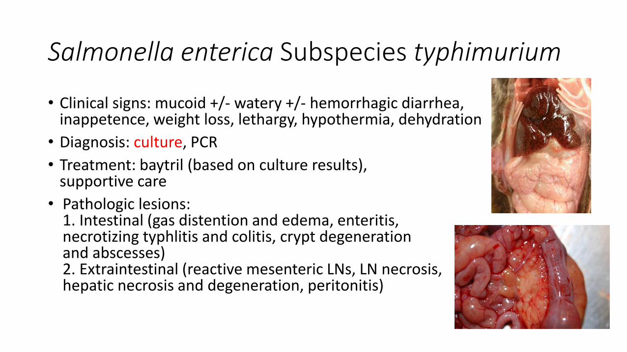
Salmonella enterica Subspecies typhimurium
• Clinical signs: mucoid +/- watery +/- hemorrhagic diarrhea, inappetence, weight loss, lethargy, hypothermia, dehydration
• Diagnosis: culture, PCR
• Treatment: baytril (based on culture results), supportive care
• Pathologic lesions:1. Intestinal (gas distention and edema, enteritis, necrotizing typhlitis and colitis, crypt degeneration and abscesses)2. Extraintestinal (reactive mesenteric LNs, LN necrosis,hepatic necrosis and degeneration, peritonitis)

Diagnostic Methods
• Culture: media matters!
1. MacConkey agar – selective for gram-negative/enteric bacteria2. Selenite broth – selective for Salmonella3. Hektoen agar – selective for Salmonella and Shigella4. Xylose-Lysine-Desoxycholate Agar (XLD) – selective for Salmonella and Shigella
• PCR: useful to screen for chronic carriers

Clostridium difficile Colitis

Marmoset Wasting Disease (MWD)
• Idiopathic lymphoplasmacytic enterocolitis and weight loss, absence of diarrhea
• Biomarkers: low serum albumin(<3.5g/dl), low BW(<325g)
• +/- association with metabolic bone disease, presumed secondary to malabsorption of vitamin D
• Treatment: budesonide, vitamin D, calcium, supportive care
Bone Disease in the Common Marmoset
• Traumatic
• Idiopathic
• Metabolic
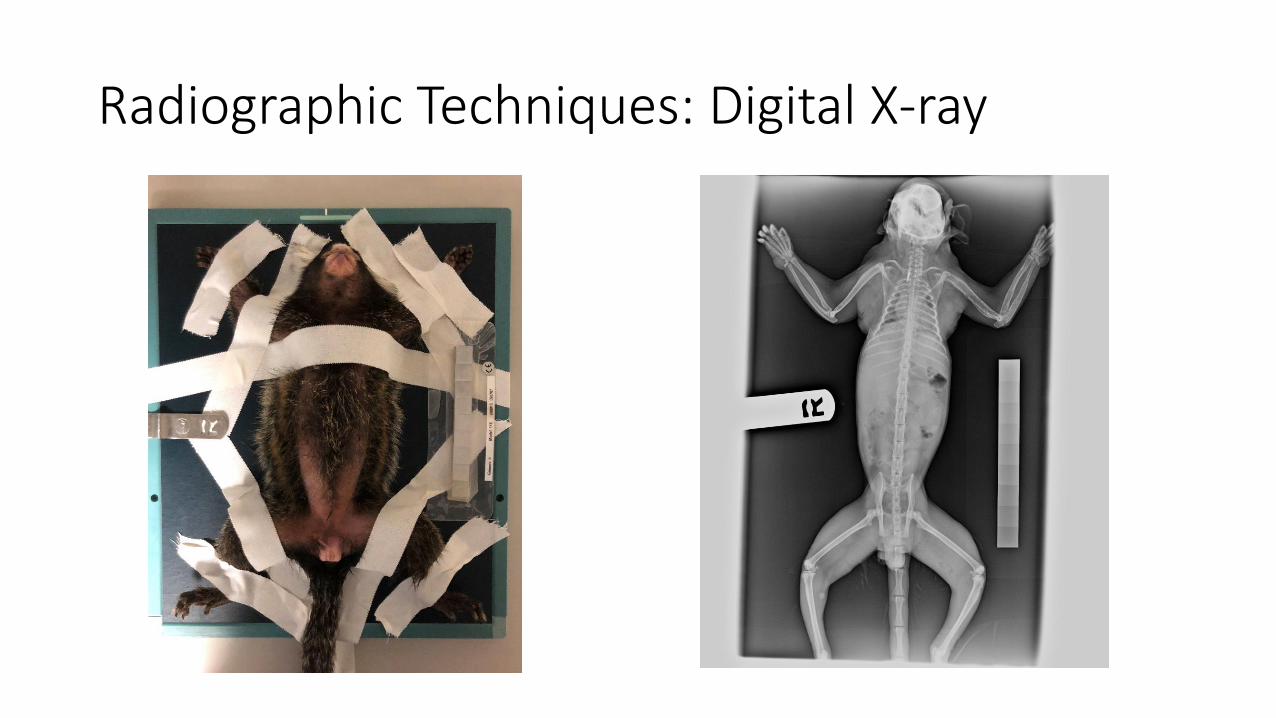
Radiographic Techniques: Digital X-ray

Radiographic Techniques: Faxitron X-ray

Traumatic Wounds and Fractures
• Fight wounds: breeding pairs, same-sex pairs or juvenile groups
• Iatrogenic: cage injuries, hand-catching and restraint

Traumatic Fractures: Surgical Fixation

Fractures: External Coaptation
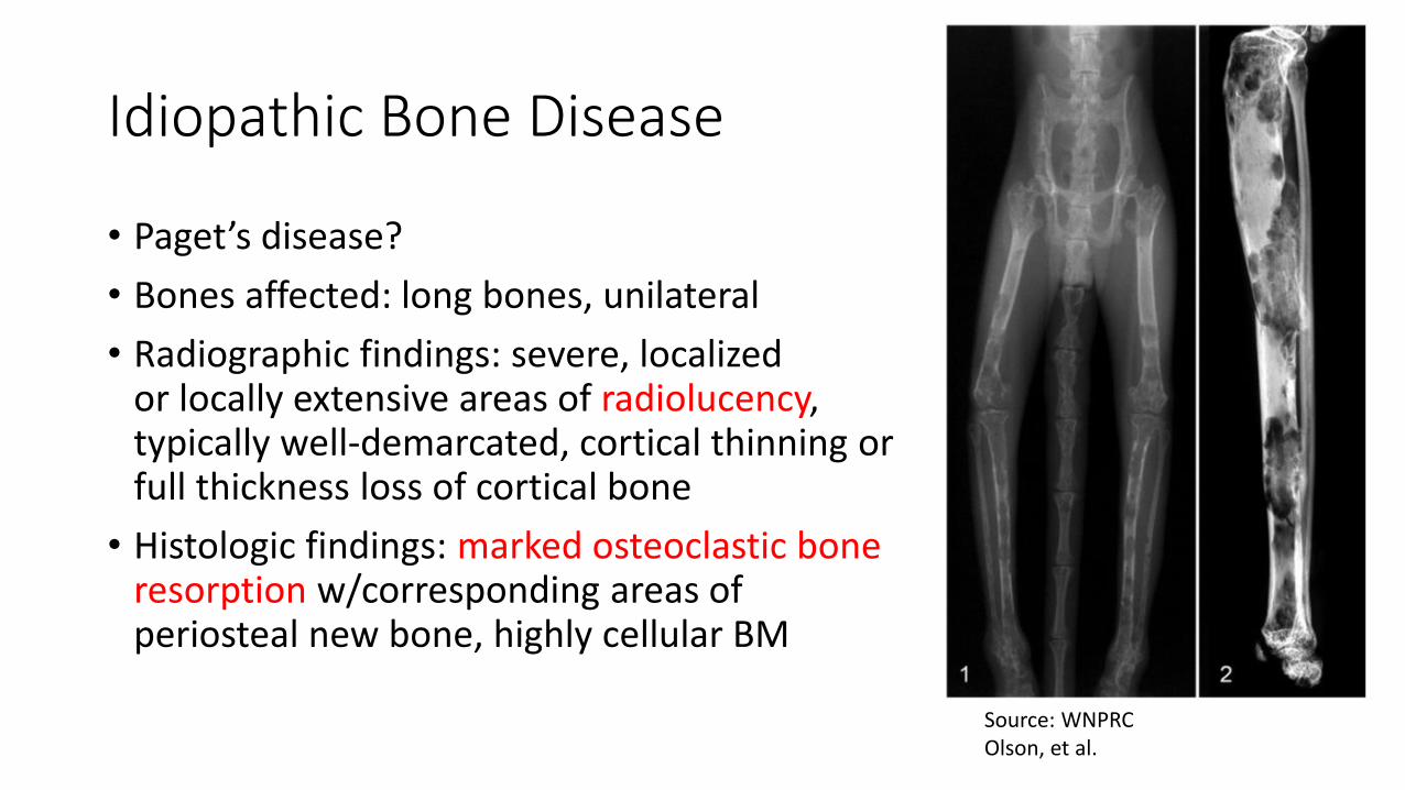
Idiopathic Bone Disease
• Paget’s disease?
• Bones affected: long bones, unilateral
• Radiographic findings: severe, localized or locally extensive areas of radiolucency, typically well-demarcated, cortical thinning or full thickness loss of cortical bone
• Histologic findings: marked osteoclastic boneresorption w/corresponding areas of periosteal new bone, highly cellular BM
Source: WNPRCOlson, et al.

Idiopathic Bone Disease
• Fibrous dysplasia (FD)?
• Reported at WNPRC and recently seen at JHU
• Bones affected: long bones, unilateral
• Radiographic findings: diffuse multilocular expansile lesions with increased bone diameter, lytic lesions (early), lack of clearly defined cortices
• Histologic findings: replacement of cortical lamellar bone by trabecular woven bone, hypocellular BM

Metabolic Bone Disease: Fibrous Dysplasia?

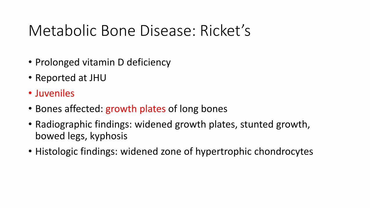
Metabolic Bone Disease: Ricket’s
• Prolonged vitamin D deficiency
• Reported at JHU
• Juveniles
• Bones affected: growth plates of long bones
• Radiographic findings: widened growth plates, stunted growth, bowed legs, kyphosis
• Histologic findings: widened zone of hypertrophic chondrocytes

Normal, age-matched Affected

Metabolic Bone Disease: Fibrous Osteodystrophy (FOD)
• Reported at JHU
• Bones affected: long bones, mandible,maxilla, vertebrae
• Radiographic findings: multifocal areasof radiolucency or “moth-eaten” lysis
• Histologic findings: increased numbersof osteoclasts, incomplete dental alveoli/tooth sockets, periosteal newbone formation
Source: JHUOlson, et al.

Source: NIH

Complications with Cranial Implants
• Margin infections: Pseudomonas aeruginosa, Burkholderia cepacia, Serratia marcescens, Klebsiella pneumoniae, Staphylococcus xylosus
• Excess granulation tissue
• Skin retraction
• Loss of implant
• Tissue growth beneath implant
• CNS disease: intra-cranial abscess,meningitis, meningoencephalitis


Other Important Diseases
• Viral: Herpes simplex 1, measles, LCMV, lymphocryptovirus
• Aging: chronic renal disease, amyloidosis, chronic liver disease, neurodegenerativedisease, obesity and Type II diabetes
• Neoplastic: lymphoma, intestinal adenocarcinoma, SCC

Dental Disease
• Tooth root abscesses, loose teeth, devitalized teeth, periodontal disease, fractures, malocclusion
• Diagnostics: oral exam, dental rads if available
• Treatments: lance and drain facial abscesses, extraction, antibiotic and anti-inflammatorymedications as indicated
Acknowledgments
• Bob Adams, DVM, DACLAM
• Eric Hutchinson, DVM, DACLAM
• Dr. Xiaoqin Wang and the Wang lab
• Jessica Lynch and Kayla Schonvisky(veterinary technicians)
• Tom Thomas, DVM, DACLAM (NIH)
• Husbandry and care staff
References• Pisarath H, Cooper T, Brice A, et al. Septicemia and Peritonitis in a Colony of Common Marmosets (Callithrix jacchus) Secondary to Klebsiella
pneumoniae infection. AALAS Contemporary Topics (2005) 44 (1): 35-37.
• Otovic P, Smith S, and Hutchinson E. The use of glucocorticoids in marmoset wasting syndrome. J Med Primatol. 44 (2015) 53-59.
• Ludlage E and Mansfield K. 2003. Clinical care and diseases of the common marmoset (Callithrix jacchus). Comp Med. 53: 369-382.
• Gozalo A. and Montoyo E. 1991. Klebsiella pneumoniae infection in a New World nonhuman primate center. Lab. Primate Newslett. 30(2): 13-20.
• Baskerville M. Canine tooth root infection as a cause of facial abscess in the common marmoset (Callithrix jacchus). Laboratory Animals (1984) 18, 115-118.
• Burke R, Whitehouse C, Taylor J, et al. Epidemiology of Invasive Klebsiella pneumoniae with Hypermucoviscosity Phenotype in a Research Colony of Nonhuman Primates. Comp Med. 2009 Dec; 59(6): 589-597.
• Tardif S, Mansfield K, Ratnam R, et al. The Marmoset as a Model of Aging and Age-Related Diseases. ILAR J. 2011 Feb 8; 52(1): 54-65.
• Power M, Ross C, Schulkin J, et al. Metabolic consequences of the early onset of obesity in common marmoset monkeys. Obesity (Silver Spring) 2013 Dec; 21(12): E592-E598.
• Kramer J, Hachey A, Wachtman L, et al. Treatment of Giardiasis in Common Marmosets (Callithrix jacchus) with Tinidazole. Comp Med 2009 Apr; 59(2): 174-179.
• Olson EJ, Shaw GC, Hutchinson EK, et al. Bone Disease in the Common Marmoset: Radiographic and Histological Findings. Vet Path 2015 Sep; 52(5): 883-93.
• Baxter VK, Shaw GC, Sotuyo NP, Carlson CS, Olson EJ, et al. (2013) Serum Albumin and Body Weight as Biomarkers for the Antemortem Identification of Bone and Gastrointestinal Disease in the Common Marmoset. PLoS ONE 8(12): e82747.