class iii malocclusion
TRANSCRIPT

UNIVERSITY OF GLASGOW
Class III Malocclusion
Personal notes
Mohammed Almuzian2/20/2013
……………………………………….

Table of Contents
Definition.................................................................................................................1
Incidence.................................................................................................................1
Classification...........................................................................................................2
Aetiology.................................................................................................................2
Features of Class III................................................................................................3
A. Skeletal features...............................................................................................3
B. Soft tissues features..........................................................................................4
C. Dental features.................................................................................................4
D. Displacements..................................................................................................4
E. Facial growth...................................................................................................4
IOTN and class III...................................................................................................8
Crossbite (2.c, 3.c, 4.c)............................................................................................8
Growth status assessment for class III patients.......................................................9
Monitoring the growth of mandible........................................................................9
Differentiation between mandibular prognathism & maxillary deficiency...........10
Treatment options for class III malocclusion........................................................12Mohammed Almuzian, University of Glasgow, 2013 1

Factors influencing treatment options...................................................................12
Summary about treatment strategies according to dental age...............................13
Reasons for early treatment of class 3 malocclusions...........................................16
Orthopaedic treatment option................................................................................17
Effect of orthopaedic appliance in class III maloculsion......................................17
Positive factors for orthopaedic treatment.............................................................17
Types of orthopaedic treatment in class III malocclusion.....................................18
1. Protraction HG...............................................................................................18
Definition...............................................................................................................18
History...................................................................................................................18
Indications.............................................................................................................19
Timing...................................................................................................................20
Effects....................................................................................................................20
Protraction face mask system................................................................................21
Evidence based short term effectiveness of PH....................................................25
Evidence based long term effectiveness of PH.....................................................26
2. Chin caps........................................................................................................26
Types: occipital pull, used for patients with mandibular prognathism or
vertical pull, used for patients with increased anterior face height.......................27
Mohammed Almuzian, University of Glasgow, 2013 2

Best patient for Chin cup therapy..........................................................................27
Ko et al (2004).......................................................................................................27
The effects of chincup therapy..............................................................................27
(Thilander 1963)....................................................................................................27
3. Reverse chin cup therapy...............................................................................28
4. Bone anchored orthopaedic appliance (Bollard miniplates, PFH supported
with miniplates).....................................................................................................28
5. Shapiro and Kokich 1984 used the same idea by inducing artificial ankylosis
and use the ankylosed teeth as anchor...................................................................29
6. Functional appliances.....................................................................................29
Camouflage (dental compensation for mild cases)...............................................31
Indications.............................................................................................................31
Techniques of camoflagable treatments................................................................32
1. Non extraction................................................................................................32
2. Extraction:......................................................................................................32
Orthognathic surgery options................................................................................35
The types of surgery most frequently used are the following...............................35
Intraoperative complications of the mandibular ramus surgery............................36
Mohammed Almuzian, University of Glasgow, 2013 3
What Factors need to be taken into Account When Planning a surgical treatment
for class III cases...................................................................................................37
1. Planning the type of surgery..........................................................................37
2. The Pre-Surgical Orthodontics in Class III?..................................................37
What Are the Aims of the Pre-Surgical Orthodontics?.........................................37
Borderline Camouflage/ Orthognathic Surgery Patients.......................................38
Summary of the evidences....................................................................................39
Mohammed Almuzian, University of Glasgow, 2013 4

Class III malocclusion
Definition
BSI 1982 defined class III incisor relationship as ‘’the lower incisal edge lies
anterior to the cingulum plateau of the the upper incisors, British Standards
Institute, 1983
The term ‘pseudo-class III’ has been coined for this situation where an
anterior displacement masking what is in fact an underlying skeletal class I base
relationship.
Incidence
1.Class III prevalence in white populations
3% UK (Foster & Day, 1974)
5% (Jones & Oliver, 2000)
5% UK (Todd and Dodd 1975)
2.Class III prevalence in Asian populations
4-14% in Asian (Lew 1993)
3.Dental anterior crossbite
Anterior crossbite in 10% of children (1993 Child Dental Health Survey)
Mohammed Almuzian, University of Glasgow, 2013 5

Classification
Lin (2007) divides class 3 malocclusion into three categories according to the
following definitions:
1. True class 3, anterior crossbite cases with bilateral buccal occlusions in class
III.
2. Class 3 subdivision, anterior crossbite cases with one of the bilateral buccal
occlusions in class 1 and the other in class 3.
3. Pseudo class 3, bilateral class 1 buccal occlusions and majority of teeth in
anterior crossbite. The pseudo class 3 malocclusion is often due to collapse of the
arch perimeter resulted from:
Caries in some Eastern societies (caries collapse)
TSD or small, missing or impacted or palatal positioning of the upper teeth
(perimeter-collapse).
Aetiology
1. Skeletal :
A. Environmental
Airway problems like enlarged tonsils & nasal blockage,
Scaring from CLP as a result of surgical repair
Hormonal like in acromegaly.
Some syndromes caused by environmental as well as genetic reasons such as
Crouzons, Aperts, and Cleidocranial dysostosis.
Mohammed Almuzian, University of Glasgow, 2013 6
B. Genetic (Litton et al 1970). 1/3 of patients with severe class III have a
parent with class III problems but there is no detected autosomal dominant or
recessive method of transmission.
2. Soft tissue : the ST indeed my act to reduce the severity of CLIII, Lower
incisor retroclination is adaptive due to soft tissue forces and tongue might
procline ULS. Exception in high angle case when there is tongue to lower lip seal
and macroglosia that worsen the CLIII.
3. Dental factors :
Rarely ULS retroclination and LLS proclination.
Hypodontia or microdontia in the upper arch
Impacted upper teeth
4. Habits : tongue to lower lip seal and macroglosia that worsen the CLIII.
Features of Class III
A. Skeletal features
Cranial base features
AP relationship
Vertical relationship
Transverse relationship
Cephalometric skeletal values
Mohammed Almuzian, University of Glasgow, 2013 7

B. Soft tissues features
C. Dental features
D. Displacements
E. Facial growth
In details
F. Skeletal features
1. Cranial base features:
Short cranial base length.
Decrease cranial base angle resulting in forwards position of mandible.
2. AP relationship
Mainly skeletal class 3 base relationships but it could be Class I or even class II
skeletal base.
Guyer, Ellis, Behrents and McNamara (1986) 55% of class 3 malocclusions
had maxillary deficiency as one of the components of the malocclusion.
Mandibular prognathsim in 45% of cases.
3. Vertical relationship
Guyer, Ellis, Behrents and McNamara (1986), 59% of class 3 malocclusions had
reduced or neutral lower facial heights and that 41% had increased lower facial
heights.
4. Transverse relationship
Mohammed Almuzian, University of Glasgow, 2013 8
The maxillary skeletal base widths were (statistically) significantly smaller in the
class 3 than in the class 1group (Chen et al 2008)
Skeletal asymmetries, particularly in conjunction with mandibular prognathsim,
are also relatively common in class III malocclusions (Severt and Proffit, 1997).
5. Cephalometric skeletal values
Reduced cranial base angle
Increased saddle angle
Obtuse gonial angle
Reduced ANB
Normal or increase MMP angle and lower face height
Increased mand length
Reduced maxillary length
G. Soft tissues features
ST not involved in aetiology but encourage dentoalveolar compensation.
However there are some features which could be found in class III case
depending in the aetiology of the problem:
1. Orbital rim hypoplasia
2. Increase scleral show
3. Check bone flattening
4. Malar hypoplasia in midface deficiency
Mohammed Almuzian, University of Glasgow, 2013 9

5. Paranasal hallowing
6. Obtuse NLA
7. Reduced incisor show at smile
8. Increase buccal corridor dark space
9. Upper lip looks thin with reduced vermilion border show while lower lip
may be full and pendulous
10. Obtuse LMA
11. Prominent chin
12. Concave or straight profile with anterior divergence.
13. Increased throat length
H. Dental features
1. Class III incisor relationship
2. Mostly CI III molar relationship could be I or even II. The same applied for
canine relationship.
3. Tendency to or full reverse OJ,
4. Reduced OB, AOB may exist
5. Max probably crowded, mandible unlikely to be so but usually spaced.
6. Incisors compensate for Skeletal base, i.e. Proclined maxillary, retroclined
mandibular incisors
Mohammed Almuzian, University of Glasgow, 2013 10
7. Transverse discrepancy expressed in a form of tendency to posterior cross
bite. It could be unilateral with or without displacement or could be bilateral
mainly without displacement and to lesser extent with displacement
I. Displacements
The displacement could be in an anterior or lateral direction or combination.
It is due to:
Unsatisfactory edge-to-edge incisor
Unsatisfactory transverse buccal segment relationship
J.Facial growth
Tends to be unfavourable i.e. backwards growth rotation.
NB: Bacceti 2007, found that the pubertal peak of mandibular growth in AP and
vertical direction occurred between stages CS3 and CS4 in (corresponding with
the eruption of canines and premolars) and CS4-CS6 late developmental stages
(corresponding with the complete eruption of second and third molars) but 2 time
more in male than female.
IOTN and class III
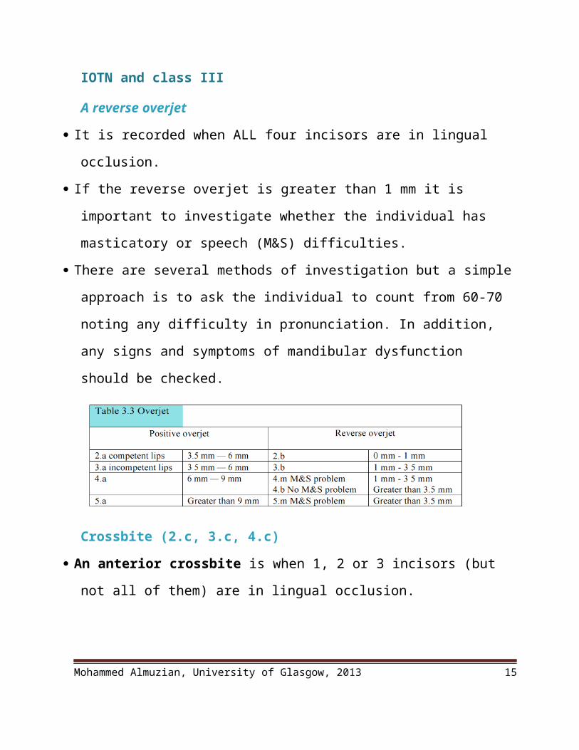
A reverse overjet
It is recorded when ALL four incisors are in lingual occlusion.
If the reverse overjet is greater than 1 mm it is important to investigate whether the
individual has masticatory or speech (M&S) difficulties.
Mohammed Almuzian, University of Glasgow, 2013 11

There are several methods of investigation but a simple approach is to ask the
individual to count from 60-70 noting any difficulty in pronunciation. In addition,
any signs and symptoms of mandibular dysfunction should be checked.
Crossbite (2.c, 3.c, 4.c)
An anterior crossbite is when 1, 2 or 3 incisors (but not all of them) are in lingual
occlusion.
A posterior crossbite is recorded when the posterior tooth or teeth are cusp to
cusp or in full crossbite in a buccal or lingual perspective.
The grade recorded depends on the severity of discrepancy between retruded
contact position (RCP) and intercuspal position (IP) (Table 3.4).
The greater the discrepancy between RCP an IP, the higher the grade
Growth status assessment for class III patients
Mandibular skeletal maturity can be assessed by means of a series of biologic
indicators:
Mohammed Almuzian, University of Glasgow, 2013 12

1. History (is the patient changing shoes)
2. Growth chart like an increase in body height (Nanda, 1955; Hunter, 1966)
3. Biological parameters like:
Skeletal maturation of the hand and wrist (Bjork, 1967) or cervical vertebral
maturation (CVM) method. Franchi 2000, Beccteti 2002 & 2005 (please
read the summary about ‘’The Cervical Vertebral Maturation’’)
Dental development and eruption (Bjork, 1967)
Chronological age
Secondary sexual features like Menarche, breast, and voice changes (Tanner
1962)
Monitoring the growth of mandible
1.Serial Clinical measurements like OJ
2.Serial Study models
3.Serial Photograph or 3D stereo photogrammetry
4.Serial Ceph (not justified)
5.Growth Treatment Response Vector (GTRV) analysis
Ngan (2005) has described this as a method of determining whether a class 3
malocclusion can be treated by camouflage or if surgical treatment will be
required at a later date.
It is calculated from two serial cephalometric radiographs at least one year apart.
Mohammed Almuzian, University of Glasgow, 2013 13
The lines AO and BO are constructed in the same way as for the Wits analysis on
the first radiograph;
The first radiograph is then superimposed on the second using the stable structures
of the cranial base.
New AO and BO are then constructed using the occlusal plane of the first
radiograph.
Horizontal growth change of maxilla is second AO-first AO
Horizontal growth change of mandible is second BO-first BO
The GTRV is then given by the following formula:
GTRV = horizontal growth change of maxilla / horizontal growth change of
mandible
The normal GTRV of patients is 0.77 – ie: normally, mandibular growth usually
exceeds maxillary growth by 23% between the ages of 8 and 16 years.
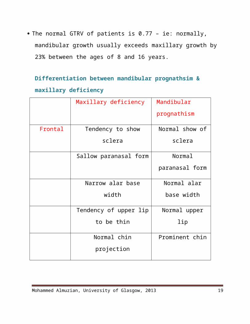
Differentiation between mandibular prognathsim & maxillary deficiency
Maxillary deficiency Mandibular
prognathism
Frontal Tendency to show sclera Normal show of sclera
Sallow paranasal form Normal paranasal
form
Mohammed Almuzian, University of Glasgow, 2013 14
Narrow alar base width Normal alar base
width
Tendency of upper lip to be
thin
Normal upper lip
Normal chin projection Prominent chin
Normal to decreased lower
facial height (LFH)
Normal, increased or
decreased lower facial
height (LFH)
Profile Nasolabial line-Subnasale:
subnasale-tip of nose ,usually
not 1:1 ratio
Normal
Nasal tip down Normal
Obtuse nasolabial angle Normal nasolabial
angle
Smiling
assessment
Less incisor visible Good
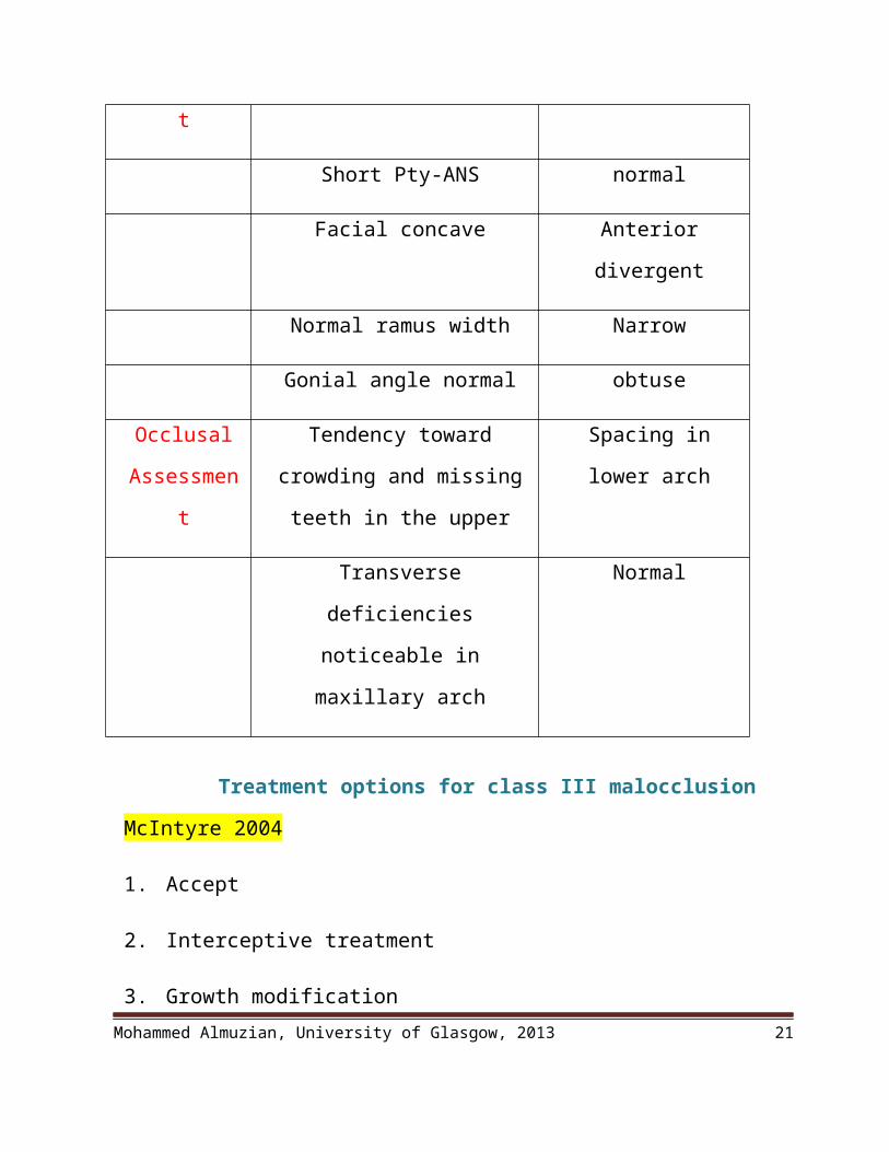
Cephalometri
c assessment
Normal to decreased total facial
height
Increased total facial
height
Short Pty-ANS normal
Facial concave Anterior divergent
Normal ramus width Narrow
Mohammed Almuzian, University of Glasgow, 2013 15

Gonial angle normal obtuse
Occlusal
Assessment
Tendency toward crowding and
missing teeth in the upper
Spacing in lower arch
Transverse deficiencies
noticeable in maxillary arch
Normal
Treatment options for class III malocclusion
McIntyre 2004
1. Accept
2. Interceptive treatment
3. Growth modification
4. Orthodontic camouflage
5. Orthodontic decompensation and orthognathic surgery
6. Compromised orthodontic treatment
Factors influencing treatment options in Class III
1. Patient concern (dental or facial concern)
2. Patient age
3. Growth
4. Medical condition
5. Patient compliance Mohammed Almuzian, University of Glasgow, 2013 16
6. Family history of class III
7. Severity of skeletal problem in AP, V & T direction
8. Clinical condition of the teeth and oral tissues.
9. Amount of the OJ &OB
10. Degree of crowding
11. Degree of compensation
12. Presence of displacement
Summary about treatment strategies according to dental age a. In primary dentition
There is no evidence to suggest that orthodontic intervention during the primary
dentition avoids, or reduces, the complexity of later orthodontic treatment.
b. In early mixed dentition
1. Incisor crossbites due to retained primary incisors: Treatment extract
retained primary teeth
2. Premature contact and mandibular displacement or incisors erupted in cross
bite relationship, then
Extract or grind cusp tips (usually primary canines)
Posterior onlay to overcome the posterior crossbite that caused
displacement.
Mohammed Almuzian, University of Glasgow, 2013 17
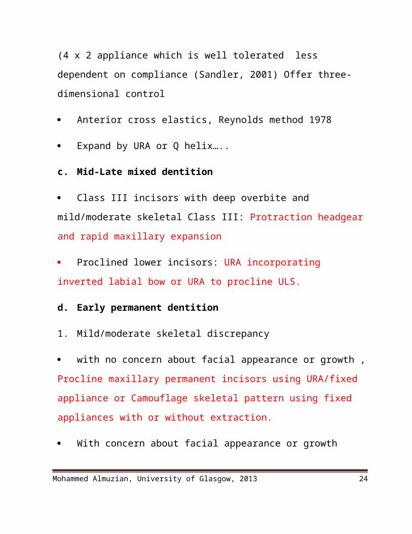
Procline maxillary permanent incisor(s) using an upper removable appliance
(URA) or a fixed appliance (4 x 2 appliance which is well tolerated less
dependent on compliance (Sandler, 2001) Offer three-dimensional control
Anterior cross elastics, Reynolds method 1978
Expand by URA or Q helix…..
c. Mid-Late mixed dentition
Class III incisors with deep overbite and mild/moderate skeletal Class III:
Protraction headgear and rapid maxillary expansion
Proclined lower incisors: URA incorporating inverted labial bow or URA to
procline ULS.
d. Early permanent dentition
1. Mild/moderate skeletal discrepancy
with no concern about facial appearance or growth , Procline maxillary
permanent incisors using URA/fixed appliance or Camouflage skeletal pattern
using fixed appliances with or without extraction.
With concern about facial appearance or growth
A. Postpone treatment decision until skeletal growth completed.
B. sometime, Align maxillary arch with fixed appliance and relieve crowding,
accepting Class III incisor relationship will require orthognathic surgery in
adulthood
2. Severe skeletal discrepancy or a concern about facial appearance
Mohammed Almuzian, University of Glasgow, 2013 18
Accept malocclusion will require combined orthodontic
treatment/orthognathic surgery in adulthood
Align maxillary arch with fixed appliance and relieve crowding, accepting
Class III incisor relationship will require orthognathic surgery in adulthood
e. Adult treatment
1. Mild/moderate skeletal discrepancy
A. no concern about facial appearance
Procline maxillary permanent incisors using URA/fixed appliance
Camouflage skeletal pattern using fixed appliances
B. Mild/moderate skeletal discrepancy –concern about facial appearance
Compromised treatment by aligning the UA with or without extraction and if
possible align lower arch on non-extraction base to keep the cop of
decompensation if the Combined orthodontic treatment/orthognathic surgery
decided later
2. Severe skeletal discrepancy with no concern about facial appearance
Compromised treatment by aligning the UA with or without extraction and if
possible align lower arch on non-extraction base to keep the cop of
decompensation if the Combined orthodontic treatment/orthognathic surgery
decided later
3. Severe skeletal discrepancy with a concern about facial appearance
Combined orthodontic treatment/orthognathic surgery
Mohammed Almuzian, University of Glasgow, 2013 19

Reasons for early treatment of class 3 malocclusions
Hägg et al (2004) and Ngan (2005) cite the reasons for early treatment as:
a. To eliminate CR-CO discrepancies which may cause
periodontal damage
occlusal wear
TMJ problems
b. To provide a more favourable environment for growth and
development of the maxilla and mandible with a reduction in dental
compensation because remodelling may occur in the joint as the postured position
which will act as functional appliance and making correction of the crossbite
more difficult at a later date
c. To provide space for the eruption of the buccal segments as a result of
proclination of the upper incisor so the canines and premolars can be guided into
a class 1 relationship
d. Psychological benefits resulting from improved dental and facial
appearance
Orthopaedic treatment option
Effect of orthopaedic appliance in class III maloculsion
Dermaut and Aelbers (1996) have reviewed the possible effects of orthopaedic
treatment in class 3 malocclusions.
1. 50% of the studies showed stimulation of maxillary growth
Mohammed Almuzian, University of Glasgow, 2013 20
2. 90% showed an inhibition of mandibular projection
In general orthopaedic appliances are more effective if cl3 is due to maxilla
retrusion than mand prognathism.
However, most of the effects are dentoalveolar in nature with maxillary incisor
proclination and mandibular retroclination.
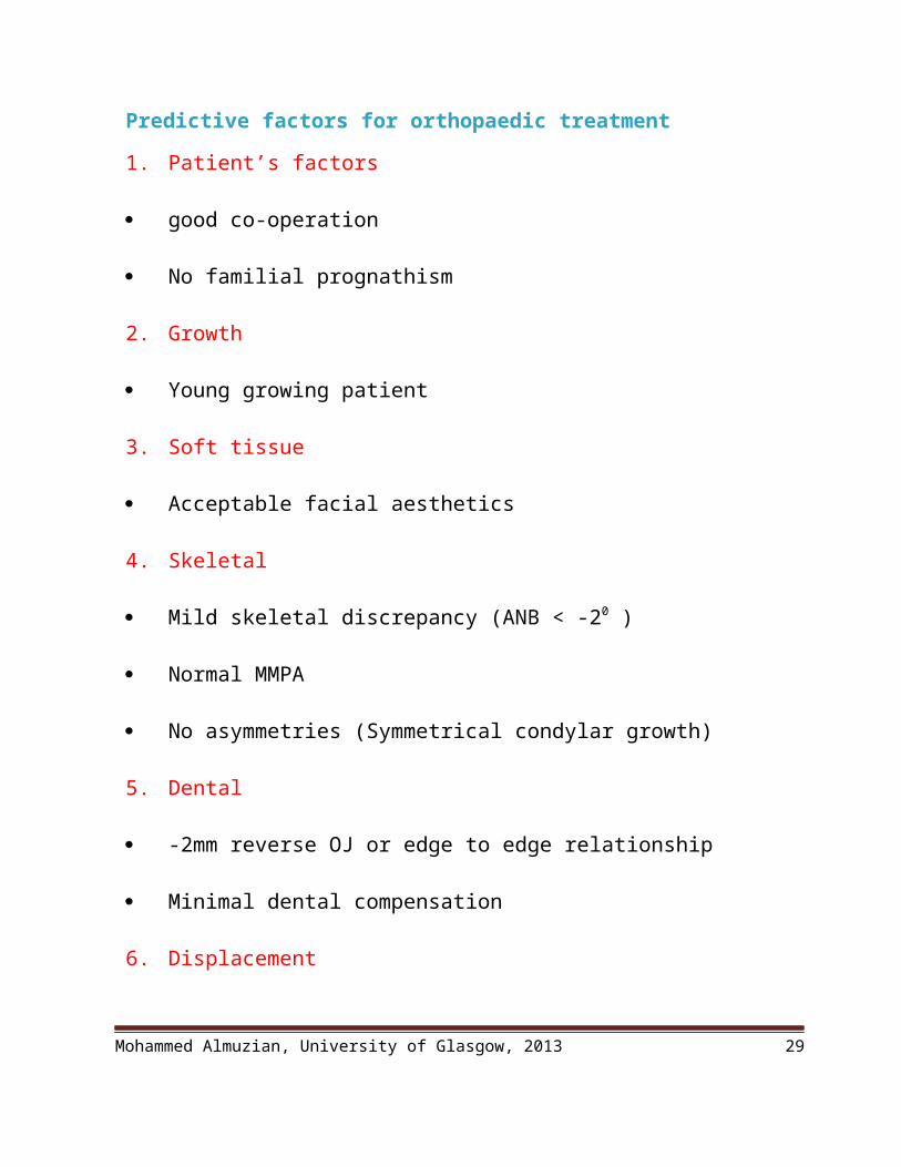
Predictive factors for orthopaedic treatment
1. Patient’s factors
good co-operation
No familial prognathism
2. Growth
Young growing patient
3. Soft tissue
Acceptable facial aesthetics
4. Skeletal
Mild skeletal discrepancy (ANB < -20 )
Normal MMPA
No asymmetries (Symmetrical condylar growth)
5. Dental
-2mm reverse OJ or edge to edge relationship
Mohammed Almuzian, University of Glasgow, 2013 21

Minimal dental compensation
6. Displacement
Functional shift
Types of orthopaedic treatment in class III malocclusion
1. Protraction HG
Definition
Means of applying anterior directed forces to teeth and/or skeletal structures from
an extra-oral source
History
The technique of maxillary protraction is based on work by Nanda (1978), with
rhesus monkeys in which he showed that a force of approximately 500g could
produce anterior displacement of the maxilla
It is appropriate to refer to this type of treatment as facemask therapy.
Indications
A. Treatment of maxillary retrusion. An ideal case would be;
1. Patient’s factors
good co-operation
No familial prognathism
Mohammed Almuzian, University of Glasgow, 2013 22

7. Growth
Young growing patient
8. Soft tissue
Acceptable facial aesthetics
9. Skeletal
Mild skeletal discrepancy (ANB < -20 )
Normal MMPA
No asymmetries (Symmetrical condylar growth)
10. Dental
-2mm reverse OJ or edge to edge relationship
Retroclined ULS
Proclined LLS
11. Displacement
Functional shift
B. Reinforcement of anterior anchorage and dental protraction allowing closure of
space from behind in patients suffering from hypodontia
C. Stabilization following maxillary osteotomy/distraction osteogenisis
D. Rotate arch segments in cleft palate patients
E. Remove hyper-anterior contact in TMJ internal derangement cases,
Mohammed Almuzian, University of Glasgow, 2013 23
Timing
1. Dental age: McNamara (1987) suggested that the optimal time for treatment is in
the early late mixed dentition, coincident with the eruption of the upper
permanent incisors.
2. Skeletal age: Baccetti et al (1998) showed that the early treatment group CVM2
showed effective forward displacement of the maxillary structures whereas the
late treatment group CVM3 showed no change compared with controls
3. Chronological age: Other investigators have suggested that for optimal
orthopaedic effects, treatment should be initiated before the patient is 9 years old
(Proffit, 2000). Mandal 2010, 2012 used it at age of 8.5-10 year.
Kim et al., 1999, meta-analysis of effectiveness of protraction HG concluded it
was less effective on patients >10yrs
Effects
1. Correction of a centric occlusion-centric relation discrepancy. This correction
happens relatively rapidly in patients with an edge to edge relationship and
associated displacement
2. Maxillary skeletal protraction, with up to 3mm of forward movement of the
maxilla possible , Mandal 2010 and 2012, showed that these effect are stable after
3 years follow-up
3. Proclination and forward movement of the maxillary dentition
4. Lingual tipping of the lower incisors
5. Redirection of mandibular growth in a downward and backward direction,
resulting in an increase in lower anterior facial height
Mohammed Almuzian, University of Glasgow, 2013 24

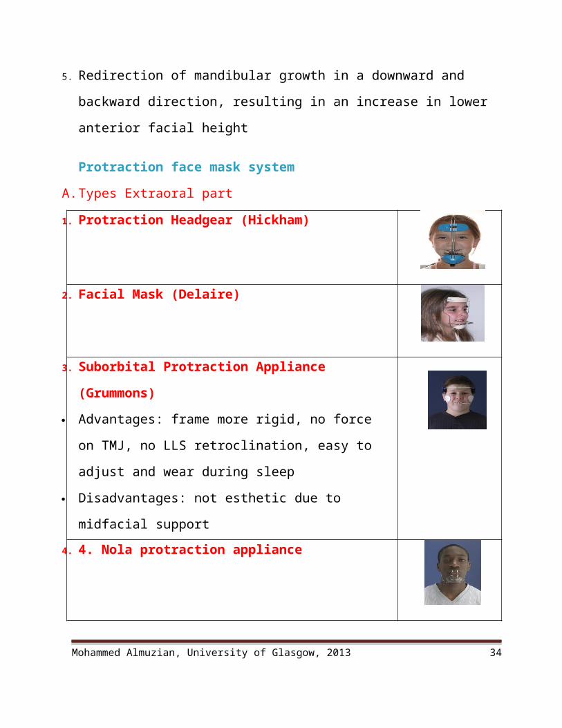
Protraction face mask system
A. Types Extraoral part
1. Protraction Headgear (Hickham)
2. Facial Mask (Delaire)
3. Suborbital Protraction Appliance (Grummons)
Advantages: frame more rigid, no force on TMJ, no LLS
retroclination, easy to adjust and wear during sleep
Disadvantages: not esthetic due to midfacial support
4. 4. Nola protraction appliance
5. Petit style face mask
The Petit style with a single central vertical bar is also well
tolerated and recent price changes have made it
economically much more attractive.
B. Intraoral part:
Mohammed Almuzian, University of Glasgow, 2013 25

1. In order to maximize the amount of skeletal change in young children, a
removable full coverage acrylic splint is used with a protraction headgear (Proffit
1986).
2. McNamara (1987) has described the use of a Biocryl and wire splint that is
bonded in the mouth. The splint material should be at least 3 mm thick with a
0.045" stainless steel wire framework. The two halves of the splint are joined by
an expansion screw. Traction hooks to receive the elastics from the headgear are
placed in the first premolar region.
3. RME with hook can be used
4. Fixed appliance
5. Some recommend using an intraoral bone plate to support the PHG force.
Systematic review to compare the dentally anchored face mask with skeletally
anchored one by Major (2012) in Canada, he found Approximately 3 mm of
horizontal A-point movement is predictably attainable with the skeletal one in
comparison to dental one..
C. Rapid maxillary expansion
Advantages (Haas 1973).
1. Sutural loosening
2. Correct transverse discrepancy that commonly associated with class III
malocclusion
3. Displace the maxillary complex anteriorly. This is due to butterfly effect of
expansion at the Midpalatal suture and because of the anterior sloping of the
facial sutures
Mohammed Almuzian, University of Glasgow, 2013 26

Evidences
1. Many clinicians use protraction with a facemask following or simultaneously
with palatal expansion, because some evidence suggests that the expansion makes
antero-posterior skeletal change more likely. Kim et al (1999)
2. There is other evidence that the expansion is optional and should be dictated by
the maxillary arch width related to the lower arch width, Vaughan 2001 and 2005.
D. Techniques
First step is to fabricate and bond/cement the rapid maxillary expansion appliance
Appliance is activated once per day until the desired increase in maxillary width
has been obtained.
If patients do not need an increase in maxillary width, the appliance is still
activated for 7-10 days to disrupt the maxillary sutural system and promote
maxillary protraction (Haas, 1965)
After the patient activated the maxillary appliance for 7-10 days protraction
headgear is fitted.
E. Force level
1. Moving maxillary anterior teeth forward: 400g per side, 12-14h/day
2. Maxillary protraction : 800g per side, 14h/day
3. Overcorrect to compensate for mandibular growth
4. Active treatment should be limited to 9-12 months because of the risk of
decalcification of the dentition
F. Force direction:
1. To avoid bite opening, place protraction elastics near maxillary bicuspids,
2. Force vector should be 15-30 degree below the horizontal
Mohammed Almuzian, University of Glasgow, 2013 27

3. To avoid irritation to the lip, use crossed elastic,
4. Pay special attention to airway and tongue posture
5. Ishii et al (1987) describe the effects of providing the protraction force from the
first molars or the premolar region. Protraction from the first molars results in
more anterior movement and a forward and upward rotation of the maxilla;
protraction from the premolars results in less forward movement but fewer
tendencies to upward and forward rotation.
G. Transitional period
After treatment objectives have been achieved, the patient can be retained with a
number of appliances:
The facemask,
FR-3 appliance
Acrylic maxillary retainer with reverse lower labial bow
Chin cup (seldom used).
H. Post protraction treatment consideration
1. As mandibular growth exceeds maxillary growth during adolescence, early Class
III correction may be lost during the teenage period. The patients and parents
should again be warned of the possibility of orthognathic treatment if growth is
unfavourable
2. Upper labial root torque during fixed appliance stage: Most class 3 patients
demonstrate considerable proclination of the upper labial segment at the end of
treatment. Catania et al (1990) recommend in his case report to use inverted U
incisor bracket to counteract the effect of proclination.
Mohammed Almuzian, University of Glasgow, 2013 28
Evidence based short term effectiveness of PH
Mandall, 2010 (similar to study of Ngan 1998)
Early Class III orthopaedic treatment with protraction face mask in patients
less than 10 years of age is skeletally and dentally effective in the short term 15
months. (After 15 months of treatment, children undergoing early facemask
therapy had 1.3 degrees more forward movement of SNA, almost 2degrees less
forward movement of SNB and an overall ANB improvement of around 2.6
degrees when compared to the control group. In addition, the overjet improved by
more than 4 mm and the relative PAR score by more than 40% in the facemask
compared to the control group. Thus, early class III protraction facemask
treatment in patients under 10 years of age would seem to be skeletally and
dentally effective in the short-term)
70% of patients had successful treatment, defined as achieving a positive
overjet.
Early treatment does not seem to confer a clinically significant psychosocial
benefit.
No TMJ problem
Evidence based long term effectiveness of PH
Mandal 2012: Early Class III orthopaedic treatment with protraction face
mask in patients less than 10 years of age is skeletally and dentally effective after
3 years of treatment.
Masucci 2011: RME/FM therapy led to successful outcomes in about 73%
of the patients. Significantly improved sagittal dentoskeletal relationships. These
Mohammed Almuzian, University of Glasgow, 2013 29

favourable changes were mainly due to significant improvements in the sagittal
position of the mandible, but the maxillary changes reverted completely in the
long term. This treatment not induces a tendency of bite opening or increased
vertical relationship.
A Cochrane review by Watkinson in 2013. This review looked at the use of
four different types of orthodontic treatment for correcting prominent lower front
teeth in children.-Facemask-Chin cup-Mandibular -Tandem traction bow
appliance. This review found some evidence that the use of a facemask appliance
can help to correct prominent lower front teeth on a short-term basis. There was
no evidence available to show whether or not these short-term changes will still
be maintained until the child is fully grown. There was not enough evidence to
support any other types of treatment for prominent lower front teeth.
2. Alternate Rapid Maxillary Expansion and Constriction (Alt-
RAMEC)
Original technique:
A protocol of maxillary protraction developed by Liou and Tsai in 2005.
It includes 3 components: a 2-hinged rapid maxillary expander, repetitive
weekly protocol of Alternate Rapid Maxillary Expansion and Constriction
(Alt-RAMEC), and intraoral maxillary protraction springs.
The innovative part of this technique is the sutural loosening accomplished
by alternating expansion with constriction for 8 weeks.
Mohammed Almuzian, University of Glasgow, 2013 30

This backand- forth motion can mobilize the maxilla and protract the
maxilla for longer distances. Liou 2005
The Alt-RAMEC protocol the maxilla is expanded for 7 consecutive days
and constricted for 7 consecutive days for 9 weeks. After the Alt-RAMEC
technique, the maxilla is protracted with protraction springs for 3 months.
The technique has been modified to use a standard Hyrax rapid maxillary expansion appliance and fixed orthodontic appliances in the lower arch, Class III
Mohammed Almuzian, University of Glasgow, 2013 31

elastics and reverse-pull headgear or facemask (Yen 2011).
The Clinical Protocol for Maxillary Protraction in a Typical Adolescent Patient With Cleft Lip and Palate
● At age 13, the patient chooses surgery or maxillary protraction.
Maxillary expansion and protraction was performed before dental alignment with
an archwire because additional space was created with arch expansion. If
maxillary dental crowding was present, extractions were deferred until after
expansion and protraction. The exceptionsto this rule were the patients who had
molars and premolars tilting in opposite directionswhich would prevent parallel
draw of bands. These patients received limited orthodontics with sectional wires
to improve the insertion and draw of a rapid palatal expander (RPE).
● Pretreatment records. Check for parallel path of insertion of RPE along anchor
teeth. Prior alignment is needed if teeth are tilted in opposite directions.
● Banding of the lower first and second molars, bonded orthodontic brackets for
the lower arch premolars and anterior teeth. Initiate leveling of mandibular
dentition. Stabilize lower dentition in a stainless steel rectangular archwire;
● Banding maxillary molars and premolar (or canines) for a Hyrax rapid palatal
expander placed high in vault of palate.
● Delivery of Hyrax expander. Demonstration of screw turns needed to expand
and constrict the Hyrax expander. Sutural loosening is initiated by activating the
appliance 2 turns in the morning and 2 turns in the evening. The expansion rate is
1 mm/d. The screw turns follow the same direction for 1 week.
Mohammed Almuzian, University of Glasgow, 2013 32

● At the end of the week, the patient returns to the clinic to demonstrate ability to
insert the swivel key into the screw of the Hyrax expander. The swivel key is
suspended in the RPE before activation to ensure that the key is fully inserted.
The reverse direction is taught so that patient so that he/she is proficient in both
directions with the swivel key.
● Expansion and constriction is alternated each week. Recall after 4 weeks.
● After 8 weeks of alternating expansion with constriction, the facemask and
Class III elastics are given to the patient to start protraction. The patients are
instructed to wear the facemask at night to “pull” the maxilla forward and wear
the
Class III elastics during the day to “hold” the results obtained by the facemask.
Expansion and constriction are continued during protraction. The facemask bar
for protraction elastics is placed at the level of the lower lip to provide
a slight downward direction of pull from the premolar bands in the Hyrax
expander. The facemask is used with elastics to the premolar bands during the
evening. Heavy force Class III elastics are placed from hooks anterior to the
mandibular canine to the maxillary first molar bands of the Hyrax expander 24
hours/day with intermittent changes before and after meals. The elastics are
stretched to their elastic limit to maximize force.
● After 2 weeks, the patient returns to demonstrate ability to use facemask and
intraoral elastics. Some correction, such as edge-to-edge occlusion should have
occurred by this time. The elastics are changed to heavier and shorter elastics as
the distance between RPE and headgear is shorter.
Mohammed Almuzian, University of Glasgow, 2013 33
● Continue the facemask and Class III elastics until the underbite is over
corrected into a Class II malocclusion by at least
3 mm. Maintain the correction with 24-hour Class III elastics.
● After 4 months, remove RPE and replace with maxillary brackets and bands for
orthodontic alignment.
● Class III elastics are used for 18 months during the arch alignment, finishing
and retention stages of treatment.
Masucci1 2014, (retrospective control trial)) (young children aged 6.1 yers) Both
the Alt-RAMEC/FM and the RME/FM protocols showed significantly favorable
effects leading to correction of the Class III malocclusion. The Alt-RAMEC/FM
protocol produced a more effective advancement of the maxilla (SNA +1.2°) and
greater intermaxillary changes (ANB +1.7°) vs. the RME/FM protocol. No
significant differences were recorded as for mandibular skeletal changes and
vertical skeletal relationships.
Effectiveness of maxillary protraction using facemask with or without maxillary
expansion: a systematic review and meta-analysis, Foersch, 2015. The
statistical analysis of treatment changes advocates a positive influence on sagittal
maxillary development, which is not primarily influenced by transverse
expansion. Dental side effects are more distinct when no expansion was carried
out. For the concept of alternating activation/deactivation of the expansion Mohammed Almuzian, University of Glasgow, 2013 34

appliance (alt-RAMEC), two articles of high methodological scoring were
identified. They indicate an enhancement of face mask treatment.The findings are
consistent with results of previous literature studies regarding the efficiency of
class III face mask treatment. A further need for more randomized controlled
studies was identified especially with regard to the new concept of alternating
maxillary expansion and compression, which showed a positive influence on the
maxillary protraction based on two studies.
Clinical relevance
Class III therapy using extraoral face mask anchorage is effective for maxillary
protraction. The recently discussed new protocols potentially improve this
treatment.
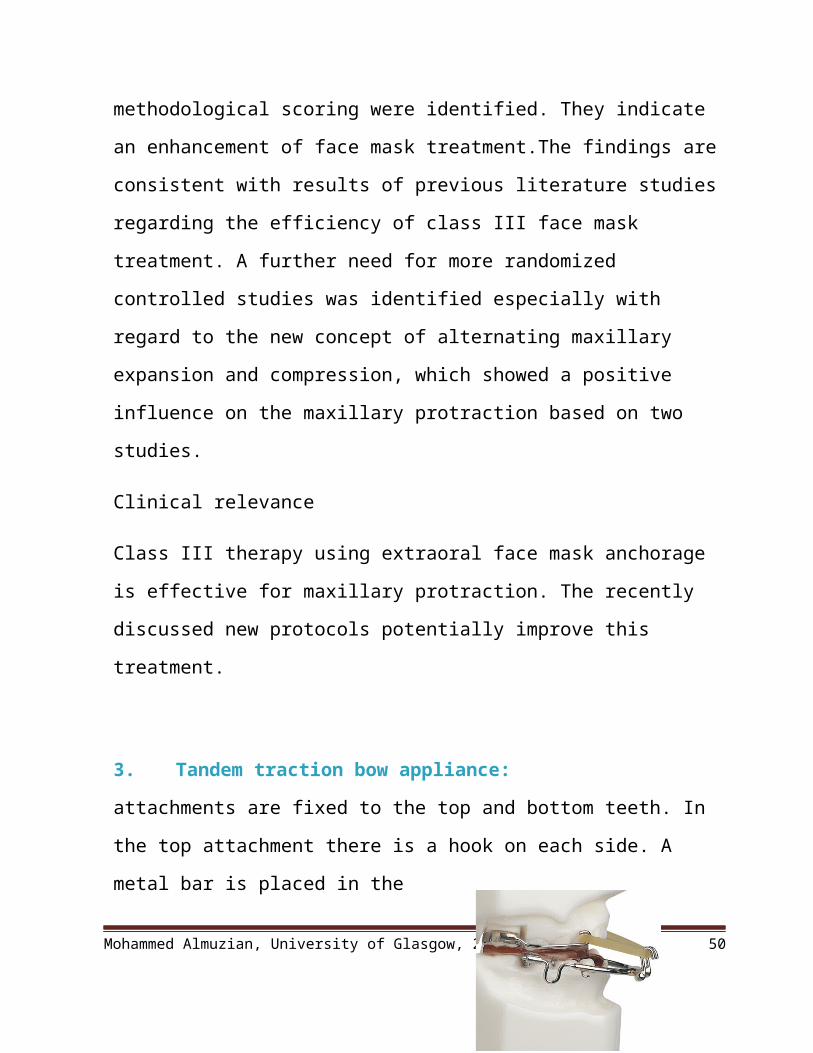
3. Tandem traction bow appliance:
attachments are fixed to the top and bottom teeth. In the top attachment there is a
hook on each side. A metal bar is placed in the lower attachment, which sits in
front of the lower teeth. An elastic band can then be placed on each side to pull
the top jaw forward and bottom jaw backwards, to correct the prominent lower
teeth
4. Chin caps
The idea of this appliance is that because the condyle is a growth site, the
growth impeded by extra-oral force (Graber, 1977).
Mohammed Almuzian, University of Glasgow, 2013 35
Despite success in animal experiments, most human studies have found little
difference in mandibular dimensions between treated and untreated subjects
(Sugawara et al, 1990).
Chincup appliances greatly improve the skeletal profile in the short term
such changes are however rarely maintained during the pubertal growth spurt
Force 500g per side 12-14 h/day for 4-5 years. Once the anterior crossbite
was corrected, the patient was instructed to wear the chin cup at least 10 hours per
day until slight Class II canine and molar relationships were established.
The best age is before canine and premolar erupt (CS2-CS3 maturity) this is
the first growth spurt of mandible, the second one when 7 and 8 erupt CS4-CS6
(Bacceti, 2005).
Types: occipital pull, used for patients with mandibular prognathsim or
vertical pull, used for patients with increased anterior face height
Best patient for Chin cup therapy
Ko et al (2004)
1.Mild Skeletal III, ability to achieve edge to edge incisors
2.Short vertical facial height (.Chincup cause clockwise rotation of the mandible.
3.Proclined or upright LLS (Chincup cause lingual tipping of the lower incisors
(Thilander 1963)
4.Absence of severe facial and dental asymmetry
Mohammed Almuzian, University of Glasgow, 2013 36

The effects of chincup therapy
(Thilander 1963)
Retardation of mandibular growth. Effective at reducing mandibular
prognathism before puberty but this is then lost with continual growth, Sugawara
et al., 1990
Remodelling of the condyle and glenoid fossa
Backward rotation of the mandible
Closure of the gonial angle
Result in lingual tipping of LLS,
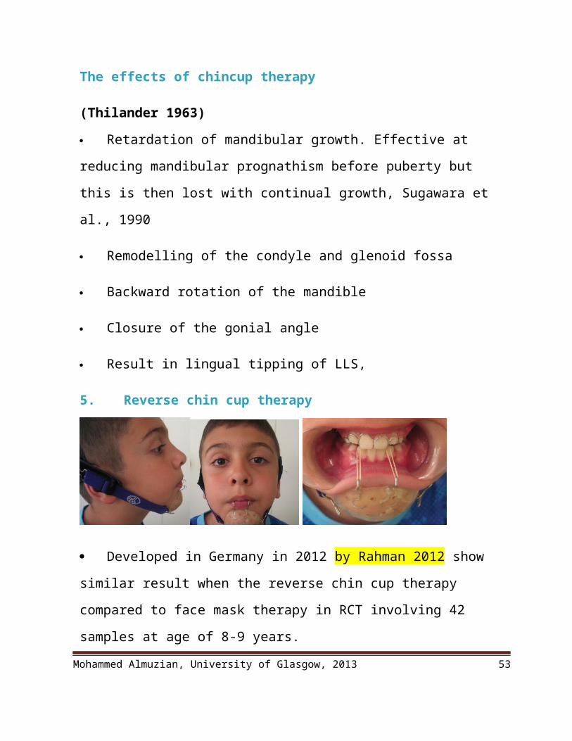
5. Reverse chin cup therapy
Developed in Germany in 2012 by Rahman 2012 show similar result when
the reverse chin cup therapy compared to face mask therapy in RCT involving 42
samples at age of 8-9 years.
Reverse chin cup therapy is able to produce forward movement of the
maxilla in the growing child associated with lingual tipping of the lower incisors
and labial tipping of the uppers.
Mohammed Almuzian, University of Glasgow, 2013 37

The point of application of protraction elastics from the upper removable
appliances was similar for both groups. All patients received the same protraction
force of 500 g per side with a 30 degree downwards pull.
The proposed advantages of the new reverse chin cup design were that it
was smaller and less bulky than other protraction appliances, therefore
encouraging children to wear it.
6. Bone anchored orthopaedic appliance (Bollard miniplates, PFH
supported with miniplates)
Plate comes in different size and form. It should
be adapted to the bone surface and fixed with 2.5mm
width and 5mm length screw.
Heavy Class 3 elastic used
Age of 9-13
Force about 150gm 24h per day. Loading start 3 weeks after plate insertion.
The major problem with this technique is the low rigidity of bone for young
patient which affect stability of bone plate and the presence of teeth follicle which
might cause problem with implant insertion. Also plate removal is problematic
bec it needs surgery and sometime the bone grow over the screw.
Success rate 92% with 3mm improvement of maxilla position and zygoma.
(Nguyen 2011) (De Clerck 2011)
This approach has two advantages:
Mohammed Almuzian, University of Glasgow, 2013 38

1. it is clearly more effective than a facemask to a maxillary splint and also
appears to produce more skeletal change than has been reported with
facemasks to anterior miniplates
2. wearing an Extraoral appliance is not necessary and nearly full-time
application of the force can be obtained.
7. Shapiro and Kokich 1984 used the same idea by inducing artificial
ankylosis and use the ankylosed teeth as anchor.
8. Functional appliances
Reverse twin block
a.The design included
Cantilever springs behind the upper incisors,
A midline expansion screw,
A lower labial bow
Intersecting blocks at 70 degrees with a vertical height of 7 mm.
Block on U4s,5s and L5s,6s
b.The patient was instructed to wear the appliance on a full-time basis initially,
activating the midline expansion screw twice a week.
c.Effects:
There is no sustained effect on growth of either the maxilla or mandible.
Reverse TB for mandibular prognatisim (Ucem 2004 claim that it produce dental
effect only) Although there is a reduction in SNB and an increase in ANB due to
Mohammed Almuzian, University of Glasgow, 2013 39
the backwards and downwards rotation of the mandible and an increase in lower
face height. Therefore, this type of treatment is inappropriate for high angle cases
with an already increased FMPA.
Seehra 2012, compare the effectiveness of Reverse Twin-Block therapy
(RTB) and protraction face mask treatment (PFM) with respect to an untreated
control in the correction of developing Class III malocclusion. Both appliances
are capable of correction of Class III dental relationships; however, the relative
skeletal and dental contributions differ. Skeletal effects, chiefly anterior maxillary
translation, predominated with PFM therapy. The RTB appliance induced Class
III correction,
Primarily as a result of dentoalveolar effects and by clockwise rotation of the
mandible.
The FR 3
They are designed to rotate the mandible downward and backward, and to
guide the eruption of the teeth so that the upper posterior teeth erupt down and
forward whilst eruption of the lower teeth is restrained. This rotates the occlusal
plane in the direction that favours correction of a class III molar relationship.
These appliances also tip the mandibular incisors lingually and the maxillary
incisors labially, introducing an element of dental camouflage for the skeletal
discrepancy.
In theory, the lip pads stretch the periosteum in a way that stimulates
forward growth of the maxilla. .
Mohammed Almuzian, University of Glasgow, 2013 40

Camouflage (dental compensation for mild cases)
Indications
1. Growth
Patient past peak growth
Non-progressive worsening of the Class III.
2. Skeletal
Class I or mild class III skeletal base relationship;
Average or reduced lower face height;
3. Dental
Average or increased overbite;
Minimal reverse OJ or edge-edge relationship
Proclined lower incisors;
Upright or retroclined upper incisors;
Molar relationship less than half unit Cl Ill
4. Soft tissues features
1. Patient not concern about the profile
2. Favourable soft tissue features
5. Displacement
Anterior displacement on closing from RCP into ICP.
Mohammed Almuzian, University of Glasgow, 2013 41

Techniques of camoflagable treatments
1. Non extraction
Expansion in upper arch to relieve crowding, eliminate crossbites and
mandibular displacements
Procline upper incisors, retrocline lower incisors (it is unwise to procline the
upper incisors beyond 120 degrees to the maxillary plane or retrocline the lower
incisors beyond 80 degrees to the mandibular plane.)
2. Extraction:
Aims of extraction
To relieve crowding or ML,
Correct incisor inclination
Correction of class III
To achieving a positive overjet
To achieving a positive overbite
To constrict the lower arch in order to correct any transverse problems
Options of extraction:
Extraction upper 5`s to maintain U lip support+ lower 4`s to allow LLS
retroclination.
Extraction of 4x4.
Mohammed Almuzian, University of Glasgow, 2013 42

Extraction of a single lower incisor: If the upper arch is well-aligned but
space is required to align and retrocline the lower incisors, extraction of a single
lower incisor can be an option (Zachrisson 1999) but it may leave some black
triangle and gingival recession. This decision depend on the presence of (large IC
distance, minor crowding, TSD in LLS, square shape L incisors not triangular). A
better approach to camouflage in patients of European descent with a moderately
severe Class III problem is extraction of one lower incisor, which prevents major
retraction of the lower teeth, while the maxillary incisors are moved facially with
some tipping allowed. The combination of upright mandibular incisors and
proclined maxillary incisors often leads to good dental occlusion rather than the
expected tooth-size problem, but a wax setup always should be done when one
lower incisor extraction is considered to verify the probably occlusal outcome.
Proffit 2013, For Asian (or rarely, other) Class III patients with major
protrusion of the lower incisors, using skeletal anchorage to move the whole
lower arch posteriorly can be quite helpful in correcting the problem. Extraction
of third molars usually is needed in order to move the mandibular dental arch
back. If second molars are extracted to facilitate distal movement, third molars
may erupt as satisfactory replacements, but this is not as likely as in the maxillary
arch and therefore is not recommended as a routine procedure.
Bracket setup
To get further proclination of ULS, use MBT in the ULS
Lingual crown torque on LLS
Contra-lateral canine brackets (to avoid LLS proclination)
Mohammed Almuzian, University of Glasgow, 2013 43

Mechanics
Lacebacks in LA (to avoid LLS proclination)
Cinch back in LA (to avoid LLS proclination)
Banding 7`s to increase posterior anchorage to retract lower dentition
Closing space on a round wire in the lower arch will facilitate
retroclination of the lower incisors.
CIII elastics (better to use short class III elastic to avoid posterior teeth
overeruption)
Avoid distal headgear forces on maxilla in C3 patients
NB: do not extract in lower arch if surgery is anticipated
Transverse problem can be addresses by:
1. URA
2. Q helix
3. RME
4. If more than 8mm, Surgically assisted RME
5. Constriction of the LA
6. AW expansion of the UA
7. Auxillary AW in the UA
Mohammed Almuzian, University of Glasgow, 2013 44

Orthognathic surgery options
The types of surgery most frequently used are the following.
1. Sagittal split ramus osteotomy (SSRO) or bilateral sagittal split osteotomy
(BSSO) to set the mandible backward
2. Intraoral vertical ramus osteotomy (IVRO) or vertical subsigmoid osteotomy
(VSSO) or vertical or oblique subcondylar osteotomy (VSO) is different names
for the same technique using an intraoral approach. This type of surgery is used to
reduce the size of the mandible (Cheung 2002). Contraindicated in predisposed
toward developing obstructive sleep apnea syndrome (OSAS) (Turnbull 2000;
Chen 2005).
3. Mandibular step osteotomy (MSO). This is a surgical technique on the
mandible that is performed in the anterior region of the mental foramen. It is
indicated for correcting the size of the mandible by using the space resulting from
the extraction of a posterior tooth or for closing spaces caused by lost posterior
teeth. MSO enables vertical and transversal modifications to the dental arches:
closure of an anterior open bite and correction of the reverse curve of Spee. This
type of osteotomy presents stepwise sectioning, which allows the bone segments
to be brought together as much as possible, thereby ensuring their stability. The
fixation is accomplished with a miniplates on each side (Cheung 2002).
Mohammed Almuzian, University of Glasgow, 2013 45
4. Surgically assisted rapid palatal expansion (SARPE) to correct the combined
transverse problems
5. Le Fort I (total maxillary osteotomy), the combination of Le Fort I and Le
Fort III, or Le Fort II in one operation or different operations.
Sugaya 2012 Cochrane review two randomized controlled trials were included in
this review. There are different types of surgery for this type of malocclusion but
only trials of mandible reduction surgery were identified. One trial compared
intraoral vertical ramus osteotomy (IVRO) with sagittal split ramus osteotomy
(SSRO) and the other trial compared vertical ramus osteotomy (VRO) with and
without osteosynthesis. Neither trial found any difference between the two
treatments. The trials did not provide adequate data for assessing effectiveness of
the techniques described
Complications of the mandibular ramus surgery
1. Fractures of the osteotomised segments,
2. Incomplete sectioning (Van Merkesteyn 1987),
3. Infection, necroses, persistent paresthesia,
4. Reduced mouth opening, nausea, airway disturbance (Yamada 2008),
5. Reduced mandibular movement range (Yazdani 2010).
6. Trauma to the inferior alveolar nerve,
Mohammed Almuzian, University of Glasgow, 2013 46
What Factors need to be taken into Account When Planning a surgical
treatment for class III cases
1. Planning the type of surgery
The required surgery is planned around the aetiology of the skeletal discrepancy
taking into account facial aesthetics, stability of the result, TMJ and airway, little
morbidity. Allows the decision to make regarding whether the maxilla is to be
advanced or the mandible set back, or a combination of these.
2. The Pre-Surgical Orthodontics in Class III?
The pre-surgical orthodontics is planned around the surgery required to achieve
optimal aesthetics with the best achievable occlusion. Three important points
need to be considered;
1. Expansion: Assessment of arch co-ordination using the pre-treatment models
in a class I position will identify the extent of any required expansion of the
maxillary arch. If minimal expansion is required, this can be achieved using the
orthodontic archwires during pre-surgical orthodontics.
2. Reverse Target overjet: The planned surgical moves for optimal aesthetics
dictate the reverse overjet required pre-surgically.
3. Inclination of the ULS which is determined by the degree of maxillary
impaction while the inclination of LLS would be determined by the amount of
autorotation.
What Are the Aims of the Pre-Surgical Orthodontics?
1. Alignment
2. Levelling and alignment of the arches.
Mohammed Almuzian, University of Glasgow, 2013 47

3. Arch co-ordination.
4. Decompensation: In this case, decompensation of the upper and lower
arches was required to produce an appropriate reverse overjet pre-surgically and
allow the desired surgical movements to be carried out to promote the desired
facial change.
5. Maintenance of the centre line with the mid-point of the chin in Lower teeth
and philtrum in the upper teeth.
Borderline Camouflage/ Orthognathic Surgery PatientsThe decision will depend on
1. Growth where there is any doubt about further skeletal growth (principally
mandibular), orthodontic camouflage should be deferred, possibly until the
remaining skeletal growth has been expressed. In class III cases with a significant
skeletal component, the mandible will tend to grow more and later than in class I
individuals (Baccetti et al, 2007).
2. Any concerns about facial appearance.
3. Medical and family history
4. Severity of the underlying skeletal problem
5. Presence or absence of functional displacement
6. Degree dentoalveolar compensation
7. Amount of crowding, OJ, OB
Mohammed Almuzian, University of Glasgow, 2013 48

8. Vertical height
9. Cephalometric Yardsticks
A. Kerr et al 1992 in Glasgow showed that surgery is indicated for patients with
ANB <-4°
Maxillary mandibular ratio = 0.84
Lower incisor inclination (LI/MP <= 83°)
Soft tissue profile Holdaway angle > = 3.5° (Holdaway angle means soft
tissue nasion-soft tissue pogonion labrale superius)
Interestingly, vertical dimension had little influence on treatment decision.
B. Stellzig-Eisenhauer et al (2002) surgery indicated when Wits analysis value
of –12.2 ± 4.3 mm or more while camouflage indicated when Wits value is -4.6 ±
1.7 mm or less.
Summary of the evidences BSI 1982 defined class III incisor relationship as ‘’the lower incisal edge lies
anterior to the cingulum plateau of the upper incisors, British Standards Institute,
1983
3% UK (Foster & Day, 1974)
Anterior crossbite in 10% of children (1993 Child Dental Health Survey)
Lin (2007) divides class 3 malocclusion into three categories
Guyer, Ellis, Behrents and McNamara (1986) 55% of class 3 malocclusions had
maxillary deficiency as one of the components of the malocclusion. Mandibular
Mohammed Almuzian, University of Glasgow, 2013 49
prognathism in 45% of cases.
Guyer, Ellis, Behrents and McNamara (1986), 59% of class 3 malocclusions had
reduced or neutral lower facial heights and that 41% had increased lower facial
heights.
The maxillary skeletal base widths were (statistically) significantly smaller in the
class 3 than in the class 1group. (Chen et al 2008)
Skeletal asymmetries, particularly in conjunction with mandibular prognathism,
are also relatively common in class III malocclusions (Severt and Proffit, 1997).
Growth status assessment for class III patients
1.Mandibular skeletal maturity can be assessed by means of a series of biologic
indicators:
2.Increase in body height (Nanda, 1955; Hunter, 1966)
3.Skeletal maturation of the hand and wrist (Bjork, 1967)
4.Dental development and eruption (Bjork, 1967)
5.Menarche, breast, and voice changes (Tanner 1962)
6.Cervical vertebral maturation (CVM) method. Franchi 2000, Beccteti 2002 & 2005
(please read the summary about ‘’The Cervical Vertebral Maturation’’)
Growth Treatment Response Vector (GTRV) analysis, Ngan (2005) has described
this as a method of determining whether a class 3 malocclusion can be treated by
camouflage or if surgical treatment will be required at a later date.
Treatment options for class III malocclusion, McIntyre 2004
Procline maxillary permanent incisor(s) using an upper removable appliance
(URA) or a fixed appliance (4 x 2 appliance which is well tolerated less
dependent on compliance (Sandler, 2001) Offer three-dimensional control
Reasons for early treatment of class 3 malocclusions, Hägg et al (2004) and Ngan
(2005) cite the reasons for early treatment
Mohammed Almuzian, University of Glasgow, 2013 50
The technique of maxillary protraction is based on work by Nanda (1978), with
rhesus monkeys in which he showed that a force of approximately 500g could
produce anterior displacement of the maxilla
Timing, Dental age:
1.McNamara (1987) suggested that the optimal time for treatment is in the early late
mixed dentition, coincident with the eruption of the upper permanent incisors.
2.Skeletal age: Baccetti et al (1998) showed that the early treatment group CVM2
showed effective forward displacement of the maxillary structures whereas the
late treatment group CVM3 showed no change compared with controls
3.Chronological age: Other investigators have suggested that for optimal orthopaedic
effects, treatment should be initiated before the patient is 9 years old ( Proffit,
2000).
4.Kim et al., 1999, meta-analysis of effectiveness of protraction HG concluded it was
less effective on patients >10yrs
Maxillary skeletal protraction, with up to 3mm of forward movement of the
maxilla possible , Mandal 2010 and 2012, showed that these effect are stable after
3 years follow-up
McNamara (1987) has described the use of a Biocryl and wire splint
Some recommend using an intraoral bone plate to support the PHG force.
Systematic review to compare the dentally anchored face mask with skeletally
anchored one by Major (2012) in Canada, he found Approximately 3 mm of
horizontal A-point movement is predictably attainable with the skeletal one in
comparison to dental one..
Many clinicians use protraction with a facemask following or simultaneously with
palatal expansion, because some evidence suggests that the expansion makes
antero-posterior skeletal change more likely. Kim et al (1999)
Mohammed Almuzian, University of Glasgow, 2013 51

There is other evidence that the expansion is optional and should be dictated by the
maxillary arch width related to the lower arch width, Vaughan 2005.
Ishii et al (1987) describe the effects of providing the protraction force from the
first molars or the premolar region. Protraction from the first molars results in
more anterior movement and a forward and upward rotation of the maxilla;
protraction from the premolars results in less forward movement but less
tendency to upward and forward rotation.
Chin caps, The idea of this appliance is that because the condyle is a growth site,
the growth impeded by extra-oral force (Graber, 1977).
Despite success in animal experiments, most human studies have found little
difference in mandibular dimensions between treated and untreated subjects
(Sugawara et al, 1990).
The effects of chincup therapy , (Thilander 1963)
Reverse chin cup therapy, Developed in Germany in 2012 by Rahman 2012 show
similar result when the reverse chin cup therapy compared to face mask therapy
in RCT involving 42 samples at age of 8-9 years.
Bone anchored orthopaedic appliance (Bollard miniplates, PFH supported with
miniplates) , Success rate 92% with 3mm improvement of maxilla position and
zygoma. (Nguyen 2011) (De Clerck 2011)
Shapiro and Kokich 1984 used the same idea by inducing artificial ankylosis and
use the ankylosed teeth as anchor.
Reverse twin block, There is no sustained effect on growth of either the maxilla or
mandible. Reverse TB for mandibular prognatisim (Ucem 2004 claim that it
produce dental effect only)
Extraction of a single lower incisor: If the upper arch is well-aligned but space is
required to align and retrocline the lower incisors, extraction of a single lower
Mohammed Almuzian, University of Glasgow, 2013 52
incisor can be an option (Zachrisson 1999) but it may leave some black triangle
and gingival recession. This decision depend on the presence of (large IC
distance, minor crowding, TSD in LLS, square shape L incisors not triangular).
Sugaya 2012 Cochrane review Two randomized controlled trials were included in
this review. There are different types of surgery for this type of malocclusion but
only trials of mandible reduction surgery were identified. One trial compared
intraoral vertical ramus osteotomy (IVRO) with sagittal split ramus osteotomy
(SSRO) and the other trial compared vertical ramus osteotomy (VRO) with and
without osteosynthesis. Neither trial found any difference between the two
treatments. The trials did not provide adequate data for assessing effectiveness of
the techniques described
Cephalometric Yardsticks, Kerr et al 1992 , Stellzig-Eisenhauer et al (2002)
Mohammed Almuzian, University of Glasgow, 2013 53