chlamydia-induced curvature of the host-cell plasma ... · chlamydia-induced curvature of the...
TRANSCRIPT

Chlamydia-induced curvature of the host-cell plasmamembrane is required for infectionSebastian Hänscha,b,1, Dominik Sponaa,1, Gido Murraa, Karl Köhrerc, Agathe Subtild, Ana Rita Furtadod,Stephan F. Lichtenthalere, Bastian Disliche,f, Katja Möllekena,2
, and Johannes H. Hegemanna,2,3
aInstitute for Functional Microbial Genomics, Heinrich-Heine-Universität Düsseldorf, Düsseldorf, Germany; bCenter of Advanced Imaging (CAI), Heinrich-Heine-Universität Düsseldorf, Düsseldorf, Germany; cBiologisch-Medizinisches Forschungszentrum (BMFZ), Genomics & Transcriptomics Laboratory (GTL),Heinrich-Heine-Universität Düsseldorf, Düsseldorf, Germany; dUnité de Biologie Cellulaire de l’Infection Microbienne, CNRS UMR3691, Institute Pasteur,Paris, France; eGerman Center for Neurodegenerative Diseases (DZNE), Helmholtz Association, 81377 München, Germany; and fInstitut für Pathologie,Universität Bern, Bern, Switzerland
Edited by Ralph R. Isberg, Tufts University School of Medicine, Boston, MA, and approved December 24, 2019 (received for review July 8, 2019)
During invasion of host cells, Chlamydia pneumoniae secretes theeffector protein CPn0678, which facilitates internalization of thepathogen by remodeling the target cell’s plasma membrane andrecruiting sorting nexin 9 (SNX9), a central multifunctional endo-cytic scaffold protein. We show here that the strongly amphipathicN-terminal helix of CPn0678 mediates binding to phospholipids inboth the plasma membrane and synthetic membranes, and is suf-ficient to induce extensive membrane tubulations. CPn0678 inter-acts via its conserved C-terminal polyproline sequence with theSrc homology 3 domain of SNX9. Thus, SNX9 is found at bacte-rial entry sites, where C. pneumoniae is internalized via EGFR-mediated endocytosis. Moreover, depletion of human SNX9 signif-icantly reduces internalization, whereas ectopic overexpression ofCPn0678–GFP results in a dominant-negative effect on endocytoticprocesses in general, leading to the uptake of fewer chlamyd-ial elementary bodies and diminished turnover of EGFR. Thus,CPn0678 is an early effector involved in regulating the endocytosisof C. pneumoniae in an EGFR- and SNX9-dependent manner.
effector protein | membrane modulation | lipid binding | endocytosis
All members of the Chlamydiaceae are obligate intracellularpathogens of humans and animals, and cause a variety of
diseases depending on the tissues they target (1). The two speciesthat affect humans are Chlamydia trachomatis and Chlamydiapneumoniae, which are the causative agents of severe urogenitalconditions, including pelvic inflammation and ectopic pregnan-cies, and respiratory disorders such as pneumonia and bronchitis,respectively (2, 3). C. pneumoniae infections are also associatedwith several chronic diseases, including asthma, Alzheimer’sdisease, multiple sclerosis, and even lung cancer (4–7).The most critical step in the life cycle of an obligate in-
tracellular bacterium is internalization into the host cell. Themost common entry strategies are 1) the “zipper” and 2) the“trigger” mechanisms. In the former, a bacterial adhesin/invasininteracts with a surface receptor, thereby activating its down-stream signaling machinery and effectively hijacking receptorendocytosis for bacterial internalization. In the latter, an initialand rather weak interaction between pathogen and host is rap-idly followed by translocation of bacterial proteins, called ef-fectors, into the host cytoplasm. These effectors modulate thehost cytoskeleton and induce extensive ruffling of the plasmamembrane (PM) to facilitate pathogen entry (8). Both mecha-nisms involve the use of bacterial proteins to manipulate essen-tial components of the endocytic machinery, such as thephosphoinositide-converting enzymes that regulate the lipidcomposition (and hence the curvature) of the PM (9, 10),adaptors and regulators like sorting nexin 9 (SNX9) that controlendocytosis and vesicle trafficking (11, 12), and finally actinpolymerization, which facilitates bacterial uptake (13, 14). In-terestingly, SNX9 harbors a membrane-curvature–sensing bin-amphiphysin-rvs (BAR) domain and binds preferentially tomembranes of high curvature (15). Using an in vitro system it has
been proposed that binding to PI(4,5)P2, the early endosomemarker PI(3)P, and domains of high membrane curvature re-cruits SNX9 in order to trigger the actin machinery and completeendocytosis (16).Internalization is preceded by stable adhesion to the host cell,
which induces intracellular signaling and recruitment of endocytosis-related proteins. Recently, host receptors like the ephrin receptor(EPHA2) or EGFR have been shown to promote adhesion of C.trachomatis, but the chlamydial interaction partner remains un-defined, as does the mechanism of entry (17, 18). ForC. pneumoniaewe have shown that the pathogen uses one of its highly diversepolymorphic membrane proteins, Pmp21, to bind and activate theEGFR (19). EGFR activation triggers the PI3 kinase, which in turnrecruits specific endocytic adaptor proteins to facilitate the EGFR-mediated endocytosis of C. pneumoniae (20). In addition to thiszipper mechanism, C. pneumoniae employs the trigger approach toenter host cells. Simultaneously with the Pmp21–EGFR interaction,C. pneumoniae secretes its TarP ortholog CPn0572 via a type IIIsecretion (T3S) system. CPn0572 then binds and polymerizes actinto enforce bacterial uptake into actin-rich structures (21).To determine whether C. pneumoniae employs other mecha-
nisms to achieve efficient internalization, we searched for newearly effector proteins involved in these processes. Here we showthat the effector CPn0678 is also a T3S substrate, and localizes tothe PM at bacterial entry sites. CPn0678 can bind phospholipids
Significance
We describe a mechanism by which the obligate intracellularpathogen Chlamydia pneumoniae induces curvature of thehost-cell plasma membrane and recruits a central componentof the endocytotic machinery. We demonstrate that a typeIII-secreted C. pneumoniae effector protein named CPn0678binds via its N-terminal amphipathic helix to negativelycharged phospholipids in the inner leaflet of the host plasmamembrane at the site of entry, and induces membrane curva-ture. Its proline-rich region then recruits SNX9 (sorting nexin 9),a key regulator of endocytosis, and the complex facilitatesuptake of C. pneumoniae into host cells.
Author contributions: K.K., K.M., and J.H.H. designed research; S.H., D.S., G.M., A.S., A.R.F.,and K.M. performed research; K.K., S.F.L., and B.D. contributed new reagents/analytic tools;S.H., D.S., G.M., A.S., A.R.F., and K.M. analyzed data; and K.M. and J.H.H. wrote the paper.
The authors declare no competing interest.
This article is a PNAS Direct Submission.
This open access article is distributed under Creative Commons Attribution-NonCommercial-NoDerivatives License 4.0 (CC BY-NC-ND).1S.H. and D.S. contributed equally to this work.2K.M. and J.H.H. contributed equally to this work.3To whom correspondence may be addressed. Email: [email protected].
This article contains supporting information online at https://www.pnas.org/lookup/suppl/doi:10.1073/pnas.1911528117/-/DCSupplemental.
www.pnas.org/cgi/doi/10.1073/pnas.1911528117 PNAS Latest Articles | 1 of 11
MICRO
BIOLO
GY
Dow
nloa
ded
by g
uest
on
July
31,
202
0

in both natural and synthetic membranes, and upon expression inhuman cells it generates membrane tubulations, and interactswith the host protein SNX9, a multifunctional protein involvedin clathrin-mediated endocytosis, membrane remodeling, andactin dynamics (12). Our data suggest that, during host-cell entry,secreted CPn0678 binds to and curves the PM, which recruitsSNX9 to the late stages of endocytosis. These findings show thatCPn0678-induced membrane curvature plays a central role in theuptake of C. pneumoniae.
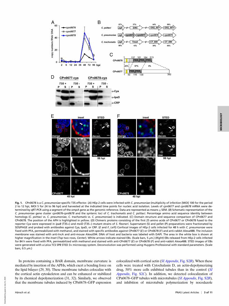
ResultCPn0678: Prototype of a T3S Chlamydial Effector Protein. A commonfeature of both chlamydial adhesins and early effectors is that theyare expressed late in the preceding infection, and displayed on theelementary body (EB) or stored within it for early secretion.Therefore, we performed a genome-wide transcriptional analysisto screen for such genes and found 88 genes to be significantly up-regulated (SI Appendix, Table S1). This set included genes forpreviously described adhesins, components, and potential sub-strates of the T3S system, and hypothetical genes. Among thelatter class, we identified a highly up-regulated cluster of threegenes comprising cpn0676, cpn0677, and cpn0678. RT-PCR con-firmed that their transcript levels increased significantly from 36 honward, with cpn0677 and cpn0678 RNAs becoming more abun-dant than cpn0676 (Fig. 1A).Comparison of the C. pneumoniae cpn0676-cpn0678 locus with
other chlamydial genomes showed that the genes are part of asyntenic locus. CPn0676 is conserved among all Chlamydia spe-cies with approximately 39 to 44% identity, while comparison ofCPn0677 and CPn0678 revealed a more complex pattern. Allother species have only one gene at this locus, while C. pneu-moniae carries two genes, cpn0677 and cpn0678 (Fig. 1B).CPn0678 itself harbors three conserved proline-rich repeats(PRR), two of which are conserved within the N-terminal seg-ment of CPn0677 and are 67% identical to each other (Fig. 1C).In-depth comparisons with other chlamydial species revealedthat these two proteins are C. pneumoniae-specific, displaying noor only low homology to other chlamydial proteomes. Thus, wegenerated a phylogenetic tree showing the relationships acrossthe chlamydial kingdom by comparing full-length CPn0678 withthe syntenic proteins of various species (SI Appendix, Fig. S1A).The most distantly related proteins, with less than 5% identity toCPn0678, are found in C. trachomatis, Chlamydia muridarum,and Chlamydia suis (Fig. 1B and SI Appendix, Fig. S1A). One ofthese is C. trachomatis TmeA (CT_694), a known T3 effectorprotein, which interacts with the host protein AHNAK and isinvolved in early steps in the C. trachomatis infection (22, 23).Although the three species are closely related, C. trachomatisTmeA shares only 50 to 55% identity with its homologs in C. suisand C. muridarum, possibly suggesting differences in theirfunctions. The syntenic proteins from Chlamydia abortus, Chla-mydia felis, Chlamydia psittaci, and Chlamydia caviae share higherlevels of identity with CPn0678 (SI Appendix, Fig. S1A), rangingbetween 34% and 40%. This is because the PRR1 and PRR2 ofCPn0678 are conserved among the homologous proteins in thesefour species (SI Appendix, Fig. S1A). Among them is SinC(G5Q_0070), an effector expressed by C. psittaci, which asso-ciates with the nuclear membrane during late stages of infection(24). These findings clearly demonstrate that this genomic locusencodes effector proteins with diverse functions in differentchlamydial species.To confirm that CPn0677 and CPn0678 are T3S effector
proteins, we used a heterologous Shigella flexneri T3S assay (25).The first 25 N-terminal amino acids of each protein were fusedto the calmodulin-dependent adenylate cyclase (Cya) reporterprotein. These constructs were then separately expressed in a S.flexneri ipaB (constitutive T3S) or mxiD (deficient in T3S)-nullstrain (26, 27). Fractionation experiments confirmed that both
proteins contained a T3S-competent signal sequence, as the re-porter constructs were detectable in the supernatants, as was thepositive control IpaD, whereas the intracellular protein CRP wasretained in the pellet (Fig. 1D). In addition, confocal microscopyof inclusions labeled with specific antibodies at 48 h post-infection (hpi) revealed that both CPn0677 (Fig. 1E and SI Ap-pendix, Fig. S1C) and CPn0678 (Fig. 1F and SI Appendix, Fig.S1C) were associated with the condensed DAPI staining char-acteristic of EBs and not with the nuclear envelope, as shown forSinC (24). In agreement with our transcriptional studies, the twoproteins were detected from 48 to 84 hpi in lysates of infectedcells (SI Appendix, Fig. S1B). Deconvolution of stimulatedemission depletion microscopy (STED) images of EBs releasedfrom bursting inclusions at 86 hpi showed a ring-like structure forboth proteins (Fig. 1 E and F). The protein signals in these ringsdo not look continuous, but seem to be enriched in certain areas,which perhaps indicates the clustered presence of the T3S systempreloaded with the effector proteins. Taken together, thesefindings indicate that both proteins are T3S effectors, which aresecreted in the early phase of infection.
CPn0678 Is a Lipid-Binding, Membrane-Tubulating Effector Protein.Based on our sequence comparisons and initial localizationstudies, we concluded that CPn0678 could be the prototype forthe C. pneumoniae effector proteins CPn0678 and CPn0677encoded by the syntenic locus, and in the following we describeour efforts to uncover the function of CPn0678. As both TmeAand SinC interact with host membranes (24, 28), we searched forpotential membrane-interacting domains and found an N-terminalregion (amino acids 47 to 64) with a strongly amphipathic char-acter. Secondary-structure predictions revealed that this regionalso carries an α-helix, so we refer to this sequence as theamphipathic helix (APH). Projected into a helical wheel, theamphipathic amino acids of the APH all face to one side of thehelix (Fig. 2A). The APH is conserved, albeit weakly, in CPn0677(amino acids 48 to 65) (Fig. 1C). To understand the function ofthe APH, we first studied the localization of CPn678–GFP ec-topically expressed in living HEp-2 cells treated with the PM dyeCellMask (Fig. 2A). Imaging revealed that CPn678–GFP localizesto the PM and induces a strong tubulation phenotype, withCPn678-lined tubules emanating from the cell membrane into thecytosol (Fig. 2A). Next, we introduced amino acid exchanges intothe APH in the full-length protein to elucidate whether thepredicted α-helix or the amphipathic amino acids are function-ally involved in the observed membrane tubulation (Fig. 2 B andC). In MutA the amino acid exchanges result in an APH with a10-fold reduction in hydrophobicity, while the predicted α-helixis retained (Fig. 2B); in the converse mutant MutB the α-helix isdestroyed while the hydrophobicity is increased (Fig. 2C). Ec-topic expression and live imaging in the presence of CellMaskrevealed that both mutants have lost their tubulation phenotypesand no PM localization could be detected (Fig. 2 B and C).To verify that the N-terminal APH is responsible for the
tubulation phenotype, we generated different deletion variantsand analyzed them under the same conditions in live imaging andfor general protein expression (Fig. 2 D and E and SI Appendix,Fig. S2A). All variants harboring the APH (CPn0678, N-terminalfragment [N-term], and ΔPRR1) show both localization to thePM and a membrane-tubulating phenotype (Fig. 2D). Quantifi-cation of phenotypes revealed that cells expressing CPn0678 orΔPRR1 show an ∼50% distribution of both phenotypes, whilecells expressing the N-term variant show membrane tubules in10% of cells; otherwise we found the protein at the PM (Fig. 2E).In contrast, localization to the PM and tubule formation wereboth lost when we deleted the APH (ΔAPH) or expressed aC-terminal fragment (C-term), indicating that the APH is indeedessential for membrane binding (Fig. 2D).
2 of 11 | www.pnas.org/cgi/doi/10.1073/pnas.1911528117 Hänsch et al.
Dow
nloa
ded
by g
uest
on
July
31,
202
0
In proteins containing a BAR domain, membrane curvature ismediated by insertion of the APHs, which exert a bending force onthe lipid bilayer (29, 30). These membrane tubules colocalize withthe cortical actin cytoskeleton and can be enhanced or stabilizedby its chemical depolymerization (31, 32). Similarly, we observedthat the membrane tubules induced by CPn0678–GFP expression
colocalized with cortical actin (SI Appendix, Fig. S2B). When thesecells were treated with Cytochalasin D, an actin-depolymerizingdrug, 50% more cells exhibited tubules than in the control (SIAppendix, Fig. S2C). In addition, we detected colocalization ofCPn0678–GFP tubules with microtubules (SI Appendix, Fig. S2B),and inhibition of microtubule polymerization by nocodazole
Fig. 1. CPn0678 is a C. pneumoniae-specific T3S effector. (A) HEp-2 cells were infected with C. pneumoniae (multiplicity of infection [MOI] 100 for the period2 to 12 hpi, MOI 5 for 24 to 96 hpi) and harvested at the indicated time points for nucleic acid isolation. Levels of cpn0677 and cpn0678 mRNA were de-termined by qRT-PCR using a segment of the ompA gene as the genomic reference. Data are represented as means ± SEM. (B) Schematic representation of theC. pneumoniae gene cluster cpn0676-cpn0678 and the syntenic loci of C. trachomatis and C. psittaci. Percentage amino acid sequence identity betweenhomologs (C. psittaci vs. C. pneumoniae, C. trachomatis vs. C. pneumoniae) is indicated. (C) Domain structure and sequence comparison of CPn0677 andCPn0678. The position of the APH is highlighted in yellow. (D) Chimeric proteins consisting of the first 25 amino acids of CPn0677 or CPn0678 fused to thereporter Cya were expressed in ipaB (T3S+) and mxiD (T3S−) mutant strains of S. flexneri. Supernatant (S) and pellet (P) preparations were fractionated bySDS/PAGE and probed with antibodies against Cya, IpaD, or CRP. (E and F, Left) Confocal images of HEp-2 cells infected for 48 h with C. pneumoniae werefixed with PFA, permeabilized with methanol, and stained with specific antibodies against CPn0677 (E) or CPn0678 (F) and anti-rabbit Alexa488. The inclusionmembrane was stained with anti-IncA and anti-mouse Alexa594. DNA of host and bacteria was labeled with DAPI. The area in the white box is shown athigher magnification in the Inset (Top two rows, Center). White arrows indicate stained EBs. (Scale bars, 5 μm.) (Right) EBs released from HEp-2 cells infectedfor 84 h were fixed with PFA, permeabilized with methanol and stained with anti-CPn0677 (E) or CPn0678 (F) and anti-rabbit Alexa488. STED images of EBswere generated with a Leica TCS SP8 STED 3× microscopy system. Deconvolution was performed using Huygens Professional with standard parameters. (Scalebars, 0.5 μm.)
Hänsch et al. PNAS Latest Articles | 3 of 11
MICRO
BIOLO
GY
Dow
nloa
ded
by g
uest
on
July
31,
202
0

treatment completely suppressed tubule formation (SI Appendix,Fig. S2C). These findings indicate that cortical actin and the mi-crotubule cytoskeleton play an important role in CPn0678-mediated membrane tubulation.Using giant unilaminar vesicles (GUVs), a model system for
biological membranes, we analyzed the membrane and lipid in-teractions of CPn0678 in more detail (Fig. 3). These GUVs arebased on 69.75 mol% dipalmitoylphosphatidylcholine (DOPC)and 25 mol% cholesterol stained with Texas red (0.25 mol%).During vesicle assembly, additional selected phosphatidylinosi-tols (PIPs) are incorporated, while in phosphatidylserine (PS)-containing GUVs the lipid composition is changed to 49.75 mol%
DOPC/25 mol% cholesterol/0.25 mol% Texas red and 20 mol%PS. Quantification of confocal images revealed that FITC-labeled recombinant CPn0678 binds to all of the tested lipids,and shows the highest affinities for PS, the most abundant neg-atively charged phospholipid in the PM (33), and PIP4, a phos-pholipid found in Golgi membranes which is the precursor ofPIP4,5 in the PM (34) (Fig. 3 A and B). Deletion of the PRR1did not significantly affect binding, whereas deletion of the APHcompletely abolished lipid interaction (Fig. 3 A and B), in-dicating that the APH mediates the interaction of CPn0678 withlipids, especially those found in the inner leaflet of the PM (35).During these analyses, we observed that GUVs incubated with
Fig. 2. The N-terminal APH of CPn0678 mediates binding to the PM and triggers tubulation of the PM. (A) The region encompassing residues 40 to 70 ofCPn0678 was analyzed for hydrophobicity, hydrophobic moment and amino acid composition using HeliQuest (heliquest.ipmc.cnrs.fr) and a segment dis-playing a with strongly amphipathic character was identified (amino acids 47 to 64, highlighted in yellow), which is presented as a helical wheel (APH) withamphipathic amino acids in yellow and polar residues in blue. Secondary-structure prediction with GORIV (https://npsa-prabi.ibcp.fr/cgi-bin/npsa_automat.pl?page=/NPSA/npsa_server.html) revealed the presence of an α-helix (amino acids 47 to 58, represented by blue “h”). (B and C) Mutational analyses wereperformed using both predictions to generate a mutant with a major loss of hydrophobicity (MutA, B) and one in which the α-helical structure was disrupted(MutB, C). Fluorescence images of living cells expressing CPn0678-GFP (A), MutA (B), or MutB (C) treated with the PM dye CellMask. (Scale bars, 10 μm; 1 μm inInsets.) (D) Schematic representation of the fragments of CPn0678 which were C terminally fused with GFP and used in live-cell imaging following treatmentwith CellMask. White arrowheads show membrane tubulations emanating from the PM. (Scale bars, 10 μm.) (E) Quantification of subcellular localization ofCpn0678, N-term and ΔPRR1 fused with GFP. Data are represented as means ± SD (n = 3).
4 of 11 | www.pnas.org/cgi/doi/10.1073/pnas.1911528117 Hänsch et al.
Dow
nloa
ded
by g
uest
on
July
31,
202
0

binding-competent CPn0678 variants developed membrane tu-bules on their surfaces, especially those containing the negativelycharged PS (Fig. 3A, white arrowheads). Thus, we generatedGUVs composed of PIP4,5 and PS to more closely approximatethe phospholipid mixture on the PM, and imaged them in theabsence or presence of different FITC-labeled CPn0678 variants.All variants harboring the APH domain (CPn0678, N-term, andΔPRR1) elicited membrane tubulation on the GUVs, whileΔAPH did not bind to the GUVs, as shown previously (Fig. 3 Cand D and Movies S1–S5). Quantification of GUVs incubatedwith CPn0678, N-term, and ΔPRR1 revealed that 90 to 100% ofthem showed membrane tubulations, while only 12% of GUVs
incubated with ΔAPH showed tubulation which is close to thevalue for control GUVs (11%) (Fig. 3D). Furthermore, we ob-served that GUVs incubated with N-term showed a marked ten-dency to burst during incubation; bursting also occurred in GUVstreated with CPn0678, but with a lower frequency (Movies S1–S5),indicating that the tubulation elicited by the APH-carrying vari-ants is strong enough to disturb the integrity of the GUVs.
CPn678-Induced Membrane Tubulation Enables the Protein to Interactwith the Endocytic Scaffold Protein SNX9. Based on our previousfindings, we hypothesized that CPn0678 is an early effector se-creted via T3S. Therefore, we next analyzed the localization of
Fig. 3. CPn0678 binds to and tubulates artificial membranes. (A and B) Lipid binding assay using FITC-labeled recombinant CPn0678 variants and Texas red-stained GUVs containing the indicated lipids. (A) Confocal images of GUVs incubated with CPn0678 variants. White arrowheads indicate membrane deformations.(Scale bars, 10 μm.) (B) Quantification of microscopic analyses of protein binding expressed as the mean fluorescence of sets of 50 individual GUVs. Data arepresented as means ± SD (n = 3). (C and D) Lipid binding assay using FITC-labeled recombinant CPn0678 variants and Texas red-stained GUVs containing PS andPIP4,5. (C) Confocal images of GUVs bound by FITC-labeled recombinant CPn0678 variants. White arrowheads indicate membrane deformations leading todisruption of GUVs, as shown in Movies S1–S5. (Scale bars 5 μm.) (D) Quantification of analyses of membrane deformation in samples containing 50 individualGUVs each. Data are presented as means ± SD (n = 2). n.s., not significantly different from CPn0678; **P value of 0.01; ***P value of 0.001.
Hänsch et al. PNAS Latest Articles | 5 of 11
MICRO
BIOLO
GY
Dow
nloa
ded
by g
uest
on
July
31,
202
0
endogenous CPn0678 at 15 min postinfection, at which timeadhesion and internalization of C. pneumoniae can be observedby colocalization of EBs with the host EGF receptor (20). Asexpected, we found that CPn0678 colocalized with EGFR atEB contact sites on the PM and in endocytic invaginationscontaining internalized EBs (Fig. 4A). We quantified and vali-dated our imaging data and 40% of EGFR+ EBs showedcolocalization with CPn0678 (Fig. 4B and SI Appendix, Fig. S3 Aand B), indicating that the protein is secreted during EGFR-dependent adhesion and internalization. We then studied thefunction of CPn0678 during the entry process. As mentionedabove, CPn0678 contains three PRR domains. Such domains areknown to mediate protein–protein interactions, most commonlywith proteins carrying Src homology 3 (SH3) domains (36, 37).SH3 domains are small modules of 50 to 60 amino acids with acharacteristic 3D structure, which recognize the minimal motifPxxP. Extending the motif with additional prolines and chargedamino acids increases the specificity of the PRR for definedtarget proteins (38). Interestingly, SH3 domains can be found inmany proteins involved in endocytosis (39). Since C. pneumoniaeutilizes EGFR-mediated endocytosis (19), we asked whethertransfected CPn0678-GFP colocalized with various SH3-containingproteins. We chose proteins directly related to EGFR-mediatedendocytosis, such as the endocytic adaptor proteins Grb2 (40),c-Cbl (41), and Cin85 (42). We also selected SNX9, a key regulatorof dynamin assembly required for efficient clathrin-mediated en-docytosis and known to be involved in actin dynamics during en-docytosis (12, 43) (SI Appendix, Fig. S4 A and B). Coexpressionstudies revealed no colocalization of CPn0678 with Cin85, c-Cbl, orGrb2, but SNX9 colocalized with CPn0678 both at the PM and inthe CPn0678-induced tubules described above (SI Appendix, Fig.S4A). In cells coexpressing both proteins, we observed that SNX9relocalized to CPn0678+ tubules. Since the latter are phenotypi-cally distinct from the tubules generated upon overexpression ofSNX9 (SI Appendix, Fig. S4C) (44), this observation is indicative ofa direct interaction. Moreover, when we immunoprecipitatedCPn0678–GFP from these coexpressing cells, we only detectedmCherry–SNX9 and none of the other SH3 proteins in theeluted fraction, in agreement with our imaging data (SI Ap-pendix, Fig. S4B).We verified the SNX9–CPn0678 interaction in pulldown ex-
periments using recombinant proteins. Here, CPn0678-His wasfound to interact directly with GST–SNX9 (Fig. 4C and SI Ap-pendix, Fig. S5) in pulldowns using either His or GST resin.SNX9 contains an SH3 domain that recognizes PRRs with apositive charge at the N terminus [consensus (R/K)xφPxxP; φ =hydrophobic amino acid], as do the PRRs found in dynamin andN-WASP (43). Since this consensus sequence also occurs in thefirst PRR motif (RPAPPQP) of CPn0678, we deleted either theSH3 domain of SNX9 (ΔSH3) or the first PRR motif in CPn0678(ΔPRR), and found that both variants had lost the ability tointeract with their respective full-length partner (Fig. 4C and SIAppendix, Fig. S5), indicating that SNX9 and CPn0678 indeedinteract via their SH3 and PRR motifs respectively. The SH3domain alone is sufficient for interaction, as it is capable ofpulling down CPn0678 on its own.Finally, to test for a direct interaction between the two pro-
teins, we performed fluorescence lifetime imaging microscopy(FLIM). In vivo, the average lifetime of CPn0678–GFP wasmeasured with and without coexpression of mCherry–SNX9.Reduction of the lifetime is caused by Förster resonance energytransfer (FRET) from a donor molecule (here CPn0678–GFP)to an acceptor molecule (here mCherry–SNX9) in close prox-imity (<10 nm) and thus indicates interaction in vivo. The donorcontrol showed an average lifetime of 2.86 ns (Fig. 4D). Incontrast, the average lifetime of CPn0678–GFP was significantlyreduced (by 256 ps) to 2.60 ns, when coexpressed with mCherry–SNX9 (Fig. 4D). Similar levels of lifetime reduction have been
observed in other protein interaction studies and for fusedproteins (45–47).Taken together, these data suggest that the APH directs
CPn0678 to the PM, where it can directly interact with SNX9 viaPRR and SH3 domains. To elucidate the hierarchy of these in-teractions during recruitment of SNX9 to the secreted chla-mydial effector, we performed further in vivo and in vitro assaysin which we measured the interaction of different CPn0678variants—either capable of binding and tubulating the mem-brane (CPn0678, N-term, and ΔPRR1) or not—with SNX9 andtwo deletion variants (ΔBAR, ΔBARΔSH3) (Fig. 4 E and F andMovies S6–S15). The BAR domain of SNX9 contains classicamphipathic helices to induce membrane tubulation, althoughSNX9 is better at sensing curvature than inducing it (44, 48).While lipid binding is maintained via the PX domain, deletion ofthe BAR domain disrupts membrane tubulation, and deletion ofthe SH3 domain inhibits all protein–protein interactions (12).Upon coexpression of CPn0678 with SNX9, colocalization ofboth proteins on membrane tubules was dependent on thepresence of the SNX9 SH3 domain, but independent of the BARdomain (Fig. 4E and SI Appendix, Fig. S4E).Furthermore, CPn0678 variants lacking the PRR1 motif (i.e.,
N-term or ΔPRR1) showed no colocalization with SNX9, whereasvariants lacking the membrane localization domain, C terminus, orAPH, interacted with SNX9, although interaction was restricted tothe cytosol (SI Appendix, Fig. S4 D and E). This indicates thatCPn0678 induces the recruitment of SNX9 to membrane struc-tures formed by the chlamydial effector. During chlamydial en-docytosis this occurs at the PM, where secreted CPn0678 locallydeforms the membrane and recruits SNX9. To test our hypothe-sis that CPn0678-induced membrane curvature indeed re-cruits SNX9, we again used GUVs composed of PS and PIP4,5(Fig. 4F and Movies S6–S15). In accordance with published data,DyLight650-labeled SNX9 did not bind to these GUVs (Fig. 4Fand Movie S6); although it is capable of binding to PIP4,5 lipids(49, 50), it does so only if the membranes are already curved (16).Strikingly, when we added FITC-labeled CPn0678 to GUVs pre-incubated with SNX9 we observed immediate binding of CPn0678and generation of membrane tubules, to which SNX9 was sub-sequently recruited (Fig. 4F and Movie S7). This recruitment isdependent on the membrane-tubulating capacity of the N-terminal APH of CPn0678, as SNX9 is recruited to the GUVsurface in the presence of membrane-modulating N-term orΔPRR1 variants but not ΔAPH (Fig. 4F and Movies S8–S10).Moreover, SNX9 recruitment to GUVs is not dependent on itsown membrane-curving domain, as we observed the same patternof recruitment of SNX9ΔBAR to CPn0678, N-term, and ΔPRR1but not ΔAPH variants (SI Appendix, Fig. S6 and Movies S11–S15). Taken together, these results show that CPn0678 inducesmembrane curvature which is then sensed by SNX9.
Internalization of C. pneumoniae Depends on the Interaction betweenCPn0678 and SNX9. To demonstrate the interdependence of en-dogenous SNX9 and CPn0678 during C. pneumoniae internali-zation, we first analyzed whether SNX9 localizes to invadingEBs. Indeed, by 15 min postinfection, we were able to detect apatch-like association of SNX9 with EBs surrounded by EGFR(Fig. 5A). Quantification and validation of the imaging datarevealed that 47% of EBs that colocalize with EGFR are alsopositive for SNX9 (Fig. 5B and SI Appendix, Fig. S3C). Next, weperformed a coimmunoprecipitation assay on cells lysed 15 minafter infection, and were able to coprecipitate CPn0678 using aspecific antibody against SNX9; conversely, SNX9 was recoveredwith an antibody directed against CPn0678 (Fig. 5C). Thesefindings are further supported by direct colocalization of bothproteins with attaching and invading C. pneumoniae EBs (Fig.5D). We analyzed 400 EBs for CPn0678 expression, quantified,and validated how many of them colocalize with SNX9. Some
6 of 11 | www.pnas.org/cgi/doi/10.1073/pnas.1911528117 Hänsch et al.
Dow
nloa
ded
by g
uest
on
July
31,
202
0

Fig. 4. CPn0678 interacts with host SNX9. (A) Colocalization of CPn0678 (stained with anti-CPn0678 and anti-rabbit Alexa488) and EGFR (stained with anti-EGFR and anti-mouse Alexa594) at bacterial entry sites at 15 min postinfection. The area in the white box is shown at higher magnification in the Inset (Right).C. pneumoniae EBs were stained with DAPI. White arrowheads indicate instances of colocalization. (Scale bar, 10 μm; 1 μm in Insets.) (B) EBs associating withEGFR were quantified for additional colocalization of CPn0678 signals shown in A. Confocal images of 300 individual EBs were analyzed. Data are presentedas means ± SD (n = 3). ***P ≤ 0.001. Colocalization was validated by estimating Pearsons correlation coefficients (SI Appendix, Fig. S3 A and B). (C) Pulldownexperiments using purified recombinant GST or GST–SNX9 variants (ΔSH3, SH3) and CPn067810His or CPn0678ΔPRR110His. Input and elution samples obtainedfrom His-pulldowns and GST-pulldowns were fractionated by SDS/PAGE and probed with anti-GST and anti-His antibodies. (D) Quantification of FLIMmeasurements of average fluorescence lifetimes, used to monitor FRET from CPn0678–GFP to mCherry–SNX9. The lifetime of donor fluorescence measured in30 to 40 cells with or without coexpression of the acceptor are depicted as a box plot generated by OriginPro. Data are presented as means ± SD (n = 3). (E)Confocal images of cells coexpressing CPn0678–GFP and mCherry–SNX9 variants (SNX9, ΔBAR, ΔBARΔSH3). Insets show regions in white squares. (Scale bars10 μm; 1 μm in Insets.) (F) Confocal images of Texas red-stained GUVs containing PS and PIP4,5 preincubated for 10 min with DyLight650-labeled SNX9 andfollowed by addition of FITC-labeled CPn0678 variants. See Movies S6–S15 for movies of each sample. (Scale bars, 5 μm.)
Hänsch et al. PNAS Latest Articles | 7 of 11
MICRO
BIOLO
GY
Dow
nloa
ded
by g
uest
on
July
31,
202
0
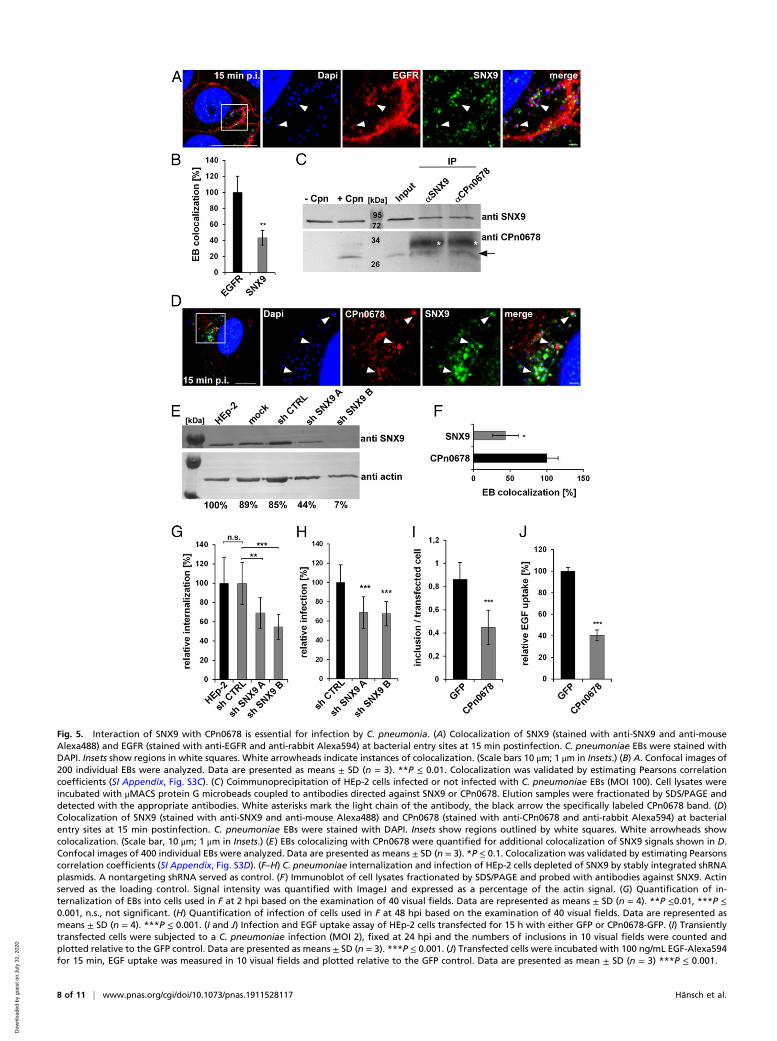
Fig. 5. Interaction of SNX9 with CPn0678 is essential for infection by C. pneumonia. (A) Colocalization of SNX9 (stained with anti-SNX9 and anti-mouseAlexa488) and EGFR (stained with anti-EGFR and anti-rabbit Alexa594) at bacterial entry sites at 15 min postinfection. C. pneumoniae EBs were stained withDAPI. Insets show regions in white squares. White arrowheads indicate instances of colocalization. (Scale bars 10 μm; 1 μm in Insets.) (B) A. Confocal images of200 individual EBs were analyzed. Data are presented as means ± SD (n = 3). **P ≤ 0.01. Colocalization was validated by estimating Pearsons correlationcoefficients (SI Appendix, Fig. S3C). (C) Coimmunoprecipitation of HEp-2 cells infected or not infected with C. pneumoniae EBs (MOI 100). Cell lysates wereincubated with μMACS protein G microbeads coupled to antibodies directed against SNX9 or CPn0678. Elution samples were fractionated by SDS/PAGE anddetected with the appropriate antibodies. White asterisks mark the light chain of the antibody, the black arrow the specifically labeled CPn0678 band. (D)Colocalization of SNX9 (stained with anti-SNX9 and anti-mouse Alexa488) and CPn0678 (stained with anti-CPn0678 and anti-rabbit Alexa594) at bacterialentry sites at 15 min postinfection. C. pneumoniae EBs were stained with DAPI. Insets show regions outlined by white squares. White arrowheads showcolocalization. (Scale bar, 10 μm; 1 μm in Insets.) (E) EBs colocalizing with CPn0678 were quantified for additional colocalization of SNX9 signals shown in D.Confocal images of 400 individual EBs were analyzed. Data are presented as means ± SD (n = 3). *P ≤ 0.1. Colocalization was validated by estimating Pearsonscorrelation coefficients (SI Appendix, Fig. S3D). (F–H) C. pneumoniae internalization and infection of HEp-2 cells depleted of SNX9 by stably integrated shRNAplasmids. A nontargeting shRNA served as control. (F) Immunoblot of cell lysates fractionated by SDS/PAGE and probed with antibodies against SNX9. Actinserved as the loading control. Signal intensity was quantified with ImageJ and expressed as a percentage of the actin signal. (G) Quantification of in-ternalization of EBs into cells used in F at 2 hpi based on the examination of 40 visual fields. Data are represented as means ± SD (n = 4). **P ≤0.01, ***P ≤0.001, n.s., not significant. (H) Quantification of infection of cells used in F at 48 hpi based on the examination of 40 visual fields. Data are represented asmeans ± SD (n = 4). ***P ≤ 0.001. (I and J) Infection and EGF uptake assay of HEp-2 cells transfected for 15 h with either GFP or CPn0678-GFP. (I) Transientlytransfected cells were subjected to a C. pneumoniae infection (MOI 2), fixed at 24 hpi and the numbers of inclusions in 10 visual fields were counted andplotted relative to the GFP control. Data are presented as means ± SD (n = 3). ***P ≤ 0.001. (J) Transfected cells were incubated with 100 ng/mL EGF-Alexa594for 15 min, EGF uptake was measured in 10 visual fields and plotted relative to the GFP control. Data are presented as mean ± SD (n = 3) ***P ≤ 0.001.
8 of 11 | www.pnas.org/cgi/doi/10.1073/pnas.1911528117 Hänsch et al.
Dow
nloa
ded
by g
uest
on
July
31,
202
0

40% were found to show an overlap between SNX9 and CPn678(Fig. 5E and SI Appendix, Fig. S3D). This indicates that, uponattachment of the EB to the host cell, CPn0678 is secreted andrecruits SNX9 to the site of internalization, where both pro-teins together facilitate the EGFR-mediated endocytosis ofC. pneumoniae.We subsequently used a cell line that stably expresses two
different short-hairpin RNAs (shRNAs) directed against SNX9and performed internalization and infection studies with thesecells (Fig. 5 F–H). Cells expressing shRNA B retained only 9%SNX9 protein and showed a 50% reduction in internalizationrate compared to control cells, while cells expressing the lesseffective shRNA A retained 67% of the protein, and the in-ternalization rate felt by 30% (Fig. 5 F and G). In infectionstudies both cell lines showed a comparable reduction of in-fection (Fig. 5H), indicating that SNX9 is essential for theC. pneumoniae internalization process.Moreover, we generated cells with a dominant-negative en-
docytosis phenotype by transiently overexpressing CPn0678–GFP, therefore trapping endogenous SNX9 in a CPn0678-boundstate (Fig. 5 I and J) at the PM. When we analyzed their capacityfor endocytosis by either exposing them to C. pneumoniae EBs orlabeled EGF, we observed a 50% reduction in inclusion forma-tion in comparison to cells expressing GFP (Fig. 5I), and a 60%reduction in uptake of EGF (Fig. 5J), showing that the chla-mydial early effector protein CPn0678 recruits SNX9 to bacte-rial entry sites, where both proteins facilitate the uptake ofC. pneumoniae into host cells in a concerted manner.
DiscussionUpon T3-mediated secretion, the effector protein CPn0678 de-scribed in this work localizes to the host PM at bacterial entrysites enriched in EGFR. CPn0678 induces membrane curvature,which is sensed by SNX9, a key regulator of endocytosis. SNX9 isthen recruited by and binds to CPn0678 via the interaction ofits SH3 domain with the class I polyproline sequence in theC-terminal segment of the chlamydial effector. We therefore referto the protein hereafter as SemC (secreted effector of membranecurvature).The BAR-PX domain of SNX9 binds preferentially to mem-
branes of high curvature (15). Indeed, recently it was shown thatSNX9 localizes to highly curved regions of late clathrin-coatedinvaginations, which coincide with PI(4,5)P2 and PI(3)P (50, 51).The SNX9 PX-BAR domain binds to PI(4,5)P2-containing ves-icles primarily via the BAR domain, and with lower affinity toPI(3)P-binding sites via the PX domain. Based on in vitro stud-ies, it was subsequently proposed that SNX9 is recruited via itsthree effectors PI(4,5)P2, PI(3)P, and membrane curvature to theclathrin-coated invagination to locally activate the actin ma-chinery and complete endocytosis (16).On the basis of our results, we propose that SemC acts to
control the size and shape of the endocytic vesicle formed fol-lowing the activation of the EGFR-mediated endocytosis ma-chinery, which is triggered by binding of the bacterial adhesinPmp21 to EGFR (19). During chlamydial infection, an adjust-ment in PM curvature is essential, because while clathrin-mediated receptor endocytosis generates vesicles with a di-ameter of ∼120 nm (52), chlamydial EBs have a diameter of∼400 nm. The significantly larger size of the developing vesicleharboring the EB exhibits significantly less curvature. SNX9binds to membranes of clathrin-coated vesicles ∼10 times betterthan to vesicles with lower curvature (16). Thus, we suggest that,once secreted, CPn0678 binds to the inner leaflet of the host cellPM and induces a level of curvature equivalent to that of thedeveloping clathrin vesicle; the curved membrane recruits SNX9,and a direct interaction of SemC with SNX9 may facilitate andstabilize this process. Recruitment of SNX9 and additionalendocytic components targeted by SNX9, such as dynamin for
neck constriction and N-WASP, enhance actin dynamics (12,43, 53). We have shown here that SemC uses its APH domain toinduce membrane curvatures in synthetic membranes, such asGUVs, which are strong enough to disrupt these vesicles. Ec-topic expression of the effector in human cells generates tubulesemanating from the PM. Formation of SemC-induced tubules onGUV membranes is essential for recruitment of SNX9, which byitself does not bind to these vesicles, and tubulation is solelydependent on the presence of the APH. Formation of membranetubules within cells is dependent on the actin and microtubulecytoskeleton. SemC-induced tubulation is intensified upon re-cruitment of SNX9, as revealed by our finding that, when over-expressed, SNX9 strongly enhances the SemC tubule phenotype.Furthermore, ectopic expression of SemC prior to infectionnegatively influences the endocytotic capacity of cells, leading tofewer internalized EBs and less endocytosed EGFR. These ob-servations imply that, during internalization, secreted SemC re-directs SNX9 function to chlamydial entry sites, which in turnpromotes the uptake of EBs.In addition, SemC may manipulate other SNX9 functions,
such as vesicle maturation, activation of dynamin for vesiclescission, and the regulation of actin assembly, which is essentialfor membrane remodeling by interaction with N-WASP or Arp2/3(12). Interestingly, due to this central role in endocytosis, SNX9is targeted in different ways by other bacterial pathogens(54, 55).Depletion of SNX9 by shRNA reduces but does not abrogate
C. pneumoniae internalization. This can likely be accounted forby continuing interaction of SemC with other members of theSNX9 family, in particular SNX18 and SNX33 (56). Moreover,this might also be indicative of the robustness of the chlamydialhost-cell entry process, which very likely relies on a variety ofchlamydial effector protein functions yet to be identified.In conclusion, we speculate that the chlamydial effector SemC
manipulates a core process of endocytosis by binding to andinducing curvature of the PM underneath invading Chlamydiae.This membrane deformation recruits the central endocytoticscaffold protein SNX9. The SemC–SNX9 interaction is direct,and together the two proteins promote formation of a vesiclethat is large enough to accommodate an EB, and stimulate itsendocytosis by locally activating the actin machinery.
Materials and MethodsAntibodies and Reagents. The primary antibody against SNX9 (OTI1E4) waspurchased fromOrigene, anti-EGFR (PA1-1110) and anti-GFP (MA5-15256)werefrom Thermo Scientific, antipenta-His (#34660) from Qiagen, anti-GST (#2622)from Cell Signaling, and anti-DsRed (sc-101526) from Santa Cruz. Anti-IncAwas a gift from G. Zhong, University of Texas Health Science Center at SanAntonio, San Antonio, TX (57), and anti-DnaK was obtained from S. Birkelund,Aalborg University, Aalborg, Denmark (58). Antibodies against Cpn0677 andCpn0678 were generated in our laboratory, as was anti-CPn0147. Mouse anti-Cya, rabbit anti-CRP, and rabbit anti-IpaD antibodies were generously do-nated by N. Guiso, A. Ullmann, and C. Parsot, Institut Pasteur, Paris, France,respectively. Secondary anti-rabbit and anti-mouse antibodies coupled toAlexa488 or Alexa594 were purchased from Thermo Scientific, and thosecoupled to alkaline phosphatase were sourced from Promega. CellMask (or-ange) and Rhodamine-Phalloidin were purchased from Thermo Scientific, andSiR-Tubulin from Spirochrome. All lipids used in this study were obtained fromAvanti Lipids, and Texas red dye, NHS-FITC and DyLight650NHS from ThermoScientific. Nocodazole and Cytochalasin D were purchased from Merck.
Growth of Chlamydia, Bacteria, and Cell Lines. C. pneumoniae GiD waspropagated in HEp-2 cells (ATCC: CCL-23). HEp-2 and HEK293-T cells stablyexpressing shRNA plasmids were cultured in DMEM supplemented with 10%FCS, MEM vitamins, and nonessential amino acids (Thermo Scientific). Chla-mydial EBs were purified using a 30% gastrographin solution (Bayer) andstored in SPG buffer (220 mM sucrose, 3.8 mM KH2PO4, 10.8 mM Na2HPO4,4.9 mM L-glutamine). All cloning was carried out by in vivo homologousrecombination in Saccharomyces cerevisiae. The Escherichia coli strains XL-1Blue (Stratagene) and BL21 (DE3, Invitrogen) were used for plasmid
Hänsch et al. PNAS Latest Articles | 9 of 11
MICRO
BIOLO
GY
Dow
nloa
ded
by g
uest
on
July
31,
202
0

amplification and protein purification, respectively. The ipaB and mxiDstrains, in which the corresponding genes (ipaB and mxiD) have been inac-tivated (26), are derived from the virulent wild-type M90T strain of S. flexneri,and were grown in Luria-Bertani (LB) medium supplemented with (0.1 mg/mL)ampicillin.
Shigella Heterologous Secretion Assay. Analysis of secreted proteins wasperformed as described previously (25). Briefly, 1 mL of a 30 °C overnightculture of S. flexneri ipaB or mxiD that had been transformed with differentCya chimeras was added to 30 mL of LB broth containing 0.1 mg/mL ampi-cillin, and incubated at 37 °C for 4 h. For experiments using full-length proteins,expression was induced by adding 150 μM isfopropyl-β-D-thiogalactopyranoside(IPTG) for the 4-h growth period. Bacteria were then harvested by centri-fugation and the supernatant was filtered through a Millipore filter (0.2 μm).To precipitate the proteins, a one-tenth volume of trichloroacetic acid wasadded to the supernatant, and both the precipitate and the bacterialpellet were resuspended in sample buffer for analysis by SDS/PAGE andimmunoblot.
Preparation and Analysis of GUVs. GUVs were prepared as described pre-viously (59). Briefly, PIP-containing lipid mixtures contain 69.75 mol% DOPC,25 mol% cholesterol, and 0.25 mol % Texas red to which 5 mol% PIPs wasadded. In PS-containing GUVs the mixture was changed to 49.75 mol%DOPC/25 mol% cholesterol/0.25 mol% Texas red/20 mol% DOPS. GUVscontaining PIP4,5 and PS were prepared by mixing 44.75 mol% DOPC,25 mol% cholesterol, 0.25 mol% Texas red, and 20 mol% DOPS with 5 mol%PIP4,5. Lipid mixtures were prepared and added to a chamber built of ITO-coated slides (Präzisions Glas & Optik) glued together with Vitrex (VitrexMedical). The cavity between the slides was filled with 10% sucrose solutionand sealed with Vitrex. The slides were connected via clamps to a frequencygenerator and an alternating voltage of 2.0 Vp-p was applied at a frequencyof 11 Hz. The GUVs were grown for 2 to 3 h in the dark at room tempera-ture. For microscopic analyses, μ-slides Angiogenesis (Ibidi) were coated for 5to 10 min at room temperature with 2 mg/mL β-casein (Merck) and washedwith PBS. Then 10 μL of GUV solution mixed with 30 μL of PBS was added tothe slides and the GUVs were allowed to settle. Then 2 μg of NHS-FITC–labeled recombinant protein was added and incubated for 15 min at roomtemperature. For binding studies with two proteins, GUVs were first
incubated with 2 μg of NHS-650–labeled recombinant protein and imagedfor 10 min at room temperature, then 2 μg of NHS-FITC–labeled recombi-nant protein was added and images were acquired for additional 10 min.Images were quantified using ImageJ. For each lipid and protein combina-tion, 50 GUVs were analyzed for their maximum fluorescence intensity onthe membrane.
Microscopy and Image Processing. General imaging was performed using aninverse Nikon TiE Live Cell Confocal C2plus equipped with a 100× TIRF ob-jective and a C2 SH C2 Scanner. All images were generated with Nikon NISElements software and quantified using ImageJ.
Gated STED measurements were performed using a TCS SP8 STED 3×(Leica) equipped with an HC PL APO CS2 100× objective (NA 1.4) at a scanspeed of 400 to 600 Hz. A pulsed white-light laser was used at 499 nm forAlexaFluor488 excitation and a continuous-wave fiber laser at 592 nm wasused for excitation depletion. The detection range of the GaAsP hybriddetector was set from 504 nm to 580 nm to collect the emitted fluorescentsignals. To further increase resolution, time gating was set from 0.5 ns to 12 ns.Finally, deconvolved STED data were calculated using standard algo-rithms implemented in the Huygens software (Huygens Professional, Sci-entific Volume Imaging) on the acquired raw data.
Statistical Analyses. Statistical analyses of FLIM imaging data were performedusing OriginPro. The data represent the mean ± SD of n experiments. Forsimple paired analyses between two groups, a Student’s t test was chosen. AP value of less than 0.01 was considered to be statistically significant.
Data Availability Statement. All data are available in the manuscript and SIAppendix.
ACKNOWLEDGMENTS.We thank M. A. McNiven for plasmids; Bernd Tebarthfor implementing the Chlamydia microarray; Astrid Engel for preparation ofprotein samples; Elena Görres and David Shi for generating mutant con-structs; and the Center of Advnced Imaging for imaging. We acknowledgegrant support from the Deutsche Forschungsgemeinschaft to J.H.H. (Project-ID 267205415) as part of CRC 1208, and funding of a graduate fellowship bythe Jürgen Manchot Foundation.
1. C. Elwell, K. Mirrashidi, J. Engel, Chlamydia cell biology and pathogenesis. Nat. Rev.Microbiol. 14, 385–400 (2016).
2. L. Newman et al., Global estimates of the prevalence and incidence of four curablesexually transmitted infections in 2012 based on systematic review and global re-porting. PLoS One 10, e0143304 (2015).
3. E. Roulis et al., Comparative genomic analysis of human Chlamydia pneumoniaeisolates from respiratory, brain and cardiac tissues. Genomics 106, 373–383 (2015).
4. W. C. Webley, D. L. Hahn, Infection-mediated asthma: Etiology, mechanisms andtreatment options, with focus on Chlamydia pneumoniae and macrolides. Respir. Res.18, 98 (2017).
5. B. J. Balin et al., Chlamydia pneumoniae: An etiologic agent for late-onset dementia.Front. Aging Neurosci. 10, 302 (2018).
6. D. Cossu, K. Yokoyama, N. Hattori, Bacteria-host interactions in multiple sclerosis.Front. Microbiol. 9, 2966 (2018).
7. P. Zhan et al., Chlamydia pneumoniae infection and lung cancer risk: A meta-analysis.Eur. J. Cancer 47, 742–747 (2011).
8. P. Cossart, A. Helenius, Endocytosis of viruses and bacteria. Cold Spring Harb. Perspect.Biol. 6, a016972 (2014).
9. M. M. Weber, R. Faris, Subversion of the endocytic and secretory pathways by bac-terial effector proteins. Front. Cell Dev. Biol. 6, 1 (2018).
10. B. A. Weigele, R. C. Orchard, A. Jimenez, G. W. Cox, N. M. Alto, A systematic explo-ration of the interactions between bacterial effector proteins and host cell mem-branes. Nat. Commun. 8, 532 (2017).
11. N. M. Alto et al., The type III effector EspF coordinates membrane trafficking by thespatiotemporal activation of two eukaryotic signaling pathways. J. Cell Biol. 178,1265–1278 (2007).
12. N. Bendris, S. L. Schmid, Endocytosis, metastasis and beyond: Multiple facets of SNX9.Trends Cell Biol. 27, 189–200 (2017).
13. M. de Souza Santos, K. Orth, Subversion of the cytoskeleton by intracellular bacteria:Lessons from Listeria, Salmonella and Vibrio. Cell. Microbiol. 17, 164–173 (2015).
14. O. L. Mooren, B. J. Galletta, J. A. Cooper, Roles for actin assembly in endocytosis.Annu. Rev. Biochem. 81, 661–686 (2012).
15. O. Pylypenko et al., A combinatorial approach to crystallization of PX-BAR unit of thehuman Sorting Nexin 9. J. Struct. Biol. 162, 356–360 (2008).
16. F. Daste et al., Control of actin polymerization via the coincidence of phosphoinosi-tides and high membrane curvature. J. Cell Biol. 216, 3745–3765 (2017).
17. P. Subbarayal et al., EphrinA2 receptor (EphA2) is an invasion and intracellular sig-naling receptor for Chlamydia trachomatis. PLoS Pathog. 11, e1004846 (2015).
18. A. L. Patel et al., Activation of epidermal growth factor receptor is required forChlamydia trachomatis development. BMC Microbiol. 14, 277 (2014).
19. K. Mölleken, E. Becker, J. H. Hegemann, The Chlamydia pneumoniae invasin proteinPmp21 recruits the EGF receptor for host cell entry. PLoS Pathog. 9, e1003325 (2013).
20. K. Mölleken, J. H. Hegemann, Acquisition of Rab11 and Rab11-Fip2-A novel strategyfor Chlamydia pneumoniae early survival. PLoS Pathog. 13, e1006556 (2017).
21. R. Zrieq, C. Braun, J. H. Hegemann, The Chlamydia pneumoniae Tarp orthologCPn0572 stabilizes host F-actin by displacement of cofilin. Front. Cell. Infect. Micro-biol. 7, 511 (2017).
22. S. Hower, K. Wolf, K. A. Fields, Evidence that CT694 is a novel Chlamydia trachomatisT3S substrate capable of functioning during invasion or early cycle development.Mol.Microbiol. 72, 1423–1437 (2009).
23. M. J. McKuen, K. E. Mueller, Y. S. Bae, K. A. Fields, Fluorescence-reported allelic ex-change mutagenesis reveals a role for Chlamydia trachomatis TmeA in invasion that isindependent of host AHNAK. Infect. Immun. 85, e00640-17 (2017).
24. S. A. Mojica et al., SINC, a type III secreted protein of Chlamydia psittaci, targets theinner nuclear membrane of infected cells and uninfected neighbors.Mol. Biol. Cell 26,1918–1934 (2015).
25. A. Subtil, C. Parsot, A. Dautry-Varsat, Secretion of predicted Inc proteins of Chlamydiapneumoniae by a heterologous type III machinery.Mol. Microbiol. 39, 792–800 (2001).
26. A. Allaoui, P. J. Sansonetti, C. Parsot, MxiD, an outer membrane protein necessary forthe secretion of the Shigella flexneri lpa invasins. Mol. Microbiol. 7, 59–68 (1993).
27. R. Ménard, P. J. Sansonetti, C. Parsot, Nonpolar mutagenesis of the ipa genes definesIpaB, IpaC, and IpaD as effectors of Shigella flexneri entry into epithelial cells.J. Bacteriol. 175, 5899–5906 (1993).
28. H. D. Bullock, S. Hower, K. A. Fields, Domain analyses reveal that Chlamydia tracho-matis CT694 protein belongs to the membrane-localized family of type III effectorproteins. J. Biol. Chem. 287, 28078–28086 (2012).
29. H. T. McMahon, E. Boucrot, Membrane curvature at a glance. J. Cell Sci. 128, 1065–1070 (2015).
30. J. Zimmerberg, M. M. Kozlov, How proteins produce cellular membrane curvature.Nat. Rev. Mol. Cell Biol. 7, 9–19 (2006).
31. T. Itoh et al., Dynamin and the actin cytoskeleton cooperatively regulate plasmamembrane invagination by BAR and F-BAR proteins. Dev. Cell 9, 791–804 (2005).
32. S. Guerrier et al., The F-BAR domain of srGAP2 induces membrane protrusions re-quired for neuronal migration and morphogenesis. Cell 138, 990–1004 (2009).
33. P. A. Leventis, S. Grinstein, The distribution and function of phosphatidylserine incellular membranes. Annu. Rev. Biophys. 39, 407–427 (2010).
34. G. R. Hammond, M. P. Machner, T. Balla, A novel probe for phosphatidylinositol4-phosphate reveals multiple pools beyond the Golgi. J. Cell Biol. 205, 113–126 (2014).
35. H. I. Ingólfsson et al., Lipid organization of the plasma membrane. J. Am. Chem. Soc.136, 14554–14559 (2014).
10 of 11 | www.pnas.org/cgi/doi/10.1073/pnas.1911528117 Hänsch et al.
Dow
nloa
ded
by g
uest
on
July
31,
202
0

36. L. J. Ball, R. Kühne, J. Schneider-Mergener, H. Oschkinat, Recognition of proline-richmotifs by protein-protein-interaction domains. Angew. Chem. Int. Ed. Engl. 44, 2852–2869 (2005).
37. N. Kurochkina, U. Guha, SH3 domains: Modules of protein-protein interactions. Bio-phys. Rev. 5, 29–39 (2013).
38. K. Saksela, P. Permi, SH3 domain ligand binding: What’s the consensus and where’sthe specificity? FEBS Lett. 586, 2609–2614 (2012).
39. M. Marsh, H. T. McMahon, The structural era of endocytosis. Science 285, 215–220(1999).
40. X. Jiang, F. Huang, A. Marusyk, A. Sorkin, Grb2 regulates internalization of EGF re-ceptors through clathrin-coated pits. Mol. Biol. Cell 14, 858–870 (2003).
41. G. D. Visser, N. L. Lill, The Cbl RING finger C-terminal flank controls epidermal growthfactor receptor fate downstream of receptor ubiquitination. Exp. Cell Res. 311, 281–293 (2005).
42. K. Haglund, N. Shimokawa, I. Szymkiewicz, I. Dikic, Cbl-directed monoubiquitinationof CIN85 is involved in regulation of ligand-induced degradation of EGF receptors.Proc. Natl. Acad. Sci. U.S.A. 99, 12191–12196 (2002).
43. R. Lundmark, S. R. Carlsson, SNX9—A prelude to vesicle release. J. Cell Sci. 122, 5–11(2009).
44. J. Park, H. Zhao, S. Chang, The unique mechanism of SNX9 BAR domain for inducingmembrane tubulation. Mol. Cells 37, 753–758 (2014).
45. D. Llères et al., Quantitative FLIM-FRET microscopy to monitor nanoscale chromatincompaction in Vivo reveals structural roles of condensin complexes. Cell Rep. 18,1791–1803 (2017).
46. Y. Long et al., Optimizing FRET-FLIM labeling conditions to detect nuclear proteininteractions at native expression levels in living Arabidopsis roots. Front. Plant Sci. 9,639 (2018).
47. S. Baumann, S. Zander, S. Weidtkamp-Peters, M. Feldbrügge, Live cell imaging ofseptin dynamics in Ustilago maydis. Methods Cell Biol. 136, 143–159 (2016).
48. S. Neumann, S. L. Schmid, Dual role of BAR domain-containing proteins in regulatingvesicle release catalyzed by the GTPase, dynamin-2. J. Biol. Chem. 288, 25119–25128(2013).
49. N. Shin et al., SNX9 regulates tubular invagination of the plasma membrane throughinteraction with actin cytoskeleton and dynamin 2. J. Cell Sci. 121, 1252–1263 (2008).
50. J. Schöneberg et al., Lipid-mediated PX-BAR domain recruitment couples localmembrane constriction to endocytic vesicle fission. Nat. Commun. 8, 15873 (2017).
51. K. A. Sochacki, A. M. Dickey, M. P. Strub, J. W. Taraska, Endocytic proteins are par-titioned at the edge of the clathrin lattice in mammalian cells. Nat. Cell Biol. 19, 352–361 (2017).
52. M. Kaksonen, A. Roux, Mechanisms of clathrin-mediated endocytosis. Nat. Rev. Mol.Cell Biol. 19, 313–326 (2018).
53. F. Soulet, D. Yarar, M. Leonard, S. L. Schmid, SNX9 regulates dynamin assembly and isrequired for efficient clathrin-mediated endocytosis. Mol. Biol. Cell 16, 2058–2067(2005).
54. R. Tapia, S. E. Kralicek, G. A. Hecht, EPEC effector EspF promotes Crumbs3 endocytosisand disrupts epithelial cell polarity. Cell. Microbiol. 19, e12757 (2017).
55. H. L. Piscatelli, M. Li, D. Zhou, Dual 4- and 5-phosphatase activities regulate SopB-dependent phosphoinositide dynamics to promote bacterial entry. Cell. Microbiol. 18,705–719 (2016).
56. J. Park et al., SNX18 shares a redundant role with SNX9 and modulates endocytictrafficking at the plasma membrane. J. Cell Sci. 123, 1742–1750 (2010).
57. J. Luo et al., Characterization of hypothetical proteins Cpn0146, 0147, 0284 & 0285that are predicted to be in the Chlamydia pneumoniae inclusion membrane. BMCMicrobiol. 7, 38 (2007).
58. S. Birkelund et al., Characterization of two conformational epitopes of the Chlamydiatrachomatis serovar L2 DnaK immunogen. Infect. Immun. 64, 810–817 (1996).
59. L. Mathivet, S. Cribier, P. F. Devaux, Shape change and physical properties of giantphospholipid vesicles prepared in the presence of an AC electric field. Biophys. J. 70,1112–1121 (1996).
Hänsch et al. PNAS Latest Articles | 11 of 11
MICRO
BIOLO
GY
Dow
nloa
ded
by g
uest
on
July
31,
202
0