cervical laminoplasty for the treatment of cervical ... · cervical laminoplasty for the treatment...
TRANSCRIPT

J Neurosurg: Spine / Volume 11 / August 2009 157
Recommendations
Indications: Cervical Spondylotic Myelopathy and OPLL. Cervical laminoplasty is recommended in the treatment of myelopathy in the setting of CSM or OPLL. Using the JOA scale, ~ 55–60% recovery rate is anticipat
ed (quality of evidence, Class III; strength of recommendation, D). The functional improvement observed after laminoplasty may be limited by duration of symptoms, severity of stenosis, severity of myelopathy, and poorly controlled diabetes as risk factors. There is conflicting evidence regarding age with 1 study citing age as a risk factor, and another not demonstrating this result (quality of evidence Class II).
Technique: Cervical Laminoplasty. Cervical laminoplasty is recommended in the treatment of myelopathy in the setting of CSM or OPLL. However, outcomes from laminoplasty are equivalent to those achieved with ACF
Cervical laminoplasty for the treatment of cervical degenerative myelopathy
Paul G. Matz, M.D.,1 Paul a. anDerson, M.D.,2 Michael W. Groff, M.D.,3 robert f. heary, M.D.,4 lanGston t. holly, M.D.,5 Michael G. Kaiser, M.D.,6 Praveen v. MuMManeni, M.D.,7 tiMothy c. ryKen, M.D.,8 tanvir f. chouDhri, M.D.,9 eDWarD J. vresilovic, M.D., Ph.D.,10 anD Daniel K. resnicK, M.D.11
1Division of Neurological Surgery, University of Alabama, Birmingham, Alabama; Departments of 2Orthopaedic Surgery and 11Neurological Surgery, University of Wisconsin, Madison, Wisconsin; 3Department of Neurosurgery, Harvard Medical School and Beth Israel Deaconess Medical Center, Boston, Massachusetts; 4Department of Neurosurgery, University of Medicine and Dentistry of New Jersey— New Jersey Medical School, Newark, New Jersey; 5Division of Neurosurgery, David Geffen School of Medicine, University of California at Los Angeles, California; 6Department of Neurological Surgery, Neurological Institute, Columbia University, New York, New York; 7Department of Neurosurgery, University of California at San Francisco, California; 8Department of Neurosurgery, University of Iowa Hospitals and Clinics, Iowa City, Iowa; 9Department of Neurosurgery, Mount Sinai School of Medicine, New York, New York; and 10Department of Orthopaedic Surgery, Milton S. Hershey Medical Center, Pennsylvania State College of Medicine, Hershey, Pennsylvania
Object. The objective of this systematic review was to use evidence-based medicine to examine the efficacy of cervical laminoplasty in the treatment of cervical spondylotic myelopathy (CSM).
Methods. The National Library of Medicine and Cochrane Database were queried using MeSH headings and keywords relevant to cervical laminoplasty and CSM. Abstracts were reviewed and studies meeting the inclusion criteria were selected. The guidelines group assembled an evidentiary table summarizing the quality of evidence (Classes I–III). Disagreements regarding the level of evidence were resolved through an expert consensus conference. The group formulated recommendations that contained the degree of strength based on the Scottish Intercollegiate Guidelines network. Validation was done through peer review by the Joint Guidelines Committee of the American Association of Neurological Surgeons and Congress of Neurological Surgeons.
Results. Cervical laminoplasty has improved functional outcome in the setting of CSM or ossification of the posterior longitudinal ligament. Using the Japanese Orthopaedic Association scale score, ~ 55–60% average recovery rate has been observed (Class III). The functional improvement observed after laminoplasty may be limited by duration of symptoms, severity of stenosis, severity of myelopathy, and poorly controlled diabetes as negative risk factors (Class II). There is conflicting evidence regarding age, with 1 study citing it as a negative risk factor, and another not demonstrating this result.
Conclusions. Cervical laminoplasty is recommended for the treatment of CSM or ossification of the posterior longitudinal ligament (Class III). (DOI: 10.3171/2009.1.SPINE08726)
Key WorDs • cervical spine • cervical spondylosis • laminoplasty • myelopathy • practice guidelines • treatment outcome
Abbreviations used in this paper: ACDF = anterior cervical discectomy and fusion; ACF = anterior cervical fusion; CSM = cer vical spondylotic myelopathy; EMG = electromyography; JOA = Japanese Orthopaedic Association; OPLL = ossification of the pos terior longitudinal ligament; ROM = range of motion.
J Neurosurg Spine 11:157–169, 2009
P. G. Matz et al.
158 J Neurosurg: Spine / Volume 11 / August 2009
and laminectomy with arthrodesis. Evidence is unclear regarding differences in complication rates between these techniques (quality of evidence, Class III; strength of recommendation, D).
Timing. There is insufficient evidence to make a recommendation regarding timing of surgery.
RationaleThe purpose of this review is to examine questions
regarding the efficacy of laminoplasty using an evidence-based approach. Cervical laminoplasty was described in the 1970s as an alternative to laminectomy in patients with myelopathy.10 The impetus for laminoplasty was the desire to decompress long segments while avoiding postlaminectomy membrane formation and/or kyphotic deformity.10 The authors of multiple reports have demonstrated that laminoplasty increases canal diameter.23,28 However, this increase in canal diameter appears to be at the expense of pain and diminished ROM.4,21,30,31,33,36,39,43
Different approaches to laminoplasty include the opendoor, the midline “Frenchwindow,” and the Zplasty techniques.10,31 Each of these techniques permits expansion of the canal while providing a dorsal laminar cover. Specific questions regarding laminoplasty include its effectiveness in improving myelopathy, its efficacy compared to other techniques for decompression, and its complication rate. Other issues include diminished ROM, pain, and preoperative predictors of clinical outcome in patients undergoing this approach.
Search CriteriaWe performed a computerized search of the National
Library of Medicine database and the Cochrane database of the literature published between 1966 and 2007. We used standard keywords along with MeSH headings. A search using the subject heading “laminoplasty” yielded 381 citations. The following subject headings were combined: “laminoplasty and outcome,” “laminoplasty and cervical spine,” “laminoplasty and myelopathy,” “laminoplasty and surgery,” and “laminoplasty and cervical stenosis.” These search terms yielded 155, 269, 266, 347, and 69 citations, respectively. Accounting for redundancy, 314 citations were acquired. We selected only citations in English, and reviewed the titles and abstracts. Additional references were culled from the reference lists of the remaining articles.
Among the studies reviewed, 46 dealt with cervical laminoplasty and outcome, including functional outcome and ROM (Table 1). These studies also assessed complication rates, pain, and prognostic factors. Sixteen of these studies compared laminoplasty to a different surgical approach. One article was a metaanalysis review of laminoplasty. The remaining studies examined functional improvement, especially myelopathy, using the JOA scale.
Scientific FoundationClinicians have used laminoplasty to treat cervical
myelopathy primarily resulting from CSM or OPLL. Quantitative outcome measures for cervical myelopathy are many. Most studies used the JOA and Nurick scales.5,29 The JOA scale is a reliable, valid, and responsive measure. Although more traditional, the Nurick scale has not been studied in as great detail.
EffectivenessMultiple studies have demonstrated the effective
ness of laminoplasty within a group of patients. Using the JOA outcome scale, the average recovery for patients was 55–65%.4,10,12,21,25,28,31,33,36,38 None of these studies used a nonoperated control group.31 Accordingly, the data supporting their conclusions is Class III. In the short term, Kihara et al.21 reported on 151 patients with myelopathy (average age of 62 years, mean symptom duration 31 months), 132 with CSM, and 19 with OPLL. Surgeries were completed over a 7month period. The mean JOA scale score increased from 8.1 to 15.2 at the 1year followup examination (p < 0.01). Multiple other Class III studies corroborated this type of shortterm improvement.31
Suda et al.38 reported on 154 patients with CSM over an 18year span who underwent Frenchwindow laminoplasty. Followup was possible in 114 patients (mean age 60 years), for an average of 5 years. The JOA scale score improved from 9.9 to 14.0 (60% improvement) in this series. To assess outcomes over a longer period of time, Seichi et al.36 reviewed 60 patients (mean age 54.5 years; 35 with OPLL and 25 with CSM) who underwent Frenchwindow laminoplasty with 10 years of followup. In the OPLL group, the JOA scale score increased from 8.6 to 12.1; similar increases were seen in the patients with CSM (improvement from 8.3 to 12.0). Late clinical worsening was observed in 11 patients (7 with OPLL and 4 with CSM). Several other Class III studies corroborated clinical improvement maintained over 530,33 and 10 years.13,18,43 However, in their study, Ogawa and colleagues30 noted functional decline as evidenced by lower JOA scale scores 5 years after laminoplasty.
PrognosisAny discussion regarding efficacy should be tem
pered by a discussion of prognosis because there likely exist patients who are not going to benefit. Conventional wisdom would argue that increasing age would be a risk factor, but laminoplasty studies have produced conflicting results. The authors of some studies have reported age to be a risk factor,18,23,35,45 while others did not come to the same conclusions.6,17,40,45,46 Kohno et al.23 examined 22 patients (mean age 60 years) with myelopathy (CSM in 12 and OPLL in 5) who underwent Frenchwindow laminoplasty and participated in followup for 5 years. The average JOA scale score recovery was 51% of the maximum. The authors stratified their results into good (> 50% recovery) and fair (< 50% recovery). The average age in the good recovery group was 56 years, compared to 64 years in the fair recovery group (p < 0.05). This study was rated Class II for prognosis because the patients were all eligible for and underwent the same treatment.

J Neurosurg: Spine / Volume 11 / August 2009
Laminoplasty for treatment of degenerative cervical myelopathy
159
In their study of 61 consecutive patients, Handa et al.6 reported results indicating that increased age was not a risk factor. Patients were stratified into those older than 70 years of age (22 patients, mean age 74 years) and those younger (39 patients, mean age 57 years). Postoperative evaluation at 1 year after opendoor laminoplasty revealed recovery rates of 59% in the younger and 62% in older group. This study was graded Class II because patients were studied as a cohort and all were eligible for the same treatment. Yamazaki et al.46 took a different approach in their study of 64 patients with CSM who underwent Frenchwindow laminoplasty. The authors stratified groups into those younger than 65 years (29 patients with a mean age of 53 years) and those older (35 patients with a mean age of 79 years). Evaluation took place over 3.5 years. In the younger group, the JOA scale score improved from 14.8 to 15.8 (62% recovery). In the older group, the JOA scale score improved from 12.0 to 14.6 (59% recovery). There was a significant difference between final JOA scale scores (15.8 vs 14.6; p < 0.05) but not recovery (62 vs 59%; p = 0.758). This study was rated Class III because the starting JOA scale score in the younger group was significantly higher than in the older group. This bias invalidated the comparison.
Less controversial were evaluation of duration and severity of symptoms. The studies undertaken by Yamazaki et al.46 (rated Class III) and Handa et al.6 (rated Class II) examined severity (mean transverse area at maximum compression and canal diameter) and duration of symptoms. Both groups found that symptom durations longer than 12 months portended a better outcome in elderly patients. Yamazaki and colleagues46 also concluded that a smaller mean transverse area negatively affected all patients; however, in the study by Handa et al.,6 more severe stenosis impaired outcome only in elderly patients.
Three Class II studies associated lower JOA scale scores with a worse outcome. In the study of Handa et al.,6 outcome relative to preoperative JOA scale scores was evaluated in the young and the elderly. A JOA scale score < 12 was associated with a worse outcome, but only in patients younger than 70 years of age. Iwasaki et al.13 studied 92 patients with OPLL who underwent opendoor laminoplasty over an 8year period. Only 64 patients were evaluated because the minimum followup was 10 years (25 patients died and 3 were lost to followup). Although the JOA scale score improved from 8.9 to 13.8, a low JOA scale score carried a strong negative prognosis (p < 0.0001). The patients who died also had a mean preoperative JOA scale score of 6.9—confirming the poor prognosis associated with severe myelopathy.13 Kamizono et al.15 reviewed 301 patients with OPLL who underwent opendoor laminoplasty (mean age 58 years, 8year followup period). The pre and postoperative JOA scale scores were evaluated along with symptom duration and exertion required by the patient’s job. A JOA scale score < 9, either pre or postoperatively, was negatively associated with returntowork, as was the extent of exertion required by the job. Duration did not affect returntowork. This study was evaluated as Class II for prognosis.
The authors of 2 Class II studies dealt with specific prognostic issues. Kawaguchi et al.19 studied 18 patients
with CSM and OPLL who had diabetes mellitus (mean age 66 years), and underwent opendoor laminoplasty. These authors compared these 18 patients with 34 patients in a control group who underwent the same therapy but did not have diabetes mellitus. Recovery for both groups was the same according to the JOA scale score (12.6 with diabetes and 13.3 without; p = 0.25). Within the diabetes group, the authors observed a negative correlation between JOA scale score and hemoglobin bA1c levels (r = −0.61, p < 0.03). This study was graded as Class II for prognosis. Baba et al.1 examined posterior cord migration in 55 patients who underwent opendoor laminoplasty. The authors used an index that examined the distance from the anterior spinal canal (posterior surface of the vertebral body) to the center of the spinal cord. Using this system, the authors reported an average shift of 55.3% relative to baseline in patients whose JOA scale scores improved > 50%, and an average shift of 27.7% relative to baseline in patients whose JOA scale scores im proved < 50%. Matsuyama et al.27 reported on 44 patients with cervical myelopathy (26 with CSM and 18 with OPLL) who underwent laminoplasty. The authors measured the crosssectional area, sagittal diameter, and transverse diameter of the spinal cord prior to surgery, immediately afterward, and 1 month postoperatively. They correlated results with JOA scale scores. Increased crosssectional area was considered “expansion.” The authors found that gradual expansion (over the course of 1 month) was associated with a 68.4% recovery rate, while a 32.6% recovery rate was seen without gradual expansion. This study was graded Class III because observers were not necessarily blinded to preoperative JOA scale scores, and the uncertainty regarding selection bias as to who was chosen for surgery.
Wada et al.44 discussed a series of 50 patients with CSM who underwent laminoplasty. This was part of an initial series of 85 patients. Ten patients were excluded and another 25 did not undergo postoperative imaging. These authors found that a transverse cord area < 40 mm2, a long duration of symptoms, and a poor anteroposterior canal ratio correlated with poor outcome. Less predictive were patient age and presence of a poor preoperative JOA scale score. Importantly, multisegmental hyperintensity on T2weighted images correlated strongly with a poor outcome (p < 0.01). This study was graded Class III due to nonblinded outcome assessment and the loss of 25 patients from the series.
Range of Motion and PainLaminoplasty has been associated with an aggregate
loss of ROM.4,21,30,31,33,36,43 However, diminished ROM has not always signified poor outcome. In the Class III study of Kihara et al.21 described above, 151 patients (132 with CSM and 19 with OPLL) underwent opendoor laminoplasty over a 7month period. As stated, the mean JOA scale score improved significantly in this cohort. Simultaneously, ROM decreased from 36.9 to 29.1° (p < 0.01). Saruhashi and colleagues reviewed 30 patients who underwent Frenchwindow laminoplasty for CSM.33 Patients were followed up for 5 years, and JOA scale scores
P. G. Matz et al.
160 J Neurosurg: Spine / Volume 11 / August 2009
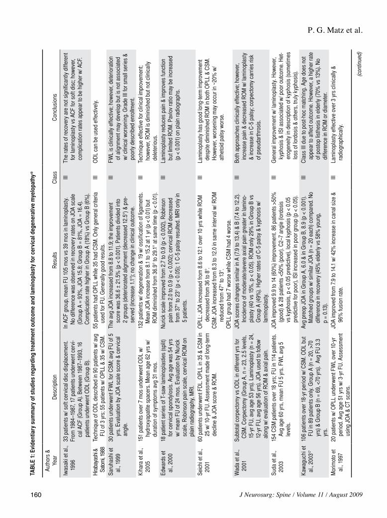
TAB
LE 1
: Evi
dent
iary
sum
mar
y of
stu
dies
rega
rdin
g tr
eatm
ent o
utco
me
and
lam
inop
last
y fo
r cer
vica
l deg
ener
ativ
e m
yelo
path
y*
Auth
ors
& Ye
arD
escr
iptio
nR
esul
tsC
lass
Con
clus
ions
Iw as
aki e
t al.,
19
9633
pat
ient
s w
/ sof
t cer
vica
l dis
c di
spla
cem
ent.
Fr om
198
4–19
87, 1
7 pa
tient
s un
derw
ent c
ervi
-ca
l AC
F (G
roup
A).
Betw
een
1987
–199
3, 1
6 pa
tient
s un
derw
ent O
DL
(Gro
up B
).
In A
CF
grou
p, m
ean
FU 1
05 m
os v
s 39
mos
in la
min
opla
sty.
N
o di
ffere
nce
was
obs
erve
d in
reco
very
rate
s on
JO
A sc
ale
(Gro
up A
= 9
3%, J
OA
15.8
; Gro
up B
= 8
1%, J
OA
= 16
.4).
Com
plic
atio
n ra
te h
ighe
r in
Gro
up A
(18%
) vs
Gro
up B
(6%
).
IIIT h
e ra
tes
of re
cove
ry a
re n
ot s
igni
fican
tly d
iffer
ent
for l
amin
opla
sty
vs A
CF
for s
oft d
isc;
how
ever
, co
mpl
icat
ion
rate
s ap
pear
to b
e hi
gher
w/ A
CF.
H ira
baya
shi &
Sa
tom
i, 198
8Te
chni
que
of O
DL
desc
ribed
in 9
0 pa
tient
s w
/ avg
FU
of 3
yrs
; 55
patie
nts
w/ O
PLL
& 35
w/ C
SM.
55 p
atie
nts
had
OPL
L w
hile
35
had
CSM
. Onl
y ge
nera
l crit
eria
us
ed fo
r FU
. Gen
eral
ly g
ood
resu
lts.
IIIO
DL
can
be u
sed
effe
ctiv
ely.
S aru
hash
i et
al.,
1999
30 p
atie
nts
unde
rwen
t FW
L fo
r CSM
; avg
FU
of 5
yr
s. E
valu
atio
n by
JO
A sc
ale
scor
e &
cerv
ical
an
gle.
Th e
avg
JOA
incr
ease
d fro
m 8
.8 to
11.
9; th
e im
prov
emen
t sc
ore
was
36.
6 ±
21.5
% (p
< 0
.001
). Pa
tient
s di
vide
d in
to
2 gr
oups
(det
erio
rate
d al
ignm
ent (
decr
ease
d 12
.5°)
& p
re-
serv
ed (i
ncre
ase
1.1°);
no
chan
ge in
clin
ical
out
com
e.
IIIF W
L is
clin
ical
ly e
ffect
ive;
how
ever
, det
erio
ratio
n of
alig
nmen
t may
dev
elop
but
is n
ot a
ssoc
iate
d w
/ clin
ical
wor
seni
ng. G
rade
III f
or s
mal
l ser
ies
& po
orly
des
crib
ed e
nrol
lmen
t.K i
hara
et a
l.,
2005
15 1
patie
nts
over
7 m
os u
nder
wen
t OD
L w
/ hy
drox
yapa
tite
spac
ers.
Mea
n ag
e 62
yrs
w/
dura
tion
of s
ympt
oms
avg
31 m
os.
13 2
patie
nts
w/ d
egen
erat
ion
& 19
w/ o
ssifi
catio
n of
liga
men
ts.
Mea
n JO
A in
crea
se fr
om 8
.1 to
15.
2 at
1 y
r (p
< 0.
01) b
ut
RO
M d
imin
ishe
d fro
m 3
6.9°
to 2
9.1°
at s
ame
time
(p <
0.0
1).
IIIT e
chni
que
effe
ctiv
e fo
r clin
ical
impr
ovem
ent;
how
ever
, RO
M is
dim
inis
hed
but n
ot c
linic
ally
de
lete
rious
.E d
war
ds e
t al
., 20
0018
pat
ient
ser
ies
of T
-saw
lam
inop
last
ies
(spl
it)
for c
ervi
cal s
pond
ylos
is. A
vg a
ge w
as 5
4 yr
s w
/ mea
n FU
of 2
4 m
os. E
valu
atio
n by
Nur
ick
scal
e, R
obin
son
pain
sca
le, c
ervi
cal R
OM
on
plai
n ra
diog
raph
y, M
RI.
Nu r
ick
scal
e im
prov
ed fr
om 2
.7 to
0.9
(p <
0.0
02),
Rob
inso
n pa
in fr
om 2
.0 to
0.9
(p <
0.0
02);
cerv
ical
RO
M d
ecre
ased
fro
m 3
7° to
23°
(p <
0.0
5); 1
C-5
pal
sy re
sulte
d. M
RI o
nly
in
5 pa
tient
s.
IIIL a
min
opla
sty
redu
ces
pain
& im
prov
es fu
nctio
n bu
t lim
ited
RO
M. P
avlo
v ra
tio m
ay b
e in
crea
sed
(p <
0.0
01) o
n pl
ain
radi
ogra
phs.
S eic
hi e
t al.,
20
0160
pat
ient
s un
derw
ent F
DL.
OPL
L in
35
& C
SM in
25
w/ 1
0-yr
FU
. Ass
essm
ent m
ade
of lo
ng-te
rm
decl
ine
& JO
A sc
ore
& R
OM
.
OP L
L: J
OA
incr
ease
d fro
m 8
.6 to
12.
1 ov
er 1
0 yr
s w
hile
RO
M
decr
ease
d fro
m 3
6 to
8°.
CS M
: JO
A in
crea
sed
from
8.3
to 1
2.0
in s
ame
inte
rval
w/ R
OM
re
duce
d fro
m 4
7° to
13°
. O
PLL
grou
p ha
d 7
wor
se w
hile
CSM
had
4 w
orse
.
IIIL a
min
opla
sty
has
good
long
-term
impr
ovem
ent
desp
ite d
imin
ishe
d R
OM
in b
oth
OPL
L &
CSM
. H
owev
er, w
orse
ning
may
occ
ur in
~20
% w
/ at
heto
id p
alsy
wor
se.
W ad
a et
al.,
20
01S u
btot
al c
orpe
ctom
y vs
OD
L in
diff
eren
t yrs
for
CSM
. Cor
pect
omy
(Gro
up A
, n =
23,
2.5
leve
ls,
15-y
r FU
, avg
age
53
yrs)
. Lam
inop
last
y (n
= 2
4,
12-y
r FU
, avg
age
56
yrs)
. JO
A us
ed to
follo
w
alon
g w
/ eva
luat
ion
of R
OM
& a
xial
pai
n.
JO A
scor
es c
hang
es s
imila
r in
A (7
.9 to
13.
4) &
B (7
.4 to
12.
2).
Inci
denc
e of
mod
erat
e/se
vere
axi
al p
ain
grea
ter i
n la
min
o-pl
asty
(40
vs 1
5%, p
< 0
.05)
. RO
M o
nly
29%
in G
roup
B v
s G
roup
A (4
9%).
Hig
her r
ates
of C
-5 p
alsy
& k
ypho
sis
w/
lam
inop
last
y.
IIIB o
th a
ppro
ache
s cl
inic
ally
effe
ctiv
e; h
owev
er,
incr
ease
pai
n &
decr
ease
d R
OM
w/ l
amin
opla
sty
& in
crea
se in
C-5
pal
sy; c
orpe
ctom
y ca
rrie
s ris
k of
pse
udar
thro
sis.
S uda
et a
l.,
2003
15 4
CSM
pat
ient
s ov
er 1
8 yr
s; F
U in
114
pat
ient
s.
Avg
age
60 y
rs, m
ean
FU 5
yrs
. FW
L av
g 5
leve
ls.
JO A
impr
oved
9.9
to 1
4 (6
0%) i
mpr
ovem
ent.
86 p
atie
nts
>50%
(g
ood)
& 2
8 pa
tient
s <5
0% (p
oor).
C2–
7 an
gle
(lord
osis
vs
kyp
hosi
s, p
< 0
.05
pred
ictiv
e), l
ocal
kyp
hosi
s (p
< 0
.05
pred
ictiv
e fo
r poo
r), IS
I inc
reas
ed in
poo
r gro
up (p
< 0
.05)
.
IIIG
ener
al im
prov
emen
t w/ l
amin
opla
sty.
How
ever
, ky
phos
is &
ISI a
ssoc
iate
d w
/ poo
r out
com
e. H
et-
erog
enei
ty in
des
crip
tion
of k
ypho
sis
(som
etim
es
loss
of l
ordo
sis
& ot
hers
, tru
ly k
ypho
sis)
.K a
wag
uchi
et
al.,
2003
17
10 6
patie
nts
over
18-
yr p
erio
d w
/ CSM
. OD
L bu
t FU
in 8
9 pa
tient
s on
ly. G
roup
A (n
= 2
0, >
70
yrs)
& G
roup
B (n
= 6
9, <
70 y
rs).
Avg
FU
3.3
yr
s.
Av g
preo
p JO
A in
Gro
up A
, 6.0
& in
Gro
up B
, 8.9
(p <
0.0
01).
Mat
ched
gro
ups
w/ s
imila
r JO
A (n
= 2
0 ea
ch) c
ompa
red.
No
diffe
renc
e in
reco
very
(45%
eld
erly
vs
58%
you
ng,
p =
0.13
).
IIIC
lass
III d
ue to
pos
t-hoc
mat
chin
g. A
ge d
oes
not
appe
ar to
affe
ct o
utco
me.
How
ever
, a h
ighe
r rat
e of
pos
top
listh
esis
in e
lder
ly (7
0% v
s 13
%).
No
diffe
renc
e in
RO
M o
r dia
met
er.
M or
imot
o et
al
., 19
9720
pat
ient
s w
/ OPL
L un
derw
ent F
WL
over
10-
yr
perio
d. A
vg a
ge 5
1 yr
s w
/ 3-y
r FU
. Ass
essm
ent
usin
g JO
A &
CT
scan
s.
JO A
impr
oved
from
7.9
to 1
4.1
w/ 4
2% in
crea
se in
can
al s
ize
& 96
% fu
sion
rate
.III
L am
inop
last
y ef
fect
ive
over
3 y
rs c
linic
ally
&
radi
ogra
phic
ally.
(con
tinue
d)
J Neurosurg: Spine / Volume 11 / August 2009
Laminoplasty for treatment of degenerative cervical myelopathy
161
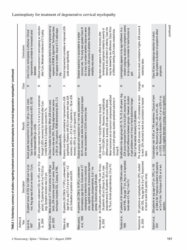
TAB
LE 1
: Evi
dent
iary
sum
mar
y of
stu
dies
rega
rdin
g tr
eatm
ent o
utco
me
and
lam
inop
last
y fo
r cer
vica
l deg
ener
ativ
e m
yelo
path
y* (c
ontin
ued)
Auth
ors
& Ye
arD
escr
iptio
nR
esul
tsC
lass
Con
clus
ions
L ee
et a
l.,
1997
25 p
atie
nt s
erie
s of
OD
L fo
r CSM
ove
r 4-y
r pe-
riod.
Avg
age
was
60
yrs
w/ d
urat
ion
1.5
yrs.
Nu r
ick
grad
e im
prov
ed fr
om 2
.76
to 1
.80
(p <
0.0
5, t-
test
). G
ait &
num
bnes
s im
prov
ed s
ubje
ctiv
ely
in 8
0%. S
C/V
B ra
tio im
prov
ed 3
8% (p
< 0
.05)
.
IIIO
v er 2
.5 y
rs, l
amin
opla
sty
is e
ffect
ive.
Clin
ical
im
prov
emen
ts c
orre
late
w/ i
ncre
ased
can
al
diam
eter
.O
gaw
a, e
t al
., 20
0413
9 pa
tient
s un
derw
ent O
DL
for O
PLL
over
14-
yr
perio
d. 7
2 pa
tient
s ha
d m
in 5
-yr F
U (9
.5-y
r av
g). S
ympt
om d
urat
ion
was
3.5
yrs
.
JO A
scor
es im
prov
ed fr
om 9
.2 to
14.
2 at
3 y
rs &
mai
ntai
ned
thro
ugh
5 (6
3% re
cove
ry) b
ut d
imin
ishe
d af
ter 5
yrs
. Li
mite
d R
OM
& C
-5 p
aral
ysis
occ
urre
d in
7%
.
IIIC
l inic
al im
prov
emen
t in
lam
inop
last
y w
/ red
uctio
n af
ter 5
yrs
. Mod
erat
e in
cide
nce
of C
-5 p
alsy
.
R at
liff &
Coo
-pe
r, 20
03M
eta-
anal
ysis
of 7
1 cl
inic
al s
erie
s w
/ 258
0 pa
-tie
nts;
JO
A ou
tcom
e us
ed in
41
serie
s. S
ever
al
tech
niqu
es in
clud
ed O
DL,
Z-p
last
y, &
FW
L.
No
Cla
ss II
stu
dies
for o
utco
me.
Whe
n JO
A sc
ore
used
, re
cove
ry ra
te w
as 5
5%. P
osto
p al
ignm
ent w
as w
orse
in
35%
w/ 1
0% k
ypho
sis.
RO
M d
imin
ishe
d by
50%
. C-5
ra
dicu
lopa
thy
in 8
%.
IIILa
min
opla
sty
serie
s te
nd to
sho
w im
prov
emen
t at
expe
nse
of R
OM
& a
lignm
ent.
Tech
niqu
e do
es
not a
ppea
r to
mat
ter.
Issu
e of
C-5
radi
culo
pa-
thy.
B aba
et a
l.,
1996
55 p
atie
nts
(38
CSM
& 1
7 O
PLL)
und
erw
ent O
DL
(age
64
yrs,
avg
FU
2.4
yrs
w/ r
ange
1–4
.2
yrs)
. Eva
luat
ion
of p
oste
rior c
ord
shift
.
Po st
erio
r cor
d m
igra
tion
corre
late
d w
/ im
prov
emen
t in
JOA
(sig
nific
ant).
Pos
terio
r shi
ft 55
.3 in
pat
ient
s w
ho h
ad J
OA
reco
very
>50
% (n
= 3
7) &
22.
7 in
pat
ient
s (n
= 1
8) w
/ JO
A re
cove
ry <
50%
(p <
0.0
2, c
hi-s
quar
e te
st).
IIC
l inic
al im
prov
emen
t cor
rela
tes
w/ i
mpr
oved
JO
A sc
ale
scor
e si
gnifi
cant
ly.
M at
suya
ma,
et
al.,
199
544
pat
ient
s (2
6 C
SM &
18
OPL
L) u
nder
wen
t la
min
opla
sty
(age
58
yrs)
. Eva
luat
ion
w/ C
T m
yelo
grap
hy to
det
erm
ine
cros
s-se
ctio
nal
area
, sag
ittal
dia
met
er, &
tran
sver
se d
iam
eter
pr
eope
rativ
ely,
pos
tope
rativ
ely,
& a
t 1 m
o po
stop
. Out
com
e us
ing
JOA
scal
e.
Gr a
dual
spi
nal c
ord
expa
nsio
n at
1 m
o w
as a
ssoc
iate
d w
/ 68
.4%
reco
very
rate
whi
le th
e ab
senc
e of
gra
dual
exp
an-
sion
was
ass
ocia
ted
w/ a
32.
6% re
cove
ry ra
te.
IIIG
r adu
al e
xpan
sion
is a
ssoc
iate
d w
/ a b
ette
r re
cove
ry ra
te. T
his
stud
y w
as g
rade
d C
lass
III
b/c
it w
as n
ot e
vide
nt w
heth
er o
bser
vers
wer
e bl
inde
d to
JO
A sc
ores
& w
heth
er th
e in
trara
ter
relia
bilit
y w
as te
sted
.
Y am
azak
i et
al.,
2003
64 C
SM p
atie
nts
unde
rwen
t FW
L ov
er 1
8 m
os;
29 p
atie
nts
<65
yrs
(Gro
up A
, 53
yrs,
mal
e pr
epon
dera
nce)
& 3
5 pa
tient
s >6
5 yr
s (G
roup
B,
79
yrs,
fem
ale
prep
onde
ranc
e). 3
.5-y
r FU
.
JO A:
Gro
up A
, 14.
815
.8 (6
2% re
cove
ry);
Gro
up B
, 12
.0
14.6
(59%
reco
very
). R
ecov
ery
not s
igni
fican
tly
diffe
rent
but
pre
- & p
osto
p JO
A sc
ale
scor
es d
iffer
ed.
Mea
n tra
nsve
rse
are
at m
axim
um c
ompr
essi
on p
redi
cted
ou
tcom
e in
bot
h gr
oups
; dur
atio
n pr
edic
ted
outc
ome
in
Gro
up B
.
IIIAg
e do
es n
ot a
ppea
r to
affe
ct re
cove
ry a
fter
lam
inop
last
y; p
rogn
osis
rela
ted
to s
ever
ity o
f st
enos
is &
als
o du
ratio
n (in
eld
erly
). C
ase
mix
m
ay b
e sk
ewed
by
sex
diffe
renc
e. C
lass
III b
/c
of d
iffer
ent s
tarti
ng J
OA
scal
es c
reat
ed b
ias.
T ana
ka e
t al.,
19
9947
pat
ient
s of
64
revi
ewed
w/ C
SM w
ho u
nder
-w
ent F
WL.
Avg
age
77
yrs.
Avg
no.
of o
pera
ted
leve
ls w
as 4
.5. A
vg 1
1-m
o FU
.
St ra
tific
atio
n in
to a
ge g
roup
s 65
–75,
76–
79, &
>80
yea
rs. A
vg
JOA
scor
e im
prov
ed fr
om 7
.6 to
9.6
(20%
reco
very
) w/ n
o si
gnifi
cant
diff
eren
ce b
etw
een
grou
ps. H
owev
er, d
urat
ion
of g
ait p
robl
ems
over
3 m
os o
r han
d dy
sfun
ctio
n lo
nger
th
an 1
yr h
as w
orse
reco
very
in 2
6 pa
tient
s.
IIILa
min
opla
sty
appe
ars
to b
e ag
e in
diffe
rent
. In
a ra
ndom
sub
grou
p, d
urat
ion
of s
ympt
oms
seem
s to
be
a ne
gativ
e co
rrela
te fo
r han
d fu
nctio
n &
gait.
K am
izon
o et
al
., 20
0330
1 pa
tient
s w
/ mye
lopa
thy
from
OPL
L st
udie
d af
ter O
DL.
Avg
age
58
w/ 8
-yr F
U. A
sses
smen
t of
retu
rn to
wor
k (fu
ll, p
artia
l, no
ne).
Pr e-
& p
osto
p JO
A sc
ores
<9,
9–1
2, >
12 c
orre
late
d w
/ ret
urn
to w
ork.
53%
retu
rned
to w
ork
but c
orre
latio
n to
hea
vier
la
bor.
II pr
ogno
-si
sR
e tur
n-to
-wor
k co
rrela
ted
w/ h
ighe
r JO
A sc
ores
&
sede
ntar
y la
bor.
S ato
mi e
t al.,
20
0120
6 pa
tient
s w
/ avg
age
of 5
7 yr
s. O
PLL
in 1
06
& C
SM in
98.
Pro
gnos
is s
tudi
ed in
80
patie
nts
w/ 5
-yr F
U m
in. T
echn
ique
of O
DL
w/ o
r w/o
sp
acer
s.
Co m
plic
atio
n ra
te 1
0.8%
w/ 7
.8%
wea
knes
s. C
losu
re o
f lam
i-na
e 1.
5%. R
ecov
ery
& ag
e (7
6% <
60 v
s 41
% >
60,
p <
0.00
1). R
ecov
ery
& du
ratio
n (7
9% <
1 yr
vs
53%
>1
yr,
p =
0.00
3); s
agitt
al d
iam
eter
or p
reop
JO
A no
t sig
nific
ant.
IIIC
l ass
III d
ue to
sub
grou
p se
lect
ion
of c
ohor
t. Ag
e of
pat
ient
& d
urat
ion
of s
ympt
oms
affe
ct
prog
nosi
s.
(con
tinue
d)

P. G. Matz et al.
162 J Neurosurg: Spine / Volume 11 / August 2009
TAB
LE 1
: Evi
dent
iary
sum
mar
y of
stu
dies
rega
rdin
g tr
eatm
ent o
utco
me
and
lam
inop
last
y fo
r cer
vica
l deg
ener
ativ
e m
yelo
path
y* (c
ontin
ued)
Auth
ors
& Ye
arD
escr
iptio
nR
esul
tsC
lass
Con
clus
ions
K ohn
o et
al.,
19
9722
pat
ient
s w
/ mye
lopa
thy
(CSM
12,
OPL
L 5,
co
mbi
ned
5) tr
eate
d w
/ FW
L ov
er 4
yrs
. Avg
ag
e 60
yrs
; FU
5 y
rs.
Av g
reco
very
was
51%
JO
A. S
tratif
ied
into
goo
d (>
50%
& fa
ir <5
0%).
Age
sign
ifica
ntly
diff
eren
t in
grou
ps (g
ood
56 y
rs
vs fa
ir 64
yrs
, p <
0.0
5). I
SI o
n M
RI l
ess
frequ
ently
see
n in
go
od (1
8% v
s 82
%).
Area
of s
pina
l can
al in
crea
sed
mor
e in
go
od g
roup
(p <
0.0
5).
III e
ffi-
ca cy
, II
prog
nosi
s
Ag e
may
affe
ct c
oncl
usio
n. A
ge a
ppea
rs to
be
nega
tive
fact
or fo
r out
com
e al
ong
w/ a
rea
of
diam
eter
. ISI
mor
e fre
quen
t in
poor
out
com
e.
W an
g et
al.,
20
0420
4 pa
tient
s w
/ CSM
und
erw
ent O
DL
over
15
yrs.
Av
g ag
e 63
& a
vg F
U 1
6 m
os.
Nu r
ick
scor
es im
prov
ed in
62%
; age
>75
yrs
did
not
app
ear
to a
ffect
out
com
e.III
La m
inop
last
y ef
fect
ive
for C
SM o
ver s
hort
term
.
K aw
aguc
hi e
t al
., 20
0318
12 6
patie
nts
w/ m
yelo
path
y (C
SM =
57,
OPL
L =
69).
OD
L. A
vg a
ge 6
0 yr
s w
/ 13.
2-yr
FU
. How
-ev
er, F
U a
vaila
ble
in o
nly
73 b
/c 4
2 pa
tient
s di
ed &
11
wer
e lo
st to
FU
.
JO A
reco
very
rate
s w
ere
sign
ifica
ntly
bet
ter w
/ age
65%
(<
60 y
rs) &
49%
(>60
yrs
). If
star
ting
JOA
scor
e <8
, no
cor-
rela
tion
w/ o
utco
me
(51
vs 5
9% J
OA
>8).
Cer
vica
l ang
le
decr
ease
d fro
m 1
3.5
to 4
.8° b
ut c
anal
dia
met
er la
rger
.
IIILa
min
opla
sty
effe
ctiv
e ov
er 1
0-yr
per
iod
but i
s as
-so
ciat
ed w
/ kyp
hosi
s. A
ge a
ppea
rs to
be
prog
-no
stic
, sev
erity
of m
yelo
path
y do
es n
ot a
ppea
r to
be
a pr
ogno
stic
fact
or. S
igni
fican
t num
bers
lo
st to
dea
th w
hich
ske
ws
conc
lusi
ons.
Iw as
aki e
t al.,
20
0292
pat
ient
s w
/ OPL
L un
derw
ent O
DL
over
8-y
r pe
riod;
64
pts
in s
erie
s si
nce
25 d
ied
& 3
lost
to
FU
. Avg
age
56
yrs
w/ m
in F
U 1
0 yr
s.
JO A
impr
oved
from
8.9
to 1
3.8;
exp
ired
patie
nts
had
preo
p JO
A 6.
9. R
ecov
ery
was
60%
at 3
–10
yrs
but r
educ
ed to
54
% a
fter 1
0 yr
s. A
ge a
t op
had
nega
tive
trend
(p =
0.0
64)
& lo
w J
OA
scor
e ha
d st
rong
neg
ativ
e tre
nd (p
< 0
.000
1).
Kyph
osis
in 8
% &
C-5
radi
culo
path
y in
5%
pos
top.
II, p
rog-
nosi
sLa
min
opla
sty
may
be
effe
ctiv
e bu
t sev
erity
of J
OA
scor
e is
neg
ativ
e ris
k fa
ctor
. Af
ter 1
0 yr
s, c
lini-
cal i
mpr
ovem
ent d
eclin
es.
H an
da e
t al.,
20
0261
con
secu
tive
patie
nts
w/ C
SM. S
tratif
ied
into
el
derly
(age
>70
yrs
, mea
n 74
; n =
22)
& y
oung
(a
ge <
70 y
rs, m
ean
57; n
= 3
9). O
DL
over
9-y
r pe
riod
w/ 1
2 m
os F
U.
El de
rly h
ad m
ean
reco
very
of 5
9% w
hile
you
ng h
ad m
ean
reco
very
of 6
2% (d
iffer
ence
not
sig
nific
ant).
In e
lder
ly, o
ut-
com
e re
late
d to
dur
atio
n of
sym
ptom
s (>
12 m
os) &
sev
erity
of
ste
nosi
s. In
you
ng, J
OA
scor
e <1
2 ne
gativ
e ris
k fa
ctor
.
III e
ffica
-cy
, II
prog
nosi
s
No
diffe
renc
e in
out
com
e ag
e ov
er 7
0 vs
you
nger
. C
lass
III f
or p
redi
ctiv
e fa
ctor
s: n
o un
iform
pr
edic
tive
fact
or. D
urat
ion
& se
verit
y of
ste
nosi
s in
eld
erly
& J
OA
scor
e in
you
ng.
K aw
aguc
hi e
t al
., 20
0019
18 p
atie
nts
w/ C
SM/O
PLL
w/ D
M (a
ge 6
6 yr
s, 3
-yr
FU) v
s 34
his
toric
al c
ontro
ls w
/o D
M (a
ge 6
5 yr
s, 4
.7-y
r FU
).
Re c
over
y 55
% w
/ DM
& 6
1% w
/o D
M b
ased
on
JOA;
how
-ev
er, n
egat
ive
corre
latio
n be
twee
n JO
A &
Hgb
A1c
(r =
0.61
, p <
0.0
3).
II pr
ogno
-si
sC
o mpa
rison
bet
wee
n D
M &
non
-DM
Cla
ss II
I due
to
his
toric
al c
ontro
ls; h
owev
er, D
M a
ppea
rs n
ot
to a
ffect
out
com
e. H
owev
er, p
oorly
con
trolle
d D
M n
egat
ivel
y af
fect
s ou
tcom
e.K a
min
sky
et
al.,
2004
20 c
onse
cutiv
e pa
tient
s (a
vg a
ge 5
3 yr
s) w
/ CSM
un
derw
ent O
DL
(A) v
s 22
mat
ched
con
trols
w
ho u
nder
wen
t lam
inec
tom
ies
(B, a
vg a
ge 5
4 yr
s). M
in F
U 3
yrs
, avg
5 y
rs.
Av g
no. o
f lev
els
deco
mpr
esse
d w
as 4
.3 in
A, &
4.6
in B
. Nu-
rick
preo
p w
as 2
.44
in A
& 3
.09
in B
(p <
0.0
001)
. Rec
over
y w
as 2
.44
1.48
(48.
6%) i
n A
& 3.
09
2.50
(17.
8%) i
n B
(p
< 0
.000
1). P
ain
leve
l red
uced
in A
(57%
vs
8%, p
<
0.00
4); h
owev
er, p
reop
pai
n le
vel s
igni
fican
tly h
ighe
r in
A.
IIIC
l ass
III d
ue to
his
toric
con
trols
& la
ck o
f sim
ilar
preo
p N
uric
k st
atus
& la
ck o
f sim
ilar p
ain
seve
r-ity
. Con
clus
ion
was
that
lam
inop
last
y pr
ovid
es
bette
r res
ults
clin
ical
ly a
t 3–5
yrs
w/ l
ess
RO
M.
It re
duce
s pa
in.
K aw
aguc
hi e
t al
., 20
0316
28 p
atie
nts
w/ C
SM u
nder
wen
t mod
ified
la
min
opla
sty
(alte
rnat
ing
spac
ers
w/ n
o bo
ne
on h
inge
, 199
7–20
00) v
s 28
his
toric
al c
ontro
ls
who
und
erw
ent O
DL
(198
2–96
); m
odifi
ed (a
ge
62.7
yrs
; FU
2.4
y); O
DL
(60.
4 yr
s, 2
.8-y
r FU
).
RO
M in
mod
ified
pre
serv
ed 7
1% v
s 48
% in
con
trols
(p
< 0
.02)
; lev
els
fuse
d in
form
er 0
.8 v
s 2.
1 in
latte
r (p
< 0
.000
1).
IIIC
l ass
III f
or h
isto
rical
con
trols
; mod
ified
tech
niqu
e pr
eser
ves
RO
M.
(con
tinue
d)

J Neurosurg: Spine / Volume 11 / August 2009
Laminoplasty for treatment of degenerative cervical myelopathy
163
TAB
LE 1
: Evi
dent
iary
sum
mar
y of
stu
dies
rega
rdin
g tr
eatm
ent o
utco
me
and
lam
inop
last
y fo
r cer
vica
l deg
ener
ativ
e m
yelo
path
y* (c
ontin
ued)
Auth
ors
& Ye
arD
escr
iptio
nR
esul
tsC
lass
Con
clus
ions
M ae
da e
t al.,
20
0244
pat
ient
s w
/ OPL
L un
derw
ent F
WL.
Avg
age
w
as 5
9 yr
s w
/ 3.2
yrs
FU
.FU
s on
ly a
vaila
ble
in 3
0 of
44
patie
nts.
Cur
vatu
re in
dex
of
Ishi
ara
decr
ease
d fro
m 1
4.5%
to 2
%. D
ecre
ase
in R
OM
w
as 4
1.3°
to 2
4°. T
his
corre
late
d w
/ dec
reas
ed fu
nctio
n on
JO
A.
IIIN
e gat
ive
corre
latio
n be
twee
n lo
ss o
f lor
dosi
s &
JOA.
H os
ono
et
al.,
1996
98 p
atie
nts
w/ C
SM (a
vg a
ge 5
8 yr
s, m
in 2
-yr
FU) u
nder
wen
t OD
L (n
= 7
2) o
r AC
F (n
= 2
6).
Lam
inop
last
y (1
984–
1990
) & A
CF
(198
2–19
84).
Pr es
ence
of a
bsen
ce o
f pai
n as
sess
ed. N
o qu
antiz
atio
n of
pa
in c
ompl
eted
. Pai
n as
sess
ed a
s nu
chal
pai
n, s
houl
-de
r pai
n, o
r sho
ulde
r spa
sm. 5
/26
(19%
) & 4
3/72
(60%
) pa
tient
s ha
d po
stop
pai
n w
/ sig
nific
antly
hig
her l
evel
s in
la
min
opla
sty
patie
nts
(p <
0.0
5).
IIIC
l ass
III d
ue to
his
toric
al c
ontro
ls. H
owev
er,
lam
inop
last
y ap
pear
s to
be
asso
ciat
ed w
/ in
crea
sed
pain
ove
r AC
F.
T ake
uchi
et
al.,
2005
Gr o
up A
was
40/
71 p
atie
nts
w/ C
SM (3
5)/O
PLL
(5) f
ollo
wed
>1
yr w
ho u
nder
wen
t C-3
lam
inec
-to
my
& C
4–7
lam
inop
last
y; G
roup
B w
as 1
6/20
pa
tient
s w
/ CSM
(9)/O
PLL
(7) w
ho u
nder
wen
t C
4–7
lam
inop
last
y w
/ 1 y
r FU
.
Ou t
com
e w
as s
ubje
ctiv
e gr
adin
g of
pai
n (n
o, m
ild, s
ever
e)
& cr
oss-
sect
iona
l are
a of
mus
cle.
Gro
up A
had
mor
e pa
tient
s w
/ no
or m
ild p
ain
(p <
0.0
2) &
atro
phy
perc
ent-
age
was
2%
vs
10%
in B
(p <
0.0
01).
IIIH
i sto
rical
con
trols
use
d si
nce
tech
niqu
e ch
ange
d af
ter 2
001;
als
o, p
ain
outc
ome
mea
sure
sub
jec-
tive.
How
ever
, pre
serv
ing
C-2
mus
cle
redu
ces
atro
phy.
S hira
ishi
et
al.,
2003
43 p
atie
nts
w/ C
SM (3
2)/O
PLL
(11)
who
und
er-
wen
t ski
p la
min
ecto
my
(afte
r 199
8); a
ge 6
9 yr
s w
/ 2-y
r FU;
Com
paris
on to
OD
L in
51
patie
nts
(CSM
36/O
PLL1
5); a
ge 6
7 yr
s w
/ 2-y
r FU
.
JO A
reco
very
was
59%
vs
60%
w/ l
amin
opla
sty;
RO
M
98%
pre
serv
ed w
/ ski
p vs
44%
in la
min
opla
sty;
Ishi
ara
curv
atur
e in
dex
11.4
1
3.4
w/ s
kip
vs 1
6.0
11.
8 in
la
min
opla
sty
(p <
0.0
5); a
troph
y w
as 1
3% in
ski
p vs
60%
in
lam
inop
last
y on
T2-
wei
ghte
d M
RI.
IIIH
i sto
rical
con
trols
(Cla
ss II
I): h
owev
er s
kip
lam
inec
tom
y re
duce
s at
roph
y &
redu
ces
risk
of
kyph
osis
.
Y one
nobu
et
al.,
1992
10 0
patie
nts
w/ C
SM o
f who
m 8
3 ha
d 2-
yr F
U; 4
1 pa
tient
s un
derw
ent A
CF
(197
6–19
83) w
hile
42
unde
rwen
t FW
L.
JO A
impr
oved
in b
oth
grou
ps (4
4% in
lam
inop
last
y &
55%
in
AC
F, n
ot s
igni
fican
t). In
sub
set w
/ can
al <
12
mm
, out
-co
mes
wer
e 55
% in
lam
inop
last
y &
59%
in A
CF.
Com
plic
a-tio
n ra
te w
as g
raft
rela
ted
& 29
% in
AC
F. L
amin
opla
sty
had
7% C
-5 ra
dicu
lopa
thy.
IIIG
r oup
s co
mpa
red
over
diff
eren
t tim
e pe
riods
(C
lass
III).
Res
ults
sho
w s
imila
r clin
ical
impr
ove-
men
t but
hig
her c
ompl
icat
ion
rate
s in
AC
F.
H at
ta e
t al.,
20
0526
con
secu
tive
patie
nts
w/ 2
–3 le
vel C
SM u
nder
-w
ent s
elec
tive
lam
inop
last
y (a
ge 6
0 yr
s, F
U 1
9 m
os) v
s hi
stor
ical
con
trols
: 25
of 5
6 (2
–3 le
vel
CSM
; age
62
yrs,
FU
38
mos
) who
und
erw
ent
OD
L.
Re c
over
y w
as 6
6% in
bot
h us
ing
JOA.
Sel
ectiv
e gr
oup
had
1.1-m
m c
ord
shift
whi
le O
DL
had
2.7-
mm
cor
d sh
ift (p
<
0.05
); C
-5 p
alsy
in la
tter w
as 8
% b
ut 0
% in
form
er.
IIIC
l ass
III d
ue to
his
toric
con
trols
. How
ever
, sel
ec-
tive
tech
niqu
e pr
oduc
es s
ame
resu
lt de
spite
le
ss c
ord
shift
. Les
s co
rd s
hift
seem
s to
be
asso
ciat
ed w
/ low
er C
-5 p
alsy
.
K om
agat
a et
al
., 20
0430
5 pa
tient
s w
/ CSM
or O
PLL
unde
rwen
t OD
L.
Eval
uatio
n w
/ pre
op E
MG
& c
urva
ture
inde
x.
230/
305
patie
nts
had
fora
min
otom
y.
3/ 11
8 pa
tient
s w
/ abn
orm
al E
MG
dev
elop
ed C
-5 p
alsy
. W
here
as 6
/140
(8.4
%) w
/ nor
mal
EM
G d
evel
oped
C-5
pa
lsy.
Cur
vatu
re in
dex
did
not a
ffect
C-5
pal
sy ra
te (4
.3%
). C
-5 p
alsy
occ
urre
d in
1/1
62 g
utte
rs w
/ for
amin
otom
y &
w/
12/2
98 g
utte
rs w
/o (p
< 0
.05)
.
IIIFo
ram
inot
omy
may
pre
vent
C-5
pal
sy b
ut s
elec
-tio
n cr
iteria
too
obtu
se, s
o C
lass
III.
Abno
rmal
EM
G d
oes
not p
redi
ct C
-5 p
alsy
.
S asa
i et a
l.,
2003
11 1
patie
nts
w/ C
SM u
nder
wen
t lam
inop
last
y.
Gro
up A
(n =
74)
who
und
erw
ent E
MG
. 11/
74
had
abno
rmal
EM
G &
und
erw
ent f
oram
inot
omy.
G
roup
B (n
= 3
7) w
ere
eval
uate
d by
diff
eren
t su
rgeo
ns &
did
not
hav
e EM
G.
In G
roup
A, 1
1/74
had
abn
orm
al E
MG
s. 0
/74
had
C-5
pal
sy.
In G
roup
B, 3
/37
had
C-5
pal
sy (p
< 0
.05)
.III
Cl a
ss II
I due
to s
elec
tion
bias
. All
C-5
pal
sies
oc-
curre
d in
gro
up s
elec
ted
by d
iffer
ent s
urge
ons.
EM
G e
valu
atio
n ap
pear
ed to
hel
p in
dica
te w
hich
pa
tient
s m
ight
ben
efit
from
fora
min
otom
y.
(con
tinue
d)

P. G. Matz et al.
164 J Neurosurg: Spine / Volume 11 / August 2009
TAB
LE 1
: Evi
dent
iary
sum
mar
y of
stu
dies
rega
rdin
g tr
eatm
ent o
utco
me
and
lam
inop
last
y fo
r cer
vica
l deg
ener
ativ
e m
yelo
path
y* (c
ontin
ued)
Auth
ors
& Ye
arD
escr
iptio
nR
esul
tsC
lass
Con
clus
ions
U em
atsu
et
al.,
1998
36 5
patie
nts
had
unde
rgon
e la
min
opla
sty;
20
patie
nts
(5.5
%) d
evel
oped
pos
top
radi
culo
pa-
thy
(mos
tly a
t C-5
) w/ t
imin
g ho
urs
to d
ays
afte
r su
rger
y.
De g
ree
of s
teno
sis,
clin
ical
sym
ptom
atic
sev
erity
, fla
tnes
s of
sp
inal
cor
d, c
ervi
cal c
urva
ture
, ant
erio
r arti
cula
r pro
cess
pr
otru
sion
did
not
affe
ct o
utco
me.
Ang
le o
f lam
ina
>60°
of
hing
e si
de &
late
ral g
utte
rs h
ad n
egat
ive
prog
nosi
s
(p <
0.0
5).
IIIM
u ltip
le te
chni
ques
util
ized
& c
ase
serie
s m
ake
this
Cla
ss II
I. To
o gr
eat a
n an
gle
is a
ssoc
iate
d w
/ cer
vica
l rad
icul
opat
hy.
C hi
ba e
t al.,
20
02R
e tro
spec
tive
revi
ew o
f 208
pat
ient
s w
ho u
n-de
rwen
t lam
inop
last
y. 1
5 pa
tient
s de
velo
ped
segm
enta
l par
alys
is. P
aral
ysis
dev
elop
ed a
n av
g of
4.6
day
s po
stop
.
11/ 1
5 pa
tient
s re
cove
red.
Pat
ient
s w
ho d
evel
oped
seg
men
tal
para
lysi
s ha
d (1
4/15
) sev
ere
preo
p dy
sest
hetic
pai
n th
at
did
not r
ecov
er. T
2 si
gnal
cha
nges
see
n on
pos
top
MR
I in
all
15 p
atie
nts
(100
%) c
ompa
red
to 5
8% in
pat
ient
s w
/o
defic
it (p
< 0
.01)
.
IIIR
e tro
spec
tive
revi
ew (C
lass
III)
corre
late
s po
stop
se
gmen
tal p
aral
ysis
w/ T
2 si
gnal
cha
nges
.
W ad
a et
al.,
19
9985
pat
ient
s w
/ CSM
des
crib
ed. 1
0 w
ere
excl
uded
in
itial
ly &
25
did
not r
ecei
ve im
agin
g. M
RIs
an
alyz
ed in
50
patie
nts
afte
r sur
gery
. Age
, JO
A sc
ore,
tran
sver
se a
rea
of s
pina
l cor
d, d
urat
ion
of s
ympt
oms
eval
uate
d.
A t ra
nsve
rse
area
>40
mm
2 at m
axim
al c
ompr
essi
on, d
ura-
tion
of s
ympt
oms,
& A
P ca
nal d
iam
eter
wer
e th
e be
st
pred
icto
rs o
f out
com
e. A
ge, p
reop
JO
A sc
ores
, & is
olat
ed
T2 s
igna
l wer
e no
t ide
al p
redi
ctor
s of
out
com
e. M
ultis
eg-
men
tal T
2 hy
perin
tens
ity w
as c
orre
late
d w
/ poo
r res
ults
(p
< 0
.01)
.
IIIM
u ltis
egm
enta
l T2
hype
rinte
nsity
is a
ssoc
iate
d w
/ po
or o
utco
me.
Thi
s st
udy
was
gra
ded
Cla
ss
III d
ue to
non
rand
omiz
ed s
elec
tion
& lim
ited
num
bers
of p
atie
nts
(50/
85) w
ho re
ceiv
ed
post
op im
agin
g.
H el
ler e
t al.,
20
01C
o mpa
rison
of l
amin
opla
sty
(n =
13)
vs
lam
inec
-to
my
+ ar
thro
desi
s (n
= 1
3) in
CSM
; orig
inal
ly
25 in
eac
h gr
oup
but n
umbe
r los
t. La
tera
l m
ass
scre
ws
utiliz
ed. A
vg a
ge 5
5 yr
s w
/ 25
mos
FU
.
Nu r
ick
impr
oved
2.3
to 1
.1 w
/ lam
inop
last
y vs
2.2
to 1
.5 w
/ fu
sion
(p >
0.0
5); l
amin
opla
sty
had
a be
tter I
shih
ara
(0.0
9 to
0.9
) ind
ex. B
oth
pres
erve
d th
e in
dex
w/ s
urge
ry.
Red
uctio
n of
RO
M:
ar
thro
desi
s: 3
611
°
la m
inop
last
y: 4
026
° (p
< 0.
002)
. Com
plic
atio
n hi
gher
w/
fusi
on.
IIILa
min
ecto
my
+ ar
thro
desi
s ha
s si
mila
r clin
ical
ou
tcom
e bu
t a g
reat
er lo
ss o
f RO
M &
incr
ease
d co
mpl
icat
ion
rate
; unc
lear
why
the
loss
of
num
bers
.
E dw
ards
et
al.,
2002
38 p
atie
nts
CSM
stu
died
retro
spec
tive
w/
mat
ched
coh
orts
Gro
up A
(13
corp
ecto
my,
be
fore
199
6) &
Gro
up B
(25
lam
inop
last
ies
of w
hich
13
chos
en, a
fter 1
996)
. OD
L in
3
patie
nts
& T-
saw
in 1
0 pa
tient
s. F
U >
40 m
os.
Nu r
ick
impr
oved
1.9
to 1
.0 in
Gro
up A
& 2
.3 to
0.8
in G
roup
B
(NS)
. Pai
n im
prov
ed to
0.5
in G
roup
A &
1.0
in G
roup
B
(NS)
. R
O M
redu
ced
from
37°
16° i
n G
roup
A &
39°
24° i
n G
roup
B
(NS)
w/ p
seud
arth
rosi
s, G
roup
A h
ad h
ighe
r com
plic
a-tio
n ra
te (9
/1).
IIIU
n cle
ar m
atch
ing
tech
niqu
e &
diffe
rent
per
iods
. Bo
th c
orpe
ctom
y &
lam
inop
last
y re
liabl
e.
Lam
inop
last
y ap
pear
s to
hav
e fe
wer
com
plic
a-tio
ns.
S aka
ura
et
al.,
2003
43 p
atie
nts
w/ c
ervi
cal d
isc
disp
lace
men
t & m
y-el
opat
hy. G
roup
A (A
CF,
n =
15/
21, a
ge 4
4 yr
s,
1984
–7).
Gro
up B
(lam
inop
last
y, n
= 1
8/22
, age
51
yrs
, 198
7–94
). Av
g FU
was
15
yrs
in G
roup
A
& 10
yrs
in G
roup
B.
Re c
over
y ra
te o
f JO
A w
as 7
1% in
Gro
up A
& 7
0% in
Gro
up
B. R
OM
mai
ntai
ned
65%
in G
roup
A &
64%
in G
roup
B.
Sim
ilar l
ate
dete
riora
tion.
IIIAn
terio
r app
roac
h as
soci
ated
w/ h
ighe
r reo
p ra
te
due
to p
seud
arth
rosi
s bu
t out
com
es s
imila
r.
K aw
akam
i et
al.,
2000
13 6
patie
nts
w/ m
yelo
path
y ov
er 1
0-yr
per
iod
(69
w/ d
isc
& 67
w/ C
SM).
ACF
in 6
0 (8
5% d
isc)
&
lam
inop
last
y in
76
(30%
) dis
c. A
CF
1–2
leve
ls,
& la
min
opla
sty
≥3 le
vels
.
Me a
n re
cove
ry in
JO
A w
as 7
1% in
dis
c pa
tient
s; s
ame
resu
lt w
/ AC
F or
lam
inop
last
y. In
CSM
, 49%
reco
very
w/ A
CF
& 58
% w
/ lam
inop
last
y (N
S). R
ecov
ery
rate
low
er in
bot
h ap
proa
ches
w/ k
ypho
tic c
ord.
IIIC
o mpa
rison
bet
wee
n m
ultip
le s
ubgr
oups
mak
es
anal
ysis
diff
icul
t. H
owev
er, a
ppro
ach
does
not
ap
pear
to m
ake
a di
ffere
nce
w/ r
espe
ct to
dis
c or
mye
lopa
thy.
Kyp
hotic
cor
d ne
gativ
e ris
k fa
ctor
.
(con
tinue
d)

J Neurosurg: Spine / Volume 11 / August 2009
Laminoplasty for treatment of degenerative cervical myelopathy
165
TAB
LE 1
: Evi
dent
iary
sum
mar
y of
stu
dies
rega
rdin
g tr
eatm
ent o
utco
me
and
lam
inop
last
y fo
r cer
vica
l deg
ener
ativ
e m
yelo
path
y* (c
ontin
ued)
Auth
ors
& Ye
arD
escr
iptio
nR
esul
tsC
lass
Con
clus
ions
H as
egaw
a et
al
., 20
0290
pat
ient
s w
/ CSM
. Age
>70
yrs
(n =
40,
27-
mo
FU) &
<60
(n =
50,
36-
mo
FU).
Ante
rior f
usio
n (n
= 3
5), l
amin
opla
sty
(n =
29)
, & la
min
ecto
my
(n =
26)
. Com
paris
on b
etw
een
tech
niqu
e &
age
grou
p (6
gro
ups)
.
No
sign
ifica
nt d
iffer
ence
s in
fina
l JO
A sc
ore
betw
een
grou
ps.
No
sign
ifica
nt d
iffer
ence
in p
reop
JO
A sc
ores
bet
wee
n gr
oups
. Com
plic
atio
n ra
te g
reat
er in
old
er v
s yo
unge
r pa
tient
s: 1
5% v
s 8%
.
IIIM
u ltip
le s
ubgr
oups
in s
erie
s. H
owev
er, a
ge d
oes
not a
ppea
r to
be a
risk
fact
or e
xcep
t for
com
-pl
icat
ions
. Als
o, te
chni
que
does
not
app
ear t
o ch
ange
con
trol o
f mye
lopa
thy.
T om
ita e
t al.,
19
88
23 p
atie
nts
w/ O
PLL
& JO
A <1
2. A
vg a
ge w
as
60 y
rs &
avg
FU
2.5
yrs
. Gro
up c
ompa
red
to
hist
oric
al c
ontro
ls o
f AC
F (n
= 1
7) &
lam
inec
-to
my
(n =
14)
.
JO A
impr
oved
from
8.5
to 1
5.25
(81%
). Th
is c
ompa
red
to
lam
inec
tom
y (7
2%) &
AC
F (6
3%).
Rad
iogr
aphi
c an
alys
is
show
ed a
n ex
pans
ion
ratio
n of
124
%.
IIILa
min
opla
sty
effe
ctiv
e w
hen
com
pare
d to
ant
erio
r fu
sion
& s
impl
e de
com
pres
sion
but
his
toric
al
cont
rols
unr
elia
ble.
Y osh
ida
et
al.,
1998
47 p
atie
nts
w/ c
onge
nita
l ste
nosi
s &
disc
dis
-pl
acem
ent;
32 p
atie
nts
had
lam
inop
last
y (o
ther
15
follo
wed
). C
ompa
red
to A
CF
(n =
44)
in
sam
e 8-
yr p
erio
d. A
CF
grou
p ha
d si
gnifi
cant
ly
larg
er A
P sp
inal
can
al d
ista
nce
& hi
gher
pre
op
JOA
scor
e.
JO A
impr
ovem
ent w
as 6
8% in
bot
h gr
oups
. In
patie
nts
w/
lam
inop
last
y, d
isc
regr
essi
on s
een
in 7
5% v
s 80
% o
f no
nope
rate
d pa
tient
s (n
=15
). In
cide
nce
of c
ompl
icat
ions
w
as re
late
d to
fusi
on &
hig
her i
n AC
F gr
oup.
IIIC
l ass
III d
ue to
mix
ed g
roup
s (d
iffer
ence
in J
OA
& de
gree
of s
teno
sis)
. Bot
h an
terio
r & p
oste
rior
tech
niqu
es e
ffect
ive.
Dis
cs te
nd to
regr
ess.
K im
ura
et a
l.,
2000
39 p
atie
nts
w/ c
ervi
cal m
yelo
path
y (C
SM 2
5,
OPL
L 8,
CD
H 6
) who
und
erw
ent s
plit-
lam
inae
la
min
opla
sty.
One
gro
up (n
= 8
, B) d
evel
oped
di
fferin
g gr
ades
of t
he b
oom
eran
g de
form
ity
whi
le th
e ot
her (
n =
31, C
) did
not
dev
elop
this
de
form
ity.
No
diffe
renc
es in
pre
- & p
osto
p JO
A w
ere
note
d. H
owev
er,
thos
e w
/ the
boo
mer
ang
defo
rmity
had
a h
ighe
r fla
tteni
ng
(sag
ittal
/tran
sver
se) r
atio
& a
sm
alle
r tra
nsve
rse
diam
eter
be
twee
n th
e sp
lit la
min
ae.
IIIAl
thou
gh th
is w
as a
coh
ort s
tudy
, onl
y 1
pers
on
com
plet
ed th
e m
easu
rem
ents
& it
was
unc
erta
in
if th
at p
erso
n w
as b
linde
d. H
owev
er, t
he
boom
eran
g de
form
ity is
mor
e lik
ely
to d
evel
op
in a
pat
ient
w/ a
sm
all f
latte
ning
ratio
& a
sm
all
trans
vers
e di
amet
er b
etw
een
split
lam
inae
.
* Th
e cr
iteria
for s
corin
g ea
ch m
anus
crip
t int
o a
clas
s ar
e de
scrib
ed in
Intro
duct
ion
and
Met
hodo
logy
: Gui
delin
es fo
r the
Sur
gica
l Man
agem
ent o
f Cer
vica
l Deg
ener
ativ
e D
isea
se, w
hich
app
ears
in
this
issu
e of
the
Jour
nal o
f Neu
rosu
rger
y: S
pine
. Abb
revi
atio
ns: A
P =
ante
ropo
ster
ior;
avg
= av
erag
e; b
/c =
bec
ause
; CD
H =
cer
vica
l dis
c he
rnia
tion;
DM
= d
iabe
tes
mel
litus
; FD
L =
Fren
ch-d
oor
lam
inop
last
y; F
U =
follo
w-u
p; F
WL
= Fr
ench
-win
dow
lam
inop
last
y; H
gbA1
c =
hem
oglo
bin
A1c;
ISI =
incr
ease
d si
gnal
inte
nsity
; NS
= no
t sig
nific
ant;
OD
L =
open
-doo
r lam
inop
last
y; S
C/V
B =
spin
al
cana
l/ver
tebr
al b
ody.
P. G. Matz et al.
166 J Neurosurg: Spine / Volume 11 / August 2009
improved significantly from a preoperative average of 8.8 to a postoperative average of 11.9 (p < 0.001). Simultaneously, alignment deteriorated in some (loss of 12.5°) and stabilized in others (gain of 1.1°). In comparing these 2 groups, the authors observed no significant difference in mean JOA scale scores. This study was graded as Class III due to a small series and poorly described methodology.
Kawaguchi and colleagues16 described a modified laminoplasty technique used in 28 patients with a mean age of 62.7 years. The surgeons placed bone spacers at every other level, with no bone placed on the hinge side. They evaluated ROM in this cohort and compared the results to those achieved in 28 historical control patients who underwent standard opendoor laminoplasty (mean age 60.4 years). The followup period was 2.5 years. Range of motion was preserved 48% in the opendoor group and 71% preservation was achieved in the modified laminoplasty group (p < 0.02). In addition, an average of only 0.8 levels were fused in the modified group compared to 2.1 in the control group (p < 0.0001). This study was graded Class III because of the use of historical controls. Shiraishi et al.37 compared skip laminectomy (in 43 patients, mean age 69 years, 32 with CSM, and 11 with OPLL) to opendoor laminoplasty (in 51 patients, mean age 67 years, 36 with CSM, and 15 with OPLL) for the treatment of cervical myelopathy. Minimum followup was 2 years, and the JOA scale recovery was similar in the 2 groups. However, ROM was 98% preserved in the skip laminectomy group compared to 44% in the laminoplasty group (p < 0.05). This study was graded Class III due to the use of historical controls.
In a Class III study, Edwards et al.4 specifically evaluated pain in 18 patients who underwent laminoplasty by splitting of the spinous process (mean age 54 years, 2year followup). The authors assessed pain with the Robinson pain scale. Pain levels significantly decreased (2.0 to 0.9; p < 0.002). In this same group, ROM diminished significantly (37 to 23°; p < 0.05). Compared to a historical control group of patients who underwent laminectomy (22 patients, mean age 54 years), Kaminsky et al.14 found that the pain level was significantly reduced (57 vs 8%, p < 0.004) with laminoplasty in 20 consecutive patients (mean age 53 years). However, this study was graded Class III because the laminoplasty group started with a significantly higher pain score than the historical control group. In a similar study, Takeuchi et al.39 modified a C3–7 laminoplasty to preserve the C2–3 muscular attachments, and compared their results in 40 patients to 16 historical control patients. The modified laminoplasty group had significantly fewer patients with severe pain (p < 0.02). They compared this technique to C4–7 laminoplasty with undercutting of the C3 lamina. In this Class III study, the authors demonstrated no difference in clinical outcomes, but did show reduced pain and muscle atrophy with this modified technique. Hosono and colleagues11 compared the presence of nuchal and shoulder pain in 72 patients who underwent laminoplasty versus a 26patient historical cohort who underwent ACF. The average age for both groups was 58 years, with a minimum 2year followup period. Nuchal or shoulder pain was present in 5 (19%)
of 26 patients who underwent ACF and 43 (60%) of 72 patients who underwent laminoplasty (p < 0.05).
ComplicationsThe most commonly described complication after
laminoplasty is arm weakness due to C5 palsy.13,30,35,43,47 Ratliff and Cooper31 reported a cumulative incidence of 8%. Satomi et al.35 reported on opendoor laminoplasty in 206 patients (106 with OPLL and 98 with CSM) of whom 80 were followed up for 5 years. In this Class III series, a complication rate of 7.8% was reported for arm weakness. Other authors have reported an incidence of 5–8%.8,13,30,47
Hatta and colleagues8 compared selective laminoplasty (in 26 patients with an average age of 60 years, and a 19month followup period) to a historical control group of 25 patients who underwent the standard opendoor technique (mean age 62 years, 38month followup period). All patients had CSM over 2–3 levels. Recovery assessed with JOA scale scores was 66% in both groups. The authors compared the posterior cord shift between the 2 groups. In the selective laminoplasty group, posterior shift was 1.1 mm, with 0% C5 palsy. In the standard historical control group, cord shift was 2.7 mm with 8% C5 palsy. Accordingly, posterior cord shift was associated with C5 palsy.8 Through selective laminoplasty, Hatta et al.8 reduced the incidence of C5 palsy while maintaining a similar level of improvement in JOA scores. This study was graded Class III because of the use of historical controls.
Sasai et al.34 reported on 111 patients who underwent laminoplasty. One group of surgeons treated 74 patients all of whom underwent preoperative EMG studies. A second group of surgeons treated 37 patients who did not undergo preoperative EMG. Eleven of 74 patients with abnormal EMG results at C5 underwent prophylactic foraminotomy. In the first group, none of the patients had postoperative radiculopathy, while postoperative radiculopathy developed in 3 patients in latter group. The authors concluded that preoperative EMG was helpful. This study was graded Class III due to the significant selection bias between the groups. Komogata and colleagues24 did not show a correlation between EMG abnormalities and C5 palsy. They reported on 305 patients, of whom 230 underwent foraminotomy in addition to laminoplasty. The study assessed patients with EMG preoperatively. Three of 118 patients with abnormal EMG results developed C5 palsy (3.5%) in contrast to 6 of 140 patients with normal EMG results (8.4%). These authors reported a reduced incidence of C5 palsy in gutters that had a foraminotomy. This study was graded Class III because of obtuse entry criteria; however, the authors did demonstrate that preoperative EMG findings did not necessarily correlate with outcome.25
Both Uematsu et al.42 and Chiba et al.2 examined the relationship of C5 postoperative palsy to clinical factors. In a series of 365 patients, Uematsu and colleagues42 reported an incidence of 5.5% for C5 palsy. These authors found the development of C5 palsy did not correlate with degree of stenosis, clinical severity of myelopathy, or the

J Neurosurg: Spine / Volume 11 / August 2009
Laminoplasty for treatment of degenerative cervical myelopathy
167
spinal curvature index. They reported that elevation of the laminae to an angle > 60° was associated with greater likelihood of C5 palsy. This study was graded Class III because of the multiple techniques used for laminoplasty. Chiba and colleagues2 described a retrospective series of 208 patients who underwent laminoplasty. Segmental motor paralysis developed in 15 patients, in most cases at C5. Weakness developed an average of 4.6 days after surgery. Fourteen of 15 patients had severe dysesthetic pain in the hands prior to surgery, a symptom that did not improve. Magnetic resonance imaging evaluation after surgery showed focal hyperintensity on T2weighted images in all 15 patients with segmental motor paralysis, in contrast to an incidence of 58% in the patients without deficit. This study was graded Class III because of its retrospective nature.
Ratliff and Cooper31 reported a cumulative incidence of postlaminoplasty kyphosis of 10%. In their study of 92 patients with OPLL, Iwasaki et al.13 conducted followup in 56 patients over a 10year period, and described a longterm kyphosis rate of 8%. Both Saruhashi et al.33 and Suda et al.38 examined postoperative kyphosis and outcome. In the Saruhashi et al.33 study described above, 30 patients with CSM underwent laminoplasty with a 5year followup period. Patients who developed kyphosis (−12.5°) had similar clinical outcomes as those who had lordosis preserved (+1.1°). This study was graded Class III because it is a small series with poorly described methodology. Suda et al.38 conducted followup in 114 patients with CSF for 5 years. The C2–7 angle was used to assess for preservation of lordosis. In this study, development of kyphosis and the presence of local kyphosis predicted poor outcome (p < 0.05).38 This study was graded Class III because of the heterogeneous definition of kyphosis the authors used—some patients began the study with loss of cervical lordosis and could not “develop” kyphosis. In a different Class III study, Maeda et al.26 reported followup in 30 of 44 patients who underwent laminoplasty for OPLL. The average patient age was 59 years, and the followup period averaged 3.2 years. The authors found that the curvature index reduced from 14.5 to 2% after laminoplasty, with ROM deteriorating from 41 to 24°. They reported a correlation between diminished ROM, diminished curvature index, and reduced JOA scale scores. Because of the significant loss of study patients, this study was graded Class III.
Kimura and colleagues22 examined the “boomerang” deformity that occasionally developed in the spinal cord with posterior shift after split laminae laminoplasty. They reviewed 39 consecutive cases of laminoplasty (in patients with CSM, OPLL, and cervical disc herniation) for cervical myelopathy. The boomerang deformity developed in 8 patients with migration of the spinal cord between the split laminae. These patients were compared with 31 patients in whom this deformity did not develop. There were no significant differences in age, pre- and postoperative JOA scale scores, or recovery rates between patients with and without this deformity. Patients who developed the “boomerang” spinal cord deformities were significantly more likely to have had a lower flattening ratio (sagittal/transverse diameter; p < 0.027) and/or a lower transverse
diameter between the split laminae (p < 0.0003). This study was graded Class III because only 1 author completed the patient measurements, and it was uncertain whether that author was blinded to the patient group.
Comparison With Other TechniquesLaminoplasty was compared to ACDF in 3 studies in
patients with singlelevel disc displacement and myelopathy.12,32,48 In the Class III studies by Iwasaki et al.12 and Sakaura et al.,32 patients underwent ACF in the beginning of the study period, and laminoplasty if diagnosed more recently. In Iwasaki and colleagues’12 study, which included 17 ACDFs and 16 laminoplasties, the JOA recovery rates were 93 and 81%, respectively. The Sakaura et al.32 study, which included 15 ACDFs and 18 laminoplasties, showed JOA score recovery rates of 71 and 70%, respectively. In a Class III study, Yoshida et al.48 compared 32 patients who had undergone laminoplasty with 44 patients who underwent ACDF. Unlike the laminoplasty group, however, the ACDF group did not have superimposed congenital stenosis. The JOA scale scores were similar between groups. The reported complication rate was higher with ACDF due to graft site complications.12,48
Laminoplasty was compared to ACF in 6 studies of treatment for CSM.3,7,20,41,43,47 In their Class III study, Wada et al.43 compared subtotal corpectomy (23 patients, mean 2.5 operated levels, average age 53 years, and a 15year followup) to opendoor laminoplasty (24 patients with an average age of 56 years, 12year followup). The JOA scale scores improved in both groups: from 7.9 to 13.4 in the corpectomy group, and from 7.4 to 12.2 in the laminoplasty group. The incidence of moderate or severe pain was greater with laminoplasty (40 vs 15%; p < 0.05), and ROM was better preserved with corpectomy (49 vs 29%). In another Class III study, Yonenobu et al.47 reported on 83 patients with CSM of whom 42 underwent Frenchwindow laminoplasty, and 41 underwent ACF. All had 2 years of followup, and the JOA scale scores improved in both groups (44% with laminoplasty and 55% with ACF). Outcomes were also similar in patients with canal stenosis (< 12 mm). The complications were higher with ACF due to graft complications (29 vs 7%). However, of the 6 studies above, not all showed a higher complication rate for the anterior approach nor did all show better preservation of ROM with an anterior approach.
Laminoplasty was compared with a variety of posterior techniques in several studies. In a Class III study, Kaminsky et al.14 compared opendoor laminoplasty in 20 patients (average age 53 years) to laminectomy in 22 patients (average age 54 years) with CSM with a 3year followup. The average number of levels decompressed was 4.3 for laminoplasty and 4.6 for laminectomy. The Nurick scores improved from 2.44 to 1.48 with laminoplasty compared to 3.09 to 2.50 for laminectomy. The difference in recovery rates (49 vs 18%) was statistically significant (p < 0.0001). However, the preoperative Nurick scores were also significantly worse for the laminectomy group (p < 0.0001). In the Class III study described above, Shiraishi et al.37 compared the results of skip laminectomy for the treatment of cervical myelopathy in 43 pa

P. G. Matz et al.
168 J Neurosurg: Spine / Volume 11 / August 2009
tients to results achieved in 51 patients who underwent opendoor laminoplasty. The JOA scale score recovery was 59% with laminectomy versus 60% with laminoplasty; however, the skip technique preserved ROM and increased the Ishiara index (from 11.4 to 13.4) compared with laminoplasty (from 16.0 to 11.8; p < 0.05).
Heller and colleagues9 compared laminoplasty (midline and opendoor) to laminectomy with arthrodesis in 13 patients each with CSM (mean patient age 55 years, 2year followup). Greater improvement in Nurick scores was observed with laminoplasty (from 2.3 to 1.1) compared to laminectomy and arthrodesis (from 2.2 to 1.5), but this trend was not statistically significant. Not surprisingly, laminoplasty was associated with greater preservation of ROM (p < 0.002).9 This study was graded Class III due to significant selection bias.
SummaryAlthough there have been no Class I or II studies
to suggest that laminoplasty is superior to nonoperative therapy for treatment of myelopathy caused by CSM or OPLL, numerous large series with longterm followup that demonstrate substantially improved neurological function when laminoplasty is used are evident. An ~ 55– 60% recovery rate (JOA scale) is anticipated. Class II evidence does suggest that duration of symptoms, severity of stenosis, severity of myelopathy, and poorly controlled diabetes are negative risk factors. There is conflicting Class II evidence regarding patient age, with 1 study citing age as a risk factor and another not demonstrating this result.
Class II evidence reveals that better JOA scale scores (both pre and postoperatively) are associated with returning to work. Laminoplasty has been linked to reduced preservation of ROM and development of C5 palsy. There is conflicting Class II evidence regarding the development of kyphosis and functional outcome, with 2 studies concluding a negative correlation between them.
There is no Class I or II evidence to suggest that laminoplasty is superior to other techniques for decompression. However, Class III evidence has shown equivalency in functional improvement between laminoplasty, ACF, and laminectomy with arthrodesis. Class III evidence is unclear regarding differences in complication rates among these techniques.
Key IssuesProspective comparison of results with laminoplasty
to results in a nonoperative group of patients with CSM would be an appropriate evaluation both of this surgical treatment and of surgery versus nonoperative treatment for this dynamic disease.
Disclosure
Administrative costs of this project were funded by the Joint Section on Disorders of the Spine and Peripheral Nerves of the American Association of Neurological Surgeons and Congress of Neurological Surgeons. No author received payment or honorarium for time devoted to this project. Dr. Resnick owns stock in Orthovita. Dr. Matz receives support from the Kyphon Grant for
Thoracolumbar Fracture Study, and an advisory honorarium from Synthes for the cadaver laboratory. Dr. Heary receives support from DePuy Spine and Biomet Spine, and receives royalties from DePuy Spine and Zimmer Spine. Dr. Groff is a consultant for DePuy Spine. Dr. Mummaneni is a consultant for and receives university grants from DePuy Spine and Medtronic, Inc., and is a patent holder in DePuy Spine. Dr. Anderson is an owner of, consultant for, and stockholder of Pioneer Surgical Technology; a consultant for and receives non–study related support from Medtronic, Inc.; and is a patent holder in Stryker. The authors report no other conflicts of interest concerning the materials or methods used in this study or the findings specified in this paper.
References
1. Baba H, Uchida K, Maezawa Y, Furusawa N, Azuchi M, Imura S: Lordotic alignment and posterior migration of the spinal cord following en bloc opendoor laminoplasty for cervical myelopathy: a magnetic resonance imaging study. J Neurol 243:626–632, 1996
2. Chiba K, Toyama Y, Matsumoto M, Maruiwa H, Watanabe M, Hirabayashi K: Segmental motor paralysis after expansive opendoor laminoplasty. Spine 27:2108–2115, 2002
3. Edwards CC II, Heller JG, Murakami H: Corpectomy versus laminoplasty for multilevel cervical myelopathy: an independent matchedcohort analysis. Spine 27:1168–1175, 2002
4. Edwards CC II, Heller JG, Silcox DH III: TSaw laminoplasty for the management of cervical spondylotic myelopathy: clinical and radiographic outcome. Spine 25:1788–1794, 2000
5. Fujiwara A, Kobayashi N, Saiki K, Kitagawa T, Tamai K, Saotome K: Association of the Japanese Orthopaedic Association score with the Oswestry Disability Index, RolandMorris Disability Questionnaire, and shortform 36. Spine 28: 1601–1607, 2003
6. Handa Y, Kubota T, Ishii H, Sato K, Tsuchida A, Arai Y: Evaluation of prognostic factors and clinical outcome in elderly patients in whom expansive laminoplasty is performed for cervical myelopathy due to multisegmental spondylotic canal stenosis. A retrospective comparison with younger patients. J Neurosurg 96:173–179, 2002
7. Hasegawa K, Homma T, Chiba Y, Hirano T, Watanabe K, Yamazaki A: Effects of surgical treatment for cervical spondylotic myelopathy in patients > or = 70 years of age: a retrospective comparative study. J Spinal Disord Tech 15:458–460, 2002
8. Hatta Y, Shiraishi T, Hase H, Yato Y, Ueda S, Mikami Y, et al: Is posterior spinal cord shifting by extensive posterior decompression clinically significant for multisegmental cervical spondylotic myelopathy? Spine 30:2414–2419, 2005
9. Heller JG, Edwards CC II, Murakami H, Rodts GE: Laminoplasty versus laminectomy and fusion for multilevel cervical myelopathy: an independent matched cohort analysis. Spine 26:1330–1336, 2001
10. Hirabayashi K, Satomi K: Operative procedure and results of expansive opendoor laminoplasty. Spine 13:870–876, 1988
11. Hosono N, Yonenobu K, Ono K: Neck and shoulder pain after laminoplasty. A noticeable complication. Spine 21:1969–1973, 1996
12. Iwasaki M, Ebara S, Miyamoto S, Wada E, Yonenobu K: Expansive laminoplasty for cervical radiculomyelopathy due to soft disc herniation. Spine 21:32–38, 1996
13. Iwasaki M, Kawaguchi Y, Kimura T, Yonenobu K: Longterm results of expansive laminoplasty for ossification of the posterior longitudinal ligament of the cervical spine: more than 10 years follow up. J Neurosurg 96:180–189, 2002
14. Kaminsky SB, Clark CR, Traynelis VC: Operative treatment of cervical spondylotic myelopathy and radiculopathy. A comparison of laminectomy and laminoplasty at five year average followup. Iowa Orthop J 24:95–105, 2004

J Neurosurg: Spine / Volume 11 / August 2009
Laminoplasty for treatment of degenerative cervical myelopathy
169
15. Kamizono J, Matsunaga S, Hayashi K, Shinkura R, Koriyama C, Sakou T, et al: Occupational recovery after opendoor type laminoplasty for patients with ossification of the posterior longitudinal ligament. Spine 28:1889–1892, 2003
16. Kawaguchi Y, Kanamori M, Ishiara H, Nobukiyo M, Seki S, Kimura T: Preventive measures for axial symptoms following cervical laminoplasty. J Spinal Disord Tech 16:497–501, 2003
17. Kawaguchi Y, Kanamori M, Ishihara H, Ohmori K, Abe Y, Kimura T: Pathomechanism of myelopathy and surgical results of laminoplasty in elderly patients with cervical spondylosis. Spine 28:2209–2214, 2003
18. Kawaguchi Y, Kanamori M, Ishihara H, Ohmori K, Nakamura H, Kimura T: Minimum 10year followup after en bloc cervical laminoplasty. Clin Orthop Relat Res 411:129–139, 2003
19. Kawaguchi Y, Matsui H, Ishihara H, Gejo R, Yasuda T: Surgical outcome of cervical expansive laminoplasty in patients with diabetes mellitus. Spine 25:551–555, 2000
20. Kawakami M, Tamaki T, Iwasaki H, Yoshida M, Ando M, Yamada H: A comparative study of surgical approaches for cervical compressive myelopathy. Clin Orthop Relat Res 381:129–136, 2000
21. Kihara S, Umebayashi T, Hoshimaru M: Technical improvements and results of opendoor expansive laminoplasty with hydroxyapatite implants for cervical myelopathy. Neurosur-gery 57:348–356, 2005
22. Kimura S, Gomibuchi F, Shimoda H, Ikezawa Y, Segawa H, Kaneko F, et al: Boomerang deformity of cervical spinal cord migrating between split laminae after laminoplasty. Eur J Spine 9:144–151, 2000
23. Kohno K, Kumon Y, Oka Y, Matsui S, Ohue S, Sakaki S: Evaluation of prognostic factors following expansive laminoplasty for cervical spinal stenotic myelopathy. Surg Neurol 48:237–245, 1997
24. Komagata M, Nishiyama M, Endo K, Ikegami H, Tanaka S, Imakiire A: Prophylaxis of C5 palsy after cervical expansive laminoplasty by bilateral partial foraminotomy. Spine J 4: 650–655, 2004
25. Lee TT, Manzano GR, Green BA: Modified open-door cervical expansive laminoplasty for spondylotic myelopathy: operative technique, outcome, and predictors for gait improvement. J Neurosurg 86:64–68, 1997
26. Maeda T, Arizono T, Saito T, Iwamoto Y: Cervical alignment, range of motion, and instability after cervical laminoplasty. Clin Orthop Relat Res 401:132–138, 2002
27. Matsuyama Y, Kawakami N, Mimatsu K: Spinal cord expansion after decompression in cervical myelopathy. Investigation by computed tomography myelography and ultrasonography. Spine 20:1657–1663, 1995
28. Morimoto T, Matsuyama T, Hirabayashi H, Sakaki T, Yabuno T: Expansive laminoplasty for multilevel cervical OPLL. J Spinal Disord 10:296–298, 1997
29. Nurick S: The pathogenesis of the spinal cord disorder associated with cervical spondylosis. Brain 95:87–100, 1972
30. Ogawa Y, Toyama Y, Chiba K, Matsumoto M, Nakamura M, Takaishi H, et al: Longterm results of expansive opendoor laminoplasty for ossification of the posterior longitudinal ligament of the cervical spine. J Neurosurg Spine 1:168–174, 2004
31. Ratliff JK, Cooper PR: Cervical laminoplasty: a critical review. J Neurosurg 98:230–238, 2003
32. Sakaura H, Hosono N, Mukai Y, Ishii T, Iwasaki M, Yoshikawa H: Longterm outcome of laminoplasty for cervical myelopathy due to disc herniation: a comparative study of laminoplasty and anterior spinal fusion. Spine 30:756–759, 2005
33. Saruhashi Y, Hukuda S, Katsuura A, Miyahara K, Asajima S, Omura K: A longterm followup study of cervical spondylotic myelopathy treated by “French window” laminoplasty. J Spinal Disord 12:99–101, 1999
34. Sasai K, Saito T, Akagi S, Kato I, Ohnari H, Iida H: Preventing C5 palsy after laminoplasty. Spine 28:1972–1977, 2003
35. Satomi K, Ogawa J, Ishii Y, Hirabayashi K: Shortterm complications and longterm results of expansive opendoor laminoplasty for cervical stenotic myelopathy. Spine J 1:26–30, 2001
36. Seichi A, Takeshita K, Ohishi I, Kawaguchi H, Akune T, Anamizu Y, et al: Longterm results of doubledoor laminoplasty for cervical stenotic myelopathy. Spine 26:479–487, 2001
37. Shiraishi T, Fukuda K, Yato Y, Nakamura M, Ikegami T: Results of skip laminectomy—minimum 2year followup study compared with opendoor laminoplasty. Spine 28:2667–2672, 2003
38. Suda K, Abumi K, Ito M, Shono Y, Kaneda K, Fujiya M: Local kyphosis reduces surgical outcomes of expansive opendoor laminoplasty for cervical spondylotic myelopathy. Spine 28:1258–1262, 2003
39. Takeuchi K, Yokoyama T, Aburakawa S, Saito A, Numasawa T, Iwasaki T, et al: Axial symptoms after cervical laminoplasty with C3 laminectomy compared with conventional C3-C7 laminoplasty: a modified laminoplasty preserving the semispinalis cervicis inserted into axis. Spine 30:2544–2549, 2005
40. Tanaka J, Seki N, Tokimura F, Doi K, Inoue S: Operative results of canalexpansive laminoplasty for cervical spondylotic myelopathy in elderly patients. Spine 24:2308–2312, 1999
41. Tomita K, Nomura S, Umeda S, Baba H: Cervical laminoplasty to enlarge the spinal canal in multilevel ossification of the posterior longitudinal ligament with myelopathy. Arch Orthop Trauma Surg 107:148–153, 1988
42. Uematsu Y, Tokuhashi Y, Matsuzaki H: Radiculopathy after laminoplasty of the cervical spine. Spine 23:2057–2062, 1998
43. Wada E, Suzuki S, Kanazawa A, Matsuoka T, Miyamoto S, Yonenobu K: Subtotal corpectomy versus laminoplasty for multilevel cervical spondylotic myelopathy: a longterm followup study over 10 years. Spine 26:1443–1448, 2001
44. Wada E, Yonenobu K, Suzuki S, Kanazawa A, Ochi T: Can intramedullary signal change on magnetic resonance imaging predict surgical outcome in cervical spondylotic myelopathy? Spine 24:455–462, 1999
45. Wang MY, Shah S, Green BA: Clinical outcomes following cervical laminoplasty for 204 patients with cervical spondylotic myelopathy. Surg Neurol 62:487–493, 2004
46. Yamazaki T, Yanaka K, Sato H, Uemura K, Tsukada A, Nose T: Cervical spondylotic myelopathy: surgical results and factors affecting outcome with special reference to age differences. Neurosurgery 52:122–126, 2003
47. Yonenobu K, Hosono N, Iwasaki M, Asano M, Ono K: Laminoplasty versus subtotal corpectomy. A comparative study of results in multisegmental cervical spondylotic myelopathy. Spine 17:1281–1284, 1992
48. Yoshida M, Tamaki T, Kawakami M, Hayashi N, Ando M: Indication and clinical results of laminoplasty for cervical myelopathy caused by disc herniation with developmental canal stenosis. Spine 23:2391–2397, 1998
Manuscript submitted October 18, 2008.Accepted January 23, 2009.Address correspondence to: Paul G. Matz, M.D., Neurosurgery
and Neurology, LLC, 232 South Woods Mill Road, Chesterfield, Missouri 63017. email: [email protected].