cd3 antibody and il2 complex combination therapy...
TRANSCRIPT
CD3 Antibody and IL-2 Complex Combination Therapy InhibitsAtherosclerosis by Augmenting a Regulatory Immune ResponseKazuyuki Kasahara, MD; Naoto Sasaki, MD, PhD;* Tomoya Yamashita, MD, PhD;* Tomoyuki Kita, MD; Keiko Yodoi, MD;Yoshihiro Sasaki, MD; Masafumi Takeda, MD, PhD; Ken-ichi Hirata, MD, PhD
Background-—Accumulating evidence suggests that the balance between pathogenic effector T cells (Teffs) and regulatory T cells(Tregs) may be important for controlling atherosclerotic disease. We hypothesized that a combination therapy with anti-CD3antibody (CD3-Ab) and IL-2/anti-IL-2 monoclonal antibody complex (IL-2 complex) aimed at increasing the ratio of Tregs to Teffswould effectively inhibit atherosclerosis in mice.
Methods and Results-—We treated apolipoprotein E-deficient mice fed a high-cholesterol diet with vehicle, CD3-Ab, IL-2 complex,or their combination. Mice receiving the combination therapy had markedly reduced atherosclerotic lesions than mice treated withCD3-Ab or IL-2 complex alone. In addition, a striking increase in the Treg/Teff ratio of lymphoid organs and atherosclerotic lesions,along with plaque stabilization characterized by decreased macrophage content and increased collagen content was observed. Thecombination treatment also markedly reduced splenic Ly6Chigh inflammatory monocytes and might induce a favorable macrophagephenotype change in atherosclerotic lesions.
Conclusions-—Our results indicate that in addition to suppressing Teff responses, enhancing Treg-mediated immune responses ismore efficacious in preventing atherosclerosis, suggesting a novel therapeutic approach for atherosclerosis. ( J Am Heart Assoc.2014;3:e000719 doi: 10.1161/JAHA.113.000719)
Key Words: atherosclerosis • immune system • inflammation • T cell
I t is now widely accepted that inflammatory conditionwithin the vessel wall is one of the most important factors
for atherosclerosis development1,2 and contributes towardplaque instability, thrombotic arterial occlusion, and severeclinical events including acute coronary syndrome and stroke.Following accumulation into the subendothelial space orintima through the activated endothelium and differentiationinto macrophages, monocytes take up modified low-densitylipoprotein (LDL) particles, and differentiate into foam cells,which secrete pro-inflammatory cytokines causing activation
of immune cells such as T cells. After antigen presentation oractivation by macrophages or dendritic cells (DCs), na€ıveCD4+ T cells differentiate into effector T cells (Teffs) such as Thelper type 1 (Th1), T helper type 2 (Th2), and T helper type 17(Th17) lineages, which all play an important role in athero-genesis in both humans and mice.2 Th1 cells are known topromote atherosclerotic disease by producing inflammatorycytokines such as interferon-c.3 However, the roles of Th2 orTh17-mediated immune responses in atherosclerosis remaincontroversial. Recently, immunoregulatory CD4+ T-cell sub-sets, namely regulatory T cells (Tregs) expressing CD25 (IL-2receptor a-chain) molecule, have been shown to play aprotective role in atherogenesis by dampening Teffresponses.4–8 The transcription factor Foxp3 (forkhead boxP3) is a master regulator and the most reliable molecularmarker for natural Tregs.9 Recent study demonstrated thatgenetic depletion of Foxp3+ Tregs increased atheroscleroticlesions in atherosclerosis-prone mice by aggravating hyper-cholesterolemia.10 In consideration of these previous studies,we believe that increasing the Treg/Teff ratio, by suppressingTeff responses and promoting Treg responses, could be apromising therapeutic approach for atherosclerotic disease.
Intravenous administration of anti-CD3-specific antibody(CD3-Ab) was shown to suppress Teff immune responses and
From the Division of Cardiovascular Medicine, Department of Internal Medicine,Kobe University Graduate School of Medicine, Kobe, Japan.
*Drs Sasaki and Yamashita contributed equally to this work.
Correspondence to: Naoto Sasaki, MD, PhD, or Tomoya Yamashita, MD,PhD, Division of Cardiovascular Medicine, Department of Internal Medicine,Kobe University Graduate School of Medicine, 7-5-1, Kusunoki-cho, Chuo-ku,Kobe 650-0017, Japan. E-mail: [email protected], [email protected]
Received December 8, 2013; accepted March 14, 2014.
ª 2014 The Authors. Published on behalf of the American Heart Association,Inc., by Wiley Blackwell. This is an open access article under the terms of theCreative Commons Attribution-NonCommercial License, which permits use,distribution and reproduction in any medium, provided the original work isproperly cited and is not used for commercial purposes.
DOI: 10.1161/JAHA.113.000719 Journal of the American Heart Association 1
ORIGINAL RESEARCH
by guest on May 15, 2018
http://jaha.ahajournals.org/D
ownloaded from
by guest on M
ay 15, 2018http://jaha.ahajournals.org/
Dow
nloaded from
by guest on May 15, 2018
http://jaha.ahajournals.org/D
ownloaded from
by guest on M
ay 15, 2018http://jaha.ahajournals.org/
Dow
nloaded from
by guest on May 15, 2018
http://jaha.ahajournals.org/D
ownloaded from
to be effective for suppressing atherosclerotic process inmice,11 autoimmune diabetes in mice and humans, and acutetransplant rejection in humans.12,13 Previous studies demon-strated induction of CD4+CD25+ Tregs, along with reducednumber of Teffs, following CD3-Ab treatment, explaining thelong-term protective effects observed in mouse models ofautoimmune diseases,12 although an increase in CD4+CD25+
Treg number was absent in atherosclerosis-prone mice.11
Despite these beneficial effects, high doses of CD3-Ab cannotbe used because of severe side effects such as low levels ofcytokine release from activated T-cells.13 Thus, in addition tothis antibody treatment, other therapeutic strategies to attainlong-term therapeutic efficacy are needed.
Recent studies have demonstrated that injection of arecombinant mouse IL-2/anti-IL-2 monoclonal antibody com-plex (IL-2 complex) could be one promising avenue for theexpansion of CD4+CD25+Foxp3+ Tregs14 and studies haveshown that this IL-2 complex therapy suppressed thedevelopment and progression of atherosclerosis15,16 andexperimental autoimmune encephalitis14 in mice withoutadverse effects. However, it is reported that Tregs cannoteffectively suppress autoimmune reactions in mouse autoim-mune disease model, if inflammation in the target organ is notregulated,17 suggesting that under inflammatory conditionssuch as hypercholesterolemia, suppression of Teff immuneresponses before expanding Tregs may result in an efficientreduction in atherosclerosis development via augmentingregulatory immune responses.
In the present study, we determined if the combinationtherapy of CD3-Ab and IL-2 complex would effectively inhibitatherosclerosis in ApoE�/� mice by enhancing regulatoryimmune responses. We propose the novel concept involvingmodulation of both effector and regulatory arms of T-cellimmune responses could be an attractive therapeuticapproach against atherosclerosis.
Methods
Animals and Experimental DesignSix-week-old ApoE�/� mice were fed a high-cholesterol dietcontaining 0.2% cholesterol and 21% fat (CLEA) and water adlibitum. For blockade of CD3, 50 lg of anti-CD3 antibody F(ab’)2 (145-2C11; Bio X cell) or 50 lg of isotype-matchedhamster immunoglobulin G F(ab’)2 (control IgG) (Bio X cell)was intravenously injected into the mice for 5 consecutivedays at 8 weeks of age. For IL-2 complex therapy, arecombinant mouse IL-2/anti-IL-2 mAb (JES6-1) complex(1 lg IL-2 plus 5 lg anti-IL-2 mAb) was given i.p. to the 9-week-old mice for 3 consecutive days, after which theyreceived once weekly from 10 to 16 weeks of age. Mice wereanaesthetized with an isoflurane and an intraperitoneal
injection of pentobarbital (30 mg/kg body weight). Micewere housed in a specific pathogen-free animal facility atKobe University, and all animal experiments were conductedin accordance to the Guidelines for Animal Experiments atKobe University School of Medicine.
Atherosclerotic Lesion AssessmentsMice were anesthetized and the aorta was perfused withsaline. The samples were cut in the ascending aorta, and theproximal samples containing the aortic sinus were embeddedin OCT compounds (Tissue-Tek; Sakura Finetek). Five con-secutive sections (10 lm thickness), spanning 550 lm ofthe aortic sinus, were collected from each mouse andstained with Oil Red O (Wako Pure Chemical Industries).Total plaque area and Oil Red O stained areas weremeasured using Image J (National Institutes of Health). Thevolume of atherosclerosis and lipid accumulation in theaortic sinus was expressed as mean size of the 5 sectionsfor each mouse. Immunohistochemistry was performed onacetone-fixed or formalin-fixed cryosections (10 lm) ofaortic roots using antibodies to identify macrophages(MOMA-2, 1:400; BMA Biomedicals), CD4+ T cells (CD4,clone H129.19, 1:100; BD Biosciences) and Foxp3+ cells(Foxp3, clone FJK-16s, 1:100; eBioscience), followed bydetection with biotinylated secondary antibodies and strep-tavidin-horseradish peroxidase. Staining with Masson’s tri-chrome was used to delineate the fibrous area. Stainedsections were observed under an All-in-one Type Fluores-cence Microscope (BZ-8000; Keyence) using the BZ AnalyzerSoftware (Keyence). Stained sections were digitally captured,and the percentage of staining (the stained area per totalatherosclerotic lesion area) was calculated. Quantitativeanalyses of CD4+ T cells and Foxp3+ cells in the athero-sclerotic lesion were performed by counting the positive-stained cells, which was divided by total plaque area.
Flow Cytometric AnalysisFlow cytometry analysis was performed by Attune AcousticFocusing Cytometer (Life Technologies) using FlowJo soft-ware (Tree Star). For Intracellular cytokine staining, cellswere stimulated with 20 ng/mL phorbol 12-myristate 13-acetate (Sigma) and 1 mmol/L ionomycin (Sigma) for 5 hourin the presence of a GolgiStop (BD Bioscience). Theantibodies used were as follows; anti-CD16/CD32 (clone2.4G2; BD Bioscience), anti-CD4 (clone H129.19; BD Biosci-ence), anti-CD25 (clone PC61; BD Bioscience), anti-CD103(clone M290; BD Bioscience), anti-GITR (clone DTA1; BDBioscience), anti-CTLA-4 (clone UC10; BD Bioscience), anti-Foxp3 (clone FJK-16s; eBioscience), anti-CD11c (clone HL3;BD Bioscience), anti-CD80 (clone 16-10A1; BD Bioscience),
DOI: 10.1161/JAHA.113.000719 Journal of the American Heart Association 2
Anti-CD3/IL-2 Complex Inhibits Atherosclerosis Kasahara et alORIG
INALRESEARCH
by guest on May 15, 2018
http://jaha.ahajournals.org/D
ownloaded from
anti-CD86 (clone GL1; BD Bioscience), anti-CD49b (cloneHMa2; BD Bioscience), anti-LAG3 (clone C9B7W; BDBioscience), anti-CD11b (clone M1/70; BD Bioscience),anti-Ly6C (clone AL-21; BD Bioscience), anti-CD115 (cloneAFS98; eBioscience), anti-F4/80 (clone BM8; eBioscience),anti-CD206 (clone C068C2; BioLegend), anti-IFNc (cloneXMG1.2; eBioscience), anti-IL-4 (clone BVD4-1D11;eBioscience), anti-IL-10 (clone JES5-16E3; eBioscience) andisotype-matched control antibodies.
Preparation of Peritoneal MacrophagesTen-week-old ApoE�/� mice of each group were treated with3% thiogycollate broth i.p. injection and sacrificed withisoflurane for peritoneal macrophage isolation after a 3-daytreatment as described previously.18 Cells were plated ontoculture dishes with RPMI medium containing 10% FBS andincubated for 3 to 4 hours at 37°C and 5% CO2. The adhesivecells were used as macrophages for FACS analysis and RT-PCR analysis.
Real-Time RT-PCR AnalysisTotal RNA was extracted from aortas or peritoneal macro-phages after perfusion with RNA later (Ambion) using theTRIzol reagent (Invitrogen). For RT, a PrimeScript RT reagentKit (Takara) was used. Quantitative PCR was performed usinga SYBER Premix Ex Taq (Takara) and an ABI PRISM 7500Sequence Detection System (Applied Biosystems) accordingto comparative threshold cycle method following manufac-turer’s protocol. The following primers were used to amplifyCD4, CD25, Foxp3, CTLA-4, iNOS, MCP-1, CXCL10, Arg I,Fizz1, Ym1, and GAPDH: CD4, 50-TCA CAC ATG AAG CAT GTCAGG-30 and 50-GCA CTG GTT AGA ATG TGA GTC TGG-30;CD25, 50-CTG ATC CCA TGT GCC AGG AA-30 and 50-AGG GCTTTG AAT GTG GCA TTG -30; Foxp3, 50-CTC ATG ATA GTG CCTGTG TCC TCA A-30 and 50-AGG GCC AGC ATA GGT GCA AG-30;CTLA-4, 50-CCT CTG CAA GGT GGA ACT CAT GTA -30 and 50-AGC TAA CTG CGA CAA GGA TCC AA-30; iNOS, 50-GCA GAGATT GGA GGC CTT GTG -30 and 50-GGG TTG TTG CTG AAC TTCCAG TC-30; MCP-1, 50-GCA TCC ACG TGT TGG CTC A-30 and50-CTC CAG CCT ACT CAT TGG GAT CA-30; CXCL10, 50-TGAATC CGG AAT CTA AGA CCA TCA A-30 and 50-AGG ACT AGCCAT CCA CTG GGT AAA G-30; Arg I, 50-GGG AAT CTG CAT GGGCAA C-30 and 50-GCA AGC CAA TGT ACA CGA TGT C-30; Fizz1,50-CAG CTG ATG GTC CCA GTG AA-30 and 50-CAA GCA CACCCA GTA GCA CTC-30; Ym1, 50-GTA GGC CTC AAC CTG GACTG-30 and 50-CGT CAA TGA TTC CTG CTC CTG-30; GAPDH, 50-TGT GTC CGT CGT GGA TCT GA-30 and 50-TTG CTG TTG AAGTCG CAG GAG-30. The amplification reactions were performedin duplicate, and the fluorescence curves were analyzed with
the software included with the ABI PRISM 7500 system.GAPDH was used as an endogenous control reference.
Statistical AnalysisData were expressed as the mean�SEM. The Mann-WhitneyU test was used to detect significant differences between 2groups. The Kruskal-Wallis test was used to detect significantdifferences when comparing more than 3 groups, followed bypost hoc Dunn’s multi-comparison test. A value of P<0.05 wasconsidered statistically significant. For statistical analysis,GraphPad Prism version 6.0 (GraphPad Software) was used.
Results
Effects of CD3 Ab, IL-2 Complex, or CombinationTherapy on CD4+ T Cell Immune ResponsesTo compare the efficacy of CD3-Ab, IL-2 complex, and thecombination therapy with CD3-Ab and IL-2 complex, 4experimental groups were made as follows: IgG plus PBS-treated (control-treated), CD3-Ab plus PBS-treated (CD3-Ab-treated), IgG plus IL-2 complex-treated (IL-2 complex-treated),CD3-Ab plus IL-2 complex-treated (combination-treated)groups. As described in Figure 1A, ApoE�/� mice wereintravenously treated with CD3-Ab or control IgG daily for 5consecutive days at 8 weeks of age, and then were intraper-itoneally treated with IL-2 complex or control PBS for 3consecutive days at 9 weeks of age. At 10 weeks of age,lymphoid cells from spleen and LNs were analyzed by flowcytometry. We first investigated the efficacy of CD3-Abtreatment on CD4+ T cells, and observed a marked reductionin CD4+ T cells in the spleen and LNs of CD3-Ab-treated mice,whereas the percentage of Foxp3+ Tregs and CD25+Foxp3+
Tregs within the CD4+ T cell population was not differentbetween control-treated and CD3-Ab-treated mice (Fig-ures 1B through 1E). These results indicate that the athero-protective effect of CD3-Ab treatment could be mainly due tothe depletion of Teffs in our experiments, although short-termtreatment with CD3-Ab has been shown to increase theproportion of Foxp3+ Tregs at later time points.13 Astreatment with IL-2 complex has been reported to systemi-cally increase Tregs but not Teffs,14,15 we next investigatedthe impact of IL-2 complex on immune responses includingTregs and Teffs according to the protocol described inFigure 1A. The percentages of CD4+ T cells in the spleen andLNs were not different between control-treated and IL-2complex-treated mice (Figures 1B and 1C). We found a trendtowards increase in CD4+Foxp3+ Tregs andCD4+CD25+Foxp3+ Tregs in the spleen and LNs of IL-2complex-treated mice compared to control-treated mice
DOI: 10.1161/JAHA.113.000719 Journal of the American Heart Association 3
Anti-CD3/IL-2 Complex Inhibits Atherosclerosis Kasahara et alORIG
INALRESEARCH
by guest on May 15, 2018
http://jaha.ahajournals.org/D
ownloaded from

A
Western diet
8 weeks6 weekst 16 weekst9 weeks 10 weeks
CD3-Abor
IgG
IL-2 complexor
PBS Control-treated group : IgG + PBSCD3-Ab-treated group : CD3-Ab + PBSIL-2 complex-treated group : IgG + IL-2 complexCombination-treated group : CD3-Ab + IL-2 complex
CB
19.5%20.5% 7.1% 10.2%
Control CD3-Ab IL-2 complex Combination
Spleen LNs
33.7%16.4% 18.9% 58.5%
CD4
CD4
Foxp
3
D E
CD4
Spleen LNs Spleen LNs
F G
ControlCD3-AbIL-2 complexCombination
Spleen LNs
Figure 1. Effect of a novel combination therapy on Teffs and Tregs. A, Experimental design is shown. Arrows withcircle represent intravenous injection with 50 lg anti-CD3 antibody and arrows with square representintraperitoneal injection with IL-2/anti-IL-2 mAb complex (1 lg IL-2 plus 5 lg anti-IL-2 mAb). For analysis ofsystemic immune responses, lymphocytes from spleen and LNs were prepared at 10 weeks of age. B,Representative results of CD4 and Foxp3 expressions in the spleen assessed by FACS. The graphs represent thepercentage of CD4+ cells within the total splenocytes and the percentage of Foxp3+ cells within the CD4+
population. C through F, The percentage of (C) CD4+ T cells within total cells, (D) Foxp3+ Tregs and (E) CD25+
Foxp3+ Tregs within the CD4+ T cell population were determined by FACS. F, The ratio of CD4+Foxp3+ Tregs toCD4+Foxp3� Teffs was also determined as Treg/Teff ratio. G, The percentage of +CD49b+LAG-3+ cells within CD4+
T cells were determined by FACS. n=5 mice per group. *P<0.05, **P<0.01, ***P<0.001. FACS indicatesfluorescence activated cell sorting; IgG, immunoglobulin G; IL, interleukin; LAG-3+, lymphocyte activation gene 3;LNs, lymph nodes; PBS, phosphate buffered saline; Teffs, effector T cells; Tregs, regulatory T cells.
DOI: 10.1161/JAHA.113.000719 Journal of the American Heart Association 4
Anti-CD3/IL-2 Complex Inhibits Atherosclerosis Kasahara et alORIG
INALRESEARCH
by guest on May 15, 2018
http://jaha.ahajournals.org/D
ownloaded from

(Figures 1B, 1D, and 1E). To examine the combined effects ofTeff elimination and Treg induction on immune responses, wetreated ApoE�/� mice fed a high-cholesterol diet with bothCD3-Ab and IL-2 complex. We found a dramatic increase inCD4+Foxp3+ Tregs and CD4+CD25+Foxp3+ Tregs in the spleenand LNs of CD3-Ab/IL-2 complex-treated mice, along with atrend towards decrease in CD4+ T cells in the spleen and LNs(Figures 1B through 1E). Notably, the Treg/Teff ratio wasmuch higher in the spleen and LNs of the mice with thecombination therapy compared with control-treated or CD3-Ab-treated mice (Figure 1F). T regulatory type 1 (Tr1) cell isanother Treg subset that does not express Foxp3 and canclearly be detected by surface markers CD49b and lympho-cyte activation gene 3 (LAG-3).19 We observed that thecombination therapy markedly increased the number ofsplenic Tr1 cells (Figure 1G). Accordingly, these resultsindicate that the combination therapy can efficiently shiftthe Treg/Teff balance to Tregs.
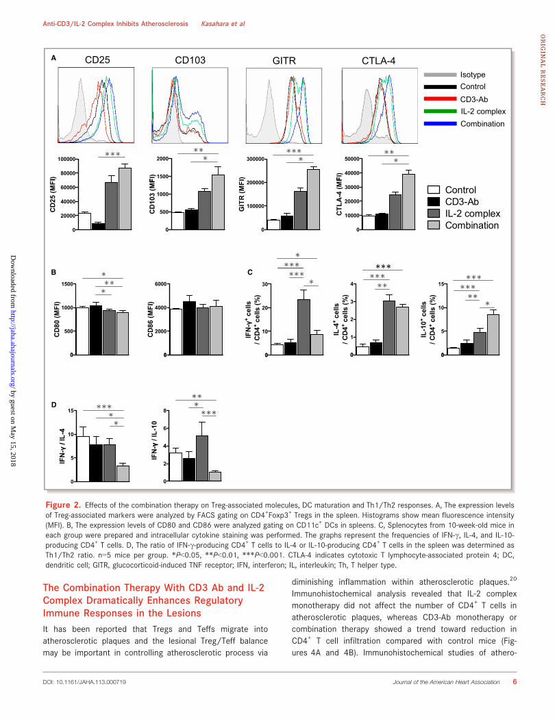
Effects of CD3 Ab, IL-2 Complex, or CombinationTherapy on Treg-Associated Molecules, DCMaturation and Th1/Th2 BalanceNext, the effects of each therapy on the expression of Treg-associated molecules in Foxp3+ Tregs were determined byflow cytometry. Notably, Foxp3+ Tregs from CD3-Ab/IL-2complex-treated mice expressed higher levels of CD25,CD103, glucocorticoid-induced TNF receptor family-relatedgene/protein (GITR), cytotoxic T lymphocyte-associatedprotein 4 (CTLA-4) compared to those from control-treatedor CD3-Ab-treated mice (Figure 2A), implying an activatedphenotype of Tregs after the combination therapy. Inaddition, a trend towards increased expression of Treg-associated molecules was seen in mice with IL-2 complexalone. We also investigated the effect of each therapy onsurface maturation markers CD80 and CD86 in splenicCD11c+ DCs. Neither CD3-Ab nor IL-2 complex monother-apy altered the expression of these maturation markers insplenic DCs, whereas we observed a modest but significantdecrease in the expression of CD80, but not CD86, insplenic DCs of CD3-Ab/IL-2 complex-treated mice (Fig-ure 2B). To determine whether the combination treatmentchanged T-cell responses and polarization, we examinedcytokine secretion from CD4+ T cells by intracellularcytokine staining. We found that IL-2 complex mono-therapydramatically increased the percentage of splenic IFN-cproducing-Th1 cells, IL-4-producing Th2 cells, and IL-10-producing CD4+ T cells compared to control-treated mice(Figure 2C). Notably, co-treatment with CD3-Ab canceledthe increase in IFN-c producing-CD4+ T cells by IL-2complex treatment, while the increase in IL-4 or IL-10-
producing-CD4+ T cells was still observed (Figure 2C). TheTh1/Th2 ratio was much lower in the spleen of the micewith the combination therapy compared with mice in othergroups (Figure 2D). Taken together, these results indicatethat the combination therapy can induce activated pheno-type of Tregs and the Th1/Th2 balance to Th2, which maycontribute to a reduction in atherosclerosis development viasuppressing inflammatory responses.
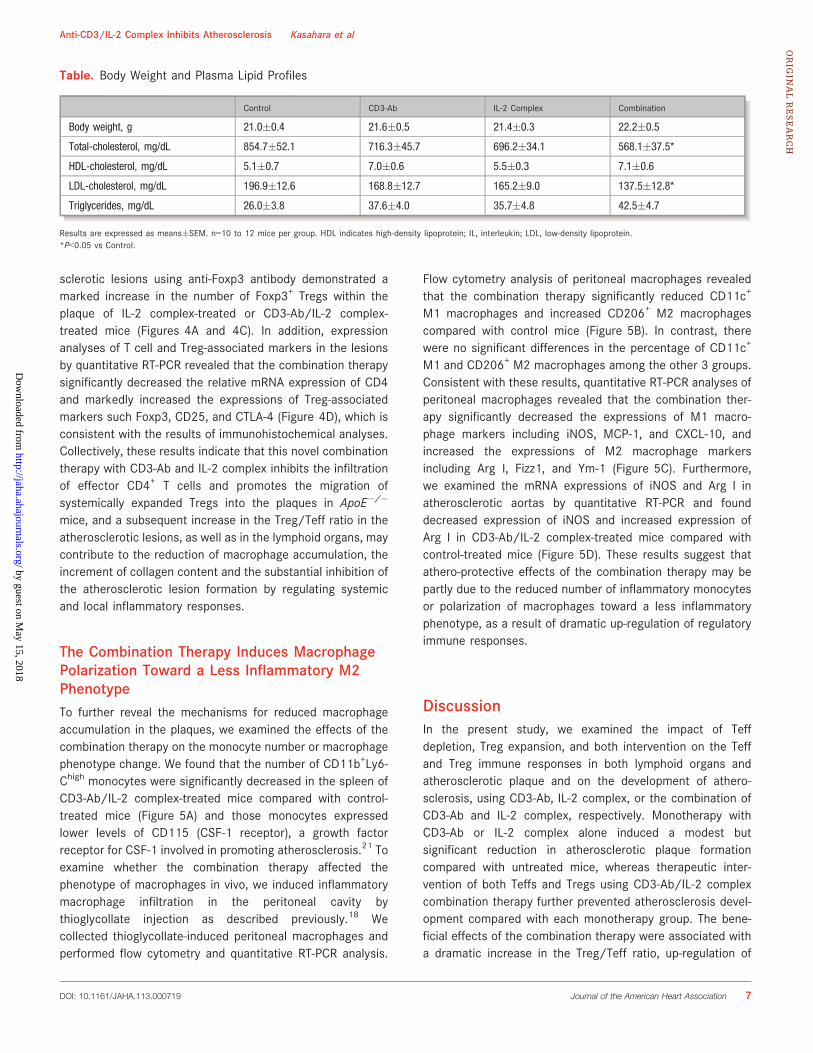
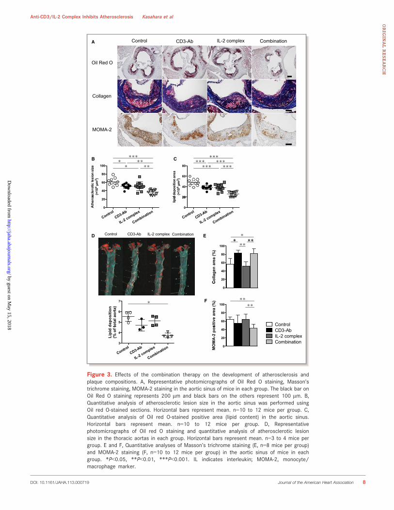
The Combination Therapy With CD3 Ab and IL-2Complex Inhibits Atherosclerotic LesionFormation and Induces Stable Plaque PhenotypeTo determine the effects of CD3-Ab, IL-2 complex, or combi-nation therapy on atherosclerosis development, we assessedthe atherosclerotic lesion formation of the 4 experimentalgroups. No adverse effects were observed in all groupsthroughout the experiments. The mice receiving combinedtherapy showed a decrease in plasma total cholesterol andLDL cholesterol levels and an increase in triglyceride levelscompared with control-treated mice, whereas no statisticaldifferences in plasma lipid profiles were detected among theother 3 groups (Table). CD3-Ab or IL-2 complex monotherapycaused a modest but significant reduction in atheroscleroticlesion formation in the aortic root compared with control-treated mice (61.6�2.89104 lm2 in control-treated mice,50.6�1.79104 lm2 in CD3-Ab-treated mice, 50.7�1.99104 lm2 in IL-2 complex-treated mice; Figures 3A and 3B).Notably, CD3-Ab/IL-2 complex-treated mice showed a furtherreduction in atherosclerotic lesion formation (37.2�1.99104 lm2, Figures 3A and 3B) compared with control-treatedand each monotherapy mice. Consistent with this, the lipidcontent of the plaques in the aortic sinus was also significantlydecreased in the CD3-Ab/IL-2 complex-treated mice comparedto control-treated and each monotherapy mice (Figure 3C). Inparallel with the cross-sectional studies, we performed en faceanalysis of thoracic aortas, revealing a significant reduction inaortic plaque burden in CD3-Ab/IL-2 complex-treated mice(3.74�0.12%) compared with control mice (5.53�0.23%)(Figure 3D).
To determine the effects of each therapy on plaquecomposition, immunohistochemical studies of atheroscleroticlesions in the aortic sinus were performed. Collagen contentsin atherosclerotic lesions were significantly increased in CD3-Ab-treated and CD3-Ab/IL-2 complex-treated mice comparedto control-treated mice (Figures 3A and 3E). Although CD3-Abor IL-2 complex monotherapy did not affect the recruitment ofmacrophages in atherosclerotic plaques, the lesions of CD3-Ab/IL-2 complex-treated mice showed a significant reductionin the macrophage accumulation compared to control mice(Figures 3A and 3F).
DOI: 10.1161/JAHA.113.000719 Journal of the American Heart Association 5
Anti-CD3/IL-2 Complex Inhibits Atherosclerosis Kasahara et alORIG
INALRESEARCH
by guest on May 15, 2018
http://jaha.ahajournals.org/D
ownloaded from
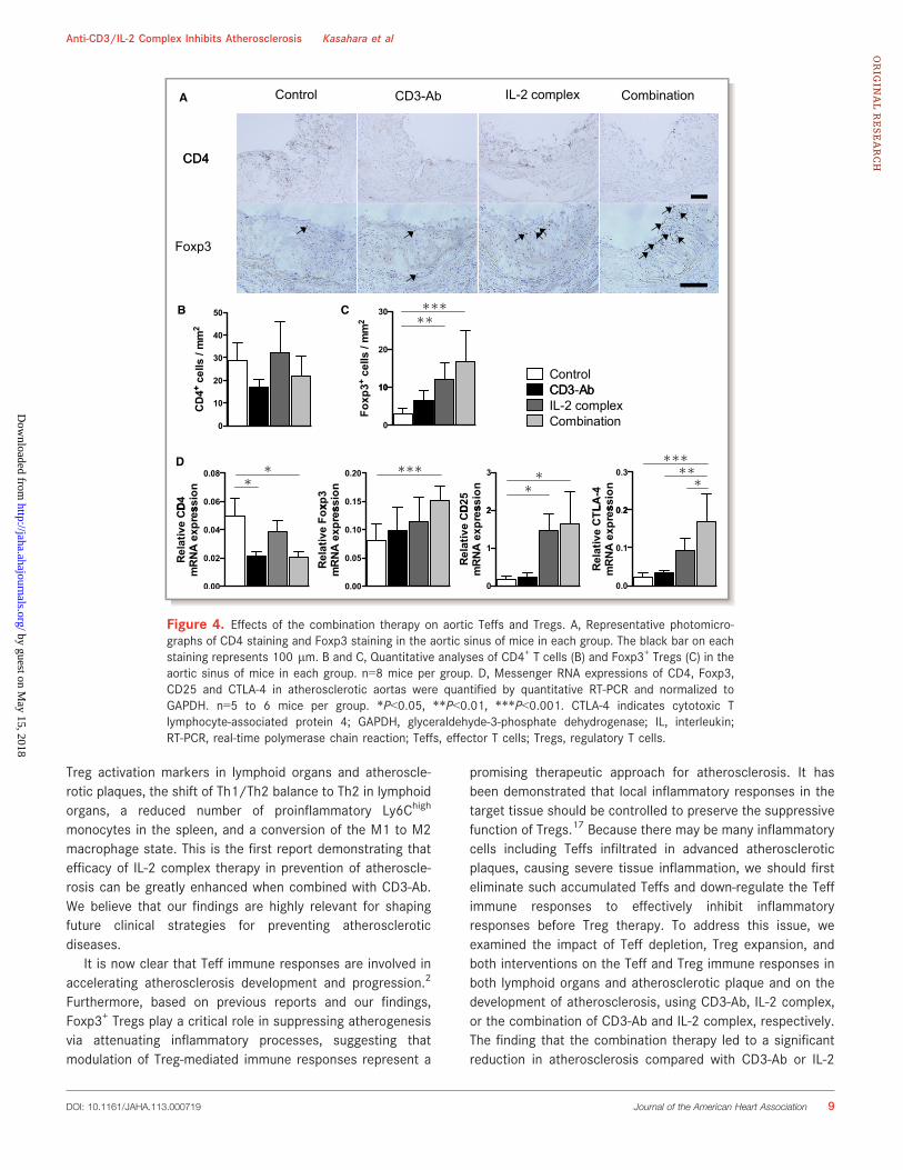
The Combination Therapy With CD3 Ab and IL-2Complex Dramatically Enhances RegulatoryImmune Responses in the LesionsIt has been reported that Tregs and Teffs migrate intoatherosclerotic plaques and the lesional Treg/Teff balancemay be important in controlling atherosclerotic process via
diminishing inflammation within atherosclerotic plaques.20
Immunohistochemical analysis revealed that IL-2 complexmonotherapy did not affect the number of CD4+ T cells inatherosclerotic plaques, whereas CD3-Ab monotherapy orcombination therapy showed a trend toward reduction inCD4+ T cell infiltration compared with control mice (Fig-ures 4A and 4B). Immunohistochemical studies of athero-
A CD25 CD103 GITR CTLA-4
ControlIsotype
CD3-AbIL-2 complexCombination
B C
ControlCD3-AbIL-2 complexCombination
0
2000
4000
6000
CD
86 (M
FI)
D
0
Figure 2. Effects of the combination therapy on Treg-associated molecules, DC maturation and Th1/Th2 responses. A, The expression levelsof Treg-associated markers were analyzed by FACS gating on CD4+Foxp3+ Tregs in the spleen. Histograms show mean fluorescence intensity(MFI). B, The expression levels of CD80 and CD86 were analyzed gating on CD11c+ DCs in spleens. C, Splenocytes from 10-week-old mice ineach group were prepared and intracellular cytokine staining was performed. The graphs represent the frequencies of IFN-c, IL-4, and IL-10-producing CD4+ T cells. D, The ratio of IFN-c-producing CD4+ T cells to IL-4 or IL-10-producing CD4+ T cells in the spleen was determined asTh1/Th2 ratio. n=5 mice per group. *P<0.05, **P<0.01, ***P<0.001. CTLA-4 indicates cytotoxic T lymphocyte-associated protein 4; DC,dendritic cell; GITR, glucocorticoid-induced TNF receptor; IFN, interferon; IL, interleukin; Th, T helper type.
DOI: 10.1161/JAHA.113.000719 Journal of the American Heart Association 6
Anti-CD3/IL-2 Complex Inhibits Atherosclerosis Kasahara et alORIG
INALRESEARCH
by guest on May 15, 2018
http://jaha.ahajournals.org/D
ownloaded from
sclerotic lesions using anti-Foxp3 antibody demonstrated amarked increase in the number of Foxp3+ Tregs within theplaque of IL-2 complex-treated or CD3-Ab/IL-2 complex-treated mice (Figures 4A and 4C). In addition, expressionanalyses of T cell and Treg-associated markers in the lesionsby quantitative RT-PCR revealed that the combination therapysignificantly decreased the relative mRNA expression of CD4and markedly increased the expressions of Treg-associatedmarkers such Foxp3, CD25, and CTLA-4 (Figure 4D), which isconsistent with the results of immunohistochemical analyses.Collectively, these results indicate that this novel combinationtherapy with CD3-Ab and IL-2 complex inhibits the infiltrationof effector CD4+ T cells and promotes the migration ofsystemically expanded Tregs into the plaques in ApoE�/�
mice, and a subsequent increase in the Treg/Teff ratio in theatherosclerotic lesions, as well as in the lymphoid organs, maycontribute to the reduction of macrophage accumulation, theincrement of collagen content and the substantial inhibition ofthe atherosclerotic lesion formation by regulating systemicand local inflammatory responses.
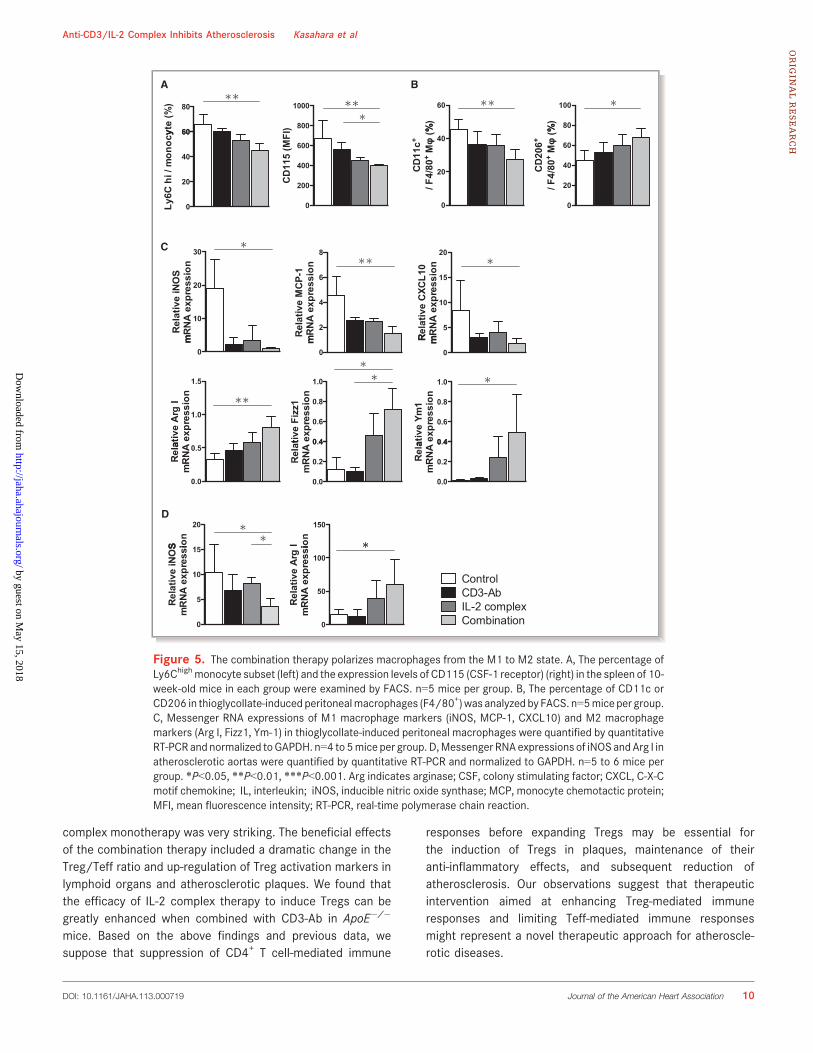
The Combination Therapy Induces MacrophagePolarization Toward a Less Inflammatory M2PhenotypeTo further reveal the mechanisms for reduced macrophageaccumulation in the plaques, we examined the effects of thecombination therapy on the monocyte number or macrophagephenotype change. We found that the number of CD11b+Ly6-Chigh monocytes were significantly decreased in the spleen ofCD3-Ab/IL-2 complex-treated mice compared with control-treated mice (Figure 5A) and those monocytes expressedlower levels of CD115 (CSF-1 receptor), a growth factorreceptor for CSF-1 involved in promoting atherosclerosis.21 Toexamine whether the combination therapy affected thephenotype of macrophages in vivo, we induced inflammatorymacrophage infiltration in the peritoneal cavity bythioglycollate injection as described previously.18 Wecollected thioglycollate-induced peritoneal macrophages andperformed flow cytometry and quantitative RT-PCR analysis.
Flow cytometry analysis of peritoneal macrophages revealedthat the combination therapy significantly reduced CD11c+
M1 macrophages and increased CD206+ M2 macrophagescompared with control mice (Figure 5B). In contrast, therewere no significant differences in the percentage of CD11c+
M1 and CD206+ M2 macrophages among the other 3 groups.Consistent with these results, quantitative RT-PCR analyses ofperitoneal macrophages revealed that the combination ther-apy significantly decreased the expressions of M1 macro-phage markers including iNOS, MCP-1, and CXCL-10, andincreased the expressions of M2 macrophage markersincluding Arg I, Fizz1, and Ym-1 (Figure 5C). Furthermore,we examined the mRNA expressions of iNOS and Arg I inatherosclerotic aortas by quantitative RT-PCR and founddecreased expression of iNOS and increased expression ofArg I in CD3-Ab/IL-2 complex-treated mice compared withcontrol-treated mice (Figure 5D). These results suggest thatathero-protective effects of the combination therapy may bepartly due to the reduced number of inflammatory monocytesor polarization of macrophages toward a less inflammatoryphenotype, as a result of dramatic up-regulation of regulatoryimmune responses.
DiscussionIn the present study, we examined the impact of Teffdepletion, Treg expansion, and both intervention on the Teffand Treg immune responses in both lymphoid organs andatherosclerotic plaque and on the development of athero-sclerosis, using CD3-Ab, IL-2 complex, or the combination ofCD3-Ab and IL-2 complex, respectively. Monotherapy withCD3-Ab or IL-2 complex alone induced a modest butsignificant reduction in atherosclerotic plaque formationcompared with untreated mice, whereas therapeutic inter-vention of both Teffs and Tregs using CD3-Ab/IL-2 complexcombination therapy further prevented atherosclerosis devel-opment compared with each monotherapy group. The bene-ficial effects of the combination therapy were associated witha dramatic increase in the Treg/Teff ratio, up-regulation of
Table. Body Weight and Plasma Lipid Profiles
Control CD3-Ab IL-2 Complex Combination
Body weight, g 21.0�0.4 21.6�0.5 21.4�0.3 22.2�0.5
Total-cholesterol, mg/dL 854.7�52.1 716.3�45.7 696.2�34.1 568.1�37.5*
HDL-cholesterol, mg/dL 5.1�0.7 7.0�0.6 5.5�0.3 7.1�0.6
LDL-cholesterol, mg/dL 196.9�12.6 168.8�12.7 165.2�9.0 137.5�12.8*
Triglycerides, mg/dL 26.0�3.8 37.6�4.0 35.7�4.8 42.5�4.7
Results are expressed as means�SEM. n=10 to 12 mice per group. HDL indicates high-density lipoprotein; IL, interleukin; LDL, low-density lipoprotein.*P<0.05 vs Control.
DOI: 10.1161/JAHA.113.000719 Journal of the American Heart Association 7
Anti-CD3/IL-2 Complex Inhibits Atherosclerosis Kasahara et alORIG
INALRESEARCH
by guest on May 15, 2018
http://jaha.ahajournals.org/D
ownloaded from
A Control CD3-Ab IL-2 complex Combination
Oil Red O
Collagen
MOMA-2
CB
D EControl CD3-Ab IL-2 complex Combination
Control
F
CD3-AbIL-2 complexCombination
Figure 3. Effects of the combination therapy on the development of atherosclerosis andplaque compositions. A, Representative photomicrographs of Oil Red O staining, Masson’strichrome staining, MOMA-2 staining in the aortic sinus of mice in each group. The black bar onOil Red O staining represents 200 lm and black bars on the others represent 100 lm. B,Quantitative analysis of atherosclerotic lesion size in the aortic sinus was performed usingOil red O-stained sections. Horizontal bars represent mean. n=10 to 12 mice per group. C,Quantitative analysis of Oil red O-stained positive area (lipid content) in the aortic sinus.Horizontal bars represent mean. n=10 to 12 mice per group. D, Representativephotomicrographs of Oil red O staining and quantitative analysis of atherosclerotic lesionsize in the thoracic aortas in each group. Horizontal bars represent mean. n=3 to 4 mice pergroup. E and F, Quantitative analyses of Masson’s trichrome staining (E, n=8 mice per group)and MOMA-2 staining (F, n=10 to 12 mice per group) in the aortic sinus of mice in eachgroup. *P<0.05, **P<0.01, ***P<0.001. IL indicates interleukin; MOMA-2, monocyte/macrophage marker.
DOI: 10.1161/JAHA.113.000719 Journal of the American Heart Association 8
Anti-CD3/IL-2 Complex Inhibits Atherosclerosis Kasahara et alORIG
INALRESEARCH
by guest on May 15, 2018
http://jaha.ahajournals.org/D
ownloaded from
Treg activation markers in lymphoid organs and atheroscle-rotic plaques, the shift of Th1/Th2 balance to Th2 in lymphoidorgans, a reduced number of proinflammatory Ly6Chigh
monocytes in the spleen, and a conversion of the M1 to M2macrophage state. This is the first report demonstrating thatefficacy of IL-2 complex therapy in prevention of atheroscle-rosis can be greatly enhanced when combined with CD3-Ab.We believe that our findings are highly relevant for shapingfuture clinical strategies for preventing atheroscleroticdiseases.
It is now clear that Teff immune responses are involved inaccelerating atherosclerosis development and progression.2
Furthermore, based on previous reports and our findings,Foxp3+ Tregs play a critical role in suppressing atherogenesisvia attenuating inflammatory processes, suggesting thatmodulation of Treg-mediated immune responses represent a
promising therapeutic approach for atherosclerosis. It hasbeen demonstrated that local inflammatory responses in thetarget tissue should be controlled to preserve the suppressivefunction of Tregs.17 Because there may be many inflammatorycells including Teffs infiltrated in advanced atheroscleroticplaques, causing severe tissue inflammation, we should firsteliminate such accumulated Teffs and down-regulate the Teffimmune responses to effectively inhibit inflammatoryresponses before Treg therapy. To address this issue, weexamined the impact of Teff depletion, Treg expansion, andboth interventions on the Teff and Treg immune responses inboth lymphoid organs and atherosclerotic plaque and on thedevelopment of atherosclerosis, using CD3-Ab, IL-2 complex,or the combination of CD3-Ab and IL-2 complex, respectively.The finding that the combination therapy led to a significantreduction in atherosclerosis compared with CD3-Ab or IL-2
CD4
Control CD3-Ab IL-2 complex CombinationA
CD4
Foxp3
B C
20
30
40
50
+ ce
lls /
mm
2
ControlCD3 Ab
D
0
10CD
4+ CD3-AbIL-2 complexCombination
Figure 4. Effects of the combination therapy on aortic Teffs and Tregs. A, Representative photomicro-graphs of CD4 staining and Foxp3 staining in the aortic sinus of mice in each group. The black bar on eachstaining represents 100 lm. B and C, Quantitative analyses of CD4+ T cells (B) and Foxp3+ Tregs (C) in theaortic sinus of mice in each group. n=8 mice per group. D, Messenger RNA expressions of CD4, Foxp3,CD25 and CTLA-4 in atherosclerotic aortas were quantified by quantitative RT-PCR and normalized toGAPDH. n=5 to 6 mice per group. *P<0.05, **P<0.01, ***P<0.001. CTLA-4 indicates cytotoxic Tlymphocyte-associated protein 4; GAPDH, glyceraldehyde-3-phosphate dehydrogenase; IL, interleukin;RT-PCR, real-time polymerase chain reaction; Teffs, effector T cells; Tregs, regulatory T cells.
DOI: 10.1161/JAHA.113.000719 Journal of the American Heart Association 9
Anti-CD3/IL-2 Complex Inhibits Atherosclerosis Kasahara et alORIG
INALRESEARCH
by guest on May 15, 2018
http://jaha.ahajournals.org/D
ownloaded from
complex monotherapy was very striking. The beneficial effectsof the combination therapy included a dramatic change in theTreg/Teff ratio and up-regulation of Treg activation markers inlymphoid organs and atherosclerotic plaques. We found thatthe efficacy of IL-2 complex therapy to induce Tregs can begreatly enhanced when combined with CD3-Ab in ApoE�/�
mice. Based on the above findings and previous data, wesuppose that suppression of CD4+ T cell-mediated immune
responses before expanding Tregs may be essential forthe induction of Tregs in plaques, maintenance of theiranti-inflammatory effects, and subsequent reduction ofatherosclerosis. Our observations suggest that therapeuticintervention aimed at enhancing Treg-mediated immuneresponses and limiting Teff-mediated immune responsesmight represent a novel therapeutic approach for atheroscle-rotic diseases.
BA
C
D
ControlCD3-AbIL-2 complexCombination
Figure 5. The combination therapy polarizes macrophages from the M1 to M2 state. A, The percentage ofLy6Chighmonocyte subset (left) and the expression levels of CD115 (CSF-1 receptor) (right) in the spleen of 10-week-old mice in each group were examined by FACS. n=5 mice per group. B, The percentage of CD11c orCD206 in thioglycollate-induced peritonealmacrophages (F4/80+)was analyzed by FACS. n=5miceper group.C, Messenger RNA expressions of M1 macrophage markers (iNOS, MCP-1, CXCL10) and M2 macrophagemarkers (Arg I, Fizz1, Ym-1) in thioglycollate-induced peritoneal macrophages were quantified by quantitativeRT-PCR and normalized toGAPDH. n=4 to 5mice per group.D,Messenger RNAexpressions of iNOSandArg I inatherosclerotic aortas were quantified by quantitative RT-PCR and normalized to GAPDH. n=5 to 6 mice pergroup. *P<0.05, **P<0.01, ***P<0.001. Arg indicates arginase; CSF, colony stimulating factor; CXCL, C-X-Cmotif chemokine; IL, interleukin; iNOS, inducible nitric oxide synthase; MCP, monocyte chemotactic protein;MFI, mean fluorescence intensity; RT-PCR, real-time polymerase chain reaction.
DOI: 10.1161/JAHA.113.000719 Journal of the American Heart Association 10
Anti-CD3/IL-2 Complex Inhibits Atherosclerosis Kasahara et alORIG
INALRESEARCH
by guest on May 15, 2018
http://jaha.ahajournals.org/D
ownloaded from

Recent studies have demonstrated that treatment with IL-2complex selectively increases CD4+CD25+Foxp3+ Tregs with-out affecting other immune cells including CD4+ T cells, CD8+
T cells, or natural killer cells, although the immune responseand activation status of helper CD4+ T cells were notexamined after treatment.14,15 However, as shown in Fig-ure 2A and 2B, we unexpectedly observed that IL-2 complexsignificantly induced the activation of both Teffs and Tregs.CD25 molecule is a component of the high-affinity IL-2receptor (IL-2R) and is functionally essential for Treg devel-opment by binding IL-2.9 However, this molecule is expressednot only on Tregs but also on activated immune cells such asTeffs, for proliferation through IL-2R signaling.22 Taking thisinto account, it is possible that IL-2 complex affects theactivation and proliferation of both Teffs and Tregs. Impor-tantly, we showed that the additional treatment with CD3-Abin IL-2 complex-treated mice abolished the up-regulation ofatherogenic Th1 immune responses, while the up-regulationof the immune responses such as IL-4 or IL-10 productionfrom CD4+ T cells remained unchanged, suggesting thataddition of CD3-Ab may enhance the efficacy and reduce therisk of side effects associated with IL-2 complex treatment.Recent evidence suggests that several subsets of Tregsdifferentiate from na€ıve T cells in the periphery under certainconditions and have similar immunological properties withthymus-derived Foxp3+ Tregs.9 Previous studies showed thatperipherally generated Tregs such as Tr1 cells or CD4+LAP+
(latency-associated peptide) Tregs inhibit atherosclerosis inatherosclerosis-prone mice by producing IL-10 or TGF-b,respectively.7,23 Our data indicate that induction of Tr1 cellsmay also contribute to the reduction of atherosclerosisfollowing CD3-Ab/IL-2 complex combination therapy.
In the present study, we found that combination therapydramatically increased not only the number of Foxp3+ Tregsbut also the expressions of Treg activation markers in bothlymphoid organs and atherosclerotic plaques. Our finding ofmarked up-regulation of CTLA-4, one of Treg activationmarkers, in Foxp3+ Tregs in CD3-Ab/IL-2 complex-treatedmice is interesting, because CTLA-4-dependent suppressionof antigen-presenting cell (APC) function including DCs byCD80 or CD86 down-regulation is supposed to be a keymechanism for Treg-mediated suppression.24 Such DCs withlow expression levels of CD80/CD86 are called “tolerogenicDCs” and have been shown to contribute to the inhibition ofatherosclerosis development and the regression of estab-lished plaques by inducing Tregs and inhibiting Teffs.25,26
Although we observed a modest decrease in the CD80expression in splenic DCs of CD3-Ab/IL-2 complex-treatedmice, whether the lesional suppression mechanisms throughthis pathway are involved in the reduction of atherosclerosisremains unclear and further studies are needed. To the best ofour knowledge, we believe that this combination therapy is
the most effective approach for specifically promotingregulatory immune responses.
A recent study has demonstrated that hypercholesterol-emia decreases the Treg/Teff ratio in atherosclerotic lesionsvia inhibiting Treg accumulation within plaques.20 This impliesthe possibility that increasing proportion of Tregs in athero-sclerotic lesions could be a hopeful strategy to dampenplaque inflammation and prevent atherosclerosis, althoughfurther extensive experiments are required to identify theexact role of intraplaque Tregs in atherogenesis. In this study,we showed that the marked reduction of atherosclerosis fromcombination therapy was associated with a dramatic increasein the Treg/Teff ratio and up-regulation of Treg activationmarkers not only in lymphoid organs but also in atheroscle-rotic plaques, implying that the beneficial effects of thecombination therapy may partly be due to the induction ofTregs with activated phenotype in atherosclerotic plaques.
Monocytes infiltration from the peripheral blood to thesubendothelial space or intima followed by macrophagedifferentiation is believed to be critical in initiation ofatherosclerosis in humans and animals. The extent ofmacrophage recruitment into atherosclerotic plaques maydepend on the cholesterol levels or monocyte number in theblood.27 In the present study, we found that the combinationtherapy significantly reduced proinflammatory Ly6Chigh mono-cytes compared to untreated or monotherapy groups,suggesting a possible contribution to decreased macrophageaccumulation in atherosclerotic plaques. We found thatthioglycollate-induced peritoneal macrophages from CD3-Ab/IL-2 complex-treated mice showed lower mRNA expres-sions of M1 markers and higher expressions of M2 markers.Regarding macrophage markers, a similar trend was observedin the atherosclerotic aortas of CD3-Ab/IL-2 complex-treatedmice. Th1 cells are reported to contribute to generation of M1macrophages, whereas Th2 cells promote generation of M2macrophages.28 In addition, IL-10 derived from Tregs inducesSTAT3 (Signal Transducer and Activator of Transcription 3)activation and drives M2 polarization in mice with severecombined immunodeficiency.29 Collectively, we suppose thatin CD3-Ab/IL-2 complex-treated mice, phenotype change ofthioglycollate-induced peritoneal macrophages toward M2phenotype may be attributable to dramatic expansion of Tregsas well as a shift from Th1 to Th2 immune responses, whichrepresents one possible explanation for the athero-protectiveeffects of the combination therapy.
In conclusion, we have demonstrated that combinedintervention using CD3-Ab and IL-2 complex induced aremarkable inhibition of atherosclerosis via dramaticallyincreasing the Treg/Teff ratio in lymphoid organs andatherosclerotic plaques, which caused macrophage polariza-tion toward a less inflammatory M2 phenotype. Our dataimply that therapeutic intervention aimed at enhancing
DOI: 10.1161/JAHA.113.000719 Journal of the American Heart Association 11
Anti-CD3/IL-2 Complex Inhibits Atherosclerosis Kasahara et alORIG
INALRESEARCH
by guest on May 15, 2018
http://jaha.ahajournals.org/D
ownloaded from

Treg-mediated immune responses and limiting Teff-mediatedimmune responses might represent a novel therapeuticapproach for atherosclerotic diseases.
AcknowledgmentsWe would like to thank Tomomi Minami for technical assistance andNaoki Kitano for critical reading of the manuscript.
Sources of FundingThis work was supported by Japan Society for the Promotion ofScience KAKENHI Grant Number 23790849 (N. Sasaki),25860601 (N. Sasaki), and 24591114 (Yamashita), researchgrants from the Global Center of Excellence (Kasahara),Suzuken Memorial Foundation (N. Sasaki), ONO MedicalResearch Foundation (N. Sasaki), Takeda ScientificFoundation (Yamashita and N. Sasaki), Senshin MedicalResearch Foundation (Yamashita), Mitsui Life Social WelfareFoundation (Yamashita), Yakult Bioscience Research Founda-tion (Yamashita), UeharaMemorial Foundation (Hirata), and TheJapan Circulation Society Translational Research Foundation(Hirata).
DisclosuresNone.
References1. Ross R. Atherosclerosis—an inflammatory disease. N Engl J Med.
1999;340:115–126.
2. Hansson GK, Hermansson A. The immune system in atherosclerosis. NatImmunol. 2011;12:204–212.
3. Gupta S, Pablo AM, Jiang X, Wang N, Tall AR, Schindler C. IFN-gammapotentiates atherosclerosis in apoE knock-out mice. J Clin Invest. 1997;99:2752–2761.
4. Ait-Oufella H, Salomon BL, Potteaux S, Robertson AK, Gourdy P, Zoll J, MervalR, Esposito B, Cohen JL, Fisson S, Flavell RA, Hansson GK, Klatzmann D, TedguiA, Mallat Z. Natural regulatory T cells control the development of atheroscle-rosis in mice. Nat Med. 2006;12:178–180.
5. Gotsman I, Grabie N, Gupta R, Dacosta R, MacConmara M, Lederer J, SukhovaG, Witztum JL, Sharpe AH, Lichtman AH. Impaired regulatory T-cell responseand enhanced atherosclerosis in the absence of inducible costimulatorymolecule. Circulation. 2006;114:2047–2055.
6. Mor A, Planer D, Luboshits G, Afek A,Metzger S, Chajek-Shaul T, Keren G, GeorgeJ. Role of naturally occurring CD4+ CD25+ regulatory T cells in experimentalatherosclerosis. Arterioscler Thromb Vasc Biol. 2007;27:893–900.
7. Sasaki N, Yamashita T, Takeda M, Shinohara M, Nakajima K, Tawa H, Usui T,Hirata K. Oral anti-CD3 antibody treatment induces regulatory T cells andinhibits the development of atherosclerosis in mice. Circulation.2009;120:1996–2005.
8. Sasaki N, Yamashita T, Takeda M, Hirata K. Regulatory T cells in atherogen-esis. J Atheroscler Thromb. 2012;19:503–515.
9. Sakaguchi S, Yamaguchi T, Nomura T, Ono M. Regulatory T cells and immunetolerance. Cell. 2008;133:775–787.
10. Klingenberg R, Gerdes N, Badeau RM, Gistera A, Strodthoff D, Ketelhuth DF,Lundberg AM, Rudling M, Nilsson SK, Olivecrona G, Zoller S, Lohmann C,
Luscher TF, Jauhiainen M, Sparwasser T, Hansson GK. Depletion of FOXP3+
regulatory T cells promotes hypercholesterolemia and atherosclerosis. J ClinInvest. 2013;123:1323–1334.
11. Steffens S, Burger F, Pelli G, Dean Y, Elson G, Kosco-Vilbois M, Chatenoud L,Mach F. Short-term treatment with anti-CD3 antibody reduces the develop-ment and progression of atherosclerosis in mice. Circulation. 2006;114:1977–1984.
12. Belghith M, Bluestone JA, Barriot S, Megret J, Bach JF, Chatenoud L. TGF-beta-dependent mechanisms mediate restoration of self-tolerance induced byantibodies to CD3 in overt autoimmune diabetes. Nat Med. 2003;9:1202–1208.
13. Chatenoud L, Bluestone JA. CD3-specific antibodies: a portal to the treatmentof autoimmunity. Nat Rev Immunol. 2007;7:622–632.
14. Webster KE, Walters S, Kohler RE, Mrkvan T, Boyman O, Surh CD, Grey ST,Sprent J. In vivo expansion of T reg cells with IL-2-mAb complexes: induction ofresistance to EAE and long-term acceptance of islet allografts withoutimmunosuppression. J Exp Med. 2009;206:751–760.
15. Dinh TN, Kyaw TS, Kanellakis P, To K, Tipping P, Toh BH, Bobik A, Agrotis A.Cytokine therapy with interleukin-2/anti-interleukin-2 monoclonal antibodycomplexes expands CD4+CD25+Foxp3+ regulatory T cells and attenuatesdevelopment and progression of atherosclerosis. Circulation. 2012;126:1256–1266.
16. Foks AC, Frodermann V, ter Borg M, Habets KL, Bot I, Zhao Y, van Eck M, vanBerkel TJ, Kuiper J, van Puijvelde GH. Differential effects of regulatory T cellson the initiation and regression of atherosclerosis. Atherosclerosis. 2011;218:53–60.
17. Korn T, Reddy J, Gao W, Bettelli E, Awasthi A, Petersen TR, Backstrom BT,Sobel RA, Wucherpfennig KW, Strom TB, Oukka M, Kuchroo VK. Myelin-specific regulatory T cells accumulate in the CNS but fail to controlautoimmune inflammation. Nat Med. 2007;13:423–431.
18. Yamashita T, Kawashima S, Hirase T, Shinohara M, Takaya T, Sasaki N, TakedaM, Tawa H, Inoue N, Hirata K, Yokoyama M. Xenogenic macrophageimmunization reduces atherosclerosis in apolipoprotein E knockout mice.Am J Physiol Cell Physiol. 2007;293:C865–C873.
19. Gagliani N, Magnani CF, Huber S, Gianolini ME, Pala M, Licona-Limon P, Guo B,Herbert DR, Bulfone A, Trentini F, Di Serio C, Bacchetta R, Andreani M,Brockmann L, Gregori S, Flavell RA, Roncarolo MG. Coexpression of CD49band LAG-3 identifies human and mouse T regulatory type 1 cells. Nat Med.2013;19:739–746.
20. Maganto-Garcia E, Tarrio ML, Grabie N, Bu DX, Lichtman AH. Dynamic changesin regulatory T cells are linked to levels of diet-induced hypercholesterolemia.Circulation. 2011;124:185–195.
21. Shaposhnik Z, Wang X, Lusis AJ. Arterial colony stimulating factor-1 influencesatherosclerotic lesions by regulating monocyte migration and apoptosis. J LipidRes. 2010;51:1962–1970.
22. Boyman O, Sprent J. The role of interleukin-2 during homeostasis andactivation of the immune system. Nat Rev Immunol. 2012;12:180–190.
23. Mallat Z, Gojova A, Brun V, Esposito B, Fournier N, Cottrez F, Tedgui A, GrouxH. Induction of a regulatory T cell type 1 response reduces the development ofatherosclerosis in apolipoprotein E-knockout mice. Circulation. 2003;108:1232–1237.
24. Takeda M, Yamashita T, Sasaki N, Hirata K. Dendritic cells in atherogenesis:possible novel targets for prevention of atherosclerosis. J Atheroscler Thromb.2012;19:953–961.
25. Takeda M, Yamashita T, Sasaki N, Nakajima K, Kita T, Shinohara M, Ishida T,Hirata K. Oral administration of an active form of vitamin D3 (calcitriol)decreases atherosclerosis in mice by inducing regulatory T cells and immaturedendritic cells with tolerogenic functions. Arterioscler Thromb Vasc Biol.2010;30:2495–2503.
26. Nakajima K, Yamashita T, Kita T, Takeda M, Sasaki N, Kasahara K, ShinoharaM, Rikitake Y, Ishida T, Yokoyama M, Hirata K. Orally administeredeicosapentaenoic acid induces rapid regression of atherosclerosis viamodulating the phenotype of dendritic cells in LDL receptor-deficient mice.Arterioscler Thromb Vasc Biol. 2011;31:1963–1972.
27. Swirski FK, Libby P, Aikawa E, Alcaide P, Luscinskas FW, Weissleder R, PittetMJ. Ly-6Chi monocytes dominate hypercholesterolemia-associated monocy-tosis and give rise to macrophages in atheromata. J Clin Invest. 2007;117:195–205.
28. Mosser DM, Edwards JP. Exploring the full spectrum of macrophage activation.Nat Rev Immunol. 2008;8:958–969.
29. Biswas SK, Mantovani A. Macrophage plasticity and interaction with lympho-cyte subsets: cancer as a paradigm. Nat Immunol. 2010;11:889–896.
DOI: 10.1161/JAHA.113.000719 Journal of the American Heart Association 12
Anti-CD3/IL-2 Complex Inhibits Atherosclerosis Kasahara et alORIG
INALRESEARCH
by guest on May 15, 2018
http://jaha.ahajournals.org/D
ownloaded from
CD3 Antibody and IL-2 Complex Combination Therapy InhibitsAtherosclerosis by Augmenting a Regulatory Immune Response
I n the article by Kasahara et al, “CD3 Antibody and IL-2Complex Combination Therapy Inhibits Atherosclerosis by
Augmenting a Regulatory Immune Response,” which pub-lished online April 22, 2014, and appeared in the April 2014issue of the journal (J Am Heart Assoc. 2014;3:e000719 doi:10.1161/JAHA.113.000719), an error in the Acknowledg-
ments has been corrected. On page 12, left column, TomomiMinami should read Satomi Minami.
The author regrets this error.The online version of the article has been updated and
is available at http://jaha.ahajournals.org/content/3/2/e000719.
J Am Heart Assoc. 2015;4:e000750 doi: 10.1161/JAHA.115.000750.
ª 2015 The Authors. Published on behalf of the American Heart Association,Inc., by Wiley Blackwell. This is an open access article under the terms of theCreative Commons Attribution-NonCommercial License, which permits use,distribution and reproduction in any medium, provided the original work isproperly cited and is not used for commercial purposes.
DOI: 10.1161/JAHA.115.000750 Journal of the American Heart Association 1
CORRECTION

Sasaki, Masafumi Takeda and Ken-ichi HirataKazuyuki Kasahara, Naoto Sasaki, Tomoya Yamashita, Tomoyuki Kita, Keiko Yodoi, Yoshihiro
Augmenting a Regulatory Immune ResponseCD3 Antibody and IL-2 Complex Combination Therapy Inhibits Atherosclerosis by
Online ISSN: 2047-9980 Dallas, TX 75231
is published by the American Heart Association, 7272 Greenville Avenue,Journal of the American Heart AssociationThe doi: 10.1161/JAHA.113.000719
2014;3:e000719; originally published April 22, 2014;J Am Heart Assoc.
http://jaha.ahajournals.org/content/3/2/e000719World Wide Web at:
The online version of this article, along with updated information and services, is located on the
/content/4/3/e000750.full.pdfAn erratum has been published regarding this article. Please see the attached page for:
for more information. http://jaha.ahajournals.orgAccess publication. Visit the Journal at
is an online only OpenJournal of the American Heart AssociationSubscriptions, Permissions, and Reprints: The
by guest on May 15, 2018
http://jaha.ahajournals.org/D
ownloaded from

CD3 Antibody and IL-2 Complex Combination Therapy InhibitsAtherosclerosis by Augmenting a Regulatory Immune Response
I n the article by Kasahara et al, “CD3 Antibody and IL-2Complex Combination Therapy Inhibits Atherosclerosis by
Augmenting a Regulatory Immune Response,” which pub-lished online April 22, 2014, and appeared in the April 2014issue of the journal (J Am Heart Assoc. 2014;3:e000719 doi:10.1161/JAHA.113.000719), an error in the Acknowledg-
ments has been corrected. On page 12, left column, TomomiMinami should read Satomi Minami.
The author regrets this error.The online version of the article has been updated and
is available at http://jaha.ahajournals.org/content/3/2/e000719.
J Am Heart Assoc. 2015;4:e000750 doi: 10.1161/JAHA.115.000750.
ª 2015 The Authors. Published on behalf of the American Heart Association,Inc., by Wiley Blackwell. This is an open access article under the terms of theCreative Commons Attribution-NonCommercial License, which permits use,distribution and reproduction in any medium, provided the original work isproperly cited and is not used for commercial purposes.
DOI: 10.1161/JAHA.115.000750 Journal of the American Heart Association 1
CORRECTION