anti-a4 integrin antibody blocks...
TRANSCRIPT
Anti-a4 Integrin Antibody Blocks Monocyte/Macrophage Traffic to theHeart and Decreases Cardiac Pathology in a SIV Infection Model of AIDSJoshua A. Walker, MS; Graham A. Beck, BA; Jennifer H. Campbell, PhD; Andrew D. Miller, DVM, DACVP; Tricia H. Burdo, PhD;Kenneth C. Williams, PhD
Background-—Cardiovascular disease (CVD), myocarditis and fibrosis are comorbidities of HIV+ individuals on durable antiretroviraltherapy (ART). Although mechanisms for these vary, monocytes/macrophages are increasingly demonstrated to be key players.
Methods and Results-—We directly blocked monocyte/macrophage traffic to the heart in an SIV model of AIDS using an anti-alpha-4 integrin antibody (natalizumab). Nineteen Rhesus macaques were SIVmac251 infected and CD8-lymphocyte depleted forthe development of rapid AIDS. Ten animals received natalizumab once a week, for 3 weeks, and were sacrificed 1 week later. Sixanimals began treatment at the time of infection (early) and the remaining 4 began treatment 28 days post-infection (late), a timepoint we have previously established when significant cardiac inflammation occurs. Nine animals were untreated controls; of these,3 were sacrificed early and 6 were sacrificed late. At necropsy, we found decreased SIV-associated cardiac pathology in latenatalizumab-treated animals, compared to untreated controls. Early and late treatment resulted in significant reductions innumbers of CD163+ and CD68+ macrophages in cardiac tissues, compared to untreated controls, and a trend in decreasingnumbers of newly recruited MAC387+ and BrdU+ (recruited) monocytes/macrophages. In late treated animals, decreasedmacrophage numbers in cardiac tissues correlated with decreased fibrosis. Early and late treatment resulted in decreasedcardiomyocyte damage.
Conclusions-—These data demonstrate a role for macrophages in the development of cardiac inflammation and fibrosis, andsuggest that blocking monocyte/macrophage traffic to the heart can alleviate HIV- and SIV-associated myocarditis and fibrosis.They underscore the importance of targeting macrophage activation and traffic as an adjunctive therapy in HIV infection. ( J AmHeart Assoc. 2015;4:e001932 doi: 10.1161/JAHA.115.001932)
Key Words: animal model • cardiomyopathy • fibrosis • HIV • myocarditis
C ombination antiretroviral therapy (cART) has increasedthe life expectancy of HIV+ individuals, but comorbidities,
including neurological,1 renal,2 bone,3 and cardiovascular (CV)disease (CVD)4–9 exist.10–12 HIV-associated CVD is a leadingcause of HIV-associated mortality, where there is a 2-foldincrease in the relative risk compared to noninfected, age-matched individuals.13 HIV-associated CVD, which includesatherosclerosis, dilated cardiomyopathy, myocarditis, and
myocardial infarction,7,14,15 likely has multiple etiologies,including toxic effects of cART,16 opportunistic infections,17
and chronic immune activation,18 but monocyte/macrophag-es are emerging as central players.
Dilated cardiomyopathy and myocarditis with HIV infectionwas evident in 40% to 50% of AIDS patients at necropsy in thepre-cART era.19 With effective cART in developed countries,incidence has decreased approximately 30%, suggesting thatdilated cardiomyopathy and myocarditis with HIV and cARThas declined.7,19,20 Despite this, magnetic resonance imagingand spectroscopy show that HIV+ individuals continue to havesubclinical myocardial disease with myocardial fibrosis andalterations in cardiac function.21,22 Recent data comparingthe rates of mortality in the pre- and post-cART era supportthese findings, where HIV+ individuals had a 6.3-fold increasein mortality resulting from cardiomyopathy and myocarditis inthe post-cART era. Additionally, a recent study examiningmyocardial and microvascular inflammation showed thatmyocarditis is still present with HIV infection.23 This suggeststhat HIV infection with effective cART can still lead toincreased cardiac fibrosis and myocarditis.
From the Department of Biology, Boston College, Chestnut Hill, MA (J.A.W.,G.A.B., J.H.C., T.H.B., K.C.W.); Section of Anatomic Pathology, Department ofBiomedical Sciences, College of Veterinary Medicine, Cornell University, Ithaca,NY (A.D.M.).
Correspondence to: Kenneth C. Williams, PhD, Department of Biology,Boston College, 140 Commonwealth Ave, Higgins Hall 468, Chestnut Hill, MA02467. E-mail: [email protected]
Received February 13, 2015; accepted June 15, 2015.
ª 2015 The Authors. Published on behalf of the American Heart Association,Inc., by Wiley Blackwell. This is an open access article under the terms of theCreative Commons Attribution-NonCommercial License, which permits use,distribution and reproduction in any medium, provided the original work isproperly cited and is not used for commercial purposes.
DOI: 10.1161/JAHA.115.001932 Journal of the American Heart Association 1
ORIGINAL RESEARCH
by guest on June 21, 2018http://jaha.ahajournals.org/
Dow
nloaded from
SIV-infected rhesus macaques are an excellent model tostudy the effects of SIV on cardiac inflammation and fibrosis.Previous work established that SIV-infected monkeys havedilated cardiomyopathy and myocarditis.24,25 Overall few SIV-or HIV-RNA or protein positive cells are found in cardiactissues, similar to what is found in HIV-infected human cardiactissues.26,27 Despite this, levels of SIV-RNA correlate withdiastolic dysfunction, underscoring the role of lentiviralinfection and cardiac dysfunction with AIDS.27 We havepreviously demonstrated that SIV-infected, CD8-lymphocyte-depleted animals develop rapid and consistent AIDS withmacrophage accumulation in the heart, cardiomyocyte dam-age, and fibrosis28 and accumulation of CD163+ macrophagescorrelated with increased fibrosis.28,29 In these studies,bromodeoxyuridine (BrdU)-labeled monocyte/macrophages,which were labeled in the bone marrow and traffic to theheart, were increased late in infection. Overall, these obser-vations support the hypothesis that macrophage activationand accumulation with SIV and HIV infection play a criticalrole in the development of cardiac pathology. To date, nostudies have directly blocked monocyte/macrophage trafficto the heart in experimental infection with SIV.
Although no studies have directly blocked monocyte/macrophage traffic to the heart with SIV or HIV infection, datafrom experimental and clinical studies that diminishedmacrophage activation or accumulation in the heart supportthe notion that these cells are major players in cardiacpathogenesis. Thus, studies that blocked chemokine (C-Cmotif) ligand 5 (CCR5) using chemokine (C-C motif) ligand 5/regulated on activation normal T-cell expressed and secretedresulted in decreased monocyte/macrophage and T-lympho-cyte accumulation,30,31 likely by indirect mechanisms. Simi-larly, blocking CCR5 with the anti-CCR5 antibody, maraviroc,resulted in reduced cardiac CD163 expression by macro-phages and prevented diastolic dysfunction in SIV-infectedmonkeys.32 Similarly, studies blocking macrophage inhibitoryfactor showed decreased T-cell and macrophage migrationand inhibition of onset of myocarditis in a rodent model ofexperimental autoimmune myocarditis.33 Treatment with thestatin, pravastatin, decreased macrophage numbers inabdominal aortic plaques of uninfected monkeys.34 Anotherstatin, atorvastatin, that did not reduce aortic inflammation inHIV+ infected individuals, did decrease the volume and high-risk features of noncalcified plaques.35 In humans withcoronary artery disease, angiotensin receptor blocker treat-ment resulted in decreased numbers of atheroscleroticlesions and significantly decreasing levels of soluble markersof inflammation C-reactive protein, interleukin-6 (IL-6), andmonocyte chemotactic protein 1.36,37
In this study, we examined whether directly blockingleukocyte and monocyte/macrophage traffic to cardiactissues with an anti-a4 integrin antibody, natalizumab,
decreased SIV-associated cardiac pathology (inflammation,fibrosis, and cardiomyocyte damage). Natalizumab is ananti-a4 antibody that binds to the a4 subunit of a4b1 anda4b7 integrins and blocks interactions between a4 and itsligands.38 Natalizumab has been used effectively to treatmultiple sclerosis39 and Crohn’s disease,40 blocking accumu-lation of lymphocytes and monocytes/macrophages in thebrain and gut, but not lymph nodes. In rodents, blocking a4integrins reduced macrophage homing to atheroscleroticplaques.41 We have shown that natalizumab treatment inSIV-infected rhesus macaques with AIDS blocked monocyte/macrophage traffic to the central nervous system (CNS) andleukocytes to the gut, resulting in decreased numbers of SIV-RNA and SIV-p28-positive cells.42 Furthermore, natalizumabtreatment of monkeys at the time of SIV infection resulted inundetectable SIV- RNA, -DNA and -p28 in the CNS and gut inthe majority of animals, as well as the absence of leukocyteinflammation. In the current study, we examined whethernatalizumab treatment decreased monocyte/macrophageaccumulation in the heart and whether such treatmentdecreased cardiac fibrosis and myocyte damage.
Methods
Ethical Treatment of AnimalsTreatment of animals in this study was in accord with theGuide for the Care and Use of Laboratory Animals, 8th edition.Animals were housed at the New England Regional PrimateCenter (NERPC; Southborough, MA), Tulane National PrimateResearch Center (TNPRC; Covington, LA), or BIOQUAL (Bal-timore, MD). The NEPRC Protocol Number for this study is04420 and the Animal Welfare Assurance Number is A3431-01. The TNPRC Number for this study is 3497 and the AnimalWelfare Assurance Number is A4499-01. Animals weremonitored daily for evidence of disease progression andchanges in appetite or behavior, with clinical support admin-istered under the direction of an attending veterinarian.
Animals, SIV Infection, and CD8 LymphocyteDepletionNineteen rhesus macaques (Macaca mulatta) were infectedwith SIV mac251 (2 ng of SIV-p27) intravenously (kindlyprovided by Ronald Desrosiers, University of Miami; Table 1).All animals were CD8-lymphocyte depleted using cM-T807, ahuman anti-CD8 antibody, administered subcutaneously(10 mg/kg) on day 6 post-infection (pi) and intravenously(5 mg/kg) on days 8 and 12 pi, as previously described.1 Tenanimals (n=4 late natalizumab treated, n=6 untreated) weresacrificed at similar time points with progression to AIDS (49to 65 days postinfection [dpi]). All CD8-lymphocyte-depleted
DOI: 10.1161/JAHA.115.001932 Journal of the American Heart Association 2
Blocking Macrophage Traffic to the Heart Walker et alORIG
INALRESEARCH
by guest on June 21, 2018http://jaha.ahajournals.org/
Dow
nloaded from
animals had high viral load at peak viremia that remainedelevated and was not different between early and late treatedanimals and controls. Six early natalizumab-treated animalswere sacrificed at 21 dpi and 3 untreated controls weresacrificed at 22 dpi. Sections of left ventricular myocardium(hereafter referred to as cardiac tissue) were analyzed by aboard-certified veterinary pathologist (A.D.M.), and scoredbased on degree of inflammation, fibrosis, and cardiomyocytedegeneration. Sections were scored as either no significantfindings (NSF), mild, moderate, or severe, based on degree ofchange in cardiac tissues, as previously described.28
Anti-a4 Integrin (Natalizumab) and BrdUAdministrationThe anti-a4 integrin monoclonal antibody (natalizumab) wasprovided by Biogen Idec (Cambridge, MA) in a sterileconcentrated solution. Natalizumab has specificity for the
a4 subunit of a4b1 and a4b7 integrins expressed on surfacesof all leukocytes, except neutrophils.43 Natalizumab wasadministered weekly for 3 weeks beginning on the day ofinfection (0 dpi; n=6) or on 28 dpi (n=4), as previouslydescribed.42 This treatment regimen maintains high levels ofnatalizumab in serum of rhesus macaques during treatment.44
To study monocyte/macrophage traffic to the heart, animalswere administered BrdU (60 mg/kg) at indicated time points(Table 1), as previously described.42
ImmunohistochemistryNumbers of macrophages and T lymphocytes present informalin-fixed, paraffin-embedded tissues were determined byimmunohistochemistry and cell counting. Cardiac tissueswere stained with antibodies against CD163 (1:250; AbDSerotec, Kidlington, UK), CD68 (1:200; Dako, Carpinteria, CA),MAC387 (1:100; Dako) macrophages, and CD3+ T lympho-
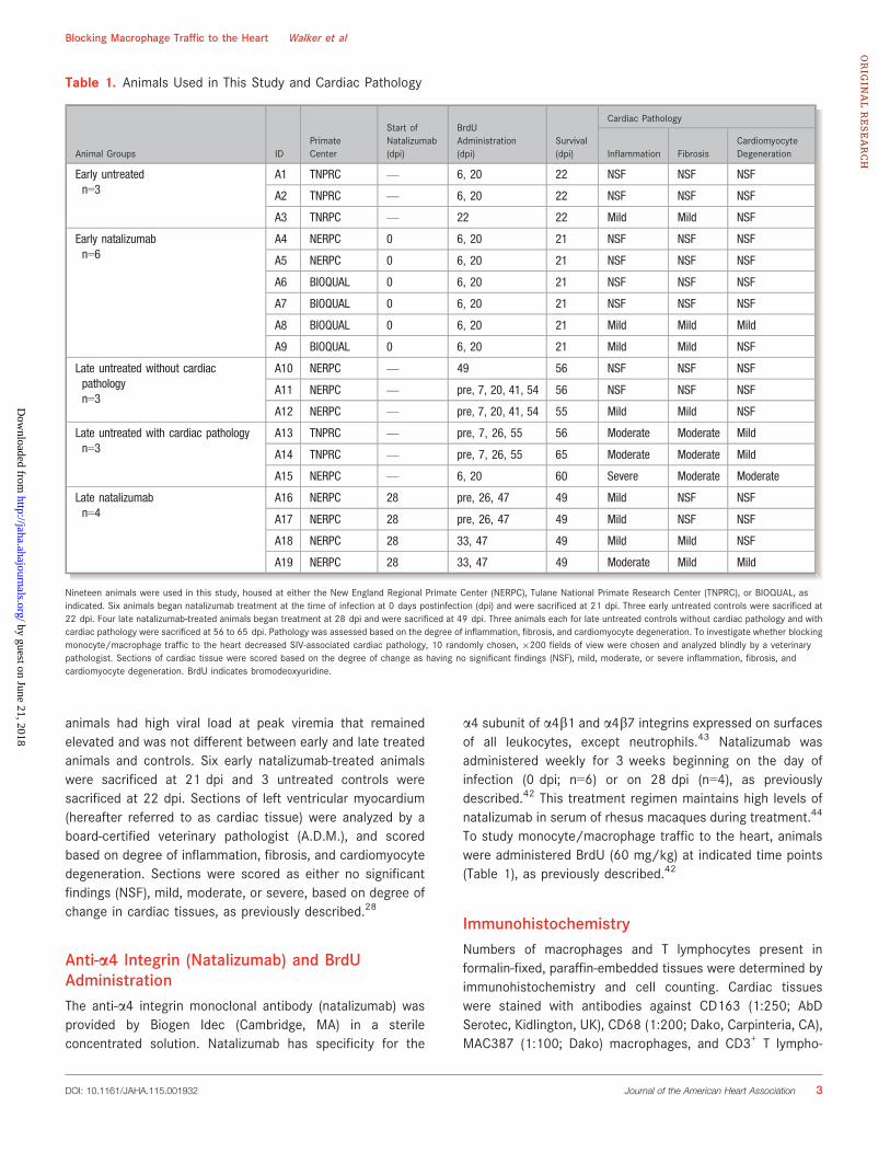
Table 1. Animals Used in This Study and Cardiac Pathology
Animal Groups IDPrimateCenter
Start ofNatalizumab(dpi)
BrdUAdministration(dpi)
Survival(dpi)
Cardiac Pathology
Inflammation FibrosisCardiomyocyteDegeneration
Early untreatedn=3
A1 TNPRC — 6, 20 22 NSF NSF NSF
A2 TNPRC — 6, 20 22 NSF NSF NSF
A3 TNRPC — 22 22 Mild Mild NSF
Early natalizumabn=6
A4 NERPC 0 6, 20 21 NSF NSF NSF
A5 NERPC 0 6, 20 21 NSF NSF NSF
A6 BIOQUAL 0 6, 20 21 NSF NSF NSF
A7 BIOQUAL 0 6, 20 21 NSF NSF NSF
A8 BIOQUAL 0 6, 20 21 Mild Mild Mild
A9 BIOQUAL 0 6, 20 21 Mild Mild NSF
Late untreated without cardiacpathologyn=3
A10 NERPC — 49 56 NSF NSF NSF
A11 NERPC — pre, 7, 20, 41, 54 56 NSF NSF NSF
A12 NERPC — pre, 7, 20, 41, 54 55 Mild Mild NSF
Late untreated with cardiac pathologyn=3
A13 TNPRC — pre, 7, 26, 55 56 Moderate Moderate Mild
A14 TNPRC — pre, 7, 26, 55 65 Moderate Moderate Mild
A15 NERPC — 6, 20 60 Severe Moderate Moderate
Late natalizumabn=4
A16 NERPC 28 pre, 26, 47 49 Mild NSF NSF
A17 NERPC 28 pre, 26, 47 49 Mild NSF NSF
A18 NERPC 28 33, 47 49 Mild Mild NSF
A19 NERPC 28 33, 47 49 Moderate Mild Mild
Nineteen animals were used in this study, housed at either the New England Regional Primate Center (NERPC), Tulane National Primate Research Center (TNPRC), or BIOQUAL, asindicated. Six animals began natalizumab treatment at the time of infection at 0 days postinfection (dpi) and were sacrificed at 21 dpi. Three early untreated controls were sacrificed at22 dpi. Four late natalizumab-treated animals began treatment at 28 dpi and were sacrificed at 49 dpi. Three animals each for late untreated controls without cardiac pathology and withcardiac pathology were sacrificed at 56 to 65 dpi. Pathology was assessed based on the degree of inflammation, fibrosis, and cardiomyocyte degeneration. To investigate whether blockingmonocyte/macrophage traffic to the heart decreased SIV-associated cardiac pathology, 10 randomly chosen, 9200 fields of view were chosen and analyzed blindly by a veterinarypathologist. Sections of cardiac tissue were scored based on the degree of change as having no significant findings (NSF), mild, moderate, or severe inflammation, fibrosis, andcardiomyocyte degeneration. BrdU indicates bromodeoxyuridine.
DOI: 10.1161/JAHA.115.001932 Journal of the American Heart Association 3
Blocking Macrophage Traffic to the Heart Walker et alORIG
INALRESEARCH
by guest on June 21, 2018http://jaha.ahajournals.org/
Dow
nloaded from
cytes (1:300; Dako). The number of macrophages that trafficto cardiac tissues was determined using a mouse monoclonalBrdU antibody (1:50), as previously described.28 Twentyrandom, nonoverlapping, 9200 microscopic fields of viewwere taken for each animal and the number of positive cells/mm2 calculated for each. Data are represented as the averagenumber of positive cells/mm2 from the 20 random fields.
Masson’s Trichrome StainPercent of collagen per tissue area used as a marker offibrosis29 was measured using a modified Massons TrichromeStain kit (Newcomer Supply, Middleton, WI), according to themanufacturer’s recommendation. Tissue sections wereimaged using a Zeiss Axio Imager M1 microscope (Zeiss,Oberkochen, Germany) using Plan-Apochromat 920/0.8 Korrobjectives, as previously described.28,45 The area of red andblue dyes corresponding to cytoplasm and collagen, respec-tively, were measured to determine the percentage of totaltissue area.
Statistical AnalysisStatistical analyses were conducted using Prism software(version 6.0; GraphPad Software Inc., La Jolla, CA). P valueswere calculated using the nonparametric Mann–Whitney t testwith significance accepted at P<0.05 when comparing earlyand late natalizumab-treated animals to early and lateuntreated controls. ANOVA was used to compare latenatalizumab-treated animals to late untreated animals withand without cardiac pathology. If the ANOVA was significant(P<0.05), then a post-hoc nonparametric Mann–Whitney t testwas performed. To determine whether changes in numbers ofmacrophages in cardiac tissues correlates with changes infibrosis, a nonparametric Spearman rank correlation was usedwhere P<0.05 was significant.
Results
Natalizumab Treatment Decreases the Frequencyand Severity of Pathology in SIV-Infected, CD8-Lymphocyte-Depleted Rhesus Macaque CardiacTissuesThe relative degree of pathology in cardiac tissues wasassessed based on levels of inflammation, fibrosis, andcardiomyocyte degeneration. Normal sections were scored ashaving no significant findings (NSF). When present, inflam-mation, fibrosis, and cardiomycocyte degeneration werescored as mild, moderate, or severe. We found no significantchanges in the pathology of cardiac tissues in earlynatalizumab-treated animals, compared to untreated controls.
Of 3 SIV-infected untreated animals sacrificed early, 21 dpi(n=3), 2 had no significant findings with regard to inflamma-tion or fibrosis. The remaining SIV-infected untreated controls,sacrificed at 21 dpi, had mild inflammation and mild fibrosis.In the early natalizumab-treated group (n=6), sacrificed at21 dpi, 4 animals had no significant findings in regard toinflammation and fibrosis and 2 had mild inflammation andfibrosis (Table 1).
Overall, natalizumab treatment decreased cardiac pathol-ogy in late treated animals (n=4), compared to late untreatedcontrols (n=6; Table 1). Three of 4 late treated animals had nosignificant findings with respect to cardiomyocyte degenera-tion, and 2 of 4 had no significant findings with regard tofibrosis. SIV-infected, untreated late control animals withcardiac pathology (n=3) had an increased severity, comparedto late treated SIV-infected animals (Table 1). Two of the latecontrols with pathology had moderate inflammation and 1 hadmild inflammation. All 3 late controls with pathology hadmoderate fibrosis, whereas 2 had mild and 1 had moderatecardiomycocyte degeneration. Compared to the late controlswith pathology, the severity of pathology in SIV-infected, latenatalizumab-treated animals (n=4) was diminished (Table 1).Three late treated animals had mild inflammation, whereasthe remaining had moderate inflammation. Two late treatedanimals had no inflammation and 2 had only mild inflamma-tion. Three late treated animals had no significant findingswith regard to cardiomyocyte degeneration and 1 had mildcardiomyocyte degeneration (Table 1).
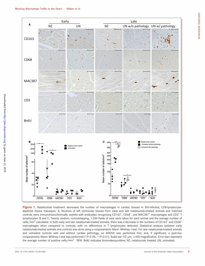
Natalizumab Treatment Decreases the Number ofMacrophages Present in Cardiac Tissue in Earlyand Late Treated AnimalsIn animals that began natalizumab treatment early at 0 dpi,there was a significant decrease in the number of CD163+ andCD68+ macrophages in heart tissues, compared to untreatedcontrols sacrificed at the same time point (Figure 1). Therewas a significant 3.35- and 3.74-fold decrease in the numbersof CD163+ and CD68+ macrophages, respectively, in cardiactissue in early natalizumab-treated animals, compared tocontrols (Figure 1B; Table 2; nonparametric Mann–Whitneyt test, P<0.05). Though not significant, there were decreasednumbers of newly infiltrating MAC387+ macrophages andCD3 T-lymphocytes in early treated animals (Figure 1B;Table 2).
Natalizumab treatment beginning on 28 dpi (late) resultedin a significant decrease in numbers of CD163+ and CD68+
macrophages, when compared to all SIV-infected untreatedlate control animals (Figure 1). We found a 3.53- and 1.19-fold decrease in numbers of CD163+ and CD68+ macrophag-es, respectively, in cardiac tissues of late natalizumab-treatedtissues, compared to controls (Table 2; nonparametric Mann–
DOI: 10.1161/JAHA.115.001932 Journal of the American Heart Association 4
Blocking Macrophage Traffic to the Heart Walker et alORIG
INALRESEARCH
by guest on June 21, 2018http://jaha.ahajournals.org/
Dow
nloaded from
A
B C
Figure 1. Natalizumab treatment decreases the number of macrophages in cardiac tissues in SIV-infected, CD8-lymphocyte-depleted rhesus macaques. A, Sections of left ventricular tissues from early and late natalizumab-treated animals and matchedcontrols were immunohistochemically stained with antibodies recognizing CD163+, CD68+, and MAC387+ macrophages and CD3+ Tlymphocytes. B and C, Twenty random, nonoverlapping, 9200 fields of view were taken for each animal and the average number ofcells/mm2 calculated. In both early and late natalizumab-treated animals, there was a decrease in the numbers of CD163+ and CD68+
macrophages when compared to controls, with no differences in T lymphocytes detected. Statistical analysis between earlynatalizumab-treated animals and controls was done using a nonparametric Mann–Whitney t test. For late natalizumab-treated animalsand untreated controls with and without cardiac pathology, an ANOVA was performed first, and, if significant, a post-hocnonparametric Mann–Whitney t test was performed (*P<0.05; **P<0.01). Scale bar=50 lm,9400 magnification. Error bars representthe average number of positive cells/mm2�SEM. BrdU indicates bromodeoxyuridine; NZ, natalizumab treated; UN, untreated.
DOI: 10.1161/JAHA.115.001932 Journal of the American Heart Association 5
Blocking Macrophage Traffic to the Heart Walker et alORIG
INALRESEARCH
by guest on June 21, 2018http://jaha.ahajournals.org/
Dow
nloaded from
Whitney t test, P<0.05; P<0.01). We next examined whetherthe numbers of macrophages in late natalizumab-treatedanimals differed between late untreated control animals withand without cardiac pathology.
Late treated animals had decreased numbers of CD163+
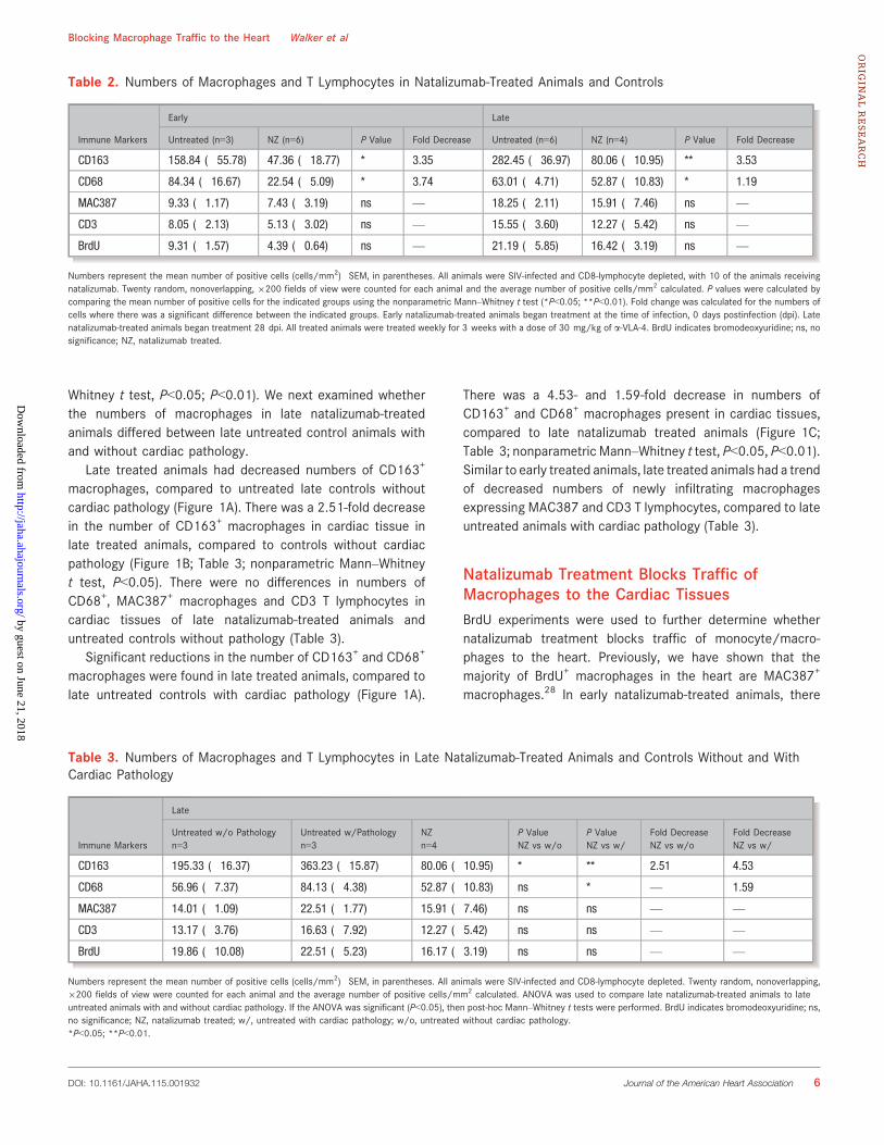
macrophages, compared to untreated late controls withoutcardiac pathology (Figure 1A). There was a 2.51-fold decreasein the number of CD163+ macrophages in cardiac tissue inlate treated animals, compared to controls without cardiacpathology (Figure 1B; Table 3; nonparametric Mann–Whitneyt test, P<0.05). There were no differences in numbers ofCD68+, MAC387+ macrophages and CD3 T lymphocytes incardiac tissues of late natalizumab-treated animals anduntreated controls without pathology (Table 3).
Significant reductions in the number of CD163+ and CD68+
macrophages were found in late treated animals, compared tolate untreated controls with cardiac pathology (Figure 1A).
There was a 4.53- and 1.59-fold decrease in numbers ofCD163+ and CD68+ macrophages present in cardiac tissues,compared to late natalizumab treated animals (Figure 1C;Table 3; nonparametric Mann–Whitney t test, P<0.05, P<0.01).Similar to early treated animals, late treated animals had a trendof decreased numbers of newly infiltrating macrophagesexpressing MAC387 and CD3 T lymphocytes, compared to lateuntreated animals with cardiac pathology (Table 3).
Natalizumab Treatment Blocks Traffic ofMacrophages to the Cardiac TissuesBrdU experiments were used to further determine whethernatalizumab treatment blocks traffic of monocyte/macro-phages to the heart. Previously, we have shown that themajority of BrdU+ macrophages in the heart are MAC387+
macrophages.28 In early natalizumab-treated animals, there
Table 3. Numbers of Macrophages and T Lymphocytes in Late Natalizumab-Treated Animals and Controls Without and WithCardiac Pathology
Immune Markers
Late
Untreated w/o Pathologyn=3
Untreated w/Pathologyn=3
NZn=4
P ValueNZ vs w/o
P ValueNZ vs w/
Fold DecreaseNZ vs w/o
Fold DecreaseNZ vs w/
CD163 195.33 (�16.37) 363.23 (�15.87) 80.06 (�10.95) * ** 2.51 4.53
CD68 56.96 (�7.37) 84.13 (�4.38) 52.87 (�10.83) ns * — 1.59
MAC387 14.01 (�1.09) 22.51 (�1.77) 15.91 (�7.46) ns ns — —
CD3 13.17 (�3.76) 16.63 (�7.92) 12.27 (�5.42) ns ns — —
BrdU 19.86 (�10.08) 22.51 (�5.23) 16.17 (�3.19) ns ns — —
Numbers represent the mean number of positive cells (cells/mm2)�SEM, in parentheses. All animals were SIV-infected and CD8-lymphocyte depleted. Twenty random, nonoverlapping,9200 fields of view were counted for each animal and the average number of positive cells/mm2 calculated. ANOVA was used to compare late natalizumab-treated animals to lateuntreated animals with and without cardiac pathology. If the ANOVA was significant (P<0.05), then post-hoc Mann–Whitney t tests were performed. BrdU indicates bromodeoxyuridine; ns,no significance; NZ, natalizumab treated; w/, untreated with cardiac pathology; w/o, untreated without cardiac pathology.*P<0.05; **P<0.01.
Table 2. Numbers of Macrophages and T Lymphocytes in Natalizumab-Treated Animals and Controls
Immune Markers
Early Late
Untreated (n=3) NZ (n=6) P Value Fold Decrease Untreated (n=6) NZ (n=4) P Value Fold Decrease
CD163 158.84 (�55.78) 47.36 (�18.77) * 3.35 282.45 (�36.97) 80.06 (�10.95) ** 3.53
CD68 84.34 (�16.67) 22.54 (�5.09) * 3.74 63.01 (�4.71) 52.87 (�10.83) * 1.19
MAC387 9.33 (�1.17) 7.43 (�3.19) ns — 18.25 (�2.11) 15.91 (�7.46) ns —
CD3 8.05 (�2.13) 5.13 (�3.02) ns — 15.55 (�3.60) 12.27 (�5.42) ns —
BrdU 9.31 (�1.57) 4.39 (�0.64) ns — 21.19 (�5.85) 16.42 (�3.19) ns —
Numbers represent the mean number of positive cells (cells/mm2)�SEM, in parentheses. All animals were SIV-infected and CD8-lymphocyte depleted, with 10 of the animals receivingnatalizumab. Twenty random, nonoverlapping, 9200 fields of view were counted for each animal and the average number of positive cells/mm2 calculated. P values were calculated bycomparing the mean number of positive cells for the indicated groups using the nonparametric Mann–Whitney t test (*P<0.05; **P<0.01). Fold change was calculated for the numbers ofcells where there was a significant difference between the indicated groups. Early natalizumab-treated animals began treatment at the time of infection, 0 days postinfection (dpi). Latenatalizumab-treated animals began treatment 28 dpi. All treated animals were treated weekly for 3 weeks with a dose of 30 mg/kg of a-VLA-4. BrdU indicates bromodeoxyuridine; ns, nosignificance; NZ, natalizumab treated.
DOI: 10.1161/JAHA.115.001932 Journal of the American Heart Association 6
Blocking Macrophage Traffic to the Heart Walker et alORIG
INALRESEARCH
by guest on June 21, 2018http://jaha.ahajournals.org/
Dow
nloaded from
were few BrdU+ cells (4.38�0.64 cells/mm2) and a trend ofdecreasing numbers of BrdU+ cells, compared to untreatedcontrols (9.31�1.57 cells/mm2; Figure 1A and 1B; Table 2).Animals that began natalizumab treatment at 28 dpi haddecreased numbers of BrdU+ cells (16.42�3.19 cells/mm2),compared to untreated animals with cardiac pathology(22.51�5.23 cells/mm2; Figure 1A and 1C; Table 3). Thenumber of BrdU+ cells in late natalizumab-treated animals didnot differ, when compared to untreated animals withoutcardiac pathology (Figure 1A and 1C; Table 3).
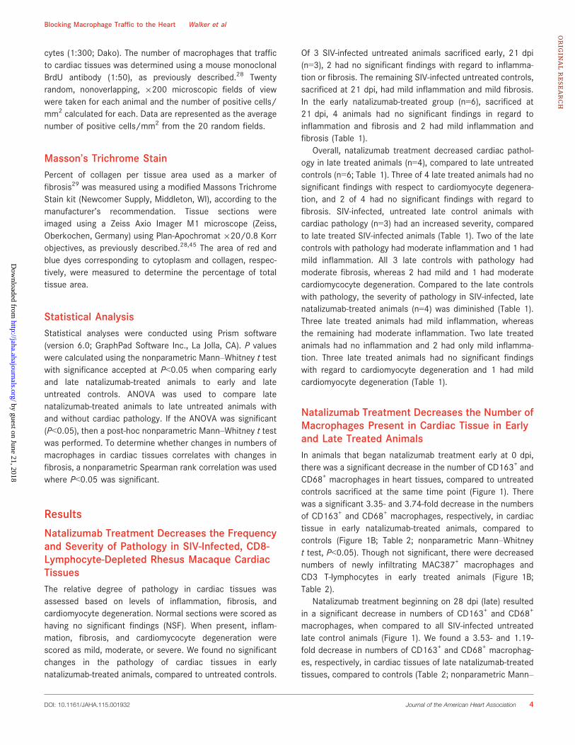
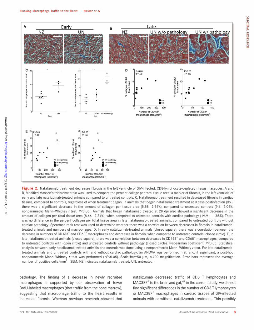
Decreased Fibrosis in Cardiac Tissues ofNatalizumab-Treated Animals Correlates WithSignificant Decreases in Macrophage NumbersUsing a modified Masson’s trichrome stain, the percentcollagen per total tissue area in cardiac tissues of natal-izumab-treated animals and untreated controls was quanti-fied (Figure 2A and 2B). Compared to controls, both earlyand late natalizumab-treated animals had decreasedamounts of collagen (Figure 2C). In early natalizumab-treatedanimals, the average percent collagen per total tissue areawas 5.58�2.56%, compared to 9.6�2.06% for untreatedcontrols, a significant 1.72-fold decrease in percent collagen(Figure 2C, left; P<0.05, nonparametric Mann–Whitneyt test).
Animals that began natalizumab treatment at 28 dpi (late)had a significantly higher average percentage of collagen pertissue area in the left ventricle (8.66�2.31%), compared toanimals that began treatment early (5.58�1.47; Figure 2C,right; P<0.05, nonparametric Mann–Whitney t test). Latenatalizumab-treated animals had no significant differences inthe percent of collagen per total tissue area, compared tountreated animals without cardiac pathology (8.66�2.31%versus 10.33�1.84%). However, when compared to untreatedanimals with cardiac pathology, there was a significantdecrease in the percentage of collagen per tissue area.Whereas late natalizumab-treated animals had an averagepercentage of collagen of 8.66�2.31%, untreated animalswith cardiac pathology had an average of 19.91�1.85%, asignificant 2.29-fold decrease in the average percent collagenper total tissue area (Figure 2C, right; P<0.05, nonparametricMann–Whitney t test).
We next examined whether there was a correlationbetween decreased fibrosis in natalizumab-treated animalsand changes in macrophage numbers in cardiac tissues ifsignificant differences in macrophage numbers were foundbetween groups. There was a correlation between increasedfibrosis and increased numbers of CD163+ (r=0.9; P<0.05)and CD68+ (r=0.86; P<0.05) macrophages in untreatedcontrols sacrificed at 21 dpi, compared to early natal-
izumab-treated animals (Figure 2D). A correlation also existedin late natalizumab-treated animals, compared to all lateuntreated controls for CD163+ (r=0.85; P<0.05), CD68+
(r=0.55; P<0.05) macrophages and fibrosis (Figure 2E).
DiscussionChronic inflammation persists within HIV-infected individualsdespite effective cART and decreased plasma viral load toundetectable levels.46–48 With chronic inflammation, there areincreased comorbidities, compared to the general non-HIV-infected population.49–51 In particular, there is an increasedincidence of CVD4 where monocytes/macrophages areincreasingly considered to play a role.52,53 Previously, wehave shown that SIV-infected, CD8-lymphocyte-depletedmonkeys have increased numbers of macrophages (CD163+,CD68+, and MAC387+) in cardiac tissues that positivelycorrelate increased fibrosis.28 In this study, we examinedwhether an anti-a4 integrin antibody, natalizumab, diminishesleukocyte and monocyte/macrophage traffic to the heartresulting in decreased fibrosis.
Natalizumab blocks the interaction between a4 integrinand its ligand, vascular cell adhesion molecule 1 (VCAM-1).38
VCAM-1 is expressed on endothelial cells of the arterial lumenwith atherosclerosis.54 Studies in mice showed that inhibitingthe interaction between a4 and VCAM-1 decreased macro-phage recruitment to atherosclerotic plaques.41 Previously,we have shown that there is a higher level of macrophagetraffic to the heart later in SIV infection (>21 dpi) and thatthere are few macrophages present in untreated animalssacrificed at 21 dpi.28 In the current study, we found thatnatalizumab treatment beginning at 0 dpi resulted indecreased numbers of CD163+ and CD68+ macrophages,compared to uninfected controls, but cardiac pathology inearly infection is minimal and most of the pathology occurredin the later stages of infection.
When compared to untreated controls with cardiacpathology, late treated animals had significant decreases inthe number of CD163+ and CD68+ macrophages. In fact, thenumbers of macrophages in late natalizumab-treated animalswere similar to untreated animals without cardiac pathology.Additionally, we found a correlation between decreasedmacrophage numbers in natalizumab-treated animals withdecreased cardiac fibrosis. Overall, these data show thatblocking monocyte/macrophage traffic to the heart alleviatesHIV- and SIV-associated cardiac pathology that resulted inreduced inflammation, fibrosis, and cardiomyocyte degener-ation.
Though not significant, we found a trend of decreasingnumbers of MAC387+ macrophages in the left ventricle of latetreated animals, compared to untreated animals with cardiac
DOI: 10.1161/JAHA.115.001932 Journal of the American Heart Association 7
Blocking Macrophage Traffic to the Heart Walker et alORIG
INALRESEARCH
by guest on June 21, 2018http://jaha.ahajournals.org/
Dow
nloaded from
pathology. The finding of a decrease in newly recruitedmacrophages is supported by our observation of fewerBrdU-labeled macrophages (that traffic from the bone marrow),suggesting that macrophage traffic to the heart results inincreased fibrosis. Whereas previous research showed that
natalizumab decreased traffic of CD3 T lymphocytes andMAC387+ to the brain and gut,42 in the current study, we did notfind significant differences in the number of CD3 T lymphocytesor MAC387+ macrophages in cardiac tissues of SIV-infectedanimals with or without natalizumab treatment. This possibly
B
C D
E
A
Figure 2. Natalizumab treatment decreases fibrosis in the left ventricle of SIV-infected, CD8-lymphocyte-depleted rhesus macaques. A andB, Modified Masson’s trichrome stain was used to compare the percent collage per total tissue area, a marker of fibrosis, in the left ventricle ofearly and late natalizumab-treated animals compared to untreated controls. C, Natalizumab treatment resulted in decreased fibrosis in cardiactissues, compared to controls, regardless of when treatment began. In animals that began natalizumab treatment at 0 days postinfection (dpi),there was a significant decrease in the amount of collagen per tissue area (5.58�2.56%), compared to untreated controls (9.6�2.06%;nonparametric Mann–Whitney t test, P<0.05). Animals that began natalizumab treated at 28 dpi also showed a significant decrease in theamount of collagen per total tissue area (8.66�2.31%), when compared to untreated controls with cardiac pathology (19.91�1.85%). Therewas no difference in the percent collagen per total tissue area in late natalizumab-treated animals, compared to untreated controls withoutcardiac pathology. Spearman rank test was used to determine whether there was a correlation between decreases in fibrosis in natalizumab-treated animals and numbers of macrophages. D, In early natalizumab-treated animals (closed square), there was a correlation between thedecrease in numbers of CD163+ and CD68+ macrophages and decreases in fibrosis, when compared to untreated controls (closed circle). E, Inlate natalizumab-treated animals (closed square), there was a correlation between decreases in CD163+ and CD68+ macrophages, comparedto untreated controls with (open circle) and untreated controls without pathology (closed circle). r=spearman coefficient, P<0.05. Statisticalanalysis between early natalizumab-treated animals and controls was done using a nonparametric Mann–Whitney t test. For late natalizumab-treated animals and untreated controls with and without cardiac pathology, an ANOVA was performed first, and, if significant, a post-hocnonparametric Mann–Whitney t test was performed (*P<0.05). Scale bar=50 lm, 9400 magnification. Error bars represent the averagenumber of positive cells/mm2�SEM. NZ indicates natalizumab treated; UN, untreated.
DOI: 10.1161/JAHA.115.001932 Journal of the American Heart Association 8
Blocking Macrophage Traffic to the Heart Walker et alORIG
INALRESEARCH
by guest on June 21, 2018http://jaha.ahajournals.org/
Dow
nloaded from
suggests that CD3+ T lymphocytes andMAC387+macrophagesuse different integrins to traffic to the heart than to the brain orgut; however, we have previously shown that MAC387+
macrophages and not CD3+ T lymphocytes correlate withincreased fibrosis in cardiac tissues.28 Our lack of finding astatistically significant reduction in the number MAC387+
macrophages may be owing to the relatively few numbers ofthose cells in cardiac tissues.
Previously, we have shown that the rate of monocyte/macrophage traffic to the heart is increased later in infection(after 48 dpi), as opposed to early infection.28 Using BrdUlabeling, we found a trend of decreased traffic of monocytes/macrophages to the heart in late natalizumab-treated animals,compared to untreated animals that developed cardiacpathology. Late natalizumab-treated animals had a similarrate of traffic of newly released monocytes/macrophages tothe heart as untreated animals without cardiac pathology. Thisprovides evidence that potentially blocking traffic of mono-cyte/macrophages later during SIV infection can alleviate SIV-associated cardiac pathology.
Although cART can decrease HIV to nondetectable levels inplasma, it does not necessarily target monocyte/macrophag-es that play a role in the development of cardiac pathologyand CVD.32 Chronic immune activation with HIV infection isposited to play a role in HIV-associated cardiovascularpathology. Previous studies show that HIV-infected individualshave increased inflammation in the ascending aorta thatcorrelates with levels of sCD163 in plasma.55 Increasedinflammation in the aorta is also been linked to high-risknoncalcified plaques that are prone to rupture.8 18-Fluorode-oxyglucose positron emission tomography imaging studieshave demonstrated that such plaque areas are comprised ofareas with accumulation of macrophages.8,56,57 Whereasmacrophage accumulation in the aorta and cardiac plaquesare critical in HIV-associated cardiac disease, it is notsurprising that also there is increased macrophage inflamma-tion in cardiac tissues at the same time. Unpublished datafrom our laboratory, using matched cardiac tissues (leftventricle) and aorta from HIV� and HIV+ individuals, showsthat, with HIV infection, there is an increased macrophageaccumulation inflammation in ventricular tissues and theaorta. Moreover, increased macrophage inflammation incardiac tissues correlates with increased fibrosis, macrophageaccumulation in the aorta, and increased aortic intima-mediathickness. To date, there are few therapies that targetmacrophages specifically or indirectly to diminish HIV-asso-ciated CV pathology. Emergent data underscore the impor-tance of such therapy strategies.
Therapeutic agents that have been successful in thetreatment noncalcified cardiac plaque in HIV+ individualsinclude 3-hydroxy-3-methylglutaryl-coenzyme A reductaseinhibitors (statins), which, fortuitously, have anti-inflammatory
effects on macrophages.58,59 Statin therapy in conjunctionwith cART reduced serum levels of inflammatory markers,including IL-6, IL-8, and tumor necrosis factor alpha, more sothan cART alone.60 Statins have similarly been used inmonkeys, where they decreased the macrophage content inplaques in the abdominal aorta.34 A recent study in HIV-infected individuals showed that statins significantlydecreased the volume on noncalcified plaques, but whetherstatin use in this study directly affected monocyte and/ormacrophage activation and traffic was not studied.35 In rodentmodels of experimental autoimmune myocarditis, rosu-vastatin-reduced numbers in macrophages, T lymphocytes,and multinucleated giant cells in the heart resulted indecreased numbers of apoptotic cardiomyocytes. The effectsof statins on myocarditis with HIV infection have not beenexamined.61
Other studies found that maraviroc treatment decreasedchemotaxis of monocyte/macrophages, in vitro,62 but inclinical studies with advanced HIV, it did not affect thedevelopment of immune reconstitution inflammatory syn-drome,63 Maraviroc is used primarily to inhibit viral replicationof R5-tropic HIV by blocking interactions between the virusand CCR5 on host cells.64,65 Studies using maraviroc in SIV-infected monkeys demonstrated fewer CD163+ macrophagesin the heart, but this could have been owing to a decreasedCD163 expression (activation) on macrophages alreadypresent in the heart and not a decrease in inflammatorycells. All together, these experiments add further evidence tothe role that monocyte/macrophages play in cardiac pathol-ogy with HIV and SIV infection, and suggest that therapiesblocking monocyte/macrophage traffic to the heart coulddiminish HIV-associated cardiac pathology.
In this study, we showed that directly blocking monocyte/macrophage traffic to cardiac tissues with natalizumabsuccessfully decreased the numbers of macrophages presentin tissues. Studies examining whether blocking traffic tovessels result in decreased high-risk vascular plaques withHIV infection are warranted. Our data suggest that studiesexamining the efficacy of blocking monocyte/macrophagetraffic, or directly targeting monocyte/macrophage activationas an adjunctive therapy with cART, should be examined withan aim to decrease HIV-associated cardiac pathology.
AcknowledgmentsThe authors thank Biogen Idec (Cambridge, MA) for providingnatalizumab for use in this study. In vivo CD8 depletion antibodieswere kindly provided by the NIH Nonhuman Primate ReagentResource. Author Contributions: Conceived and designed experi-ments: Walker, Campbell, Burdo, Williams. Performed the experi-ments: Walker, Beck. Analyzed the data: Walker, Beck. Scoring ofcardiac tissue sections: Miller. Wrote the article: Walker, Burdo,Williams.
DOI: 10.1161/JAHA.115.001932 Journal of the American Heart Association 9
Blocking Macrophage Traffic to the Heart Walker et alORIG
INALRESEARCH
by guest on June 21, 2018http://jaha.ahajournals.org/
Dow
nloaded from
Sources of FundingThis work was supported by the following National Institutesof Health (NIH) grants: R01 NS040237 (Williams) and R01NS082116 (Burdo).
DisclosuresNone.
References1. Williams K, Westmoreland S, Greco J, Ratai E, Lentz M, Kim WK, Fuller RA, Kim
JP, Autissier P, Sehgal PK, Schinazi RF, Bischofberger N, Piatak M, Lifson JD,Masliah E, Gonzalez RG. Magnetic resonance spectroscopy reveals thatactivated monocytes contribute to neuronal injury in SIV neuroAIDS. J ClinInvest. 2005;115:2534–2545.
2. Mocroft A, Kirk O, Gatell J, Reiss P, Gargalianos P, Zilmer K, Beniowski M, ViardJP, Staszewski S, Lundgren JD. Chronic renal failure among HIV-1-infectedpatients. AIDS. 2007;21:1119–1127.
3. Cazanave C, Dupon M, Lavignolle-Aurillac V, Barthe N, Lawson-Ayayi S,Mehsen N, Mercie P, Morlat P, Thiebaut R, Dabis F; Groupe d’EpidemiologieClinique du SeA. Reduced bone mineral density in HIV-infected patients:prevalence and associated factors. AIDS. 2008;22:395–402.
4. Ho JE, Hsue PY. Cardiovascular manifestations of HIV infection. Heart.2009;95:1193–1202.
5. Zaaqoq AM, Khasawneh FA, Smalligan RD. Cardiovascular complications ofHIV-associated immune dysfunction. Cardiol Res Pract. 2015;2015:1–8.
6. Freiberg MS, Chang CC, Kuller LH, Skanderson M, Lowy E, Kraemer KL, ButtAA, Bidwell Goetz M, Leaf D, Oursler KA, Rimland D, Rodriguez Barradas M,Brown S, Gibert C, McGinnis K, Crothers K, Sico J, Crane H, Warner A, GottliebS, Gottdiener J, Tracy RP, Budoff M, Watson C, Armah KA, Doebler D, Bryant K,Justice AC. HIV infection and the risk of acute myocardial infarction. JAMAIntern Med. 2013;173:614–622.
7. Esser S, Gelbrich G, Brockmeyer N, Goehler A, Schadendorf D, Erbel R,Neumann T, Reinsch N. Prevalence of cardiovascular diseases in HIV-infectedoutpatients: results from a prospective, multicenter cohort study. Clin ResCardiol. 2013;102:203–213.
8. Tawakol A, Lo J, Zanni MV, Marmarelis E, Ihenachor EJ, MacNabb M, Wai B,Hoffmann U, Abbara S, Grinspoon S. Increased arterial inflammation relates tohigh-risk coronary plaque morphology in HIV-infected patients. J AcquirImmune Defic Syndr. 2014;66:164–171.
9. Zanni MV, Schouten J, Grinspoon SK, Reiss P. Risk of coronary heart disease inpatients with HIV infection. Nat Rev Cardiol. 2014;11:728–741.
10. Ray M, Logan R, Sterne JA, Hernandez-Diaz S, Robins JM, Sabin C, Bansi L, vanSighem A, de Wolf F, Costagliola D, Lanoy E, Bucher HC, von Wyl V, Esteve A,Casbona J, del Amo J, Moreno S, Justice A, Goulet J, Lodi S, Phillips A, Seng R,Meyer L, Perez-Hoyos S, Garcia de Olalla P, Hernan MA. The effect ofcombined antiretroviral therapy on the overall mortality of HIV-infectedindividuals. AIDS. 2010;24:123–137.
11. Coquet I, Pavie J, Palmer P, Barbier F, Legriel S, Mayaux J, Molina JM,Schlemmer B, Azoulay E. Survival trends in critically ill HIV-infected patients inthe highly active antiretroviral therapy era. Crit Care. 2010;14:R107.
12. Guaraldi G, Orlando G, Zona S, Menozzi M, Carli F, Garlassi E, Berti A, Rossi E,Roverato A, Palella F. Premature age-related comorbidities among HIV-infectedpersons compared with the general population. Clin Infect Dis. 2011;53:1120–1126.
13. Hemkens LG, Bucher HC. HIV infection and cardiovascular disease. Eur Heart J.2014;35:1373–1381.
14. Barbaro G, Fisher SD, Lipshultz SE. Pathogenesis of HIV-associated cardio-vascular complications. Lancet Infect Dis. 2001;1:115–124.
15. Patan�e S, Marte F, Sturiale M, Dattilo G, Albanese A. Myocarditis andcardiomyopathy HIV associated. Int J Cardiol. 2011;146:e56–e57.
16. Friis-Møller N, Reiss P, Sabin CA, Weber R, d’Arminio Monforte A, El-Sadr W,Thi�ebaut R, De Wit S, Kirk O, Fontas E, Law MG, Phillips A, Lundgren JD. Classof antiretroviral drugs and the risk of myocardial infarction. N Engl J Med.2007;356:1723–1735.
17. Liu R, Moroi M, Yamamoto M, Kubota T, Ono T, Funatsu A, Komatsu H,Tsuji T, Hara H, Hara H, Nakamura M, Hirai H, Yamaguchi T. Presence andseverity of Chlamydia pneumoniae and Cytomegalovirus infection in
coronary plaques are asssociated with acute coronary syndromes. IntHeart J. 2006;47:511–519.
18. Stein JH, Hsue PY. Inflammation, immune activation, and CVD risk inindividuals with HIV infection. JAMA. 2012;308:405–406.
19. Khunnawat C, Mukerji S, Havlichek D Jr, Touma R, Abela GS. Cardiovascularmanifestations in human immunodeficiency virus-infected patients. Am JCardiol. 2008;102:635–642.
20. Silwa K, Carrington MJ, Becker A, Thienemann F, Ntsekhe M, Stewart S.Contribution of the human immunodeficiency virus acquired immunodeficiencysyndrome epidemic to de novo presentations of heart disease in the Heart ofSoweto Study cohort. Eur Heart J. 2012;33:866–874.
21. Holloway CJ, Ntusi N, Suttie J, Mahmod M, Wainwright E, Clutton G, HancockG, Beak P, Tajar A, Piechnik SK, Schneider JE, Angus B, Clarke K, Dorrell L,Neubauer S. Comprehensive cardiac magnetic resonance imaging andspectroscopy reveal a high burden of myocardial disease in HIV patients.Circulation. 2013;128:814–822.
22. Cheruvu S, Holloway CJ. Cardiovascular disease in human immunodeficiencyvirus. Intern Med J. 2014;44:315–324.
23. Frustaci A, Petrosillo N, Vizza D, Francone M, Badagliacca R, Verardo R, FedeleF, Ippolito G, Chimenti C. Myocardial and microvascular inflammation/infection in patients with HIV-associated pulmonary artery hypertension. AIDS.2014;28:2541–2549.
24. Shannon RP. SIV cardiomyopathy in non-human primates. Trends CardiovascMed. 2011;11:242–246.
25. Shannon RP, Simon MA, Mathier MA, Geng YJ, Mankad S, Lackner AA. Dilatedcardiomyopathy associated with simian AIDS in nonhuman primates. Circu-lation. 2000;101:185–193.
26. Yearley JH, Pearson C, Carville A, Shannon RP, Mansfield K. Phenotypicvariation in myocardial macrophage populations suggests a role for macro-phage activation in SIV-associated cardiac disease. AIDS Res Hum Retrovirus-es. 2007;23:512–524.
27. Kelly KM, Tarwater PM, Karper JM, Bedja D, Queen SE, Tunin RS, Adams RJ,Kass DA, Mankowski JL. Diastolic dysfunction is associated with myocardialviral load in simian immunodeficiency virus-infected macaques. AIDS.2012;26:815–823.
28. Walker JA, Sulciner ML, Nowicki KD, Miller AD, Burdo TH, Williams KC.Elevated numbers of CD163+ macrophages in hearts of simian immunode-ficiency virus-infected monkeys correlate with cardiac pathology and fibrosis.AIDS Res Hum Retroviruses. 2014;30:685–694.
29. Brower GL, Gardner JD, Forman MF, Murray DB, Voloshenyuk T, Levick SP,Janicki JS. The relationship between myocardial extracellular matrix remod-eling and ventricular function. Eur J Cardiothorac Surg. 2006;30:604–610.
30. Schall TJ, Bacon K, Toy KJ, Goeddel DV. Selective attraction of monocytes andT lymphocytes of the memory phenotype by cytokine RANTES. Nature.1990;347:669–671.
31. Montecucco F, Braunersreuther V, Lenglet S, Delattre BM, Pelli G, Buatois V,Guilhot F, Galan K, Vuilleumier N, Ferlin W, Fischer N, Vallee JP, Kosco-Vilbois M, Mach F. CC chemokine CCL5 plays a central role impacting infarctsize and post-infarction heart failure in mice. Eur Heart J. 2012;33:1964–1974.
32. Kelly KM, Tocchetti CG, Lyashkov A, Tarwater PM, Bedja D, Graham DR, BeckSE, Metcalf Pate KA, Queen SE, Adams RJ, Paolocci N, Mankowski JL. CCR5inhibition prevents cardiac dysfunction in the SIV/macaque model of HIV. J AmHeart Assoc. 2014;3:e000874 doi: 10.1161/JAHA.114.000874.
33. Matsui Y, Okamoto H, Jia N, Akino M, Uede T, Kitabatake A, Nishihira J.Blockade of macrophage migration inhibitory factor ameliorates experimentalautoimmune myocarditis. J Mol Cell Cardiol. 2004;37:557–566.
34. Sukhova GK. Statins reduce inflammation in atheroma of nonhuman primatesindependent of effects on serum cholesterol. Arterioscler Thromb Vasc Biol.2002;22:1452–1458.
35. Lo J, Lu MT, Ihenachor EJ, Wei J, Looby SE, Fitch KV, Oh J, Zimmerman CO,Hwang J, Abbara S, Plutzky J, Robbins G, Tawakol A, Hoffmann U, GrinspoonSK. Effects of statin therapy on coronary artery plaque volume and high-riskplaque morphology in HIV-infected patients with subclinical atherosclerosis: arandomised, double-blind, placebo-controlled trial. Lancet HIV. 2015;2:e52–e63.
36. Fliser D, Buchholz K, Haller H. Antiinflammatory effects of angiotensin IIsubtype 1 receptor blockade in hypertensive patients with microinflammation.Circulation. 2004;110:1103–1107.
37. Navalkar S, Parthasarathy S, Santanam N, Khan BV. Irbesartan, an angiotensintype 1 receptor inhibitor, regulates markers of inflammation in patients withpremature atherosclerosis. J Am Coll Cardiol. 2001;37:440–444.
38. Yu Y, Schurpf T, Springer TA. How natalizumab binds and antagonizes alpha4integrins. J Biol Chem. 2013;288:32314–32325.
DOI: 10.1161/JAHA.115.001932 Journal of the American Heart Association 10
Blocking Macrophage Traffic to the Heart Walker et alORIG
INALRESEARCH
by guest on June 21, 2018http://jaha.ahajournals.org/
Dow
nloaded from
39. Polma CH, O’Connor PW, Havrdova E, Hutchinson M, Kappos L, Miller DH,Phillips JT, Lublin FD, Giovannoni G, Wajgt A, Toal M, Lynn F, Panzara MA,Sandrock AW. A randomized, placebo-controlled trial of natalizumab forrelapsing multiple sclerosis. N Engl J Med. 2006;354:899–910.
40. Kane SV, Horst S, Sandborn WJ, Becker B, Neis B, Moscandrew M, Hanson KA,Tremaine WJ, Bruining DH, Faubion WA, Pardi DS, Harmsen WS, ZinsmeisterAR, Loftus EV. Natalizumab for moderate to severe Crohn’s disease in clinicalpractice: the Mayo Clinic Rochester experience. Inflamm Bowel Dis.2012;18:2203–2208.
41. Patel SS, Thiagarajan R, Willerson JT, Yeh ETH. Inhibition of 4 integrin andICAM-1 markedly attenuate macrophage homing to atherosclerotic plaques inApoE-deficient mice. Circulation. 1998;97:75–81.
42. Campbell JH, Ratai E-M, Autissier P, Nolan DJ, Tse S, Miller AD, Gonz�alez RG,Salemi M, Burdo TH, Williams KC. Anti-a4 antibody treatment blocks virustraffic to the brain and gut early, and stabilizes CNS injury late in infection.PLoS Pathog. 2014;10:e1004533.
43. Stuve O, Marra CM, Jerome KR, Cook L, Cravens PD, Cepok S, Frohman EM,Phillips JT, Arendt G, Hemmer B, Monson NL, Racke MK. Immune surveillancein multiple sclerosis patients treated with natalizumab. Ann Neurol.2006;59:743–747.
44. Sasseville VG, Newmna W, Brodie SJ, Hesterberg P, Pauley D, Ringler DJ.Monocyte adhesion to endothelium in simian immunodeficiency virus-inducedaids encephalitis is mediated by vascular cell adhesion molecule-1/alpha 4beta 1 integrin interactions. Am J Pathol. 1994;144:27–40.
45. Ruifrok AC, Johnston DA. Quantification of histochemical staining by colordeconvolution. Anal Quant Cytol. 2001;23:291–299.
46. Lambotte O, Taoufik Y, de Goer MG, Wallon C, Goujard C, Delfraissy JF.Detection of infectious HIV in circulating monocytes from patients onprolonged highly active antiretroviral therapy. J Acquir Immune Defic Syndr.2000;23:114–119.
47. Dornadula G, Zhang H, VanUitert B, Stern J, Livornese L Jr, Ingerman MJ, WitekJ, Kedanis RJ, Natkin J, DeSimone J, Pomerantz RJ. Residual HIV-1 RNA in bloodplasma of patients taking suppressive highly active antiretroviral therapy.JAMA. 1999;282:1627–1632.
48. Lichtfuss GF, Cheng WJ, Farsakoglu Y, Paukovics G, Rajasuriar R, VelayudhamP, Kramski M, Hearps AC, Cameron PU, Lewin SR, Crowe SM, Jaworowski A.Virologically suppressed HIV patients show activation of NK cells andpersistent innate immune activation. J Immunol. 2012;189:1491–1499.
49. Neuhaus J, Angus B, Kowalska JD, La Rosa A, Sampson J, Wentworth D,Mocroft A, Insight S; Groups Es. Risk of all-cause mortality associated withnonfatal aids and serious non-AIDS events among adults infected with HIV.AIDS. 2010;24:697–706.
50. Rodriguez-Penney AT, Iudicello JE, Riggs PK, Doyle K, Ellis RJ, Letendre SL,Grant I, Woods SP; Group HIVNRPH. Co-morbidities in persons infected withHIV: increased burden with older age and negative effects on health-relatedquality of life. AIDS Patient Care STDS. 2013;27:5–16.
51. Anzinger JJ, Butterfield TR, Angelovich TA, Crowe SM, Palmer CS. Monocytesas regulators of inflammation and HIV-related comorbidities during cart.J Immunol Res. 2014;2014:569819.
52. Medbury HJ, James V, Ngo J, Hitos K, Wang Y, Harris D, Fletcher JP. Differingassociation of macrophage subsets with atherosclerotic plaque stability. IntAngiol. 2013;32:74–84.
53. Pamukcu B, Lip GYH, Devitt A, Griffiths H, Shantsila E. The role of monocytesin atherosclerotic coronary artery disease. Ann Med. 2010;42:394–403.
54. OBrien KD, Allen MD, McDonald TO, Chait A, Harlan JM, Fishbein D, McCarty J,Ferguson M, Hudkins K, Benjamin CD, Lobb R, Alpers CE. Vascular celladhesion molecule-1 is expressed in human coronary atherosclerotic plaques.Implications for the mode of progression of advanced coronary atheroscle-rosis. J Clin Invest. 1993;92:945–951.
55. Subramanian S, Tawakol A, Abbara S, Wei J, Vijayakumar J, Corsini E,Abdelbaky A, Zanni MV, Hoffman U, Williams KC, Lo J, Grinspoon SK. Arterialinflammation in patients with HIV. JAMA. 2012;308:379–386.
56. Abdelbaky A, Corsini E, Figueroa AL, Subramanian S, Fontanez S, Emami H,Hoffmann U, Narula J, Tawakol A. Early aortic valve inflammation precedescalcification: a longitudinal FDG-PET/CT study. Atherosclerosis. 2015;238:165–172.
57. Tahara N, Mukherjee J, de Haas HJ, Petrov AD, Tawakol A, Haider N, Tahara A,Constantinescu CC, Zhou J, Boersma HH, Imaizumi T, Nakano M, Finn A, FayadZ, Virmani R, Fuster V, Bosca L, Narula J. 2-deoxy-2-[18F]fluoro-D-mannosepositron emission tomography imaging in atherosclerosis. Nat Med.2014;20:215–219.
58. Giguere JF, Tremblay MJ. Statin compounds reduce human immunodeficiencyvirus type 1 replication by preventing the interaction between virion-associated host intercellular adhesion molecule 1 and its natural cell surfaceligand LFA-1. J Virol. 2004;78:12062–12065.
59. Calza L, Trapani F, Bartoletti M, Manfredi R, Colangeli V, Borderi M, Grossi G,Motta R, Viale P. Statin therapy decreases serum levels of high-sensitivity C-reactive protein and tumor necrosis factor-alpha in HIV-infected patientstreated with ritonavir-boosted protease inhibitors. HIV Clin Trials.2012;13:153–161.
60. Calza L, Vanino E, Salvadori C, Manfredi R, Colangeli V, Cascavilla A, Di BariMA, Motta R, Viale P. Tenofovir/emtricitabine/efavirenz plus rosuvastatindecrease serum levels of inflammatory markers more than antiretroviral drugsalone in antiretroviral therapy-naive HIV-infected patients. HIV Clin Trials.2014;15:1–13.
61. Liu X, Li B, Wang W, Zhang C, Zhang M, Zhang Y, Xia Y, Dong Z, Guo Y, An F.Effects of HMG-CoA reductase inhibitor on experimental autoimmunemyocarditis. Cardiovasc Drugs Ther. 2012;26:121–130.
62. Rossi R, Lichtner M, De Rosa A, Sauzullo I, Mengoni F, Massetti AP,Mastroianni CM, Vullo V. In vitro effect of anti-human immunodeficiency virusCCR5 antagonist maraviroc on chemotactic activity of monocytes, macro-phages and dendritic cells. Clin Exp Immunol. 2011;166:184–190.
63. Sierra-Madero JG, Ellenberg SS, Rassool MS, Tierney A, Belaunzar�an-Zamudio PF, L�opez-Mart�ınez A, Pi~neir�ua-Men�endez A, Montaner LJ, Azzoni L,Ben�ıtez CR, Sereti I, Andrade-Villanueva J, Mosqueda- G�omez JL, RodriguezB, Sanne I, Lederman MM. Effect of the CCR5 antagonist maraviroc on theoccurrence of immune reconstitution inflammatory syndrome in HIV(CADIRIS): a double-blind, randomised, placebo-controlled trial. Lancet HIV.2014;1:e60–e67.
64. MacArthur RD, Novak RM. Maraviroc: the first of a new class of antiretroviralagents. Clin Infect Dis. 2008;47:236–241.
65. Soriano V, Geretti AM, Perno CF, Fatkenheuer G, Pillay D, Reynes J, TambussiG, Calvez V, Alcami J, Rockstroh J. Optimal use of maraviroc in clinical practice.AIDS. 2008;22:2231–2240.
DOI: 10.1161/JAHA.115.001932 Journal of the American Heart Association 11
Blocking Macrophage Traffic to the Heart Walker et alORIG
INALRESEARCH
by guest on June 21, 2018http://jaha.ahajournals.org/
Dow
nloaded from
Kenneth C. WilliamsJoshua A. Walker, Graham A. Beck, Jennifer H. Campbell, Andrew D. Miller, Tricia H. Burdo and
Cardiac Pathology in a SIV Infection Model of AIDS4 Integrin Antibody Blocks Monocyte/Macrophage Traffic to the Heart and Decreasesα−Anti
Online ISSN: 2047-9980 Dallas, TX 75231
is published by the American Heart Association, 7272 Greenville Avenue,Journal of the American Heart AssociationThe doi: 10.1161/JAHA.115.0019322015;4:e001932; originally published July 16, 2015;J Am Heart Assoc.
http://jaha.ahajournals.org/content/4/7/e001932World Wide Web at:
The online version of this article, along with updated information and services, is located on the
for more information. http://jaha.ahajournals.orgAccess publication. Visit the Journal at
is an online only OpenJournal of the American Heart AssociationSubscriptions, Permissions, and Reprints: The
by guest on June 21, 2018http://jaha.ahajournals.org/
Dow
nloaded from