muscular hydatidosis cyst relapse - jscimed central · in the case of hydatidosis, man becomes the...
TRANSCRIPT
CentralBringing Excellence in Open Access
Annals of Clinical Pathology
Cite this article: Marcano AL, Ramia Ángel JM, de la Plaza Llamas R, Adel F, Aguilar JG, et al. (2016) Muscular Hydatidosis Cyst Relapse. Ann Clin Pathol 4(1): 1061.
*Corresponding author
A.J López Marcano, Department of Surgery, University Hospital of Guadalajara, Guadalajara, Spain, C/ Eunate 10, 5-2, Madrid 28050 , Tel: 609869259; Email:
Submitted: 31 December 2015
Accepted: 19 January 2016
Published: 20 January 2016
ISSN: 2373-9282
Copyright© 2016 Marcano et al.
OPEN ACCESS
Keywords•Muscular Hydatidosis•Relapse•Echinococcosis
Case Report
Muscular Hydatidosis Cyst RelapseAylhin López Marcano*, José Manuel Ramia Ángel, Roberto de la Plaza Llamas, Farah Adel, Jhonny Gonzáles Aguilar, Yuri Rodrigues*, Anibal Medina VelascoDepartment of Surgery, University Hospital of Guadalajara, Spain
Abstract
Hydatidosis is a worldwide spread zoonoses caused mainly by the Echinococcus granulosus, where humans become the intermediary host through contact with a definitive host (commonly a dog) or through intake of food or water contaminated with Echinococcus eggs. Clinically speaking it usually presents itself as a hydatid cyst often developed in the liver due to the fact that it is where it reaches first through portal circulation. In order to reach other organs like the muscle it will still need to surpass the pulmonary filter.
We report a case of muscular hydatidosis relapse in a 63 years-old male patient, with a history of hydatid cyst resection in left thigh 10 years ago, presenting a new tumor in the same region with 5 years of progressive growing evolution. Soft parts ultrasound (US) shows oval lesions with ecogenic content and a daughter vesicules on the inside. Abdominal ultrasound and CT scan show two rounded hypodense images, one in segment IV and another one in segment VII, which confirm we are dealing with simple cysts. We perform a full surgical excision of the lesion. Histologic analysis reveals a hydatid cyst with intra and pericystic foreign body granulomatous reaction.
The suspicion of muscular hydatidosis is crucial when we come across a progressively growing bland parts mass in a hydatidosis endemic area. A correct pre-surgery diagnosis will avoid inadequate treatment that enables recurrence.
INTRODUCTIONHydatidosis is a worldwide occurrence zoonosis caused
mainly during the larval phase of the Echinococcus granulosus tapeworm. It occurs frequently in great shepherding regions, especially the Mediterranean, Africa, South America, Middle East, Australia and New Zealand [1].
The most frequently affected organ by hydatidosis is the liver (75% of the cases), followed by the lung (15%), while the remaining 10% of cysts occurs elsewhere, mainly due to hematogenic dissemination. The Muscular Hydatidic Cyst (MHC) is quite infrequent, only 1% to 4% of total cases of hydatidosis. MHC Diagnosis is missed often because can be mistaken with soft tissue tumors [2]. We report a case of MHC recurrence and review the diagnostic options.
METHODOLOGY AND MATERIALS
Clinic Case
Male, 63 years old, with history of MHC surgical excision (method used unknown) in left thigh 10 years ago, consulted for new growing tumor that has been developing in the same location for 5 years. Palpation reveals two tumors in the vicinity
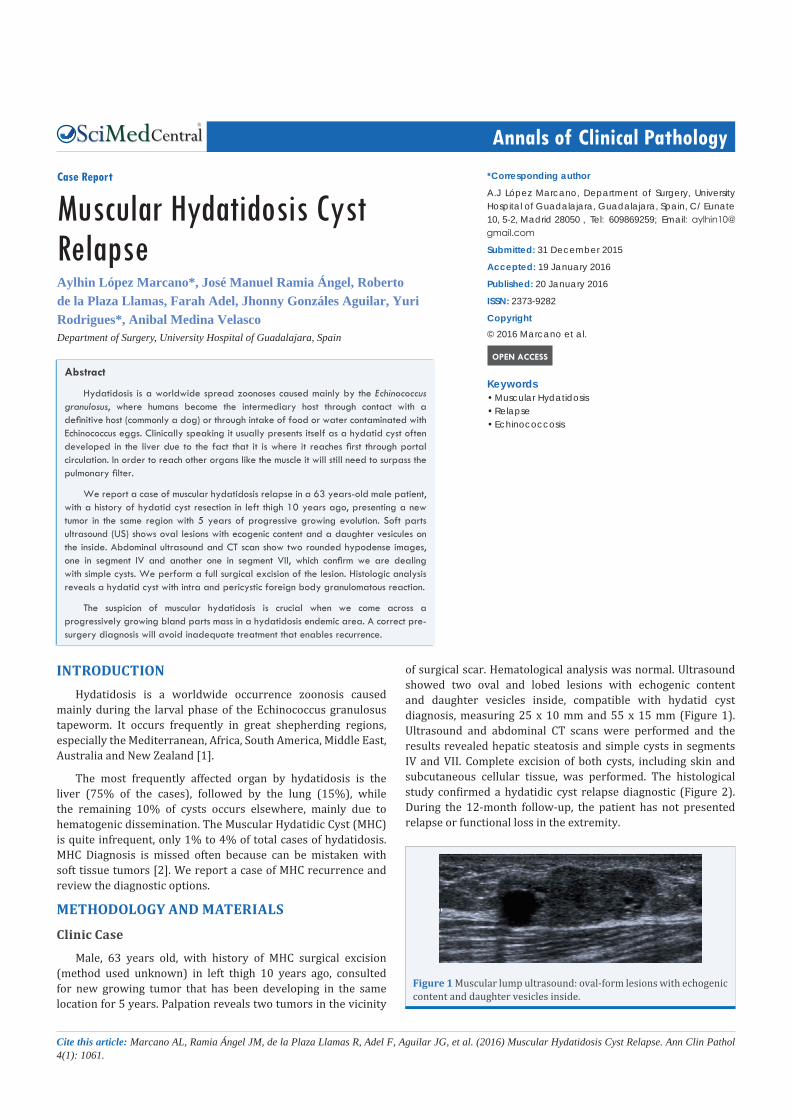
of surgical scar. Hematological analysis was normal. Ultrasound showed two oval and lobed lesions with echogenic content and daughter vesicles inside, compatible with hydatid cyst diagnosis, measuring 25 x 10 mm and 55 x 15 mm (Figure 1). Ultrasound and abdominal CT scans were performed and the results revealed hepatic steatosis and simple cysts in segments IV and VII. Complete excision of both cysts, including skin and subcutaneous cellular tissue, was performed. The histological study confirmed a hydatidic cyst relapse diagnostic (Figure 2). During the 12-month follow-up, the patient has not presented relapse or functional loss in the extremity.
Figure 1 Muscular lump ultrasound: oval-form lesions with echogenic content and daughter vesicles inside.

CentralBringing Excellence in Open Access
Marcano et al. (2016)Email:
Ann Clin Pathol 4(1): 1061 (2016) 2/3
DISCUSSION AND RESULTSIn the case of hydatidosis, man becomes the intermediary
host through contact with a definitive host (commonly a dog) or through intake of food or water contaminated with Echinococcus eggs. These eggs hatch in the intestine, the parasite pass across the intestinal wall and reach the liver through the portal venous system. Once there, if the parasites are not destroyed by macrophages a hepatic hydatid cyst will be formed [1-5]. For this reason, the liver is the most commonly affected organ by hydatidosis (75% of the patients). If it manages to surpass the hepatic filter, the parasite may reach the lungs through venous circulation, this is why the lungs are the second most affected organs (15%). Furthermore, if the parasite manages to cross the pulmonary filter –which only occurs in 10% of the patients– it may disseminate through arterial circulation to other parts of the body, affecting organs such as brain, heart, kidney, ureter, spleen, utero, fallopian tubes, mesentery, pancreas, bones and muscles [2-5].
The existence of hydatid cysts located in muscles, especially in lower extremities, as sole and primary presentation of hydatid infestation is very infrequent. The first case reported in musculoskeletal hydatidosis bibliography dates back to 1699 [6]. The low incidence of MHC is due to the fact that the muscle is not a favorable environment for a larva to grow because of the toxic effect of the lactic acid and the continuous muscular contractions, which prevent it from fixing easily. If MHC occurs, the muscle tends to encapsulate and isolate the larva, creating a thick layer of fibro-angioblastic connective tissue with numerous vesicles inside.
Clinically, the MHC manifests itself as a spherical mass that increases the size of the limb, with feeling of tension and heaviness, usually without pain except when it compresses the nerve stem or blood vessels [6,7]. In the case reported, the patient found a swelling growing slowly over 5 years without any other symptoms as there were no vascular or neural compressions.
The previous history of hydatidosis made the diagnosis of MHC easier in this case. But in the case of primary MHC, diagnosis can be difficult and can only be made through a combination of clinical, serological and radiological parameters. Serology in hydatidosis is not always positive for MHC and is more often negative in the
absence of pulmonary and hepatic diseases [8]. In ultrasound and CT scans, MHC is observed as a multi-vesicular lesion, which is characteristic but not pathognomonic [4]. The identification of small vesicles inside confirms the diagnosis of MHC [4]. The most suitable diagnostic technique in MHC is the MRI, because it allows differential diagnosis with soft tissue swelling [4]. We did not require it because the characteristic ultrasound image and the fact that it was a recurrent MHC suggested the diagnosis. Fine-needle aspiration must be avoided if there is suspicion of MHC in view of risk of dissemination and anaphylactic reaction [6]. The diagnosis can be confirmed by the histological studies. Whenever MHC is found, the other sites of frequent occurrence (liver and lungs) must be screened through imaging studies. The treatment for MHC is complete pericystectomy whenever feasible. Due to the thickness of the MHC connective tissue, as we have previously observed, excision must be done meticulously to avoid the rupture of the cyst wall. The major intra-operative complication is the rupture of the MHC because the release of daughter vesicles and intra-cystic liquid may cause the dissemination of the disease and/or the anaphylaxis [9]. Considering that the previous surgery performed on this patient is unknown we assume it was an incomplete excision. In complicated cases where radical surgery may cause serious functional deficit, incomplete resections can be performed but with high risk of recurrence. The use of antihelminthics is strongly recommended for patients with multiple, recurrent or inaccessible MHC [10].
CONCLUSIONIt is important for surgeons to have high index of suspicion
of muscular hydatidosis whenever a patient present with a progressive soft tissue swelling with no other major systemic diseases in a hydatidosis endemic area for making an appropriate preoperative diagnosis and avoid incomplete treatment as well as the risk of dissemination or recurrence of the disease as a result of the diagnose using needle punctures.
REFERENCES1. Pedrosa I, Saíz A, Arrazola J, Ferreirós J, Pedrosa CS. Hydatid disease:
radiologic and pathologic features and complications. Radiographics. 2000; 20: 795-817.
2. Rodop O, Arpacyoglu M.Ö, Akmaz I, Kiral A. Primary hydatidosis of the peripheral muscles: Treatment by surgery and chemotherapy. Eur J Plast Surg 2001; 24: 261-263.
3. Casero RD, Costas MG, Menso E. An unusual case of hydatid disease: localization to the gluteus muscle. Clin Infect Dis. 1996; 23: 395-396.
4. Navarro AC, Torcal J, García-Álvarez F, Burdio F, Tejero E, Güemes A, et al. Hidatidosis muscular primaria. Cir Esp. 2002; 72:147-145.
5. Tercero Gutiérrez MJ, Olalla Herbosa R. Hidatidosis: una zoonosis de distribución mundial. Offarm 2008; 27: 88-94.
6. Burdío F, Tejero E, Güemes A, Salinas JC, Lozano R, Navarro AC et al. Hidatidosis muscular primaria recidivante de vasto interno. Cir Esp. 2000; 68: 264-265.
7. Alimehmeti R, Seferi A, Rroji A, Alimehmeti M. Saphenous neuropathy due to large hydatid cyst within long adductor muscle: case report and literature review. J Infect Dev Ctries. 2012; 6: 531-535.
8. Agulló Bonus A, Alcalá-Santaella R. Hidatidosis muscular. A propósito de tres casos. Rev Esp Reumatol. 2002; 29: 4-6.
Figure 2 Histopathology: 1. Cyst 2. Multinucleated cells 3. Pericyst 4. Muscle.

CentralBringing Excellence in Open Access
Marcano et al. (2016)Email:
Ann Clin Pathol 4(1): 1061 (2016) 3/3
Marcano AL, Ramia Ángel JM, de la Plaza Llamas R, Adel F, Aguilar JG, et al. (2016) Muscular Hydatidosis Cyst Relapse. Ann Clin Pathol 4(1): 1061.
Cite this article
9. Charalambous GK, Katergiannakis VA, Manouras AJ. Three cases of primary hydatidosis of the gluteus muscle: our experience in clinical, diagnostic and treatment aspects. Chirurgia (Bucur). 2014; 109: 555-558.
10. Acar A, Rodop O, Yenilmez E, Baylan O, Oncül O. [Case report: primary localization of a hydatid cyst in the adductor brevis muscle]. Turkiye Parazitol Derg. 2009; 33: 174-176.