case 5 - ivra
TRANSCRIPT
Case 5Dr. Ana Pinto, DVM, MS, PhDResident in Diagnostic Imaging
Dr. Chee Kin Lim, DVM, BVSc(Hons), MMedVet (Diag Im), FMCVS (Vet Imaging), Dipl ECVDI Clinical Associate Professor, Diagnostic Imaging
Signalment and History
• 17 y male neutered Labrador retriever• Diagnosed with laryngeal paralysis 6-8 months ago by rDVM• Presented respiratory distress, cough, inspiratory stridor, and lethargy
over the last month• History of vomiting & regurgitation
Physical Exam Findings• T: 102.1o F PR: 110 beats/min RR: 60 breaths/min• Respiratory distress; harsh lung sounds in all fields with referred
upper airway noise• Regurgitation and hypersalivation
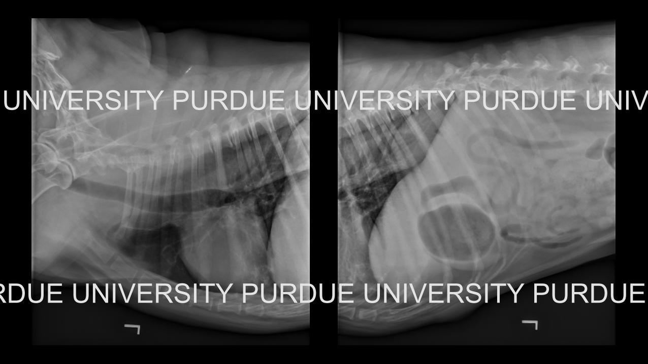
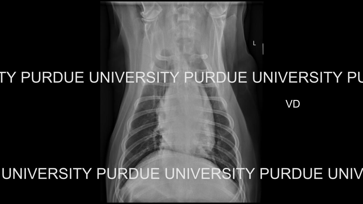
Thoracic radiographs are available.
1) Describe your radiological findings2) List your diagnosis/differential diagnoses
PURDUE UNIVERSITY PURDUE UNIVERSITY PURDUE UNIVERSITY PURDUE
UNIVERSITY
PURDUE
UNIVERSITY PURDUE UNIVERSITY PURDUE UNIVERSITY PURDUE
UNIVERSITY

PURDUE UNIVERSITY PURDUE UNIVERSITY PURDUE UNIVERSITY PURDUE UNIVERSITY
PURDUE
UNIVERSITY PURDUE UNIVERSITY PURDUE UNIVERSITY PURDUE
UNIVERSITY
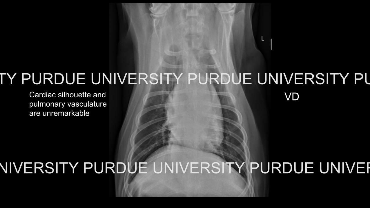
VD
PURDUE UNIVERSITY PURDUE UNIVERSITY PURDUE UNIVERSITY PURDUE UNIVERSITY
PURDUE
UNIVERSITY PURDUE UNIVERSITY PURDUE UNIVERSITY PURDUE
UNIVERSITY

Additional viewPURDUE UNIVERSITY
PURDUE UNIVERSITY

Abdominal compression view
PURDUE UNIVERSITY PURDUE UNIVERSITY PURDUE UNIVERSITY PURDUE
UNIVERSITY
PURDUE
UNIVERSITY PURDUE UNIVERSITY PURDUE UNIVERSITY PURDUE
UNIVERSITY
Radiological FindingsPURDUE UNIVERSITY
PURDUE UNIVERSITY
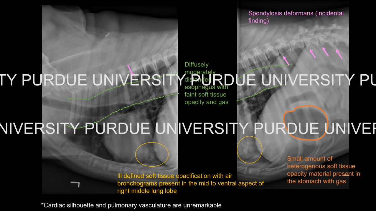
Diffusely moderately distended esophagus with faint soft tissue opacity and gas
Ill defined soft tissue opacification with air bronchograms present in the mid to ventral aspect of right middle lung lobe
Small amount of heterogenous soft tissue opacity material present in the stomach with gas
*Cardiac silhouette and pulmonary vasculature are unremarkable
Spondylosis deformans (incidental finding)
PURDUE UNIVERSITY PURDUE UNIVERSITY PURDUE UNIVERSITY PURDUE UNIVERSITY
PURDUE UNIVERSITY PURDUE UNIVERSITY PURDUE UNIVERSITY PURDUE UNIVERSITY
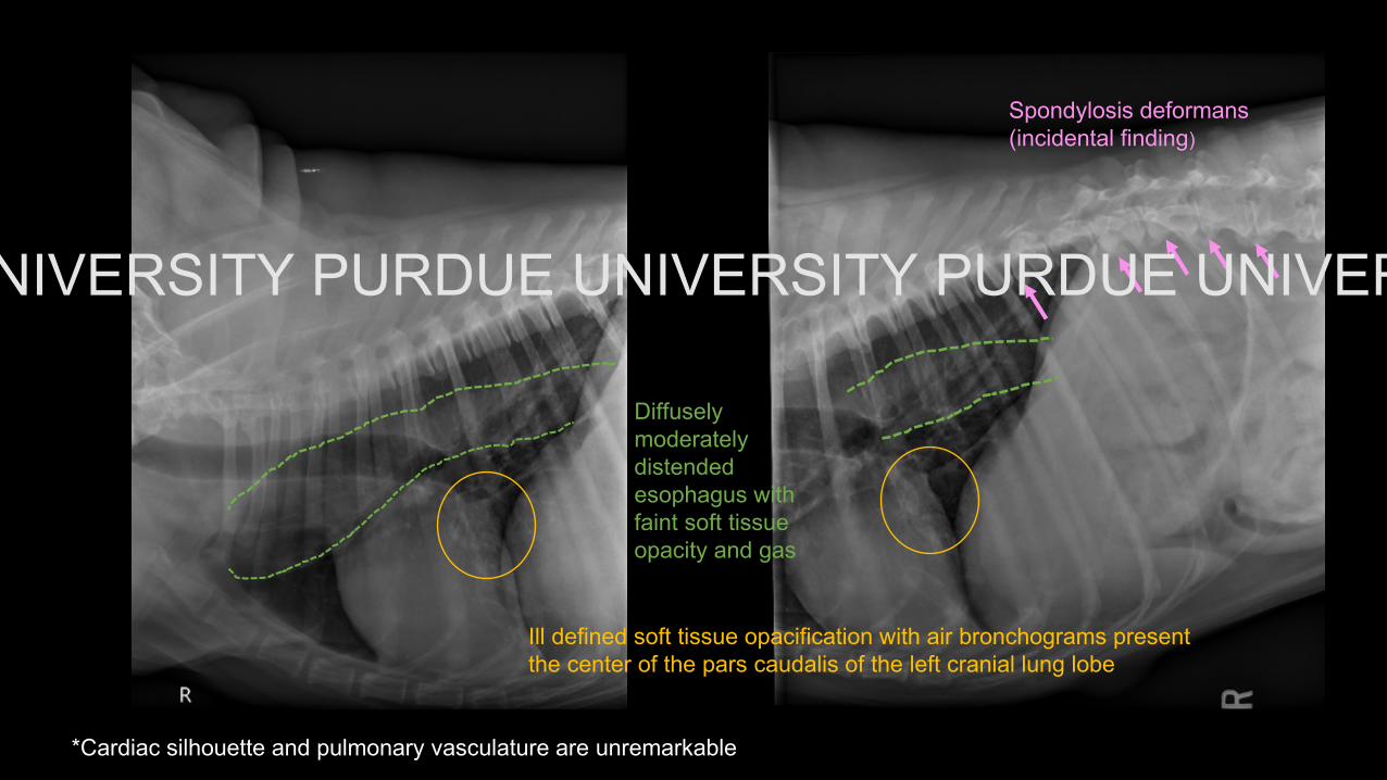
Diffusely moderately distendedesophagus withfaint soft tissueopacity and gas
Spondylosis deformans (incidental finding)
*Cardiac silhouette and pulmonary vasculature are unremarkable
Ill defined soft tissue opacification with air bronchograms present the center of the pars caudalis of the left cranial lung lobe
PURDUE UNIVERSITY PURDUE UNIVERSITY PURDUE UNIVERSITY PURDUE UNIVERSITY
PURDUE UNIVERSITY
Cardiac silhouette and pulmonary vasculature are unremarkable
VDPURDUE UNIVERSITY PURDUE UNIVERSITY PURDUE UNIVERSITY PURDUE UNIVERSITY
PURDUE
UNIVERSITY PURDUE UNIVERSITY PURDUE UNIVERSITY PURDUE
UNIVERSITY
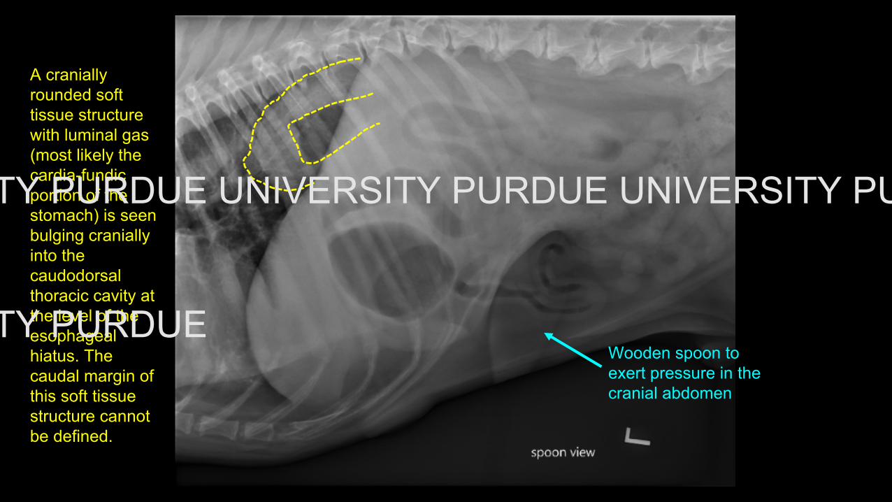
A cranially rounded soft tissue structure with luminal gas (most likely the cardia-fundic portion of the stomach) is seen bulging cranially into the caudodorsalthoracic cavity at the level of theesophagealhiatus. The caudal margin of this soft tissue structure cannot be defined.
Wooden spoon to exert pressure in the cranial abdomen
PURDUE UNIVERSITY PURDUE UNIVERSITY PURDUE UNIVERSITY PURDUE UNIVERSITY
PURDUE UNIVERSITY PURDUE UNIVERSITY
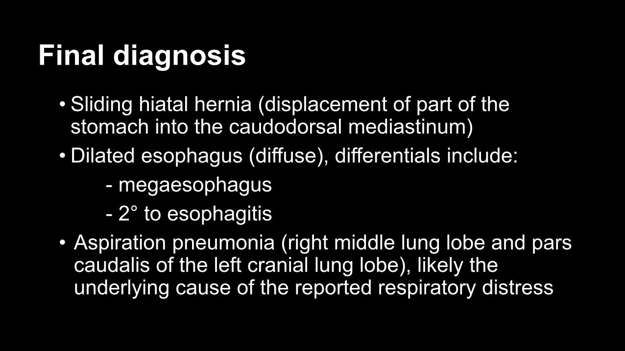
• Sliding hiatal hernia (displacement of part of thestomach into the caudodorsal mediastinum)
• Dilated esophagus (diffuse), differentials include:- megaesophagus- 2° to esophagitis
• Aspiration pneumonia (right middle lung lobe and parscaudalis of the left cranial lung lobe), likely theunderlying cause of the reported respiratory distress
Final diagnosis
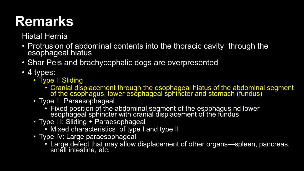
RemarksHiatal Hernia• Protrusion of abdominal contents into the thoracic cavity through the
esophageal hiatus• Shar Peis and brachycephalic dogs are overpresented• 4 types:
• Type I: Sliding• Cranial displacement through the esophageal hiatus of the abdominal segment
of the esophagus, lower esophageal sphincter and stomach (fundus)• Type II: Paraesophageal
• Fixed position of the abdominal segment of the esophagus nd loweresophageal sphincter with cranial displacement of the fundus
• Type III: Sliding + Paraesophageal• Mixed characteristics of type I and type II
• Type IV: Large paraesophageal• Large defect that may allow displacement of other organs—spleen, pancreas,
small intestine, etc.

RemarksHiatal Hernia• Clinical Signs
• mild anorexia, hypersalivation, regurgitation, vomiting, hematemesis, anddyspnea
• Diagnosis• radiography or fluoroscopy; intermittent nature can make diagnosis
challenging; abdominal compression with a wooden spoon can be veryhelpful!
• computed tomography• Treatment
• Medical treatment of esophagitis• Surgical correction

References• Callan, M. B. et al. Congenital esophageal hiatal hernia in the Chinese Shar-
Pei Dog. Journal of Veterinary Internal Medicine. 1993;7:210–215.• Guiot, L. P., Lansdowne, J. L., Rouppert, P. & Stanley, B. J. Hiatal hernia in
the dog: A clinical report of four chinese Shar Peis. Journal of the AmericanAnimal Hospital Association. 2008;44:335–341.
• Reeve EJ, Sutton D, Friend EJ, Warren-Smith CMR. Documenting theprevalence of hiatal hernia and oesophageal abnormalities inbrachycephalic dogs using fluoroscopy. J Small Anim Pract.2017;58(12):703-708.
• Lodzinska, J., Culshaw, G., Hall, J. L., Schwarz, T. & Liuti, T. CT diagnosis ofintermittent type IV paraoesophageal hernia in a dog. Vet Rec Case Rep2018;6:e000692. doi: 10.1136/vetreccr-2018-000692