canine distemper virus: an emerging disease in wild - mbio
TRANSCRIPT

Canine Distemper Virus: an Emerging Disease in Wild EndangeredAmur Tigers (Panthera tigris altaica)
Tracie A. Seimon,a Dale G. Miquelle,a Tylis Y. Chang,b Alisa L. Newton,a Irina Korotkova,c Galina Ivanchuk,c Elena Lyubchenko,c
Andre Tupikov,d Evgeny Slabe,e Denise McAloosea
Wildlife Conservation Society, Bronx, New York, USAa; Albert Einstein College of Medicine of Yeshiva University, Bronx, New York, USAb; Primorskaya State AgriculturalAcademy, Ussurisk, Russiac; Bureau of Forensic Examinations, Ussurisk Department of Health Services, Ussurisk, Russiad; Terney County Veterinarian Services, PrimorskiiKrai, Russiae
ABSTRACT Fewer than 500 Amur tigers (Panthera tigris altaica) remain in the wild. Due to low numbers and their solitary andreclusive nature, tiger sightings across their range in the Russian Far East and China are rare; sightings of sick tigers are rarerstill. Serious neurologic disease observed in several wild tigers since 2001 suggested disease emergence in this endangered spe-cies. To investigate this possibility, histology, immunohistochemistry (IHC), in situ hybridization (ISH), and reversetranscription-PCR (RT-PCR) were performed on tissues from 5 affected tigers that died or were destroyed in 2001, 2004, or 2010.Our results reveal canine distemper virus (CDV) infection as the cause of neurologic disease in two tigers and definitively estab-lish infection in a third. Nonsuppurative encephalitis with demyelination, eosinophilic nuclear viral inclusions, and positiveimmunolabeling for CDV by IHC and ISH were present in the two tigers with available brain tissue. CDV phosphoprotein (P)and hemagglutinin (H) gene products were obtained from brains of these two tigers by RT-PCR, and a short fragment of CDV Pgene sequence was detected in lymph node tissue of a third tiger. Phylogenetically, Amur tiger CDV groups with an Arctic-likestrain in Baikal seals (Phoca siberica). Our results, which include mapping the location of positive tigers and recognition of acluster of cases in 2010, coupled with a lack of reported CDV antibodies in Amur tigers prior to 2000 suggest wide geographicdistribution of CDV across the tiger range and recent emergence of CDV as a significant infectious disease threat to endangeredAmur tigers in the Russian Far East.
IMPORTANCE Recognition of disease emergence in wildlife is a rare occurrence. Here, for the first time, we identify and character-ize a canine distemper virus (CDV), the second most common cause of infectious disease death in domestic dogs and a viral dis-ease of global importance in common and endangered carnivores, as the etiology of neurologic disease and fatal encephalitis inwild, endangered Amur tigers. We establish that in 2010 CDV directly or indirectly killed ~1% of Amur tigers. Location of posi-tive cases over an expansive geographic area suggests that CDV is widely distributed across the tiger range. Interspecies interac-tions are increasing as human populations grow and expand into wildlife habitats. Identifying animal reservoirs for CDV andidentifying the CDV strains that are transmissible to and among wildlife species, including Amur tigers and sympatric criticallyendangered Amur leopards (Panthera pardus orientalis), is essential for guiding conservation and mitigation efforts.
Received 30 May 2013 Accepted 9 July 2013 Published 13 August 2013
Citation Seimon TA, Miquelle DG, Chang TY, Newton AL, Korotkova I, Ivanchuk G, Lyubchenko E, Tupikov A, Slabe E, McAloose D. 2013. Canine distemper virus: an emergingdisease in wild endangered Amur tigers (Panthera tigris altaica). mBio 4(4):e00410-13. doi:10.1128/mBio.00410-13.
Editor Michael Buchmeier, University of California, Irvine
Copyright © 2013 Seimon et al. This is an open-access article distributed under the terms of the Creative Commons Attribution-Noncommercial-ShareAlike 3.0 Unportedlicense, which permits unrestricted noncommercial use, distribution, and reproduction in any medium, provided the original author and source are credited.
Address correspondence to Denise McAloose, [email protected].
Since the beginning of the 20th century, tiger populations havecollapsed from an estimated 100,000 tigers among 9 subspe-
cies to fewer than 3,500 individuals (1, 2), and all extant subspeciesare currently listed as endangered or critically endangered by theInternational Union for the Conservation of Nature (http://www.iucn.org/). Poaching, decimation of the tiger prey base, andhabitat fragmentation have all contributed to tiger decline (1, 3,4). For the Amur tiger (Panthera tigris altaica) subspecies, 400 to500 animals remain in the wild and represent one of the mostendangered cat populations on the planet (2). Usually reclusiveand seldom observed, adult Amur tigers have been seen unchar-acteristically entering villages and wandering onto roadways in theRussian Far East (RFE) since 2001. Even more unusual is abnor-mal neurologic behavior in the tigers. These observations, along
with the diagnosis of morbillivirus infection in a wild Amur tigerin 2004 (histology and immunohistochemistry [IHC] findingswere previously described [5]), a cluster of wild tigers with abnor-mal neurologic behavior in 2010, and a localized rapid and unex-pected decline of tigers in the Sikhote-Alin biosphere reserve(SABR) in the RFE since 2009 (6), led to concerns about an infec-tious disease and in particular canine distemper virus (CDV) as anemerging threat to Amur tigers. Our aim was to identify the causeof neurologic disease and death in endangered Amur tigers in theRFE, establish etiologic linkages between cases over a wide geo-graphic and temporal range, and understand the significance ofthe disease in tiger conservation.
Tissues collected during necropsy procedures from five adult,free-ranging tigers that died naturally or were destroyed in the
OBSERVATION
July/August 2013 Volume 4 Issue 4 e00410-13 ® mbio.asm.org 1
on April 9, 2019 by guest
http://mbio.asm
.org/D
ownloaded from

RFE in 2001, 2004, or 2010 were available for histopathology, IHCstaining, in situ hybridization (ISH), and reverse transcription-PCR (RT-PCR) testing. Brain tissue, critical in assessing CDV in-fection, was available from two tigers (Pt2004 and Pt2010-3);lung, a primary site of CDV replication, was available from alltigers.
Histologic processing of formalin-fixed tissues was performedusing routine methods. Five to eighteen of twenty-two differenttissue types (adipose tissue, adrenal gland, artery, brain, heart,kidney, large intestine, liver, lung, lymph node, ovary, pancreas,peripheral nerve, salivary gland, skeletal muscle, spleen, stomach,small intestine, testis, tongue, trachea, or urinary bladder) wereavailable from each animal for histologic review. Bright-field mi-croscopy was performed using a Leica DM2500 microscope (LeicaMicrosystems Wetzlar GmbH, Wetzlar, Germany).
IHC for canine distemper virus antigen was performed using aprimary monoclonal IgG1 anti-CDV surface envelope antibody asdescribed previously and included positive and negative controls(5). Bright-field microscopy was performed as described above.
For ISH, probes to a 600-bp nucleotide region of the phospho-protein (P) gene of canine distemper virus were designed by Pa-nomics (Affymetrix, Inc., Santa Clara, CA). This region corre-sponds to nucleotides 1926 to 2526 of the CDV genome (GenBankaccession no. AF378705). ISH using Fast Red staining was per-formed using the Panomics QuantiGene View RNA kit forformalin-fixed paraffin-embedded sections according to the man-ufacturer’s protocol (product QV0050, QuantiGene ViewRNAFFPE; Affymetrix, Inc., Santa Clara, CA,) and as described previ-ously (7). Sections were counterstained with hematoxylin. Dupli-cate sections were run without the probe as a negative control.Bright-field microscopy was performed as described above.
For canine distemper virus RT-PCR, RNA was extracted usinga standard protocol for xylene deparaffinization of formalin-fixed,paraffin-embedded tissue (a total of 50 �m [10 sections, 5 �meach] of tissue; RNeasy FFPE kit [Qiagen Inc., Valencia CA]).Primer sets were designed from the following regions of the CDVgenome: P gene, MorbF, 2132 to 2151; MorbR, 2560 to 2541;CDVF4, 2206 to 2228; and CDVR3, 2319 to 2297; and hemagglu-tinin (H) gene, CDVH2-F, 8593 to 8619; CDVH2-R, 8842 to 8821;CDVH3-R, 8883 to 8864; CDVH4-R, 8868 to 8850; CDV-HF,8521 to 8541; and CDV-HR, 8836 to 8815. Nucleotide positionswere based on CDV strain A75/17 (GenBank accession no.AF164967). Primers were purchased from Life Technologies(Norwalk, CT). CDV-positive raccoon and fox brain were used ascontrol tissues. No-template negative controls were included in allassays. One-step RT-PCR amplification of CDV P or H gene re-gions was performed using a standard protocol (Qiagen Inc., Va-lencia CA). RT-PCR reactions were carried out using an initial50°C RT step for 30 min, using an annealing temperature of 45°Cfor 45 s, and 40 cycles. PCR products of correct molecular weightwere purified using the ExoSAP-IT reagent (Affymetrix, SantaClara, CA) or the Qiagen MinElute gel extraction kit and directlysequenced in the forward and reverse directions using an ABI3730x DNA analyzer for capillary electrophoresis and fluorescentdye terminator detection (Genewiz Inc., South Plainfield, NJ).Sequences were trimmed, aligned, and subjected to analysis usingthe BLASTn and BLASTx search tools to determine the identitiesof the viral sequences (GenBank, National Center for Biotechnol-ogy Information). The nucleotide sequences of the H gene weretranslated and aligned using the Geneious alignment tool (Ge-
neious Pro 5.1.7 software; Biomatters Ltd., Auckland, New Zea-land) and analyzed for amino acid polymorphisms at positions530 and 549 in the SLAM receptor binding region of the H gene.
To determine the phylogenetic relationships of tiger CDVs toeach other and to other CDV viruses and morbilliviruses, nucleo-tide sequences for the P and H genes from the tigers and represen-tative CDV strains were aligned (GenBank, National Center forBiotechnology Information; http://www.ncbi.nlm.nih.gov) (Ge-neious Pro 5.1.7 software; Biomatters Ltd., Auckland, New Zea-land). Pairwise identities were obtained by PAUP analysis to cre-ate a running P-distance pairwise comparison matrix (PAUPplugin in Geneious Pro). Bayesian analysis was performed usingMrBayes 3.1 plugin in Geneious Pro using gamma-distributedrate variation and an HKY85 substitution model (8). The first 25%of a 1,100,000 chain length was discarded as burn-in, and 4 heatedchains were run with a subsampling frequency of 200. Rinderpestvirus (accession no. AF132934) was used as an outgroup. Treeswere finalized and labeled (FigTree v1.3.1 software [Andrew Ram-baut, Institute of Evolutionary Biology, University of Edinburgh,2006 to 2009; http://tree.bio.ed.ac.uk/]). Posterior probability val-ues were calculated.
Between January and June 2010, three adult free-ranging Amurtigers (Panthera tigris altaica) (Pt2010-1, Pt2010-2, and Pt2010-3)entered villages in the RFE (Fig. 1A and B). Each was killed(Pt2010-1 and Pt2010-3) or died naturally (Pt-2010-2) after ex-hibiting abnormal neurologic behavior (disorientation, lack of re-sponse to stimulation, and/or nonaggressive fearlessness). Prior to2010, two other free-ranging Amur tigers (Pt2001 and Pt2004)were captured and died after exhibiting similar neurologic behav-ior (Fig. 1A and B). Four of the five tigers were emaciated orshowed extreme weight loss at the time of death (Fig. 1B).
Brain tissue was available from Pt2010-3 and Pt2004 (histologyand IHC for the latter were previously reported [5], and tissue wasreprocessed and reviewed for this article). Histologic lesions in thebrains were identical and consisted of nonsuppurative viral en-cephalitis with severe demyelination. Brightly eosinophilic neuro-nal and glial cell nuclear viral inclusions and positive immunohis-tochemical staining in these cell types for an envelope componentof CDV were also seen (Fig. 1C and D, respectively). The findingswere severe and were sufficient to result in the clinically observedneurologic behavior in both cases and natural death in Pt2004 (5).Mild or moderate lymphoid depletion was seen in lymph nodes ofPt2010-1 and Pt2001, respectively, and moderate lymphoid deple-tion was seen in spleens from Pt-2001, Pt2004, and Pt2010-1. In-tralesional viral RNA was confirmed in both tiger brains using ISHto a 600-bp segment of the CDV P gene (Fig. 1E and F). Viralinclusions, IHC staining, or ISH consistent with CDV infectionwere not seen in nonneural tissues, including lung or lymphoidtissues, from any of the tigers (data not shown). Concurrent,transmissible infectious disease was not seen.
Extracted RNA from select formalin-fixed paraffin-embedded(FFPE) tissues was analyzed by RT-PCR for morbillivirus andCDV phosphoprotein P and H genes using multiple primer sets.Positive results were obtained in 3 of 5 tigers: Pt2004 (histologicand IHC description previously reported [5]), Pt2010-2, and Pt-2010-3. CDV P gene products ranging in size from 114 bp to430 bp and a 291-bp H gene product were recovered from thebrains of both Pt2004 and Pt2010-3. In Pt2010-2, lymph nodetissue was positive for a 114-bp fragment of the CDV P gene andwas H gene negative. Possible reasons for failure to recover H gene
Seimon et al.
2 ® mbio.asm.org July/August 2013 Volume 4 Issue 4 e00410-13
on April 9, 2019 by guest
http://mbio.asm
.org/D
ownloaded from

sequence from this tiger include RNA degradation due to autolysisand/or cross-linking due to formalin fixation and/or prolongedformalin fixation prior to RT-PCR. In addition to the possibilityof true negatives, these complications could have prevented iden-tification of positive cases among the remaining two tigers (Pt2001
and Pt2010-1) or additional tissues in positive cases. Not havingaccess to brain, the optimal target tissue in these neurologic tigers,may also explain the failure to identify additional positive tigers.
Gene product sequences from Pt2004, Pt2010-2, and Pt2010-3were aligned with sequences of representative morbilliviruses,
FIG 1 Geographical distribution (A) and historical information (B) for tigers in the Russian Far East that died or were killed due to abnormal neurologicbehavior in 2001, 2004, or 2010. (C) Tiger Pt2010-3: hematoxylin-and-eosin-stained section of brain with neuronal intranuclear eosinophilic viral inclusions(arrow). (D) Tiger Pt2010-3: positive immunohistochemical staining of neurons with monoclonal IgG primary antibody to CDV viral envelope protein antigen(arrows) (fast-red staining). (E and F) Positive in situ hybridization (fast red) of probes to CDV P gene sequence in CDV-infected neurons in tiger Pt2004 (E) andtiger Pt2010-3 (F). Bar � 50 �m in all images.
Canine Distemper Virus in Endangered Tigers
July/August 2013 Volume 4 Issue 4 e00410-13 ® mbio.asm.org 3
on April 9, 2019 by guest
http://mbio.asm
.org/D
ownloaded from
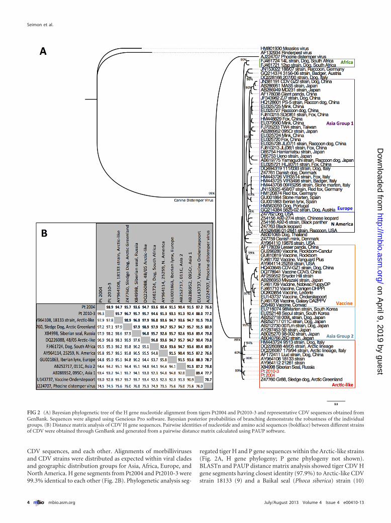
CDV sequences, and each other. Alignments of morbillivirusesand CDV strains were distributed as expected within viral cladesand geographic distribution groups for Asia, Africa, Europe, andNorth America. H gene segments from Pt2004 and Pt2010-3 were99.3% identical to each other (Fig. 2B). Phylogenetic analysis seg-
regated tiger H and P gene sequences within the Arctic-like strains(Fig. 2A, H gene phylogeny; P gene phylogeny not shown).BLASTn and PAUP distance matrix analysis showed tiger CDV Hgene segments having closest identity (97.9%) to Arctic-like CDVstrain 18133 (9) and a Baikal seal (Phoca siberica) strain (10)
FIG 2 (A) Bayesian phylogenetic tree of the H gene nucleotide alignment from tigers Pt2004 and Pt2010-3 and representative CDV sequences obtained fromGenBank. Sequences were aligned using Geneious Pro software. Bayesian posterior probabilities of branching demonstrate the robustness of the individualgroups. (B) Distance matrix analysis of CDV H gene sequences. Pairwise identities of nucleotide and amino acid sequences (boldface) between different strainsof CDV were obtained through GenBank and generated from a pairwise distance matrix calculated using PAUP software.
Seimon et al.
4 ® mbio.asm.org July/August 2013 Volume 4 Issue 4 e00410-13
on April 9, 2019 by guest
http://mbio.asm
.org/D
ownloaded from
(Fig. 2B). Our results indicate that tiger CDV is an Arctic-likestrain similar to those from Greenland (11), China (12), Russia(10), and the United States (9).
The critical amino acid residues G530 and Y549 in the SLAMreceptor binding domain of the CDV hemagglutinin protein havebeen shown to determine host cell tropism in vitro. G ¡ N, R, orD or Y ¡ H substitution at the 530 or 549 residue, respectively, isproposed to be associated with CDV transmission from domesticdogs (Canis lupus familiaris) and disease emergence in novel hostspecies (13). Both tiger sequences lacked the Y549 ¡ H substitu-tion but contained an N residue at position G530. Because theG530 ¡ N substitution is a consistent finding in Arctic-like strainsin general, including those in dogs and wildlife (12–14), we cannotattribute a recent substitution event at this residue to diseaseemergence in tigers. If this amino acid is under positive selectivepressure, the change may have occurred through a dog-to-wildlifetransmission prior to 1988, when Arctic-like strains, which in-clude the G530 ¡ N mutation, were first detected in Baikal sealsand sled dogs in Greenland (14). Subsequent reintroduction ofvirus with this substitution into the domestic dog population mayexplain why the substitution is a predominant synapomorphy inthe Arctic-like lineage. Another interesting finding was threeunique amino acid changes (V538 ¡ I, T548 ¡ M, and D570 ¡N) in the tiger H gene sequence that have not been observed pre-viously in Arctic-like strains. These findings suggest that the tigerArctic-like CDV is distinct; however, additional informationabout Arctic-like strains is needed to be confident in this conclu-sion.
CDV is the second most common cause of infectious diseasedeath in domestic dogs and is a significant viral disease of globalimportance in common and endangered wild carnivores (15). It isa multihost pathogen, and interactions with and disease transmis-sion from abundant wildlife reservoir species, such as raccoondogs (Nyctereutes procyonoides) or domestic dogs, are likely to beas important, if not more important, for disease transmission andpopulation effect than infection among tigers alone due to lowtiger numbers and population density (16). In the RFE, little ap-pears to be known about the distribution and strains of CDV thatare circulating in domestic dogs and wildlife. However, our iden-tification of positive tiger CDV cases separated by 200 km to300 km suggests wide distribution for the Arctic-like CDV strainthat infects and kills Amur tigers.
Low rates of vaccination and CDV infection are present indomestic dogs in Russia, and direct transmission of CDV frominfected, unvaccinated dogs to tigers is a significant concern, sinceAmur tigers are known to encounter and kill domestic dogs (17).In one survey, only 16% of village dogs were vaccinated againstCDV and 58% of unvaccinated dogs were seropositive for anti-bodies to the virus, indicating high endemic exposure (18). In thesame report, 15% of wild tigers (n � 40) sampled between 2000and 2004 were seropositive for CDV antibodies, with no seropos-itive tigers detected prior to 2000 (n � 27) (18); both Pt2004 andPt2010-3 were seropositive for antibodies to CDV (1:256; virusneutralization [VN] � 1:4 positive threshold value) two (5) andthree (data not shown) months, respectively, prior to their deaths.
CDV is a preventable infectious disease, and vaccination strat-egies, all of which have limitations and significant challenges in awildlife setting (19, 20), are likely to be considered for protectingendangered Amur tigers. Because dogs are a known CDV reser-voir, one strategy is to vaccinate domestic dogs to decrease trans-
mission risk to susceptible wildlife. This strategy was initiated inthe Serengeti ecosystem in 2003 in response to several significantCDV mortality events in lions (Panthera leo) (21). The successof this strategy to date is unclear, since at least one CDV out-break has occurred since initiation of the vaccination program(21). A second strategy is direct wildlife vaccination, which be-cause of small numbers of animals, limited range, and knownhigh disease-associated mortality is a critical component in con-servation programs for the endangered black-footed ferret(Mustela nigripes) (22) and critically endangered Santa CatalinaIsland fox (Urocyon littoralis catalinae) (23). Vaccination withrecombinant vectored vaccines has been safely used and isthe recommendation for captive tigers in Association of Zoosand Aquariums (AZA)-accredited zoos (http://www.aazv.org/displaycommon.cfm?an�1&subarticlenbr�273) and for nondo-mestic canid and other wildlife species (modified-live vaccines caninduce disease and should not be used). Recombinant vectoredvaccines may provide an option in wild tiger vaccination strate-gies, which in addition to safety must also consider efficacy, prac-ticality, limitations, cost, and unintended consequences of vacci-nation (including increased disease susceptibility to CDV or otherpathogens) in target or nontarget species (19, 20).
Infectious disease as the cause of population decline or (lesscommonly) extinction in free-ranging wildlife is a recognizedthreat to species survival; however, our ability to identify theseevents and their significance as they are occur and in time to mit-igate their effects is rare (24). The exact timing of CDV emergencein the RFE Amur tiger population is speculative. The absence ofpositive serology prior to 2000 (18), lack of documented observa-tions of neurologically ill tigers by scientists (5, 6, 18) or peopleliving in tiger range (personal communication, Igor Gregorivich)prior to 2001, and a cluster of cases in 2010 suggest CDV emer-gence after 2000 (whether earlier individual cases or previouswaves of tiger CDV infection and mortality occurred but wereundetected prior to 2000 remains to be rigorously investigated).Additionally, in 2010 alone, CDV infection directly or indirectlykilled approximately 1.0% of wild Amur tigers (2 adults and 3abandoned cubs). These deaths reflect the immediate, direct ef-fects of CDV infection and more than likely underestimate actualCDV-related deaths. In addition and at the population level, thelong-term impact of losing reproductively active animals, espe-cially females like Pt2010-3, will exceed the direct effects of indi-vidual animal infection alone through lost productivity of boththe dam and her offspring (25).
Our study is the first to confirm and genetically characterize aCDV that is killing wild, endangered Amur tigers in the RFE. Ourresults indicate that tiger CDV is an Arctic-like strain similar toCDV in Baikal seals in Russia and domestic dogs. Our report il-lustrates the importance of long-term wildlife monitoring andhealth surveillance in identifying emerging threats in endangeredspecies. It also shows how through these efforts we are afforded anopportunity to develop and implement mitigation activities, in-cluding identification of CDV reservoir species and considerationand assessment of vaccination strategies, to reduce disease risk inAmur tigers and sympatric critically endangered Amur leopards(Panthera pardus orientalis).
Nucleotide sequence accession numbers. Tiger CDV P and Hgene sequences were deposited in GenBank (accession numbersKC579363 [Pt2004; H gene], KC579361 [Pt2004; P gene], andKC579362 [Pt2010-3; H gene]). Accession numbers for tiger-
Canine Distemper Virus in Endangered Tigers
July/August 2013 Volume 4 Issue 4 e00410-13 ® mbio.asm.org 5
on April 9, 2019 by guest
http://mbio.asm
.org/D
ownloaded from
derived sequences and all other sequences are presented in thefigures.
ACKNOWLEDGMENTS
Funding was generously provided by the Dunemere Foundation.We thank Melissa Miller and Judy St. Leger for the positive-control
CDV tissues and John Goodrich, Kathy Quigley, Charles Leathers, AlfredNgbokoli, Daniel Friedman, Damien Joly, Enkhtuvshin Shiilegdamba,Kate Jenks, and Jamie Phillips for materials, advice, and logistical support.Special thanks go to Carol Oddoux, our Russian colleagues and the fieldstaff that participated on the necropsy teams, and Séamus Maclennan andMartin Gilbert for reviewing the manuscript.
REFERENCES1. Dinerstein E, Loucks C, Wikramanayake E, Ginsberg J, Sanderson E,
Seidensticker J, Forrest J, Bryja G, Heydlauff A, Klenzendorf S, Leim-Gruber P, Mills J, O’Brien TG, Shrestha M, Simons R, Songer M. 2007.The fate of wild tigers. BioScience 57:508 –514.
2. Henry P, Miquelle D, Sugimoto T, McCullough DR, Caccone A, Rus-sello MA. 2009. In situ population structure and ex situ representation ofthe endangered Amur tiger. Mol. Ecol. 18:3173–3184.
3. Kinnaird MG, Sanderson EW, O’Brien TG, Wibisono HT, Woolmer G.2003. Deforestation trends in a tropical landscape and implications forendangered large mammals. Conserv. Biol. 17:245–257.
4. Smith JL, Tunhikorn S, Tanhan S, Simcharoen S, Kanchanasaka B.1999. Metapopulation structure of tigers in Thailand, p 166 –175. In Se-idensticker J, Christie S, Jackson P (ed), Riding the tiger: tiger conserva-tion in human-dominated landscapes. Cambridge University Press, Cam-bridge, United Kingdom.
5. Quigley KS, Evermann JF, Leathers CW, Armstrong DL, Goodrich J,Duncan NM, Miquelle DG. 2010. Morbillivirus infection in a wild Sibe-rian tiger in the Russian Far East. J. Wildl. Dis. 46:1252–1256.
6. Smirnov EN, Miquelle DG, Zaumyslova OY. 2012. Population dynamicsof the Amur tiger in Sikhote-Alin Zapovednik: 1966 –2011, p. 159 –178. InSikhote-Alin Biosphere Region: condition of ecosystems and theircomponents: a volume of scientific work for the 75th anniversary of theSikhote-Alin Reserve. DalNauka, Vladivostok, Russia. (In Russian.)
7. Seimon TA, McAloose D, Raphael B, Honkavuori KS, Chang T, Hirsch-berg DL, Lipkin WI. 2012. A novel herpesvirus in 3 species of pheasants:mountain peacock pheasant (Polyplectron inopinatum), Malayan peacockpheasant (Polyplectron malacense), and Congo peafowl (Afropavo congen-sis). Vet. Pathol. 49:482– 491.
8. Huelsenbeck JP, Ronquist F. 2001. MRBAYES: Bayesian inference ofphylogenetic trees. Bioinformatics 17:754 –755.
9. Pardo ID, Johnson GC, Kleiboeker SB. 2005. Phylogenetic characteriza-tion of canine distemper viruses detected in naturally infected dogs inNorth America. J. Clin. Microbiol. 43:5009 –5017.
10. Mamaev LV, Denikina NN, Belikov SI, Volchkov VE, Visser IK, Flem-ing M, Kai C, Harder TC, Liess B, Osterhaus AD, Barrett T. 1995.Characterisation of morbilliviruses isolated from Lake Baikal seals (Phocasibirica). Vet. Microbiol. 44:251–259.
11. Bohm J, Blixenkrone-Møller M, Lund E. 1989. A serious outbreak ofcanine distemper among sled-dogs in northern Greenland. Arctic Med.Res. 48:195–203.
12. Martella V, Cirone F, Elia G, Lorusso E, Decaro N, Campolo M,Desario C, Lucente MS, Bellacicco AL, Blixenkrone-Møller M, Carmi-chael LE, Buonavoglia C. 2006. Heterogeneity within the hemagglutiningenes of canine distemper virus (CDV) strains detected in Italy. Vet. Mi-crobiol. 116:301–309.
13. McCarthy AJ, Shaw MA, Goodman SJ. 2007. Pathogen evolution anddisease emergence in carnivores. Proc. Biol. Sci. 274:3165–3174.
14. Bolt G, Jensen TD, Gottschalck E, Arctander P, Appel MJ, Buckland R,Blixenkrone-Møller M. 1997. Genetic diversity of the attachment (H)protein gene of current field isolates of canine distemper virus. J. Gen.Virol. 78:367–372.
15. Deem SL, Spelman LH, Yates RA, Montali RJ. 2000. Canine distemperin terrestrial carnivores: a review. J. Zoo Wildl. Med. 31:441– 451.
16. Craft ME, Hawthorne PL, Packer C, Dobson AP. 2008. Dynamics of amultihost pathogen in a carnivore community. J. Anim. Ecol. 77:1257–1264.
17. Goodrich JM, Seryodkin IV, Miquelle DG, Beriznuk SI. 2011. Conflictsbetween Amur (Siberian) tigers and humans in the Russian Far East. Biol.Conserv. 144:584 –592.
18. Goodrich JM, Quigley KS, Lewis JC, Astafiev AA, Slabi EV, MiquelleDG, Smirnov EN, Kerley LL, Armstrong DL, Quigley HB, HornockerMG. 2012. Serosurvey of free-ranging Amur tigers in the Russian Far East.J. Wildl. Dis. 48:186 –189.
19. Chauvenet AL, Durant SM, Hilborn R, Pettorelli N. 2011. Unintendedconsequences of conservation actions: managing disease in complex eco-systems. PLoS ONE 6:e28671. doi: 10.1371/journal.pone.0028671.
20. Munson L, Terio KA, Ryser-Degiorgis MP, Lane EP, Courchamp F.2010. Wild felid diseases: conservation implications and managementstrategies, p 237–259. In MacDonald DW, Loveridge AJ (ed), Biology andconservation of wild felids. Oxford University Press, Oxford, United King-dom.
21. Craft ME. 2010. Ecology of infectious diseases in Serengeti lions, p263–282. In MacDonald DW, Loveridge AJ (ed), Biology and conserva-tion of wild felids. Oxford University Press, Oxford, United Kingdom.
22. Holmes BE. 2008. A review of black-footed ferret reintroduction innorthwest Colorado, 2001–2006, p . Technical Note 426. U.S. Departmentof the Interior, Bureau of Land Management, White River Field Office,Meeker, CO43.
23. Timm SF, Munson L, Summers BA, Terio KA, Dubovi EJ, RupprechtCE, Kapil S, Garcelon DK. 2009. A suspected canine distemper epidemicas the cause of a catastrophic decline in Santa Catalina Island foxes (Uro-cyon littoralis catalinae). J. Wildl. Dis. 45:333–343.
24. MacPhee RD, Greenwood AD. 2013. Infectious disease, endangerment,and extinction. Int. J. Evol. Biol. 2013:571939. http://dx.doi.org/10.1155/2013/571939.
25. Kerley LL, Goodrich JM, Miquelle DG, Smirnov EN, Quigley HB,Hornocker MG. 2003. Reproductive parameters of wild female Amur(Siberian) tigers (Panthera tigris altaica). J. Mammal. 84:288 –298.
Seimon et al.
6 ® mbio.asm.org July/August 2013 Volume 4 Issue 4 e00410-13
on April 9, 2019 by guest
http://mbio.asm
.org/D
ownloaded from