natural and immune cytolysis of canine distemper virus- infected
TRANSCRIPT

INFECTION AND IMMUNITY, June 1980, p. 724-734 Vol. 28, No. 30019-9567/80/06-0724/1 1$02.00/0
Natural and Immune Cytolysis of Canine Distemper Virus-Infected Target Cells
WILLIAM R. SHEK, RONALD D. SCHULTZt AND MAX J. G. APPELJames A. Baker Institute for Animal Health, New York State College of Veterinary Medicine, Cornell
University, Ithaca, New York 14853
Natural and immune cytolysis of canine distemper virus (CDV)-infected targetcells in vitro is described. Lymphocytes expressing natural cytotoxicity werefound in specific-pathogen-free beagle dogs and in beagle-coonhound crossesbefore vaccination with CDV and indefinitely after vaccination, when the ephem-eral immune lymphocyte-mediated cytotoxicity (ILMC) had declined. In contrastto the natural lymphocyte-mediated cytotoxicity, the ILMC was geneticallyrestricted, could not be blocked by CDV-specific antibody, and was effectiveagainst measles virus-infected as well as CDV-infected target cells. Lymphocytepopulations were depleted of Fc receptor and surface immunoglobulin-bearingcells by resetting techniques and tested in comparison. An antibody-dependentcell-mediated cytotoxicity was demonstrated against CDV-infected target cellsthat were preincubated with CDV antibody when Fc receptor-bearing lympho-cytes were not removed. The ILMC was measurable for approximately 10 days,beginning at 6 days post-vaccination. In contrast, CDV antibody measured byvirus neutralization and humoral cytotoxicity was detectable by 6 days postvac-cination and persisted at peak levels for at least 5 months.
The importance of the immune response inrecovery from viral infection has been demon-strated both clinically and experimentally (re-viewed in reference 52). The mechanisms bywhich the immune response promotes recoveryfrom viral infection, including lysis of infectedcells, virolysis, and virus neutralization, dependon the mode of virus replication and viral mor-phology (42). Viruses that code for proteins ca-pable of integrating into the cell membrane ren-der cells susceptible to cytolysis (22). Cytolysiscan be effected by natural killer (NK) cells (24),cytotoxic T lymphocytes (CTL) (15), antibody-dependent cell-mediated cytotoxicity (ADCC)(44), or humoral cytolysis (31). The principalbenefits of cytolysis to the host include controlof cell-associated virus infections (31), destruc-tion of infected cells before infectious virus istransmitted (1, 54), and exposure of intracellularvirions to neutralization.Canine distemper virus (CDV), a morbillivirus
closely related to rinderpest and measles viruses,replicates by budding from the cell membraneafter integration of viral glycoproteins (5, 6, 28).Consequently, CDV-infected cells are excellenttargets for cytolysis. The invariable associationof a strong immune response with recovery fromcanine distemper suggests that host immuno-competence might alter disease pathogenesis.
t Present address: Department of Microbiology, School ofVeterinary Medicine, Auburn, AL 36830.
Besides a humoral immune response (4, 32),cellular reactions may be important (27, 33, 34).We have demonstrated humoral cytotoxicity
by immune serum and complement, ADCC,natural lymphocyte-mediated cytotoxicity(NLMC), and genetically restricted T-cell(immune lymphocyte) -mediated cytotoxicity(ILMC) against target cells infected with CDV.
MATERIALS AND METHODS
Virus preparation and animal inoculation. TheRockborn strain of CDV (CDV-Ro) in the 38th cellpassage in primary dog kidney cells and lyophilizeddog kidney cell-adapted measles virus (MV) vaccinefor dogs (Bio-Ceutic, St. Joseph, Mo.), containing 104IU per dose, were used for vaccination of dogs and forinfection of target cells.
Eight 12-week-old specific-pathogen-free (SPF)beagles (series 1 and series 3) and three 12-week-oldbeagle-coonhound crosses (series D) were inoculatedsubcutaneously with 3 x 1045 50% tissue culture infec-tive doses of CDV-Ro in Eagle minimal essential me-dium (MEM). One beagle-coonhound cross (D-12) wasinoculated intramuscularly with three doses of MVvaccine followed 3 weeks later by intranasal exposureto 2 x 1045 50% tissue culture infective doses of theSnyder-Hill strain of CDV (CDV-SH).Antiserum preparation. Hyperimmune serum
against CDV was prepared by immunizing dogs withFormalin-inactivated CDV, challenging with CDV-SHintranasally 6 weeks later, and harvesting the serum14 days postexposure (4). A fluorescein conjugate ofthis serum was made by standard procedures. For
724
Dow
nloa
ded
from
http
s://j
ourn
als.
asm
.org
/jour
nal/i
ai o
n 05
Feb
ruar
y 20
22 b
y 42
.3.9
4.43
.
CYTOLYSIS OF CDV-INFECTED TARGET CELLS 725
lymphocyte-mediated cytotoxicity (LMC) blocking ex-periments, this serum was precipitated with 33% (vol/vol) saturated (NH4)2SO4 followed by further purifi-cation on diethylaminoethylcellulose (anti-CDV im-munoglobulin). Canine parainfluenza-simian virus 5(SV5)-specific immunoglobulin (anti-SV5 immuno-globulin) was prepared in an identical manner. Sheepanti-dog (Fab')2 (ShADFab') and swine anti-sheepgamma globulin (SwAShG) were prepared by intra-dermal inoculation of a pepsin digest of canine im-munoglobulin G and sheep gamma globulin [33% (vol/vol) saturated (NH4)2SO4 precipitate] in Freund com-plete adjuvant. The animals were reinoculated withthe appropriate antigen in saline 4 weeks after theinitial inoculation, and serum was collected 2 weekslater. Fluorescein-conjugated rabbit anti-sheepgamma globulin (RAShG) and swine anti-humangamma globulin (SwAHuG) were purchased fromNordic Diagnostics (London, England). Cerebrospinalfluid from a subacute sclerosing panencephalitis pa-tient with an MV-neutralizing antibody titer of 1:2,000was kindly provided by V. ter Meulen (Institut furVirologie und Immunbiologie, Universitat Wurzburg,West Germany).LMC assay. Target cells were prepared from the
testicles of the experimental dogs by trypsinization.Medium consisted of equal amounts of McCoy 5Amedium and Leibovitz L-15 medium (GIBCO Labo-ratories, Grand Island, N.Y.) with antibiotics (penicil-lin, 500 U/ml, and streptomycin, 1 mg/ml [Pfizer Inc.,New York, N.Y.]; and amphotericin B, 2.5 ytg/ml [E.R. Squibb & Sons, Princeton, N.J.]) and 10% fetal calfserum (FCS) (MLA-10). Secondary cultures were in-oculated with CDV-Ro or MV and detached withethylenediaminetetraacetic acid-trypsin 4 days laterwhen cytopathic effect initially became apparent. In-fected cells were frozen in small portions in mediumwith 20% FCS and 7% dimethyl sulfoxide and storedin liquid nitrogen. Target cells were thawed, washed,placed in MLA-10, and allowed to attach overnight toflat-bottom 96-well microplates (Linbro, Hamden,Conn.). Cell concentration was adjusted to have ap-proximately 5 x 10³ attached target cells per well.SV5-infected target cells were prepared by infectingattached dog testicle cells in microplates and incuba-ting for 3 days before use. From 40 to 90% of the CDV-Ro- or SV5-infected target cells and 40 to 55% of theMV-infected cells had viral surface antigen detectableby fluorescent antibody.
Target cells were labeled by adding 2.5 ,uCi ofNa51CrO4- (New England Nuclear Corp., Boston,Mass.) to each well in a volume of 0.025 ml of MEMsupplemented with 2 mM glutamine (MicrobiologicalAssociates, Bethesda, Md.), antibiotics, and 10% FCS(MEMGA-10), and then incubating the microplatesfor 90 min at 37°C (5% CO2 in air). Labeled target cellswere rinsed twice with Eagle MEM supplementedwith lactalbumin (GIBCO), antibiotics, and 2% FCS(EgLA-2) and returned to 37°C for 60 to 120 min.Subsequently the target cells were washed a third timebefore effector cells were added.Lymphocytes were prepared from 10 to 20 ml of
defibrinated venous blood. Phagocytic cells were re-moved by adding 0.2 g of carbonyl iron powder, grade420 (GAF, New York, N.Y.) per 10 ml of blood and
incubating the mixture at 37°C for 45 min. The bloodwas then centrifuged at 500 x g for 10 min, and thesupernatant serum was removed and saved for serol-ogy. The top 2 ml of packed cells, which containedmost of the lymphocytes, was removed and diluted in3 ml of Hanks balanced salt solution. The suspensionwas layered onto 2 ml of Ficoll-Paque (PharmaciaFine Chemicals, Piscataway, N.J.) and then centri-fuged (400 x g for 25 min) (8). The lymphocyte bandat the HBSS-Ficoll-Paque interface was washed twiceand counted in a hemacytometer after the few remain-ing erythrocytes had been lysed with 0.83% NH4Cl.Cells so prepared either were used directly in the assayor were further separated.
Fc receptor (FcR)-bearing cells were removed byresetting erythrocyte-antibody (EA) complexes withthe lymphocytes and separating the resetting fromnonrosetting cells on Ficoll-Paque (53). Briefly, 0.3 mlof an 8% suspension of sheep erythrocytes (SRBC)coated with rabbit anti-SRBC (Difco Laboratories,Detroit, Mich.) (EA) was mixed with 107 lymphocytesand pelleted (350 x g, 5 min). The cell pellet wasincubated at 37°C for 30 min, gently resuspended, andcentrifuged on Ficoll-Paque for 15 min at 400 x g.Lymphocytes depleted of FcR-bearing cells were col-lected at the Ficoll-medium interface and suspendedin MEMGA-10 at the appropriate concentration.
Surface immunoglobulin (SIg)-bearing cells weredepleted according to the resetting procedure devel-oped by Parish et al. (43). SwAShG coupled to SRBCwith CrCl was rosetted with lymphocytes preincu-bated with ShADFab'. Rosettes were removed onFicoll-Paque. The percentage of rosette-forming cellswas determined in a hemacytometer on samplesstained with a crystal violet glutaraldehyde solution(43).Lymphocyte viability, tested by trypan blue dye
exclusion, was always greater than 95%. After carbonyliron-Ficoll-Paque treatment, more than 99% of thecells were mononuclear, and less than 1% were perox-idase positive by the procedure of Kaplow (30). Ofthese cells, approximately 15% formed Fc rosettes and20% formed SIg rosettes.
SIg resetting reduced the number of SIg-positivecells from approximately 15% to less than 1% detect-able by indirect fluorescence (53), using ShADFab'followed by fluorescein-conjugated RAShG. Afterbeing fixed in absolute methanol for 4 min and coveredwith phosphate-buffered glycerol, cells were examinedon a Zeiss fluorescence microscope (Brinkmann In-struments Inc., Westbury, N.Y.), using an oil dark-field condenser.Lymphocyte preparations were added to microplate
wells at effector-target cell (E:T) ratios of 100:1 to 10:1 (105 to 104 lymphocytes per well) in 0.2 ml ofMEMGA-10. The incubation period was from 6 h to12 h at 39°C, 5% CO2 in air. 5'Cr release was stoppedby placing the microplates at 4°C for 15 to 30 min.Supernatants were completely removed, and the re-
maining cells were dissolved with 2% (wt/vol) sodiumdodecyl sulfate. Both supernatants and dissolved cellswere placed in polypropylene sample tubes (Sarstedt,New Brunswick, N.J.), and the radioactivity was de-termined in total 10-min counts in a Beckman Gamma4000 counting system (Beckman Instruments, Inc.,
VOL. 28, 1980
Dow
nloa
ded
from
http
s://j
ourn
als.
asm
.org
/jour
nal/i
ai o
n 05
Feb
ruar
y 20
22 b
y 42
.3.9
4.43
.
726 SHEK, SCHULTZ, AND APPEL
Irvine, Calif.).Measurement of LMC. The total 5"Cr released
(TCR) during the assay period was computed as fol-lows: TCR = [supernatant + (SDS-dissolved cells +supernatant)] X 100. The percentage of spontaneousrelease (SR) was the TCR in the absence of effectorlymphocytes. Cytotoxicity caused by the presence oflymphocytes was computed as follows: LMC = TCR- SR. If the LMC was caused by nonimmune lympho-cytes, it was termed natural lymphocyte-mediated cy-totoxicity (NLMC). After exposure to CDV, the per-centage of ILMC was computed as follows: ILMC =LMC - NLMC.Humoral cytotoxicity. Humoral cytotoxicity was
performed in 96-well round-bottom microplates, using5"Cr-labeled CDV-infected primary dog kidney cells astarget cells and guinea pig serum as a source of com-plement. To each well was added 0.05 ml of a 1:3canine serum dilution. Subsequently, 5.0 x 10⁴ targetcells suspended in 0.05 ml of medium and 0.05 ml of a1:4 dilution of guinea pig serum were added. Theserum, target cells, and complement were allowed toreact for 2 h at 370C in an atmosphere of 5% CO2 inair. The percentage of specific cytotoxicity (SC) wascomputed as follows:
total 5"Cr released
SC = - spontaneous 5"Cr released x 100total 5"Cr added/well
- spontaneous 51Cr released
Neutralizing antibody titers were determined as re-ported by Appel and Robson (7).
RESULTSNLMC. Lymphocytes from dogs that were not
exposed to CDV and that had no neutralizingantibody were cytotoxic for CDV-Ro-infecteddog testicle cells but not for uninfected or MV-infected cells. This NLMC was detectable at thesame level against both allogeneic and autolo-gous target cells.Variation in the level ofNLMC caused by
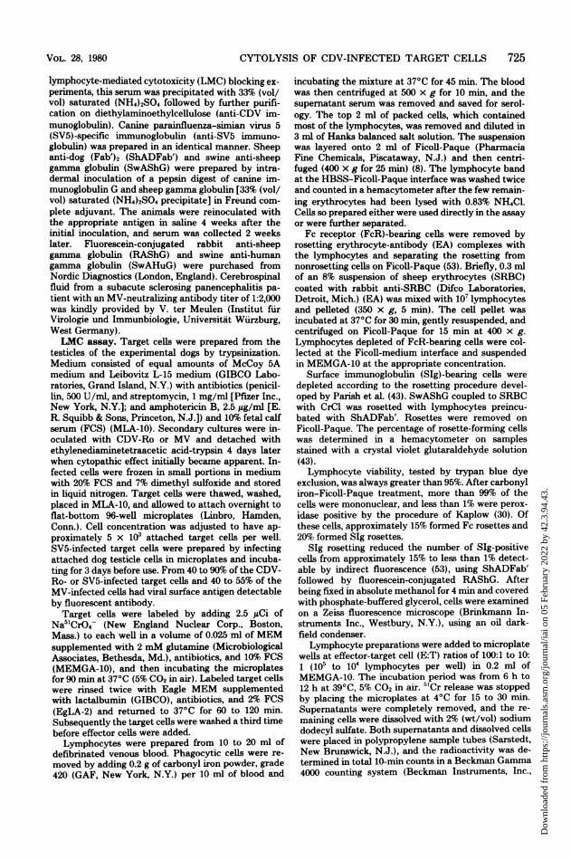
lymphocytes from different dogs. Differ-ences in the level of NLMC (Fig. 1) caused bylymphocytes from different dogs were particu-larly evident in the coonhound-beagle crosses.NLMC ranged from as low as 0% to as high as20% after a 6-h incubation at an E:T ratio of 100:1. Lymphocytes from dogs D2, D4, and D6caused significantly (P < 0.001) lower levels ofcytotoxicity than did lymphocytes from dogs D3,D9, D10, Dll, D12, and D13 (Fig. 1).Recognition of CDV antigen. As stated
above, NLMC was detected only against CDV-infected target cells and therefore appeared torequire recognition of CDV antigen. To supportthe conclusion that NLMC was CDV specific,CDV antibody (anti-CDV immunoglobulin) wasused to coat viral antigen on the target cellsurface and thereby block NLMC (Table 1). Aproblem in this experiment was ADCC activity
20
cc
.10-
U
-5
2 4 6 12 13 3 9 10 11
Effector Lymphocytes, D-Series
FIG. 1. Variation ofNLMC caused by lymphocytesfrom SPF dogs against CDV-infected target cells.Lymphocytes from dogs listed on the x axis wereassayed twice for NLMC on target cells from dogsD2, D4, D6, D7, D8, D10, D12, D13 (E:T ratio = 100:1, 6-h incubation). The bars represent the sampledeviation.
in the presence of CDV antibody. To reduceADCC against target cells pretreated with anti-CDV immunoglobulin, FcR-bearing cells weredepleted by EA resetting as described. Thisprocedure did not reduce NLMC. However,when target cells were pretreated with 5 or 10mg of anti-CDV immunoglobulin per ml, NLMCby lymphocytes that were depleted of FcR-bear-ing cells was almost completely inhibited (P <0.001). In contrast, reduction of cytotoxicity bylymphocytes not depleted of FcR-bearing cellswas less marked or reversed. This increase incytotoxicity by lymphocytes not depleted of EArosette-forming cells was believed to be ADCC,caused by FcR-bearing lymphocytes in the pres-ence of anti-CDV immunoglobulin. Conse-quently, the antibody-mediated inhibition ofNLMC was masked by ADCC. We did not at-tempt to separate FcR-bearing cells for a directdemonstration of ADCC.
Pretreatment of target cells with anti-SV5immunoglobulin or various dilutions of SPF ca-nine serum did not influence NLMC (Table 1).
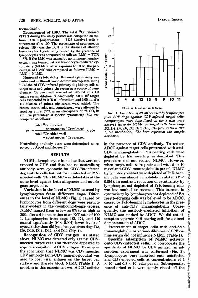
Specific adsorption of NLMC activityonto CDV-infected cells. To corroborate thespecificity of NLMC for CDV antigen, an ad-sorption experiment was performed (Fig. 2).Lymphocytes were adsorbed onto uninfectedand CDV-infected cells at concentrations of 1X 10⁷ and 0.5 x 10⁷ cells per ml. Subsequently,nonadsorbed cells were gently rinsed off the
INFECT. IMMUN.
Dow
nloa
ded
from
http
s://j
ourn
als.
asm
.org
/jour
nal/i
ai o
n 05
Feb
ruar
y 20
22 b
y 42
.3.9
4.43
.

CYTOLYSIS OF CDV-INFECTED TARGET CELLS 727
TABLE 1. Effect ofCDV and SV5 antibody and SPF canine serum on LMC caused by nonimmunelymphocytes before and after removal of FcR-bearing lymphocytes by EA resetting
Lympho- LMC
fcoymese Target cell pretreatment" Concn (mg/ml) Before EA After EA ro-ADCCries: resetting setting
3C Control 12.9 (1.4)' 12.8 (0.35) 0.1 (1.0)Anti-CDV immuno- 0.1 14.9 (4.1) 9.5 (1.9) 5.4 (2.3)e
globulin1.0 6.5 (1.1) 2.1 (2.1) 4.4 (1.0)"5.0 10.7 (0.64) 1.0 (0.78) 9.7 (0.14)
10.0 6.0 (1.1) -1.8 (1.8) 7.8 (0.64)Df Control 19.0 (4.7) 19.4 (3.3) -0.4 (1.3)
Anti-CDV immuno- 0.1 22.2 (5.7) 21.8 (2.2) 0.4 (4.0)globulin
1.0 15.2 (2.8) 15.4 (0.85) -0.2 (2.0)5.0 12.0 (0.8) 2.8 (2.3) 9.2 (3.1)10.0 14.4 (2.3) 3.0 (1.3) 11.4 (3.0)
Anti-SV5 immuno- 5.0 19.2 (2.9) 21.1 (4.8) -1.9 (3.2)globulin
SPF serum Undiluted 19.5 (3.5) 21.1 (2.6) -1.6 (-1.9)1:19 dilution 19.1 (5.3) 21.8 (2.2) -2.7 (2.0)
"To each microplate well containing "Cr-labeled CDV-infected target cells was added 0.05 ml of the indicatedconcentration of antibody or dilution of SPF canine serum. After incubating for 15 min at room temperature,lymphocytes were added.bADCC = LMC before EA resetting - LMC after EA resetting.E:T ratio = 50:1, 8-h incubation period, n = 4.
d Numbers in parentheses indicate sample deviation.eP < 0.001.fE:T ratio = 100:1, 8-h incubation period, n = 4.
adsorbing monolayers and assayed on 5"Cr-la-beled CDV-infected target cells for cytotoxicactivity. NLMC was significantly reduced (P <0.001) after adsorption onto CDV-infected cellsbut not after adsorption on uninfected cells. Aswould be expected, adsorption was more com-plete at the lower concentration, i.e., 0.5 x 10⁷cells per ml.To determine whether this adsorption was
CDV specific, CDV-infected monolayers werepretreated with 5 mg of anti-CDV immunoglob-ulin per ml. This pretreatment inhibited (P <0.001) adsorption of NLMC, albeit incompletely(Fig. 2). This experiment supported the hypoth-esis that lymphocytes, which caused NLMC,bound to serologically detectable CDV antigen.ILMC. Six days after vaccination with CDV-
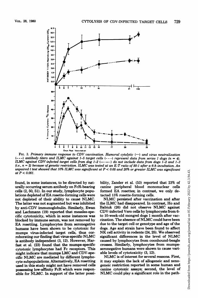
Ro, an ephemeral increase in LMC appearedthat was maximal between 7 to 9 days aftervaccination and usually decreased to insignifi-cant levels (i.e., >10%) by day 14 (Fig. 3). Incontrast, neutralizing and cytotoxic antibody,also detectable by day 6, persisted at undimin-ished titers for at least 5 months. This patternhad two exceptions. ILMC by dog 1-3 lympho-cytes against allogeneic 1-5 target cells was notmeasurable until 7 days after vaccination,whereas ILMC by dog 1-2 lymphocytes againstautologous target cells persisted for at least 5
months. All 11 vaccinates displayed significant(P < 0.001) ILMC of between 30 and 50%. EAand SIg resetting of lymphocytes to remove Kand B cells did not diminish ILMC (Table 2).
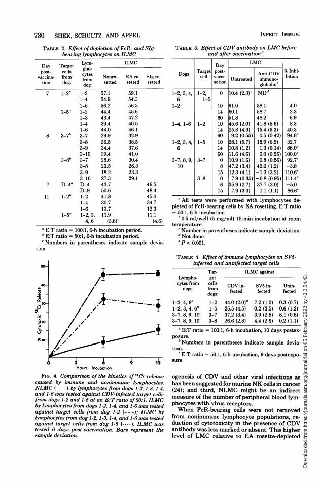
Differentiation between NLMC and ILMCon the basis of kinetics and specificity. (i)Kinetics. NLMC increased linearly over a 12-hincubation period and caused only low level ofcytotoxicity. Conversely, ILMC was maximal by6 h (Fig. 4).
(ii) Specificity: recognition of nonsero-logically detectable CDV and MV antigens.Data presented above demonstrated thatNLMC could be blocked by CDV antibody. Incontrast, when anti-CDV immunoglobulin wasused to block LMC after vaccination, the per-centage of inhibition was contingent upon thelevel of ILMC (Table 3). When the level ofILMC was high, a smaller part of the totalcytotoxicity was caused by NLMC. However, asILMC decreased, NLMC contributed a higherpercentage of the total LMC. Consequently, asthe level of ILMC decreased, the percentage ofinhibition caused by anti-CDV immunoglobulinincreased. When LMC decreased to prevaccina-tion levels, anti-CDV immunoglobulin com-pletely inhibited cytotoxicity. This suggestedthat NLMC persisted after vaccination and thatILMC did not require recognition of serolog-
VOL. 28, 1980
Dow
nloa
ded
from
http
s://j
ourn
als.
asm
.org
/jour
nal/i
ai o
n 05
Feb
ruar
y 20
22 b
y 42
.3.9
4.43
.
728 SHEK, SCHULTZ, AND APPEL
FIG. 2. Adsorption of NLMC activity by CDV-in-fected cell monolayers and inhibition of adsorptionwith CDVantibody. The adsorption ofNLMC activityonto CDV-infected (AI) and uninfected (ANI) alloge-neic dog testicle monolayers wasperformed in 24-wellmicroplates. Monolayers were preincubated for 15min with 0.2 ml ofmedium with (AIA) or without (AI)anti-CDV immunoglobulin (5 mg/ml). Subsequentlythe monolayers were overlaid with 0.4-ml suspensionsof nonimmune lymphocytes (four dogs in D series)containing 1 x 10' (1.0) or 0.5 x 10⁷ (0.5) cells/ml andincubated for 2 h at 39°C in an atmosphere of5% CO2in air, to allow adsorption. Nonadsorbed cells were
gently resuspended, washed, and tested forNLMC on
CDV-infected 5"Cr-labeled target cells at an E:T ratioof 100:1 after an 8-h incubation period.
ically detectable CDV antigen.Although serologically detectable CDV anti-
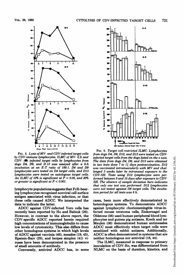
gens were not recognized by immune cytotoxiclymphocytes, ILMC was virus specific. Immunelymphocytes did not cause cytolysis of SV5-in-fected or uninfected target cells (Table 4). Con-versely, CDV-Ro-immune lymphocytes did lyseMV-infected target cells (Fig. 5). The appear-ance and duration of MV- and CDV-specificILMC were similar, although the latter consist-ently reached higher levels.Recognition of target cell surface anti-
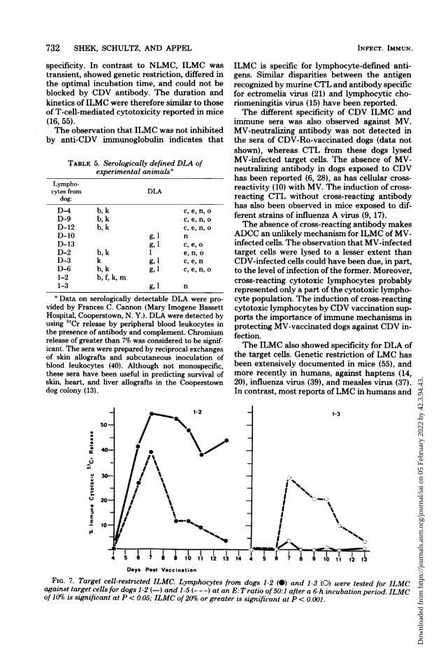
gens. As stated previously, NLMC was detectedequally well against autologous and allogeneiccells. Conversely, ILMC was, in certain in-stances, detectable only against autologous tar-get cells and allogeneic target cells possessingthe appropriate dog leukocyte antigens (DLA).
Reciprocal genetic restriction between dogsD4, D9, and D12 versus D10 and D13 (P <
0.005) (Fig. 6) corresponded to data on serolog-ically detectable DLA (Table 5). Dogs D9, D12,and D13 shared n, c, e, and o determinants.However, only D4, D9, and D12 lymphocytespossessed b and k, whereas D10 and D13 lym-phocytes were lysed by antisera specific for gand 1. Dog 1-2 lymphocytes continued to showstrong ILMC against infected autologous targetcells after activity against dog 1-5 target cellsdisappeared (Fig. 7). Dog 1-2 ILMC against au-tologous target cells persisted anomalously forat least 5 months. Conversely, dog 1-3 lympho-cytes did not lyse target cells from dog 1-2. Thisalso corresponds to data in Table 5, showing alack of shared DLA on lymphocytes from dogs1-2 and 1-3. Lysis of MV-infected target cellswas also genetically restricted.
DISCUSSIONSeveral types of CDV-specific LMC were
demonstrated, including NLMC, ADCC, and ge-netically restricted ILMC. Differentiating be-tween these types of cell-mediated cytotoxicitywas facilitated by control of experimental con-ditions, including preparation of relatively purelymphocyte populations, cryopreservation of al-logeneic and autologous target cells, and the useofSPF dogs. The latter is particularly importantbecause many studies of cell-mediated cytolysisin humans involved individuals of unknown im-munological status and lacked adequate control.The study of cell-mediated cytolysis in dogs hasthe advantages of adequate negative control,known time of exposure to the virus, and use ofan outbred species to corroborate data obtainedfrom experiments with inbred laboratory ani-mals.As was determined by blocking and adsorp-
tion experiments, cytolysis mediated by nonim-mune lymphocytes was specific for serologicallydetectable CDV antigens. Lysis of noninfectedfibroblasts observed with human effector cells(46, 51) was not detected, nor was there cytolysisof MV-infected target cells. This specificity forviral antigens is analogous to murine NK cell-mediated cytolysis of target cells infected withtype C virus (25).
Recognition of serologically detectable viralantigens has also been reported for human lym-phocytes, perhaps reflecting an NLMC reaction.Inhibition of human antiviral cell-mediated cy-totoxicity by immune serum has been reportedfor mumps (3, 48), measles (19, 38), and rubella(49) viruses. The requirements of high E:T ratiosand long incubation periods to demonstrate lowlevels of NLMC are consistent with many pub-lished reports on virus-specific cell-mediated cy-totoxicity in humans (3, 35, 38, 48, 49).The specificity of human NK cells has been
INFECT. IMMUN.
Dow
nloa
ded
from
http
s://j
ourn
als.
asm
.org
/jour
nal/i
ai o
n 05
Feb
ruar
y 20
22 b
y 42
.3.9
4.43
.
CYTOLYSIS OF CDV-INFECTED TARGET CELLS
5 5.5-
5.0-
4.0)
b3.5-
45 +IIH761 1121 14 20 251
lIAI- + I '.
Days Post VacinatlsoFIG. 3. Primary immune response to CDV vaccination. Humoral cytolytic (-) and virus neutralization
(---) antibody titers and ILMC against 1-5 target cells (- --) represent data from series 1 dogs (n = 4).ILMC against CDV infected target cells from dog 1-2 (-*-*) do not include data from dogs 1-2 and 1-3(i.e., n = 2) because ofgenetic restriction. ILMC was tested at an E:T ratio of 50:1 after a 6-h incubation. Anunpaired t test showed that 10% ILMC was significant at P c 0.05 and 20% or greater ILMC was significantatP< O.001.
found, in some instances, to be directed by nat-urally occurring serum antibody on FcR-bearingcells (2, 50, 51). In our study, lymphocyte popu-lations depleted ofEA rosette-forming cells werenot depleted of their ability to cause NLMC.The latter was not augmented but was inhibitedby anti-CDV immunoglobulin. Similarly, Ewanand Lachmann (19) reported that measles-spe-cific cytotoxicity, which in some instances wasblocked by immune serum, was not removed byEA resetting. Lymphocytes from seronegativehumans have been shown to be cytotoxic formumps virus-infected target cells, thus cor-roborating our finding that virus-specific NLMCis antibody independent (3, 12). However, Har-fast et al. (23) found that the mumps-specificcytotoxic lymphocytes had Fc receptors. Thismight indicate that mumps LMC and CDV-spe-cific NLMC are mediated by different lympho-cyte subpopulations. Alternatively, EA resettingused in this study might not have removed cellspossessing low-affinity FcR which were respon-sible for NLMC. In support of the latter possi-
bility, Zander et al. (53) reported that 23% ofcanine peripheral blood mononuclear cellsformed EA rosettes; in contrast, we only de-tected 15% rosette-forming cells.NLMC persisted after vaccination and after
the ILMC had disappeared. In contrast, Ho andBabiuk (26) did not observe NLMC againstCDV-infected Vero cells by lymphocysts from 6-to 10-week-old mongrel dogs 1 month after vac-cination. The absence ofNLMC could have beendue to the target cell or genotype and age of thedogs. Age and strain have been found to affectNK cell activity in rodents (24, 25). We observedsignificant differences in the level of NLMCcaused by lymphocytes from coonhound-beaglecrosses. Similarly, lymphocytes from mumps-seronegative humans were shown to cause vari-able levels of cytotoxicity (3, 12).NLMC is of interest for several reasons. First,
it may explain the lack of allogeneic and xeno-geneic restriction reported in some human andcanine cytotoxic assays; second, the level ofNLMC could play a significant role in the path-
VOL. 28, 1980 729
Dow
nloa
ded
from
http
s://j
ourn
als.
asm
.org
/jour
nal/i
ai o
n 05
Feb
ruar
y 20
22 b
y 42
.3.9
4.43
.
730 SHEK, SCHULTZ, AND APPEL
TABLE 2. Effect of depletion ofFcR- and SIg-bearing lymphocytes on ILMC
Day Target pho- ILMCpost- cells po
vaccina- from cytes Nonro- EA ro- SIg ro-tion dog: from setted setted setted
dog:7 1-2" 1-2 57.1 59.1
1-4 54.9 54.31-6 56.2 56.3
1-5a 1-2 44.4 45.61-3 43.4 47.21-4 39.4 40.51-6 44.0 46.1
8 3-7b 3-7 29.9 32.93-8 36.5 38.53-9 34.4 37.63-10 39.4 41.0
3-8b 3-7 28.6 30.43-8 23.5 26.33-9 18.2 23.33-16 27.3 29.1
7 D-4" D-4 43.7 46.5D-9 50.6 48.4
11 1-2b 1-2 41.8 45.01-4 30.7 34.71-6 13.7 12.3
1-5b 1-2, 3, 11.9 11.14, 6 (3.8)c (4.6)
" E:T ratio = 100:1, 6-h incubation period.bE:T ratio = 50:1, 6-h incubation period.Numbers in parentheses indicate sample devia-
tion.
Hours Incubation
FIG. 4. Comparison of the kinetics of 5"Cr releasecaused by immune and nonimmune lymphocytes.NLMC ( ) by lymphocytes from dogs 1-2, 1-3, 1-4,and 1-6 was tested against CDV-infected target cellsfrom dogs 1-2 and 1-5 at an E:T ratio of 50:1. ILMCby lymphocytes from dogs 1-2, 1-4, and 1-6 was testedagainst target cells from dog 1-2 (---); ILMC bylymphocytes from dog 1-2, 1-3, 1-4, and 1-6 was testedagainst target cells from dog 1-5 (- -*.). ILMC was
tested 6 days post-vaccination. Bars represent thesample deviation.
TABLE 3. Effect ofCDV antibody on LMC beforeand after vaccination'
Day LMC
Target post- Anti-CDV % Inhi-ogs cell vacci- bitionUntreated immuno-nation globulin'
1-2, 3, 4, 1-2, 0 10.4 (2.3) NDd6 1-5
1-2 10 61.0 58.1 4.014 60.1 58.7 2.360 51.8 48.2 6.9
1-4, 1-6 1-2 10 45.6 (2.0) 41.8 (5.6) 8.314 25.8 (4.3) 15.4 (5.3) 40.360 9.2 (0.55) 0.5 (0.42) 94.6'
1-2, 3, 4, 1-5 10 28.1 (5.7) 18.9 (6.9) 32.76 14 10.8 (1.2) 1.3 (0.14) 88.0'
60 11.6 (4.6) 0.0 (0.26) 100.oe3-7, 8, 9, 3-7 0 10.9 (1.6) 0.8 (0.56) 92.7e
10 8 47.2 (3.4) 49.0 (1.2) -3.815 12.3 (4.1) -1.3 (3.2) 110.6'
3-8 0 7.9 (0.35) -0.8 (0.95) 111.4e6 35.9 (2.7) 37.7 (3.0) -5.015 7.9 (3.0) 1.1 (1.1) 86.6e
a All tests were performed with lymphocytes de-pleted of FcR-bearing cells by EA resetting. E:T ratio= 50:1, 6-h incubation.
'0.5 ml/well (5 mg/ml) 15-min incubation at roomtemperature.
c Number in parentheses indicate sample deviation.d Not done.P < 0.001.
TABLE 4. Effect of immune lymphocytes on SV5-infected and uninfected target cells
Tar- ILMC against:Lympho- getcytes from cells CDV in- SV5 in- Unin-
dogs: from fected fected fecteddogs:
1-2, 4, 6a 1-2 44.0 (2.0) 7.2 (1.2) 0.3 (0.7)1-2, 3, 4, 6a 1-5 25.5 (4.5) 0.2 (3.5) 0.6 (1.2)3-7, 8, 9, 10' 3-7 37.2 (3.4) 3.9 (2.8) 8.1 (0.8)3-7, 8, 9, 10 3-8 26.6 (2.8) 4.4 (2.6) 0.2 (1.1)
a RT ratio = 100:1, 6-h incubation, 10 days postex-posure.
b Numbers in parentheses indicate sample devia-tion.
c E:T ratio = 50:1, 6-h incubation, 9 days postexpo-sure.
ogenesis of CDV and other viral infections ashas been suggested for murineNK cells in cancer(24); and third, NLMC might be an indirectmeasure of the number of peripheral blood lym-phocytes with virus receptors.When FcR-bearing cells were not removed
from nonimmune lymphocyte populations, re-duction of cytotoxicity in the presence of CDVantibody was less marked or absent. This higherlevel of LMC relative to EA rosette-depleted
INFECT. IMMUN.
Dow
nloa
ded
from
http
s://j
ourn
als.
asm
.org
/jour
nal/i
ai o
n 05
Feb
ruar
y 20
22 b
y 42
.3.9
4.43
.
CYTOLYSIS OF CDV-INFECTED TARGET CELLS 731
_o 40
inkE
a20
5 7 9 10 12 13 14 1
Days Post Vaccination
FIG. 5. Lysis ofMV- and CDV-infected target cellsby CDV-immune lymphocytes. ILMC ofMV- (0) andCDV- (0) infected target cells by lymphocytes fromdogs D4, D9, and D-13 was assayed after a 6-hincubation at an E:T ratio of 100:1. D4 and D9lymphocytes were tested on D4 target cells, and D13lymphocytes were tested on autologous target cells.An ILMC of 10% is significant at P < 0.05, and 20%or greater is significant at P < 0.001.
lymphocyte populations suggests that FcR-bear-ing lymphocytes recognized nonviral cell surfaceantigen associated with virus infection, or thatthese cells caused ADCC. We interpreted thedata to indicate the latter.ADCC against CDV-infected Vero cells has
recently been reported by Ho and Babiuk (26).However, in contrast to the above report, theCDV-specific ADCC reported herein requiredhigh concentrations of immunoglobulin to causelow levels of cytotoxicity. This also differs fromother homologous systems in which high levelsofADCC against vaccinia (46), measles (19, 45),Epstein-Barr (29), and herpes simplex (41) vi-ruses have been demonstrated in the presenceof small amounts of antibody.
Conversely, antiviral ADCC has, in some
FoIAILLLI
"_
I1@-- '7mS,>,
C3YbbW-da. iq CaI * IIa
FIG. 6. Target cell-restricted ILMC. Lymphocytesfrom dogs D4, D9, D1Z, and D13 were tested on CDV-infected target cells from the dogs listed on the x axis.The data from dogs D4, D9, and D13 were obtainedin two tests done 7 to 11 days postvaccination. D12was vaccinated intramuscularly with MV and chal-lenged 3 weeks later by intranasal exposure to theCDV-SH. Tests using D12 lymphocytes were per-formed between 9 and 15 days after exposure to CDV-SH. The absence of sample deviation bars indicatesthat only one test was performed. D12 lymphocyteswere not tested against D8 target cells. The incuba-tion period for all tests was 6 h.
cases, been more effectively demonstrated inheterologous systems. To demonstrate ADCCagainst lymphocytic choriomeningitis virus-in-fected mouse teratoma cells, Zinkernagel andOldstone (56) used human peripheral blood lym-phocytes and guinea pig antisera. Kreth and terMeulen (36) demonstrated human antimneaslesADOC most effectively when target cells weresensitized with rabbit antisera. Additionally,ADOC is often demonstrated in systems that areneither homologous nor antiviral (18, 44).The ILMO, measured in response to primary
inoculation of CDV-Ro, was differentiated fromNLMC on the basis of duration, kinetics, and
VOL. 28, 1980
Dow
nloa
ded
from
http
s://j
ourn
als.
asm
.org
/jour
nal/i
ai o
n 05
Feb
ruar
y 20
22 b
y 42
.3.9
4.43
.
732 SHEK, SCHULTZ, AND APPEL
specificity. In contrast to NLMC, ILMC wastransient, showed genetic restriction, differed inthe optimal incubation time, and could not beblocked by CDV antibody. The duration andkinetics of ILMC were therefore similar to thoseof T-cell-mediated cytotoxicity reported in mice(16, 55).The observation that ILMC was not inhibited
by anti-CDV immunoglobulin indicates that
TABLE 5. Serologically defined DLA ofexperimental animals
Lympho-cytes from DLA
dog:D-4 b, k c, e, n, oD-9 b,k c,e,n,oD-12 b, k c, e, n, oD-10 g,I nD-13 g,l c,e,oD-2 b, k I e, n, oD-3 k g, l c, e, nD-6 b, k g, 1 c, e, n, o1-2 b,f,k,m1-3 g,l n
a Data on serologically detectable DLA were pro-vided by Frances C. Cannon (Mary Imogene BassettHospital, Cooperstown, N. Y.). DLA were detected byusing 5"Cr release by peripheral blood leukocytes inthe presence of antibody and complement. Chromiumrelease of greater than 7% was considered to be signif-icant. The sera were prepared by reciprocal exchangesof skin allografts and subcutaneous inoculation ofblood leukocytes (40). Although not monospecific,these sera have been useful in predicting survival ofskin, heart, and liver allografts in the Cooperstowndog colony (13).
s- / 250-
U 20- \
E- 10-St L'
ILMC is specific for lymphocyte-defined anti-gens. Similar disparities between the antigenrecognized by marine CTL and antibody specificfor ectromelia virus (21) and lymphocytic cho-riomeningitis virus (15) have been reported.The different specificity of CDV ILMC and
immune sera was also observed against MV.MV-neutralizing antibody was not detected inthe sera of CDV-Ro-vaccinated dogs (data notshown), whereas CTL from these dogs lysedMV-infected target cells. The absence of MV-neutralizing antibody in dogs exposed to CDVhas been reported (6, 28), as has cellular cross-reactivity (10) with MV. The induction of cross-reacting CTL without cross-reacting antibodyhas also been observed in mice exposed to dif-ferent strains of influenza A virus (9, 17).The absence of cross-reacting antibody makes
ADCC an unlikely mechanism for ILMC of MV-infected cells. The observation that MV-infectedtarget cells were lysed to a lesser extent thanCDV-infected cells could have been due, in part,to the level of infection of the former. Moreover,cross-reacting cytotoxic lymphocytes probablyrepresented only a part of the cytotoxic lympho-cyte population. The induction of cross-reactingcytotoxic lymphocytes by CDV vaccination sup-ports the importance of immune mechanisms inprotecting MV-vaccinated dogs against CDV in-fection.The ILMC also showed specificity for DLA of
the target cells. Genetic restriction of LMC hasbeen extensively documented in mice (55), andmore recently in humans, against haptens (14,20), influenza virus (39), and measles virus (37).In contrast, most reports ofLMC in humans and
Days Post VaccinationFIG. 7. Target cell-restricted ILMC. Lymphocytes from dogs 1-2 (0) and 1-3 (0) were tested for ILMC
against target cells for dogs 1-2 (-) and 1-5 (- - -) at an E:T ratio of50:1 after a 6-h incubation period. ILMCof 10%c is significant at P < 0.05; ILMC of 20%o or greater is significant at P < 0.001.
INFECT. IMMUN.
Dow
nloa
ded
from
http
s://j
ourn
als.
asm
.org
/jour
nal/i
ai o
n 05
Feb
ruar
y 20
22 b
y 42
.3.9
4.43
.

CYTOLYSIS OF CDV-INFECTED TARGET CELLS 733
recent reports on CDV (33, 34) and vacciniavirus (27) in dogs have failed to demonstrategenetic restriction.
Reciprocal genetic restriction in the D seriesoccurred between dogs that had common DLA.These common DLA might be unimportant astargets for ILMC. Murine virus-specific CTLrecognize only gene products of the K and D lociof the H-2 complex (55). Alternatively, thesecommon DLA might be recognized by CTL butnot by distemper-immune CTL. Preferential cy-tolysis of antigen associated with certain H-2haplotypes has been documented for tumor vi-ruses (11, 47).The duration, kinetics, and specificity of
ILMC made it unlikely that antibody was in-volved. CDV antibody persisted long after ILMChad become insignificant. ADCC demonstratedbefore vaccination was much lower that the peakILMC. Moreover, ILMC appeared to be geneti-cally restricted and not to recognize serologicallydefined CDV antigen. This is corroborated bydata showing that depletion of SIg- and FcR-bearing cells did not diminish ILMC.
In the present study, NLMC, ILMC, ADCC,and humoral cytotoxic responses in dogs afterCDV vaccination have been reported. Studiesare in progress to determine the significance ofcellular versus humoral immune responses inrecovery or viral persistence in dogs after expo-sure to virulent CDV.
ACKNOWLEDGMENTS
This work was supported by Public Health Service grantNS 14342 from the National Institutes of Health.
The assistance of Mary Beth Metzgar, Lincoln Adams, andAnn Signore is greatly appreciated.
LITERATURE CITED
1. Ada, G. L., D. C. Jackson, R. V. Blanden, R. Tha Hla,and N. A. Bowern. 1976. Changes in the surface ofvirus infected cells recognized by cytotoxic T cells. I.Minimal requirements for lysis of ectromelia-infectedP-815 cells. Scand. J. Immunol. 5:23-30.
2. Akira, D., and M. Takasugi. 1977. Loss of specific cell-mediated cytotoxicity with adsorption of natural anti-bodies from serum. Int. J. Cancer 19:747-755.
3. Andersson, T., V. Stejskal, and B. Harfast. 1978. Anin vitro method for study of human lymphocyte cyto-toxicity against mumps virus-infected target cells. J.Immunol. 114:237-243.
4. Appel, M. J. G. 1969. Pathogenesis of canine distemper.1969. Am. J. Vet. Res. 30:1167-1182.
5. Appel, M. J. G., E. P. J. Gibbs, S. J. Martin, V. terMeulen, B. K. Rima, J. R. Stephenson, and W. P.Taylor. 1980. Morbillivirus diseases of animals andman, in press. In E. Kurstak (ed.), Comparative diag-nosis of viral diseases, vol. III: Animal and relatedviruses. Academic Press Inc., New York.
6. Appel, M. J. G., and J. H. Gillespie. 1972. Caninedistemper virus. Virol. Monogr. 11:1-96.
7. Appel, M. J. G., and D. S. Robson. 1973. A microneu-tralization test for canine distemper virus. Am. J. Vet.Res. 34:1459-1463.
8. Boyum, A. 1968. Isolation of mononuclear cells and gran-ulocytes from human blood. Scand. J. Clin. Lab. Invest.21(Suppl. 97):77-89.
9. Braciale, T. J. 1977. Immunologic recognition of influ-enza virus-infected cells. II. Expression of influenza Amatrix protein on the infected cell surface and its rolein recognition by cross-reactive cytotoxic T cells. J.Exp. Med. 146:673-689.
10. Brown, A. L., and R. E. McCarthy. 1974. Relationshipbetween measles and canine distemper viruses deter-mined by delayed type hypersensitivity reactions indogs. Nature (London) 248:344-345.
11. Bubbers, J. E., S. Chen, and F. Lilly. 1978. Nonrandominclusion of H-2K and H-2D antigens in friend virusparticles from mice of various strains. J. Exp. Med. 147:340-351.
12. Chiba, Y., J. L. Dzierba, A. Morag, and P. L. Ogra.1976. Cell-mediated immune response to mumps virusinfection in man. J. Immunol. 116:12-15.
13. Dausset, J., F. T. Rappaport, F. D. Cannon, and J.W. Ferrebee. 1971. Histocompatibility studies in aclosely bred colony of dogs. III. Genetic definition of theDL-A system of canine histocompatibility, with partic-ular reference to comparative immunogenicity of themajor transplantable organs. J. Exp. Med. 134:1222-1237.
14. Dickmeiss, E., B. Soeberg, and A. Svejgaard. 1977.Human cell-mediated cytotoxicity against modified tar-get cells is restricted by HLA. Nature (London) 270:526-628.
15. Doherty, P. C., R. V. Blanden, and R. M. Zinkernagel.1976. Specificity of virus-immune effector T cells for H-2K or H-2D compatible interactions: implication for H-antigen diversity. Transplant. Rev. 29:89-124.
16. Doherty, P., R. B. Effros, and J. Bennink. 1978. Cell-mediated immunity in virus infection: influenza virusand the problem of self-non self discrimination. Per-spect. Virol. 10:73-89.
17. Effros, R. B., J. Bennink, and P. C. Doherty. 1978.Characteristics of secondary cytotoxic T-cell responsesin mice infected with influenza A viruses. Cell. Immunol.36:345-353.
18. Evans, R. L., L. Chess, H. Levine, and S. F. Schloss-man. 1978. Antibody-dependent cellular cytotoxicityby allosensitized human T-cells. J. Exp. Med. 157:605-610.
19. Ewan, P. W., and P. J. Lachmann. 1977. Demonstrationof T-cell and K-cell cytotoxicity against measles-in-fected cells in normal subjects, multiple sclerosis andsubacute sclerosing panencephalitis. Clin. Exp. Immu-nol. 30:22-31.
20. Friedman, S. M., N. Neyhard, and L. Chess. 1978. Cell-mediated lympholysis of trinitrophenyl-derivatized au-tologous human cells: in vitro triggering by nonspecificsignals. J. Immunol. 120:630-637.
21. Gardner, I. E., N. A. Bowern, and R. V. Blanden.1975. Cell-mediated cytotoxicity against ectromelia vi-rus-infected target cells. III. Role of H-2 gene complex.Eur. J. Immunol. 5:122-127.
22. Gething, M. J., V. Koszinowski, and M. Waterfield.1978. Fusion of Sendai virus with the target cell mem-brane is required for T-cell cytotoxicity. Nature (Lon-don) 274:689-693.
23. Harfast, B. T., T. Andersson, and P. Perlmann. 1975.Human lymphocyte cytotoxicity against mumps virus-infected target cells. Requirement for non-T cells. J.Immunol. 114:1820-1823.
24. Herberman, R. B., and H. T. Holden. 1978. Naturalcell-mediated immunity. Adv. Cancer Res. 27:305-377.
25. Herbermnan, R. B., M. E. Nunn, and D. H. Laurin.1975. Natural cytotoxic reactivity of mouse lymphoidcells against syngeneic and allogeneic tumors in distri-bution of reactivity and specificity. Int. J. Cancer 16:
VOL. 28, 1980
Dow
nloa
ded
from
http
s://j
ourn
als.
asm
.org
/jour
nal/i
ai o
n 05
Feb
ruar
y 20
22 b
y 42
.3.9
4.43
.

734 SHEK, SCHULTZ, AND APPEL
216-239.26. Ho, C.-K., and L. A. Babiuk. 1979. Immune mechanisms
against canine distemper. I. Identification of K cellagainst canine distemper virus infected target cells invitro. Immunology 37:231-239.
27. Ho, C.-K., L. A. Babiuk, and B. T. Rouse. 1978. Immuneeffector cell activity in canines: failure to demonstrategenetic restriction in direct antiviral cytotoxicity. Infect.Immun. 19:18-25.
28. Imagawa, D. T. 1968. Relationships among measles, ca-nine distemper, and rinderpest. Prog. Med. Virol. 10:160-193.
29. Joseph, B. S., N. R. Cooper, and M. B. A. Oldstone.1975. Immunologic injury of cultured cells infected withmeasles virus. II. Role of IgG antibody and the alter-native complement pathway. J. Exp. Med. 141:761-774.
30. Kaplow, L. S. 1965. Simplified myeloperoxidase strainusing benzidine dihydrochloride. Blood 26:215-219.
31. Kibler, R., and V. ter Meulen. 1975. Antibody-mediatedcytotoxicity after measles virus infection. J. Immunol.114:93-98.
32. Krakowka, S., R. Olsen, A. Confer, A. Koestner, andB. McCullough. 1975. Serologic response to caninedistemper viral antigens in gnotobiotic dogs infectedwith canine distemper virus. J. Infect. Dis. 132:384-392.
33. Krakowka, S., and A. L. Wallace. 1979. Lymphocyte-associated immune responses to canine distemper andmeasles viruses in distemper-infected gnotobiotic dogs.Am. J. Vet. Res. 40:669-672.
34. Krakowka, S., A. L. Wallace, and A. Koestner. 1978.Syncytia inhibition by immune lymphocytes: in vitrotest for immunity to canine distemper. J. Clin. Micro-biol. 7:292-297.
35. Kreth, W. H., M. Y. Kackell, and V. ter Meulen. 1975.Demonstration of in vitro lymphocyte-mediated cyto-toxicity against measles virus in SSPE. J. Immunol.114:1042-1046.
36. Kreth, W. H., and V. ter Meulen. 1977. Cell-mediatedcytotoxicity against measles virus in SSPE. I. Enhance-ment by antibody. J. Immunol. 118:291-295.
37. Kreth, H. W., V. ter Meulen, and G. Eckert. 1979.Demonstration of HLA restricted killer cells in patientswith acute measles. Med. Microbiol. Immunol. 165:203-214.
38. Labowski, R. J., R. Edelman, R. Rustigian, and J. A.Bellanti. 1974. Studies of cell-mediated immunity tomeasles virus by in vitro lymphocyte-mediated cytotox-icity. J. Infect. Dis. 129:233-239.
39. McMichael, A. 1978. HLA restriction of human cytotoxicT lymphocytes specific for influenza virus: poor recog-nition of virus associated with HLA A2. J. Exp. Med.148:1458-1467.
40. Mollen, N. D. St. John, F. D. Cannon, and J. W.Ferrebee. 1968. Lymphocyte typing in allografted bea-gles. Transplantation 6:939.
41. Moller-Larsen, A., I. Heron, and S. Haahr. 1977. Cell-mediated cytotoxicity to herpes-infected cells in hu-mans: dependence on antibodies. Infect. Immun. 16:43-47.
42. Notkin, A. L., and D. L. Lodmell. 1975. Cellular immuneresponses in viral infections. Perspect. Virol. 9:115-130.
43. Parish, C. R., S. M. Kirov, N. Bowern, and R. V.Blanden. 1974. A one-step procedure for separatingmouse T and B lymphocytes. Eur. J. Immunol. 4:808-815.
44. Pearson, G. R. 1978. In vitro and in vivo investigationson antibody-dependent cellular cytotosicity. Curr. Top.Microbiol. Immunol. 80:65-96.
45. Perrin, L. H., A. Tishon, and M. B. A. Oldstone. 1977.Immunologic injury in measles virus infection. III. Pres-ence and characterization of human cytotoxic lympho-cytes. J. Immunol. 118:282-290.
46. Perrin, L. H., R. M. Zinkernagel, and M. B. A. Old-stone. 1977. Immune response in humans after vacci-nation with vaccinia virus: generation of a virus-specificcytotoxic activity by human peripheral lymphocytes. J.Exp. Med. 146:949-969.
47. Pfizenmaier, K., G. Trinchieri, D. Solter, and B. B.Knowles. 1978. Mapping of H-2 genes associated withT-cell-mediated cytotoxic responses to SV40-tumor-as-sociated specific antigens. Nature (London) 274:691-693.
48. Rola-Pleszczynski, M., M. M. Vincent, S. A. Hensen,J. Walser, M. Crawford, and J. A. Bellanti. 1976.Chromium-release microassay, technique for cell-me-diated immunity to mumps virus: correlation with hu-moral and delayed type skin hypersensitivity responses.J. Infect. Dis. 134:546-551.
49. Steele, R. W., S. A. Hensen, M. M. Vincent, D. A.Fuccillo, and J. A. Bellanti. 1973. A 51Cr microassaytechnique for cell-mediated immunity to viruses. J.Immunol. 110:1502-1510.
50. Takasugi, J., Y. Koide, and M. Takasugi. 1978. Recon-stitution of natural cell-mediated cytotoxicity with spe-cific antibodies. Eur. J. Immunol. 7:887-892.
51. Timonen, T., and E. Saksela. 1977. Human natural cell-mediated cytotoxicity against fetal fibroblasts. I. Gen-eral characteristics and the cytotoxic activity. Cell. Im-munol. 33:340-352.
52. Woodruff, J. F., and J. J. Woodruff. 1975. T lympho-cyte interaction with virus and virus-infected tissue.Prog. Med. Virol. 19:120-160.
53. Zander, A. R., N. Boopalam, and R. B. Epstein. 1975.Surface markers on canine lymphocytes. Transplant.Proc. 7:369-393, 1975.
54. Zinkernagel, R. M., and A. Althage. 1977. Antiviralprotection by virus-immune cytotoxic T cells: infectedtarget cells are lysed before infectious virus progeny isassembled. J. Exp. Med. 145:644-651.
55. Zinkernagel, R. M., and P. C. Doherty. 1977. Majortransplantation antigens, viruses and specificity of sur-veillance T cells. Contemp. Top. Immunobiol. 7:170-220.
56. Zinkernagel, R. M., and M. B. A. Oldstone. 1976. Cellsthat express viral antigens but lack H-2 determinantsare not lysed by immune thymus-derived lymphocytesbut are lysed by other antiviral immune attack mecha-nisms. Proc. Natl. Acad. Sci. U.S.A. 73:3666-3670.
INFECT. IMMUN.
Dow
nloa
ded
from
http
s://j
ourn
als.
asm
.org
/jour
nal/i
ai o
n 05
Feb
ruar
y 20
22 b
y 42
.3.9
4.43
.