candidaalbicans pseudomonas aeruginosa interaction ... · and fonzi, 2001). interaction of c ......
TRANSCRIPT
REVIEWpublished: 26 February 2016
doi: 10.3389/fphys.2016.00064
Frontiers in Physiology | www.frontiersin.org 1 February 2016 | Volume 7 | Article 64
Edited by:
Naim Akhtar Khan,
Université de Bourgogne, France
Reviewed by:
Gérard Lizard,
Université de Bourgogne, France
Aziz Hichami,
Université de Bourgogne, France
*Correspondence:
Carolina H. Pohl
Specialty section:
This article was submitted to
Lipidology,
a section of the journal
Frontiers in Physiology
Received: 13 November 2015
Accepted: 11 February 2016
Published: 26 February 2016
Citation:
Fourie R, Ells R, Swart CW,
Sebolai OM, Albertyn J and Pohl CH
(2016) Candida albicans and
Pseudomonas aeruginosa Interaction,
with Focus on the Role of
Eicosanoids. Front. Physiol. 7:64.
doi: 10.3389/fphys.2016.00064
Candida albicans and Pseudomonasaeruginosa Interaction, with Focuson the Role of EicosanoidsRuan Fourie 1, Ruan Ells 1, 2, Chantel W. Swart 1, Olihile M. Sebolai 1, Jacobus Albertyn 1 and
Carolina H. Pohl 1*
1 Pathogenic Yeast Research Group, Department of Microbial, Biochemical and Food Biotechnology, University of the Free
State, Bloemfontein, South Africa, 2National Control Laboratory, University of the Free State, Bloemfontein, South Africa
Candida albicans is commonly found in mixed infections with Pseudomonas aeruginosa,
especially in the lungs of cystic fibrosis (CF) patients. Both of these opportunistic
pathogens are able to form resistant biofilms and frequently infect immunocompromised
individuals. The interaction between these two pathogens, which includes physical
interaction as well as secreted factors, is mainly antagonistic. In addition, research
suggests considerable interaction with their host, especially with immunomodulatory lipid
mediators, termed eicosanoids. Candida albicans and Pseudomonas aeruginosa are
both able to utilize arachidonic acid (AA), liberated from the host cells during infection,
to form eicosanoids. The production of these eicosanoids, such as Prostaglandin E2,
by the host and the pathogens may affect the dynamics of polymicrobial infection and
the outcome of infections. It is of considerable importance to elucidate the role of
host-produced, as well as pathogen-produced eicosanoids in polymicrobial infection.
This review will focus on in vitro as well as in vivo interaction between C. albicans and
P. aeruginosa, paying special attention to the role of eicosanoids in the cross-talk between
host and the pathogens.
Keywords: Candida albicans, co-infection, eicosanoid, interaction, prostaglandin, Pseudomonas aeruginosa
INTRODUCTION
Recently it has become increasingly evident that microorganisms are not only found as free floatingcells, but exist as surface associated, structured and cooperative consortia, called biofilms (Douglas,2003; Hentzer et al., 2003; Burmølle et al., 2006; Harriott and Noverr, 2011). In addition, thesecommunities are embedded in an extracellular matrix of self-produced polymeric material. Inthese interactive organizations for microorganisms secreted factors and physical proximity enablemetabolic interactions (Diaz et al., 2014). This often involves interkingdom interactions necessaryfor ecological balance and survival of certain species (Rinzan, 2009).
Pseudomonas aeruginosa is a Gram-negative, aerobic rod colonizing a remarkable assortment ofniches, including aquatic environments, terrestrial environments and eukaryotic organisms (Pier,1985; Tan et al., 1999). It is an opportunistic pathogen, frequently isolated from healthy humans aspart of the human microbiota and is commonly found in mixed infections with the yeast, Candidaalbicans (Kaleli et al., 2006). Candida albicans is found as part of the normal microbiota of theskin, gastrointestinal tract and female genital tract (Morales and Hogan, 2010) and is a major causeof opportunistic infections ranging from superficial to fatal systemic infections (Sandven, 2000).

Fourie et al. C. albicans and P. aeruginosa Interaction
Fungal infections have become increasingly troublesome in thepast decades, especially in immunocompromised patients and inthe hospital setting, with C. albicans being the most frequentlyisolated fungal pathogen and the most commonly isolatedbloodstream pathogen (Rinzan, 2009). Selective pressure ofnutrient limitation and competition between bacteria and fungiregulate the colonization of potential pathogenicmicroorganismssuch as C. albicans and P. aeruginosa, with a disruption in thisequilibrium resulting in infection by opportunistic pathogens(Calderone and Fonzi, 2001).
These two microorganisms have tendencies to formpolymicrobial biofilms and as such play extensive roles innosocomial infections, infection in immunocompromisedindividuals and especially in cystic fibrosis (CF) patients (El-Azizi et al., 2004; Bianchi et al., 2008; McAlester et al., 2008).This review, therefore, aims to evaluate the complex cross-kingdom relationship of these two pathogens and the impressiveinteraction and communication between them as well as thecollateral damage to hosts caught in the cross-fire. Additionally,special attention will be given to the known immunomodulatorylipids produced by both of these microorganisms as well as thehost and the role this may play during infection.
PATHOGENESIS OF PSEUDOMONAS
AERUGINOSA
Pseudomonas aeruginosa possesses numerous virulence factorsincluding exotoxin A, proteases and lipases, released by a typeII secretion system (Xcp regulon), as well as exotoxins exoS,T, U, and Y, secreted into host cells by a type III secretionsystem (Hogardt et al., 2004). Interestingly, it was found that P.aeruginosa possesses two type II secretory pathways, previouslynot seen in one organism (Ball et al., 2002). Additionally,pyoverdine, rhamnolipids, lipopolysaccharide (LPS) and pilialso form part of this formidable pathogen’s virulence arsenal(Gilligan, 1991; Méar et al., 2013). A study by Bianchi et al.(2008) showed that P. aeruginosa impairs the engulfment ofapoptotic cells through the action of yet another virulencefactor, the phenazine, pyocyanin (PYO) (Gibson et al., 2009).Interestingly, it has been shown that multiple drug resistantstrains of P. aeruginosa show decreased production of PYO,and thus have a reduction in virulence (Fuse et al., 2013).As previously mentioned, P. aeruginosa forms biofilms, anda universal model for the formation of P. aeruginosa biofilmformation was suggested (O’Toole et al., 2000; Klausen et al.,2003). According to this model, P. aeruginosa cells move bymeans of flagella to an adequate surface and movement alongthis surface is accomplished through type IV-pili. Cells aggregate,leading to microcolony formation. During maturation, largemushroom-shaped structures are formed. Klausen et al. (2003)proposed an alternate model, with evidence indicating thatflagella do not play a role in the attachment of P. aeruginosacells. The formation of P. aeruginosa biofilms are, however, highlydependent on the carbon source. Additionally, the circumstancesduring growth, such as flow vs. stationary growth, might elicitlarge morphological changes.
In addition to the previously mentioned factors, the resistanceof P. aeruginosa to antimicrobial agents is key to its pathogeniccapabilities. Various mechanisms for antibiotic resistance inP. aeruginosa biofilms have been proposed (Drenkard, 2003).These include the reduced transport of antimicrobial agentsin the biofilm due to extracellular matrix and accompaniednutrient and oxygen limitation of cells deeply embedded inthe biofilm. This causes a decrease in metabolic activity of thecells. Antibiotic resistant persisters embedded in the biofilmstructure, stress responses of the cells, efflux pumps andquorum sensing among cells may all contribute to the increasedresistance observed in bacterial biofilms. Evidence also suggestthat a protein, PvrR, regulates susceptibility and resistancephenotypes of P. aeruginosa (Drenkard and Ausubel, 2002;Benamara et al., 2011). An impact on lipid composition is alsospeculated due to differential protein expression in biofilms.In this regard, Benamara et al. (2011) examined the effectof biofilm formation on inner membrane lipid compositionin P. aeruginosa that indicated a reduced amount of unevennumbered phospholipids. In addition, an increase in longchain phosphatidylethanolamines was observed, suggesting anincrease in bilayer lipid stability and a decrease in membranefluidity.
PATHOGENESIS OF CANDIDA ALBICANS
Candida albicans is a dimorphic yeast, meaning that both yeastand hyphal morphology is shown, with a tendency to formdrug resistant biofilms (Ramage et al., 2001). The ability ofthis microorganism to switch between the planktonic singleyeast cell and hyphal morphologies has a major influence onits virulence (Andes et al., 2004; Pierce, 2005; Bruzual et al.,2007; Brand et al., 2008; Gil-Bona et al., 2015). In addition tothis morphological plasticity, the aggressiveness of C. albicanscolonization is due to a collection of other virulence factors.These include adhesins (biomolecules that enable binding to hostcells or host cell ligands), lipolytic and proteolytic enzymes andphenotypic switching (white to opaque switching) (Calderoneand Fonzi, 2001). Interaction of C. albicans with the hostis largely accomplished by contact with the C. albicans cellsurface and subsequent biofilm formation (Gow and Hube,2012). Ramage et al. (2001) investigated the formation ofC. albicans biofilms through visualization of the biofilms atvarious stages of development and monitoring the metabolicactivity of the cells. Through scanning electron microscopy(SEM) and confocal scanning laser microscopy (CSLM) a densenetwork of hyphae and yeast cells in a matrix of exopolymericmaterial was visualized in the matured biofilm. This study alsotested the effect of antifungal drugs on Candida biofilms andplanktonic C. albicans cells. It was found that cells in thebiofilm had a 250-fold increase in resistance against fluconazole.Dumitru et al. (2004) argued that this increased resistancemight be due to the hypoxic conditions found in biofilms, withanaerobically grown C. albicans also showing resistance againstcertain antifungal agents. Interestingly, a study by Kuhn et al.(2002) evaluated a range of antifungal agents againstCandida and
Frontiers in Physiology | www.frontiersin.org 2 February 2016 | Volume 7 | Article 64
Fourie et al. C. albicans and P. aeruginosa Interaction
found that sub-inhibitory concentrations of certain antifungalselicited alterations in biofilm formation.
INTERACTION BETWEEN PSEUDOMONAS
AERUGINOSA AND CANDIDA ALBICANS
IN VITRO
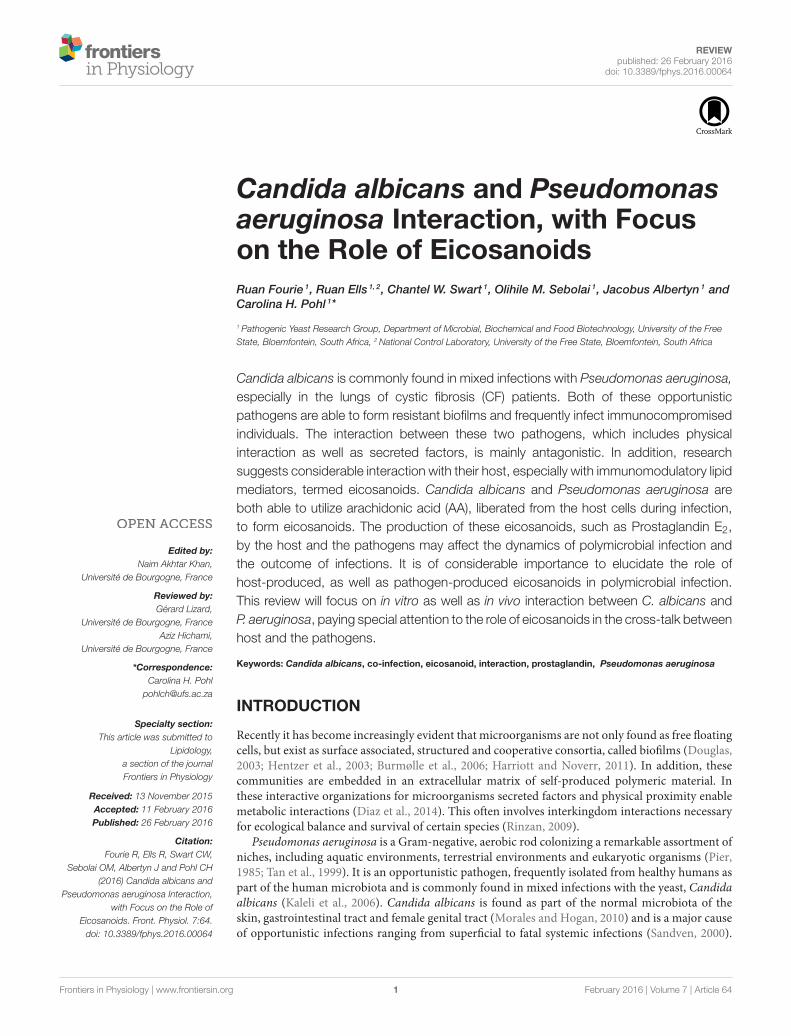
Physical/Direct InteractionSeveral studies were conducted to evaluate the interaction ofP. aeruginosa and C. albicans. The antagonistic interaction ofC. albicans and P. aeruginosawas examined by Brand et al. (2008)who showed that P. aeruginosa cells kill C. albicans hyphal cells,but not C. albicans yeast cells. The deadly effect on C. albicans isthought to be due to PYO, which alters the cell wall of C. albicans(Kerr et al., 1999). Further research into this interaction providedevidence that there is a difference in P. aeruginosa mediatedC. albicans killing among different morphotypes of C. albicans(Rinzan, 2009). Increased susceptibility to the killing effect ofP. aeruginosa was seen with filamentous cells compared toplanktonic counterparts, similar to the study by Brand et al.(2008), as well as a reversion of germ tube formation in thepresence of P. aeruginosa. Further analysis of this interactionindicated that attachment of P. aeruginosa and C. albicanskilling is mediated by lectin-carbohydrate interaction, type IVpili as well as mannans. The authors also speculated on thepossible involvement of O-linked mannans in the survival ofC. albicans yeast cells during combined incubation, as wasproposed previously (Brand et al., 2008; Rinzan, 2009). InFigure 1, scanning electron micrographs of a dual species biofilmwith C. albicans and P. aeruginosa is seen showing extensivecolonization of C. albicans cells by P. aeruginosa.
Indirect InteractionRole of Pseudomonas aeruginosa Quorum Sensing
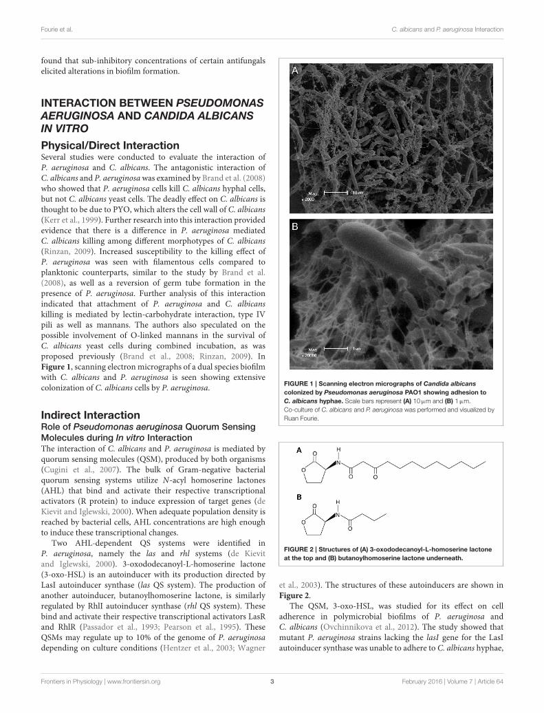
Molecules during In vitro InteractionThe interaction of C. albicans and P. aeruginosa is mediated byquorum sensing molecules (QSM), produced by both organisms(Cugini et al., 2007). The bulk of Gram-negative bacterialquorum sensing systems utilize N-acyl homoserine lactones(AHL) that bind and activate their respective transcriptionalactivators (R protein) to induce expression of target genes (deKievit and Iglewski, 2000). When adequate population density isreached by bacterial cells, AHL concentrations are high enoughto induce these transcriptional changes.
Two AHL-dependent QS systems were identified inP. aeruginosa, namely the las and rhl systems (de Kievitand Iglewski, 2000). 3-oxododecanoyl-L-homoserine lactone(3-oxo-HSL) is an autoinducer with its production directed byLasI autoinducer synthase (las QS system). The production ofanother autoinducer, butanoylhomoserine lactone, is similarlyregulated by RhlI autoinducer synthase (rhl QS system). Thesebind and activate their respective transcriptional activators LasRand RhlR (Passador et al., 1993; Pearson et al., 1995). TheseQSMs may regulate up to 10% of the genome of P. aeruginosadepending on culture conditions (Hentzer et al., 2003; Wagner
FIGURE 1 | Scanning electron micrographs of Candida albicans
colonized by Pseudomonas aeruginosa PAO1 showing adhesion to
C. albicans hyphae. Scale bars represent (A) 10µm and (B) 1µm.
Co-culture of C. albicans and P. aeruginosa was performed and visualized by
Ruan Fourie.
FIGURE 2 | Structures of (A) 3-oxododecanoyl-L-homoserine lactone
at the top and (B) butanoylhomoserine lactone underneath.
et al., 2003). The structures of these autoinducers are shown inFigure 2.
The QSM, 3-oxo-HSL, was studied for its effect on celladherence in polymicrobial biofilms of P. aeruginosa andC. albicans (Ovchinnikova et al., 2012). The study showed thatmutant P. aeruginosa strains lacking the lasI gene for the LasIautoinducer synthase was unable to adhere to C. albicans hyphae,
Frontiers in Physiology | www.frontiersin.org 3 February 2016 | Volume 7 | Article 64

Fourie et al. C. albicans and P. aeruginosa Interaction
while a P. aeruginosa strain without the mutation was able toadhere to C. albicans cells. The study suggested that 3-oxo-HSLis needed for the adherence of P. aeruginosa cells to C. albicanshyphae, because 3-oxo-HSL is needed for the production ofsurface adherence proteins on P. aeruginosa cells. A study byMcAlester et al. (2008) showed that if cell free supernatantfrom high 3-oxo-HSL producing P. aeruginosa strains is addedto C. albicans cultures, the yeast to hyphal switch is inhibited.Pseudomonas aeruginosa strains that produced low amounts of3-oxo-HSL did not inhibit the yeast to hyphal switch when thesupernatants of their cultures were added to C. albicans cultures,suggesting that 3-oxo-HSL affects yeast morphology in a dosedependent manner. To ensure that the 3-oxo-HSL was the causeof the inhibition of morphological switch, pure 3-oxo-HSL wasadded to a C. albicans culture with the same results obtained.The reaction of C. albicans toward 3-oxo-HSL may lead to thedispersal of C. albicans cells in the presence of P. aeruginosa(Morales and Hogan, 2010; Ovchinnikova et al., 2012). Thesestudies thus show that AHLs are not only important for bacterialcommunication, but are responsible for considerable interactionwith other microorganisms such as C. albicans.
In addition to AHLs, a QS signal, 2-heptyl-3-hydroxyl-4-quinolone signal or Pseudomonas quinolone signal (PQS),was later identified and this molecule is released in the lateexponential phase (Pesci et al., 1999; Lépine et al., 2003). Theproduction of PQS can be induced by the LasI/R system andinhibited by the RhlI/R system (De Sordi and Mühlschlegel,2009). Strikingly, PQS was shown to have both a damaging effecton P. aeruginosa through a pro-oxidative effect, as well as an anti-inflammatory effect (Haussler and Becker, 2008). The authorsspeculate that this contradictory effect drives survival of thefittest through selection of phenotypic variants able to survivein stressful conditions and molding populations sufficientlyadapted. It also modulates swarming motility of P. aeruginosa(Déziel et al., 2004; Ha et al., 2011). In addition to PQS, itsimmediate precursor, 2-heptyl-4-quinolone (HHQ), has beenshown to repressC. albicans biofilm formation (Reen et al., 2011).
PQS (Figure 3A) induces the formation of several virulencefactors, including phenazine compounds (Phelan et al., 2014).Of these, PYO or methyl-1-hydroxyphenazine (Figure 3B), isthe best studied. Pyocyanin is a chloroform soluble compoundwith a blue color (Cox, 1986). It has been shown to be aQSM produced in the early stationary phase (Hernandez et al.,2004; Price-Whelan et al., 2007) and a study by Dietrichet al. (2006) suggested that PYO is a terminal signalingmolecule that controls its own cycling. It plays a major rolein maintaining NADH/NAD+ ratio stability in P. aeruginosacells when they encounter oxygen limiting conditions due tothe limited fermentation capability of Pseudomonas (Dietrichet al., 2006). Pyocyanin can then act as an alternate terminalelectron acceptor and decrease the NADH/NAD+ ratio in thestationary phase of growth. It can then later be reoxidizedby oxygen when it becomes available and this could be amechanism for the production of reactive oxygen species (ROS).Phenazines, including PYO, phenazine-1-carboxylic acid, 1-hydroxyphenazine and phenazine-1-carboxamide play extensiveroles in the interaction between Pseudomonas species and
FIGURE 3 | Structures of (A) Pseudomonas quinolone signal at the top
and (B) pyocyanin underneath.
eukaryotes, including fungal microorganisms (Kaleli et al., 2006;Phelan et al., 2014).
In addition, PYO has antimicrobial activity against a widerange of cells including a bactericidal effect against a wide varietyof bacterial species with Gram-positive species being moresusceptible than Gram-negative species (Hassan and Fridovich,1980). Interestingly, Pseudomonas species seem to be resistant tothis bactericidal effect (Baron and Rowe, 1981). It is also toxicto eukaryotic cells (O’Malley et al., 2003). The mechanism ofthis effect is due to the ability of this compound to undergonon-enzymatic redox cycling intracellularly, resulting in thegeneration of ROS (Gloyne et al., 2011). Another effect of PYOis the reduction in cyclic adenosine monophosphate (cAMP)(Kerr et al., 1999). This enables PYO to inhibit the shiftfrom yeast to hyphal morphology in C. albicans because theyeast-mycelium transition is promoted by increased levels ofintracellular cAMP. Gibson et al. (2009) observed a red pigmentwith the co-incubation of P. aeruginosa and C. albicans producedduring close proximity of the yeast and bacterial cells. Thispigment was localized in fungal cells. The authors speculatethat C. albicans enzymes participate in the formation of thisproduct intracellularly. The precursor of this red pigment wasidentified as 5-methylphenazine-1-carboxylic acid (5-MPCA)through the use of P. aeruginosa strains with disruptions inthe phenazine biosynthesis pathway. The presence of the redpigmented compound was linked to significant repression ofC. albicans viability. In addition, a recent study indicated thatthe phenazines, phenazine-1-ol, phenazine-1-carboxylic acid andphenazine-1-carboxamide, has a synergistic effect with threeantifungals: fluconazole, itraconazole and clotrimazole againstCandida species (Kumar et al., 2014). This then suggeststhat the presence of phenazine producing organisms such asPseudomonas can drastically alter the treatment of simultaneousfungal infection.
Frontiers in Physiology | www.frontiersin.org 4 February 2016 | Volume 7 | Article 64

Fourie et al. C. albicans and P. aeruginosa Interaction
FIGURE 4 | Structure of farnesol.
The Role of Candida albicans Quorum Sensing
Molecules during In vitro InteractionCandida albicans has also been shown to produce QSMs (Hornbyet al., 2001). The QSM, farnesol (Figure 4), inhibits germtube formation and also caused a morphological shift frompredominant mycelial state to predominant yeast morphology,indicating the effect as 3-oxo-HSL. The effect on the morphologyof C. albicans is thought to be due to inhibition of theRas1-controlled pathway involved in hyphal growth (Moralesand Hogan, 2010). Recently, farnesol was identified to attractmacrophages in hosts (Hargarten et al., 2015). The authorsspeculate that engulfment and movement of these immunecells then aid in dissemination as macrophages are killed byC. albicans after engulfment. Farnesol induces the generationof ROS which could play a role in the competition of C.albicans with bacteria. Resistance of C. albicans to oxidativestress has also been shown to be linked, in part, to farnesol(Westwater et al., 2005). A study by Cugini et al. (2007) indicatedthat farnesol inhibits P. aeruginosa PQS and a subsequentvirulence factor, PYO, whose production is controlled by PQS,in a dose dependent manner. Interestingly, there was no effecton the overall growth of P. aeruginosa. Additionally, whenP. aeruginosa was co-cultured with C. albicans, reduction inPQS and PYO produced by P. aeruginosa was also observed,suggesting that high enough concentrations of farnesol isproduced by C. albicans to exert an effect on P. aeruginosa.Later, the same research group found that C. albicans and itssecreted factors increase PQS and butanoylhomoserine lactonein lasR defective mutants of P. aeruginosa, with a downstreamincrease in phenazine production (Cugini et al., 2010). Theauthors speculated that oxidative stress may trigger downstreamquorum sensing pathways.
As seen with proteomic analysis by Jones-Dozier (2008), apossible decrease in virulence of P. aeruginosa is evident whenexposed to farnesol, due to alterations in protein expressionof P. aeruginosa subjected to this compound. In addition todecreasing PQS and PYO production, farnesol inhibits swarmingmotility in P. aeruginosa (McAlester et al., 2008). Rhamnolipids,a class of glycolipids, play a role in swarming motility and hasbeen implicated as playing part in the development of ventilatorassociated pneumonia (VAP) (Köhler et al., 2010). Due to thefact that rhamnolipid production is partly regulated by PQS, themechanism of decreased swarming motility may be due to thereduction in PQS production by farnesol.
Other Factors Influencing In vitro Interaction
Iron availabilityIn a study by Purschke et al. (2012), C. albicans exhibited alower metabolic activity in mixed biofilms with P. aeruginosa
when compared to single species biofilms. Secretome analysis ofthe proteins of the single vs. mixed species biofilms revealed anoverall increase of secreted proteins of mixed species biofilmsof C. albicans and P. aeruginosa compared to the single speciescounterparts. This increase was largely found to be due toincreased secreted proteins by P. aeruginosa. Interestingly, a largeproportion of the increased protein production was attributedto a siderophore, pyoverdine, specific to Pseudomonas. Thisincrease in pyoverdine was thought to be due to the increasediron utilization by the two species in the mixed biofilm. Thiswas confirmed by the addition of iron, which abolished theproduction of pyoverdine. The authors speculated sequestrationof available iron by pyoverdine results in decreased availability toC. albicans, although C. albicans is able to utilize iron bound tocertain other microbial siderophores. Recent evidence suggeststhat this phenomenon may not be of importance during in vivointeraction (Lopez-Medina et al., 2015). In this study, C. albicanssecreted factors significantly reduced pyoverdine and anothersiderophore, pyochelin, expression by P. aeruginosa duringgastrointestinal colonization in a murine model. This decrease ofexpression by P. aeruginosa was linked to diminished virulenceof P. aeruginosa with growth unaffected. The authors suspect theheterogeneity of the biofilms between in vivo and in vitro studiesmay cause the differential results.
Recently, Trejo-Hernández et al. (2014) found that hypoxiainfluences the ability of P. aeruginosa to inhibit C. albicansfilamentation in vitro compared to aerobic conditions. Thiswas attributed to decreased AHL produced by P. aeruginosain the presence of C. albicans. Previously, it was shownthat hypoxic conditions promote filamentation in C. albicansand reduces farnesol production and response to it (Dumitruet al., 2004). Additionally, the authors also speculated thatcompetition for ironmay also be greater during hypoxia (Synnottet al., 2010; Trejo-Hernández et al., 2014). Therefore, both theinteraction of P. aeruginosa with C. albicans, the concentrationof oxygen and iron competition influences the productionof AHLs (Trejo-Hernández et al., 2014). The authors alsofound that proteins known to play roles in iron uptake inP. aeruginosa through siderophores were upregulated in mixedbiofilms, confirming previous observations. Additionally, ironsupplementation increased the growth of P. aeruginosa in singleandmixed biofilms, with this effect not seen withC. albicans. Thisincrease in growth of the bacterium may increase the destructionof the fungal population. Lamont et al. (2002) indicated thatpyoverdine may act as a signaling molecule to modulate othervirulence factors including exotoxin A and pyoverdine itself.Because pyoverdine production is increased in mixed biofilms,the virulence of P. aeruginosamight also be upregulated in mixedbiofilms (Trejo-Hernández et al., 2014). Additionally, PQS aswell as products of the PQS system including rhamnolipids andPYO, were upregulated. A significant increase in P. aeruginosamutability frequency was seen with a large number of antibioticresistant mutant phenotypes arising over time. The authorsspeculate that the decreased catalase activity observed in mixedbiofilms may result in increased oxidative stress, concomitantlyincreasing mutability. In the case of C. albicans, the same trendwas seen with hypermutability arising with a high frequencyof antimicrobial resistant phenotypes, possibly attributed to
Frontiers in Physiology | www.frontiersin.org 5 February 2016 | Volume 7 | Article 64

Fourie et al. C. albicans and P. aeruginosa Interaction
the increased oxidative stress caused by PYO produced by P.aeruginosa. Additionally, C. albicans iron dependant processes,including aerobic respiration, were downregulated. Glycolyticenzyme activity in C. albicanswas also altered, with PYO possiblyattributing to this and leading to other pathways for energyutilization. To confirm the increased virulence of C. albicans andP. aeruginosa in mixed biofilms, the authors used a rat infectionmodel. Candida albicans was shown to promote pathogenicityof P. aeruginosa. Therefore, the ability of these pathogens tocompete for iron may alter population dynamics and influencethe nature of the interaction.
Bacterial cell wall componentsIn addition to various secreted factors produced by P. aeruginosain polymicrobial interaction (Holcombe et al., 2010), bacterialLPS were shown to have adverse effects on Candida spp.biofilms (Bandara et al., 2010). The same group later confirmedthese results by evaluating the effect of P. aeruginosa LPSon C. albicans (Bandara et al., 2013). The study suggested adecrease of C. albicans filamentation and biofilm metabolicactivity, including glycolysis, and growth with the addition ofhigh concentrations of P. aeruginosa LPS. In addition to this,peptidoglycan was shown to trigger filamentation in C. albicans(Xu et al., 2008).
EthanolChen et al. (2014) evaluated the effect of C. albicans producedethanol on P. aeruginosa and found that ethanol stimulatedadhesion and biofilm formation of P. aeruginosa. In addition,swarming motility by P. aeruginosa decreased and a stimulationof phenazine derivatization and production of 5-MPCA byP. aeruginosa in the presence of ethanol was observed. Theauthors speculate that there is a positive feedback loop whereC. albicans ethanol production increases P. aeruginosa 5-MPCAproduction and biofilm formation. In turn, 5-MPCA stimulatesethanol production in C. albicans (Morales et al., 2013).
Extracellular DNAA recent study also identified extracellular DNA as a largefactor in biofilm formation by C. albicans (Sapaar et al.,2014). Low amounts of extracellular DNA (1.0µg/mL) wasshown to promote biofilm formation and increase biofilmstability, whereas higher concentrations (10µg/mL) hamperedthe formation of biofilms by C. albicans as well as the stabilityof the biofilms. The study also indicated that the source of theextracellular DNA, whether it is C. albicans, or from bacterialsources such as P. aeruginosa, does not matter. This increase inbiofilm formation by C. albicans due to extracellular DNA mayincrease the virulence of the fungus. Evidence also suggest thatthe concentration of extracellular DNA can reach 4 mg/mL inCF patient sputum samples, raising the question if this facet ofinteraction might have clinical relevance (Sapaar et al., 2014).
A summary of several facets of interaction between C. albicansand P. aeruginosa can be seen in Figure 5.
INTERACTION BETWEEN PSEUDOMONAS
AERUGINOSA AND CANDIDA ALBICANS
IN VIVO
A high number of C. albicans nosocomial infections arepolymicrobial with P. aeruginosa a frequent co-isolate in bloodstream infections and pneumonia (Lindsay and Hogan, 2014).Kerr (1994) was the first to describe the anticandidal activity ofP. aeruginosa in vivo. The study evaluated lung infection of threesurgery patients postoperatively with inhibition of C. albicansgrowth seen after P. aeruginosa colonization. This inhibitionwas confirmed with the regrowth of C. albicans seen aftereradication of P. aeruginosa, even with fluconazole treatment.Additional in vitro susceptibility experiments confirmed thesuppression of Candida growth by P. aeruginosa. Gupta et al.(2005) evaluated 300 burn patients over 2 years and foundrepression of Candida spp. in the presence of P. aeruginosa.Several studies also indicate that prior colonization of Candidamay promote susceptibility of the host to P. aeruginosa infection(Roux et al., 2009; Hamet et al., 2012; Xu et al., 2014).Nseir et al. (2007) reported that antifungal treatment duringCandida spp. tracheobronchial colonization may be associatedwith reduced risk for P. aeruginosa colonization. The case isstrengthened by Azoulay et al. (2006), who reported a possiblelink between Candida colonization of the respiratory tract and anincreased risk for Pseudomonas VAP. Roux et al. (2013) reportedthat the Th1-Th17 immune response, associated with Candidacolonization, caused a decrease in P. aeruginosa phagocytosisby alveolar macrophages, the primary innate immune responseagainst bacterial invasion. The adaptive Th1-Th17 responseevoked by C. albicans invasion, is characterized by an increase ininterferon È(IFNÈ), as well as an increase in interleukin-17 (IL-17) and decrease in IL-2. The authors suggest that the increasein IFNÈ, associated with the Th1-Th17 response, is responsiblefor the inhibition of bacterial phagocytosis, through inhibitingexpression of scavenger receptors on alveolar macrophages.
Remarkably, contradictory results to the notion thatP. aeruginosa infection is more aggressive after prior C. albicanscolonization, was provided by Ader et al. (2011). In a murinemodel, C. albicans short term colonization prior to P. aeruginosacolonization caused a reduction in P. aeruginosa bacterialload compared to the absence of C. albicans colonization(Ader et al., 2011). Additionally, a reduction in P. aeruginosainduced lung injury was observed with the prior colonization ofC. albicans. Interestingly, this effect was reversed with treatmentby the antifungal caspofungin during C. albicans colonization.Candida albicans initiates alveolar innate immunity in a murinemodel, protecting the host against subsequent P. aeruginosainfection (Mear et al., 2014). The authors showed that priorC. albicans infection induces interleukin-17 (IL-17) and IL-22 secretion through innate lymphoid cell recruitment. Thecytokines produced, induce the production of antimicrobialpeptides as well as the mobilization of phagocytic cells. Ina murine gut model, C. albicans secreted factors inhibitedexpression of siderophores as well as cytotoxic moleculesby P. aeruginosa, reducing the virulence of the bacteria(Lopez-Medina et al., 2015). Due to this, increased survival of
Frontiers in Physiology | www.frontiersin.org 6 February 2016 | Volume 7 | Article 64

Fourie et al. C. albicans and P. aeruginosa Interaction
FIGURE 5 | Illustration of competition between Candida albicans and Pseudomonas aeruginosa. Pseudomonas aeruginosa attaches to C. albicans hyphae
and kills hyphal cells through secreted hydrolytic enzymes such as hemolytic phospholipase C (PlcH) and phenazines such as pyocyanin and
5-methylphenazine-1-carboxylic acid (5-MPCA). 3-oxo-homoserine lactone produced by P. aeruginosa and phenazines inhibit filamentation by C. albicans, similar to
farnesol, produced by C. albicans. Pseudomonas aeruginosa lipopolysaccharide (LPS) inhibits C. albicans filamentation. Ethanol production is increased by the
fungus, inhibiting the motility of P. aeruginosa (adapted from Lindsay and Hogan, 2014).
the host was observed during co-incubation of P. aeruginosawith C. albicans. Interestingly, Neely et al. (1986) demonstratedincreased mortality in a murine model when C. albicans infectionwas preceded by P. aeruginosa. This reciprocal effect may alsobe due to alterations in innate immune response, as Faureet al. (2014) reported that the P. aeruginosa type III secretionsystem induced IL-18 secretion causing substantial neutrophilrecruitment and host cell damage, and decreased IL-17 secretionin a mouse model, possibly leading to the reduced clearance ofpathogens.
The co-infection of P. aeruginosa and C. albicans has beenwell documented in cystic fibrosis (CF) patients. CF is one ofthe most commonly encountered autosomal recessive disordersfound, with the occurrence varying in race (Andersen, 1938). Thedisease is caused by a genetic disorder where a mutation existsin the CF transmembrane conductance regulator (CFTR) gene(Delhaes et al., 2012). CF is a disease with two pathophysiologicalproperties, namely pancreatic insufficiency with malnutritionand also airway infections due to extremely viscous secretions(Andersen, 1938). The increased viscosity of lung secretionsis thought to be due to the increased sodium absorptionof the respiratory epithelium and the defective regulation ofchloride ion secretion (Gilligan, 1991). This is thought to be the
reason why CF patients have comparatively dehydrated surfaceliquid which leads to defective mucociliary clearance. The thickbronchial mucus traps viral particles, fungal spores and bacteriaand provides a suitable environment for the fungal spores andbacteria to grow, causing infection (Delhaes et al., 2012). Ninetypercent of deaths in CF are due to pulmonary dysfunctionand in effect, chronic airway infection (Gilligan, 1991). A studyby Güngör et al. (2013) evaluated the most prevalent fungalcolonization in Turkish CF patients. The most prevalent fungalmicroorganisms isolated from these CF patients, were shown tobe C. albicans at 62.5% (30 patients). In addition, fungal-bacterialco-colonization in the CF patients was shown to be 98.2% inthese C. albicans infections. The most frequent bacterial co-colonizer of CF patients with C. albicans infections was found tobe Staphylococcus aureus (53.57%), with P. aeruginosa at 48.21%,Staphylococcus maltophilia at 16.07% and Haemophilus influenzaat 5.97%. Other similar studies have also implicated S. aureusand P. aeruginosa as the most prevalent bacterial species isolated(Valenza et al., 2008; Williamson et al., 2011). Several studiesaddressed the correlation between C. albicans and P. aeruginosain CF infection, with a recent study suggesting a significantcorrelation (Conrad and Bailey, 2015). Discrepancies may arisedue to method of analysis and the population analyzed.
Frontiers in Physiology | www.frontiersin.org 7 February 2016 | Volume 7 | Article 64

Fourie et al. C. albicans and P. aeruginosa Interaction
PRODUCTION AND ROLE OF OXYLIPINSDURING INFECTION
Lipids have crucial cellular significance, forming membranes,as well as acting as cellular signals (reviewed by Tehlivetset al., 2007). The latter is of great importance in multicellulareukaryotic organisms. Although lipids as signaling moleculeshave beenwell studied inmammals and plants, their roles in fungiare not as well characterized.
Arachidonic acid (AA) or 5,8,11,14-eicosatetraenoicacid (Figure 6A), a major constituent of the mammalianphospholipids, together with its wide range of metabolites(termed eicosanoids), have substantial roles as lipid signals(Chilton et al., 1996), including modulation of the innateimmune response (Rodríguez et al., 2014). As the constituentof cellular membranes, it is predominantly found in the sn2position of phospholipids with incorporation into inflammatorycells through CoA-dependant acyl transferases.
The metabolism of AA to form a plethora of lipidmediators involve various pathways in vertebrates, includingthe action of cyclooxygenases (COX), lipoxygenases (LOX),monooxygenases (CYP450), and non-enzymatic (NE) pathways.Cyclooxygenases, or prostaglandin endoperoxide synthases, areenzymes catalyzing the insertion of two oxygen atoms in AA(Marnett et al., 1999). In mammalian cells, two isoforms exist,namely, COX-1, which is constitutively expressed, and COX-2, which is inducible. The initial reaction of AA oxidation,mediated by COX, yields prostaglandin G2 (PGG2) (Rodríguezet al., 2014). Through peroxidase activity, PGG2 is reduced toprostaglandinH2 (PGH2), which serves as a precursor for variousother immunomodulatory compounds, including prostaglandinsas well as thromboxanes. For example, further action byProstaglandin E synthase converts PGH2 to Prostaglandin E2(PGE2) (Figure 6D).
Lipoxygenases are a large group of dioxygenases that catalyzeoxygen insertion into polyunsaturated fatty acids in animals,plants as well as microorganisms (Kuhn and O’Donnell, 2006;Brodhun and Feussner, 2011). The reaction of oxygenationconsists of various steps, starting with hydrogen abstraction,followed by radical rearrangement and the insertion of oxygen.In addition to oxygenation and hydroperoxidation, LOX alsocatalyze the synthesis of leukotrienes, lipoxins and hepoxilinsthrough the combination of various enzymatic activities (Kuhnand O’Donnell, 2006; Dennis and Norris, 2015). Variousisoforms of LOX exist with different stereospecificity andactivities, for example 12/15-LOX catalyze oxygenation andhydroperoxidation of PUFAs at the 12 or 15 carbon position.Various studies suggest that the 12/15-LOX can be inducedby interleukin-4 (IL4) and IL13, which are important inthe Th2 response. Additionally, 12/15-LOX is inhibited byinterferon È. The low expression of 12/15-LOX has been linkedto pro-inflammatory responses and has been implicated inairway functions. Prostaglandin E2 has also been argued toinduce the expression of 12/15-LOX in neutrophils throughcAMP elevation. Various products of LOX have also beenimplicated in anti-inflammatory responses in neutrophils. ManyLOX products, including hydroperoxy-eicosatetraenoic acids
(HPETEs) and hydroxy-eicosatetraenoic acids (HETEs) areintermediate products leading to the formation of lipoxins andleukotrienes (Dennis and Norris, 2015). The interaction of COX-and LOX-derived lipid mediators as well as the combination ofthese two pathways leads to the modulation of the inflammatoryresponse (Dennis and Norris, 2015). The non-steroidal anti-inflammatory drug (NSAID), acetylsalicylic acid (ASA) (alsoknown as aspirin), was shown to acetylate COX isozymesleading to the formation of 15(R)-HETE, which acts as LOXsubstrate for the formation of lipoxins (Serhan, 2002). Theselipoxins are potent anti-inflammatory molecules, inhibitingneutrophil recruitment and leukotriene formation. In additionto these pathways, cytochrome P450s are also responsible for theformation of epoxyeicosatetraenoic acids (EETs) from AA, withconcurrent modification to diHETEs, playing differential effectson the host.
The effects of eicosanoids on mammalian cells are dependenton the type of target tissue and the physiological state of thesetissues (Dennis and Norris, 2015). Considerable research isbeing done to determine the eicosanoids that play a role inhost protection against pathogens during infection as they canenhance the clearance of pathogens. For this review, the focus willfall on lipid mediators in terms of C. albicans and P. aeruginosainfection.
Role of Mammalian Oxylipins duringPseudomonas aeruginosa InfectionSeveral studies have addressed the effect of various invadingpathogens on the production of PGE2 to gain a betterunderstanding of the immunological aspects of infection. Inmammalian cells, PGE2 is formed through the metabolism ofAA by the action of cyclooxygenase (COX-1 or COX-2) tofirst form PGH2, followed by PGE synthases to form PGE2(Murakami et al., 2003). Immune cells are the main source ofPGE2, although this compound is also produced by various othercell types (Kalinski, 2012; Agard et al., 2013). It elicits a responsein mammalian cells through activation of four receptors,designated EP1–EP4, with the effect dependent on the receptoractivated.
Pseudomonas aeruginosa pulmonary infection is associatedwith an overproduction of PGE2 and concurrent decrease inphagocytosis by alveolar macrophages (Ballinger et al., 2006;Agard et al., 2013). This increase in PGE2 is due to the largeamount of AA released during P. aeruginosa infection, mediatedby ExoU, an intracellular phospholipase (König et al., 1996; Salibaet al., 2005; Sadikot et al., 2007; Agard et al., 2013). As suchit plays a crucial role in initial infection and infiltration by P.aeruginosa. The absence of ExoU in P. aeruginosa was linkedto diminished severity of infection and PGE2 production. Theimportance of PGE2 during P. aeruginosa infection was seenwhen a COX-2 inhibitor was employed, resulting in a decreasein severity of infection by this pathogen (Sadikot et al., 2007).Several other virulence factors also elicit changes in PGE2 levels.The QSM 3-oxo-HSL produced by P. aeruginosa was shown toinduce COX-2 and, therefore, PGE2 production in human lungfibroblasts (Smith et al., 2002). Similarly, P. aeruginosa PYO
Frontiers in Physiology | www.frontiersin.org 8 February 2016 | Volume 7 | Article 64

Fourie et al. C. albicans and P. aeruginosa Interaction
FIGURE 6 | Structures of (A) arachidonic acid (AA) at the top, followed by (B) 3-hydroxyeicosatetraenoic acid (3-HETE); (C) 15-HETE; (D) Prostaglandin
E2 at the bottom.
and LPS increased the release of PGE2 in urothelial cells in aconcentration dependent manner (McDermott et al., 2013).
Role of Pseudomonas aeruginosa
Oxylipins during InfectionA number of microorganisms including bacteria are able toform eicosanoids (Lamacka and Sajbidor, 1995). Although thepresence of LOX in plants and animals has long been known,their presence in lower eukaryotes and prokaryotes has onlyrecently been established, with P. aeruginosa one of the fewbacteria with typical LOX genes. Pseudomonas aeruginosa hasbeen found to possess a secretable 15-LOX, homologous tomammalian LOX, producing 15-HETE (Figure 6C) which issimilar to host 15-HETE and elicits anti-inflammatory effectson the host, through acting as a substrate for lipoxin formation(Serhan, 2002; Vance et al., 2004). The formation of theselipoxins may alter the severity of infection through inhibitingneutrophil recruitment and generation of leukotrienes (Serhan,2002). In addition to 15-HETE, P. aeruginosa is able to producea range of products including dihydroxy unsaturated fatty
acids such as 7,10-dihydroxy-8(E)-octadecenoic acid (DOD)(Hou, 2008). In addition, the production of prostaglandinsand prostaglandin-analog compounds have been identified inP. aeruginosa (Lamacka and Sajbidor, 1995), however, theeffect of these compounds during infection has not beenaddressed.
Role of Mammalian Oxylipins duringCandida albicans InfectionThe alteration of immune response is not unique to bacteria, butplays a significant role as a virulence factor during C. albicansinfection. Candida albicans is able to metabolize AA, liberatedfrom host cells by host as well as yeast-derived phospholipaseA2 (Castro et al., 1994; Filler et al., 1994; Brash, 2001; Niewerthand Korting, 2001; Theiss et al., 2006; Parti et al., 2010).In addition, C. albicans can induce significant production ofPGE2 by mammalian cells (Filler et al., 1994; Deva et al.,2001). Candida albicans mannans, forming part of the C.albicans cell wall, can directly induce PGE2 production bymammalian cells (Smeekens et al., 2010). In addition, the
Frontiers in Physiology | www.frontiersin.org 9 February 2016 | Volume 7 | Article 64

Fourie et al. C. albicans and P. aeruginosa Interaction
induction of the release of AA and the eicosanoid metabolitesfrom alveolar macrophages has been shown to be partlyregulated by mannose and β-glucan receptors interacting withfungal cell wall components and have also been shown toinhibit phagocytosis during C. albicans infection (Castro et al.,1994).
Eradication of infection by the immune system is highlydependent on the balance of Th1 and Th2 responses (Romani,2000). The Th1 response is associated with the removal ofpathogens through phagocytosis, in contrast to the hamperingof this effect during the Th2 response. Prostaglandin E2is able to modulate this balance and frequently promotescolonization of pathogens such as C. albicans, as well as causingtissue eosinophilia. Prostaglandin E2 production in response toC. albicans invasion also induces the protective Th-17 responsein mammalian systems (Smeekens et al., 2010).
Role of Candida albicans Oxylipins duringInfectionAA can be used as a carbon source by C. albicans (Devaet al., 2000). The authors also identified a HETE produced byC. albicans from AA namely 3,18-diHETE, that is associated withhyphal forms and may play a role in adhesion during infection(Deva et al., 2000; Ells et al., 2012). Additionally, the authorsshowed that most AA is metabolized by non-mitochondrialpathways, concurrent with the speculation that fatty acids aremetabolized by peroxisomal β-oxidation in yeasts. Acetylsalicylicacid-sensitive 3-HETE (Figure 6B), produced by C. albicans wasalso speculated to be linked to C. albicans morphogenesis (Devaet al., 2001). 3-HETE can also serve as substrate for COX-2in mammalian cells to form 3-hydroxyeicosanoids due to thesimilarity to AA (Ciccoli et al., 2005). Further analysis of one ofthese metabolites, 3-OH-PGE2, indicated a similar or even morerobust effect on mammalian cells eliciting a pro-inflammatoryresponse.
The anti-inflammatory lipid Resolvin E1, which Candidaalbicans is able to produce from eicosapentaenoic acid, alsoplays a role in the interspecies signaling (Haas-Stapleton et al.,2007). This compound was shown to protect C. albicans againsthost immunity at low doses, however, at higher concentration,this protective effect is lost. This is thought to facilitate thecommensal carriage of C. albicans as part of the humanmicrobiome.
An important aspect of the determination of C. albicansvirulence was the identification of a PGE2 cross reactivecompound produced by C. albicans from exogenous AA (Noverret al., 2001). This compound was later identified as PGE2,identical to the host product (Erb-Downward and Noverr, 2007).This was especially surprising as C. albicans does not possesshomologs to COX present in mammalian cells responsible forthe formation of PGE2. The involvement of various enzymes orhomologous enzymes was speculated in the production of PGE2.A multicopper oxidase homolog (Fet3), fatty acid desaturasehomolog (Ole2) and cytochrome P450s has been shown tobe involved in the production of PGE2 by C. albicans (Erb-Downward and Noverr, 2007; Ells et al., 2011; Krause et al.,
2015). Biofilm formation in C. albicans as well as germ tubeformation has been shown to be enhanced by the addition ofsynthetic PGE2. In addition, various COX-inhibitors such as ASAdrastically affect the formation of biofilms and germ tubes (Alemand Douglas, 2004). Interestingly, the simultaneous addition ofPGE2 abolished the inhibitory effect of ASA on C. albicansbiofilm formation. In a later study by the same researchers,a comparison of PGE2 production by C. albicans planktoniccells and biofilms showed a significant increase in PGE2production of C. albicans biofilms (Alem and Douglas, 2005). Inaddition, various COX-inhibitors, namely ASA, diclofenac andetodolac, significantly decreased the production of PGE2 by C.albicans biofilms. These findings suggest a possible link betweenprostaglandin production and biofilm formation. Deva et al.(2001) reported significant decrease in yeast to hyphal transitionby C. albicans with the addition of ASA. This morphologicaleffect was attributed to the possible suppression of 3(R)-hydroxy-oxylipins.
Figure 7 indicates the interaction of C. albicans and P.aeruginosa with host cells with the focus on eicosanoidproduction. In the figure, arrows indicate the production orincrease of an eicosanoid, whereas a question mark indicates anunknown effect or interaction.
Considering the information regarding the direct andindirect interaction of C. albicans and P. aeruginosa, aswell as the information regarding the role of eicosanoidsduring single species infection, several questions can beasked relating to polymicrobial infection with C. albicans andP. aeruginosa.
Role of Oxylipins in Polymicrobial InfectionAlthough information is available regarding the eicosanoidsproduced, and their effect during single species infection,very little is known regarding the effect of microbially-produced eicosanoids on co-infecting pathogens, as well as onthe host.
In a study by Peters and Noverr (2013), the role of eicosanoidsduring polymicrobial infection in another microbial interactionbecame evident. In this study, a murine model was used toelucidate the effect of polymicrobial interactions usingC. albicansand Staphylococcus aureus. Through their work, it was evidentthat polymicrobial infections with these organisms resultedin a significant increase in morbidity and mortality duringmurine peritoneal infection, with this effect not seen with singlespecies infection. In addition, disease progression and microbialload in infected mice was significantly higher, compared tomonomicrobial infections. The authors also detected a significantincrease in pro-inflammatory chemokines released, as wellas recruitment of polymorphonucleocytes. To determine theeffect of the pro-inflammatory response on infection dynamics,indomethacin was used. Indomethacin, a non-selective COXinhibitor, similar to ASA, caused a significant reduction inmorbidity and mortality in polymicrobial infection, as wellas a reduction in cytokine release. Indomethacin also causeda significant reduction in microbial load during co-infection.This effect was not seen with monomicrobial infection. In vitroexperiments showed that this effect was not due to inhibition of
Frontiers in Physiology | www.frontiersin.org 10 February 2016 | Volume 7 | Article 64

Fourie et al. C. albicans and P. aeruginosa Interaction
FIGURE 7 | Interaction of Candida albicans and Pseudomonas aeruginosa with host cells highlighting the production of eicosanoids. Arrows indicate
production/increase. Question marks indicate uncharacterized interactions (AA, Arachidonic acid; PLA2,Phospholipase A2; PGs, Prostaglandins; PGE2,
Prostaglandin
E2; ExoU, Exotoxin U; PYO, pyocyanin; LPS, lipopolysaccharide; 3-HETE, 3-hydroxyeicosatetraenoic acid; 3-oxo-HLS, 3-oxododecanoyl-L-homoserine lactone;
15-LOX, 15-Lipoxygenase).
growth of C. albicans and S. aureus. The authors also observed asignificant increase in PGE2 release by the host cells in responseto polymicrobial infection with indomethacin also causing asignificant reduction in PGE2. The authors suggested that PGE2may play a role in the non-protective pro-inflammatory responseduring polymicrobial infection, as PGE2 induces the releaseof several cytokines. To further determine the effect of PGE2in co-infection, mice were co-infected with C. albicans andS. aureus and treated with indomethacin as well as PGE2. Underthis circumstance, high morbidity and mortality was observedin the mice, although they were treated with indomethacin.In the absence of infection, with only the administration ofindomethacin and PGE2, no mortality was observed. Further,the administration of PGE2 caused a significant increase inmicrobial burden during co-infection. During this study, theproduction of eicosanoids, such as PGE2, by C. albicans itselfwas not addressed. Interestingly, Krause et al. (2015) indicatedthat C. albicans PGE2 production significantly enhances S.aureus biofilm growth in vitro. The authors also found thatco-incubation of C. albicans with S. aureus did not elicit anincrease in PGE2 production by C. albicans. Nothing is knownyet regarding the eicosanoid production of C. albicans and P.aeruginosa during co-infection.
CONCLUSIONS
In addition to the ample evidence supporting the interactionof C. albicans and P. aeruginosa, not only with their host,
but also with each other, it is evident that the interaction ismultifaceted. Various virulence factors such as morphogenesis,hypermutabililty and secreted factors (including lipid mediators)affect and damage hosts to facilitate rapid and aggressivecolonization and infection. Any disequilibrium in host defenses,such as in CF, immune disorders, and breaching of host barriers,is rapidly exploited by these opportunistic pathogens.
On their own, C. albicans and P. aeruginosa pose a risk tocompromised hosts. Recent studies, however, illuminate thatthe interaction of these microorganisms are antagonistic withsubstantial damage caused to the host during the chemical warthey play part in. Host immunity also plays a large role in damageto its own tissue due to radical generation by the innate immuneresponse and various lipid mediators. This is even more evidentin the work by Peters and Noverr (2013). It is also evident thatvarious secreted factors of C. albicans (including farnesol andvarious hydrolytic enzymes) and P. aeruginosa (including AHL’s,PQS, PYO and various peptides) form a large amount of radicalsand can elicit oxidative damage to each other, as well as the host.
Although the role of lipid mediators between single pathogensand hosts has been studied, a gap in knowledge still existsregarding the effect of lipid mediators in polymicrobial infectionof C. albicans and P. aeruginosa. Polymicrobial infection by C.albicans and S. aureus, however, highlight the importance oflipid mediators, such as PGE2, in infection by multiple microbes.Due to the fact that C. albicans and P. aeruginosa are bothable to produce significant amounts of prostaglandins and othereicosanoids from exogenous AA, this could affect the dynamicsof this co-infection as well as host survival during infection.
Frontiers in Physiology | www.frontiersin.org 11 February 2016 | Volume 7 | Article 64

Fourie et al. C. albicans and P. aeruginosa Interaction
This warrants investigation to fully understand the interaction ofC. albicans and P. aeruginosa in terms of eicosanoids, as well asthe effect of these eicosanoids in the host during co-infection.
AUTHOR CONTRIBUTIONS
RF compiled the information, co-wrote the manuscript andapproved the final version submitted. RE, CW, OS, JA providedscholarly input in placing the literature into context, editedthe manuscript and approved the final version submitted. CP
provided scholarly input in placing the literature into context, co-wrote the manuscript and approved the final version submitted
FUNDING
The financial assistance of the National Research Foundation(NRF) (Grant: CPRR 93485) toward this research is herebyacknowledged. Opinions expressed and conclusions arrived at,are those of the author and are not necessarily to be attributedto the NRF.
REFERENCES
Ader, F., Jawhara, S., Nseir, S., Kipnis, E., Faure, K., Vuotto, F., et al. (2011). Shortterm Candida albicans colonization reduces Pseudomonas aeruginosa-relatedlung injury and bacterial burden in a murine model. Crit. Care 15:R150. doi:10.1186/cc10276
Agard, M., Asakrah, S., and Morici, L. A. (2013). PGE2 suppression of innateimmunity during mucosal bacterial infection. Front. Cell. Infect. Microbiol.
3:45. doi: 10.3389/fcimb.2013.00045Alem, M. A. S., and Douglas, L. J. (2004). Effects of aspirin and other nonsteroidal
anti-inflammatory drugs on biofilms and planktonic cells of Candida albicans.Antimicrob. Agents. Chemother. 48, 41–47. doi: 10.1128/AAC.48.1.41-47.2004
Alem, M. A. S., and Douglas, L. J. (2005). Prostaglandin production duringgrowth of Candida albicans biofilms. J. Med. Microbiol. 54, 1001–1005. doi:10.1099/jmm.0.46172-0
Andersen, D. H. (1938). Cystic fibrosis of the pancreas and its relation to celiacdisease: a clinical and pathologic study. Am. J. Dis. Child. 56, 344–399. doi:10.1001/archpedi.1938.01980140114013
Andes, D., Nett, J., Oschel, P., Albrecht, R., Marchillo, K., and Pitula, H.(2004). Development and characterization of an in vivo central venouscatheter Candida albicans biofilm model. Infect. Immun. 72, 6023–6031. doi:10.1128/IAI.72.10.6023-6031.2004
Azoulay, E., Timsit, J.-F., Tafflet, M., de Lassence, A., Darmon, M., Zahar, J.-R.,et al. (2006). Candida colonization of the respiratory tract and subsequentPseudomonas ventilator-associated pneumonia. Chest 129, 110–117. doi:10.1378/chest.129.1.110
Ball, G., Durand, É., Lazdunski, A., and Filloux, A. (2002). A novel type IIsecretion system in Pseudomonas aeruginosa. Mol. Cell. Biochem. 43, 475–485.doi: 10.1046/j.1365-2958.2002.02759.x
Ballinger, M. N., Aronoff, D. M., McMillan, T. R., Cooke, K. R., Olkiewicz, K.,Toews, G. B., et al. (2006). Critical role of prostaglandin E2 overproductionin impaired pulmonary host response following bone marrow transplantation.J. Immunol. 177, 5499–5508. doi: 10.4049/jimmunol.177.8.5499
Bandara, H. M. H. N., K Cheung, B. P., Watt, R. M., Jin, L. J., andSamaranayake, L. P. (2013). Pseudomonas aeruginosa lipopolysaccharideinhibits Candida albicans hyphae formation and alters gene expression duringbiofilm development.Mol. Oral. Microbiol. 28, 54–69. doi: 10.1111/omi.12006
Bandara, H. M. H. N., Lam, O. L. T., Watt, R. M., Jin, L. J., and Samaranayake,L. P. (2010). Bacterial lipopolysaccharides variably modulate in vitro biofilmformation of Candida species. J. Med. Microbiol. 59, 1225–1234. doi:10.1099/jmm.0.021832-0
Baron, S. S., and Rowe, J. J. (1981). Antibiotic action of pyocyanin. Antimicrob.
Agents Chemother. 20, 814–820. doi: 10.1128/AAC.20.6.814Benamara, H., Rihouey, C., Jouenne, T., and Alexandre, S. (2011). Impact of the
biofilm mode of growth on the inner membrane phospholipid compositionand lipid domains in Pseudomonas aeruginosa. Biochim. Biophys. Acta 1808,98–105. doi: 10.1016/j.bbamem.2010.09.004
Bianchi, S. M., Prince, L. R., McPhillips, K., Allen, L., Marriott, H. M., Taylor,G. W., et al. (2008). Impairment of apoptotic cell engulfment by pyocyanin,a toxic metabolite of Pseudomonas aeruginosa. Am. J. Respir. Crit. Care. Med.
177, 35–43. doi: 10.1164/rccm.200612-1804OC
Brand, A., Barnes, J. D., Mackenzie, K. S., Odds, F. C., and Gow, N. A. R. (2008).Cell wall glycans and soluble factors determine the interactions between thehyphae of Candida albicans and Pseudomonas aeruginosa. FEMS Microbiol.
Lett. 287, 48–55. doi: 10.1111/j.1574-6968.2008.01301.xBrash, A. R. (2001). Arachidonic acid as a bioactive molecule. J. Clin. Investig. 107,
1339–1345. doi: 10.1172/JCI13210Brodhun, F., and Feussner, I. (2011). Oxylipins in fungi. FEBS 278, 1047–1063. doi:
10.1111/j.1742-4658.2011.08027.xBruzual, I., Riggle, P., Hadley, S., and Kumamoto, C. A. (2007). Biofilm formation
by fluconazole-resistant Candida albicans strains is inhibited by fluconazole.J. Antimicrob. Chemother. 59, 441–450. doi: 10.1093/jac/dkl521
Burmølle, M., Webb, J. S., Rao, D., Hansen, L. H., Sørensen, S. J., andKjelleberg, S. (2006). Enhanced biofilm formation and increased resistanceto antimicrobial agents and bacterial invasion are caused by synergisticinteractions in multispecies biofilms. Appl. Environ. Microbiol. 72, 3916–3923.doi: 10.1128/AEM.03022-05
Calderone, R. A., and Fonzi, W. A. (2001). Virulence factors of Candida albicans.Trends. Microbiol. 9, 327–335. doi: 10.1016/S0966-842X(01)02094-7
Castro, M., Ralston, N. V. C., Morgenthaler, T. I., Rohrbach, M. S., andLimper, A. H. (1994). Candida albicans stimulates arachidonic acid liberationfrom alveolar macrophages through alpha-mannan and beta-glucan cell wallcomponents. Infect. Immun. 62, 3138–3145.
Chen, A. I., Dolben, E. F., Okegbe, C., Harty, C. E., Golub, Y., Thao, S., et al. (2014).Candida albicans ethanol stimulates Pseudomonas aeruginosaWspR-controlledbiofilm formation as part of a cyclic relationship involving phenazines. PLoSPathog. 10:e1004480. doi: 10.1371/journal.ppat.1004480
Chilton, F. H., Fonteh, A. N., Surette, M. E., Triggiani, M., and Winkler, J. D.(1996). Control of arachidonate levels within inflammatory cells. Biochim.
Biophys. Acta 1299, 1–15. doi: 10.1016/0005-2760(95)00169-7Ciccoli, R., Sahi, S., Singh, S., Prakash, H., Zafiriou, M.-P., Ishdorj, G.,
et al. (2005). Oxygenation by COX-2 (cyclo-oxygenase-2) of 3-HETE (3-hydroxyeicosatetraenoic acid), a fungal mimetic of arachidonic acid, producesa cascade of novel bioactive 3-hydroxyeicosanoids. Biochem. J. 390, 737–747.doi: 10.1042/BJ20041995
Conrad, D. J., and Bailey, B. A. (2015). Multidimensional clinical phenotypingof an adult cystic fibrosis patient population. PLoS ONE 10:e0122705. doi:10.1371/journal.pone.0122705
Cox, C. D. (1986). Role of pyocyanin in the acquisition of iron from transferrin.Infect. Immun. 52, 263–270.
Cugini, C., Calfee, M.W., Farrow, J. M. III, Morales, D. K., Pesci, E. C., and Hogan,D. A. (2007). Farnesol, a common sesquiterpene, inhibits PQS productionin Pseudomonas aeruginosa. Mol. Microbiol. 65, 896–906. doi: 10.1111/j.1365-2958.2007.05840.x
Cugini, C., Morales, D. K., and Hogan, D. A. (2010). Candida albicans-produced farnesol stimulates Pseudomonas quinolone signal production inLasR-defective Pseudomonas aeruginosa strains. Microbiology 156, 3096–3107.doi: 10.1099/mic.0.037911-0
de Kievit, T. R., and Iglewski, B. H. (2000). Bacterial quorum sensing in pathogenicrelationships. Infect. Immun. 68, 4839–4849. doi: 10.1128/IAI.68.9.4839-4849.2000
Delhaes, L., Monchy, S., Fréalle, E., Hubans, C., Salleron, J., Leroy, S., et al.(2012). The airway microbiota in cystic fibrosis: a complex fungal and bacterial
Frontiers in Physiology | www.frontiersin.org 12 February 2016 | Volume 7 | Article 64

Fourie et al. C. albicans and P. aeruginosa Interaction
community-implications for therapeutic management. PLoS ONE 7:e36313.doi: 10.1371/journal.pone.0036313
Dennis, E. A., and Norris, P. C. (2015). Eicosanoid storm in infection andinflammation. Nat. Rev. Immunol. 15, 511–523. doi: 10.1038/nri3859
De Sordi, L., and Mühlschlegel, F. A. (2009). Quorum sensing and fungal-bacterial interactions inCandida albicans: a communicative network regulatingmicrobial coexistence and virulence. FEMS Yeast Res. 9, 990–999. doi:10.1111/j.1567-1364.2009.00573.x
Deva, R., Ciccoli, R., Kock, L., and Nigam, S. (2001). Involvement of aspirin-sensitive oxylipins in vulvovaginal candidiasis. FEMS Microbiol. Lett. 198,37–43. doi: 10.1111/j.1574-6968.2001.tb10616.x
Deva, R., Ciccoli, R., Schewe, T., Kock, J. L. F., and Nigam, S. (2000). Arachidonicacid stimulates cell growth and forms a novel oxygenated metabolite inCandida albicans. Biochim. Biophys. Acta 1486, 299–311. doi: 10.1016/S1388-1981(00)00073-1
Déziel, E., Lépine, F., Milot, S., He, J., Mindrinos, M. N., Tompkins,R. G., et al. (2004). Analysis of Pseudomonas aeruginosa 4-hydroxy-2-alkylquinolines (HAQs) reveals a role for 4-hydroxy-2-heptylquinoline in cell-to-cell communication. Proc. Natl. Acad. Sci. U.S.A. 101, 1339–1344. doi:10.1073/pnas.0307694100
Diaz, P. I., Strausbaugh, L. D., andDongari-Bagtzoglou, A. (2014). Fungal-bacterialinteractions and their relevance to oral health: linking the clinic and the bench.Front. Cell. Infect. Microbiol. 4:101. doi: 10.3389/fcimb.2014.00101
Dietrich, L. E. P., Price-Whelan, A., Petersen, A.,Whiteley, M., and Newman, D. K.(2006). The phenazine pyocyanin is a terminal signalling factor in the quorumsensing network of Pseudomonas aeruginosa. Mol. Microbiol. 61, 1308–1321.doi: 10.1111/j.1365-2958.2006.05306.x
Douglas, L. J. (2003). Candida biofilms and their role in infection. Trends
Microbiol. 11, 30–36. doi: 10.1016/S0966-842X(02)00002-1Drenkard, E. (2003). Antimicrobial resistance of Pseudomonas aeruginosa biofilms.
Microbes Infect. 5, 1213–1219. doi: 10.1016/j.micinf.2003.08.009Drenkard, E., and Ausubel, F. M. (2002). Pseudomonas biofilm formation and
antibiotic resistance are linked to phenotypic variation. Nature 416, 740–743.doi: 10.1038/416740a
Dumitru, R., Hornby, J. M., and Nickerson, K. W. (2004). Defined anaerobicgrowth medium for studying Candida albicans basic biology and resistanceto eight antifungal drugs. Antimicrob. Agents Chemother. 48, 2350–2354. doi:10.1128/AAC.48.7.2350-2354.2004
El-Azizi, M. A., Starks, S. E., and Khardori, N. (2004). Interactions of Candidaalbicanswith other Candida spp. and bacteria in the biofilms. J. Appl. Microbiol.
96, 1067–1073. doi: 10.1111/j.1365-2672.2004.02213.xElls, R., Kock, J. L. F., Albertyn, J., Kemp, G., and Pohl, C. H. (2011). Effect of
inhibitors of arachidonic acid metabolism on prostaglandin E2 production byCandida albicans and Candida dubliniensis biofilms.Med. Microbiol. Immunol.
200, 23–28. doi: 10.1007/s00430-010-0169-7Ells, R., Kock, J. L. F., Albertyn, J., and Pohl, C. H. (2012). Arachidonic acid
metabolites in pathogenic yeasts. Lipids Health Dis. 11:100. doi: 10.1186/1476-511X-11-100
Erb-Downward, J. R., and Noverr, M. C. (2007). Characterization of prostaglandinE2 production by Candida albicans. Infect. Immun. 75, 3498–3505. doi:10.1128/IAI.00232-07
Faure, E., Mear, J.-B., Faure, K., Normand, S., Couturier-Maillard, A., Grandjean,T., et al. (2014). Pseudomonas aeruginosa type-3 secretion system dampens hostdefense by exploiting the NLRC4-coupled inflammasome. Am. J. Respir. Crit.
Care. Med. 189, 799–811. doi: 10.1164/rccm.201307-1358OCFiller, S. G., Ibe, B. O., Ibrahim, A. S., Ghannoum, M. A., Rai, J. U., and Edwards,
J. E. (1994). Mechanisms by which Candida albicans induces endothelial cellprostaglandin synthesis. Infect. Immun. 62, 1064–1069.
Fuse, K., Fujimura, S., Kikuchi, T., Gomi, K., Iida, Y., Nukiwa, T., et al. (2013).Reduction of virulence factor pyocyanin production in multidrug-resistantPseudomonas aeruginosa. J. Infect. Chemother. Off. J. Japan. Soc. Chemother.
19, 82–88. doi: 10.1007/s10156-012-0457-9Gibson, J., Sood, A., and Hogan, D. A. (2009). Pseudomonas aeruginosa-Candida
albicans interactions: localization and fungal toxicity of a phenazine derivative.Appl. Environ. Microbiol. 75, 504–513. doi: 10.1128/AEM.01037-08
Gil-Bona, A., Parra-Giraldo, C. M., Hernáez, M. L., Reales-Calderon, J. A., Solis,N. V., Filler, S. G., et al. (2015). Candida albicans cell shaving uncovers newproteins involved in cell wall integrity, yeast to hypha transition, stress response
and host–pathogen interaction. J. Prot. 127, 340–351. doi: 10.1016/j.jprot.2015.06.006
Gilligan, P. H. (1991). Microbiology of airway disease in patients with cysticfibrosis. Clin. Microbiol. 4, 35–51.
Gloyne, L. S., Grant, G. D., Perkins, A. V., Powell, K. L., McDermott, C. M.,Johnson, P. V., et al. (2011). Pyocyanin-induced toxicity in A549 respiratorycells is causally linked to oxidative stress. Toxicol. Vitr. 25, 1353–1358. doi:10.1016/j.tiv.2011.05.004
Gow, N. A. R., and Hube, B. (2012). Importance of the Candida albicans cell wallduring commensalism and infection. Curr. Opin. Microbiol. 15, 406–412. doi:10.1016/j.mib.2012.04.005
Güngör, Ö., Tamay, Z., Güler, N., and Erturan, Z. (2013). Frequency of fungi inrespiratory samples from Turkish cystic fibrosis patients.Mycoses 56, 123–129.doi: 10.1111/j.1439-0507.2012.02221.x
Gupta, N., Haque, A., Mukhopadhyay, G., Narayan, R. P., and Prasad, R. (2005).Interactions between bacteria and Candida in the burn wound. Burns 31,375–378. doi: 10.1016/j.burns.2004.11.012
Ha, D., Merritt, J. H., Hampton, T. H., Hodgkinson, J. T., Janecek,M., Spring, D. R.,et al. (2011). 2-Heptyl-4-Quinolone, a precursor of the Pseudomonas quinolonesignal molecule, modulates swarming motility in Pseudomonas aeruginosa.J. Bacteriol. 193, 6770–6780. doi: 10.1128/JB.05929-11
Haas-Stapleton, E. J., Lu, Y., Hong, S., Arita, M., Favoreto, S., Nigam,S., et al. (2007). Candida albicans modulates host defense bybiosynthesizing the pro-resolving mediator resolvin E1. PLoS ONE 2:e1316.doi:10.1371/journal.pone.0001316
Hamet, M., Pavon, A., Dalle, F., Pechinot, A., Prin, S., Quenot, J.-P., et al. (2012).Candida spp. airway colonization could promote antibiotic-resistant bacteriaselection in patients with suspected ventilator-associated pneumonia. IntensiveCare Med. 38, 1272–1279. doi: 10.1007/s00134-012-2584-2
Hargarten, J. C., Moore, T. C., Petro, T. M., Nickerson, K. W., and Atkin,A. L. (2015). Candida albicans quorum sensing molecules stimulate mousemacrophage migration. Infect. Immun. 83, 3857–3864. doi: 10.1128/IAI.00886-15
Harriott, M. M., and Noverr, M. C. (2011). Importance of Candida-bacterialpolymicrobial biofilms in disease. Trends Microbiol. 19, 557–563. doi:10.1016/j.tim.2011.07.004
Hassan, H. M., and Fridovich, I. (1980). Mechanism of the antibiotic action ofpyocyanine. J. Bacteriol. 141, 156–163.
Haussler, S., and Becker, T. (2008). The Pseudomonas Quinolone Signal (PQS)balances life and death in Pseudomonas aeruginosa populations. PLoS Pathog.
4:e1000166. doi: 10.1371/journal.ppat.1000166Hentzer, M., Wu, H., Andersen, J. B., Riedel, K., Rasmussen, T. B., Bagge, N., et al.
(2003). Attenuation of Pseudomonas aeruginosa virulence by quorum sensinginhibitors. EMBO J. 22, 3803–3815. doi: 10.1093/emboj/cdg366
Hernandez, M. E., Kappler, A., and Newman, D. K. (2004). Phenazines and otherredox-active antibiotics promote microbial mineral reduction. Appl. Environ.Microbiol. 70, 921–928. doi: 10.1128/AEM.70.2.921-928.2004
Hogardt, M., Roeder, M., Schreff, A. M., Eberl, L., and Heesemann, J. (2004).Expression of Pseudomonas aeruginosa exoS is controlled by quorum sensingand RpoS.Microbiology 150, 843–851. doi: 10.1099/mic.0.26703-0
Holcombe, L. J., McAlester, G., Munro, C. A., Enjalbert, B., Brown, A. J. P.,Gow, N. A. R., et al. (2010). Pseudomonas aeruginosa secreted factors impairbiofilm development in Candida albicans. Microbiology 156, 1476–1486. doi:10.1099/mic.0.037549-0
Hornby, J. M., Jensen, E. C., Lisec, A. D., Tasto, J. J., Jahnke, B., Shoemaker,R., et al. (2001). Quorum sensing in the dimorphic fungus Candida albicans
is mediated by farnesol. Appl. Environ. Microbiol. 67, 2982–2992. doi:10.1128/AEM.67.7.2982-2992.2001
Hou, C. T. (2008). New bioactive fatty acids. Asia. Pac. J. Clin. Nutr. 17, 192–195.Jones-Dozier, S. L. (2008). Proteomic Analysis of the Response of Pseudomonas
aeruginosa PAO1 to the Cell to Cell Signaling Molecule Trans, Trans-Farnesol
of Candida albicans. Department of Biology, Georgia State University, Atlanta,USA.
Kaleli, I., Cevahir, N., Demir, M., Yildirim, U., and Sahin, R. (2006). Anticandidalactivity of Pseudomonas aeruginosa strains isolated from clinical specimens.Mycoses 50, 74–78. doi: 10.1111/j.1439-0507.2006.01322.x
Kalinski, P. (2012). Regulation of immune responses by prostaglandin E2.J. Immunol. 188, 21–28. doi: 10.4049/jimmunol.1101029
Frontiers in Physiology | www.frontiersin.org 13 February 2016 | Volume 7 | Article 64

Fourie et al. C. albicans and P. aeruginosa Interaction
Kerr, J. R. (1994). Suppression of fungal growth exhibited by Pseudomonas
aeruginosa. J. Clin. Microbiol. 32, 525–527.Kerr, J. R., Taylor, G. W., Rutman, A., Hoiby, N., Cole, P. J., and Wilson, R. (1999).
Pseudomonas aeruginosa pyocyanin and 1-hydroxyphenazine inhibit fungalgrowth. J. Clin. Pathol. 52, 385–387. doi: 10.1136/jcp.52.5.385
Klausen, M., Heydorn, A., Ragas, P., Lambertsen, L., Aaes-Jørgensen, A., Molin, S.,et al. (2003). Biofilm formation by Pseudomonas aeruginosa wild type, flagellaand type IV pili mutants. Mol. Microbiol. 48, 1511–1524. doi: 10.1046/j.1365-2958.2003.03525.x
Köhler, T., Guanella, R., Carlet, J., and van Delden, C. (2010). Quorumsensing-dependent virulence during Pseudomonas aeruginosa colonisation andpneumonia in mechanically ventilated patients. Thorax 65, 703–710. doi:10.1136/thx.2009.133082
König, B., Jaeger, K. E., Sage, A. E., Vasil, M. L., and König, W. (1996). Role ofPseudomonas aeruginosa lipase in inflammatory mediator release from humaninflammatory effector cells (platelets, granulocytes, and monocytes). Infect.Immun. 64, 3252–3258.
Krause, J., Geginat, G., and Tammer, I. (2015). Prostaglandin E2 from Candida
albicans stimulates the growth of Staphylococcus aureus in mixed biofilms. PLoSONE 10:e0135404. doi: 10.1371/journal.pone.0135404
Kuhn, D. M., George, T., Chandra, J., Mukherjee, P. K., and Ghannoum, M.A. (2002). Antifungal susceptibility of Candida biofilms: unique efficacy ofamphotericin B lipid formulations and echinocandins. Antimicrob. Agents
Chemother. 46, 1773–1780. doi: 10.1128/AAC.46.6.1773-1780.2002Kuhn, H., and O’Donnell, V. B. (2006). Inflammation and immune
regulation by 12/15-lipoxygenase. Prog. Lipid. Res. 45, 334–356. doi:10.1016/j.plipres.2006.02.003
Kumar, S. N., Nisha, G. V., Sudaresan, A., Venugopal, V. V., Sree Kumar, M. M.,Lankalapalli, R. S., et al. (2014). Synergistic activity of phenazines isolated fromPseudomonas aeruginosa in combination with azoles against Candida species.Med. Mycol. 52, 480–488. doi: 10.1093/mmy/myu01
Lamacka, M., and Sajbidor, J. (1995). The occurrence of prostaglandins and relatedcompounds in lower organisms. Prostaglandins Leukot. Essiential Fat. acids 52,357–364. doi: 10.1016/0952-3278(95)90062-4
Lamont, I. L., Beare, P. A., Ochsner, U., Vasil, A. I., and Vasil, M. L. (2002).Siderophore-mediated signaling regulates virulence factor production inPseudomonas aeruginosa. Proc. Natl. Acad. Sci. U.S.A. 99, 7072–7077. doi:10.1073/pnas.092016999
Lépine, F., Déziel, E., Milot, S., and Rahme, L. G. (2003). A stable isotopedilution assay for the quantification of the Pseudomonas quinolone signal inPseudomonas aeruginosa cultures. Biochim. Biophys. Acta 1622, 36–41. doi:10.1016/S0304-4165(03)00103-X
Lindsay, A. K., and Hogan, D. A. (2014). Candida albicans: molecular interactionswith Pseudomonas aeruginosa and Staphylococcus aureus. Fungal Biol. Rev. 28,85–96. doi: 10.1016/j.fbr.2014.10.002
Lopez-Medina, E., Fan, D., Coughlin, L. A., Ho, E. X., Lamont, I. L., Reimmann,C., et al. (2015). Candida albicans inhibits Pseudomonas aeruginosa virulencethrough suppression of pyochelin and pyoverdine biosynthesis. PLoS Pathog.
11:e1005129. doi: 10.1371/journal.ppat.1005129Marnett, L. J., Rowlinson, S. W., Goodwin, D. C., Kalgutkar, A. S., and
Lanzo, C. A. (1999). Arachidonic acid oxygenation by COX-1 and COX-2:mechanisms of catalysis and inhibition. J. Biol. Chem. 274, 22903–22906. doi:10.1074/jbc.274.33.22903
McAlester, G., O’Gara, F., andMorrissey, J. P. (2008). Signal-mediated interactionsbetween Pseudomonas aeruginosa and Candida albicans. J. Med. Microbiol. 57,563–569. doi: 10.1099/jmm.0.47705-0
McDermott, C., Chess-Williams, R., Mills, K. A., Kang, S. H., Farr, S.E., Grant, G. D., et al. (2013). Alterations in acetylcholine, PGE2 andIL6 release from urothelial cells following treatment with pyocyanin andlipopolysaccharide. Toxicol. Vitr. 27, 1693–1698. doi: 10.1016/j.tiv.2013.04.015
Mear, J. B., Gosset, P., Kipnis, E., Faure, E., Dessein, R., Jawhara, S., et al. (2014).Candida albicans airway exposure primes the lung innate immune responseagainst Pseudomonas aeruginosa infection through innate lymphoid cellrecruitment and interleukin-22-associated mucosal response. Infect. Immun.
82, 306–315. doi: 10.1128/IAI.01085-13Méar, J.-B., Kipnis, E., Faure, E., Dessein, R., Schurtz, G., Faure, K., et al. (2013).
Candida albicans and Pseudomonas aeruginosa interactions: more than an
opportunistic criminal association? Médecine Mal. Infect. 43, 146–151. doi:10.1016/j.medmal.2013.02.005
Morales, D. K., Grahl, N., Okegbe, C., Dietrich, L. E. P., Jacobs, N. J., and Hogan,D. A. (2013). Control of Candida albicans metabolism and biofilm formationby Pseudomonas aeruginosa phenazines.MBio 4:e00526-12. doi: 10.1128/mBio.00526-12
Morales, D. K., and Hogan, D. A. (2010). Candida albicans interactions withbacteria in the context of human health and disease. PLoS Pathog. 6:e1000886.doi: 10.1371/journal.ppat.1000886
Murakami, M., Nakashima, K., Kamei, D., Masuda, S., Ishikawa, Y., Ishii, T.,et al. (2003). Cellular prostaglandin E2 production by membrane-boundprostaglandin E synthase-2 via both cyclooxygenases-1 and -2. J. Biol. Chem.
278, 37937–37947. doi: 10.1074/jbc.M305108200Neely, A. N., Law, E. J., and Holder, I. A. (1986). Increased susceptibility to lethal
Candida Infections in burned mice preinfected with Pseudomonas aeruginosa
or pretreated with proteolytic enzymes. Infect. Immun. 52, 200–204.Niewerth, M., and Korting, H. C. (2001). Phospholipases of Candida albicans.
Mycoses 367, 361–367. doi: 10.1046/j.1439-0507.2001.00685.xNoverr, M. C., Phare, S. M., Toews, G. B., Coffey, M. J., and Huffnagle, G.
B. (2001). Pathogenic yeasts Cryptococcus neoformans and Candida albicans
produce immunomodulatory prostaglandins. Infect. Immun. 69, 2957–2963.doi: 10.1128/IAI.69.5.2957-2963.2001
Nseir, S., Jozefowicz, E., Cavestri, B., Sendid, B., Di Pompeo, C., Dewavrin, F., et al.(2007). Impact of antifungal treatment on Candida-Pseudomonas interaction: apreliminary retrospective case-control study. Intensive Care Med. 33, 137–142.doi: 10.1007/s00134-006-0422-0
O’Malley, Y. Q., Abdalla, M. Y., McCormick, M. L., Reszka, K. J., Denning, G. M.,and Britigan, B. E. (2003). Subcellular localization of Pseudomonas pyocyanincytotoxicity in human lung epithelial cells. Am. J. Physiol. Lung Cell. Mol.
Physiol. 284, L420–L430. doi: 10.1152/ajplung.00316.2002O’Toole, G., Kaplan, H. B., and Kolter, R. (2000). Biofilm formation
as microbial development. Annu. Rev. Microbiol. 54, 49–79. doi:10.1146/annurev.micro.54.1.49
Ovchinnikova, E. S., Krom, B. P., van der Mei, H. C., and Busscher, H. J. (2012).Force microscopic and thermodynamic analysis of the adhesion betweenPseudomonas aeruginosa and Candida albicans. Soft Matter 8, 6454–6461. doi:10.1039/c2sm25100k
Parti, R. P., Loper, R., Brown, G. D., Gordon, S., Taylor, P. R., Bonventre, J. V., et al.(2010). Cytosolic phospholipase A2 activation by Candida albicans in alveolarmacrophages: role of dectin-1. Am. J. Respir. Cell. Mol. Biol. 42, 415–423. doi:10.1165/rcmb.2009-0110OC
Passador, L., Cook, J. M., Gambello, M. J., Rust, L., and Iglewski, B. H. (1993).Expression of Pseudomonas aeruginosa virulence genes requires cell-to-cellcommunication. Science 260, 1127–1130. doi: 10.1126/science.8493556
Pearson, J. P., Passador, L., Iglewski, B. H., and Greenberg, E. P. (1995). A secondN-acylhomoserine lactone signal produced by Pseudomonas aeruginosa. Proc.Natl. Acad. Sci. U.S.A. 92, 1490–1494. doi: 10.1073/pnas.92.5.1490
Pesci, E. C., Milbank, J. B. J., Pearson, J. P., McKnight, S., Kende, A. S.,Greenberg, E. P., et al. (1999). Quinolone signaling in the cell-to-cellcommunication system of Pseudomonas aeruginosa. Proc. Natl. Acad. Sci.U.S.A. 96, 11229–11234. doi: 10.1073/pnas.96.20.11229
Peters, B. M., and Noverr, M. C. (2013). Candida albicans-Staphylococcus aureuspolymicrobial peritonitis modulates host innate immunity. Infect. Immun. 81,2178–2189. doi: 10.1128/IAI.00265-13
Phelan, V. V., Moree, W. J., Aguilar, J., Cornett, D. S., Koumoutsi, A.,Noble, S. M., et al. (2014). Impact of a transposon insertion in phzF2
on the specialized metabolite production and interkingdom interactionsof Pseudomonas aeruginosa. J. Bacteriol. 196, 1683–1693. doi: 10.1128/JB.01258-13
Pier, G. B. (1985). Pulmonary disease associated with Pseudomonas aeruginosa incystic fibrosis: current status of the host-bacterium interaction. J. Infect. Dis.151, 575–580. doi: 10.1093/infdis/151.4.575
Pierce, G. E. (2005). Pseudomonas aeruginosa, Candida albicans, and device-related nosocomial infections: implications, trends, and potential approachesfor control. J. Ind. Microbiol. Biotechnol. 32, 309–318. doi: 10.1007/s10295-005-0225-2
Price-Whelan, A., Dietrich, L. E. P., and Newman, D. K. (2007). Pyocyaninalters redox homeostasis and carbon flux through central metabolic
Frontiers in Physiology | www.frontiersin.org 14 February 2016 | Volume 7 | Article 64

Fourie et al. C. albicans and P. aeruginosa Interaction
pathways in Pseudomonas aeruginosa PA14. J. Bacteriol. 189, 6372–6381. doi:10.1128/JB.00505-07
Purschke, F. G., Hiller, E., Trick, I., and Rupp, S. (2012). Flexible survivalstrategies of Pseudomonas aeruginosa in biofilms result in increased fitnesscompared with Candida albicans. Mol. Cell. Proteomics 11, 1652–1669. doi:10.1074/mcp.M112.017673
Ramage, G., VandeWalle, K., Wickes, B. L., and López-Ribot, J. L. (2001).Characteristics of biofilm formation by Candida albicans. Rev. Iberoam. Micol.
18, 163–170.Reen, F. J., Mooij, M. J., Holcombe, L. J., McSweeney, C. M., McGlacken,
G. P., Morrissey, J. P., et al. (2011). The Pseudomonas quinolone signal(PQS), and its precursor HHQ, modulate interspecies and interkingdombehaviour. FEMS Microbiol. Ecol. 77, 413–428. doi: 10.1111/j.1574-6941.2011.01121.x
Rinzan, F. F. (2009). Pseudomonas Aeruginosa-Candida albicans Interactions from
Ecological and Molecular Perspectives. Department of Biology, Georgia StateUniversity, Atlanta, USA.
Rodríguez, M., Domingo, E., Municio, C., Alvarez, Y., Hugo, E., Fernández, N.,et al. (2014). Polarization of the innate immune response by prostaglandinE2: a puzzle of receptors and signals. Mol. Pharmacol. 85, 187–197. doi:10.1124/mol.113.089573
Romani, L. (2000). Innate and adaptive immunity in Candida albicans infectionsand saprophytism. J. Leukoc. Biol. 68, 175–179.
Roux, D., Gaudry, S., Dreyfuss, D., El-Benna, J., de Prost, N., Denamur, E.,et al. (2009). Candida albicans impairs macrophage function and facilitatesPseudomonas aeruginosa pneumonia in rat. Crit. Care Med. 37, 1062–1067. doi:10.1097/CCM.0b013e31819629d2
Roux, D., Gaudry, S., Khoy-Ear, L., Aloulou, M., Phillips-Houlbracq, M., Bex, J.,et al. (2013). Airway fungal colonization compromises the immune systemallowing bacterial pneumonia to prevail. Crit. Care Med. 41, 191–199. doi:10.1097/ccm.0b013e31828a25d6
Sadikot, R. T., Zeng, H., Azim, A. C., Joo, M., Dey, S. K., Breyer, R. M., et al. (2007).Bacterial clearance of Pseudomonas aeruginosa is enhanced by the inhibition ofCOX-2. Eur. J. Immunol. 37, 1001–1009. doi: 10.1002/eji.200636636
Saliba, A. M., Nascimento, D. O., Silva, M. C. A., Assis, M. C., Gayer, C.R. M., Raymond, B., et al. (2005). Eicosanoid-mediated proinflammatoryactivity of Pseudomonas aeruginosa ExoU. Cell. Microbiol. 7, 1811–1822. doi:10.1111/j.1462-5822.2005.00635.x
Sandven, P. (2000). Epidemiology of candidemia. Rev. Iberoam. Micol. 17, 73–81.Sapaar, B., Nur, A., Hirota, K., Yumoto, H., Murakami, K., Amoh, T.,
et al. (2014). Effects of extracellular DNA from Candida albicans andpneumonia-related pathogens on Candida biofilm formation and hyphaltransformation. J. Appl. Microbiol. 116, 1531–1542. doi: 10.1111/jam.12483
Serhan, C. N. (2002). Lipoxins and aspirin-triggered 15-epi-lipoxin biosynthesis:an update and role in anti-inflammation and pro-resolution. ProstaglandinsOther Lipid. Mediat. 68–69, 433–455. doi: 10.1016/S0090-6980(02)00047-3
Smeekens, S. P., van de Veerdonk, F. L., van der Meer, J. W. M.,Kullberg, B. J., Joosten, L. A. B., and Netea, M. G. (2010). TheCandida Th17 response is dependent on mannan- and beta-glucan-induced prostaglandin E2. Int. Immunol. 22, 889–895. doi: 10.1093/intimm/dxq442
Smith, R. S., Harris, S. G., Phipps, R., and Iglewski, B. (2002). The Pseudomonas
aeruginosa quorum-sensing molecule N-(3-Oxododecanoyl) homoserinelactone contributes to virulence and induces inflammation in vivo. J. Bacteriol.184, 1132–1139. doi: 10.1128/jb.184.4.1132-1139.2002
Synnott, J. M., Guida, A., Mulhern-Haughey, S., Higgins, D. G., and Butler, G.(2010). Regulation of the hypoxic response in Candida albicans. Eukaryot. Cell9, 1734–1746. doi: 10.1128/EC.00159-10
Tan, M. -W., Mahajan-Miklos, S., and Ausubel, F. M. (1999). Killing ofCaenorhabditis elegans by Pseudomonas aeruginosa used to model mammalianbacterial pathogenesis. Proc. Natl. Acad. Sci. U.S.A. 96, 715–720. doi:10.1073/pnas.96.2.715
Tehlivets, O., Scheuringer, K., and Kohlwein, S. D. (2007). Fatty acid synthesisand elongation in yeast. Biochim. Biophys. Acta 1771, 255–270. doi:10.1016/j.bbalip.2006.07.004
Theiss, S., Ishdorj, G., Brenot, A., Kretschmar, M., Lan, C.-Y., Nichterlein, T., et al.(2006). Inactivation of the phospholipase B gene PLB5 in wild-type Candida
albicans reduces cell-associated phospholipase A2 activity and attenuatesvirulence. Int. J. Med. Microbiol. 296, 405–420. doi: 10.1016/j.ijmm.2006.03.003
Trejo-Hernández, A., Andrade-Domínguez, A., Hernández, M., and Encarnación,S. (2014). Interspecies competition triggers virulence andmutability inCandidaalbicans – Pseudomonas aeruginosamixed biofilms. ISME J. 8, 1974–1988. doi:10.1038/ismej.2014.53
Valenza, G., Tappe, D., Turnwald, D., Frosch, M., König, C., Hebestreit, H., et al.(2008). Prevalence and antimicrobial susceptibility of microorganisms isolatedfrom sputa of patients with cystic fibrosis. J. Cyst. Fibros. 7, 123–127. doi:10.1016/j.jcf.2007.06.006
Vance, R. E., Hong, S., Gronert, K., Serhan, C. N., and Mekalanos, J. J. (2004).The opportunistic pathogen Pseudomonas aeruginosa carries a secretablearachidonate 15-lipoxygenase. Proc. Natl. Acad. Sci. U.S.A. 101, 2135–2139. doi:10.1073/pnas.0307308101
Wagner, V. E., Bushnell, D., Passador, L., Brooks, A. I., and Iglewski, B. H. (2003).Microarray analysis of Pseudomonas aeruginosa quorum-sensing regulons:effects of growth phase and environment. J. Bacteriol. 185, 2080–2095. doi:10.1128/JB.185.7.2080-2095.2003
Westwater, C., Balish, E., and Schofield, D. A. (2005). Candida albicans-conditioned medium protects yeast cells from oxidative stress: a possible linkbetween quorum sensing and oxidative stress resistance. Eukaryot. Cell 4,1654–1661. doi: 10.1128/EC.4.10.1654-1661.2005
Williamson, D. R., Albert, M., Perreault, M. M., Delisle, M.-S., Muscedere,J., Rotstein, C., et al. (2011). The relationship between Candida speciescultured from the respiratory tract and systemic inflammation in critically illpatients with ventilator-associated pneumonia.Can. J. Anesth. 58, 275–284. doi:10.1007/s12630-010-9439-5
Xu, L., Wang, F., Shen, Y., Hou, H., Liu, W., Liu, C., et al. (2014). Pseudomonas
aeruginosa inhibits the growth of pathogenic fungi: in vitro and in vivo studies.Exp. Ther. Med. 7, 1516–1520. doi: 10.3892/etm.2014.1631
Xu, X., Teck Ho Lee, R., Fang, H., Wang, Y., Li, R., Zou, H., et al. (2008).Bacterial peptidoglycan triggers Candida albicans hyphal growth by directlyactivating the adenylyl cyclase Cyr1p. Cell Host Microbe 4, 28–39. doi:10.1016/j.chom.2008.05.014
Conflict of Interest Statement: The authors declare that the research wasconducted in the absence of any commercial or financial relationships that couldbe construed as a potential conflict of interest.
The reviewer AH and handling Editor declared a common affiliation andthe handling Editor states that the process nevertheless met the standards of a fairand objective review.
Copyright © 2016 Fourie, Ells, Swart, Sebolai, Albertyn and Pohl. This is an open-
access article distributed under the terms of the Creative Commons Attribution
License (CC BY). The use, distribution or reproduction in other forums is permitted,
provided the original author(s) or licensor are credited and that the original
publication in this journal is cited, in accordance with accepted academic practice.
No use, distribution or reproduction is permitted which does not comply with these
terms.
Frontiers in Physiology | www.frontiersin.org 15 February 2016 | Volume 7 | Article 64