functions of candidaalbicans cell wall glycosidases dfg5p and in ... · candidiasis involving the...
TRANSCRIPT
Submitted 13 February 2018Accepted 30 August 2018Published 28 September 2018
Corresponding authorAbhiram Maddi, [email protected]
Academic editorJürg Bähler
Additional Information andDeclarations can be found onpage 19
DOI 10.7717/peerj.5685
Copyright2018 Mancuso et al.
Distributed underCreative Commons CC-BY 4.0
OPEN ACCESS
Functions of Candida albicans cell wallglycosidases Dfg5p and Dcw1p in biofilmformation and HOG MAPK pathwayRyan Mancuso1, Jennifer Chinnici1, Charlene Tsou1, Sujay Busarajan1,Raveena Munnangi1 and Abhiram Maddi2
1Periodontics & Endodontics, State University of New York at Buffalo, Buffalo, NY, United States of America2Periodontics & Endodontics, Oral Biology, State University of New York at Buffalo, Buffalo, NY,United States of America
ABSTRACTBackground. Candida albicans is a commensal fungus that inhabits the oral mucosalsurface and causes oral and systemic candidiasis. Oral candidiasis most commonlyoccurs in patients with AIDS, denture wearers and newborn children. Systemiccandidiasis occurs mainly in immunocompromised patients and patients admittedto hospitals for prolonged periods. C. albicans homologous genes, DFG5 and DCW1,encode for two closely related cell wall proteinswith putative glycosyltransferase enzymeactivity and C-terminal GPI-anchors. Past studies have shown that individual DFG5and DCW1 mutations are viable but simultaneous deletion of DFG5 and DCW1 inC. albicans results in lethality. However, the exact functions of these cell wall basedenzymes, which represent potential drug targets, are not understood.Methods. C. albicans DFG5/DCW1 heterologous and conditional double mutantstrains were assessed for growth and biofilm formation in comparison to wild typeand parental strains. Cell wall and heat stress susceptibility of the mutant and controlstrains were assessed using agar spotting assays. Growth was assessed under normaland osmotic stress conditions along with light microscopy imaging. Biofilm dry weightand microscopic imaging analysis of biofilms was performed. Hypha formation inresponse to serum was analyzed using light microscopy imaging. Western blot analysisof mutant strains and control strains was performed to assess Hog1 basal levels andphosphorylation status.Results. Analysis of the heterologous mutants indicated that Dfg5p is more importantfor growth while Dcw1p appeared to play a role in cell wall integrity response. Theconditional double mutant was observed to be less resistant to cell wall stress. However,growth of the mutants was similar under control and osmotic stress conditions.The mutants were also able to grow similar to wild type under heat stress. Biofilmformation was reduced in the mutants where DFG5 was deleted or suppressed. Hyphalmorphogenesis was reduced although germ tube formationwas observed in the biofilmsof the mutant strains. Basal Hog1 protein levels were reduced or absent in the DFG5and DCW1 mutants. However, osmotic stress was able to induce Hog1 protein levelscomparable to wild type. Hog1 phosphorylation appeared to be slightly reducedalthough not significantly. In addition to biofilm assays, serum dose response imaginganalysis indicated that hyphae formation in DFG5 and DCW1 mutants was defective.
How to cite this article Mancuso et al. (2018), Functions of Candida albicans cell wall glycosidases Dfg5p and Dcw1p in biofilm formationand HOGMAPK pathway . PeerJ 6:e5685; DOI 10.7717/peerj.5685
Conclusions. These data indicate that DFG5 and DCW1 are required for hyphalmorphogenesis and biofilm formation in C. albicans. These functions may be regulatedvia basal Hog1 MAPK which is required for transcriptional regulation of chitinsynthesis.
Subjects Microbiology, Dentistry, Infectious Diseases, Women’s HealthKeywords Candida albicans, DFG5, DCW1, Hog1, Biofilms
INTRODUCTIONA great majority of all fungal infections in humans are caused by Candida albicans, afungus that occurs in various morphologies including yeast, hyphae, pseudohyphae andchlamydiospores (Veses & Gow, 2009; Sudbery, 2011; Noble, Gianetti & Witchley, 2017).Normally C. albicans exists as a commensal in the human body and causes the diseasecandidiasis under certain conditions. The conditions that result in candidiasis includeprolonged antibiotic treatment, immunosuppressive conditions that arise due to geneticdisorders or drug therapy, HIV infection, medical and dental prostheses and dry mouth(Bondaryk, Kurzatkowski & Staniszewska, 2013; Cassone & Cauda, 2012). Oral mucosalcandidiasis involving the mouth occurs in millions of people worldwide. Disseminatedcandidiasis occurs in patients who are immunocompromised due to prolonged hospitalbased treatments and has an associated high mortality rate (Nguyen et al., 2012; Bondaryk,Kurzatkowski & Staniszewska, 2013). Additionally, there is an alarming rise in the incidenceof antifungal drug resistance which warrants the need for novel antifungal drugs and drugtargets (Bondaryk, Kurzatkowski & Staniszewska, 2013).
Dfg5p and Dcw1p represent homologous cell wall proteins with an N-terminal signalpeptide, a GPI-anchor and enzymatic domains for putative glycosidase/mannosidasefunctions in C. albicans. These proteins are primarily located in the cell membrane(Spreghini et al., 2003). The genes that encode these proteins are highly conserved amongseveral ascomycetes fungi and are known to have redundant functions. Past studies in theyeast Saccharomyces cerevisiae have shown that simultaneous DFG5 and DCW1 deletionis lethal indicating that the cell wall proteins/enzymes encoded by these genes have acritical and redundant role in cell functions (Kitagaki et al., 2002; Kitagaki, Ito & Shimoi,2004). Similarly studies in C. albicans have shown that simultaneous deletion of the DFG5and DCW1 genes is lethal indicating that these cell wall enzymes are critical for normalgrowth and survival (Spreghini et al., 2003). Additionally, DFG5 deletion leads to a defectin hyphal formation in alkaline pH. Based on the observation that the expression ofHWP1, a well-known cell wall gene expressed only in hyphae, was dependent upon thepresence of Dfg5p, it has been thought to be involved in signal transduction mechanismstied into the cell wall integrity pathway (Spreghini et al., 2003). Our studies in N. crassademonstrated that the dfg51/dcw11 double mutant is unable to cross-link cell wall proteininto the cell wall matrix, and that the integrity of the cell wall is compromised (Maddi, Fu& Free, 2012). These initial studies in N. crassa are significant as this fungus primarily hasa hyphae/filamentous morphology and the resulting defect in filamentous growth in the
Mancuso et al. (2018), PeerJ, DOI 10.7717/peerj.5685 2/23

dfg51/dcw11mutant indicates the important evolutionary role played by these glycosidasesin filamentation (Maddi, Fu & Free, 2012). We have also shown that C. albicans Dfg5p andDcw1p are involved in cell wall protein cross-linking within the cell wall (Ao et al., 2015).In a recent study in S. cerevisiae it was observed that Hog1 and Slt2 signaling pathwaysare affected in the DFG5 deletion background (Nasution et al., 2015). However, the roleof DFG5 and DCW1 in biofilm formation and Hog1 signaling mechanisms has not beenstudied in the pathogenic fungus C. albicans.
In S. cerevisiae, HOGMAPK signaling pathway mainly functions in response to osmoticstress. However, HOG response also functions in cell wall integrity response and oxidativestress response in S. cerevisiae and C. albicans (Kruppa & Calderone, 2006). In C. albicans,HOG response to stress is not only important for cell survival under stress but also criticalfor cell survival in vivo (Arana et al., 2007). More importantly HOG pathway is requiredfor virulence in a mouse model of candidiasis (Alonso-Monge et al., 1999). In S. cerevisiae,the HOG pathway has sensor proteins and histidine kinases, Sln1 and Sho1 that are locatedin the cell membrane and act as upstream regulators of osmotic stress (Brewster & Gustin,2014). However, more recent studies have shown that in the pathogenic C. albicans, onlythe Sln1 dependent pathway activates Hog1 MAPK (Román, Nombela & Pla, 2005; Kruppa& Calderone, 2006; Cheetham et al., 2007). However, null mutant of Sln1 in C. albicansis not susceptible to osmotic stress (Nagahashi et al., 1998). Investigation of two otherhistidine kinases Nik1 and Hk1, along with Sln1 found that all three histidine kinansesfunctioned independent of Hog1 (Román, Nombela & Pla, 2005). Interestingly, Sln1,Nik1 and Hk1 deletion mutations in C. albicans lead to defects in hyphal morphogenesis(Yamada-Okabe et al., 1999). This clearly shows that in C. albicans, the MAPK signalingcircuits are much different and more complicated than S. cerevisiae and this may providean evolutionary advantage to this opportunistic pathogen. However, the exact physicalmechanism of osmosensing or phenotypic switching is not clearly understood (Brewster& Gustin, 2014). We hypothesized that the cell wall mannosidases, Dfg5p and Dcw1p,act as the mechanical switch between HOG osmotic response and hyphal morphogenesisdepending on environmental cues.
The study of DFG5 and DCW1 in C. albicans by Spreghini et al. (2003), utilized severalsingle mutant strains and double mutant strains including homozyogous, heterozygousand conditional mutants. In that study, the single mutants of DFG5 and DCW1 werewell characterized and were found to be viable. Only the dfg51/dfg51 single mutant wasfound to have a filamentous growth defect at alkaline pH, indicating that these geneshave redundant and compensatory functions. However, the exact functions of Dfg5p andDcw1p are still not known. In our study we utilized the heterozygous and the conditionaldouble mutants of DFG5 and DCW1 in order to characterize the redundant functions ofthese genes. These heterozygous and conditional mutants include the ES1, ES195 and theES195 conditional mutant (Table 1). The ES195 conditional mutant especially is powerfulin that it takes the mutant strain to the very brink of deletion without causing lethality. Toour knowledge this is the first study to determine the role of C. albicans DFG5 and DCW1in biofilm formation as well as in Hog1 MAPK signaling mechanisms.
Mancuso et al. (2018), PeerJ, DOI 10.7717/peerj.5685 3/23

Table 1 Candida albicans strains used in this study.
Group Strain Genotype
Control 1 SC5314 Wild typeControl 2 BWP17 Wild type parentalControl 3 DAY185 Wild type parental (URA Reintegrated)Test 1 ES1 DFG5 homozygous deletion strain with one copy of DCW1Test 2 ES195 DFG5 and DCW1 deletion with 1 copy of DFG5 expressed
using a Methionine promoterTest 3 ES195+Met/Cys Conditional DFG5/DCW1 mutant 5mMMethionine
and 2mM Cysteine added for 1 h to media for 85% DFG5repression
MATERIALS & METHODSStrains and growth conditionsFor wild type, SC5314 strain of Candida albicanswas used. In addition, parental WT strainsBWP17 (URA- or uridine auxotroph) and reintegrated strain DAY185 (URA+) were usedin some experiments. These strains along with the test strains, ES1 and ES195, wereprovided as a kind gift from Dr. Aaron Mitchell (Carnegie Mellon University, Pittsburgh,PA, USA) and have also been deposited at the fungal genetics stock center (FGSC). Otherstrains—hog11, sln11, nik11 and hk11 were obtained from the Candida albicans kinaseplates (Mitchell Kinase plates 1 & 2, FGSC). The ES1 and ES195 strains have been previouslydescribed (Spreghini et al., 2003). ES1 has a dfg51/dfg51::dcw11/DCW1 genotype. ES195has a dfg51/dfg51::dcw11/dcw11 genotype, but also contains an ectopic copy of theDFG5coding region with the upstream MET3 regulatory elements. ES195 is viable when grownin the absence of methionine and cysteine (when the chimeric copy of DFG5 is expressed),but stops growing when the chimeric gene is turned off by adding methionine and cysteineto the medium. The strains were cultured in Yeast Nitrogen Base (YNB) medium withammonium sulfate and 2% glucose adjusted to pH 7. Synthetic complete supplementmixture (MP Biomedicals, Santa Ana, CA, USA) was added as aminoacid supplement toYNB. A total of 5 mMMethionine and 2 mM cysteine were added to the medium for ES195strain for conditional repression (85%) of the chimeric MET3::DFG5 gene to generate aDfg5p-deficient condition.
Spotting and growth assays with and without 1 M sorbitolWT (SC5314), WT (BWP13), ES1 and ES195 were plated in YNB with 5 mM methionineand 2 mM cysteine and with or without 1 M Sorbitol. Plates were incubated at 30 ◦C for48 h. Spotting assays were performed and images were taken at 24 and 48 h. For assessmentof osmotic stress in liquid cultures wild type, parental and mutant strains strains werecultured in YNB or YNB with 1 M sorbitol for 24 h and OD600 readings were obtained at0, 2, 4, 6, 8, 12 and 24 h. To the cultures containing ES195 conditional mutant, methionineand cysteine were added, for a final concentration of 5 mM and 2 mM respectively, to shutoff the ectopic copy of DFG5. The growth assays were performed in triplicates on at leastthree different days. The OD600 readings were graphed against time in Microsoft Excel.
Mancuso et al. (2018), PeerJ, DOI 10.7717/peerj.5685 4/23
Cell wall stress testsTo determine if the mutants were affected in the synthesis of the cell wall, growth testsin the presence of cell wall stress reagents were carried out as described previously (Aoet al., 2015). Overnight cultures ofWT, ES1, and ES195 strains were inoculated from frozenstocks in YNB and cultured overnight at 30 ◦Cwith shaking at 225 RPM. The cell counts forthe overnight cultures were determined using a hemocytometer. The cells were transferredto fresh YNB (pH 7) for a final concentration of 1× 10⁶ cells/mL. To the tubes containingES195, methionine and cysteine were added, for a final concentration of 5 mM and 2 mMrespectively, to shut off the ectopic copy of DFG5. Cultures were incubated at 30 ◦C for1 h with shaking at 225 RPM. A 1:10 dilution series was made with each culture. 5 ul eachof the undiluted, 1:10, 1:100, and 1:1,000 dilution samples were spotted onto YNB pH7plates containing one of the following cell wall stress agents: Calcofluor White (20 µg/mL),Caspofungin (0.25 µg/mL), Congo Red (1 µg/mL), 100 µg/mL SDS (100 µg/mL), orSorbitol (1 M). Concentrations of stress agents were based on MIC values as describedpreviously (Ao et al., 2015; Nikolaou et al., 2009; Heilmann et al., 2013). In addition, justfor the 1 M sorbitol experiments WT (SC5314), WT (BWP13), ES1 and ES195 were platedin YNB with 5 mM methionine and 2 mM cysteine. Plates were incubated at 30 ◦C for48 h. Spotting assays were also done under heat stress at 39 ◦C. Images were taken at 24and 48 h. The ability of the ES1 and ES195 strains to grow in the presence of these cell wallstress agents was observed and compared with the growth of the WT strain.
Biofilm biomass analysisOvernight cultures of WT, ES1, and ES195 strains were inoculated from frozen stocksin YNB and cultured overnight at 30 ◦C with shaking at 225 RPM. The cell countsfor the overnight cultures were determined using a hemocytometer. The cells weretransferred to fresh YNB (pH 7) supplemented with 20% FBS for a final concentration of1 × 106 cells/mL. To the tubes containing ES195, methionine and cysteine were added fora final concentration of 5 mM and 2 mM respectively, to shut off the ectopic copy ofDFG5.The cultures were transferred to six well polystyrene culture plates (Falcon, Corning, NY),2 mL per well, and incubated at 37 ◦C statically for 24 hrs. After incubation, the mediaover the resulting biofilms was carefully removed and the biofilms were washed once with1X PBS. They were then removed with a pipette and additional 1X PBS to pre-weighedmicrofuge tubes as described before (Li et al., 2013). The samples were centrifuged to pelletthe cells and remove most of the liquid to facilitate drying. The sample tubes were openedand placed in a desiccator jar with anhydrous calcium chloride used as the desiccant. Drycell mass was quantified after∼3 days using and analytical weighing scale (Mettler Toledo).
Microscopy imaging of biofilmsCultures of wild type and mutant C. albicans strains were inoculated from frozen stocksand grown overnight. The concentrations of the overnight cultures were determined andthen used to inoculate a culture with the starting concentration of 1 × 10⁶ cells/ml foreach strain in a total volume of 6 ml of YNB supplemented with 20% Fetal Bovine Serum(FBS). Methionine and Cysteine, at a final concentration of 5 mM and 2 mM respectively,
Mancuso et al. (2018), PeerJ, DOI 10.7717/peerj.5685 5/23
were added to ES195 to shut off the ectopic copy of DFG5. Each culture was transferred touncoated 6-well polystyrene culture plates (2 ml/well) and incubated statically for 24 h at37 ◦C. After incubation, the media was carefully removed and the biofilms were washedonce with 1x PBS. Images of the biofilms were taken using a Nikon Eclipse TE2000-Umicroscope and the Spot Advance software Version 4.0.4 at a total magnification of 200×.The 20× objective was used for the imaging, the oculars were 10×.
Microscopy imaging under osmotic stress with 0.4 M NaClCultures of wild type and mutant C. albicans strains were inoculated from frozen stocksand grown overnight. The concentrations of the overnight cultures were diluted 1:10 infresh YNB with or without 0.4 M NaCl for a concentration of 1 × 10⁷ cells/ml for eachstrain in a total volume of 1 ml. Methionine and Cysteine, at a final concentration of 5 mMand 2 mM respectively, were added to ES195 to shut off the ectopic copy of DFG5. Cellswere incubated statically at 37 ◦C for 1 h. Drops of each culture were put on slides andimages were taken using a Nikon Eclipse TE2000-U microscope and the Spot Advancesoftware Version 4.0.4 at a total magnification of 200×. The 20× objective was used forthe imaging, the oculars were 10×.
Analysis of Hog1 MAPK basal levels and phosphorylation statusOvernight cultures of WT (SC5314), WT parental (BWP17), ES1, and ES195 strains wereinoculated from frozen stocks in YNB with complete supplement mixture (CSM) andcultured overnight at 30 ◦C with shaking at 225 RPM. The cell counts for the overnightcultures were determined using a hemocytometer. The overnight cultures were added tofresh YNB for a total volume of 100 mL for each strain. The cells were allowed to grow toabout mid-log phase (around 5 × 10⁷ cells/mL). To induce phosphorylation of Hog1 andassess levels of activated protein, NaCl was added to each culture for a final concentrationof 0.4 M and allowed to incubate with shaking for 5 min. Control cultures (no NaCl added)were used to assess total Hog1 levels. Methionine (5 mM) and Cysteine (2 mM) were addedto ES195 to shut off the ectopic copy of DFG5. The cells were harvested by centrifugationat 5,860× g for 10 min and washed twice with cold 1X PBS. Cell pellets, approximately 300ul, were transferred to microfuge tubes in order to pulverize them and prepare cell extracts.Two volumes of additional 1X PBS was added to each sample (600 ul) along with 1 volumeof 0.5 mm zirconium oxide beads (300 ul) (Next Advance, Troy, NY, USA). Cells werethen pulverized in a Bullet Blender Storm 24 (Next Advance, Troy, NY, USA) followingthe recommended settings for C. albicans (Speed 10 for 3 min). The samples were placedon ice for 5 min and subjected to one more cycle in the Bullet Blender. After pulverizing,the samples were centrifuged for 2 min at 12,000× g. The supernatants, containing thecell extracts, were transferred to new tubes and subjected to a DC Protein Assay (BioRad,Hercules, CA, USA) to determine protein concentration. The cell extract protein (10 µg) ofnon-induced cells were subjected to SDS PAGE gel electrophoresis for protein separation.The protein gels were the subjected to Western transfer to PVDF and then Western blotanalysis using anti-pHog1 antibody (Cell Signaling Technology, Danvers, MA, USA) formeasuring phosphorylated Hog1 was performed as described previously (Adhikari &
Mancuso et al. (2018), PeerJ, DOI 10.7717/peerj.5685 6/23
Cullen, 2014; Cullen, 2015). Anti-Rabbit HRP conjugated secondary antibody was usedto detect the primary antibody. The blot was then stripped and then reprobed using theanti-Hog1 antibody (Santacruz Biotechnology, Santacruz, CA) for detecting total Hog1. AnECL Clarity Kit (BioRad, Hercules, CA, USA) was used with Image Lab 5.2.1 software andthe Gel Doc XR+ (BioRad, Hercules, CA, USA) to image the western blots and determineband intensities for later analysis. Band intensities for phosphorylated Hog1 in ES1 andES195 and WT (SC5314) strains were compared to that of the total Hog1 for triplicateexperiments on the same blot using ECL analysis. Anti-pHog1 blot for WT (SC5314) andWT parental (BWP13) was run to determine if phosphorylation of Hog1 was affected inthe parental strain. Coommassie staining of the blot was performed for checking loadingcontrols. The blot was then destained prior to Western blot analysis.
Hypha formation in response to serumOvernight cultures of WT, ES1, and ES195 strains were inoculated from frozen stocks inYNB and cultured overnight at 30 ◦C with shaking at 225 RPM. The cell counts for theovernight cultures were determined using a hemocytometer. The cells were transferredto fresh YNB alone and YNB supplemented with 4%, 10% and 20% Fetal Bovine Serum(FBS) (Seradigm) for a final concentration of 2 × 10⁷ cells/mL. To the tubes containingES195, methionine and cysteine were added for a final concentration of 5 mM and 2 mMrespectively, to shut off the ectopic copy of DFG5. The tubes were incubated statically at37 ◦C for 4 h in six well plates. Light microscopy imaging was performed using a NikonEclipse TE2000-U microscope and the Spot Advance software Version 4.0.4 at a totalmagnification of 200×.
Statistical analysisStatistical analysis was performed using Microsoft Excel on a Windows operating system.Each experimental group had a triplicate of samples. Experimental groups were comparedusing Student’s t -test for two samples assuming equal variances. A p-Value of <0.01 wasconsidered significant.
RESULTSDFG5 and DCW1 heterologous mutations lead to variable growthdefectsA 24 h culture of the wild type and mutant strains revealed that the mutantstrains ES1(dfg51/dfg51::dcw11/DCW1) had a slight growth defect while ES195(dfg51/dfg51::dcw11/dcw11/MET3::DFG5) with methionine and cysteine had a majorgrowth defect. On the other hand, ES195 without methionine and cysteine grew normallyand comparably to the wild type strains. This indicates that there is a variable rescueof growth when these genes are deleted with one copy of the other gene present. Thisdata indicates that DFG5 may be required for growth more than DCW1 despite theirredundant functions. On the other hand, the conditional repression (85%) of DFG5 inES195 + Met/Cys leads to a severe defect, confirming that the simultaneous deletion ofboth DFG5 and DCW1 is lethal. This data also shows that the genetic background of the
Mancuso et al. (2018), PeerJ, DOI 10.7717/peerj.5685 7/23

Figure 1 DFG5 andDCW1 heterologous mutants have growth defects. Strains were cultured in YNB at30 degrees Celsius with shaking over 24 h. OD600 readings were obtained at 0, 2, 4, 6, 8, 12 and 24 h timepoints. To the cultures containing ES195 conditional mutant, methionine and cysteine were added for afinal concentration of 5 mM and 2 mM respectively, to shut off theMet3DFG5 copy.
Full-size DOI: 10.7717/peerj.5685/fig-1
parental wild type strain (BWP17) and the reintegrated strain (DAY185) does not affecttheir growth rate in comparison to the wild type strain (SC5314) (Fig. 1).
DFG5 and DCW1 heterologous mutants have a differential responseto cell wall stress and heat stressThe oral cavity has a pH of 7 generally. In this experiment we performed spotting assaysto test the ability of ES1 and ES195 mutant strains in withstanding cell wall stress andtemperature stress at pH7. We did not include the conditional mutant ES195+Met/Cyshere as it has a severe growth defect and is unable to withstand cell wall stress. The abilityof the mutants and the WT (SC5314) to withstand cell wall stress was determined usingcalcoflour white, caspofungin, congo red and SDS. The ES1 mutant behaved very similarto WT and was able to recover well by 48 h in all agents except for calcofluor white. Onthe other hand, the ES195 mutant strain was affected by all cell wall stress agents includingcalcoflour white, caspofungin, congo red and SDS at 24 h (Fig. 2A). The ES195 strain wasable to recover by 48 h only in the presence of Congo Red. Generally, fungi respond tocell wall stress by activating the cell wall integrity response. Thus, although there is anincreased susceptibility of the mutant strains initially at 24 h, they tend to recover by 48 h.The differences between the two mutant strains indicates that DFG5 and DCW1 may havedifferent functions. The mutant strains were also affected by temperature stress (39 ◦C) at24 h but they recovered completely by 48 h (Fig. 2B). The better recovery of ES1 and theincreased susceptibility of ES195, to cell wall stress indicates that DCW1 may play a moreimportant role in cell wall integrity.
Mancuso et al. (2018), PeerJ, DOI 10.7717/peerj.5685 8/23

Figure 2 DFG5 andDCW1mutations increase susceptibility to cell wall stress but are not affected byelevated heat stress. WT and mutant strains were spotted on YNB agar in the presence of (A) cell wallstress agents or (B) elevated temperature.
Full-size DOI: 10.7717/peerj.5685/fig-2
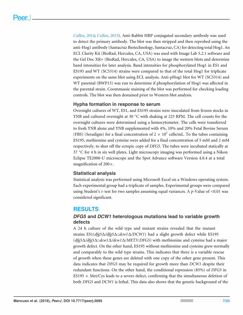
DFG5 and DCW1 mutants are unaffected by osmotic stressThe ability to withstand osmotic stress is critical for cell survival. Osmotic stress responseis regulated by HOG MAPK pathway in S. cerevisiae. The response to osmotic stress canbe tested by growing cells in the presence of 1 M sorbitol. Spotting assays were performedon YNB agar plates for wild type and mutant strains, in the presence or absence of 1 Msorbitol (Fig. 3). The YNB plates were prepared with methionine and cysteine. Spottingassays revealed that the growth of the mutants was similar under control (no stress plate)and stress (1 M sorbitol) conditions. Additionally, we performed growth assays to comparethe growth rates ofWT (SC5314),WT parental (BWP17), URA+ parental strain (DAY185),ES1, ES195 and ES195+Met/Cyst (conditional mutant) in the presence or absence of 1 Msorbitol over a 24 h period. After 24 h, both WT and mutant strains appeared to havesignificantly decreased growth rates in the presence of 1 M sorbitol as compared to control(no stress) cultures (Fig. 4). The ES195 strain without Met+Cys seemed to grow normallycompared to WT strains. Also the ES1 and the ES195 + Met/Cys (conditional mutant)strains seemed to have similar recovery rates in the presence of 1 M sorbitol as comparedto WT strain. This data indicates that the simultaneous deletion of DFG5 and DCW1 doesnot affect the ability to overcome osmotic stress, but the recovery to stress could possiblybe slower due to a growth defect. Additionally, this experiment further clearly shows thatthe parental strain BWP17 grows similar to WT (SC5314) strain in 1 M sorbitol and thusis not affected by its parental genetic background.
DFG5 and DCW1 mutants exhibit abnormal phenotypesLight microscopic imaging of mutants revealed abnormal phenotypes under controlconditions.DFG5 andDCW1mutants, ES1, ES195 and ES195 conditional mutant appearedto have a cell separation defect (Fig. 5A). This phenotype was somewhat similar to thehog11mutant which appeared to have a much more severe cell separation defect (Fig. 5A).Additionally, under osmotic stress conditions, the ES195 mutant appeared to have an
Mancuso et al. (2018), PeerJ, DOI 10.7717/peerj.5685 9/23
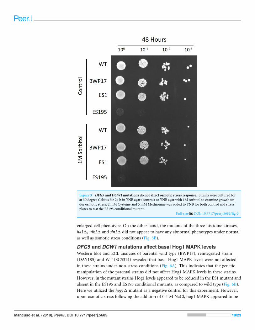
Figure 3 DFG5 andDCW1mutations do not affect osmotic stress response. Strains were cultured forat 30 degree Celsius for 24 h in YNB agar (control) or YNB agar with 1M sorbitol to examine growth un-der osmotic stress. 2 mM Cysteine and 5 mMMethionine was added to YNB for both control and stressplates to test the ES195 conditional mutant.
Full-size DOI: 10.7717/peerj.5685/fig-3
enlarged cell phenotype. On the other hand, the mutants of the three histidine kinases,hk11, nik11 and sln11 did not appear to have any abnormal phenotypes under normalas well as osmotic stress conditions (Fig. 5B).
DFG5 and DCW1 mutations affect basal Hog1 MAPK levelsWestern blot and ECL analyses of parental wild type (BWP17), reintegrated strain(DAY185) and WT (SC5314) revealed that basal Hog1 MAPK levels were not affectedin these strains under non-stress conditions (Fig. 6A). This indicates that the geneticmanipulation of the parental strains did not affect Hog1 MAPK levels in these strains.However, in the mutant strains Hog1 levels appeared to be reduced in the ES1 mutant andabsent in the ES195 and ES195 conditional mutants, as compared to wild type (Fig. 6B).Here we utilized the hog11 mutant as a negative control for this experiment. However,upon osmotic stress following the addition of 0.4 M NaCl, hog1 MAPK appeared to be
Mancuso et al. (2018), PeerJ, DOI 10.7717/peerj.5685 10/23
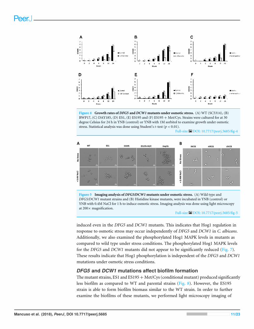
Figure 4 Growth rates ofDFG5 andDCW1mutants under osmotic stress. (A) WT (SC5314), (B)BWP17, (C) DAY185, (D) ES1, (E) ES195 and (F) ES195+Met/Cys. Strains were cultured for at 30degree Celsius for 24 h in YNB (control) or YNB with 1M sorbitol to examine growth under osmoticstress. Statistical analysis was done using Student’s t -test (p< 0.01).
Full-size DOI: 10.7717/peerj.5685/fig-4
Figure 5 Imaging analysis ofDFG5/DCW1mutants under osmotic stress. (A) Wild type andDFG5/DCW1mutant strains and (B) Histidine kinase mutants, were incubated in YNB (control) orYNB with 0.4M NaCl for 1 h to induce osmotic stress. Imaging analysis was done using light microscopyat 200×magnification.
Full-size DOI: 10.7717/peerj.5685/fig-5
induced even in the DFG5 and DCW1 mutants. This indicates that Hog1 regulation inresponse to osmotic stress may occur independently of DFG5 and DCW1 in C. albicans.Additionally, we also examined the phosphorylated Hog1 MAPK levels in mutants ascompared to wild type under stress conditions. The phosphorylated Hog1 MAPK levelsfor the DFG5 and DCW1 mutants did not appear to be significantly reduced (Fig. 7).These results indicate that Hog1 phosphorylation is independent of the DFG5 and DCW1mutations under osmotic stress conditions.
DFG5 and DCW1 mutations affect biofilm formationThemutant strains, ES1 and ES195+Met/Cys (conditional mutant) produced significantlyless biofilm as compared to WT and parental strains (Fig. 8). However, the ES195strain is able to form biofilm biomass similar to the WT strain. In order to furtherexamine the biofilms of these mutants, we performed light microscopy imaging of
Mancuso et al. (2018), PeerJ, DOI 10.7717/peerj.5685 11/23
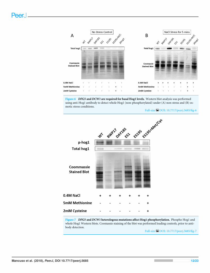
Figure 6 DFG5 andDCW1 are required for basal Hog1 levels. Western blot analysis was performedusing anti-Hog1 antibody to detect whole Hog1 (non-phosphorylated) under (A) non-stress and (B) os-motic stress conditions.
Full-size DOI: 10.7717/peerj.5685/fig-6
Figure 7 DFG5 andDCW1 heterologous mutations affect Hog1 phosphorylation. Phospho Hog1 andwhole Hog1 Western blots. Coomassie staining of the blot was performed loading controls, prior to anti-body detection.
Full-size DOI: 10.7717/peerj.5685/fig-7
Mancuso et al. (2018), PeerJ, DOI 10.7717/peerj.5685 12/23
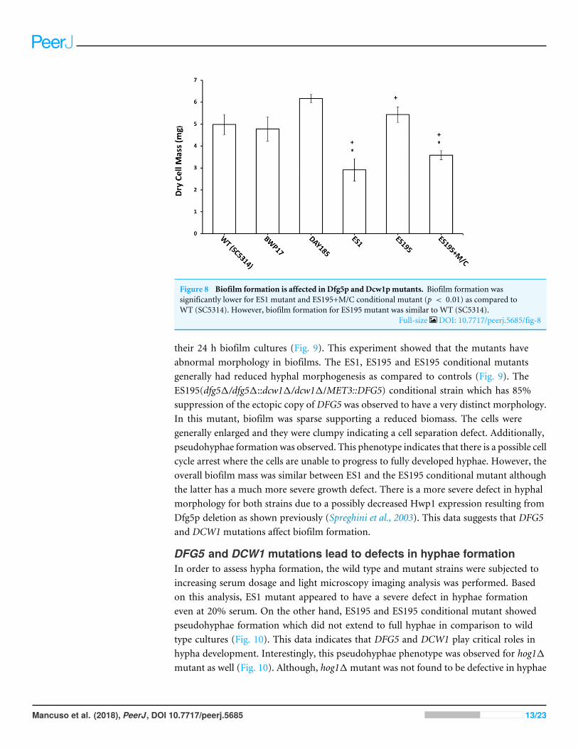
Figure 8 Biofilm formation is affected in Dfg5p and Dcw1pmutants. Biofilm formation wassignificantly lower for ES1 mutant and ES195+M/C conditional mutant (p < 0.01) as compared toWT (SC5314). However, biofilm formation for ES195 mutant was similar to WT (SC5314).
Full-size DOI: 10.7717/peerj.5685/fig-8
their 24 h biofilm cultures (Fig. 9). This experiment showed that the mutants haveabnormal morphology in biofilms. The ES1, ES195 and ES195 conditional mutantsgenerally had reduced hyphal morphogenesis as compared to controls (Fig. 9). TheES195(dfg51/dfg51::dcw11/dcw11/MET3::DFG5) conditional strain which has 85%suppression of the ectopic copy of DFG5 was observed to have a very distinct morphology.In this mutant, biofilm was sparse supporting a reduced biomass. The cells weregenerally enlarged and they were clumpy indicating a cell separation defect. Additionally,pseudohyphae formation was observed. This phenotype indicates that there is a possible cellcycle arrest where the cells are unable to progress to fully developed hyphae. However, theoverall biofilm mass was similar between ES1 and the ES195 conditional mutant althoughthe latter has a much more severe growth defect. There is a more severe defect in hyphalmorphology for both strains due to a possibly decreased Hwp1 expression resulting fromDfg5p deletion as shown previously (Spreghini et al., 2003). This data suggests that DFG5and DCW1 mutations affect biofilm formation.
DFG5 and DCW1 mutations lead to defects in hyphae formationIn order to assess hypha formation, the wild type and mutant strains were subjected toincreasing serum dosage and light microscopy imaging analysis was performed. Basedon this analysis, ES1 mutant appeared to have a severe defect in hyphae formationeven at 20% serum. On the other hand, ES195 and ES195 conditional mutant showedpseudohyphae formation which did not extend to full hyphae in comparison to wildtype cultures (Fig. 10). This data indicates that DFG5 and DCW1 play critical roles inhypha development. Interestingly, this pseudohyphae phenotype was observed for hog11mutant as well (Fig. 10). Although, hog11 mutant was not found to be defective in hyphae
Mancuso et al. (2018), PeerJ, DOI 10.7717/peerj.5685 13/23
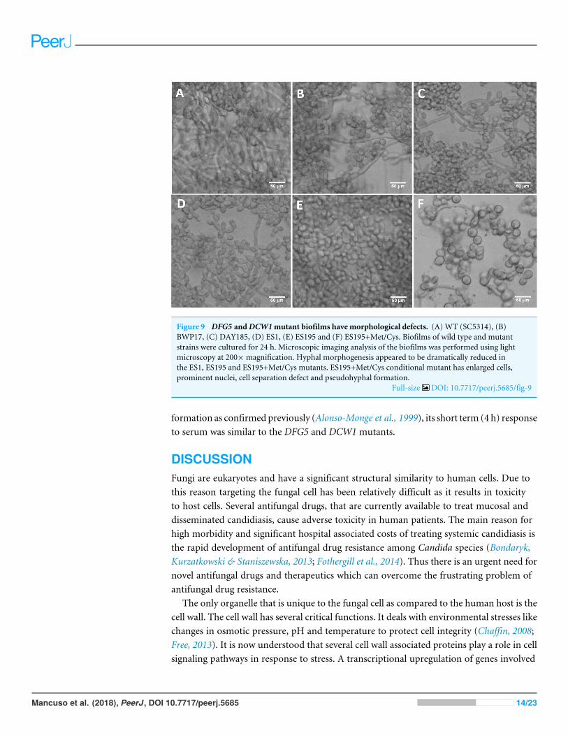
Figure 9 DFG5 andDCW1mutant biofilms have morphological defects. (A) WT (SC5314), (B)BWP17, (C) DAY185, (D) ES1, (E) ES195 and (F) ES195+Met/Cys. Biofilms of wild type and mutantstrains were cultured for 24 h. Microscopic imaging analysis of the biofilms was performed using lightmicroscopy at 200×magnification. Hyphal morphogenesis appeared to be dramatically reduced inthe ES1, ES195 and ES195+Met/Cys mutants. ES195+Met/Cys conditional mutant has enlarged cells,prominent nuclei, cell separation defect and pseudohyphal formation.
Full-size DOI: 10.7717/peerj.5685/fig-9
formation as confirmed previously (Alonso-Monge et al., 1999), its short term (4 h) responseto serum was similar to the DFG5 and DCW1 mutants.
DISCUSSIONFungi are eukaryotes and have a significant structural similarity to human cells. Due tothis reason targeting the fungal cell has been relatively difficult as it results in toxicityto host cells. Several antifungal drugs, that are currently available to treat mucosal anddisseminated candidiasis, cause adverse toxicity in human patients. The main reason forhigh morbidity and significant hospital associated costs of treating systemic candidiasis isthe rapid development of antifungal drug resistance among Candida species (Bondaryk,Kurzatkowski & Staniszewska, 2013; Fothergill et al., 2014). Thus there is an urgent need fornovel antifungal drugs and therapeutics which can overcome the frustrating problem ofantifungal drug resistance.
The only organelle that is unique to the fungal cell as compared to the human host is thecell wall. The cell wall has several critical functions. It deals with environmental stresses likechanges in osmotic pressure, pH and temperature to protect cell integrity (Chaffin, 2008;Free, 2013). It is now understood that several cell wall associated proteins play a role in cellsignaling pathways in response to stress. A transcriptional upregulation of genes involved
Mancuso et al. (2018), PeerJ, DOI 10.7717/peerj.5685 14/23
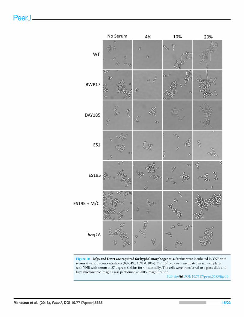
Figure 10 Dfg5 and Dcw1 are required for hyphal morphogenesis. Strains were incubated in YNB withserum at various concentrations (0%, 4%, 10% & 20%). 2× 10⁷ cells were incubated in six well plateswith YNB with serum at 37 degrees Celsius for 4 h statically. The cells were transferred to a glass slide andlight microscopic imaging was performed at 200×magnification.
Full-size DOI: 10.7717/peerj.5685/fig-10
Mancuso et al. (2018), PeerJ, DOI 10.7717/peerj.5685 15/23
in maintaining cell wall integrity occurs in response to signal transduction (Chaffin, 2008;Dichtl, Samantaray & Wagener, 2016). MAPK signaling pathways are among the varioussignaling pathways that regulate cell wall biogenesis and integrity. Most importantly, thecell wall plays a critical role in disease pathogenesis as well as in protecting the pathogenfrom the host immune system (Cullen & Edgerton, 2016).
The cell wall is a complex structure made of carbohydrates and cell wall mannoproteins.The carbohydrates form an extracellular matrix in which the mannoproteins are cross-linked. The cell wall proteins play important roles in cell physiology as well as in diseasepathogenesis. The extracellular matrix is also needed for biofilm formation. Biofilmformation is an important virulence factor for pathogenic fungi in causing local andsystemic disease (Costa-Orlandi et al., 2017). In C. albicans, various genes are involved inadhesion, extracellular matrix formation, quorum sensing and morphogenesis of biofilms(Fox & Nobile, 2012; Finkel & Mitchell, 2011).Moreover, inC. albicans the yeast and hyphaeforms have been found to have unique roles in biofilm formation (Finkel & Mitchell, 2011).
The cell wall proteins, Dfg5p andDcw1p, are predictedmannosidases/glycosyl hydrolases(gh-76 family). They have been implicated in the cross-linking of cell wall proteins in the cellwall. There are three known ways of cross-linking cell wall proteins in the cell wall matrixof C. albicans: (1) by a possible Dfg5p/Dcw1p-mediated cross-linking of N-linked outerchain mannan to the cell wall glucans described in this application, (2) by cross-linkingthe GPI anchor to the cell wall glucans through an alkaline-sensitive linkage, and (3) PIR(proteins with internal repeats) can be cross-linked into the cell walls by a linkage betweena glutamine residue in the PIR repeat and β-1,3-glucan (Free, 2013; Xie & Lipke, 2010). Thisredundancy in cell wall protein cross-linking is thought to help ensure the formation of afunctional cell wall and maintain its integrity. However, the exact enzymatic mechanismsof Dfg5p and Dcw1p have not been demonstrated experimentally.
In this study we examined the functions of Dfg5p and Dcw1p in biofilm formation andHog1MAPK signaling by using heterologousmutant strains and conditionalmutant strains(Table 1). Using the aboveC. albicansmutant strains we have previously confirmed that theDfg5p andDcw1pmannanases (cross-linking enzymes) function in cell wall biogenesis inC.albicans (Ao et al., 2015).We have also showed previously that theDFG5/DCW1 conditionalmutants have a cell separation growth phenotype. The mutants are hypersensitive tocell wall stress reagents and to treatment with lyticase (β-glucanase), indicating thatthe mutant cell walls are weaker than wild type cell walls (Ao et al., 2015). The DFG5and DCW1 mutants produced cell walls containing reduced levels of cell wall proteinsand released cell wall proteins into the growth medium. A carbohydrate analysis of theDFG5/DCW1mutant cell walls showed that the mannose levels were significantly reduced,indicating a reduced incorporation of cell wall proteins in the wall (Ao et al., 2015). Thesecharacteristics are similar to our observations of the Neurospora crassa DFG51/dcw11mutants, and demonstrate that DFG5 and DCW1 in C. albicans function in cross-linkingcell wall proteins into the cell wall. However, the substrates of these enzymes and their exactmechanisms of cross-linking cell wall proteins into the cell wall are yet to be determined.But structural studies of the α-mannanases (GH-76 enzymes), have revealed that these
Mancuso et al. (2018), PeerJ, DOI 10.7717/peerj.5685 16/23
enzymes are mainly involved in catalysis and could be potential targets for anti-fungal drugdevelopment (Thompson et al., 2015).
We hypothesized that the lethality of the dfg51/dcw11 mutants in S. cerevisiae and C.albicans may be a manifestation of additional roles that Dfg5p and Dcw1p play in signaltransductionpathways. This has beendemonstrated especially in S. cerevisiae. Recent studieshave shown that the Hog1 and Slt2 signaling pathways are activated in the dfg51 mutantof S. cerevisiae (Nasution et al., 2015). It was also demonstrated in the same study that theexpression of genes related to these signaling pathways was altered in the dfg51 mutantof S. cerevisiae, by using RNA sequencing analysis (Nasution et al., 2015). Our preliminarystudies have focused on C. albicans DFG5 andDCW1 functions at pH 7 which is commonlypresent in the oral cavity. Also ES195 mutant has defective growth patterns under variouscell wall stress conditions but exhibit elevated heat tolerance, which is concurrent withstudies in S. cerevisiae (Fig. 2). The defect in the ability to overcome cell wall stress couldbe the result of a weakened cell wall. However, growth of the mutants was not affected byosmotic stress (1 M Sorbitol) (Figs. 3 and 4). Phenotypic analysis of the DFG5 and DCW1mutants indicated a defect in cell separation which was similar to hog11 mutant undernon-stress conditions (Fig. 5). However, there were minor differences in phenotypes of themutants with and without osmotic stress, indicating that the DFG5 and DCW1 mutationsare not indispensable for osmotic stress. We further investigated whether Hog1 MAPKlevels were altered in the mutants and found that the basal Hog1 MAPK levels were eitherreduced or absent in the DFG5 and DCW1mutants (Fig. 6A). However, upon 0.4 M NaClstress, Hog1 MAPK was induced in the DFG5 and DCW1 mutants (Fig. 6B). We furtherinvestigated if Hog1 phosphorylation is being affected in these mutants under osmoticstress and found that p-hog1 was slightly reduced in the mutants but not significantly(Fig. 6B) confirming that the DFG5 and DCW1 mutations may not affect osmotic stressresponse via HOG pathway. Taken together, this data indicates that DFG5 and DCW1 arenot required forHOGdependent osmotic stress response. However, by affecting basal Hog1MAPK levels, DFG5 and DCW1may possibly regulate other important functions of Hog1.
Interestingly the HOG pathway under normal or non-stress conditions plays a criticalrole in chitin synthesis (Munro et al., 2007). C. albicans has four genes encoding chitinsynthases—CHS1, CHS2, CHS3 and CHS8 with redundant functions in order to ensurecell wall fitness. Of these CHS1, CHS3 and CHS8 have ATF/CREB elements for Sko1transcription factor (Munro et al., 2007). Studies in both S. cerevisiae and C. albicans haveshown that hog1 MAPK regulates sko1 transcription factor (Proft et al., 2005;Munro et al.,2007). It was also found that in the hog11 mutant, CHS1, CHS2 and CHS8 transcriptionwas downregulated and CHS3 was upregulated (Munro et al., 2007). Calcoflour white is acell wall stress agent that inhibits chitin synthesis and thus causes a compensatory increasein transcriptional regulation of chitin synthases. In this study we show that the ES1 andES195 conditional mutant are unable to recover by 48 h in the presence of Calcoflour whiteas compared to wild type (Fig. 2). This could be due to the defect in Hog1 basal levels inthe DFG5 and DCW1mutants. This indicates that DFG5 and DCW1mutations may affectchitin synthesis via HOG MAPK pathway. This could further lead to a weakened cell walland increased susceptibility to cell wall perturbing agents.
Mancuso et al. (2018), PeerJ, DOI 10.7717/peerj.5685 17/23
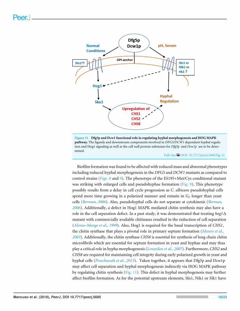
Figure 11 Dfg5p and Dcw1 functional role in regulating hyphal morphogenesis and HOGMAPKpathway. The ligands and downstream components involved in DFG5/DCW1 dependent hyphal regula-tion and Hog1 signaling as well as the cell wall protein substrates for Dfg5p and Dcw1p are to be deter-mined.
Full-size DOI: 10.7717/peerj.5685/fig-11
Biofilm formation was found to be affected with reducedmass and abnormal phenotypesincluding reduced hyphal morphogenesis in theDFG5 andDCW1mutants as compared tocontrol strains (Figs. 8 and 9). The phenotype of the ES195+Met/Cys conditional mutantwas striking with enlarged cells and pseudohyphae formation (Fig. 9). This phenotypepossibly results from a delay in cell cycle progression as C. albicans pseudohyphal cellsspend more time growing in a polarized manner and remain in G2 longer than yeastcells (Berman, 2006). Also, pseudohyphal cells do not separate at cytokinesis (Berman,2006). Additionally, a defect in Hog1 MAPK mediated chitin synthesis may also have arole in the cell separation defect. In a past study, it was demonstrated that treating hog11mutant with commercially available chitinases resulted in the reduction of cell separation(Alonso-Monge et al., 1999). Also, Hog1 is required for the basal transcription of CHS1,the chitin synthase that plays a pivotal role in primary septum formation (Munro et al.,2003). Additionally, the chitin synthase CHS8 is essential for synthesis of long chain chitinmicrofibrils which are essential for septum formation in yeast and hyphae and may thusplay a critical role in hyphamorphogenesis (Lenardon et al., 2007). Furthermore,CHS2 andCHS8 are required for maintaining cell integrity during early polarized growth in yeast andhyphal cells (Preechasuth et al., 2015). Taken together, it appears that Dfg5p and Dcw1pmay affect cell separation and hyphal morphogenesis indirectly via HOG MAPK pathwayby regulating chitin synthesis (Fig. 11). This defect in hyphal morphogenesis may furtheraffect biofilm formation. As for the potential upstream elements, Sln1, Nik1 or Hk1 have
Mancuso et al. (2018), PeerJ, DOI 10.7717/peerj.5685 18/23
been shown to be independent of Hog1 under osmotic stress (Yamada-Okabe et al., 1999;Román, Nombela & Pla, 2005). Yet the mutants of these histidine kinases have a defect inhyphal morphogenesis. Thus, Sln1, Nik1 and Hk1 may be involved in regulating Hog1MAPK under non-stress conditions or in response to serum, pH etc as the mutants of thesehistidine kinases have defects in hyphal morphogenesis (Yamada-Okabe et al., 1999).
CONCLUSIONSWe conclude that Dfg5p and Dcw1p have distinct functions. While Dfg5p is importantfor hyphal morphogenesis, Dcw1p could be important for maintaining cell wall integrity.Additionally, these cell wall mannosidases may function in biofilm formation via hyphalregulation and also activate the HOG MAPK pathway to possibly regulate chitin synthesisas depicted in our working model (Fig. 11). As Hog1 is thought to be downstream ofSln1, Nik1 and Hk1, the Dfg5p and Dcw1p cell wall mannosidases could be interactingwith these upstream elements. This interaction may involve enzymatic modifications ofglycosylation on the osmosensing histidine kinases like Sln1, Nik1 and Hk1 resulting insignal transduction. Located in the cell wall and having enzymatic functions, Dfg5p andDcw1p are readily accessible to therapeutic agents and could be ideal targets for novelantifungal drugs. Thus, it is not only necessary but novel to further investigate the signalingfunctions of these GPI-anchored cell wall enzymes. Our future studies will focus on theenzymatic mechanisms of these mannosidases.
ACKNOWLEDGEMENTSWewould like to thank Dr. Paul Cullen, Associate Professor, Biological Sciences, Universityat Buffalo, for kindly providing us the antibodies against hog1 and phog1. We would alsolike to thank Mr. Jason Chwirut, University at Buffalo for preparing the figures in thismanuscript.
ADDITIONAL INFORMATION AND DECLARATIONS
FundingThe authors received no funding for this work.
Competing InterestsThe authors declare there are no competing interests.
Author Contributions• Ryan Mancuso, Jennifer Chinnici, Charlene Tsou, Sujay Busarajan and RaveenaMunnangi conceived and designed the experiments, performed the experiments,analyzed the data, contributed reagents/materials/analysis tools, prepared figures and/ortables, authored or reviewed drafts of the paper, approved the final draft.• AbhiramMaddi conceived and designed the experiments, analyzed the data, contributedreagents/materials/analysis tools, prepared figures and/or tables, authored or revieweddrafts of the paper, approved the final draft.
Mancuso et al. (2018), PeerJ, DOI 10.7717/peerj.5685 19/23
Data AvailabilityThe following information was supplied regarding data availability:
The raw data are included in the Supplemental Files.
Supplemental InformationSupplemental information for this article can be found online at http://dx.doi.org/10.7717/peerj.5685#supplemental-information.
REFERENCESAdhikari H, Cullen PJ. 2014.Metabolic respiration induces AMPK- and Ire1p-
dependent activation of the p38-Type HOGMAPK pathway. PLOS Genetics10(10):e1004734 DOI 10.1371/journal.pgen.1004734.
Alonso-Monge R, Navarro-García F, Molero G, Diez-Orejas R, Gustin M, Pla J,SánchezM, Nombela C. 1999. Role of the mitogen-activated protein kinaseHog1p in morphogenesis and virulence of Candida albicans. Journal of Bacteriology181(10):3058–3068.
Ao J, Chinnici JL, Maddi A, Free SJ. 2015. The N-linked outer chain mannans andthe dfg5p and dcw1p endo-α-1, 6-mannanases are needed for incorporation ofCandida albicans glycoproteins into the cell wall. Eukaryotic Cell 14:792–803DOI 10.1128/EC.00032-15.
Arana DM, Alonso-Monge R, Du C, Calderone R, Pla J. 2007. Differential susceptibilityof mitogen-activated protein kinase pathway mutants to oxidative-mediated killingby phagocytes in the fungal pathogen Candida albicans. Cellular Microbiology9(7):1647–1659 DOI 10.1111/j.1462-5822.2007.00898.x.
Berman J. 2006.Morphogenesis and cell cycle progression in Candida albicans. CurrentOpinion in Microbiology 9(6):595–601 DOI 10.1016/j.mib.2006.10.007.
BondarykM, KurzątkowskiW, StaniszewskaM. 2013. Antifungal agents commonlyused in the superficial and mucosal candidiasis treatment: mode of action andresistance development. Advances in Dermatology and Allergology 30:293–301DOI 10.5114/pdia.2013.38358.
Brewster JL, Gustin MC. 2014.Hog1: 20 years of discovery and impact. Science Signaling7(343):re7 DOI 10.1126/scisignal.2005458.
Cassone A, Cauda R. 2012. Candida and candidiasis in HIV-infected patients: wherecommensalism, opportunistic behavior and frank pathogenicity lose their borders.AIDS 26:1457–1472 DOI 10.1097/QAD.0b013e3283536ba8.
ChaffinWL. 2008. Candida albicans cell wall proteins.Microbiology and MolecularBiology Reviews 72:495–544 DOI 10.1128/MMBR.00032-07.
Cheetham J, Smith DA, Da Silva Dantas A, Doris KS, PattersonMJ, Bruce CR,Quinn J. 2007. A single MAPKKK regulates the Hog1 MAPK pathway in thepathogenic fungus Candida albicans.Molecular Biology of the Cell 18(11):4603–4614DOI 10.1091/mbc.e07-06-0581.
Costa-Orlandi CB, Sardi JCO, Pitangui NS, De Oliveira HC, Scorzoni L, GaleaneMC,Medina-Alarcón KP, MeloWCMA,MarcelinoMY, Braz JD, Fusco-Almeida AM,
Mancuso et al. (2018), PeerJ, DOI 10.7717/peerj.5685 20/23
Mendes-Giannini MS. 2017. Fungal biofilms and polymicrobial diseases. Journal ofFungi 3(2):E22 DOI 10.3390/jof3020022.
Cullen PJ. 2015. Evaluating the activity of the filamentous growth mitogen-activatedprotein kinase pathway in yeast. Cold Spring Harbor Protocols 3:276–283.
Cullen PJ, EdgertonM. 2016. Unmasking fungal pathogens by studying MAPK-dependent cell wall regulation in Candida albicans. Virulence 7(5):502–505DOI 10.1080/21505594.2016.1177695.
Dichtl K, Samantaray S, Wagener J. 2016. Cell wall integrity signalling in humanpathogenic fungi. Cellular Microbiology 18:1228–1238 DOI 10.1111/cmi.12612.
Finkel JS, Mitchell AP. 2011. Genetic control of Candida albicans biofilm development.Nature Reviews. Microbiology 9:109–118 DOI 10.1038/nrmicro2475.
Fothergill AW, Sutton DA, McCarthy DI, Wiederhold NP. 2014. Impact of newantifungal breakpoints on antifungal resistance in Candida species. Journal of ClinicalMicrobiology 52:994–997 DOI 10.1128/JCM.03044-13.
Fox EP, Nobile CJ. 2012. A sticky situation: untangling the transcriptional networkcontrolling biofilm development in Candida albicans. Transcription 3:315–322DOI 10.4161/trns.22281.
Free SJ. 2013. Fungal cell wall organization and biosynthesis. Advances in Genetics81:33–82 DOI 10.1016/B978-0-12-407677-8.00002-6.
Heilmann CJ, Sorgo AG, Mohammadi S, Sosinska GJ, De Koster CG, Brul S, De KoningLJ, Klis FM. 2013. Surface stress induces a conserved cell wall stress responsein the pathogenic fungus Candida albicans. Eukaryotic Cell 12(2):254–264DOI 10.1128/EC.00278-12.
Kitagaki H, Ito K, Shimoi H. 2004. A temperature-sensitive dcw1 mutant of Saccha-romyces cerevisiae is cell cycle arrested with small buds which have aberrant cell walls.Eukaryotic Cell 3:1297–1306 DOI 10.1128/EC.3.5.1297-1306.2004.
Kitagaki H,WuH, Shimoi H, Ito K. 2002. Two homologous genes, DCW1 (YKL046c)and DFG5, are essential for cell growth and encode glycosylphosphatidylinositol(GPI)-anchored membrane proteins required for cell wall biogenesis in Saccha-romyces cerevisiae.Molecular Microbiology 46:1011–1022DOI 10.1046/j.1365-2958.2002.03244.x.
KruppaM, Calderone R. 2006. Two-component signal transduction in human fungalpathogens. FEMS Yeast Research 6(2):149–159 DOI 10.1111/j.1567-1364.2006.00024.x.
LenardonMD,Whitton RK, Munro CA, Marshall D, GowNAR. 2007. Individualchitin synthase enzymes synthesize microfibrils of differing structure at specificlocations in the Candida albicans cell wall.Molecular Microbiology 66:1164–1173DOI 10.1111/j.1365-2958.2007.05990.x.
Li R, Kumar R, Tati S, Puri S, EdgertonM. 2013. Candida albicans flu1-mediated effluxof salivary histatin 5 reduces its cytosolic concentration and fungicidal activity.Antimicrobial Agents and Chemotherapy 57:1832–1839 DOI 10.1128/AAC.02295-12.
Maddi A, Fu C, Free SJ. 2012. The Neurospora crassa dfg5 and dcw1 genes encode α-1,6-mannanases that function in the incorporation of glycoproteins into the cell wall.PLOS ONE 7(6):e38872 DOI 10.1371/journal.pone.0038872.
Mancuso et al. (2018), PeerJ, DOI 10.7717/peerj.5685 21/23
Munro CA, Selvaggini S, De Bruijn I, Walker L, LenardonMD, Gerssen B, Milne S,Brown AJ, GowNAR. 2007. The PKC, HOG and Ca2+ signalling pathways co-ordinately regulate chitin synthesis in Candida albicans.Molecular Microbiology63:1399–1413 DOI 10.1111/j.1365-2958.2007.05588.x.
Munro CA,Whitton RK, Hughes HB, Rella M, Selvaggini S, GowNAR. 2003. CHS8—afourth chitin synthase gene of Candida albicans contributes to in vitro chitin synthaseactivity, but is dispensable for growth. Fungal Genetics and Biology 40:146–158DOI 10.1016/S1087-1845(03)00083-5.
Nagahashi S, Mio T, Ono N, Yamada-Okabe T, ArisawaM, Bussey H, Yamada-OkabeH. 1998. Isolation of CaSLN1 and CaNIK1, the genes for osmosensing histidinekinase homologues, from the pathogenic fungus Candida albicans.Microbiology144(Pt 2):425–432.
Nasution O, Lee J, Srinivasa K, Choi IG, Lee YM, Kim E, ChoiW, KimW. 2015.Loss of DFG5 glycosylphosphatidylinositol-anchored membrane protein confersenhanced heat tolerance in Saccharomyces cerevisiae. Environmental Microbiology17:2721–2734 DOI 10.1111/1462-2920.12649.
Nguyen KA, Zmeter G, Claris O, Kassai B. 2012. Epidemiology of invasive Candidainfection in a neonatal intensive care unit in France. Acta Paediatrica 101:e137–9.
Nikolaou E, Agrafioti I, StumpfM, Quinn J, Stansfield I, Brown AJ. 2009. Phylogeneticdiversity of stress signaling pathways in fungi. BMC Evolutionary Biology 9:44DOI 10.1186/1471-2148-9-44.
Noble SM, Gianetti BA,Witchley JN. 2017. Candida albicans cell-type switchingand functional plasticity in the mammalian host. Nature Reviews. Microbiology15(2):96–108 DOI 10.1038/nrmicro.2016.157.
Preechasuth K, Anderson JC, Peck SC, Brown AJ, GowNA, LenardonMD. 2015. Cellwall protection by the Candida albicans class I chitin synthases. Fungal Genetics andBiology 82:264–276 DOI 10.1016/j.fgb.2015.08.001.
Proft M, Gibbons FD, CopelandM, Roth FP, Struhl K. 2005. Genomewide identificationof Sko1 target promoters reveals a regulatory network that operates in responseto osmotic stress in Saccharomyces cerevisiae. Eukaryotic Cell 4(8):1343–1352DOI 10.1128/EC.4.8.1343-1352.2005.
Román E, Nombela C, Pla J. 2005. The Sho1 adaptor protein links oxidative stress tomorphogenesis and cell wall biosynthesis in the fungal pathogen Candida albicans.Molecular and Cellular Biology 25(23):10611–10627DOI 10.1128/MCB.25.23.10611-10627.2005.
Spreghini E, Davis DA, Subaran R, KimM,Mitchell AP. 2003. Roles of Candida albicansDfg5p and Dcw1p cell surface proteins in growth and hypha formation. EukaryoticCell 2:746–755 DOI 10.1128/EC.2.4.746-755.2003.
Sudbery PE. 2011. Growth of Candida albicans hyphae. Nature Reviews. Microbiology9(10):737–748 DOI 10.1038/nrmicro2636.
Thompson AJ, Speciale G, Iglesias-Fernández J, Hakki Z, Belz T, Cartmell A, SpearsRJ, Chandler E, Temple MJ, Stepper J, Gilbert HJ, Rovira C,Williams SJ, DaviesGJ. 2015. Evidence for a boat conformation at the transition state of GH76 α-1,
Mancuso et al. (2018), PeerJ, DOI 10.7717/peerj.5685 22/23

6-mannanases—key enzymes in bacterial and fungal mannoprotein metabolism.Angewandte Chemie 54:5378–5382 DOI 10.1002/anie.201410502.
Veses V, GowNA. 2009. Pseudohypha budding patterns of Candida albicans.MedicalMycology 47(3):268–275 DOI 10.1080/13693780802245474.
Xie X, Lipke PN. 2010. On the evolution of fungal and yeast cell walls. Yeast 27:479–488DOI 10.1002/yea.1787.
Yamada-Okabe T, Mio T, Ono N, Kashima Y, Matsui M, ArisawaM, Yamada-Okabe H.1999. Roles of three histidine kinase genes in hyphal development and virulence ofthe pathogenic fungus Candida albicans. Journal of Bacteriology 181(23):7243–7247.
Mancuso et al. (2018), PeerJ, DOI 10.7717/peerj.5685 23/23