brain and cognition - neurociencia cognitiva · brain activity (14 compassion-eliciting pictures...
TRANSCRIPT
Brain and Cognition xxx (2011) xxx–xxx
Contents lists available at ScienceDirect
Brain and Cognition
journal homepage: www.elsevier .com/ locate /b&c
Perception of suffering and compassion experience: Brain gender disparities
Roberto E. Mercadillo a, José Luis Díaz b, Erick H. Pasaye a,c, Fernando A. Barrios a,⇑a Institute of Neurobiology, Universidad Nacional Autónoma de México, Querétaro, Mexicob School of Medicine, Universidad Nacional Autónoma de México, México D.F., Mexicoc National Institute of Neurology and Neurosurgery ‘‘Manuel Velasco Suárez’’, México D.F., Mexico
a r t i c l e i n f o a b s t r a c t
Article history:Accepted 24 March 2011Available online xxxx
Keywords:EmotionCognitionMoralCompassionEmpathyGenderFunctional imagingSocial neuroscienceNurtureCulture
0278-2626/$ - see front matter � 2011 Elsevier Inc. Adoi:10.1016/j.bandc.2011.03.019
⇑ Corresponding author. Address: Institute of NeuroAutónoma de México, Campus UNAM Juriquilla, BlvdQRO 76230, Mexico. Fax: +52 442 238 1046.
E-mail address: [email protected] (F.A. Barrios
Please cite this article in press as: Mercadillo, R(2011), doi:10.1016/j.bandc.2011.03.019
Compassion is considered a moral emotion related to the perception of suffering in others, and resultingin a motivation to alleviate the afflicted party. We compared brain correlates of compassion-evokingimages in women and men. BOLD functional images of 24 healthy volunteers (twelve women and twelvemen; age = 27 ± 2.5 y.o.) were acquired in a 3T magnetic resonance scanner while subjects viewed pic-tures of human suffering previously verified to elicit compassion and indicated their compassionateexperience by finger movements. Functional analysis revealed that while women manifested activationin areas involved in basic emotional, empathic, and moral processes, such as basal regions and cingulateand frontal cortices, activation in men was restricted mainly to the occipital cortex and parahippocampalgyrus. These findings suggest that compassion and its moral elements constitute gender-relative subjec-tive phenomena emerging from differently evolved neural mechanisms and socially learned features pos-sibly related to nurturing skills.
� 2011 Elsevier Inc. All rights reserved.
1. Introduction
The interactions between moral and emotional cognition havebeen recently studied in terms of moral emotions, which refer toa kind of emotions elicited during the perception of social trans-gressions encoded in attitudes and beliefs, and frequently implyan empathic component to infer the mental states in others(Bennett & Matthews, 2000; Kagan, 2005; Moll, De Oliveira-Souza,& Zahn, 2008; Nichols, 2002). In contrast to basic emotions that areelicited by stimuli that motivate clearly adaptive behaviors, such asfleeing in response to fear or attack elicited by anger (Ekman, 1993;Izard, 1992), moral emotions are related to the welfare of society asa whole or of individuals different from the person who experi-ences the emotion (Haidt, 2003).
Neurobiological approaches to moral emotions and reasoninghave applied neuroimaging techniques to identify regional brainactivity while the subjects process visual scenes, auditory state-ments, or moral dilemmas. The findings indicate activation in sub-cortical regions related to the experience of basic emotions, such asthe amygdala, thalamus, insular cortex, and upper midbrain, butalso in cortical regions linked to complex cognition, such as the
ll rights reserved.
biology, Universidad Nacional. Juriquilla 3001, Querétaro,
).
. E., et al. Perception of suffering
prefrontal and orbitofrontal cortices and the superior temporal sul-cus (Casebeer, 2003; Greene & Haidt, 2002; Moll, de Oliveira-Sou-za, Bramati, & Grafman, 2002; Moll & Schulkin, 2009; Young &Koenigs, 2007).
Because it is strongly related to social cooperation (Damasio,2006), compassion can be considered a moral emotion usually elic-ited by witnessing the suffering of others and resulting in a moti-vation to alleviate their perceived affliction (Haidt, 2003; Lazarus,1991). Neuroimaging studies focused in the experience of compas-sionate attitudes and decisions of prosocial actions report neuralactivity related with theory of mind, empathy, and decision mak-ing involving the medial and inferior prefrontal cortex, temporo-parietal junction, cingulate cortex, insula and midbrain (Kedia,Berthoz, Wessa, Hilton, & Martinot, 2008; Kim et al., 2009; Lutz,Brefczynski-Lewis, Johnstone, & Davidson, 2008; Moll et al., 2007).
In a recent validation of compassion-evoking pictures, Merca-dillo and coworkers found that compassion experience includesattributes of basic negative emotions, such as displeasure and higharousal. Both women and men reported similar appraisals relatedto valence, arousal, and dominance when viewing compassion-evoking pictures representing human suffering in different con-texts. Nevertheless, women’s appraisals indicated a more intenseexperience of compassion and arousal when the compassion-evok-ing pictures depicted scenes of illness in particular, and reportedlower dominance and higher arousal than men when viewing so-cially neutral pictures used as control stimuli (Mercadillo, Barrios,& Diaz, 2007).
and compassion experience: Brain gender disparities. Brain and Cognition
2 R.E. Mercadillo et al. / Brain and Cognition xxx (2011) xxx–xxx
Although gender approaches are not reported in the neurobiol-ogy of compassion, some neuropsychological studies have showngender differences in moral brain processing, emotional experi-ence, and cognitive functions in which women manifest a patternof brain activity related with emotional experience and strategiesin resting electroencephalographic recordings (Jausovec & Jauso-vec, 2005). Also, women exhibit stronger defensive reactivity thanmen when viewing aversive pictures (Bradley, Codispoti, Cuthbert,& Lang, 2001) and report less control over the experience related toviewing pictures representing moral transgressions, such as inten-tion to harm and frustration of goals (Javela, Mercadillo, & Ramirez,2008). Neurobiological approaches report that women manifest amore intense activation of emotional-control brain areas whenprocessing memories or sounds, while men manifest activation ofcortical cognitive-control related areas (Canli, Desmond, Zhao, &Gabrieli, 2002; Flores-Gutierrez et al., 2009; Koch et al., 2007).Men exhibit more activation in frontal and temporal regions re-lated to social learning and emotional concepts when viewing fa-cial expressions depicting contempt, while women manifestactivation in the insula when faces represent disgust (Aleman &Swart, 2008). Also, it has been suggested that women exhibit amore intense empathic sensitivity when viewing chimerical faces(Rueckert & Naybar, 2008) and moral-emotional understandingwhen perceiving unpleasant pictures, as inferred from their activ-ity in insular and cingulate cortices (Harenski, Antonenko, Shane, &Kiehl, 2008). In reference to moral reasoning, women profuselyelaborate moral judgments oriented to the care of others, whereasmen tend to adjust their assessment based on a sense of duty(Bjorklund, 2003; Self & Olivarez, 1993). It has also been reportedthat women are more empathic in conflict-resolution situations,and that men manifest a lower activation of brain areas relatedto empathy when they observe a painful stimulus received by aperson known to be unfair (de Wied, Branje, & Meeus, 2007; Singeret al., 2006).
Biological research on cognitive and emotional processes re-quires consistency between subjective reports and their relatedphysiological changes (Mauss, Levenson, McCarter, Wilhelm, &Gross, 2005). Because women report more intense compassionand emotional appraisals related to emotions when viewing ges-tures and scenes of suffering, neurophysiological differences canbe expected. The aim of the present study was to analyze genderdifferences in subjective compassion responses and their neurobio-logical foundations. We expected that, in response to experimen-tally evoked compassion experiences, women would showgreater and more diverse neural activation than men in brain re-gions related to emotional experience, empathy, and prosocial ac-tions such as prefrontal and cingulate cortex, insula, and temporo–parietal junction.
2. Materials and methods
2.1. Preceding behavioral data and pilot study
In preparation for the functional MRI investigation, a behavioralstudy was applied using first-rate visual compassion-elicitingstimuli (Mercadillo, Barrios, & Diaz, 2007). Eighty-four pictureswere selected from the International Affective Picture System(IAPS) and rated, as proposed in this archive, in the dimensionsof valence, arousal, and dominance, where 1 indicates the lowestand 9 the highest rating. A compassion dimension was addedwhere a score of 1 represented null compassion and 9 the greatestcompassionate experience. With this method, 28 pictures evokinghigh compassion were obtained, and 28 pictures representing non-compassion-evoking stimuli were selected as controls, 14 depict-ing objects and landscapes and 14 showing customary social
Please cite this article in press as: Mercadillo, R. E., et al. Perception of suffering(2011), doi:10.1016/j.bandc.2011.03.019
scenes, such as people drinking coffee (information about the rat-ings of valence, arousal, dominance and compassion dimensions ineach visual stimulus are presented in Tables 1–12 of the Supple-mentary material).
Prior to the MRI scanning presented in this research, six volun-teers participated in a pilot study conducted to determine theappropriate mode to obtain the emotional appraisals during thefMRI session. Since the participants reported difficulty in remem-bering the 1–9 appraisal range and expressing the experience ofmore than one emotional dimension when viewing each picture,we decided to record only the absence or presence of compassion-ate feelings when viewing each picture. The subjects were in-structed to press a key with one finger when they experienced acompassionate feeling or with the contralateral finger if they didnot. The reaction time results obtained from the E-Prime responserecord (E-Prime responses achievement, Psychology SoftwareTools, Inc. Pittsburgh PA.) indicated that viewing the picture fortwo seconds was enough to produce the requested appraisal signal.Since repeated viewing of an image likely reduces the reportedemotional experience, we decided to obtain the response only onceduring the MRI scanning to avoid habituation to the stimuli.
2.2. Participants
A different set of 24 healthy subjects in the same age and edu-cational range as the behavioral study volunteers (twelve womenand twelve men, mean age (M) = 27, standard deviation(s.d.) = 2.5 and mean of 15 years of education) participated in thefMRI sessions. In agreement with the procedures authorized bythe Institutional Review Board (IRB), the ethical principles recom-mended by the American Psychological Association (O’Donohue &Ferguson, 2003), and the World Medical Association Declaration ofHelsinki, all subjects gave written informed consent after the nat-ure of the experiment was explained to them. Participants werestrongly right-handed as measured by the Edinburgh HandednessInventory and in good general health as confirmed by a clinicalinterview. The Mexican electronic version of the Symptom CheckList 90 (González-Santos, Mercadillo, Graff, & Barrios, 2007) anda clinical interview verified the absence of current neurologicaland mental disorders. No subject was taking any medication orwas paid for participating. As was already pointed out, no partici-pant had previously seen the pictures they viewed during thestudy. All the imaging studies were conducted at the NeuroimagingUnit of the National Institute of Neurology and Neurosurgery, Mex-ico City.
2.3. Task parameters
The functional MRI session consisted of series conformed by100 pictures alternating compassion- and non-compassion-evok-ing stimuli selected from a validated set of the International Affec-tive Picture System (Lang, Bradley, & Curberth, 2005; Mercadillo,Barrios, & Diaz, 2007). Since compassion was reported by viewingexpressions or situations manifested in social contexts, it is neces-sary to distinguish the neurocognitive functions related with thevisual analysis of social context that may not involve compassion-ate processes per se. Thus, we decided to present two functionalruns of visual stimuli contrasting compassion with both non-socialand social conditions in an event-related design. The Compassion-Objects series contrasted the compassion-correlated brain activity(14 compassion-eliciting pictures depicting suffering in differentcontexts such as war scenes, sad facial expressions, and famine)from the activity related with viewing non-human scenes (14 emo-tionally neutral pictures representing objects or landscapes thatwere repeated to form a neutral background of 86 stimuli). TheCompassion-Social series contrasted the compassion-correlated
and compassion experience: Brain gender disparities. Brain and Cognition
R.E. Mercadillo et al. / Brain and Cognition xxx (2011) xxx–xxx 3
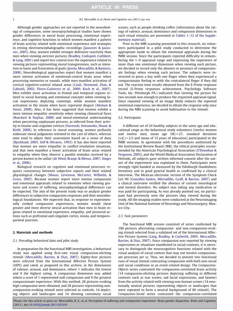
brain activity (14 compassion-eliciting pictures depicting sufferingin different contexts) from the activity related with viewing non-compassion-evoking social scenes (14 emotionally neutral socialpictures such as people walking or waiting for the bus repeatedto form a socially neutral background of 86 stimuli). Compassion-and non-compassion-evoking stimuli used in one series were to-tally different than the stimuli used in the other series and werepseudo-randomly distributed so that compassion-evoking pictureswere separated by 12–25 s. Pictures were projected centrally every2500 ms followed by a fixation cross for 500 ms. The sequence isillustrated in Fig. 1.
Participants saw the stimuli through the IFIS Presentation Re-sponse System (In Vivo Inc., Orlando, FL) screen situated in frontof their eyes over the head coil. The presentation-response para-digm was executed and synchronized using E-Prime (PsychologySoftware Tools, Inc., Pittsburg, PA). The presentation was synchro-nized in the same manner for all the subjects.
The IFIS response system was utilized to record the subjects’ re-sponses, verifying a compassionate/non-compassionate experienceelicited during the observation of each of the pictures. In order toneutralize the average brain activation areas related to finger mo-tor responses, 12 of the participants (six women, six men) were in-structed to press a button with their right index finger if theyexperienced a compassionate feeling and a button with their leftindex finger in the absence of a compassionate feeling. The other12 participants were instructed to respond in an inverse manner.In order to verify the proper understanding of the task, participantspracticed in a mock procedure inside the scanner before the MRIsession viewing 10 pictures different from those used in the studyand by indicating their experience using the IFIS response system.
2.5. Behavioral analysis
The frequency of compassionate experiences indicated by thesubjects during the two series of stimuli through the finger move-ment in the IFIS response system was obtained for each partici-pant. These responses given by both genders were comparedapplying a Mann–Whitney U test to estimate different experiencesreported by genders.
Fig. 1. Event-related design used in the presentation of visual stimuli. The seriesconsisted of 100 pictures: 14 compassion-eliciting pictures depicting suffering indifferent contexts (stimuli of interest) and 86 neutral pictures representing objectsor common social scenes (base stimuli). Each stimulus was presented for 2500 msfollowed by a fixation cross for 500 ms. Stimuli of interest were randomly presentedat 12–25 s intervals.
Please cite this article in press as: Mercadillo, R. E., et al. Perception of suffering(2011), doi:10.1016/j.bandc.2011.03.019
2.6. Functional image acquisition and analysis
Anatomical and functional pulse sequences covering the wholebrain were performed on a 3.0 Tesla GE scanner (General ElectricMedical Systems, Milwaukee, WI). Anatomical images were ac-quired using a high resolution 3D SPGR (spoiled gradient se-quence): 140 slices, TR = 24 ms, TE = 5 ms, flip angle = 30�, with1 � 1 � 1 mm3 resolution voxels. For the functional image acquisi-tion a BOLD EPI-GRE (blood-oxygen level dependent echo planarimaging gradient-echo) sequence was acquired over 30, 5-mmthick slices with no gap and 4 � 4 mm2 in plane resolution;TR = 3000 ms, TE = 30 ms, flip angle = 90�, FOV = 24 cm. All fMRIdata were transferred to offline workstations using DICOM formatand all the image analysis was executed using SPM5 (WellcomeDepartment of Imaging Neuroscience, http://www.fil.ion.ucl.a-c.uk/spm/).
Pre-statistics image analysis included time slice correction tosynchronize for inter-slice time difference; realignment for headmovement; normalization for framing all the brain volume imagesinto the MNI standard (Montreal Neurological Institute anatomicalbrain template); and spatial smoothing to limit size effect (Friston,2007).
The stimuli of interest in our event-related design were the 14compassion-evoking pictures distributed along each functionalrun. Since we performed a pseudo-randomized presentation, weextracted from the E-Prime record the onset vector for each subjectindicating the event or stimulus of interest distributed along 100time frames. The General Linear Model description matrix for eachsubject was defined using the 14 recorded onset vectors with zeroduration and including the motion correction parameters calcu-lated during the realignment process as multiple regressors. Con-trast between conditions (Compassion vs. Objects pictures andCompassion vs. Non-compassion-Social pictures) was estimatedas a first level statistical analysis for each subject. To estimatethe average activation for all subjects in each functional series, asecond level statistical analysis was estimated with a one-samplet-test using the first level contrast images of each subject. The sameprocedure was executed to estimate the average activation for wo-men and for men. To contrast Gender vs. Gender we performed asecond level two-sample t-test using the first level contrasts of fe-males and males. Clustering was estimated by thresholding withFalse Discovery Rate (FDR) correction at p = 0.05 (illustrations ofthe matrix analyses executed in SPM 5 are presented in the Fig. 1of the Supplementary material).
Cluster centroid coordinates of brain activation were estimatedin SPM, and brain structure and function related data was acquiredusing the Talairach Deamon Client system (Lancaster et al., 2000)obtaining the approximate Brodmann area.
3. Results and discussion
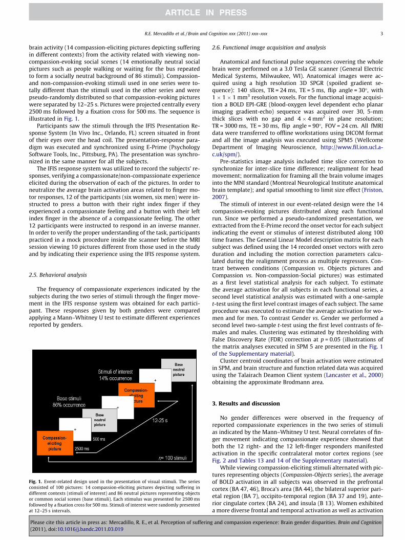
No gender differences were observed in the frequency ofreported compassionate experiences in the two series of stimulias indicated by the Mann–Whitney U test. Neural correlates of fin-ger movement indicating compassionate experience showed thatboth the 12 right- and the 12 left-finger responders manifestedactivation in the specific contralateral motor cortex regions (seeFig. 2 and Tables 13 and 14 of the Supplementary material).
While viewing compassion-eliciting stimuli alternated with pic-tures representing objects (Compassion-Objects series), the averageof BOLD activation in all subjects was observed in the prefrontalcortex (BA 47, 46), Broca’s area (BA 44), the bilateral superior pari-etal region (BA 7), occipito-temporal region (BA 37 and 19), ante-rior cingulate cortex (BA 24), and insula (B 13). Women exhibiteda more diverse frontal and temporal activation as well as activation
and compassion experience: Brain gender disparities. Brain and Cognition
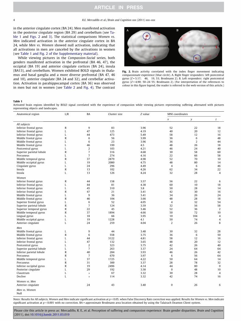
Fig. 2. Brain activity correlated with the index finger movement indicatingcompassionate experience (blue circle). A. Right finger responders: left postcentralgyrus (Z = 5.17; �46, �19, 53; Brodmann 2). B. Left responders: right postcentralgyrus (Z = 4.90; 50–24 55; Brodmann 2). (For interpretation of the references tocolour in this figure legend, the reader is referred to the web version of this article.)
4 R.E. Mercadillo et al. / Brain and Cognition xxx (2011) xxx–xxx
in the anterior cingulate cortex (BA 24). Men manifested activationin the posterior cingulate region (BA 29) and cerebellum (see Ta-ble 1 and Figs. 2 and 3). The statistical comparisons Women vs.Men indicated activation in the anterior cingulate cortex in BA24, while Men vs. Women showed null activation, indicating thatall activations in men are canceled by the activations in women(see Table 1 and Fig. 2 of the Supplementary material).
While viewing pictures in the Compassion-Social series, bothgenders manifested activation in the prefrontal (BA 46, 47), theoccipital (BA 19) and anterior cingulate cortices (BA 24), insula(BA13), and cerebellum. Women exhibited BOLD signals in thala-mus and basal ganglia and a more diverse prefrontal (BA 47, 46and 10), anterior cingulate (BA 24 and 32), and cerebellar activa-tion. Activation in parahippocampal cortex (BA 36) was observedin men but not in women (see Table 2 and Fig. 4). The contrast
Table 1Activated brain regions identified by BOLD signal correlated with the experience of compassion while viewing pictures representing suffering alternated with picturesrepresenting objects and landscapes.
Anatomical region L/R BA Cluster size Z value MNI coordinates
x y z
All subjectsInferior frontal gyrus R 9 83 3.96 52 22 18Inferior frontal gyrus L 47 125 4.19 �40 20 �12Inferior frontal gyrus L 44 473 5.49 �58 12 16Middle frontal gyrus R 6 56 3.99 36 �6 48Middle frontal gyrus L 6 23 3.96 �36 �2 44Middle frontal gyrus L 46 199 4.5 �48 26 18Postcentral gyrus L 2 103 4.21 �46 �24 40Superior parietal lobule R 7 316 4.68 30 �58 60Precuneus L 7 274 4.16 �22 �66 58Middle temporal gyrus R 37 2879 4.98 52 �70 10Middle occipital gyrus L 19 2080 4.75 �48 �80 14Cingulate gyrus L 24 296 4.49 �4 6 46Insula R 13 102 4.26 62 �34 22Insula L 13 126 4.24 �32 28 �4
WomenInferior frontal gyrus R 44 158 3.57 56 22 6Inferior frontal gyrus L 44 81 4.38 �60 10 18Inferior frontal gyrus L 45 310 3.8 �50 28 14Inferior frontal gyrus R 47 21 3.41 42 30 �16Middle frontal gyrus L 9 36 3.41 �34 26 24Middle frontal gyrus R 46 104 3.66 48 28 18Superior frontal gyrus L 6 52 4.05 �4 32 54Superior parietal lobule R 7 124 3.59 32 �56 58Superior temporal gyrus R 22 45 3.46 52 �36 2Middle temporal gyrus R 37 1894 4.66 50 �72 10Lingual gyrus L 18 66 3.95 �10 �104 �4Middle occipital gyrus L 19 1228 4.1 �50 �74 �4Anterior cingulate L 24 259 4.04 2 26 8
MenMiddle frontal gyrus L 9 44 3.48 �30 32 28Middle frontal gyrus R 6 558 3.75 36 �6 50Inferior frontal gyrus L 44 3122 4.81 �58 12 16Inferior frontal gyrus L 47 132 3.65 �48 20 �12Postcentral gyrus L 2 323 3.75 �42 �26 40Superior parietal lobule L 7 263 3.37 �24 �64 64Inferior parietal lobule R 40 284 3.93 46 �26 42Precuneus R 7 670 3.97 6 �56 64Middle temporal gyrus L 37 1725 4.22 �50 �64 14Precuneus R 31 300 3.37 28 �78 32Inferior occipital gyrus R 19 2496 4.14 44 �80 0Posterior cingulate L 29 192 3.58 0 �48 10Claustrum L ⁄ 67 3.32 �30 28 �4Declive L ⁄ 114 3.32 �42 �74 �16
Women vs. MenAnterior cingulate 24 43 3.40 0 26 6
Men vs. WomenNull
Notes: Results for All subjects, Women and Men indicate significant activation at p < 0.05. when False Discovery Rate correction was applied. Results for Women vs. Men indicatesignificant activation at p < 0.001 with no correction. BA = approximate Brodmann area location obtained by using the Talairach Deamon Client system.
Please cite this article in press as: Mercadillo, R. E., et al. Perception of suffering and compassion experience: Brain gender disparities. Brain and Cognition(2011), doi:10.1016/j.bandc.2011.03.019
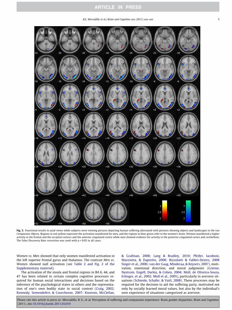
Fig. 3. Functional results in axial views while subjects were viewing pictures depicting human suffering alternated with pictures showing objects and landscapes in the runCompassion-Objects. Regions in red-yellow represent the activation manifested for men, and the regions in blue-green refer to the women’s brain. Women manifested a higheractivity in the frontal and the occipital cortices and the anterior cingulated cortex while men showed evidence for activity in the posterior cingulated cortex and cerebellum.The False Discovery Rate correction was used with p = 0.05 in all cases.
R.E. Mercadillo et al. / Brain and Cognition xxx (2011) xxx–xxx 5
Women vs. Men showed that only women manifested activation inthe left superior frontal gyrus and thalamus. The contrast Men vs.Women showed null activation (see Table 2 and Fig. 2 of theSupplementary material).
The activation of the insula and frontal regions in BA 6, 44, and47 has been related to certain complex cognitive processes re-quired for human social interactions and decisions based on theinference of the psychological states in others and the representa-tion of one’s own bodily state in social context (Craig, 2002;Kennedy, Semendeferi, & Courchesne, 2007; Knutson, McClellan,
Please cite this article in press as: Mercadillo, R. E., et al. Perception of suffering(2011), doi:10.1016/j.bandc.2011.03.019
& Grafman, 2008; Lang & Bradley, 2010; Pfeifer, Iacoboni,Mazziotta, & Dapretto, 2008; Rizzolatti & Fabbri-Destro, 2008Singer et al., 2006; van der Gaag, Minderaa, & Keysers, 2007), moti-vation, emotional direction, and moral judgments (Greene,Nystrom, Engell, Darley, & Cohen, 2004; Moll, de Oliveira-Souza,Eslinger, et al., 2002; Moll et al., 2005), particularly in aversive sit-uations (Schienle, Schafer, & Vaitl, 2008). These processes may berequired for the decision to aid the suffering party, motivated notonly by socially learned moral values, but also by the individual’sown experience of situations categorized as aversive.
and compassion experience: Brain gender disparities. Brain and Cognition
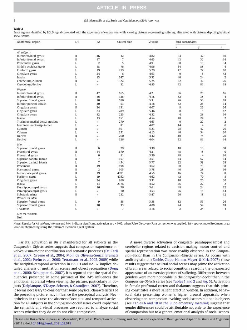
Table 2Brain regions identified by BOLD signal correlated with the experience of compassion while viewing pictures representing suffering, alternated with pictures depicting habitualsocial scenes.
Anatomical region L/R BA Cluster size Z value MNI coordinates
x y z
All subjectsInferior frontal gyrus R 46 32 4.82 54 32 10Inferior frontal gyrus R 47 7 4.63 42 32 �14Postcentral gyrus L 2 5 4.9 �60 �18 34Middle occipital gyrus L 18 76 4.96 �34 �88 4Fusiform gyrus L 19 719 5.29 �42 �72 �10Cingulate gyrus L 24 6 4.63 0 �8 42Insula L 13 247 5.32 �40 24 �2Cerebellum/culmen R ⁄ 1322 5.75 32 �42 �26Cerebellum/declive L ⁄ 32 4.85 �32 �86 �18
WomenInferior frontal gyrus R 47 165 4.2 36 20 �16Inferior frontal gyrus R 46 126 4.18 52 38 4Superior frontal gyrus L 10 590 5.3 �26 56 18Inferior parietal lobule L 40 53 4.18 �42 �28 34Cingulate gyrus R 24 131 4.07 8 22 26Cingulate gyrus L 24 289 5.46 0 �8 42Cingulate gyrus L 32 225 4.32 �4 28 36Insula L 13 151 4.54 �40 24 �2Thalamus medial dorsal nucleus L ⁄ 270 4.63 0 �22 2Lentiform nucleus/putamen L ⁄ 81 4.07 �14 4 6Culmen R ⁄ 1501 5.23 28 �42 �26Culmen L ⁄ 251 4.13 �40 �54 �20Declive R ⁄ 200 4.32 10 �74 �20Declive L ⁄ 326 4.04 �32 �86 �14
MenSuperior frontal gyrus R 6 29 3.39 10 16 68Precentral gyrus R 44 1670 4.3 48 18 0Precentral gyrus L 6 51 3.39 �48 4 26Superior parietal lobule R 7 157 3.51 34 �52 54Superior parietal lobule L 7 454 3.77 �22 �58 68Precuneus R 19 198 3.62 30 �76 38Postcentral gyrus R 2 305 3.78 46 �28 66Inferior occipital gyrus R 19 4991 4.55 46 �74 �8Fusiform gyrus L 19 4752 4.62 �42 �70 �8Cingulate gyrus R 24 266 3.31 10 20 30Insula L 13 2376 4.52 �36 22 �2Parahippocampal gyrus R 36 76 3.6 48 �24 �12Parahippocampal gyrus L ⁄ 70 3.53 �26 �18 �14Substania nigra R ⁄ 232 3.47 20 �18 �12Women vs. MenSuperior frontal gyrus L 9 88 3.38 �12 56 26Superior frontal gyrus L 10 33 4.00 �24 54 18Thalamus L ⁄ 7 3.16 2 �24 4
Men vs. WomenNull
Notes: Results for All subjects, Women and Men indicate significant activation at p < 0.05. when False Discovery Rate correction was applied. BA = approximate Brodmann arealocation obtained by using the Talairach Deamon Client system.
6 R.E. Mercadillo et al. / Brain and Cognition xxx (2011) xxx–xxx
Parietal activation in BA 7 manifested for all subjects in theCompassion-Objects series suggests that compassion experience in-volves visuo-motor coordination and semantic processes (De Carliet al., 2007; Greene et al., 2004; Moll, de Oliveira-Souza, Bramatiet al., 2002; Prehn et al., 2008; Tettamanti et al., 2002, 2009) whilethe occipital-temporal activation in BA 19 and BA 37 implies de-tailed analysis of mutilation scenes and object recognition (Donget al., 2000; Schupp et al., 2007). It is reported that the spatial fre-quencies presented in some pictures of the IAPS influences theemotional appraisal when viewing the picture, particularly in ob-jects (Delplanque, N’Diaye, Scherer, & Grandjean, 2007). Therefore,it seems necessary to consider that some physical characteristics ofthe preceding picture may influence the perceptual analysis. Nev-ertheless, in this case, the absence of occipital and temporal activa-tion for all subjects in the Compassion-Social series could imply thatthe semantic and visual processes are required to analyze socialscenes whether they do or do not elicit compassion.
Please cite this article in press as: Mercadillo, R. E., et al. Perception of suffering(2011), doi:10.1016/j.bandc.2011.03.019
A more diverse activation of cingulate, parahippocampal andcerebellar regions related to decision making, motor control, andspatial representation was more readily observed in the Compas-sion-Social than in the Compassion-Objects series. As occurs withauditory stimuli (Zaehle, Clapp, Hamm, Meyer, & Kirk, 2007), theseresults suggest that neutral social scenes may prime the activationof brain areas related to social cognition regarding the unexpectedappearance of an aversive picture of suffering. Differences betweengenders were more noticeable in the Compassion-Social than in theCompassion-Objects series (see Tables 1 and 2 and Fig. 5). Activationin female prefrontal cortex and thalamus suggests that this prim-ing constitutes a more salient effect in women. In addition, behav-ioral data presenting women’s higher arousal appraisals whenobserving non-compassion-evoking social scenes but not in objects(see Tables 6 and 10 in the Supplementary material) suggest thatgender differences could be attributable not only to the experienceof compassion but to a general emotional analysis of social scenes.
and compassion experience: Brain gender disparities. Brain and Cognition
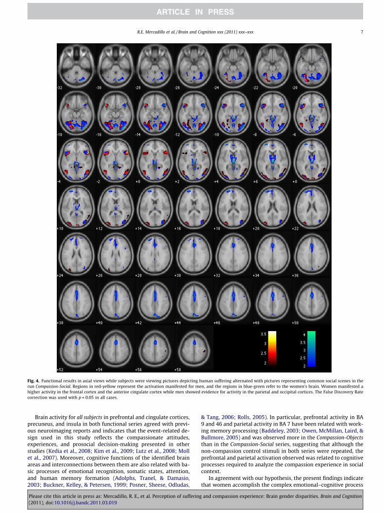
Fig. 4. Functional results in axial views while subjects were viewing pictures depicting human suffering alternated with pictures representing common social scenes in therun Compassion-Social. Regions in red-yellow represent the activation manifested for men, and the regions in blue-green refer to the women’s brain. Women manifested ahigher activity in the frontal cortex and the anterior cingulate cortex while men showed evidence for activity in the parietal and occipital cortices. The False Discovery Ratecorrection was used with p = 0.05 in all cases.
R.E. Mercadillo et al. / Brain and Cognition xxx (2011) xxx–xxx 7
Brain activity for all subjects in prefrontal and cingulate cortices,precuneus, and insula in both functional series agreed with previ-ous neuroimaging reports and indicates that the event-related de-sign used in this study reflects the compassionate attitudes,experiences, and prosocial decision-making presented in otherstudies (Kedia et al., 2008; Kim et al., 2009; Lutz et al., 2008; Mollet al., 2007). Moreover, cognitive functions of the identified brainareas and interconnections between them are also related with ba-sic processes of emotional recognition, somatic states, attention,and human memory formation (Adolphs, Tranel, & Damasio,2003; Buckner, Kelley, & Petersen, 1999; Posner, Sheese, Odludas,
Please cite this article in press as: Mercadillo, R. E., et al. Perception of suffering(2011), doi:10.1016/j.bandc.2011.03.019
& Tang, 2006; Rolls, 2005). In particular, prefrontal activity in BA9 and 46 and parietal activity in BA 7 have been related with work-ing memory processing (Baddeley, 2003; Owen, McMillan, Laird, &Bullmore, 2005) and was observed more in the Compassion-Objectsthan in the Compassion-Social series, suggesting that although thenon-compassion control stimuli in both series were repeated, theprefrontal and parietal activation observed was related to cognitiveprocesses required to analyze the compassion experience in socialcontext.
In agreement with our hypothesis, the present findings indicatethat women accomplish the complex emotional–cognitive process
and compassion experience: Brain gender disparities. Brain and Cognition
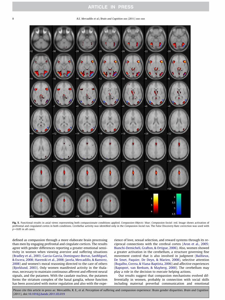
Fig. 5. Functional results in axial views representing both compassionate conditions applied. Compassion-Objects: blue; Compassion-Social: red. Image shows activation ofprefrontal and cingulated cortex in both conditions. Cerebellar activity was identified only in the Compassion-Social run. The False Discovery Rate correction was used withp = 0.05 in all cases.
8 R.E. Mercadillo et al. / Brain and Cognition xxx (2011) xxx–xxx
defined as compassion through a more elaborate brain processingthan men by engaging prefrontal and cingulate cortices. The resultsagree with gender differences reporting a greater emotional sensi-tivity in women when viewing aversive and suffering situations(Bradley et al., 2003; Garcia-Garcia, Dominguez-Borras, SanMiguel,& Escera, 2008; Harenski et al., 2008; Javela, Mercadillo, & Ramirez,2008) and women’s moral reasoning directed to the care of others(Bjorklund, 2003). Only women manifested activity in the thala-mus, necessary to maintain continuous afferent and efferent neuralsignals, and the putamen. With the caudate nucleus, the putamenforms the striatum complex of the basal ganglia, whose functionhas been associated with motor regulation and also with the expe-
Please cite this article in press as: Mercadillo, R. E., et al. Perception of suffering(2011), doi:10.1016/j.bandc.2011.03.019
rience of love, sexual selection, and reward systems through its re-ciprocal connections with the cerebral cortex (Aron et al., 2005;Bianchi-Demicheli, Grafton, & Ortigue, 2006). Also, women showeda greater activation in the cerebellum, a structure governing finemovement control that is also involved in judgment (Baillieux,De Smet, Paquier, De Deyn, & Marien, 2008), selective attention(Bugalho, Correa, & Viana-Baptista, 2006) and affective experiences(Rapoport, van Reekum, & Mayberg, 2000). The cerebellum mayplay a role in the decision to execute helping actions.
Our results suggest that compassion mechanisms evolved dif-ferentially in women, probably in connection with social skillsincluding maternal preverbal communication and emotional
and compassion experience: Brain gender disparities. Brain and Cognition
R.E. Mercadillo et al. / Brain and Cognition xxx (2011) xxx–xxx 9
responses to helpless offspring (Campbell, 2008; Febo, Numan, &Ferris, 2005; Leibenluft, Gobbini, Harrison, & Haxby, 2004; Lenziet al., 2009). They could also be explained by differential culturalexpectations influencing moral judgments (Reeder, Kumar,Hesson-McInnis, & Trafimow, 2002; Tilley, 2004). This occurs inMexican society, where differential sexual education reinforces inwomen the expressions of emotions related with inferring sadnessor pain states and promotes the development of behaviors involv-ing the care of people. Alternatively, the socially accepted mascu-line role emphasizes behaviors based in norms and rejectsexpressions believed to be feminine (Díaz-Guerrero, 1984, 1994;Saldívar-Hernández, Ramos-Lira, & Romero, 2008). The apparentincongruence between a similarly reported compassion experienceand the BOLD activations of men and women may reflect differentcognitive and emotional mechanisms eliciting a similar appraisalbehavior.
Some experimental restrictions may influence the interpreta-tion of the brain activity presented in this work. Arousal appraisalsmanifested higher scores in compassion stimuli than non-compas-sion evoked pictures used as control, so brain activity correlatedwith compassion could reflect the influence of arousal. In thissense, although compassion generally involves aversive situationsqualified as high arousal, it could be essential, for future studies,to control the experimental conditions presenting similar arousalqualities to cancel the effect of this dimension. Similarly, as ex-plained in the discussion of the occipital and temporal activity re-lated with the visual analysis, it is necessary to consider thephysical characteristics of the stimuli in order to differentiate thecompassion process from the visual processes per se. Also, to im-prove this design it is possible to consider stimuli novelty in allcases to avoid possible effects related with mnemonic functions.
Further research of reported compassionate experience and re-lated neurobiological mechanisms will require an integral neuro-cognitive approach capable of discriminating possible causalfactors of the observed gender differences. It would be importantto study compassion by considering qualitative differences inexperimental designs that come closer to real life situations (Todo-rov, Harris, & Fiske, 2006) and by defining the features of the visualstimuli that elicit empathy and motivate compassionate intentionsand behaviors.
Funding
This study was supported by PAPIIT grant IN106707 Grant andCONACYT scholarship No. 213635 in the Posgrado en CienciasBiomédicas, Universidad Nacional Autónoma de México.
Acknowledgments
We thank Peter J. Lang, Margaret M. Bradley, and Bruce N. Cuth-bert, of the NIMH Center for the Study of Emotion and Attention,for the database and pictures of the International Affective Pic-ture System (IAPS). We also thank Dr. Dorothy Pless, Dr. MagdaGiordano and Dr. Michael C. Jeziorski for their corrections and ad-vice, Dr. Perla Salgado, Institute of Neurology and Neurosurgery‘‘Manuel Velasco Suárez’’, and Leopoldo González-Santos, Instituteof Neurobiology Universidad Nacional Autónoma de México, fortechnical support in the acquisition and analyses of anatomicaland functional images, and the volunteers who agreed to partici-pate in this study.
Appendix A. Supplementary material
Supplementary data associated with this article can be found, inthe online version, at doi:10.1016/j.bandc.2011.03.019.
Please cite this article in press as: Mercadillo, R. E., et al. Perception of suffering(2011), doi:10.1016/j.bandc.2011.03.019
References
Adolphs, R., Tranel, D., & Damasio, A. R. (2003). Dissociable neural systems forrecognizing emotions. Brain and Cognition, 52(1), 61–69.
Aleman, A., & Swart, M. (2008). Sex differences in neural activation to facialexpressions denoting contempt and disgust. PLoS ONE, 3(11), e3622.
Aron, A., Fisher, H., Mashek, D. J., Strong, G., Li, H., & Brown, L. L. (2005). Reward,motivation, and emotion systems associated with early-stage intense romanticlove. Journal of Neurophysiology, 94(1), 327–337.
Baddeley, A. (2003). Working memory: Looking back and looking forward. NatureReview. Neuroscience, 4(10), 829–839.
Baillieux, H., De Smet, H. J., Paquier, P. F., De Deyn, P. P., & Marien, P. (2008).Cerebellar neurocognition: Insights into the bottom of the brain. ClinicalNeurology and Neurosurgery, 110(8), 763–773.
Bennett, M., & Matthews, L. (2000). The role of second-order belief-understandingand social context in children’s self-attribution of social emotions. SocialDevelopment, 9(1), 126–130.
Bianchi-Demicheli, F., Grafton, S. T., & Ortigue, S. (2006). The power of love on thehuman brain. Social Neuroscience, 1(2), 90–103.
Bjorklund, F. (2003). Differences in the justification of choices in moral dilemmas:Effects of gender, time pressure and dilemma seriousness. Scandinavian Journalof Psychology, 44(5), 459–466.
Bradley, M. M., Codispoti, M., Cuthbert, B. N., & Lang, P. J. (2001). Emotion andmotivation I: Defensive and appetitive reactions in picture processing. Emotion,1(3), 276–298.
Bradley, M. M., Sabatinelli, D., Lang, P. J., Fitzsimmons, J. R., King, W., & Desai, P.(2003). Activation of the visual cortex in motivated attention. BehavioralNeuroscience, 117(2), 369–380.
Buckner, R. L., Kelley, W. M., & Petersen, S. E. (1999). Frontal cortex contributes tohuman memory formation. Nature Neuroscience, 2(4), 311–314.
Bugalho, P., Correa, B., & Viana-Baptista, M. (2006). Role of the cerebellum incognitive and behavioural control: Scientific basis and investigation models.Acta Médica Portuguesa, 19(3), 257–267.
Campbell, A. (2008). Attachment, aggression and affiliation: The role of oxytocin infemale social behavior. Biological Psychology, 77(1), 1–10.
Canli, T., Desmond, J. E., Zhao, Z., & Gabrieli, J. D. (2002). Sex differences in the neuralbasis of emotional memories. Proceedings of the National Academy of Sciences ofthe United States of America, 99(16), 10789–10794.
Casebeer, W. D. (2003). Moral cognition and its neural constituents. Nature Review.Neuroscience, 4(10), 840–846.
Craig, A. D. (2002). How do you feel? Interoception: The sense of the physiologicalcondition of the body. Nature Review. Neuroscience, 3(8), 655–666.
Damasio, A. (2006). En busca de Spinoza. Neurobiología de la emoción y lossentimientos. (J. Ros, Trans.). Barcelona: Crítica.
De Carli, D., Garreffa, G., Colonnese, C., Giulietti, G., Labruna, L., Briselli, E., et al.(2007). Identification of activated regions during a language task. MagneticResonance Imaging, 25(6), 933–938.
de Wied, M., Branje, S. J., & Meeus, W. H. (2007). Empathy and conflict resolutionin friendship relations among adolescents. Aggressive Behavior, 33(1), 48–55.
Delplanque, S., N’Diaye, K., Scherer, K., & Grandjean, D. (2007). Spatial frequencies oremotional effects? A systematic measure of spatial frequencies for IAPS picturesby a discrete wavelet analysis. Journal of Neuroscience Methods, 165(1), 144–150.
Díaz-Guerrero, R. (1984). Contemporary psychology in Mexico. Annual Review ofPsychology, 35, 83–112.
Díaz-Guerrero, R. (1994). Psicología del mexicano: Descubrimiento de laetnopsicología. México: Trillas.
Dong, Y., Fukuyama, H., Honda, M., Okada, T., Hanakawa, T., Nakamura, K., et al.(2000). Essential role of the right superior parietal cortex in Japanese kanamirror reading: An fMRI study. Brain, 123(Pt 4), 790–799.
Ekman, P. (1993). Facial expression and emotion. The American Psychologist, 48(4),384–392.
Febo, M., Numan, M., & Ferris, C. F. (2005). Functional magnetic resonance imagingshows oxytocin activates brain regions associated with mother-pup bondingduring suckling. The Journal of Neuroscience, 25(50), 11637–11644.
Flores-Gutierrez, E. O., Diaz, J. L., Barrios, F. A., Guevara, M. A., Del Rio-Portilla, Y.,Corsi-Cabrera, M., et al. (2009). Differential alpha coherence hemisphericpatterns in men and women during pleasant and unpleasant musical emotions.International Journal of Psychophysiology, 71(1), 43–49.
Friston, K. (2007). Statistical parametric mapping. In K. Friston, J. Ashburner, S. J.Kiebel, T. Nichols, & W. Penny (Eds.), Statistical parametric mapping (pp. 10–31).Oxford: Academic Press.
Garcia-Garcia, M., Dominguez-Borras, J., SanMiguel, I., & Escera, C. (2008).Electrophysiological and behavioral evidence of gender differences in themodulation of distraction by the emotional context. Biological Psychology, 79(3),307–316.
González-Santos, L., Mercadillo, R. E., Graff, A., & Barrios, F. A. (2007). Versióncomputarizada para la aplicación del Listado de Síntomas 90 (SCL 90) y delInventario de Tempramento y Carácter (ITC). Salud Mental, 30(4), 31–40.
Greene, J., & Haidt, J. (2002). How (and where) does moral judgment work? Trends inCognitive Sciences, 6(12), 517–523.
Greene, J. D., Nystrom, L. E., Engell, A. D., Darley, J. M., & Cohen, J. D. (2004). Theneural bases of cognitive conflict and control in moral judgment. Neuron, 44(2),389–400.
and compassion experience: Brain gender disparities. Brain and Cognition
10 R.E. Mercadillo et al. / Brain and Cognition xxx (2011) xxx–xxx
Haidt, J. (2003). The moral emotions. In R. J. Davidson, K. Scherer, & H. Goldsmith(Eds.), Handbook of affective sciences (pp. 852–870). Oxford: Oxford UniversityPress.
Harenski, C. L., Antonenko, O., Shane, M. S., & Kiehl, K. A. (2008). Gender differencesin neural mechanisms underlying moral sensitivity. Social Cognitive andAffective Neuroscience, 3(4), 313–321.
Izard, C. E. (1992). Basic emotions, relations among emotions, and emotion–cognition relations. Psychological Review, 99, 561–565.
Jausovec, N., & Jausovec, K. (2005). Sex differences in brain activity related togeneral and emotional intelligence. Brain and Cognition, 59(3), 277–286.
Javela, J. J., Mercadillo, R. E., & Ramirez, J. (2008). Anger and associated experiencesof sadness, fear, valence, arousal, and dominance evoked by visual scenes.Psychological Reports, 103(3), 663–681.
Kagan, J. (2005). Human morality and temperament. Nebraska Symposium onMotivation, 51, 1–32.
Kedia, G., Berthoz, S., Wessa, M., Hilton, D., & Martinot, J. L. (2008). An agent harms avictim: A functional magnetic resonance imaging study on specific moralemotions. Journal of Cognitive Neuroscience, 20(10), 1788–1798.
Kennedy, D. P., Semendeferi, K., & Courchesne, E. (2007). No reduction of spindleneuron number in frontoinsular cortex in autism. Brain and Cognition, 64(2),124–129.
Kim, J. W., Kim, S. E., Kim, J. J., Jeong, B., Park, C. H., Son, A. R., et al. (2009).Compassionate attitude towards others’ suffering activates the mesolimbicneural system. Neuropsychologia, 47(10), 2073–2081.
Knutson, K. M., McClellan, E. M., & Grafman, J. (2008). Observing social gestures: AnfMRI study. Experimental Brain Research, 188(2), 187–198.
Koch, K., Pauly, K., Kellermann, T., Seiferth, N. Y., Reske, M., Backes, V., et al. (2007).Gender differences in the cognitive control of emotion: An fMRI study.Neuropsychologia, 45(12), 2744–2754.
Lancaster, J. L., Woldorff, M. G., Parsons, L. M., Liotti, M., Freitas, C. S., Rainey, L., et al.(2000). Automated Talairach atlas labels for functional brain mapping. HumanBrain Mapping, 10(3), 120–131.
Lang, P. J., & Bradley, M. M. (2010). Emotion and the motivational brain. BiologicalPsychology, 84(3), 437–450.
Lang, P. J., Bradley, M. M., & Curberth, B. (2005). International affective picture system(IAPS): Instruction manual and affective ratings. Gainesville, FL: University ofFlorida.
Lazarus, R. S. (1991). Progress on a cognitivemotivational–relational theory ofemotion. The American Psychology, 46(8), 819–834.
Leibenluft, E., Gobbini, M. I., Harrison, T., & Haxby, J. V. (2004). Mothers’ neuralactivation in response to pictures of their children and other children. BiologicalPsychiatry, 56(4), 225–232.
Lenzi, D., Trentini, C., Pantano, P., Macaluso, E., Iacoboni, M., Lenzi, G. L., et al. (2009).Neural basis of maternal communication and emotional expression processingduring infant preverbal stage. Cerebral Cortex, 19(5), 1124–1133.
Lutz, A., Brefczynski-Lewis, J., Johnstone, T., & Davidson, R. J. (2008). Regulation ofthe neural circuitry of emotion by compassion meditation: Effects of meditativeexpertise. PLoS ONE, 3(3), e1897.
Mauss, I. B., Levenson, R. W., McCarter, L., Wilhelm, F. H., & Gross, J. J. (2005). The tiethat binds? Coherence among emotion experience, behavior, and physiology.Emotion, 5(2), 175–190.
Mercadillo, R. E., Barrios, F. A., & Diaz, J. L. (2007). Definition of compassion-evokingimages in a Mexican sample. Perceptual and Motor Skills, 105(2), 661–676.
Moll, J., de Oliveira-Souza, R., Bramati, I. E., & Grafman, J. (2002). Functionalnetworks in emotional moral and nonmoral social judgments. Neuroimage, 16(3Pt 1), 696–703.
Moll, J., de Oliveira-Souza, R., Eslinger, P. J., Bramati, I. E., Mourao-Miranda, J.,Andreiuolo, P. A., et al. (2002b). The neural correlates of moral sensitivity: Afunctional magnetic resonance imaging investigation of basic and moralemotions. The Journal of Neuroscience, 22(7), 2730–2736.
Moll, J., de Oliveira-Souza, R., Garrido, G. J., Bramati, I. E., Caparelli-Daquer, E. M.,Paiva, M. L., et al. (2007). The self as a moral agent: Linking the neural bases ofsocial agency and moral sensitivity. Social Neuroscience, 2(3–4), 336–352.
Moll, J., de Oliveira-Souza, R., Moll, F. T., Ignacio, F. A., Bramati, I. E., Caparelli-Daquer, E. M., et al. (2005). The moral affiliations of disgust: A functional MRIstudy. Cognitive and Behavioral Neurology, 18(1), 68–78.
Please cite this article in press as: Mercadillo, R. E., et al. Perception of suffering(2011), doi:10.1016/j.bandc.2011.03.019
Moll, J., De Oliveira-Souza, R., & Zahn, R. (2008). The neural basis of moral cognition:sentiments, concepts, and values. Annals of the New York Academy of Sciences,1124, 161–180.
Moll, J., & Schulkin, J. (2009). Social attachment and aversion in human moralcognition. Neuroscience and Biobehavioral Reviews, 33(3), 456–465.
Nichols, S. (2002). Norms with feeling: Towards a psychological account of moraljudgment. Cognition, 84(2), 221–236.
O’Donohue, W. T., & Ferguson, K. E. (2003). Handbook of professional ethics forpsychologists: Issues, questions, and controversies. USA: Sage Publications Inc..
Owen, A. M., McMillan, K. M., Laird, A. R., & Bullmore, E. (2005). N-back workingmemory paradigm: A meta-analysis of normative functional neuroimagingstudies. Human Brain Mapping, 25(1), 46–59.
Pfeifer, J. H., Iacoboni, M., Mazziotta, J. C., & Dapretto, M. (2008). Mirroring others’emotions relates to empathy and interpersonal competence in children.Neuroimage, 39(4), 2076–2085.
Posner, M. I., Sheese, B. E., Odludas, Y., & Tang, Y. (2006). Analyzing and shapinghuman attentional networks. Neural Networks, 19(9), 1422–1429.
Prehn, K., Wartenburger, I., Meriau, K., Scheibe, C., Goodenough, O. R., Villringer, A.,et al. (2008). Individual differences in moral judgment competence influenceneural correlates of socio-normative judgments. Social Cognitive and AffectiveNeuroscience, 3(1), 33–46.
Rapoport, M., van Reekum, R., & Mayberg, H. (2000). The role of the cerebellum incognition and behavior: A selective review. The Journal of Neuropsychiatry andClinical Neurosciences, 12(2), 193–198.
Reeder, G. D., Kumar, S., Hesson-McInnis, M. S., & Trafimow, D. (2002). Inferencesabout the morality of an aggressor: The role of perceived motive. Journal ofPersonality and Social Psychology, 83(4), 789–803.
Rizzolatti, G., & Fabbri-Destro, M. (2008). The mirror system and its role in socialcognition. Current Opinion in Neurobiology, 18(2), 179–184.
Rolls, E. T. (2005). Emotion explained. New York: Oxford University Press.Rueckert, L., & Naybar, N. (2008). Gender differences in empathy: The role of the
right hemisphere. Brain and Cognition, 67(2), 162–167.Saldívar-Hernández, G., Ramos-Lira, L., & Romero, M. (2008). Qué es la coerción
sexual? significado, tácticas e interpretación en jóvenes universitarios de laciudad de México. Salud Mental, 31(1), 45–51.
Schienle, A., Schafer, A., & Vaitl, D. (2008). Individual differences in disgust imagery:A functional magnetic resonance imaging study. Neuroreport, 19(5), 527–530.
Schupp, H. T., Stockburger, J., Codispoti, M., Junghofer, M., Weike, A. I., & Hamm, A.O. (2007). Selective visual attention to emotion. The Journal of Neuroscience,27(5), 1082–1089.
Self, D. J., & Olivarez, M. (1993). The influence of gender on conflicts of interest inthe allocation of limited critical care resources: Justice versus care. Journal ofCritical Care, 8(1), 64–74.
Singer, T., Seymour, B., O’Doherty, J. P., Stephan, K. E., Dolan, R. J., & Frith, C. D.(2006). Empathic neural responses are modulated by the perceived fairness ofothers. Nature, 439(7075), 466–469.
Tettamanti, M., Alkadhi, H., Moro, A., Perani, D., Kollias, S., & Weniger, D. (2002).Neural correlates for the acquisition of natural language syntax. Neuroimage,17(2), 700–709.
Tettamanti, M., Rotondi, I., Perani, D., Scotti, G., Fazio, F., Cappa, S. F., et al. (2009).Syntax without language: Neurobiological evidence for cross-domain syntacticcomputations. Cortex, 45(7), 825–838.
Tilley, J. (2004). Justifaying reasons, motivating reasons and agent relativism inethics. Philosophical Studies, 1, 27.
Todorov, A., Harris, L. T., & Fiske, S. T. (2006). Toward socially inspired socialneuroscience. Brain Research, 1079(1), 76–85.
van der Gaag, C., Minderaa, R. B., & Keysers, C. (2007). Facial expressions: What themirror neuron system can and cannot tell us. Social Neuroscience, 2(3–4),179–222.
Young, L., & Koenigs, M. (2007). Investigating emotion in moral cognition: A reviewof evidence from functional neuroimaging and neuropsychology. British MedicalBulletin, 84, 69–79.
Zaehle, T., Clapp, W. C., Hamm, J. P., Meyer, M., & Kirk, I. J. (2007). Induction of LTP-like changes in human auditory cortex by rapid auditory stimulation: An FMRIstudy. Restorative Neurology and Neuroscience, 25(3–4), 251–259.
and compassion experience: Brain gender disparities. Brain and Cognition