bones of the hindlimb - Állatorvostudományi egyetem€¦ · bones of the pelvic limb (ossa membri...
TRANSCRIPT
BONES OF THE HINDLIMB
Andrea Heinzlmann
University of Veterinary Medicine Budapest
Department of Anatomy and Histology
1st Oktober 2019

BONES OF THE HINDLIMB
COMPOSED OF:
1. PELVIC GIRDLE (CINGULUM MEMBRI PELVINI)
2. THIGH
3. LEG (CRUS)
4. FOOT (PES)
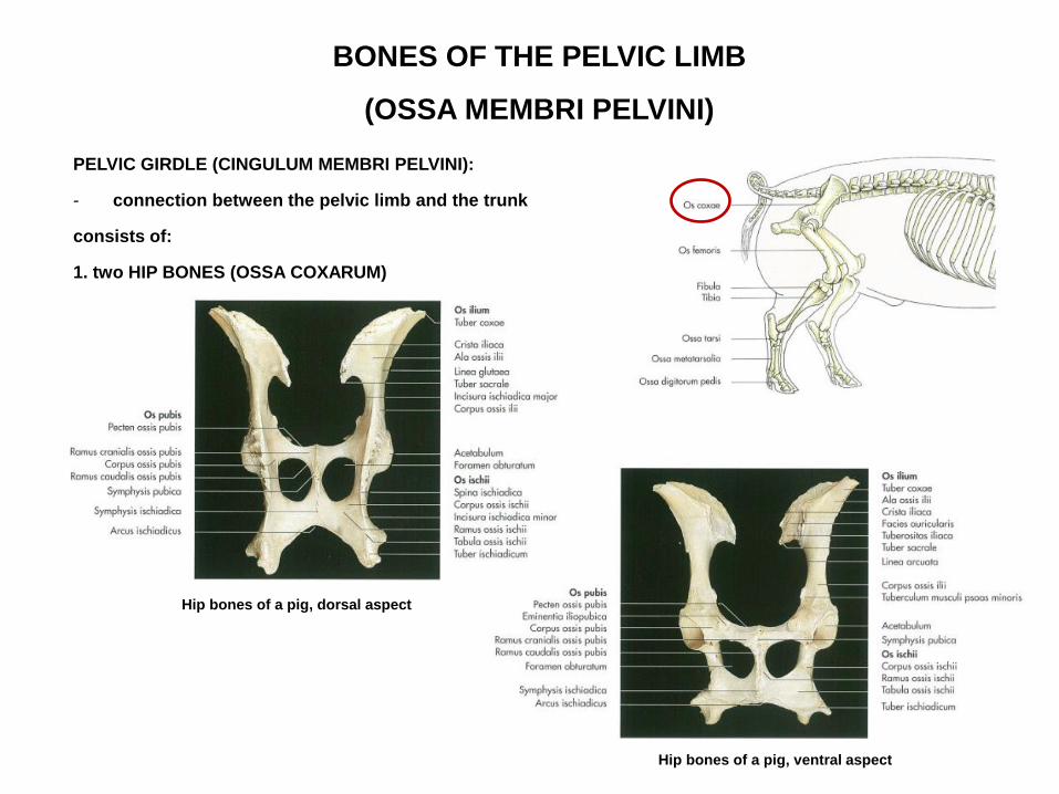
BONES OF THE PELVIC LIMB
(OSSA MEMBRI PELVINI)
PELVIC GIRDLE (CINGULUM MEMBRI PELVINI):
- connection between the pelvic limb and the trunk
consists of:
1. two HIP BONES (OSSA COXARUM)
Hip bones of a pig, ventral aspect
Hip bones of a pig, dorsal aspect
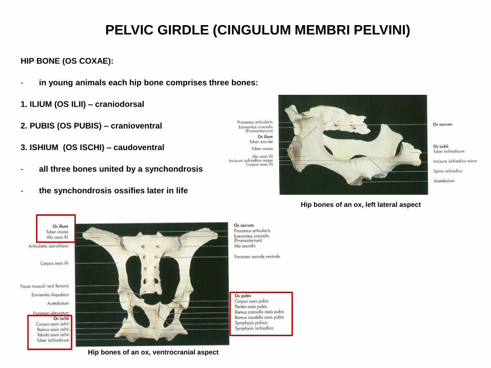
PELVIC GIRDLE (CINGULUM MEMBRI PELVINI)
HIP BONE (OS COXAE):
- in young animals each hip bone comprises three bones:
1. ILIUM (OS ILII) – craniodorsal
2. PUBIS (OS PUBIS) – cranioventral
3. ISHIUM (OS ISCHI) – caudoventral
- all three bones united by a synchondrosis
- the synchondrosis ossifies later in life
Hip bones of an ox, left lateral aspect
Hip bones of an ox, ventrocranial aspect

PELVIC GIRDLE (CINGULUM MEMBRI PELVINI)
HIP BONE (OS COXAE):
ACETABULUM:
- ilium, pubis and ischium meet at the acetabulum
Hip bones of a dog, right lateral aspect
Left acetabulum of a horse, lateral aspect
Left acetabulum of an ox, lateral aspect

PELVIC GIRDLE (CINGULUM MEMBRI PELVINI)
HIP BONE (OS COXAE):
SYMPHYSIS PELVINA:
- the two hip bones united ventrally at the symphysis pelvina by a fibrocartilaginous joint ossified with
advancing age
- in females the fibrocartilage of the symphysis becomes loosened during pregnancy by action of hormones
Hip bones of a horse, ventrocranial aspect
Hip bones of an ox, ventrocranial aspect

PELVIC GIRDLE (CINGULUM MEMBRI PELVINI)
HIP BONE (OS COXAE):
SYMPHYSIS PELVINA:
- in females the fibrocartilage of the symphysis becomes loosened during pregnancy by action of hormones
http://pchorse.se/index.php/en/articles/topic-of-the-month/topics-topics/4395-mars2017-eng

PELVIC GIRDLE (CINGULUM MEMBRI PELVINI)
HIP BONE (OS COXAE):
SYMPHYSIS PELVINA divided into:
1. SYMPHYSIS PUBICA – cranial
2. SYMPHYSIS ISCHIADICA - caudal
Hip bones of a horse, ventrocranial aspect
Hip bones of an ox, ventrocranial aspect

PELVIC GIRDLE (CINGULUM MEMBRI PELVINI)
HIP BONE (OS COXAE):
SYMPHYSIS PELVINA:
CRISTA SYMPHYSIALIS:
- median crest on tzhe ventral surface of symphysis pelvina
- in bo
https://www.medicalexpo.com/prod/sawbones-pacific-research-labs/product-103604-821765.html

PELVIC GIRDLE (CINGULUM MEMBRI PELVINI)
OS SACRUM:
- lies dorsally between the two hip bones
- articulates with the two iliac bones
Hip bones of a dog, right lateral aspect
Hip bones of an ox, ventrocranial aspect

PELVIC GIRDLE (CINGULUM MEMBRI PELVINI)
BONY PELVIS:
formed by the:
1. two hip bones
2. sacrum
3. first few caudal veretebrae
Hip bones of an ox, ventrocranial aspect
Hip bones of an ox, ventrocranial aspect

PELVIC GIRDLE (CINGULUM MEMBRI PELVINI)
OS ILIUM :
consists of:
1. ALA OSSIS ILII (cranial wing)
2. CORPUS OSSIS ILII:
– contributes caudally to the formation of acetabulum
Hip bones of a horse, ventral aspect
Hip bones of the horse, dorsal aspect
Hip bones of an ox, ventrocranial aspect

PELVIC GIRDLE (CINGULUM MEMBRI PELVINI)
OS ILIUM :
POSITION OF THE ALA OSSIS ILII:
1. in bo, eq – nearly horizontal – so the gluteal surface directed dorsally
2. in small ruminants – sagittal, so the gluteal surface directed drsolaterally
3. in carnivores – lateral, so the gluteal surface directed laterally
Hip bones of a dog, right lateral aspect
Hip bones of an ox, ventrocranial aspect
Hip bones of a horse, left lateral aspect
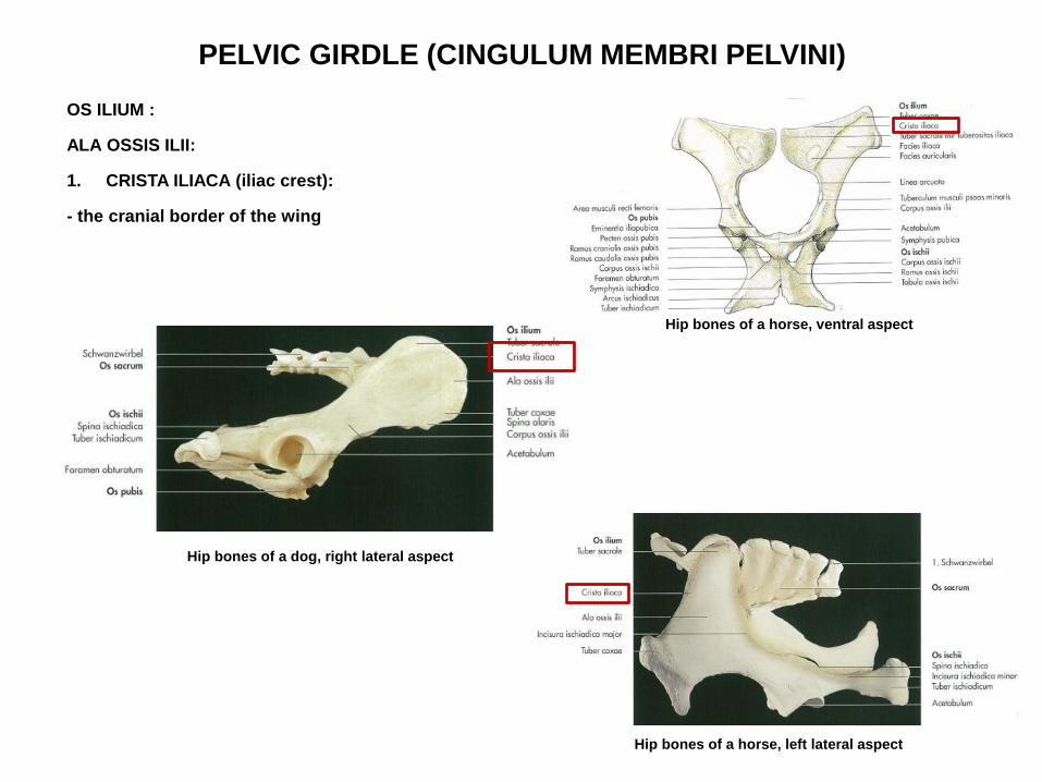
PELVIC GIRDLE (CINGULUM MEMBRI PELVINI)
OS ILIUM :
ALA OSSIS ILII:
1. CRISTA ILIACA (iliac crest):
- the cranial border of the wing
Hip bones of a dog, right lateral aspect
Hip bones of a horse, left lateral aspect
Hip bones of a horse, ventral aspect
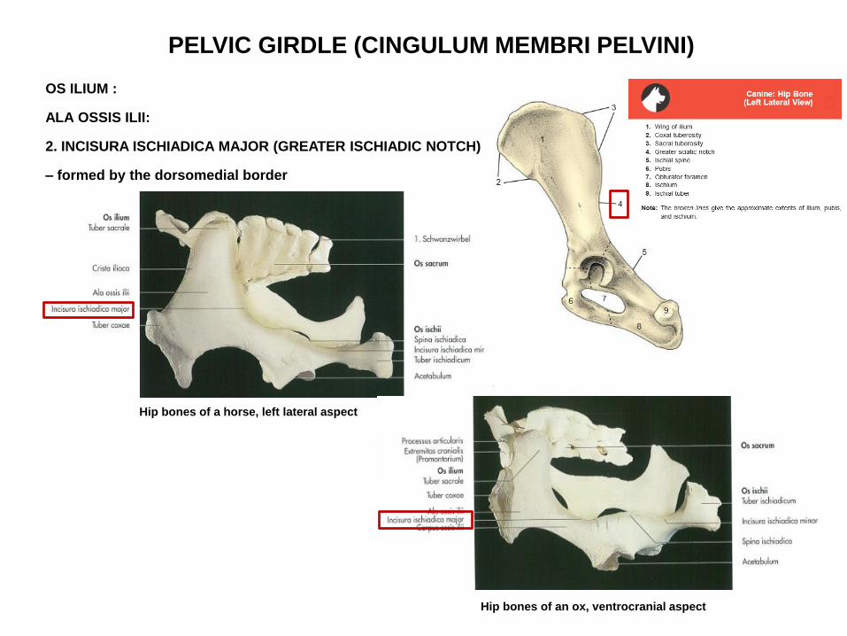
PELVIC GIRDLE (CINGULUM MEMBRI PELVINI)
OS ILIUM :
ALA OSSIS ILII:
2. INCISURA ISCHIADICA MAJOR (GREATER ISCHIADIC NOTCH)
– formed by the dorsomedial border
Hip bones of a horse, left lateral aspect
Hip bones of an ox, ventrocranial aspect
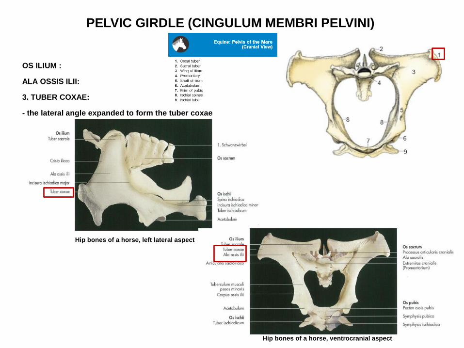
PELVIC GIRDLE (CINGULUM MEMBRI PELVINI)
OS ILIUM :
ALA OSSIS ILII:
3. TUBER COXAE:
- the lateral angle expanded to form the tuber coxae
Hip bones of a horse, left lateral aspect
Hip bones of a horse, ventrocranial aspect
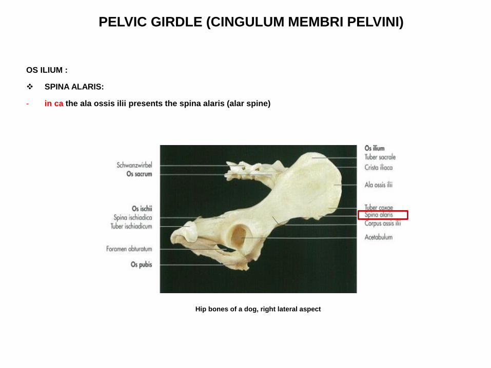
PELVIC GIRDLE (CINGULUM MEMBRI PELVINI)
OS ILIUM :
SPINA ALARIS:
- in ca the ala ossis ilii presents the spina alaris (alar spine)
Hip bones of a dog, right lateral aspect

PELVIC GIRDLE (CINGULUM MEMBRI PELVINI)
OS ILIUM :
in ca.
SPINA ILIACA VENTRALIS CRANIALIS (1.)
Spina
alaris
1.
https://www.youtube.com/watch?v=J92_W8GzIh4

PELVIC GIRDLE (CINGULUM MEMBRI PELVINI)
OS ILIUM :
ALA OSSIS ILII:
4. TUBER SACRALE:
- the medial angle thickened to form the tuber sacrale
Hip bones of a horse, left lateral aspect
Hip bones of a horse, ventrocranial aspect

PELVIC GIRDLE (CINGULUM MEMBRI PELVINI)
OS ILIUM :
ALA OSSIS ILII:
4. TUBER SACRALE:
- in ca the tuber sacrale divides into:
a. Spina iliaca dorsalis cranialis
b. Spina iliaca dorsalis caudalis
15. Spina iliaca dorsalis cranialis
16. Spina iliaca dorsalis caudalis
15.
16.
https://www.youtube.com/watch?v=J92_W8GzIh4

PELVIC GIRDLE (CINGULUM MEMBRI PELVINI)
OS ILIUM :
ALA OSSIS ILII:
5. FACIES GLUTEA (gluteal surface)
• outer surface
• area for attachement of the gluteal muscles
Ca.
11. Greater sciatic notch
12. Lesser sciatic notch
https://www.youtube.com/watch?v=J92_W8GzIh4
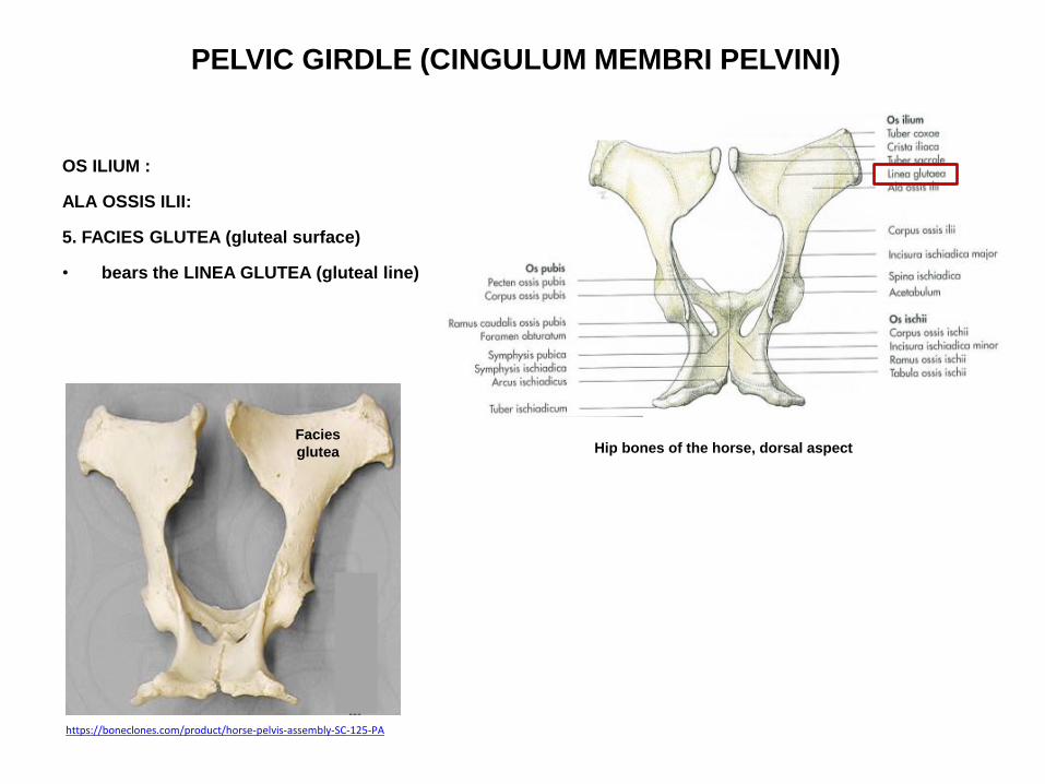
PELVIC GIRDLE (CINGULUM MEMBRI PELVINI)
OS ILIUM :
ALA OSSIS ILII:
5. FACIES GLUTEA (gluteal surface)
• bears the LINEA GLUTEA (gluteal line)
Hip bones of the horse, dorsal aspect
https://boneclones.com/product/horse-pelvis-assembly-SC-125-PA
Facies
glutea
PELVIC GIRDLE (CINGULUM MEMBRI PELVINI)
OS ILIUM :
ALA OSSIS ILII:
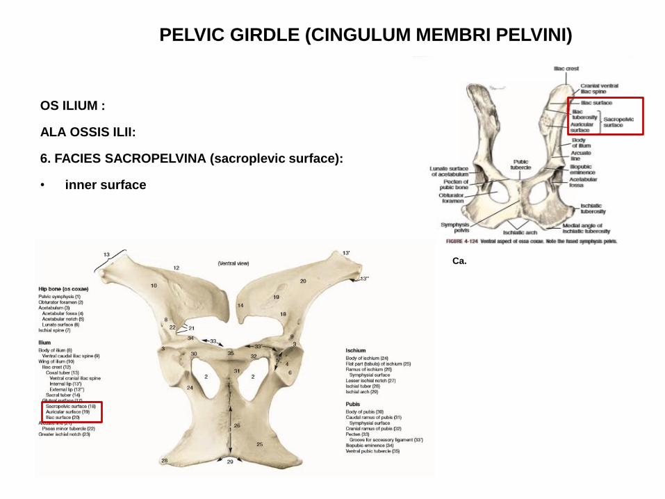
6. FACIES SACROPELVINA (sacroplevic surface):
• inner surface
Ca.

PELVIC GIRDLE (CINGULUM MEMBRI PELVINI)
OS ILIUM :
ALA OSSIS ILII:
6. FACIES SACROPELVINA
presents:
1. FACIES ILIACA (iliac surface):
– laterally situated, smooth
- attachment for the lumbar muscles
Hip bones of a horse, ventrocranial aspect
PELVIC GIRDLE (CINGULUM MEMBRI PELVINI)
OS ILIUM :
ALA OSSIS ILII:
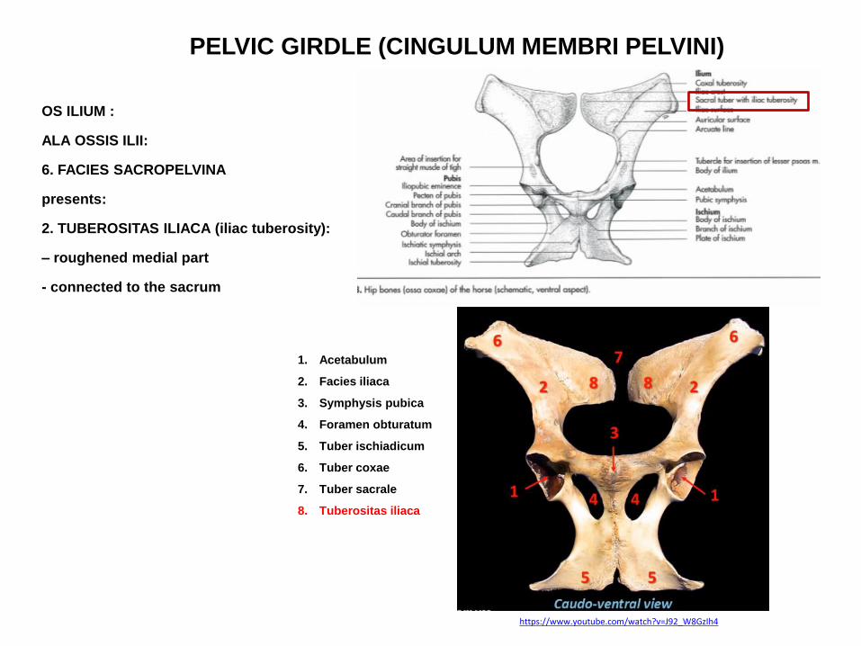
6. FACIES SACROPELVINA
presents:
2. TUBEROSITAS ILIACA (iliac tuberosity):
– roughened medial part
- connected to the sacrum
1. Acetabulum
2. Facies iliaca
3. Symphysis pubica
4. Foramen obturatum
5. Tuber ischiadicum
6. Tuber coxae
7. Tuber sacrale
8. Tuberositas iliaca
https://www.youtube.com/watch?v=J92_W8GzIh4

PELVIC GIRDLE
(CINGULUM MEMBRI PELVINI)
OS ILIUM
ALA OSSIS ILII:
6. FACIES SACROPELVINA
presents:
3. FACIES AURICULARIS (auricular surface):
– ear – shaped articular surface
- covered by cartilage
- articulates with the auricular surface of the wing of the sacrum
Facies
auricularis Tuberositas
iliaca
https://hu.pinterest.com/pin/496592296403870116/
Sacrum, Eq., lateral aspect

PELVIC GIRDLE (CINGULUM MEMBRI PELVINI)
OS ILIUM :
CORPUS OSSIS ILII:
- bears LINEA ARCUATA (arcuate line)
Hip bones of a horse, ventrocranial aspect
Linea
arcuata
http://vanat.cvm.umn.edu/ungDissect/Lab05/Img5-2.html
Eq. https://www.tankonyvtar.hu/en/tartalom/tkt/haziallatok/ch02.html
PELVIC GIRDLE
(CINGULUM MEMBRI PELVINI)
OS ILIUM :
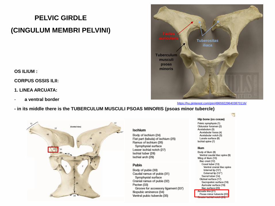
CORPUS OSSIS ILII:
1. LINEA ARCUATA:
- a ventral border
- in its middle there is the TUBERCULUM MUSCULI PSOAS MINORIS (psoas minor tubercle)
Facies
auricularis Tuberositas
iliaca
https://hu.pinterest.com/pin/496592296403870116/
Tuberculum
musculi
psoas
minoris
PELVIC GIRDLE (CINGULUM MEMBRI PELVINI)
OS ILIUM :
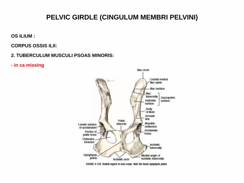
CORPUS OSSIS ILII:
2. TUBERCULUM MUSCULI PSOAS MINORIS:
- in ca missing
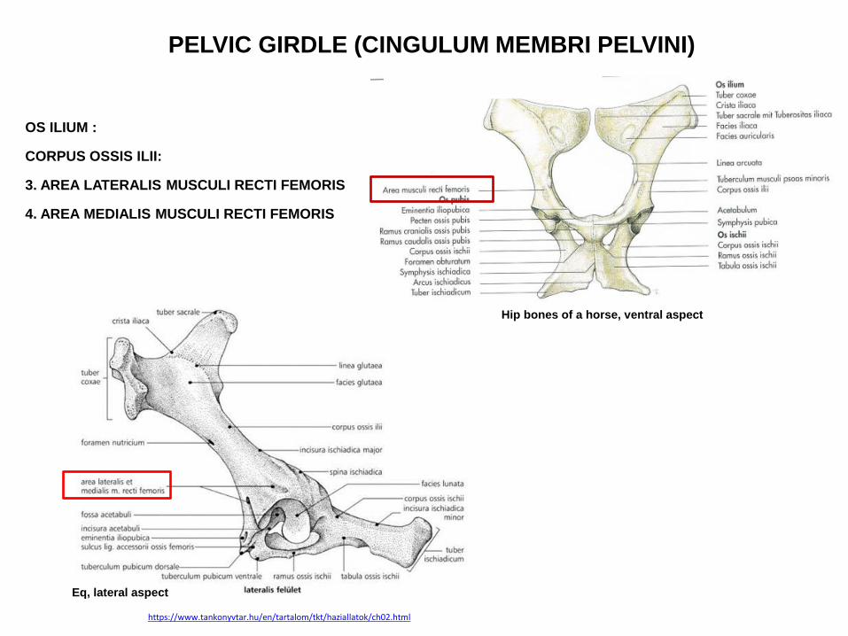
PELVIC GIRDLE (CINGULUM MEMBRI PELVINI)
OS ILIUM :
CORPUS OSSIS ILII:
3. AREA LATERALIS MUSCULI RECTI FEMORIS
4. AREA MEDIALIS MUSCULI RECTI FEMORIS
Hip bones of a horse, ventral aspect
Eq, lateral aspect
https://www.tankonyvtar.hu/en/tartalom/tkt/haziallatok/ch02.html
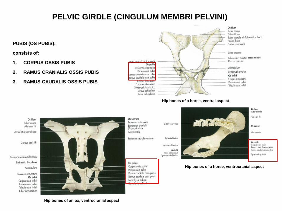
PELVIC GIRDLE (CINGULUM MEMBRI PELVINI)
PUBIS (OS PUBIS):
consists of:
1. CORPUS OSSIS PUBIS
2. RAMUS CRANIALIS OSSIS PUBIS
3. RAMUS CAUDALIS OSSIS PUBIS
Hip bones of an ox, ventrocranial aspect
Hip bones of a horse, ventral aspect
Hip bones of a horse, ventrocranial aspect
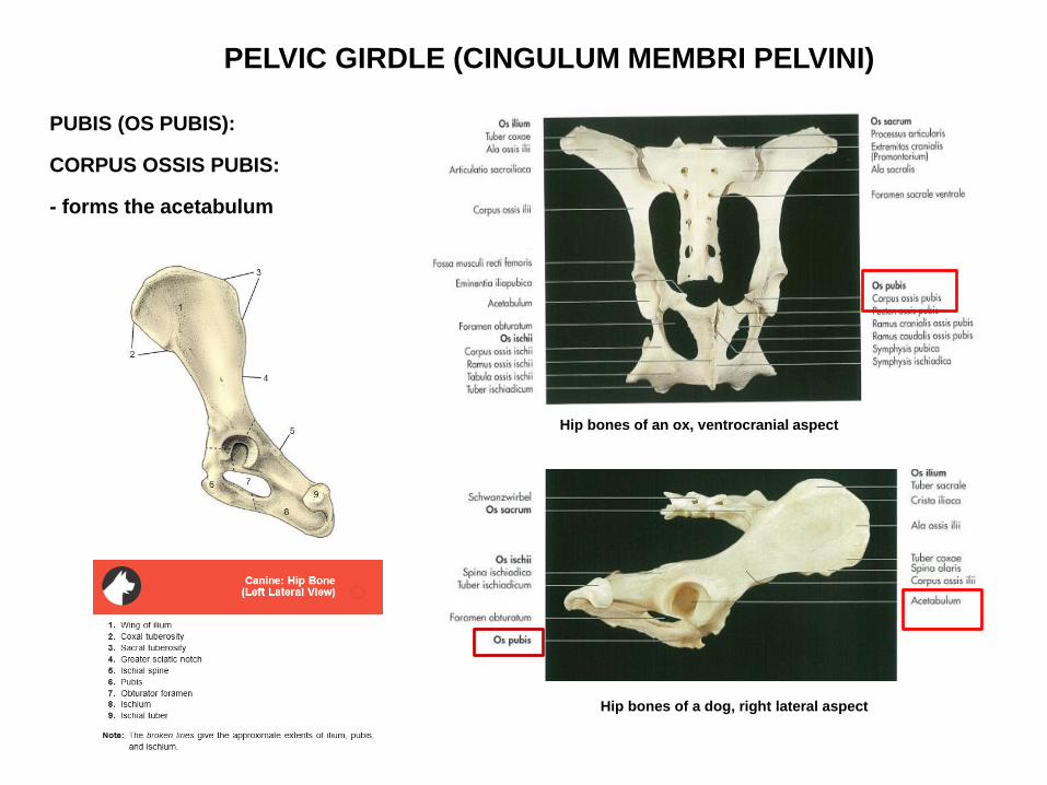
PELVIC GIRDLE (CINGULUM MEMBRI PELVINI)
PUBIS (OS PUBIS):
CORPUS OSSIS PUBIS:
- forms the acetabulum
Hip bones of an ox, ventrocranial aspect
Hip bones of a dog, right lateral aspect

PELVIC GIRDLE (CINGULUM MEMBRI PELVINI)
PUBIS (OS PUBIS):
RAMUS CRANIALIS OSSIS PUBIS:
- its cranial border knows as the:
PECTEN OSSIS PUBIS
Hip bones of an ox, ventrocranial aspect
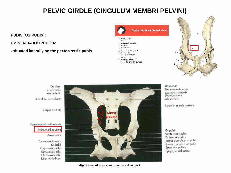
PELVIC GIRDLE (CINGULUM MEMBRI PELVINI)
PUBIS (OS PUBIS):
EMINENTIA ILIOPUBICA:
- situated laterally on the pecten ossis pubis
Hip bones of an ox, ventrocranial aspect
Linea
arcuata

PELVIC GIRDLE (CINGULUM MEMBRI PELVINI)
PUBIS (OS PUBIS):
SULCUS LIGAMENTI ACCESSORII OSSIS FEMORIS:
- in eq
- on the ventral surface of the pecten ossis pubis
http://vanat.cvm.umn.edu/ungDissect/Lab05/Img5-5.html
PELVIC GIRDLE
(CINGULUM MEMBRI PELVINI)
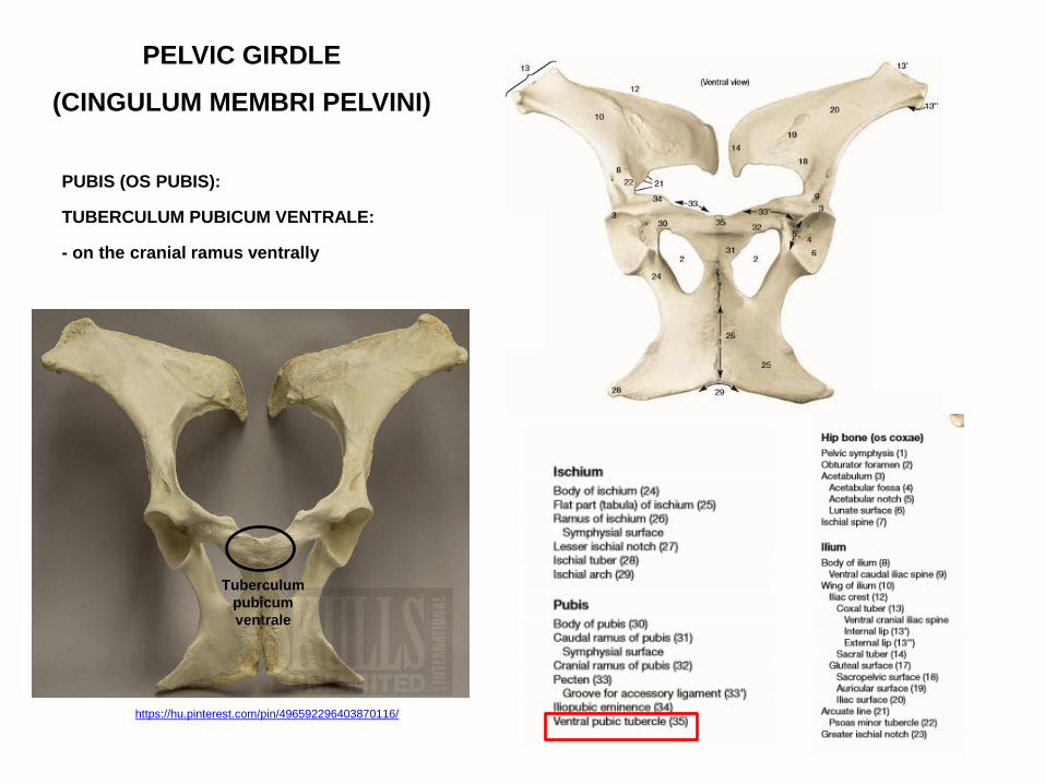
PUBIS (OS PUBIS):
TUBERCULUM PUBICUM VENTRALE:
- on the cranial ramus ventrally
https://hu.pinterest.com/pin/496592296403870116/
Tuberculum
pubicum
ventrale
PELVIC GIRDLE (CINGULUM MEMBRI PELVINI)
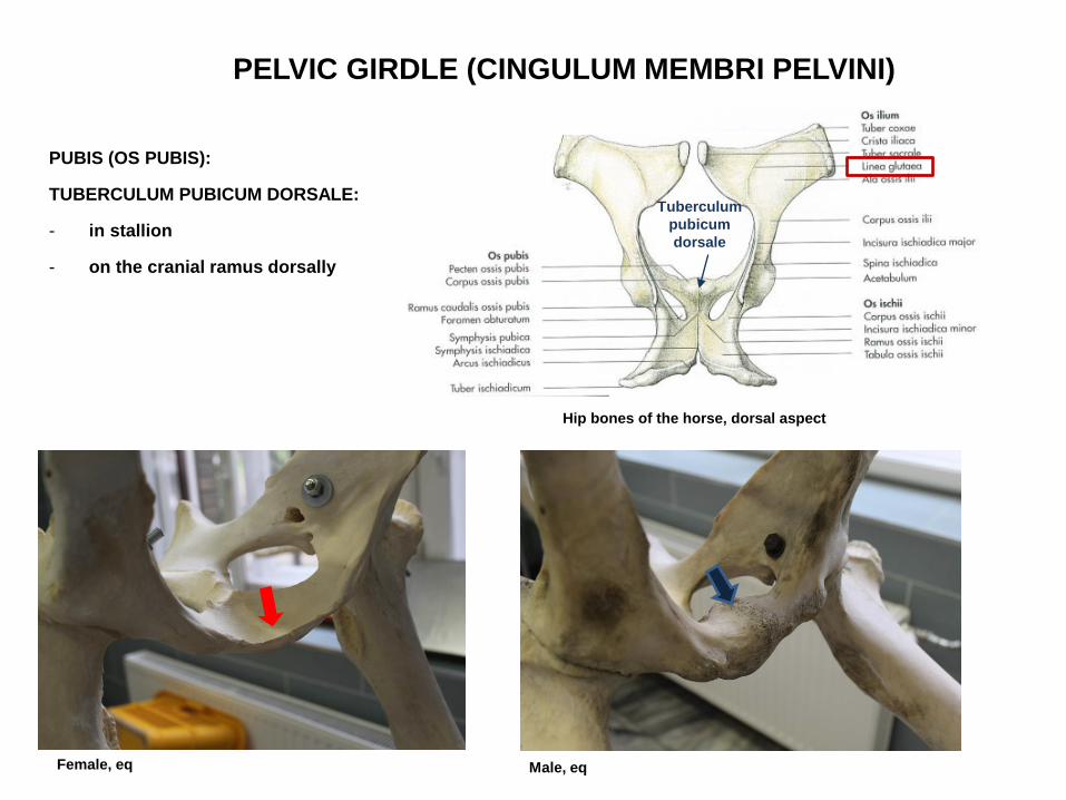
PUBIS (OS PUBIS):
TUBERCULUM PUBICUM DORSALE:
- in stallion
- on the cranial ramus dorsally
Hip bones of the horse, dorsal aspect
Tuberculum
pubicum
dorsale
Female, eq Male, eq
PELVIC GIRDLE (CINGULUM MEMBRI PELVINI)
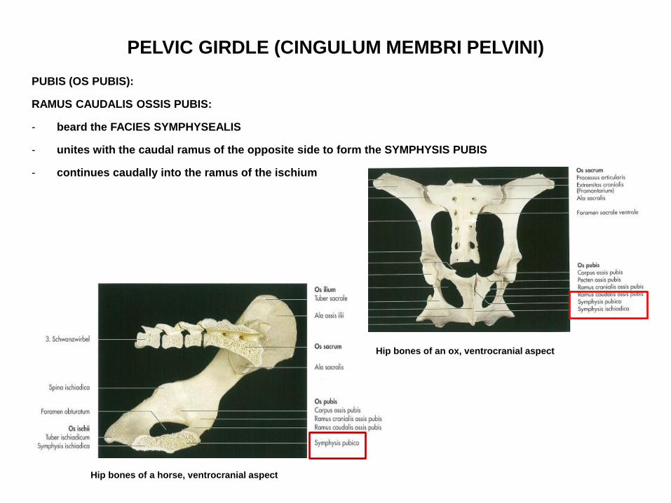
PUBIS (OS PUBIS):
RAMUS CAUDALIS OSSIS PUBIS:
- beard the FACIES SYMPHYSEALIS
- unites with the caudal ramus of the opposite side to form the SYMPHYSIS PUBIS
- continues caudally into the ramus of the ischium
Hip bones of a horse, ventrocranial aspect
Hip bones of an ox, ventrocranial aspect

PELVIC GIRDLE (CINGULUM MEMBRI PELVINI)
ISCHIUM (OS ISCHII):
consists of:
1. CORPUS OSSIS ISCHII
2. TABULA OSSIS ISCHII
3. RAMUS OSSIS ISCHII
Hip bones of an ox, ventrocranial aspect
https://www.tankonyvtar.hu/en/tartalom/tkt/haziallatok/ch02.html Eq, medial aspect Eq, lateral aspect

PELVIC GIRDLE (CINGULUM MEMBRI PELVINI)
ISCHIUM (OS ISCHII):
CORPUS OSSIS ISCHII:
- participates in the formation of acetabulum
Hip bones of a dog, right lateral aspect
Hip bones of an ox, ventrocranial aspect

PELVIC GIRDLE (CINGULUM MEMBRI PELVINI)
SPINA ISCHIADICA:
- the corpus expanded dorsally to form the spina ischiadica
- situated caudal to the incisura ischiadica major
- behind it lies the INCISURA ISCHIADICA MINOR (lesser sciatic notch)
Hip bones of a horse, left lateral aspect
Hip bones of an ox, ventrocranial aspect
Hip bones of a dog, right lateral aspect
Incisura ischiadica major
PELVIC GIRDLE
(CINGULUM MEMBRI PELVINI)
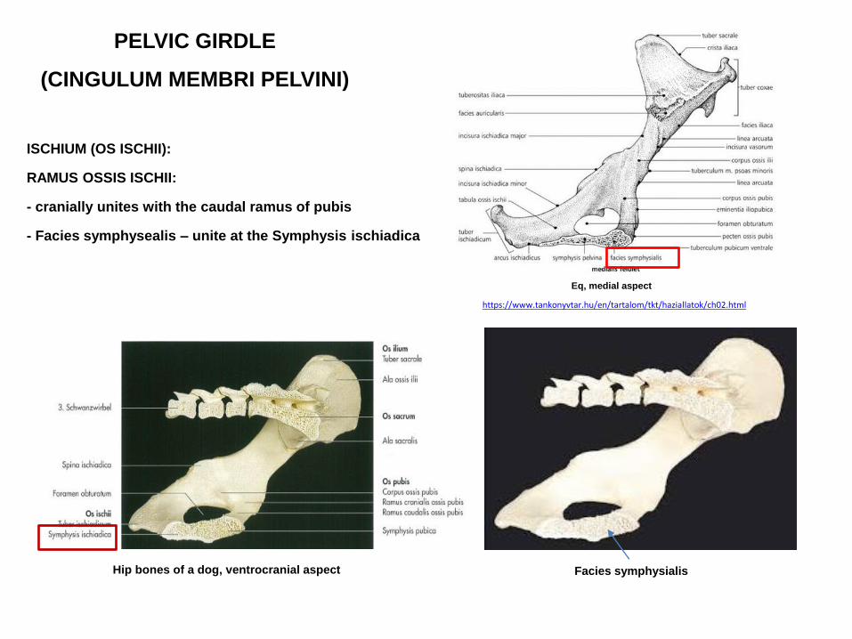
ISCHIUM (OS ISCHII):
RAMUS OSSIS ISCHII:
- cranially unites with the caudal ramus of pubis
- Facies symphysealis – unite at the Symphysis ischiadica
Hip bones of a dog, ventrocranial aspect
https://www.tankonyvtar.hu/en/tartalom/tkt/haziallatok/ch02.html
Eq, medial aspect
Facies symphysialis

PELVIC GIRDLE
(CINGULUM MEMBRI PELVINI)
ISCHIUM (OS ISCHII):
TABULA OSSIS ISCHII:
- form the TUBER ISCHIADICUM
Hip bones of a dog, ventrocranial aspect
http://vanat.cvm.umn.edu/ungDissect/Lab05/Img5-16.html
Tabula
ossis
ischii
https://www.tankonyvtar.hu/en/tartalom/tkt/haziallatok/ch02.html
Eq, medial aspect

PELVIC GIRDLE (CINGULUM MEMBRI PELVINI)
ISCHIUM (OS ISCHII):
TUBER ISCHIADICUM:
1. in eq, ca – presents a thickened ridge
2. in bo it is trituberculate
Hip bones of a dog, ventrocranial aspect
Hip bones of a horse, left lateral aspect
Hip bones of an ox, ventrocranial aspect
bo, tuber ischiadicum
eq, tuber ischiadicum
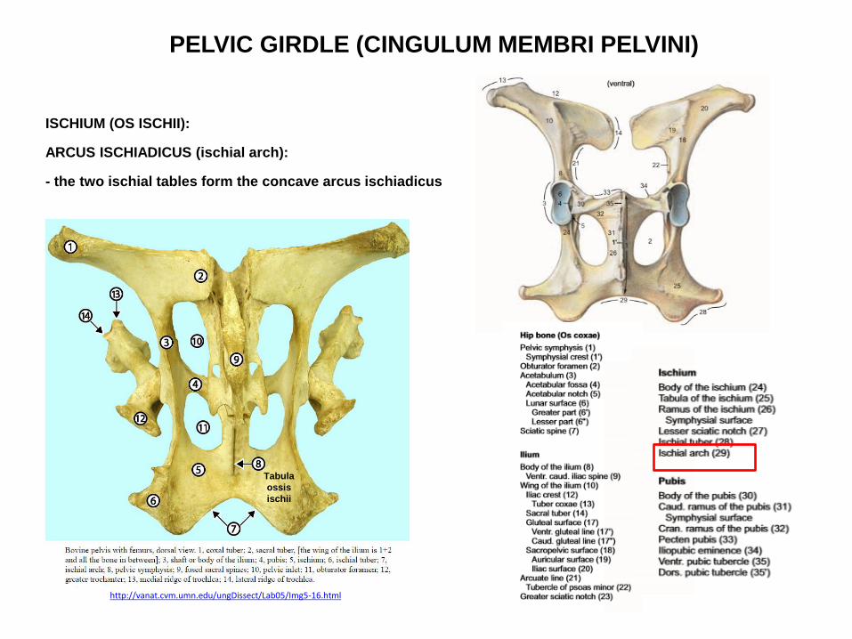
PELVIC GIRDLE (CINGULUM MEMBRI PELVINI)
ISCHIUM (OS ISCHII):
ARCUS ISCHIADICUS (ischial arch):
- the two ischial tables form the concave arcus ischiadicus
http://vanat.cvm.umn.edu/ungDissect/Lab05/Img5-16.html
Tabula
ossis
ischii
PELVIC GIRDLE (CINGULUM MEMBRI PELVINI)
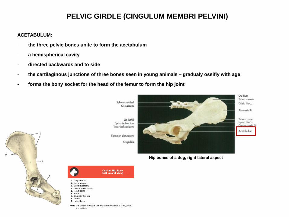
ACETABULUM:
- the three pelvic bones unite to form the acetabulum
- a hemispherical cavity
- directed backwards and to side
- the cartilaginous junctions of three bones seen in young animals – gradualy ossifiy with age
- forms the bony socket for the head of the femur to form the hip joint
Hip bones of a dog, right lateral aspect

PELVIC GIRDLE (CINGULUM MEMBRI PELVINI)
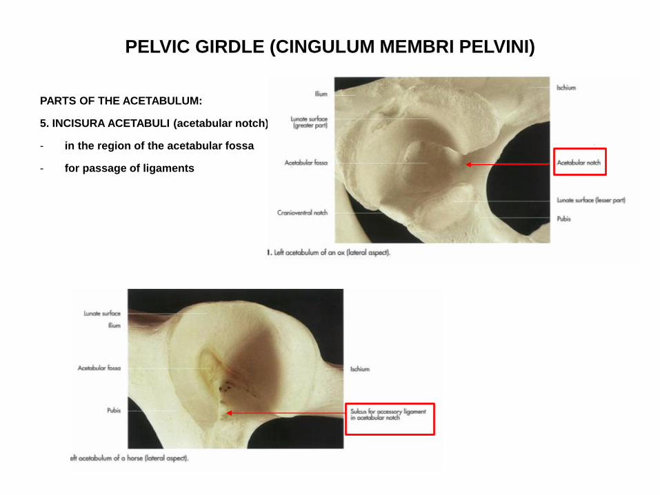
PARTS OF THE ACETABULUM:
1.MARGO ACTEABULI
2.FACIES LUNATA:
– ring – like articular part
- covered by cartilage
Margo
acetabuli
Margo acetabuli
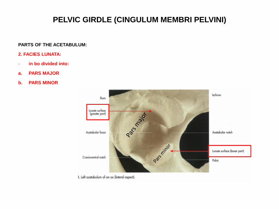
PELVIC GIRDLE (CINGULUM MEMBRI PELVINI)
PARTS OF THE ACETABULUM:
2. FACIES LUNATA:
- in bo divided into:
a. PARS MAJOR
b. PARS MINOR
PELVIC GIRDLE (CINGULUM MEMBRI PELVINI)
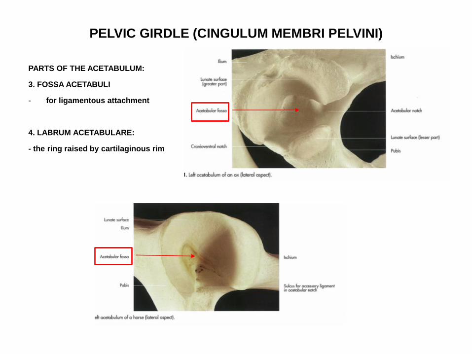
PARTS OF THE ACETABULUM:
3. FOSSA ACETABULI
- for ligamentous attachment
4. LABRUM ACETABULARE:
- the ring raised by cartilaginous rim
PELVIC GIRDLE (CINGULUM MEMBRI PELVINI)
PARTS OF THE ACETABULUM:
5. INCISURA ACETABULI (acetabular notch)
- in the region of the acetabular fossa
- for passage of ligaments

PELVIC GIRDLE
(CINGULUM MEMBRI PELVINI)
PUBIS (OS PUBIS):
FORAMEN OBTURATUM (obturator foramen):
formed by:
a) the corpus and the ramus cranialis of pubis form the cranial margin
b) the ramus caudalis ossis pubis form the medial margin
c) Ischium forms the caudal, medial, lateral boundaries
- SULCUS OBTURATORIUS
Hip bones of a dog, ventrocranial aspect
http://vanat.cvm.umn.edu/ungDissect/Lab05/Img5-16.html
Tabula
ossis
ischii
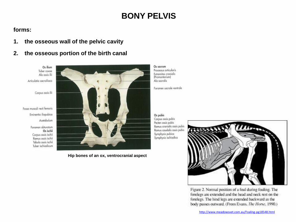
BONY PELVIS
forms:
1. the osseous wall of the pelvic cavity
2. the osseous portion of the birth canal
Hip bones of an ox, ventrocranial aspect
http://www.meadowsvet.com.au/Foaling-pg18548.html

BONY PELVIS
demarcated:
a. dorsally by :
• the sacrum
• the first 3-4 caudal vertebrae
b. ventrally by:
• the pubis,
• the ischium
c. on either side by:
• the ilium
• the body of the ischium
• the spina ischiadica
Hip bones of a horse, left lateral aspect

BONY PELVIS
demarcated:
d. the broad pelvic ligament
- absent in ca
e. musculature
http://vanat.cvm.umn.edu/ungDissect/Lab05/Img5-17.html
http://vanat.cvm.umn.edu/ungDissect/Lab05/Img5-1.html
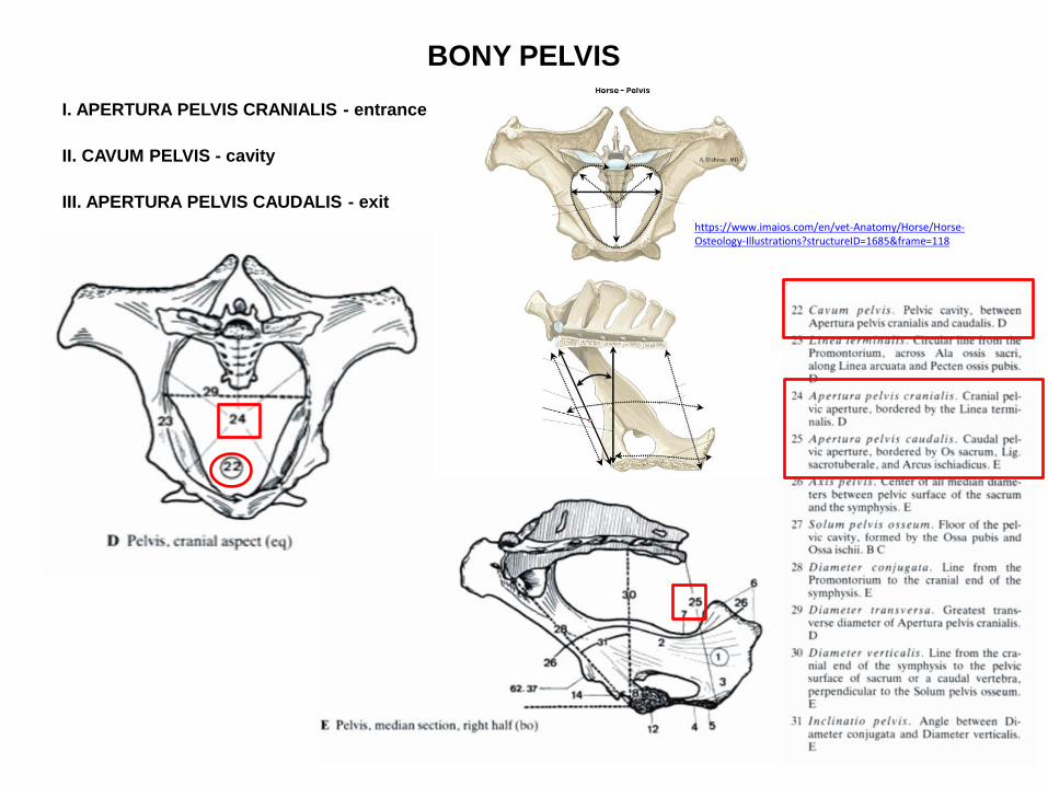
BONY PELVIS
I. APERTURA PELVIS CRANIALIS - entrance
II. CAVUM PELVIS - cavity
III. APERTURA PELVIS CAUDALIS - exit
https://www.imaios.com/en/vet-Anatomy/Horse/Horse-Osteology-Illustrations?structureID=1685&frame=118

BONY PELVIS
I. APERTURA PELVIS CRANIALIS:
- the inlet of the pelvic cavity
- formed by the LINEA TERMINALIS
23.
24.
https://www.imaios.com/en/vet-Anatomy/Horse/Horse-Osteology-Illustrations?structureID=1685&frame=118
Hip bones of a horse, ventrocranial aspect
24.
23.
24.
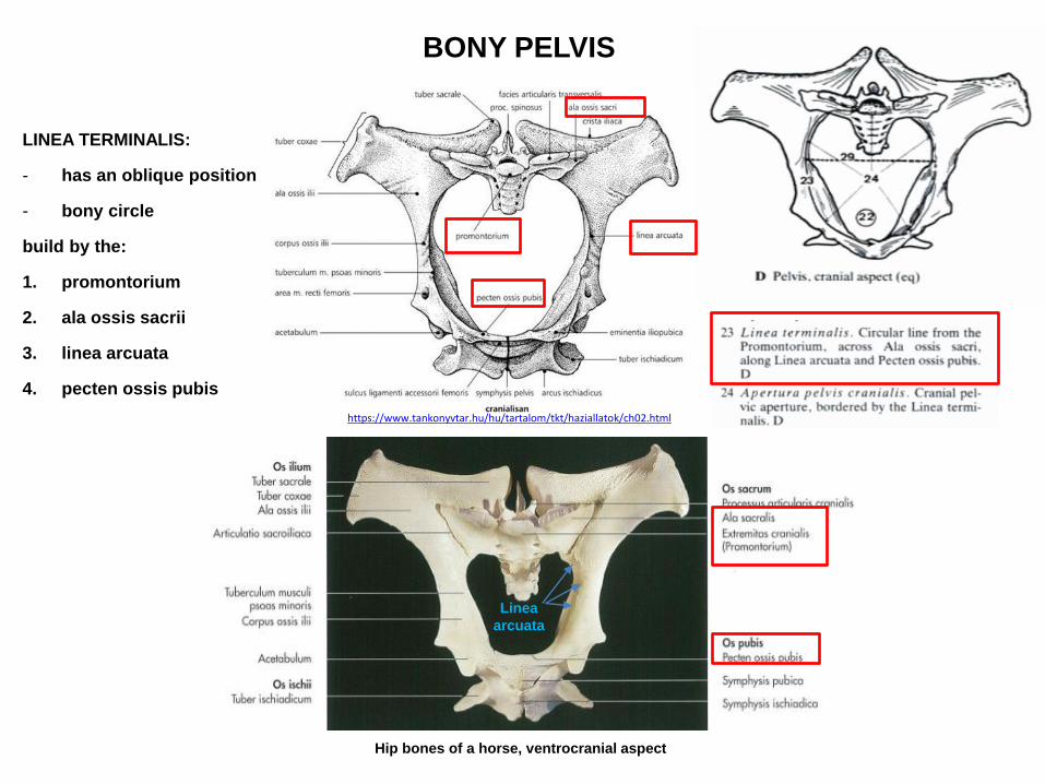
BONY PELVIS
LINEA TERMINALIS:
- has an oblique position
- bony circle
build by the:
1. promontorium
2. ala ossis sacrii
3. linea arcuata
4. pecten ossis pubis
Hip bones of a horse, ventrocranial aspect
Linea
arcuata
https://www.tankonyvtar.hu/hu/tartalom/tkt/haziallatok/ch02.html

BONY PELVIS
II. CAVUM PELVIS:
- lies behind the linea terminalis
- not separated from the abdominal cavity
Hip bones of a horse, ventrocranial aspect
24.

BONY PELVIS
III. APERTURA PELVIS CAUDALIS:
- the pelvic outlet
- narrower than the inlet
bounded ventrally by the:
a. arcus ischiadicus
b. tuberculum ischiadicum
Hip bones of a horse, left lateral aspect
http://vanat.cvm.umn.edu/ungDissect/Lab05/Img5-16.html
Tabula
ossis
ischii
https://commons.wikimedia.org/wiki/File:Horse_pelvis_02.jpg
25.
BONY PELVIS
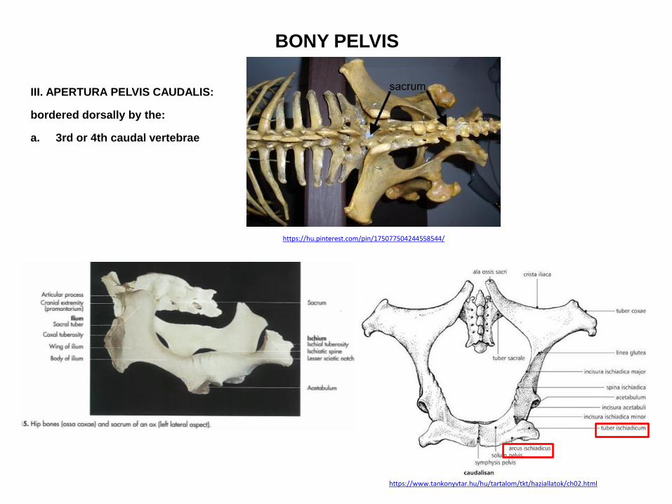
III. APERTURA PELVIS CAUDALIS:
bordered dorsally by the:
a. 3rd or 4th caudal vertebrae
https://hu.pinterest.com/pin/175077504244558544/
https://www.tankonyvtar.hu/hu/tartalom/tkt/haziallatok/ch02.html

BONY PELVIS
III. APERTURA PELVIS CAUDALIS:
Bordered laterally by the:
a. broad pelvic ligament in bo, eq
b. ligamentum sacrotuberale in ca
http://vanat.cvm.umn.edu/ungDissect/Lab05/Img5-1.html
https://twitter.com/juancarpalacio/status/457464399868227584
Canine pelvis with modelled
sacrotuberous ligament
http://www.onlineveterinaryanatomy.net/content/canine-pelvis-modelled-sacrotuberous-ligament

BONY PELVIS
III. APERTURA PELVIS CAUDALIS:
closed by:
- the DIAPHRAGMA PELVIS
Ca.
Ca.

BONY PELVIS
DIAPHRAGMA PELVIS:
- musculomembranous structure
- closes the aptertura pelvis caudalis
Ca.
Ca.
BONY PELVIS
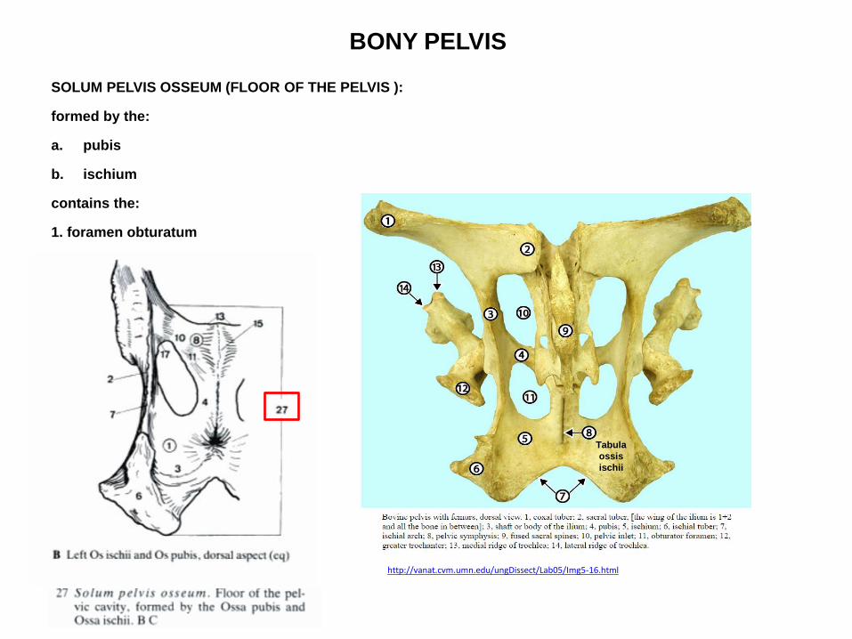
SOLUM PELVIS OSSEUM (FLOOR OF THE PELVIS ):
formed by the:
a. pubis
b. ischium
contains the:
1. foramen obturatum
http://vanat.cvm.umn.edu/ungDissect/Lab05/Img5-16.html
Tabula
ossis
ischii

DIAMETERS OF THE BONY PELVIS
important for evaluation of the functional structure of the pelvis
1. CONJUGATA VERA
2. CONJUGATA DIAGONALIS
3. DIAMETER VERTICALIS
4. DIAMETER TRANSVERSA

DIAMETERS OF THE BONY PELVIS
CONJUGATA VERA (diameter conjugata):
- the median line taken from the promontorium to cranial end of the pelvic symphysis
- provides an indication of the height of the pelvic inlet
Hip bones of a dog, ventrocranial aspect
DIAMETERS OF THE BONY PELVIS
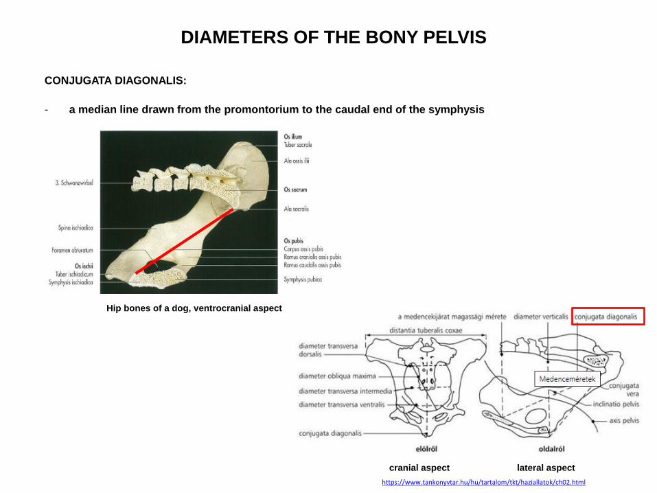
CONJUGATA DIAGONALIS:
- a median line drawn from the promontorium to the caudal end of the symphysis
Hip bones of a dog, ventrocranial aspect
cranial aspect lateral aspect
https://www.tankonyvtar.hu/hu/tartalom/tkt/haziallatok/ch02.html

DIAMETERS OF THE BONY PELVIS
DIAMETER VERTICALIS:
- a vertical line taken from the cranial end of the pelvic symphysis to the ventral surface of the sacrum or
the caudal vertebrae
- gives the true height of the pelvic cavity
Hip bones of a dog, ventrocranial aspect
Caudal
vertebrae
DIAMETERS OF THE BONY PELVIS
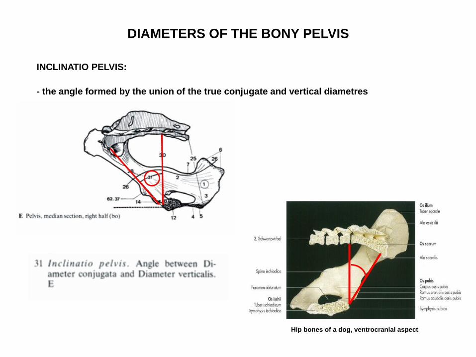
INCLINATIO PELVIS:
- the angle formed by the union of the true conjugate and vertical diametres
Hip bones of a dog, ventrocranial aspect

DIAMETERS OF THE BONY PELVIS
a. DIAMETER TRANSVERSA of the Apertura pelvis cranialis
- represents the greatest diameter of the terminal line
- taken between the two psoas tubercules
b. DIAMETER TRANSVERSA of the Apertura pelvis caudalis:
- the distance between the medial ends of the two ischiatic tuberosities
cranial aspect
https://www.tankonyvtar.hu/hu/tartalom/tkt/haziallatok/ch02.html

SEXUAL DIFFERENCE OF PELVIS
http://vanat.cvm.umn.edu/ungDissect/Lab05/Img5-4.html

SKELETON OF THE THIGH (SKELETON FEMORIS)
- formed by the OS FEMORIS (FEMUR)
- distally the femur releated in front to the patella
FEMUR:
- largest, long bone
1. PROXIMAL EPIPHYSIS
2. SHAFT (DIAPHYSIS, CORPUS FEMORIS)
3. DISTAL EPIPHYSIS
http://www.boneid.net/product/dog-canis-lupus-familiaris-right-femur-posterior-view/
Ca, right femur, caudal aspect
1.
2.
3.
Ca, right femur, cranial aspect

SKELETON OF THE THIGH (SKELETON FEMORIS)
OS FEMORIS (FEMUR)
http://www.boneid.net/product/dog-canis-lupus-familiaris-right-femur-posterior-view/
ca, right femur, cranial aspect caudal aspect eq, left femur, cranial aspect caudal aspect bo, left femur, cranial aspect caudal aspect
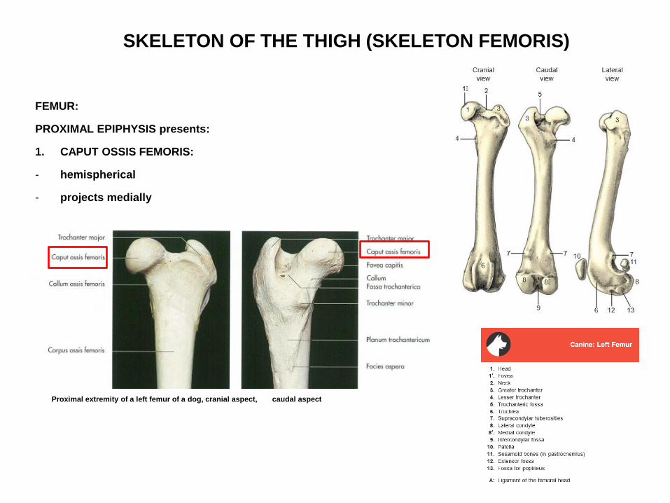
SKELETON OF THE THIGH (SKELETON FEMORIS)
FEMUR:
PROXIMAL EPIPHYSIS presents:
1. CAPUT OSSIS FEMORIS:
- hemispherical
- projects medially
Proximal extremity of a left femur of a dog, cranial aspect, caudal aspect

SKELETON OF THE THIGH (SKELETON FEMORIS)
FEMUR:
PROXIMAL EPIPHYSIS presents:
1. CAPUT OSSIS FEMORIS:
bears the FOVEA CAPITIS:
• attachment for the ligamentum capitis femoris
Proximal extremity of a left femur of a dog, cranial aspect, caudal aspect
http://vanat.cvm.umn.edu/ungDissect/Lab05/Img5-6.html
Eq.

SKELETON OF THE THIGH (SKELETON FEMORIS)
FEMUR:
PROXIMAL EPIPHYSIS presents:
2. COLLUM FEMORIS:
- in ca
Proximal extremity of a left femur of a dog, cranial aspect, caudal aspect

SKELETON OF THE THIGH
(SKELETON FEMORIS)
FEMUR:
PROXIMAL EPIPHYSIS presents:
3. TROCHANTER MAJOR:
- lies lateral to the head
IN HORSE divides into:
a. Pars cranialis
b. Pars caudalis
Proximal extremity of a left femur of a dog, cranial aspect, caudal aspect
http://vanat.cvm.umn.edu/ungDissect/Lab05/Img5-6.html
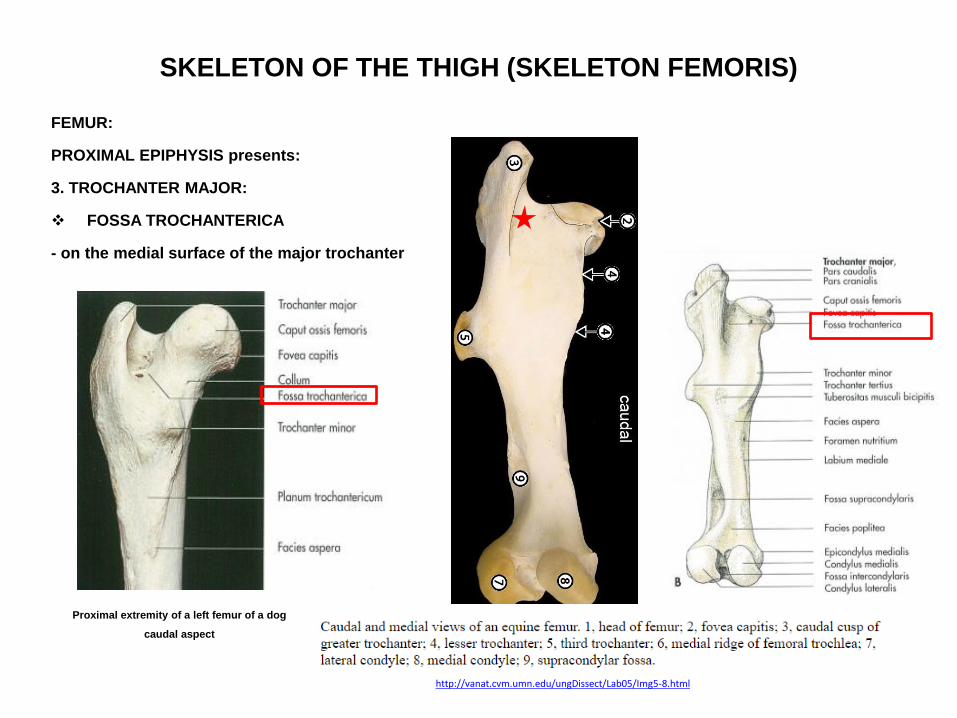
SKELETON OF THE THIGH (SKELETON FEMORIS)
FEMUR:
PROXIMAL EPIPHYSIS presents:
3. TROCHANTER MAJOR:
FOSSA TROCHANTERICA
- on the medial surface of the major trochanter
Proximal extremity of a left femur of a dog
caudal aspect
http://vanat.cvm.umn.edu/ungDissect/Lab05/Img5-8.html

SKELETON OF THE THIGH (SKELETON FEMORIS)
FEMUR:
PROXIMAL EPIPHYSIS presents:
4. TROCHANTER MINOR:
- located on the medial aspect of the shaft
- located distal to the head
Proximal extremity of a left femur of a dog
caudal aspect
http://vanat.cvm.umn.edu/ungDissect/Lab05/Img5-8.html
Bo, left femur caudal aspect
Trochanter
minor
Trochanter
major

SKELETON OF THE THIGH (SKELETON FEMORIS)
FEMUR:
PROXIMAL EPIPHYSIS presents:
5. LINEA INTERTROCHANTERICA:
- between the major and minor trochanter
- cranially
- poorly marked
http://qu.edu.iq/el/pluginfile.php/85109/mod_resource/content/1/The%20femur.pdf

SKELETON OF THE THIGH (SKELETON FEMORIS)
FEMUR:
PROXIMAL EPIPHYSIS presents:
6. CRISTA INTERTROCHANTERICA:
- caudally
- between the two trochanters
Bo, left femur caudal aspect
Proximal extremity of a left femur of a dog
caudal aspect

SKELETON OF THE THIGH (SKELETON FEMORIS)
FEMUR:
CORPUS OSSIS FEMORIS presents:
TUBEROSITAS GLUTEA:
- in carnovores
Proximal extremity of a left femur of a dog
caudal aspect

SKELETON OF THE THIGH (SKELETON FEMORIS)
FEMUR:
CORPUS OSSIS FEMORIS presents:
IN HORSE:
TROCHANTER TERTIUS:
- on the lateral surface of the shaft
- distal to the greater trochanter
http://vanat.cvm.umn.edu/ungDissect/Lab05/Img5-8.html
http://vanat.cvm.umn.edu/ungDissect/Lab05/Img5-7.html
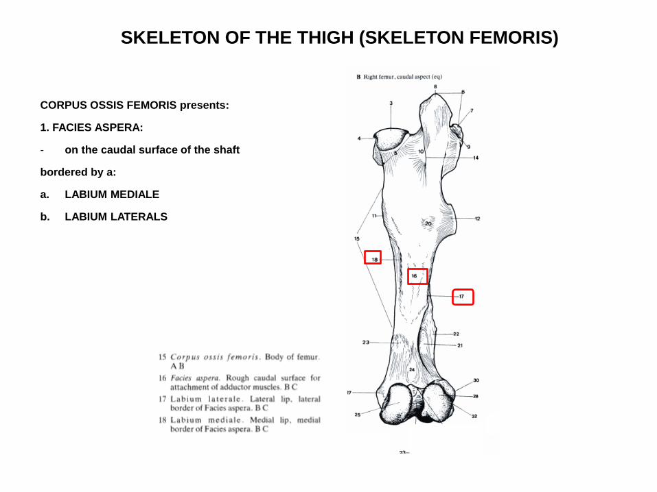
SKELETON OF THE THIGH (SKELETON FEMORIS)
CORPUS OSSIS FEMORIS presents:
1. FACIES ASPERA:
- on the caudal surface of the shaft
bordered by a:
a. LABIUM MEDIALE
b. LABIUM LATERALS

SKELETON OF THE THIGH (SKELETON FEMORIS)
FEMUR:
CORPUS OSSIS FEMORIS presents:
2. FACIES POPLITEA:
- the labium med. et lat. diverge at the distal end of the shaft to form the outline the facies poplitea
http://vanat.cvm.umn.edu/ungDissect/Lab05/Img5-8.html
Facies poplitea
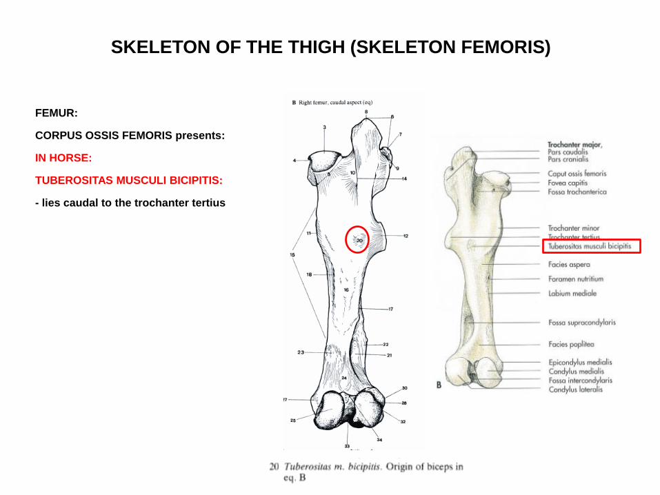
SKELETON OF THE THIGH (SKELETON FEMORIS)
FEMUR:
CORPUS OSSIS FEMORIS presents:
IN HORSE:
TUBEROSITAS MUSCULI BICIPITIS:
- lies caudal to the trochanter tertius

SKELETON OF THE THIGH (SKELETON FEMORIS)
FEMUR:
CORPUS OSSIS FEMORIS presents:
IN HORSE:
FOSSA SUPRACONDYLARIS:
- lies on the lateral aspect of the distal shaft
- increases the area of origin of the superficial digital flexor muscle
http://vanat.cvm.umn.edu/ungDissect/Lab05/Img5-8.html
Distal extremity of the right femur of a horse
distolateral aspect
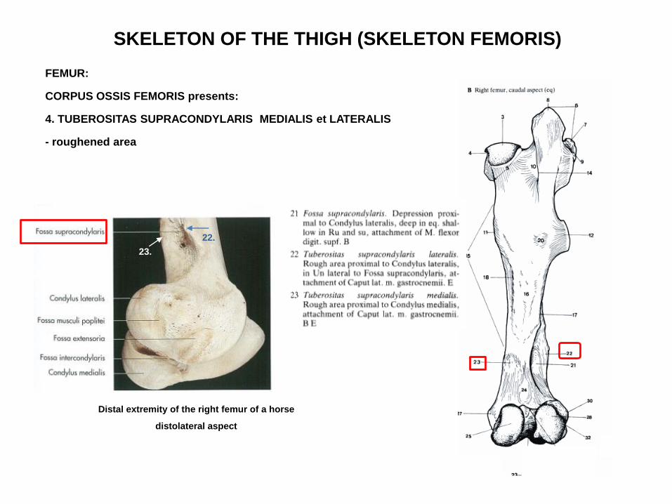
SKELETON OF THE THIGH (SKELETON FEMORIS)
FEMUR:
CORPUS OSSIS FEMORIS presents:
4. TUBEROSITAS SUPRACONDYLARIS MEDIALIS et LATERALIS
- roughened area
22.
Distal extremity of the right femur of a horse
distolateral aspect
22.
23.

SKELETON OF THE THIGH (SKELETON FEMORIS)
FEMUR
DISTAL EPIPHYSIS presents:
1. CONDYLUS MEDIALIS
2. CONDYLUS LATERALIS
3. FOSSA INTERCONDYLARIS:
- between the two condyles
Distal extremity of the right femur of a horse
distolateral aspect
Distal extremity of the left femur of a dog, cranial aspect caudal aspect
http://vanat.cvm.umn.edu/ungDissect/Lab05/Img5-8.html
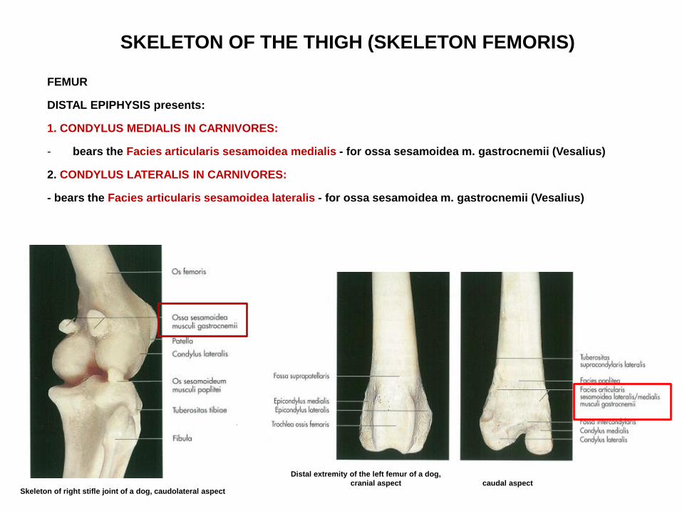
SKELETON OF THE THIGH (SKELETON FEMORIS)
FEMUR
DISTAL EPIPHYSIS presents:
1. CONDYLUS MEDIALIS IN CARNIVORES:
- bears the Facies articularis sesamoidea medialis - for ossa sesamoidea m. gastrocnemii (Vesalius)
2. CONDYLUS LATERALIS IN CARNIVORES:
- bears the Facies articularis sesamoidea lateralis - for ossa sesamoidea m. gastrocnemii (Vesalius)
Distal extremity of the left femur of a dog,
cranial aspect caudal aspect Skeleton of right stifle joint of a dog, caudolateral aspect
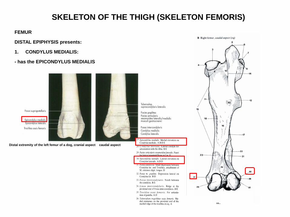
SKELETON OF THE THIGH (SKELETON FEMORIS)
FEMUR
DISTAL EPIPHYSIS presents:
1. CONDYLUS MEDIALIS:
- has the EPICONDYLUS MEDIALIS
Distal extremity of the left femur of a dog, cranial aspect caudal aspect
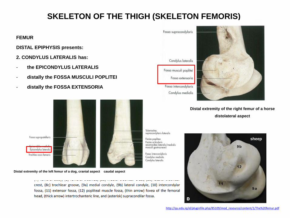
SKELETON OF THE THIGH (SKELETON FEMORIS)
FEMUR
DISTAL EPIPHYSIS presents:
2. CONDYLUS LATERALIS has:
- the EPICONDYLUS LATERALIS
- distally the FOSSA MUSCULI POPLITEI
- distally the FOSSA EXTENSORIA
Distal extremity of the left femur of a dog, cranial aspect caudal aspect
Distal extremity of the right femur of a horse
distolateral aspect
http://qu.edu.iq/el/pluginfile.php/85109/mod_resource/content/1/The%20femur.pdf
sheep
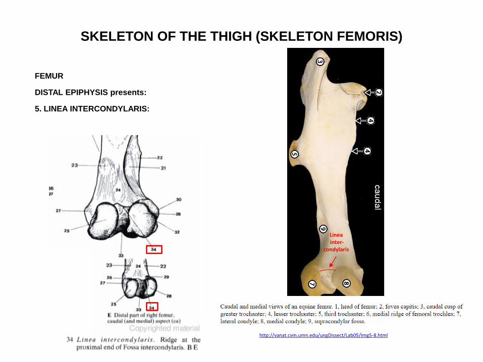
SKELETON OF THE THIGH (SKELETON FEMORIS)
FEMUR
DISTAL EPIPHYSIS presents:
5. LINEA INTERCONDYLARIS:
http://vanat.cvm.umn.edu/ungDissect/Lab05/Img5-8.html
Linea inter-
condylaris

SKELETON OF THE THIGH (SKELETON FEMORIS)
FEMUR
DISTAL EPIPHYSIS presents:
6. TROCHLEA OSSIS FEMORIS:
- cranially
- comprises two ridges separated by a groove
Distal extremity of the left femur of a dog,
cranial aspect caudal aspect
http://qu.edu.iq/el/pluginfile.php/85109/mod_resource/content/1/The%20femur.pdf

SKELETON OF THE THIGH (SKELETON FEMORIS)
FEMUR
DISTAL EPIPHYSIS presents:
6. TROCHLEA OSSIS FEMORIS:
- the medial ridge is larger on bo, eq
- in eq the medial ridge extends as the TUBERCULUM TROCHLEAE OSSIS FEMORIS
Left femur of a horse, cranial aspect Eq, left femur, cranial aspect

SKELETON OF THE THIGH
(SKELETON FEMORIS)
PATELLA:
- a sesamoid bone
- embedded in the ligamentum patellae
Skeleton of the right stifle joint of a dog, cranial aspect
Distal extremity of the left femur, patella, proximal extremity of the tibia of a horse
lateral aspect medial aspect
http://vanat.cvm.umn.edu/ungDissect/Lab06/Img6-2.html
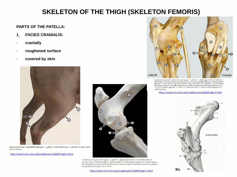
SKELETON OF THE THIGH (SKELETON FEMORIS)
PARTS OF THE PATELLA:
1. FACIES CRANIALIS:
- cranially
- roughened surface
- covered by skin
http://vanat.cvm.umn.edu/ungDissect/Lab06/Img6-1.html
http://vanat.cvm.umn.edu/ungDissect/Lab06/Img6-2.html
http://vanat.cvm.umn.edu/ungDissect/Lab06/Img6-3.html
Bo.
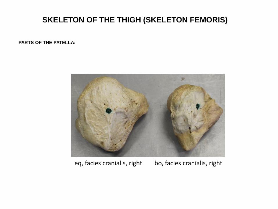
SKELETON OF THE THIGH (SKELETON FEMORIS)
PARTS OF THE PATELLA:
eq, facies cranialis, right bo, facies cranialis, right

SKELETON OF THE THIGH (SKELETON FEMORIS)
PARTS OF THE PATELLA:
2. BASIS:
- proximally
- attachament for muscles
https://www.tankonyvtar.hu/hu/tartalom/tkt/haziallatok/ch02.html
eq.
bo.
su.
ca.

SKELETON OF THE THIGH (SKELETON FEMORIS)
PARTS OF THE PATELLA:
3. APEX:
- distally
- attachament for muscles
https://www.tankonyvtar.hu/hu/tartalom/tkt/haziallatok/ch02.html
eq.
bo.
su.
ca.
left
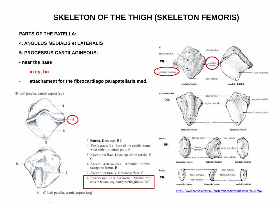
SKELETON OF THE THIGH (SKELETON FEMORIS)
PARTS OF THE PATELLA:
4. ANGULUS MEDIALIS et LATERALIS
5. PROCESSUS CARTILAGINEOUS:
- near the base
- in eq, bo
- attachament for the fibrocartilago parapatellaris med.
https://www.tankonyvtar.hu/hu/tartalom/tkt/haziallatok/ch02.html
eq.
bo.
su.
ca.
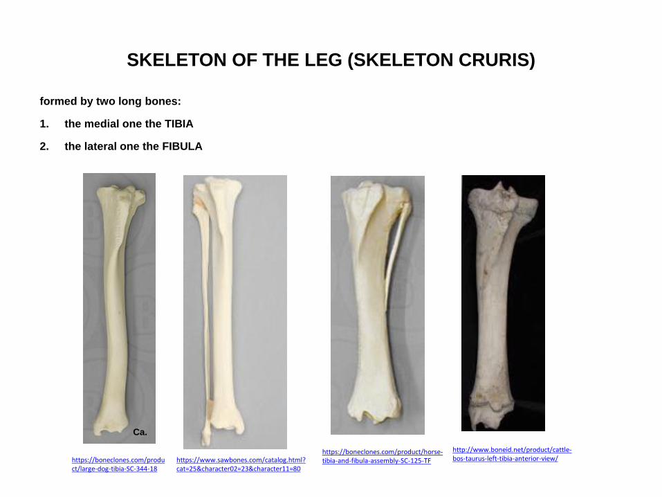
SKELETON OF THE LEG (SKELETON CRURIS)
formed by two long bones:
1. the medial one the TIBIA
2. the lateral one the FIBULA
https://boneclones.com/product/horse-tibia-and-fibula-assembly-SC-125-TF
Ca.
https://boneclones.com/product/large-dog-tibia-SC-344-18
https://www.sawbones.com/catalog.html?cat=25&character02=23&character11=80
http://www.boneid.net/product/cattle-bos-taurus-left-tibia-anterior-view/
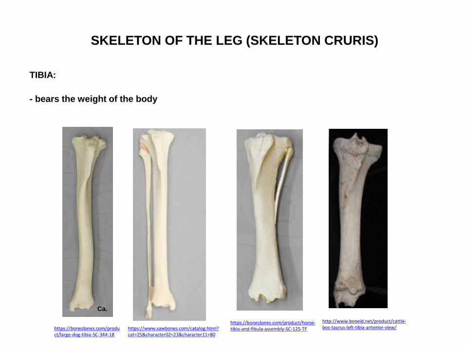
SKELETON OF THE LEG (SKELETON CRURIS)
TIBIA:
- bears the weight of the body
https://boneclones.com/product/horse-tibia-and-fibula-assembly-SC-125-TF
Ca.
https://boneclones.com/product/large-dog-tibia-SC-344-18
https://www.sawbones.com/catalog.html?cat=25&character02=23&character11=80
http://www.boneid.net/product/cattle-bos-taurus-left-tibia-anterior-view/

SKELETON OF THE LEG (SKELETON CRURIS)
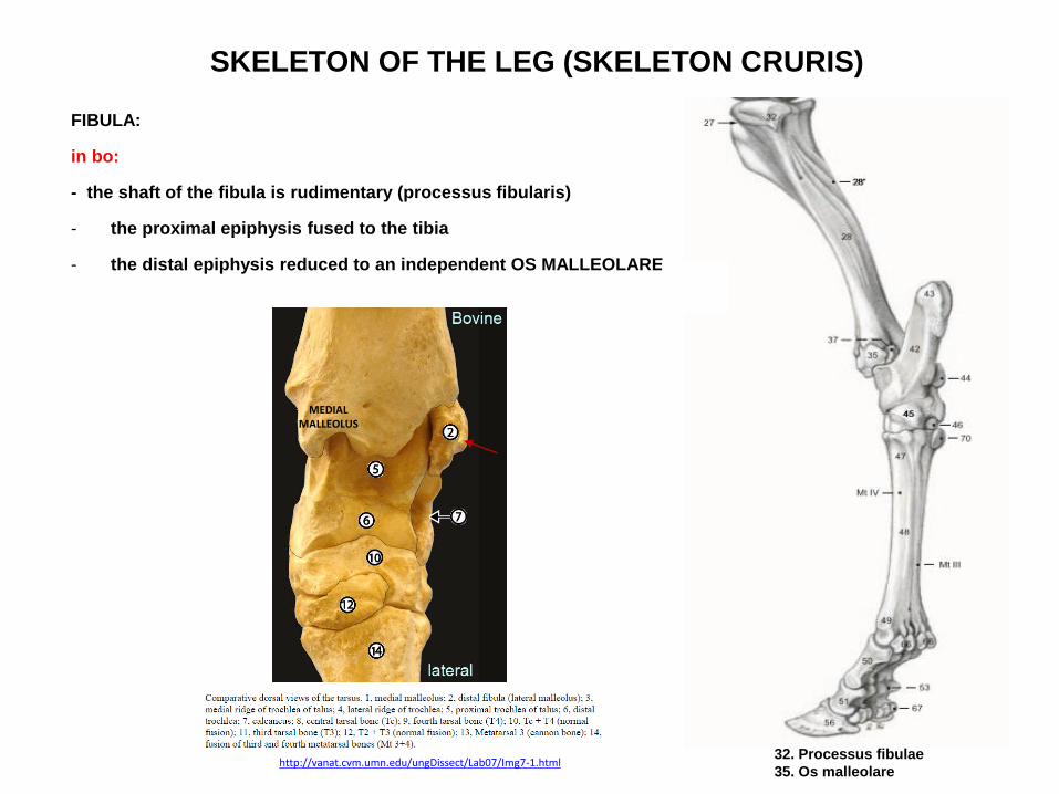
FIBULA:
in ca:
- the fibula is as long as the tibia
Ca.
https://boneclones.com/product/large-dog-tibia-SC-344-18
https://www.sawbones.com/catalog.html?cat=25&character02=23&character11=80
Ca.
http://www.boneid.net/product/dog-canis-lupus-familiaris-left-fibula-lateral-view/
SKELETON OF THE LEG (SKELETON CRURIS)
FIBULA:
in bo:
- the shaft of the fibula is rudimentary (processus fibularis)
- the proximal epiphysis fused to the tibia
- the distal epiphysis reduced to an independent OS MALLEOLARE
http://vanat.cvm.umn.edu/ungDissect/Lab07/Img7-1.html 32. Processus fibulae
35. Os malleolare
MEDIAL MALLEOLUS

SKELETON OF THE LEG (SKELETON CRURIS)
FIBULA:
in eq:
- extends as a free splint – like bone as far as the middle of the shaft of the tibia
- the distal epiphysis represented by a fusion with the tibia as the LATERAL MALLEOLUS
http://vanat.cvm.umn.edu/ungDissect/Lab07/Img7-1.html
MEDIAL MALLEOLUS
fibula
malleulus lat.

SKELETON OF THE LEG (SKELETON CRURIS)
SPATIUM INTEROSSEUM CRURIS:
- separates the two bones
a) in cat - wide
b) in dog – narrow
c) in eq – the space presents only proximally
d) in bo – no fibula = no space
Ca.
Spatium
interosseum
cruris
Bo. Eq.

SKELETON OF THE LEG (SKELETON CRURIS)
TIBIA:
1. PROXIMAL EPIPHYSIS
2. SHAFT (DIAPHYSIS, CORPUS)
3. DISTAL EPIPHYSIS
Ca.
https://boneclones.com/product/large-dog-tibia-SC-344-18
1.
2.
3.
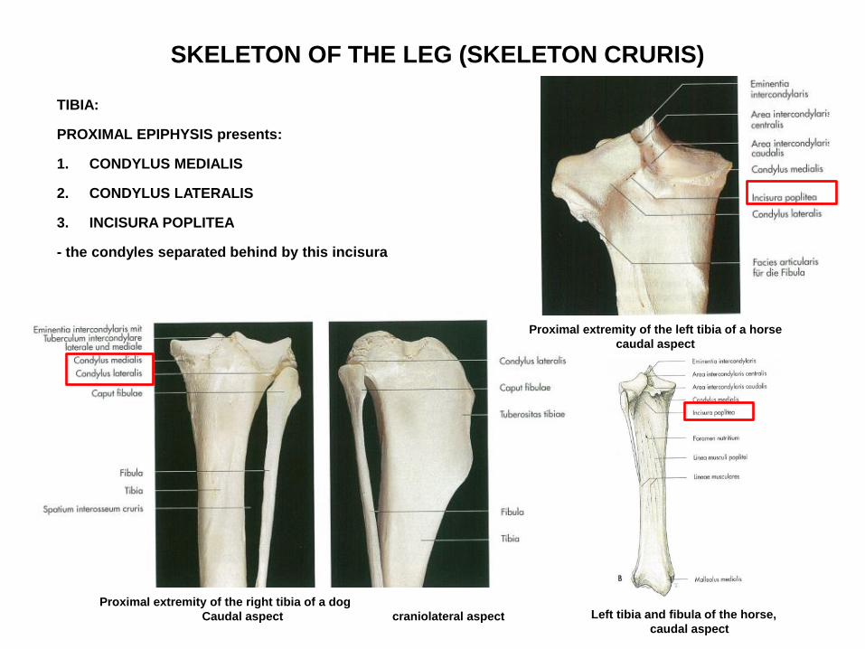
SKELETON OF THE LEG (SKELETON CRURIS)
TIBIA:
PROXIMAL EPIPHYSIS presents:
1. CONDYLUS MEDIALIS
2. CONDYLUS LATERALIS
3. INCISURA POPLITEA
- the condyles separated behind by this incisura
Proximal extremity of the right tibia of a dog
Caudal aspect craniolateral aspect
Proximal extremity of the left tibia of a horse
caudal aspect
Left tibia and fibula of the horse,
caudal aspect
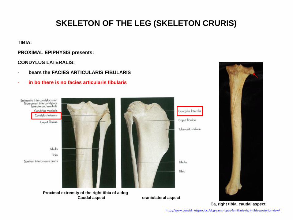
SKELETON OF THE LEG (SKELETON CRURIS)
TIBIA:
PROXIMAL EPIPHYSIS presents:
CONDYLUS LATERALIS:
- bears the FACIES ARTICULARIS FIBULARIS
- in bo there is no facies articularis fibularis
Proximal extremity of the right tibia of a dog
Caudal aspect craniolateral aspect
http://www.boneid.net/product/dog-canis-lupus-familiaris-right-tibia-posterior-view/
Ca, right tibia, caudal aspect

SKELETON OF THE LEG (SKELETON CRURIS)
TIBIA:
PROXIMAL EPIPHYSIS presents:
4. FACIES ARTICULARIS PROXIMALIS:
- on the condyles
- incongruent articular surface
- articulates via the menisci with the condyli femoris
Proximal extremity of the left tibia of a horse
caudal aspect
Facies
articularis
prox.
Facies
articularis prox.
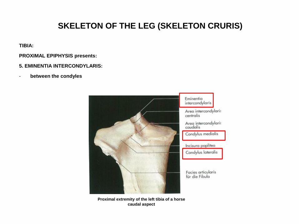
SKELETON OF THE LEG (SKELETON CRURIS)
TIBIA:
PROXIMAL EPIPHYSIS presents:
5. EMINENTIA INTERCONDYLARIS:
- between the condyles
Proximal extremity of the left tibia of a horse
caudal aspect
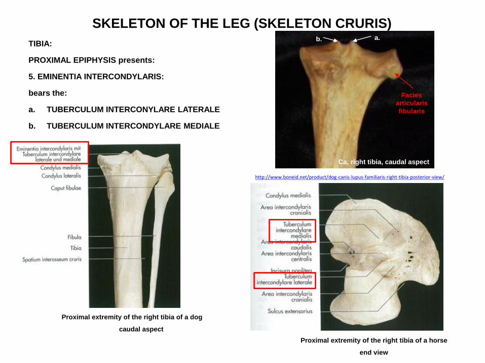
SKELETON OF THE LEG (SKELETON CRURIS)
TIBIA:
PROXIMAL EPIPHYSIS presents:
5. EMINENTIA INTERCONDYLARIS:
bears the:
a. TUBERCULUM INTERCONYLARE LATERALE
b. TUBERCULUM INTERCONDYLARE MEDIALE
Proximal extremity of the right tibia of a dog
caudal aspect
http://www.boneid.net/product/dog-canis-lupus-familiaris-right-tibia-posterior-view/
Ca, right tibia, caudal aspect
Facies
articularis
fibularis
a. b.
Proximal extremity of the right tibia of a horse
end view
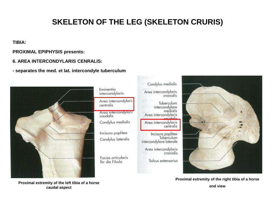
SKELETON OF THE LEG (SKELETON CRURIS)
TIBIA:
PROXIMAL EPIPHYSIS presents:
6. AREA INTERCONDYLARIS CENRALIS:
- separates the med. et lat. intercondyle tuberculum
Proximal extremity of the left tibia of a horse
caudal aspect
Proximal extremity of the right tibia of a horse
end view
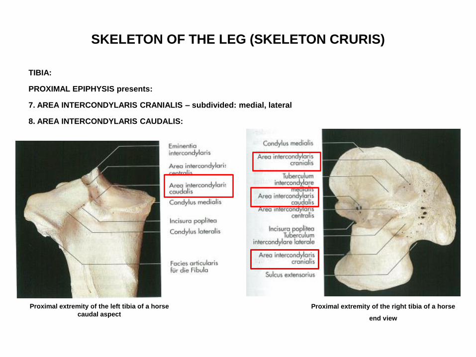
SKELETON OF THE LEG (SKELETON CRURIS)
TIBIA:
PROXIMAL EPIPHYSIS presents:
7. AREA INTERCONDYLARIS CRANIALIS – subdivided: medial, lateral
8. AREA INTERCONDYLARIS CAUDALIS:
Proximal extremity of the left tibia of a horse
caudal aspect
Proximal extremity of the right tibia of a horse
end view
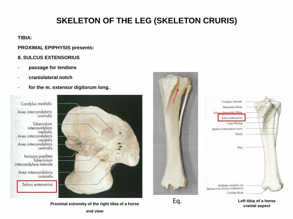
SKELETON OF THE LEG (SKELETON CRURIS)
TIBIA:
PROXIMAL EPIPHYSIS presents:
8. SULCUS EXTENSORIUS
- passage for tendons
- craniolateral notch
- for the m. extensor digitorum long.
Proximal extremity of the right tibia of a horse
end view
Eq. Left tibia of a horse
cranial aspect
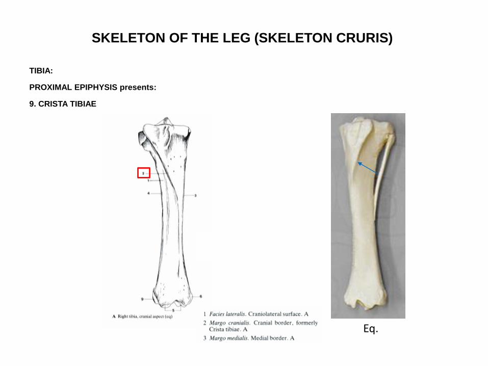
SKELETON OF THE LEG (SKELETON CRURIS)
TIBIA:
PROXIMAL EPIPHYSIS presents:
9. CRISTA TIBIAE
Eq.

SKELETON OF THE LEG (SKELETON CRURIS)
TIBIA:
PROXIMAL EPIPHYSIS presents:
IN RUMINANTS:
PROCESSUS FIBULARIS
- caput fibulae rudiment, and fused wirth the tibia
32. Processus fibulae
35. Os malleolare
Proc.
fibularis
Os malleolare
Bo, right tibia, cranial aspect https://www.flickr.com/photos/jrochester/38736449362
Crista
tibiae
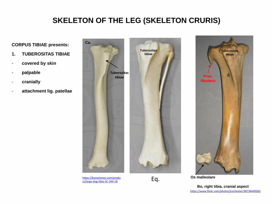
SKELETON OF THE LEG (SKELETON CRURIS)
CORPUS TIBIAE presents:
1. TUBEROSITAS TIBIAE
- covered by skin
- palpable
- cranially
- attachment lig. patellae
Proc.
fibularis
Os malleolare
Bo, right tibia, cranial aspect https://www.flickr.com/photos/jrochester/38736449362
Tuberositas
tibiae
Eq.
Tuberositas
tibiae
Ca.
https://boneclones.com/product/large-dog-tibia-SC-344-18
Tuberositas tibiae
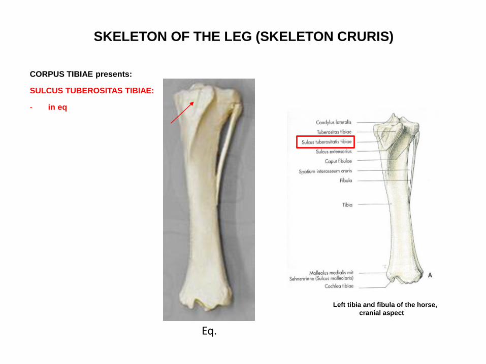
SKELETON OF THE LEG (SKELETON CRURIS)
CORPUS TIBIAE presents:
SULCUS TUBEROSITAS TIBIAE:
- in eq
Eq.
Left tibia and fibula of the horse,
cranial aspect

SKELETON OF THE LEG (SKELETON CRURIS)
CORPUS TIBIAE presents:
2. MARGO CRANIALIS (Crista tibiae)
3. FACIES MEDIALIS
4. FACIES LATERALIS
5. FACIES CAUDALIS
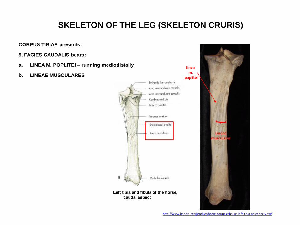
SKELETON OF THE LEG (SKELETON CRURIS)
CORPUS TIBIAE presents:
5. FACIES CAUDALIS bears:
a. LINEA M. POPLITEI – running mediodistally
b. LINEAE MUSCULARES
Left tibia and fibula of the horse,
caudal aspect
http://www.boneid.net/product/horse-equus-caballus-left-tibia-posterior-view/
Linea m.
poplitei
Lineae musculares

SKELETON OF THE LEG (SKELETON CRURIS)
DISTAL EPIPHYSIS presents:
1. COCHLEA TIBIAE
- forms the articular surface between the tibia and the tarsal bones
Left tibia and fibula of the horse,
cranial aspect caudal aspect
Distal extremity of the right tibia and fibula of a dog
caudal aspect
Ca.
https://boneclones.com/product/large-dog-tibia-SC-344-18
eq., cochlea tibiae
Bo, cochlea tibiae
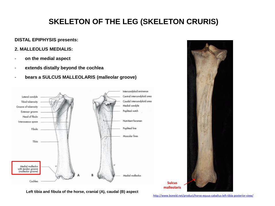
SKELETON OF THE LEG (SKELETON CRURIS)
DISTAL EPIPHYSIS presents:
2. MALLEOLUS MEDIALIS:
- on the medial aspect
- extends distally beyond the cochlea
- bears a SULCUS MALLEOLARIS (malleolar groove)
Left tibia and fibula of the horse, cranial (A), caudal (B) aspect http://www.boneid.net/product/horse-equus-caballus-left-tibia-posterior-view/
Sulcus malleolaris

SKELETON OF THE LEG (SKELETON CRURIS)
DISTAL EPIPHYSIS presents:
3. INCISURA FIBULARIS:
- laterally
- for articulation with the fibula
- in eq, bo no incisura fibularis
Distal extremity of the right tibia and fibula of a dog
caudal aspect
Ca.
https://boneclones.com/product/large-dog-tibia-SC-344-18
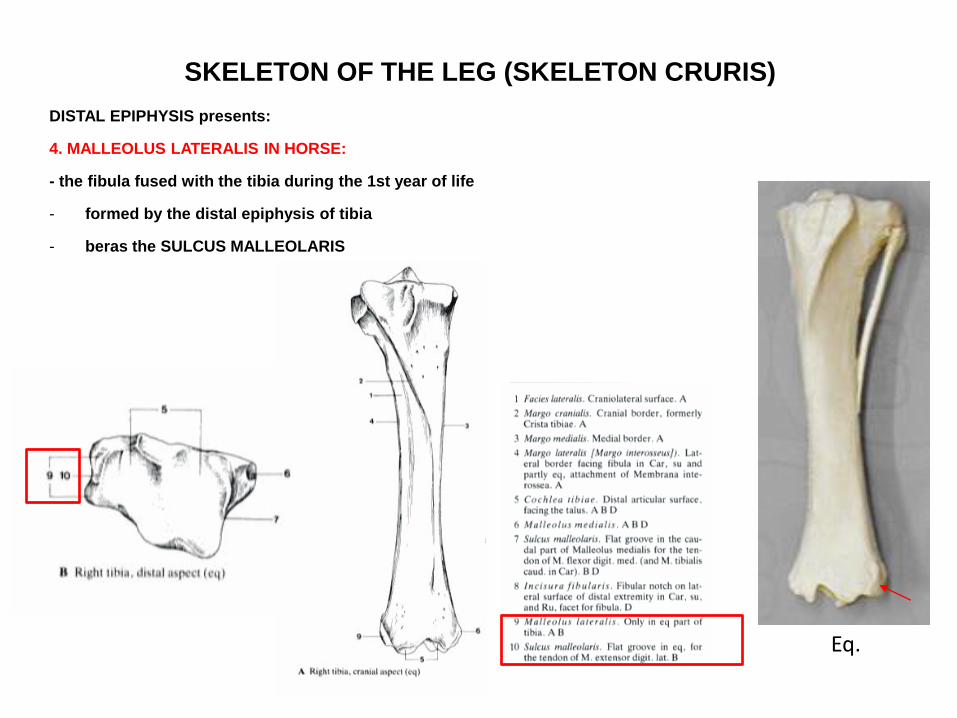
SKELETON OF THE LEG (SKELETON CRURIS)
DISTAL EPIPHYSIS presents:
4. MALLEOLUS LATERALIS IN HORSE:
- the fibula fused with the tibia during the 1st year of life
- formed by the distal epiphysis of tibia
- beras the SULCUS MALLEOLARIS
Eq.

SKELETON OF THE LEG (SKELETON CRURIS)
DISTAL EPIPHYSIS presents:
IN RUMINANTS:
- FACIES ARTICULARIS LATERALIS – articulates with the lateral malleolus – OS MALLEOLARE
- the lateral malleolus is the distal rudiment of the fibula
32. Processus fibulae
35. Os malleolare
Proc.
fibularis
Os malleolare
Bo, right tibia, cranial aspect
https://www.flickr.com/photos/jrochester/38736449362
Crista
tibiae
https://www.researchgate.net/figure/Maleollar-bone-in-camel-A-and-cow-B_fig2_268254773
Maleollar bone in cow (B)
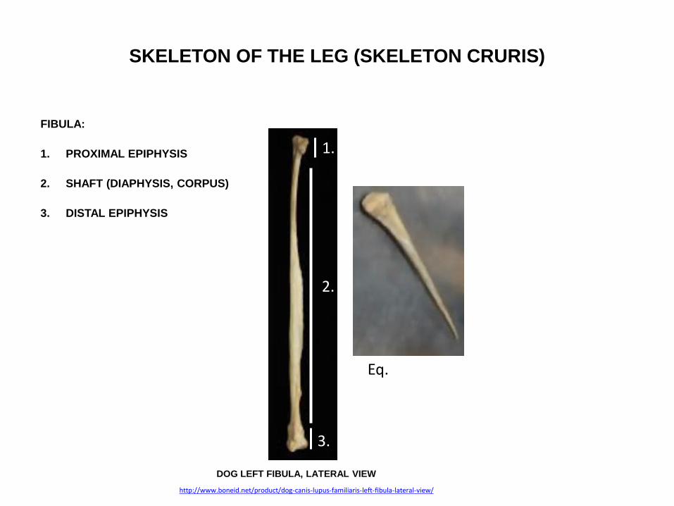
SKELETON OF THE LEG (SKELETON CRURIS)
FIBULA:
1. PROXIMAL EPIPHYSIS
2. SHAFT (DIAPHYSIS, CORPUS)
3. DISTAL EPIPHYSIS
DOG LEFT FIBULA, LATERAL VIEW
http://www.boneid.net/product/dog-canis-lupus-familiaris-left-fibula-lateral-view/
1.
2.
3.
Eq.

SKELETON OF THE LEG (SKELETON CRURIS)
FIBULA:
PROXIMAL EPIPHYSIS forms the CAPUT FIBULAE
CAPUT FIBULAE beras:
- FACIES ARTICULARIS CAPITIS FIBULAE – for the articulation with the lateral condyle of the tibia
Proximal extremity of the right tibia of a dog
caudal aspect craniolateral aspect
Su.
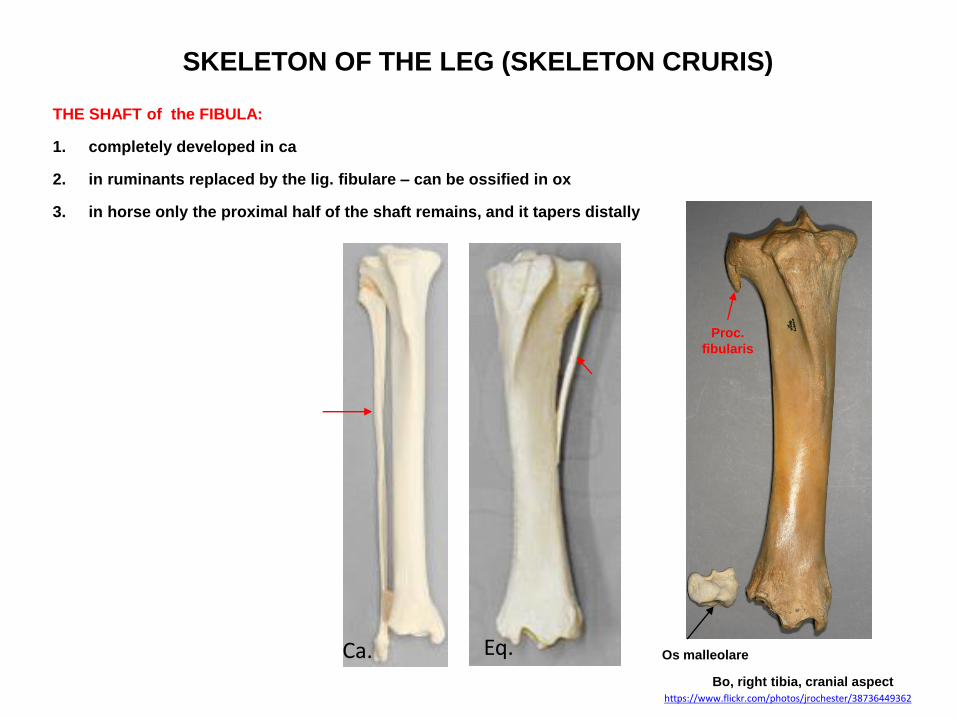
SKELETON OF THE LEG (SKELETON CRURIS)
THE SHAFT of the FIBULA:
1. completely developed in ca
2. in ruminants replaced by the lig. fibulare – can be ossified in ox
3. in horse only the proximal half of the shaft remains, and it tapers distally
Proc.
fibularis
Os malleolare
Bo, right tibia, cranial aspect https://www.flickr.com/photos/jrochester/38736449362
Eq. Ca.
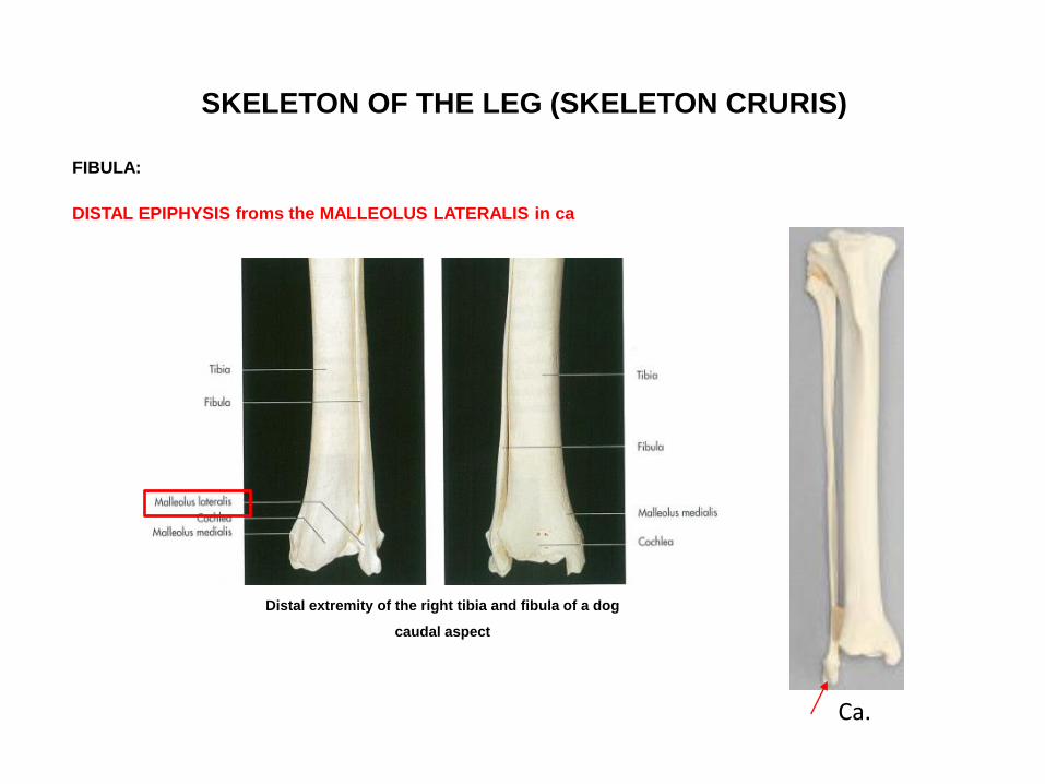
SKELETON OF THE LEG (SKELETON CRURIS)
FIBULA:
DISTAL EPIPHYSIS froms the MALLEOLUS LATERALIS in ca
Ca.
Distal extremity of the right tibia and fibula of a dog
caudal aspect

SKELETON OF THE LEG (SKELETON CRURIS)
FIBULA:
MALLEOLUS LATERALIS bears:
a. SULCUS MALLEOLARIS in ru.
b. SULCUS TENDINIS M. PERONEI LONGI in ca.
c. SULCUS TENDINUM MM. EXTENSORIS DIGIT. LAT. et PERONEUS BREVIS in ca.
Su, left fibula
Caudomedial aspect
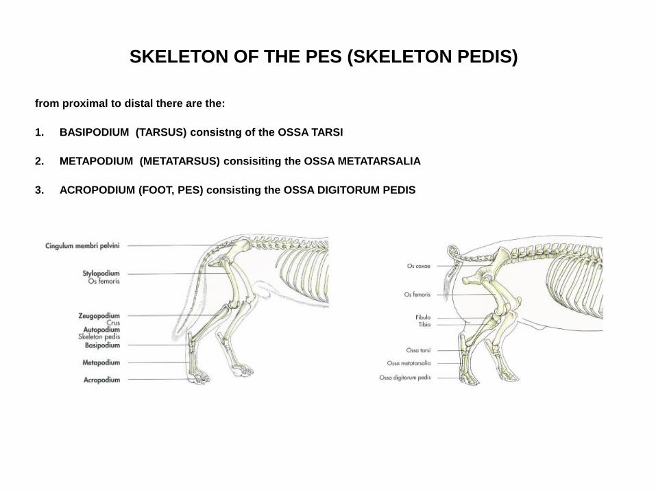
SKELETON OF THE PES (SKELETON PEDIS)
from proximal to distal there are the:
1. BASIPODIUM (TARSUS) consistng of the OSSA TARSI
2. METAPODIUM (METATARSUS) consisiting the OSSA METATARSALIA
3. ACROPODIUM (FOOT, PES) consisting the OSSA DIGITORUM PEDIS
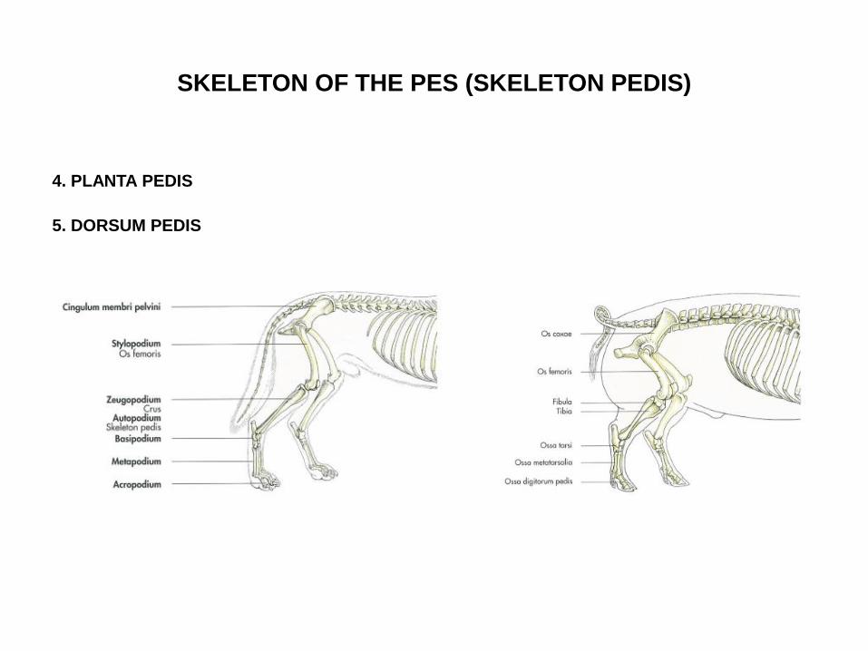
SKELETON OF THE PES (SKELETON PEDIS)
4. PLANTA PEDIS
5. DORSUM PEDIS

SKELETON OF THE TARSUS
consists of three rows of the tarsal bones:
1. PROXIMAL or CRURAL ROW
2. MIDDLE or INTERTARSAL ROW
3. DISTAL or METATARSAL ROW
Ca. Su. Bo. Eq.

SKELETON OF THE TARSUS
PROXIMAL or CRURAL ROW consists of:
1. TALUS
2. CALCANEUS
Ca. Su. Bo. Eq.
http://vanat.cvm.umn.edu/ungDissect/Lab07/Img7-3.html Skeleton of the right tarsus of a dog, plantar aspect
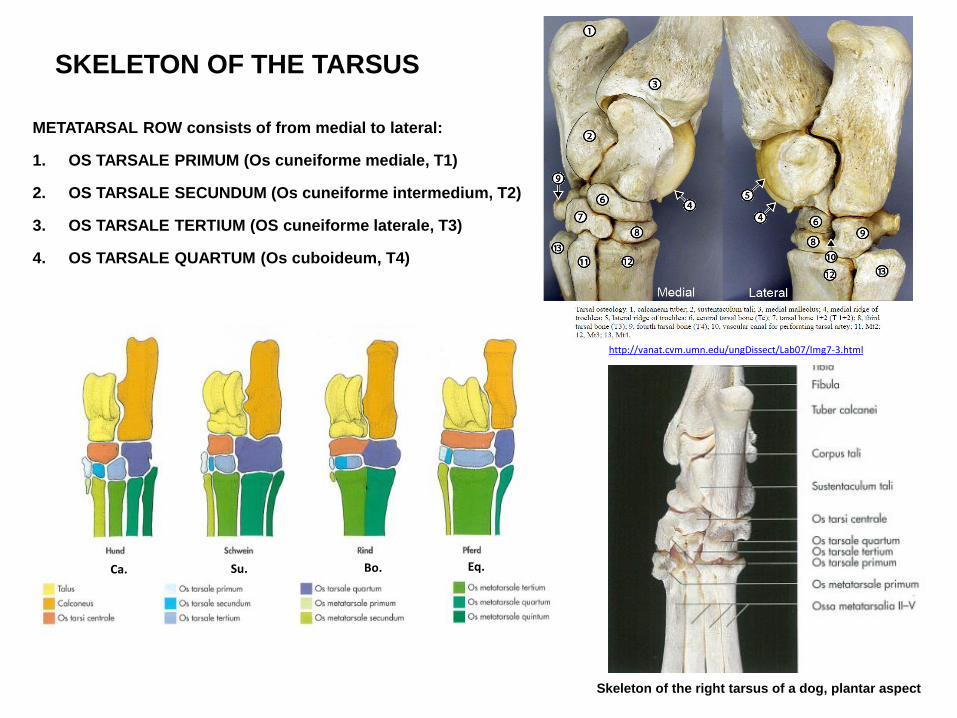
SKELETON OF THE TARSUS
METATARSAL ROW consists of from medial to lateral:
1. OS TARSALE PRIMUM (Os cuneiforme mediale, T1)
2. OS TARSALE SECUNDUM (Os cuneiforme intermedium, T2)
3. OS TARSALE TERTIUM (OS cuneiforme laterale, T3)
4. OS TARSALE QUARTUM (Os cuboideum, T4)
Ca. Su. Bo. Eq.
http://vanat.cvm.umn.edu/ungDissect/Lab07/Img7-3.html
Skeleton of the right tarsus of a dog, plantar aspect
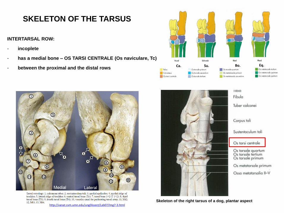
SKELETON OF THE TARSUS
INTERTARSAL ROW:
- incoplete
- has a medial bone – OS TARSI CENTRALE (Os naviculare, Tc)
- between the proximal and the distal rows
http://vanat.cvm.umn.edu/ungDissect/Lab07/Img7-3.html Skeleton of the right tarsus of a dog, plantar aspect
Ca. Su. Bo. Eq.
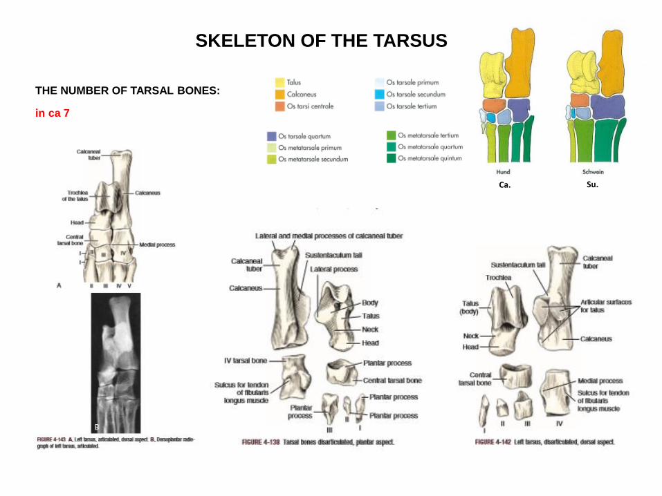
SKELETON OF THE TARSUS
THE NUMBER OF TARSAL BONES:
in ca 7
Ca. Su.
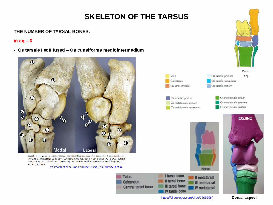
SKELETON OF THE TARSUS
THE NUMBER OF TARSAL BONES:
in eq – 6
- Os tarsale I et II fused – Os cuneiforme mediointermedium
Eq.
https://slideplayer.com/slide/3898358/
http://vanat.cvm.umn.edu/ungDissect/Lab07/Img7-3.html
Dorsal aspect
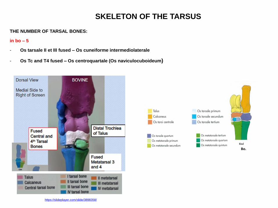
SKELETON OF THE TARSUS
THE NUMBER OF TARSAL BONES:
in bo – 5
- Os tarsale II et III fused – Os cuneiforme intermediolaterale
- Os Tc and T4 fused – Os centroquartale (Os naviculocuboideum)
https://slideplayer.com/slide/3898358/
Bo.

SKELETON OF THE TARSUS
https://slideplayer.com/slide/3898358/

SKELETON OF THE TARSUS
TALUS
- on the medial side
PARTS OF THE TALUS:
1. CORPUS TALI
2. CAPUT TALI
3. COLLUM TALI in ca, betwenn the caput and corpus tali
Ca., plantar aspect Ca., dorsal aspect
Caput tali
Collum tali
Trochlea
tali
http://www.boneid.net/product/bobcat-lynx-rufus-left-astragalus-superior-view/
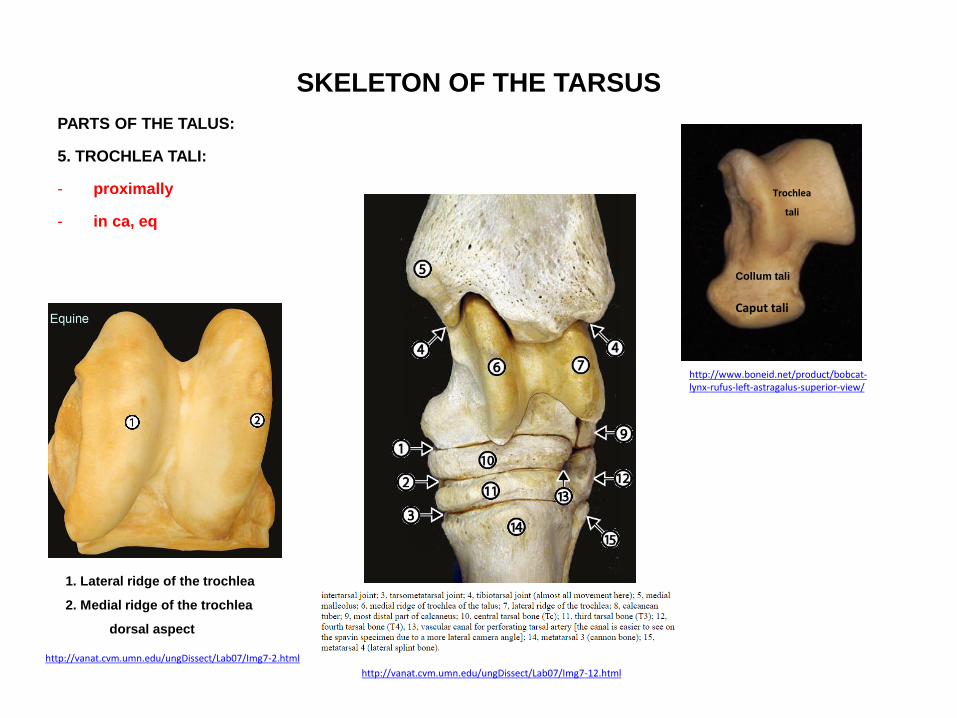
SKELETON OF THE TARSUS
PARTS OF THE TALUS:
5. TROCHLEA TALI:
- proximally
- in ca, eq
http://vanat.cvm.umn.edu/ungDissect/Lab07/Img7-12.html
http://vanat.cvm.umn.edu/ungDissect/Lab07/Img7-2.html
1. Lateral ridge of the trochlea
2. Medial ridge of the trochlea
dorsal aspect
Caput tali
Collum tali
Trochlea
tali
http://www.boneid.net/product/bobcat-lynx-rufus-left-astragalus-superior-view/

SKELETON OF THE TARSUS
PARTS OF THE TALUS:
5. TROCHLEA TALI PROXIMALIS:
- proximally
- in bo
6. TROCHLEA TALI DISTALIS:
- in bo
http://vanat.cvm.umn.edu/ungDissect/Lab07/Img7-1.html
http://vanat.cvm.umn.edu/ungDissect/Lab07/Img7-2.html
3. Trochlea tali prox.
4. Trochlea tali distalis
dorsal aspect

SKELETON OF THE TARSUS
PARTS OF THE TALUS:
6. FACEIS ARTICULARIS CALCANEAE:
- on the plantar and lateral surface
- articulates with the bones of the intetarsal row
9.
9.
http://www.boneid.net/product/dog-canis-lupus-familiaris-left-astragalus-inferior-view/
9.
9. Sulcus tarsi
eq, plantar aspect

SKELETON OF THE TARSUS
PARTS OF THE TALUS:
7. FACEIS ARTICULARIS NAVICULARIS:
- in eq, ca
- articulates with the navicular bone
http://vanat.cvm.umn.edu/ungDissect/Lab07/Img7-3.html
12.
eq

SKELETON OF THE TARSUS
PARTS OF THE TALUS:
8. SULCUS TALI:
- in eq, ca
- on the plantar surface
- forms with the sulcus calcanei the SINUS TARSI
http://www.boneid.net/product/dog-canis-lupus-familiaris-left-astragalus-inferior-view/
10.

SKELETON OF THE TARSUS
PARTS OF THE TALUS:
TUBERCULUM TALI:
- in eq
- medial tubercle for ligament attachament
http://vanat.cvm.umn.edu/ungDissect/Lab07/Img7-3.html
http://vanat.cvm.umn.edu/ungDissect/Lab07/Img7-2.html
1. Lateral ridge of the trochlea
2. Medial ridge of the trochlea
dorsal aspect

SKELETON OF THE TARSUS
PROXIMAL or CRURAL ROW consists of:
2. CALCANEUS:
- on the lateral side
- behind the talus
- forms the basis of the heel
Ca., plantar aspect
Ca., dorsal aspect
Skeleton of the left tarsus of a horse
lateral aspect medial aspect
eq
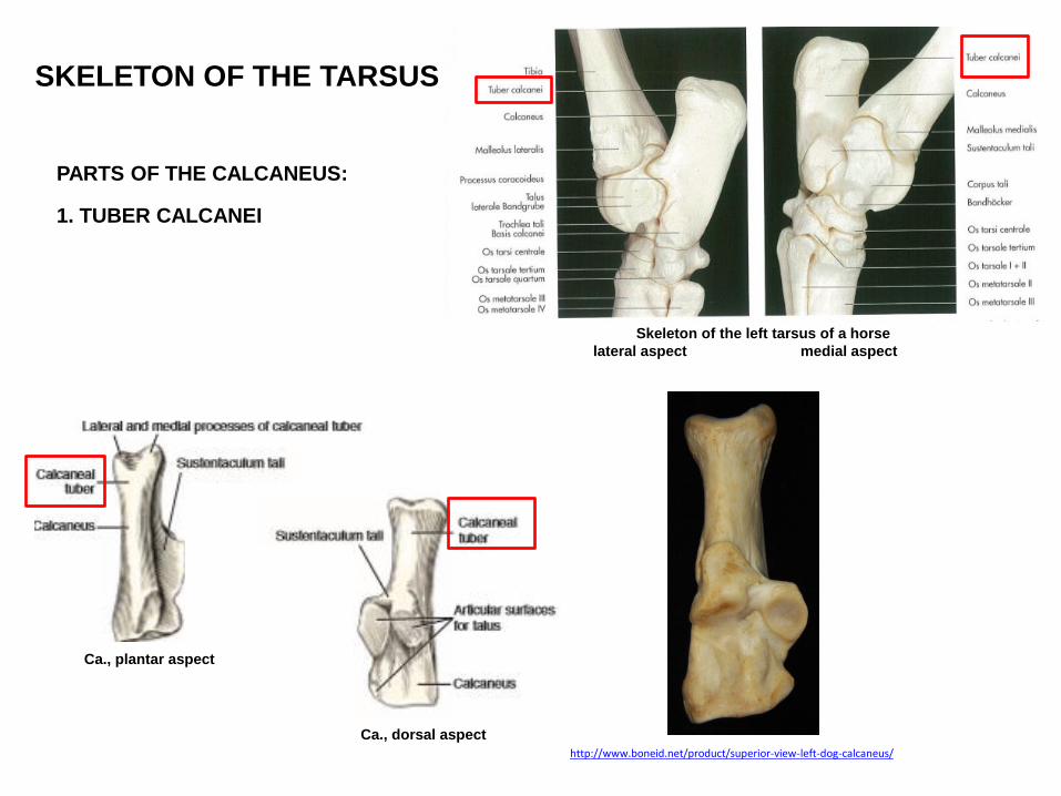
SKELETON OF THE TARSUS
PARTS OF THE CALCANEUS:
1. TUBER CALCANEI
Skeleton of the left tarsus of a horse
lateral aspect medial aspect
Ca., plantar aspect
Ca., dorsal aspect
http://www.boneid.net/product/superior-view-left-dog-calcaneus/
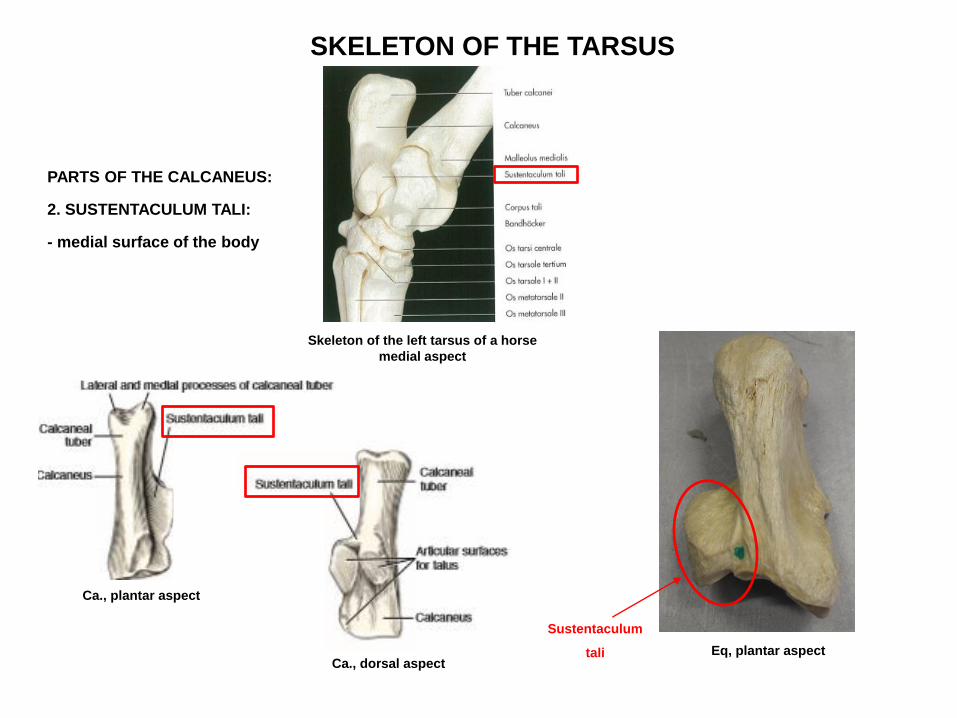
SKELETON OF THE TARSUS
PARTS OF THE CALCANEUS:
2. SUSTENTACULUM TALI:
- medial surface of the body
Ca., plantar aspect
Ca., dorsal aspect
Skeleton of the left tarsus of a horse
medial aspect
Sustentaculum
tali Eq, plantar aspect
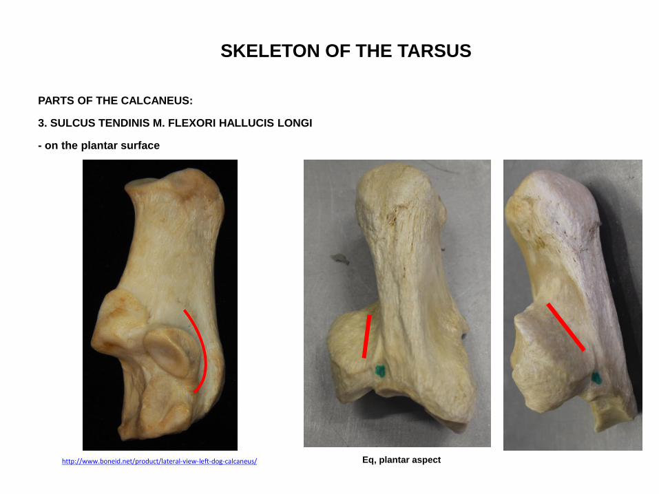
SKELETON OF THE TARSUS
PARTS OF THE CALCANEUS:
3. SULCUS TENDINIS M. FLEXORI HALLUCIS LONGI
- on the plantar surface
http://www.boneid.net/product/lateral-view-left-dog-calcaneus/ Eq, plantar aspect

SKELETON OF THE TARSUS
PARTS OF THE CALCANEUS:
4. PROCESSUS CORACOIDEUS:
- projects toward the talus
Skeleton of the left tarsus of a horse
lateral aspect
http://vanat.cvm.umn.edu/ungDissect/Lab07/Img7-3.html
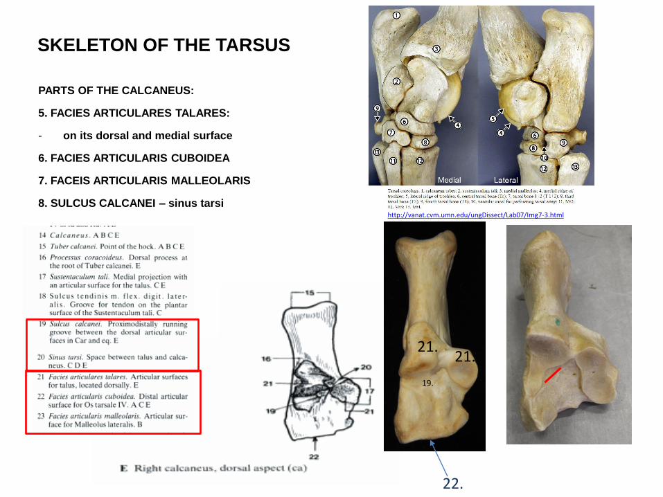
SKELETON OF THE TARSUS
PARTS OF THE CALCANEUS:
5. FACIES ARTICULARES TALARES:
- on its dorsal and medial surface
6. FACIES ARTICULARIS CUBOIDEA
7. FACEIS ARTICULARIS MALLEOLARIS
8. SULCUS CALCANEI – sinus tarsi
21. 21.
22.
http://vanat.cvm.umn.edu/ungDissect/Lab07/Img7-3.html
19.

SKELETON OF THE TARSUS SINUS TARSI:
-space between talus and calcaneus
- build by sulcus tali et calcanei
CANALIS TARSI:
- vascular canal for the perforating tarsal vessels
- in Un
- between 3rd and 4th tarsal bones
20. Sinus tarsi 33. Canalis tarsi
Sinus
tarsi
Canalis
tarsi
eq
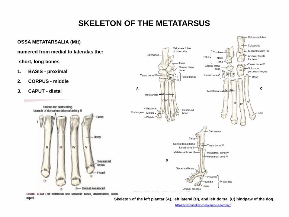
SKELETON OF THE METATARSUS
OSSA METATARSALIA (Mtt)
numered from medial to lateralas the:
-short, long bones
1. BASIS - proximal
2. CORPUS - middle
3. CAPUT - distal
Skeleton of the left plantar (A), left lateral (B), and left dorsal (C) hindpaw of the dog.
https://veteriankey.com/canine-anatomy/
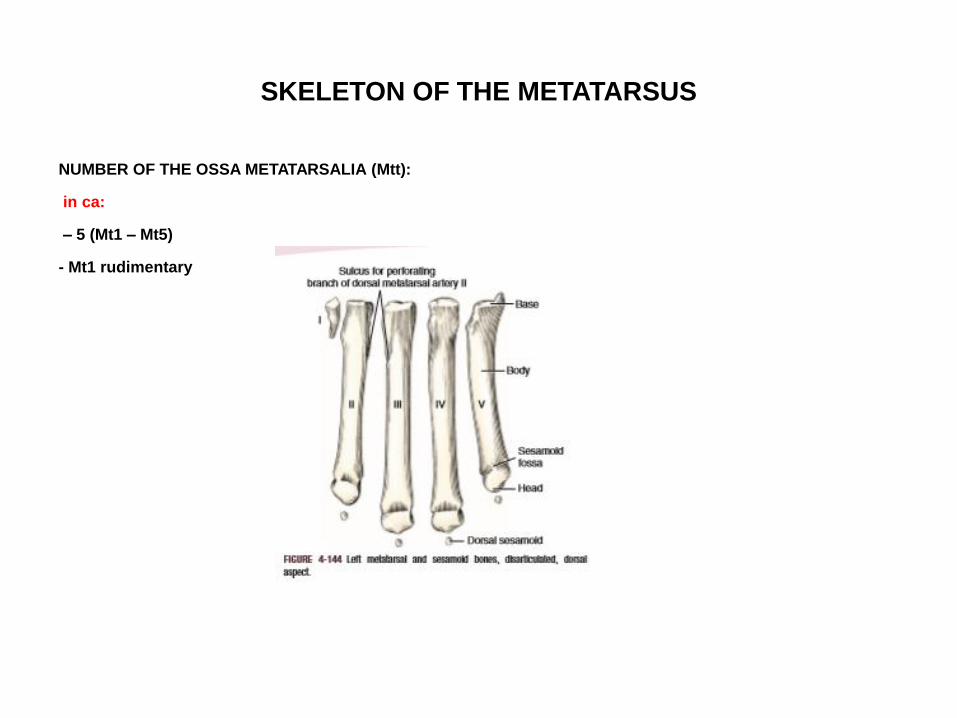
SKELETON OF THE METATARSUS
NUMBER OF THE OSSA METATARSALIA (Mtt):
in ca:
– 5 (Mt1 – Mt5)
- Mt1 rudimentary
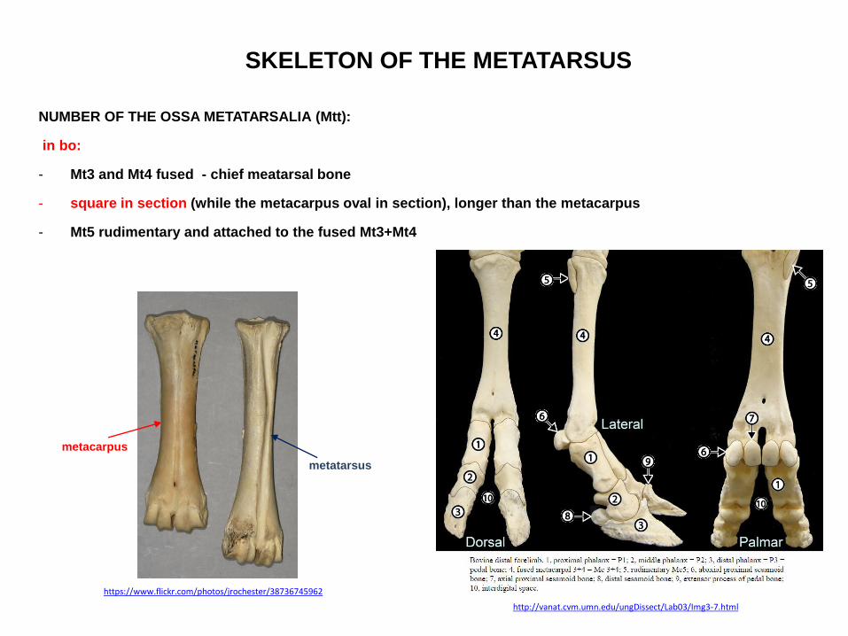
SKELETON OF THE METATARSUS
NUMBER OF THE OSSA METATARSALIA (Mtt):
in bo:
- Mt3 and Mt4 fused - chief meatarsal bone
- square in section (while the metacarpus oval in section), longer than the metacarpus
- Mt5 rudimentary and attached to the fused Mt3+Mt4
http://vanat.cvm.umn.edu/ungDissect/Lab03/Img3-7.html
https://www.flickr.com/photos/jrochester/38736745962
metacarpus
metatarsus

SKELETON OF THE METATARSUS
NUMBER OF THE OSSA METATARSALIA (Mtt):
in bo:
on chief meatarsal bone:
1. Sulcus longitudinalis dorsalis
2. Sulcus longitudinalis plantaris
3. Canalis metatarsi proximalis
4. Canalis metatarsi distalis
5. Incisura intertrochlearis
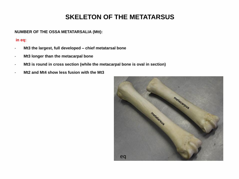
SKELETON OF THE METATARSUS
NUMBER OF THE OSSA METATARSALIA (Mtt):
in eq:
- Mt3 the largest, full developed – chief metatarsal bone
- Mt3 longer than the metacarpal bone
- Mt3 is round in cross section (while the metacarpal bone is oval in section)
- Mt2 and Mt4 show less fusion with the Mt3
eq
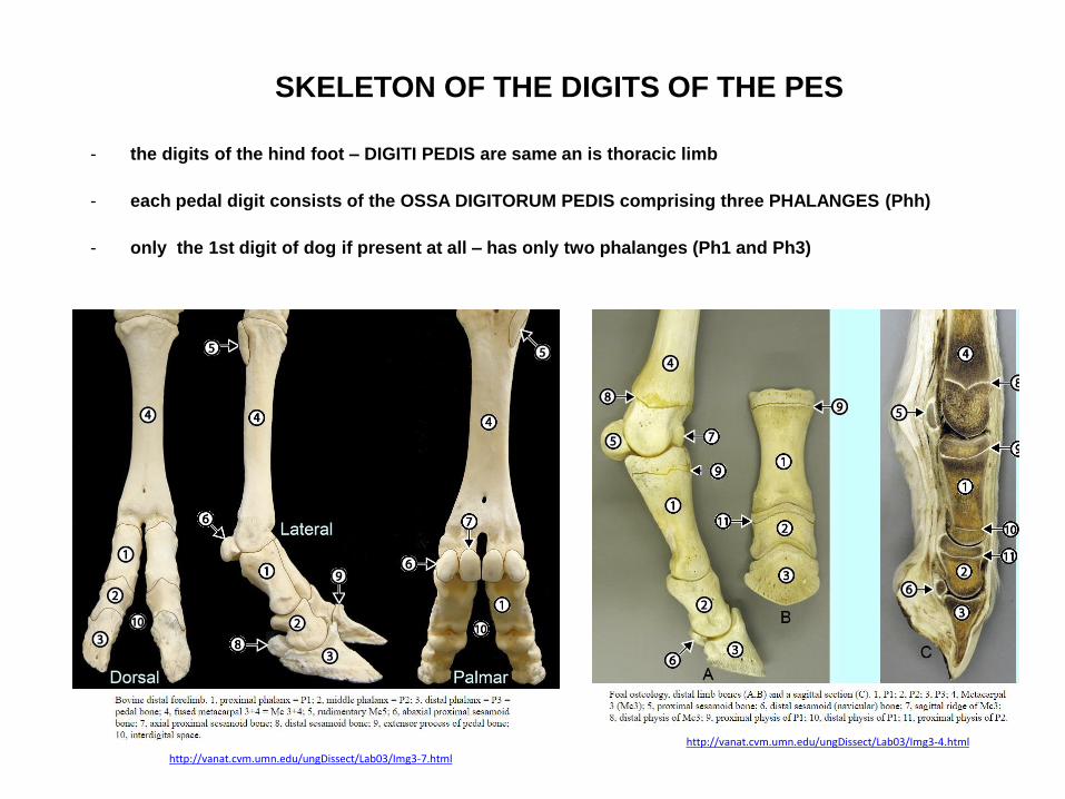
SKELETON OF THE DIGITS OF THE PES
- the digits of the hind foot – DIGITI PEDIS are same an is thoracic limb
- each pedal digit consists of the OSSA DIGITORUM PEDIS comprising three PHALANGES (Phh)
- only the 1st digit of dog if present at all – has only two phalanges (Ph1 and Ph3)
http://vanat.cvm.umn.edu/ungDissect/Lab03/Img3-7.html
http://vanat.cvm.umn.edu/ungDissect/Lab03/Img3-4.html

BIBLIOGRAPHIE
1. R. Nickel, A. Shummer, E. Steiferle: Lehrbuch der Anatomie der Haustiere Band III., 2.
Auflage
2. Klaus-Dieter Budras, Patrick H. McCarthy , Wolfgang Fricke : Renate Richter Anatomy of the
Dog, 5th revised Edition
3. Klaus-Dieter Budras , W.O.Sack, Sabine Röck : Anatomy of the Horse 5th revised Edition
4. Klaus – Dieter Budras, Rober E. Habel: Bovine Anatomy, 1st Edition
5. Miller’s Anatomy of the dog, 4th Edition
6. König – Liebich: Anatomie der Haussäugetiere, 4. Auflage
7. König – Liebich: Veterinary Anatomy of Domestic Mammals, 4th Edition
8. Saunders W.B: Veterinary Anatomy Flash Cards, 2nd Revised edition