bone density ppt
TRANSCRIPT
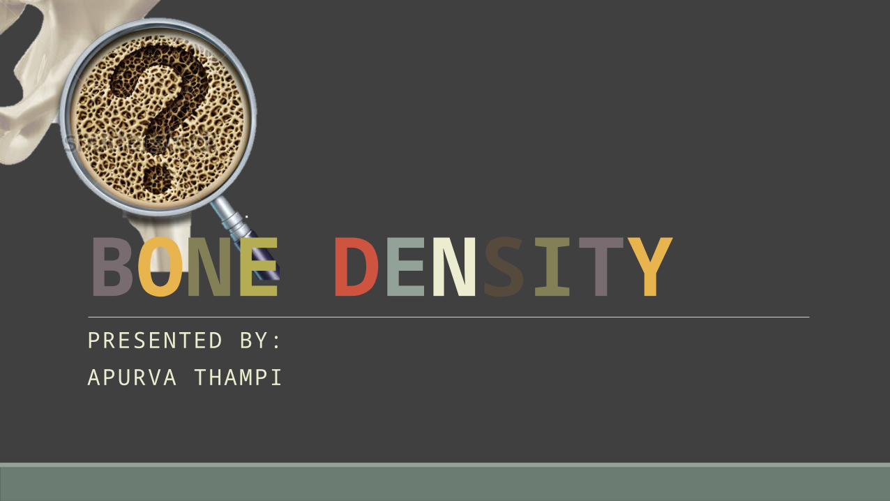
PRESENTED BY:APURVA THAMPI
BONE DENSITY

Contents •Introduction•Bone morphology•Bone physiology•Influence of bone density on implant success rates•Aetiology of various bone density•Bone classification schemes•Bone density classification - Misch
•Bone density location•Radiographic assessment of bone density•Tactile sense - bone density•Scientific rationale•Effect of bone density on surgical approach and healing•Case studies•Conclusion•References
Basic bone biology in implantology

05/01/2023
Textbookof human histology , Inderbir Singh ; 5th ed
4
Bone
33% organic67% inorganic hydroxyapetite
28% collagen
5% non collagenous protiens

There are 4 types of cells in bone tissue….
Oste
opro
geni
tor
cells• Unspecialise
d cells• Develop into
osteoblasts• Found in
periosteum, endosteum and in canals of vital teeth
Oste
obla
sts • Formation of
bone• Role in
calcification• Synthesis of
protien
Oste
ocla
sts • Responsible
for bone resorption
Oste
ocyt
es • Maintenance
of bone• Exchange of
calcium between bone and ECF
TEXTBOOKOF HUMAN HISTOLOGY , INDERBIR SINGH ; 5TH ED

Can be broadly classified into :
Compact bone
Trabecular bone
TEXTBOOKOF HUMAN HISTOLOGY , INDERBIR SINGH ; 5TH ED

What is lamellar bone?Structure of adult bone
Made up of layers – lamellae – thin plate of bone consisting of collagen fibres and mineral salts
Lacunae – between each lamellae
Each lacuna consists of one osteocyte
Canaliculi spread out from each lacuna
A
B
C
Unit of bone - lamellus
Bone acquires thickness by stacking of lamellus
Between adjoining lamellae – spaces called lacunae - Occupied by osteocytes
TEXTBOOKOF HUMAN HISTOLOGY , INDERBIR SINGH ; 5TH ED

What is woven bone?Osteocyte in lacuna canaliculi
Collagen fibres present in bundles – at random
Interlaced – woven bone
All newly formed bone
Abnormal persistence of woven bone – Paget’s disease

Compact bone VS Trabecular bone

Osteon of compact bone
Trabeculae of spongy bone
Haversian canals
Volkmann’s canal
periosteum
osteon
canaliculi
lamellae
Lacunae containing ostecytes

Compact boneLamellae
arranged in concentric circles –
surround - Haversian
canals
Occupied by blood vessels and nerves
Haversian canal +
lamellae = osteon or haversian
system
Between adjoining osteons – interstitial lamellae
At the surface – lamellae are
parallel – circumferential lamellae
TEXTBOOKOF HUMAN HISTOLOGY , INDERBIR SINGH ; 5TH ED

Trabecular boneBony plates or rods –
meshwork – trabeculae
Made of number of lamellae
Enclose wide spaces filled with bone
marrow – receive nutrition

Bone physiology

Calcium metabolism
Contemporary implant dentistry, Carl E Misch, 3rd edition
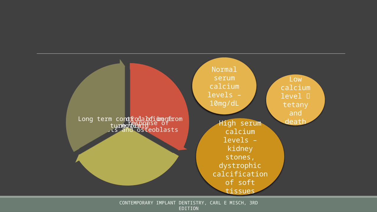
Rapid influx of calcium from bone fluidShort term response of osteoclasts
and osteoblastsLong term control of bone turnover
Normal serum
calcium levels –
10mg/dL
Low calcium level tetany
and death
High serum calcium levels – kidney stones,
dystrophic calcification of
soft tissues

Dec in calcium levels – transport of ions to
osteocytes
Calciferol enhances pumping of calcium ions from cells into
ECF
Net flux of Ca ions
PTH + calciferol + calcitonin
Transiently suppresses bone
resorption
Profound effect on skeleton
PTH is the primary regulator – mean bone age
Important determinant of
fragility
Instantaneous regulation (within
seconds)Short term regulation
Long term regulation
CONTEMPORARY IMPLANT DENTISTRY, CARL E MISCH, 3RD EDITION

Calcium conservation
Kidney excretes phophates by minimising loss of calcium Renal dysfunction – high risk for osseous manipulative procedures – renal osteodystrophy
Body spends 300mg calcium per day – recovered by absorption from gut – depends of Vit D
Kidney is the primary calcium
conservation organ of the
body
CONTEMPORARY IMPLANT DENTISTRY, CARL E MISCH, 3RD EDITION
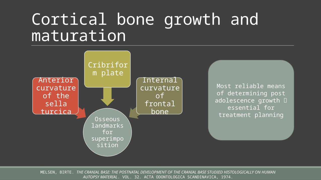
Cortical bone growth and maturation
Osseous landmarks
for superimpo
sition
Anterior curvature
of the sella
turcica
Cribriform plate
Internal curvature of frontal
bone
Most reliable means of determining post
adolescence growth essential for treatment
planning
MELSEN, BIRTE. THE CRANIAL BASE: THE POSTNATAL DEVELOPMENT OF THE CRANIAL BASE STUDIED HISTOLOGICALLY ON HUMAN AUTOPSY MATERIAL. VOL. 32. ACTA ODONTOLOGICA SCANDINAVICA, 1974.

Influence of bone density on implant success rates

Anterior mandibl
eAnterior maxilla
Posterior
mandible
Posterior maxilla
Position / Arch location Quality of
bone dependent
on the position
CONTEMPORARY IMPLANT DENTISTRY, CARL E MISCH, 3RD EDITION
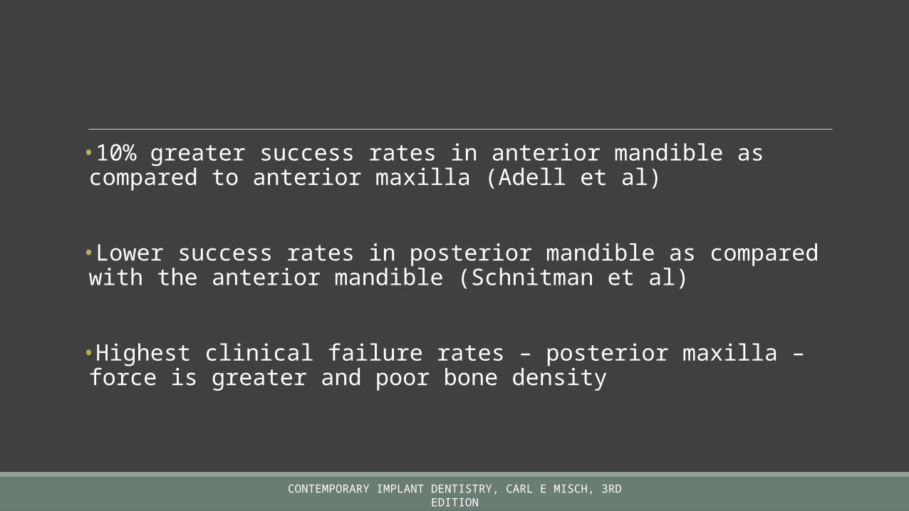
•10% greater success rates in anterior mandible as compared to anterior maxilla (Adell et al)
•Lower success rates in posterior mandible as compared with the anterior mandible (Schnitman et al)
•Highest clinical failure rates – posterior maxilla – force is greater and poor bone density
CONTEMPORARY IMPLANT DENTISTRY, CARL E MISCH, 3RD EDITION

Quality of bone
CONTEMPORARY IMPLANT DENTISTRY, CARL E MISCH, 3RD EDITION

Aetiology of various bone density

Hormones
Vitamins
Mechanical influences
Duration of edentulous
ness
Changes in bone -
adaptability
CONTEMPORARY IMPLANT DENTISTRY, CARL E MISCH, 3RD EDITION

“Every change in the form and function of bone or of its function alone is followed by certain definite changes in the internal architecture, and equally definite alteration in its external conformation in accordance with mathematical laws”
Wolff - 1892

Adaptive phenomena Alteration of mechanical forces and strain development within the bone density evolves as a result of mechanical deformation from microstrain
MODELLING Independent sites of formation and resorption
Results in change in shape and size of bone
REMODELLING Resorption and formation at the same site
Replaces previously existing bone
CONTEMPORARY IMPLANT DENTISTRY, CARL E MISCH, 3RD EDITION

The maxilla is a force distribution unit and mandible is a force absorption
unitCONTEMPORARY IMPLANT DENTISTRY, CARL E MISCH, 3RD
EDITION

The trabecular bone in dentate mandible is more coarse compared to the maxilla
CONTEMPORARY IMPLANT DENTISTRY, CARL E MISCH, 3RD EDITION

Anterior mandibl
e
Posterior maxilla
Density change after tooth loss.
• Initial density• Flexure and torsion• Parafunction before
extraction
NEUFELD JO: CHANGES IN THE TRABECULAR PATTERN OF THE MANDIBLE FOLLOWING THE LOSS OF TEETH, J PROSTHET DENT 685-697, 1958

ORBAN B: ORAL HISTOLOGY AND EMBRYOLOGY, ED 3, ST LOUIS, 1953, MOSBY

Based on Frosts’s mechanostat theory
50 1500 3000 10000+Acute Disusewindow
Adaptedwindow
MildOverloadwindow
PathologicOverloadwindow
Spontaneous fracture
Stress F/A
Strain O
Strain
Acute disuse window : lowest microstrain amount
Adapted window : ideal physiologic loading zone
Mild overload zone : cause microfracture; triggers an increase in bone remodelling – more woven bonePathologic overload : increased fatigue fractures, remodelling and bone resorption
FROST, H. M. "MECHANICAL ADAPTATION. FROST’S MECHANOSTAT THEORY." STRUCTURE, FUNCTION, AND ADAPTATION OF COMPACT BONE (1989): 179-81.

Acute disuse window•Loses mineral density•Disuse atrophy – modelling for new bone inhibited•Net loss of bone•Microstrain – 0 – 50•Cortical bone density decrease – 40% and trabecular bone density decrease – 12%
CONTEMPORARY IMPLANT DENTISTRY, CARL E MISCH, 3RD EDITION

Adapted window phase•50 – 1500 microstrain•Equilibrium of modelling and remodelling•“homeostatic window of health”•18% trabecular bone and 2-5% cortical bone•Ideally desired around an endosteal implant

Mild overload zone•1500 – 3000 microstrain•Greater rate of fatigue microfracture•Bone strength and density decreases•State of bone when endosteal implant is overloaded•Repair – woven bone is weaker than lamellar – “safety range” for bone strength is reduced
CONTEMPORARY IMPLANT DENTISTRY, CARL E MISCH, 3RD EDITION

Pathologic overload zone•Microstrains <3000 units•Physical fracture of cortical bone•Formation of fibrous tissue•Marginal bone loss in implant overloading – implant failure

Bone classification schemes in implant dentistry

Linkow in 1970 : Class I
• Ideal• Evenly
spaced trabeculae with small cancellated spaces
Class II• Less
uniformity• Larger
cancellated spaces
• Large marrow filled spaces exist
CONTEMPORARY IMPLANT DENTISTRY, CARL E MISCH, 3RD EDITION

Lekholm and Zarb in 1985:Quality 1
• Homogenous compact bone
Quality 2• Thick layer of
compact bone around a core of dense trabecular bone
Quality 3• Thin layer of
cortical bone around dense trabecular bone
• Favorable strength
Quality 4• Thin layer of
cortical bone around a coreof low density trabecular bone
CONTEMPORARY IMPLANT DENTISTRY, CARL E MISCH, 3RD EDITION
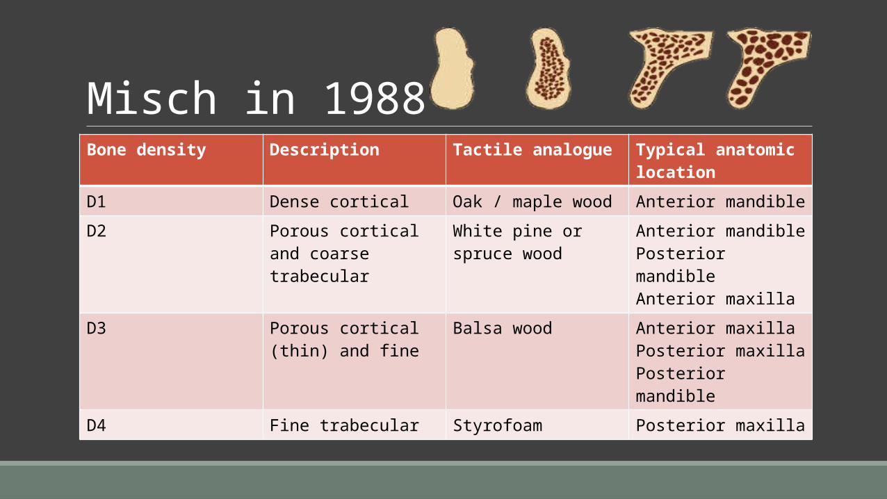
Misch in 1988Bone density Description Tactile analogue Typical anatomic
locationD1 Dense cortical Oak / maple wood Anterior mandibleD2 Porous cortical and
coarse trabecularWhite pine or spruce wood
Anterior mandiblePosterior mandibleAnterior maxilla
D3 Porous cortical (thin) and fine
Balsa wood Anterior maxillaPosterior maxillaPosterior mandible
D4 Fine trabecular Styrofoam Posterior maxilla

D5 type of bone exists – most
immature bone – found in a
developing sinus graft.
CONTEMPORARY IMPLANT DENTISTRY, CARL E MISCH, 3RD EDITION

To be continued…

Bone density location

Location of bone density types (% occurance)Bone Anterior
maxillaPosterior maxilla
Anterior mandible
Posterior mandible
D1 0 0 6 3
D2 25 10 66 50
D3 65 50 25 46
D4 10 40 3 1
CONTEMPORARY IMPLANT DENTISTRY, CARL E MISCH, 3RD EDITION

CONTEMPORARY IMPLANT DENTISTRY, CARL E MISCH, 3RD EDITION

D1 Bone• Incresed torsion / flexure • Div A Kennedy’s class IV• Antr/postr mandible – lingual cortex
D2 Bone• Partially edentulous antr/postr mandible (premolar)
• Single tooth or 2 teeth missing
D3 Bone• Most common in maxilla• Also present in posterior mandible
D4 Bone• Softest bone• Posterior maxilla – after sinus augmentation or iliac crest bone graft
CONTEMPORARY IMPLANT DENTISTRY, CARL E MISCH, 3RD EDITION
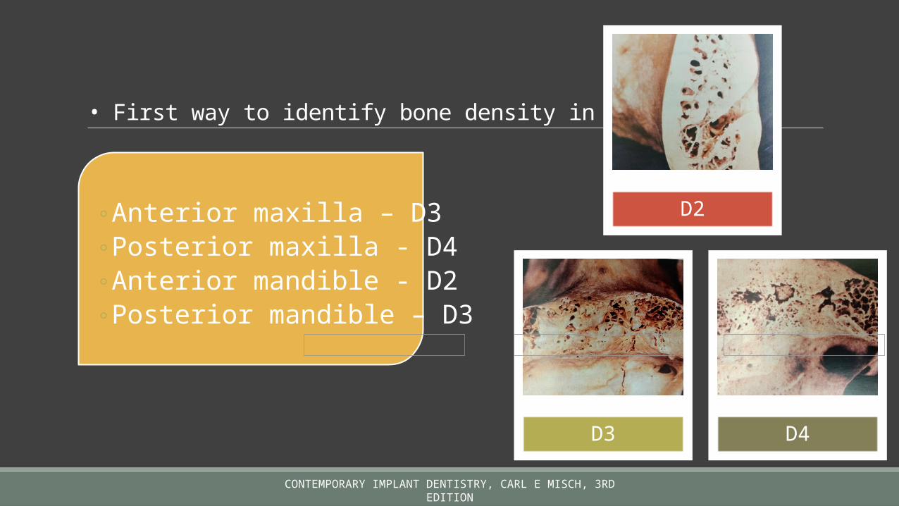
• First way to identify bone density in implant site
◦ Anterior maxilla – D3◦ Posterior maxilla - D4◦ Anterior mandible - D2◦ Posterior mandible – D3
D2
D3 D4
CONTEMPORARY IMPLANT DENTISTRY, CARL E MISCH, 3RD EDITION

Radiographic bone density

IOPAR
OPGs
• Lateral cortical plates obscure the trabecular bone density
• More subtle changes cannot be qualified

Correlation between Misch bone density classification and Hounsfield units….
Type of bone
Hounsfield units
D1 >1250 HUD2 850 – 1250 HUD3 350 - 850 HUD4 150 – 350 HUD5 <150 HU
SOGO, MOTOFUMI, ET AL. "ASSESSMENT OF BONE DENSITY IN THE POSTERIOR MAXILLA BASED ON HOUNSFIELD UNITS TO ENHANCE THE INITIAL STABILITY OF IMPLANTS." CLINICAL IMPLANT DENTISTRY AND RELATED RESEARCH 14.S1 (2012): E183-E187.
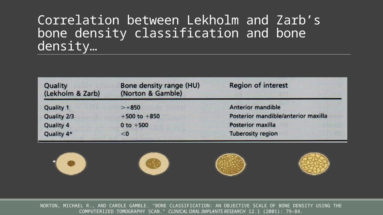
Correlation between Lekholm and Zarb’s bone density classification and bone density…
NORTON, MICHAEL R., AND CAROLE GAMBLE. "BONE CLASSIFICATION: AN OBJECTIVE SCALE OF BONE DENSITY USING THE COMPUTERIZED TOMOGRAPHY SCAN." CLINICAL ORAL IMPLANTS RESEARCH 12.1 (2001): 79-84.

Failure in mandible –
higher Hounsfield
units
• Lack of vascularisation
• Overheating
ROTHMAN, STEPHEN LG, MELVYN S. SCHWARZ, AND NEIL I. CHAFETZ. "HIGH-RESOLUTION COMPUTERIZED TOMOGRAPHY AND NUCLEAR BONE SCANNING IN THE DIAGNOSIS OF POSTOPERATIVE STRESS FRACTURES OF THE MANDIBLE: A CLINICAL REPORT." INTERNATIONAL JOURNAL OF ORAL &
MAXILLOFACIAL IMPLANTS 10.6 (1995).

Bone density – tactile sense

Bone density Description Tactile analogue Typical anatomic location
D1 Dense cortical Oak / maple wood Anterior mandibleD2 Porous cortical and
coarse trabecularWhite pine or spruce wood
Anterior mandiblePosterior mandibleAnterior maxilla
D3 Porous cortical (thin) and fine
Balsa wood Anterior maxillaPosterior maxillaPosterior mandible
D4 Fine trabecular Styrofoam Posterior maxilla

Scientific rationale for a bone density - based treatment plan

Bone strength and
density
Bone elastic modulus and
density
Bone density and implant bone contact
interface
Bone density and stress transfer
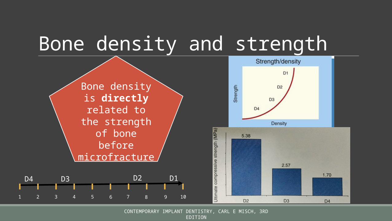
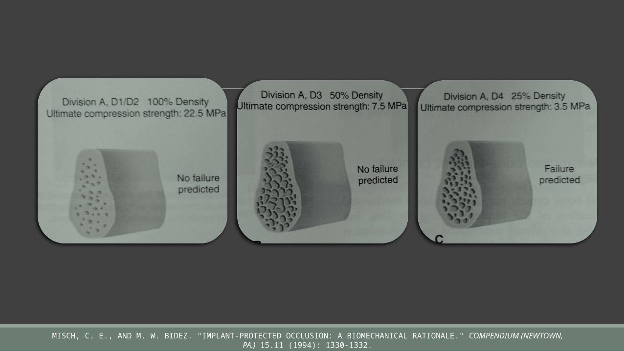
Bone density and strength
Bone density is directly
related to the strength of bone before
microfracture
CONTEMPORARY IMPLANT DENTISTRY, CARL E MISCH, 3RD EDITION
1 2 43 5 6 7 8 9 10
D1D2D3D4
MISCH, C. E., AND M. W. BIDEZ. "IMPLANT-PROTECTED OCCLUSION: A BIOMECHANICAL RATIONALE." COMPENDIUM (NEWTOWN, PA.) 15.11 (1994): 1330-1332.
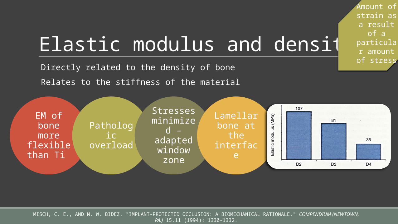
Elastic modulus and density Directly related to the density of bone Relates to the stiffness of the material
Amount of strain as a result of a particular amount of
stress
EM of bone more
flexible than Ti
Pathologic
overload
Stresses minimize
d – adapted window
zone
Lamellar bone at
the interface
MISCH, C. E., AND M. W. BIDEZ. "IMPLANT-PROTECTED OCCLUSION: A BIOMECHANICAL RATIONALE." COMPENDIUM (NEWTOWN, PA.) 15.11 (1994): 1330-1332.

Ti - D1 bone interface – very little
microstrain
Ti – D4 bone interface – pathologic overload
MISCH, C. E., AND M. W. BIDEZ. "IMPLANT-PROTECTED OCCLUSION: A BIOMECHANICAL RATIONALE." COMPENDIUM (NEWTOWN, PA.) 15.11 (1994): 1330-1332.

Bone density and bone-implant contact percentage Area less area = greater stress
D1 bone has greatest BIC
D2 has 65 – 75% BIC
D3 bone has 40 – 50% BIC
D5 bone has 30% BIC
CONTEMPORARY IMPLANT DENTISTRY, CARL E MISCH, 3RD EDITION

Bone density and stress transfer
Different stress contours for different types of
bone
Bone implant contact
Bone density
Elastic modulu
s
Crestal bone loss and early implant
failure due to
increased stress
D1 bone
Stress is of lesser
magnitude Highest
strains near the crest
D2 bone
Sustains greater strain intensity of
stress extends farther apically
D4 bone
Greatest crestal strain magnitude of
strain is further apical
Adapted window Mild overload Implant failure
CONTEMPORARY IMPLANT DENTISTRY, CARL E MISCH, 3RD EDITION

A nutshell….Each bone density has different strengths
Bone density affects elastic modulus
Density differences result in difference in BIC
Different stress-strain distribution at a B-I interface
CONTEMPORARY IMPLANT DENTISTRY, CARL E MISCH, 3RD EDITION

Effect of bone density on treatment planning

Modifications in treatment plan
Prosthetic factors
Implant surface
conditionImplant number
Need of progressive loading
Implant design
Implant size
CONTEMPORARY IMPLANT DENTISTRY, CARL E MISCH, 3RD EDITION
Aim : decrease strain in the bone thereby decrease microfracture increase SA

Dense cortical D1 BoneAnalogous to oak or
maple wood
Almost all dense cortical bone
Mostly seen in anterior mandible and
sometimes in posterior mandible

Advantages/disadvantages of D1 bone
Highly mineralizedExcellent bone strengthBest implant bone contactLess force transmission to apical thirds
Implant crown ratio>1Less blood supply – not regenerativeEasily overheatedImplant height limited to less than 12mm
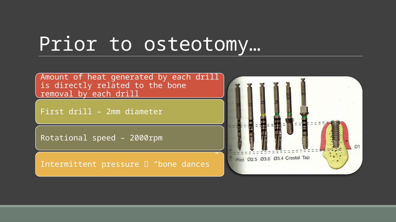
Prior to osteotomy…Amount of heat generated by each drill is directly related to the bone removal by each drill
First drill – 2mm diameter
Rotational speed – 2000rpm
Intermittent pressure “bone dances”

Osteotomy preparation in D1 bone
• Ext/Int irrigation• Intermittent pressure• Pause 3-5mins• New drills• Incremental drill sequence
Overheating
• Primarily from periosteum
• Minimal reflection• Precise approximation
Blood supply• Greater width• Greater height• Slower speed used
Final osteotomy
drill

• Short of full osteotomy depth
• Allows passive implant fit
• Removed drill remnants
Bone tap
• Unthread ½ turn to relieve internal stresses
Final implant
placement • Slower healing rate• 5 months to achieve
mature interface
healing
• 3-4 months• May use immediate
loading
Stage II recovery

D2 bone Dense-to-thick porpus cortical and carse trabeculae
Hounsfield values – 750-1250 units
Analogous to spruce or white pine wood
Occurs mostly in anterior mandible and posterior
mandible
Ideal implant dimension – 4mm diameter ; 12 mm height

Advantages/disadvantages
Excellent implant surface healingSecure initial rigid interfaceIntrabony blood supply

Osteotomy preparation in D2 bone
Rotation of drill – 2500 rpm Ext/int irrigation used Pause every 5-10 seconds – pumping motion Drill sequence similar to D1 bone Crestal bone drills should be used – reduce mechanical trauma Bone tap – engages lateral or apical cortical bone

Healing
Excellent blood supply
Initial rigid fixation
Lamellar bone
interface < 60% - 4 months healing interval
Abutment placement
may commence

D3 boneThinner porous cortical boneHounsfield values – 375 – 750 HUAnalogous to balsa woodFound in anterior maxilla and anterior mandible/maxillaIdeal implant dimension – 4X12Roughened implant body – acid etched or resorbable blast media

Advantages/ disadvantages
Time and difficulty for preparation is minimalBlood supply is excellentHighest survival rate

Disadvantages of D3 boneBone anatomy• Anterior maxilla is narrow
Osteotomy• Lateral perforation• Oversize by mistake• Apical perforation
BIC• 50%
Implant placement• One time• In level with crestal bone
Implant design• TPS or Hydroxyapatite coated• Costly• Threaded• Greater SA• Press fit
Healing• 6 months• Progressive loading more important than D1 or D2

D4 boneLeast density – no
cortical crestal bone
Found in posterior molar region
Analogous to stiff Styrofoam
Ideal Implant height – 14 mm (min 12
mm)


DisadvantagesDifficult to obtain rigid fixationRotating drills not to be used apart from pilot drillOsteotomes may be used to compress osteotomy siteCortical bone in the opposite landmark to be engaged (if any)Increase number of implants to improve load distributionNo cantilever advocated

Summary Densities vary depending of the location of edentulous ridge and the period of edentulousness
D1 is the strongest bone - 10 times greater than D4 Minimum of 12mm height of implant required for initial stability Additional bone healing and incremental loading will improve bone density

Conclusion Bone remodels in relationto the forces excerted upon it – density varies

References Textbookof human histology , Inderbir Singh ; 5th ed Contemporary implant dentistry, Carl E Misch, 3rd edition Orban B: Oral histology and embryology, ed 3, St Louis, 1953, Mosby Melsen, Birte. The cranial base: the postnatal development of the cranial base studied histologically on human autopsy material. Vol. 32. Acta Odontologica Scandinavica, 1974.
Neufeld JO: changes in the trabecular pattern of the mandible following the loss of teeth, J Prosthet Dent 685-697, 1958
Frost, H. M. "Mechanical adaptation. Frost’s mechanostat theory." Structure, function, and adaptation of compact bone (1989): 179-81

Sogo, Motofumi, et al. "Assessment of bone density in the posterior maxilla based on Hounsfield units to enhance the initial stability of implants." Clinical implant dentistry and related research 14.s1 (2012): e183-e187.
Norton, Michael R., and Carole Gamble. "Bone classification: an objective scale of bone density using the computerized tomography scan." Clinical oral implants research 12.1 (2001): 79-84.
Misch, C. E., and M. W. Bidez. "Implant-protected occlusion: a biomechanical rationale." Compendium (Newtown, Pa.) 15.11 (1994): 1330-1332.