bone mineral density studies - bcbsks · bone mineral density studies ... quantitative ultrasound...
TRANSCRIPT

Bone Mineral Density Studies Page 1 of 46
Current Procedural Terminology © American Medical Association. All Rights Reserved.
Contains Public Information
Medical Policy An independent licensee of the Blue Cross Blue Shield Association
Title: Bone Mineral Density Studies Professional Institutional Original Effective Date: May 20, 1986 Original Effective Date: June 1, 2007 Revision Date(s): May 3, 1997; February 9, 1999; July 22, 1999; February 23, 2000; August 23, 2000; March 28, 2001; July 30, 2002; October 31, 2002; January 27, 2004; May 13, 2004; October 18, 2004; December 10, 2004; June 21, 2005; August 16, 2006; November 28, 2006; May 1 2007; May 13, 2011; December 9, 2011; April 13, 2012; October 4, 2013; May 13, 2015; July 8, 2015; December 8, 2015; May 25, 2016; October 1, 2016; April 12, 2017
Revision Date(s): May 13, 2011; December 9, 2011, April 13, 2012; October 4, 2013; May 13, 2015; July 8, 2015; December 8, 2015; May 25, 2016; October 1, 2016; April 12, 2017
Current Effective Date: April 12, 2017 Current Effective Date: April 12, 2017 State and Federal mandates and health plan member contract language, including specific provisions/exclusions, take precedence over Medical Policy and must be considered first in determining eligibility for coverage. To verify a member's benefits, contact Blue Cross and Blue Shield of Kansas Customer Service. The BCBSKS Medical Policies contained herein are for informational purposes and apply only to members who have health insurance through BCBSKS or who are covered by a self-insured group plan administered by BCBSKS. Medical Policy for FEP members is subject to FEP medical policy which may differ from BCBSKS Medical Policy.
The medical policies do not constitute medical advice or medical care. Treating health care providers are independent contractors and are neither employees nor agents of Blue Cross and Blue Shield of Kansas and are solely responsible for diagnosis, treatment and medical advice. If your patient is covered under a different Blue Cross and Blue Shield plan, please refer to the Medical Policies of that plan.
Bone Mineral Density Studies Page 2 of 46
Current Procedural Terminology © American Medical Association. All Rights Reserved.
Contains Public Information
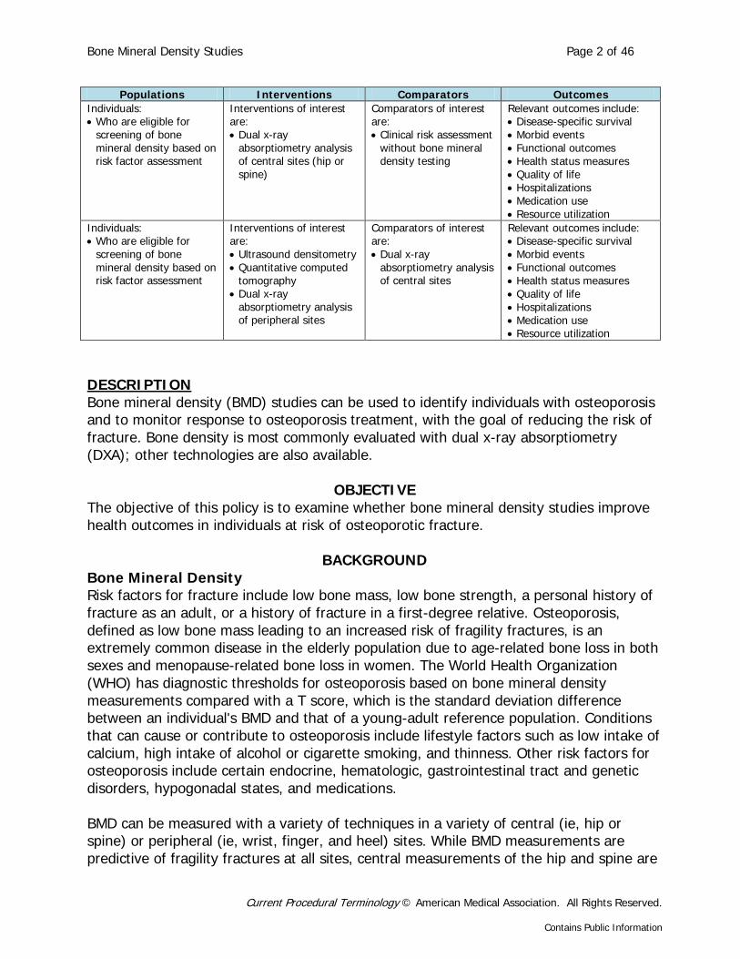
Populations Interventions Comparators Outcomes Individuals: • Who are eligible for
screening of bone mineral density based on risk factor assessment
Interventions of interest are: • Dual x-ray
absorptiometry analysis of central sites (hip or spine)
Comparators of interest are: • Clinical risk assessment
without bone mineral density testing
Relevant outcomes include: • Disease-specific survival • Morbid events • Functional outcomes • Health status measures • Quality of life • Hospitalizations • Medication use • Resource utilization
Individuals: • Who are eligible for
screening of bone mineral density based on risk factor assessment
Interventions of interest are: • Ultrasound densitometry • Quantitative computed
tomography • Dual x-ray
absorptiometry analysis of peripheral sites
Comparators of interest are: • Dual x-ray
absorptiometry analysis of central sites
Relevant outcomes include: • Disease-specific survival • Morbid events • Functional outcomes • Health status measures • Quality of life • Hospitalizations • Medication use • Resource utilization
DESCRIPTION Bone mineral density (BMD) studies can be used to identify individuals with osteoporosis and to monitor response to osteoporosis treatment, with the goal of reducing the risk of fracture. Bone density is most commonly evaluated with dual x-ray absorptiometry (DXA); other technologies are also available.
OBJECTIVE The objective of this policy is to examine whether bone mineral density studies improve health outcomes in individuals at risk of osteoporotic fracture.
BACKGROUND Bone Mineral Density Risk factors for fracture include low bone mass, low bone strength, a personal history of fracture as an adult, or a history of fracture in a first-degree relative. Osteoporosis, defined as low bone mass leading to an increased risk of fragility fractures, is an extremely common disease in the elderly population due to age-related bone loss in both sexes and menopause-related bone loss in women. The World Health Organization (WHO) has diagnostic thresholds for osteoporosis based on bone mineral density measurements compared with a T score, which is the standard deviation difference between an individual's BMD and that of a young-adult reference population. Conditions that can cause or contribute to osteoporosis include lifestyle factors such as low intake of calcium, high intake of alcohol or cigarette smoking, and thinness. Other risk factors for osteoporosis include certain endocrine, hematologic, gastrointestinal tract and genetic disorders, hypogonadal states, and medications. BMD can be measured with a variety of techniques in a variety of central (ie, hip or spine) or peripheral (ie, wrist, finger, and heel) sites. While BMD measurements are predictive of fragility fractures at all sites, central measurements of the hip and spine are

Bone Mineral Density Studies Page 3 of 46
Current Procedural Terminology © American Medical Association. All Rights Reserved.
Contains Public Information
the most predictive. Fractures of the hip and spine (ie, vertebral fractures) are also considered to be the most clinically relevant. BMD is typically expressed as a T score. The utility of screening BMD measurements can be established by demonstrating that screening identifies a population at increased risk of fracture and that, by treating those at-risk individuals, the rate of fractures is reduced thereby lowering fracture-related morbidity and mortality. These potential benefits of screening should outweigh the risks of screening (radiation exposure) or false positives (initiation of unnecessary treatment). Osteoporosis Treatment Treatment of osteoporosis includes both lifestyle measures (eg, increased intake of calcium and vitamin D, exercise, smoking cessation) and pharmacologic measures. Current pharmacologic options include bisphosphonates such as alendronate (ie, Fosamax), selective estrogen receptor modulators such as raloxifene (ie, Evista), the recombinant human parathyroid hormone teriparatide (ie, Forteo), and calcitonin. An updated 2014 systematic review funded by the Agency for Healthcare Research and Quality found good-quality evidence that bisphosphonates, denosumab, teriparatide, and raloxifene reduce fracture risk in postmenopausal women with BMD in the osteoporotic range and/or preexisting hip or vertebral fracture.1 The decision to perform bone density assessment should be based on an individual’s fracture risk profile and skeletal health assessment. In addition to age, sex, and BMD, risk factors included in the WHO Fracture Risk Assessment (FRAX) Tool2 are:
• Low body mass index; • Parental history of hip fracture; • Previous fragility fracture in adult life (ie, occurring spontaneously or a fracture
arising from trauma, which, in a healthy individual, would not have resulted in a fracture);
• Current smoking or 3 or more units of alcohol daily, where a unit is equivalent to a standard glass of beer (285 mL), a single measure of spirits (30 mL), a medium-sized glass of wine (120 mL), or 1 measure of an aperitif (60 mL);
• A disorder strongly associated with osteoporosis, which includes rheumatoid arthritis, type I (insulin-dependent) diabetes, osteogenesis imperfecta in adults, untreated long-standing hyperthyroidism, hypogonadism or premature menopause (<45 years), chronic malnutrition or malabsorption, and chronic liver disease;
• Current exposure to oral glucocorticoids or exposure to oral glucocorticoids for more than 3 months at a dose of prednisolone 5 mg daily or more (or equivalent doses of other glucocorticoids).
A 2011 joint position statement from the International Society for Clinical Densitometry and the International Osteoporosis Foundation included the official position that FRAX with BMD predicts risk of fracture better than clinical risk factors or BMD alone. In addition, the joint position statement indicated that measurements other than BMD or T score at the femoral neck by DXA are not recommended for use with FRAX.3 The FRAX

Bone Mineral Density Studies Page 4 of 46
Current Procedural Terminology © American Medical Association. All Rights Reserved.
Contains Public Information
tool does not include a recommendation about which patients to further assess or treat. The FRAX website states that this is a matter of clinical judgment and recommendations may vary by country.2 Measurement Tools The following technologies are most commonly used to measure BMD. Dual X-Ray Absorptiometry Dual x-ray absorptiometry (DXA) is probably the most commonly used technique to measure BMD because of its ease of use, low radiation exposure, and its ability to measure BMD at both the hip and spine. DXA can also be used to measure peripheral sites, such as the wrist and finger. DXA generates 2 x-ray beams of different energy levels to scan the region of interest and measures the difference in attenuation as the low- and high-energy beams pass through the bone and soft tissue. The low-energy beam is preferentially attenuated by bone, while the high-energy beam is attenuated by both bone and soft tissue. This difference in attenuation between the 2 beams allows for correction for the irregular masses of soft tissue, which surround the spine and hip, and therefore the measurement of bone density at those sites. Quantitative Computed Tomography Quantitative computed tomography (QCT) depends on the differential absorption of ionizing radiation by calcified tissue and is used for central measurements only. Compared with DXA, QCT is less readily available and associated with relatively high radiation exposure and relatively high cost. Ultrasound Densitometry Ultrasound densitometry is a technique for measuring BMD at peripheral sites, typically the heel but also the tibia and phalanges. Compared with osteoporotic bone, normal bone demonstrates higher attenuation of the ultrasound wave and is associated with a greater velocity of the wave passing through bone. Ultrasound densitometry has no radiation exposure, and machines may be purchased for use in an office setting. These techniques dominate BMD testing. Single- and dual-photon absorptiometry and radiographic absorptiometry are now rarely used and may be considered obsolete.
REGULATORY STATUS Several devices that measure bone density have been cleared for marketing by the U.S. Food and Drug Administration (FDA) through the 510(k) process. Some examples include:
• QCT-Bone Mineral Phantom (Image Analysis) in 1985 • Quantitative Digital Radiography (QDR™; Hologic) x-ray bone densitometer using
dual x-ray absorptiometry in 1987 • Lunar DPX bone densitometer (now GE Lunar DPX NT®; GE Healthcare) in 1988

Bone Mineral Density Studies Page 5 of 46
Current Procedural Terminology © American Medical Association. All Rights Reserved.
Contains Public Information
• Accudxa2 Bone Mineral Density Assessment System (Lone Oak Medical Technologies, Doylestown, PA) in 2012.
In addition, some ultrasound bone sonometers have been approved by FDA through the premarket approval (PMA) process. One example is the Sahara® Clinical Bone Sonometer (Hologic), which received approval in March 1998. Its intended use is for quantitative ultrasound measurement of the calcaneus (heel bone), the results of which can be used in conjunction with other clinical risk factors as an aid in the diagnosis of osteoporosis and medical conditions leading to reduced bone density, and ultimately in the determination of fracture risk. FDA product codes: KGI, MUA.

Bone Mineral Density Studies Page 6 of 46
Current Procedural Terminology © American Medical Association. All Rights Reserved.
Contains Public Information
POLICY Initial or repeat bone mineral density (BMD) measurement is not indicated unless the results will influence treatment decisions. A. An initial measurement of central (hip/spine) BMD using dual x-ray absorptiometry
may be considered medically necessary to assess fracture risk and the need for pharmacologic therapy in both women and men who are considered at risk for osteoporosis. BMD testing may be indicated under the following conditions:
1. Women age 65 and older, regardless of other risk factors; 2. Men age 70 and older, regardless of other risk factors; 3. Younger postmenopausal women about whom there is a concern based on
their risk factors (see risk factors); 4. Men age 50-70 about whom there is a concern based on their risk factors (see
risk factors); 5. Adults with a condition or taking a medication associated with low bone mass
or bone loss, to include: a) Anorexia nervosa b) Chronic renal failure c) Hyperparathyroidism d) Prolonged immobilization e) Radiographic evidence of osteopenia f) Malignancies g) Organ transplantation h) Aluminum-containing antacids i) Anti-seizure medications (only some), such as Dilantin or phenobarbital j) Aromatase inhibitors such as Arimidex, Aromasin, and Femara k) Cancer chemotherapeutic drugs l) Cyclosporine A and FK506 (Tacrolimus) m) Gonadotropin-releasing hormone (GnRH), such as Lupron or Zoladex n) Heparin, chronic use o) Methotrexate p) Proton pump inhibitors (PPIs), prescription strength (not OTC), taken
chronically q) Selective serotonin reuptake inhibitors (SSRIs), such as Lexapro, Prozac, or
Zoloft r) Tamoxifen (premenopausal use) s) Thyroid hormone in excess
Risk Factors (applies to A3 and A4) In addition to age, sex, and BMD, risk factors included in the World Health Organization Fracture Risk Assessment (FRAX) Tool1 are: 1. Low body mass index (BMI of 20 or less);

Bone Mineral Density Studies Page 7 of 46
Current Procedural Terminology © American Medical Association. All Rights Reserved.
Contains Public Information
2. Parental history of hip fracture; 3. Previous fragility fracture in adult life (ie, occurring spontaneously or a fracture
arising from trauma which, in a healthy individual, would not have resulted in a fracture);
4. Current smoking or alcohol 3 or more units per day, where a unit is equivalent to a standard glass of beer (285 mL), a single measure of spirits (30 mL), a medium-sized glass of wine (120 mL), or 1 measure of an aperitif (60 mL);
5. A disorder strongly associated with osteoporosis. These include rheumatoid arthritis, type I (insulin dependent) diabetes, osteogenesis imperfecta in adults, untreated long-standing hyperthyroidism, hypogonadism or premature menopause (<45 years), chronic malnutrition or malabsorption, and chronic liver disease;
6. Current exposure to oral glucocorticoids or the patient has been exposed to oral glucocorticoids for more than 3 months at a dose of prednisolone of 5 mg daily or more (or equivalent doses of other glucocorticoids).
B. Regular (not more frequent than every 2–3 years) serial measurements of central
(hip/spine) BMD using dual x-ray absorptiometry to monitor treatment response may be considered medically necessary when the information will affect treatment decisions such as duration of therapy.
C. Repeat measurement of central (hip/spine) BMD using dual x-ray absorptiometry for
individuals who do not require pharmacologic treatment may be considered medically necessary at an interval not more frequent than every 3–5 years; the interval depends on patient risk factors.
D. An initial measurement of central (hip/spine) BMD using dual x-ray absorptiometry may be considered medically necessary in patients who are to undergo hip resurfacing procedures.
E. Ultrasound densitometry is considered not medically necessary. As discussed
further in the Rationale section, it is unknown whether this technology can be used to predict response to pharmacologic therapy (ie, reduce fractures).
F. Quantitative Computed Tomography (QCT) is considered not medically
necessary.
G. Peripheral measurement can identify patients with low bone mass, but does not predict response to pharmacologic therapy and is not a substitute for central DXA measurements. Therefore, central DXA (hip/spine) is required for both the initial diagnosis and repeat BMD assessments.
Peripheral measurement of BMD is considered not medically necessary except:

Bone Mineral Density Studies Page 8 of 46
Current Procedural Terminology © American Medical Association. All Rights Reserved.
Contains Public Information
• when the hip/spine or hip/hip cannot be done or the patient is over the table limit for weight;
• for hyperparathyroidism, where the forearm is essential for diagnosis
Policy Guidelines 1. Ultrasound densitometry is an office-based technology. As discussed further in the
Rationale section, it is unknown whether this technology can be used to predict response to pharmacologic therapy (ie, reduce fractures).
2. Dual x-ray absorptiometry (DXA) of axial central sites (ie, hip and spine) is the most commonly used technique, but peripheral (appendicular) DXA and quantitative computed tomography scanning are sometimes used, based on local availability. Peripheral measurement can identify patients with low bone mass but does not predict response to pharmacologic therapy and is not a substitute for central DXA measurements. Therefore, central DXA (hip/spine) is required for both the initial diagnosis and repeat bone mineral density (BMD) assessments.
3. In pediatric patients, total body calcium is preferred because it helps reduce following patients with growing bones. This applies to pediatric patients who are not skeletally mature as documented by nonclosure of growth plates (eg, 15 years of age or younger).
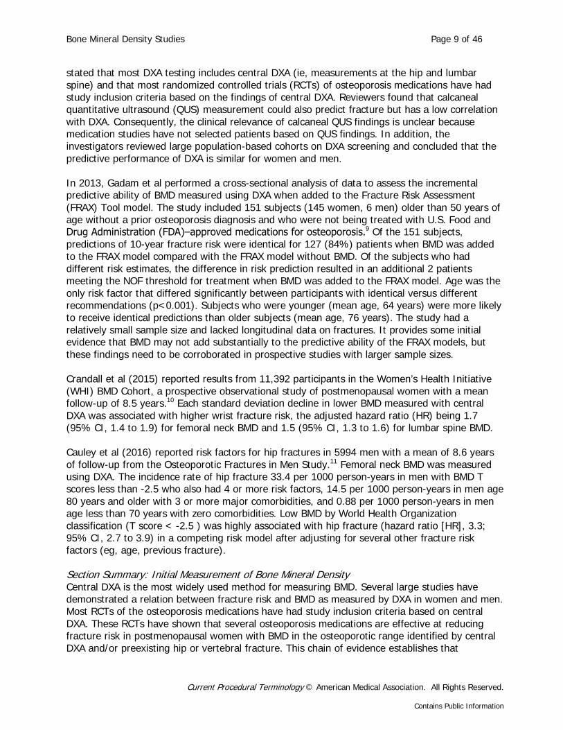
RATIONALE The most recent literature review was performed through January 25, 2017. Following is a summary of the key literature to date. Initial Measurement Of Bone Mineral Density Early versions of this evidence review were based in part on 1998 guidelines from the National Osteoporosis Foundation (NOF) and 2 TEC Assessments (1999, 2002).4-6 Because no data were available from randomized screening trials, the TEC Assessments focused on the evaluating the utility of bone mineral density (BMD) measurement in selecting patients for pharmacologic treatment to reduce risk of fracture. The TEC Assessments concluded that while both dual x-ray absorptiometry (DXA) and ultrasound densitometry were equivalent in predicting fracture risk, the 2 techniques appeared to identify different populations of at-risk patients. In addition, calcaneal ultrasound densitometry did not meet TEC criteria as a technique to predict response to pharmacologic therapy. A 2005 meta-analysis of data from 9891 men and 29,082 women (from 12 cohort studies in Europe and Canada) found that BMD measurement at the femoral neck with DXA was a strong predictor of hip fractures for both sexes.7 At age 65 years, the relative risk for osteoporotic fractures increased by 1.4 (95% confidence interval [CI], 1.3 to 1.5) in men and by 1.4 (95% CI, 1.3 to 1.5) in women for each standard deviation decrease in BMD. Data for measurement of BMD using ultrasound and peripheral DXA were available from 3 cohorts. The predictive ability of these devices was less than that of DXA measurements at the femoral neck. A systematic review of the evidence to update U.S. Preventive Services Task Force (USPSTF) recommendations on screening for osteoporosis was published in 2010.8 USPSTF reviewers
Bone Mineral Density Studies Page 9 of 46
Current Procedural Terminology © American Medical Association. All Rights Reserved.
Contains Public Information
stated that most DXA testing includes central DXA (ie, measurements at the hip and lumbar spine) and that most randomized controlled trials (RCTs) of osteoporosis medications have had study inclusion criteria based on the findings of central DXA. Reviewers found that calcaneal quantitative ultrasound (QUS) measurement could also predict fracture but has a low correlation with DXA. Consequently, the clinical relevance of calcaneal QUS findings is unclear because medication studies have not selected patients based on QUS findings. In addition, the investigators reviewed large population-based cohorts on DXA screening and concluded that the predictive performance of DXA is similar for women and men. In 2013, Gadam et al performed a cross-sectional analysis of data to assess the incremental predictive ability of BMD measured using DXA when added to the Fracture Risk Assessment (FRAX) Tool model. The study included 151 subjects (145 women, 6 men) older than 50 years of age without a prior osteoporosis diagnosis and who were not being treated with U.S. Food and Drug Administration (FDA)‒approved medications for osteoporosis.9 Of the 151 subjects, predictions of 10-year fracture risk were identical for 127 (84%) patients when BMD was added to the FRAX model compared with the FRAX model without BMD. Of the subjects who had different risk estimates, the difference in risk prediction resulted in an additional 2 patients meeting the NOF threshold for treatment when BMD was added to the FRAX model. Age was the only risk factor that differed significantly between participants with identical versus different recommendations (p<0.001). Subjects who were younger (mean age, 64 years) were more likely to receive identical predictions than older subjects (mean age, 76 years). The study had a relatively small sample size and lacked longitudinal data on fractures. It provides some initial evidence that BMD may not add substantially to the predictive ability of the FRAX models, but these findings need to be corroborated in prospective studies with larger sample sizes. Crandall et al (2015) reported results from 11,392 participants in the Women’s Health Initiative (WHI) BMD Cohort, a prospective observational study of postmenopausal women with a mean follow-up of 8.5 years.10 Each standard deviation decline in lower BMD measured with central DXA was associated with higher wrist fracture risk, the adjusted hazard ratio (HR) being 1.7 (95% CI, 1.4 to 1.9) for femoral neck BMD and 1.5 (95% CI, 1.3 to 1.6) for lumbar spine BMD. Cauley et al (2016) reported risk factors for hip fractures in 5994 men with a mean of 8.6 years of follow-up from the Osteoporotic Fractures in Men Study.11 Femoral neck BMD was measured using DXA. The incidence rate of hip fracture 33.4 per 1000 person-years in men with BMD T scores less than -2.5 who also had 4 or more risk factors, 14.5 per 1000 person-years in men age 80 years and older with 3 or more major comorbidities, and 0.88 per 1000 person-years in men age less than 70 years with zero comorbidities. Low BMD by World Health Organization classification (T score < -2.5 ) was highly associated with hip fracture (hazard ratio [HR], 3.3; 95% CI, 2.7 to 3.9) in a competing risk model after adjusting for several other fracture risk factors (eg, age, previous fracture). Section Summary: Initial Measurement of Bone Mineral Density Central DXA is the most widely used method for measuring BMD. Several large studies have demonstrated a relation between fracture risk and BMD as measured by DXA in women and men. Most RCTs of the osteoporosis medications have had study inclusion criteria based on central DXA. These RCTs have shown that several osteoporosis medications are effective at reducing fracture risk in postmenopausal women with BMD in the osteoporotic range identified by central DXA and/or preexisting hip or vertebral fracture. This chain of evidence establishes that

Bone Mineral Density Studies Page 10 of 46
Current Procedural Terminology © American Medical Association. All Rights Reserved.
Contains Public Information
measuring BMD with central DXA improves health outcomes. There is less evidence for other methods of measuring BMD. BMD measured with other techniques may also be associated with fracture risk but has not been used to select participants for trials of osteoporosis medications.
Repeat Measurement of BMD for Individuals without Osteoporosis on Initial Screen In the analysis from the WHI BMD cohort (2015) previously described, changes in central DXA were examined over a 3-year period.10 A total of 9172 women had baseline and year 3 measures of femoral neck BMD and 9216 women had baseline and year 3 measures of lumbar spine. Decrease in femoral neck BMD between baseline and year 3 was associated with increased risk of subsequent wrist fracture (HR=1.2; 95% CI, 1.0 to 1.3; p=0.03). Change in lumbar spine BMD was not associated with wrist fracture by year 3 (HR=1.1; 95% CI, 0.95 to 1.2; p=0.22). Leslie et al (2016) reported on repeat BMD measurements in clinical practice for fracture risk assessment from a large clinical BMD database for Manitoba, Canada, of women and men ages 50 years and older from 1990 to 2009.12 BMD was measured in the hip, lumbar spine, and femoral neck using DXA. A total of 50,215 participants had 1 BMD measurement, 14,619 had a second measurement, 4722 had a third measurement, and 1500 had a fourth measurement. The mean time between measurements was 4.2 years. Total hip BMD was predictive of major osteoporotic fracture at each time point. The association between BMD and major osteoporotic fracture was similar for the first and second BMD measurements: adjusted hazard ratios per standard deviation were 1.5 (95% CI, 1.3 to 1.6) and 1.6 (95% CI, 1.5 to 1.8), respectively. The hazard ratio for the second measurement was similar when stratified by preceding change in BMD or osteoporosis therapy. A 2013 study by Berry et al did not find that changes in BMD 4 years after initial measurement added substantially to the prediction of fracture risk in untreated subjects.13 The authors included 210 women and 492 men (mean age, 75 years) from the Framingham Osteoporosis Study, a population-based cohort study, who had 2 BMD measurements (mean, 3.7 years apart) and did not have a hip fracture before the second test. BMD of the femoral neck was measured using a dual-photon absorptiometer from 1987 through 1991 and DXA from 1992 through 1999. Median follow-up was 9.6 years after the second BMD test. During this time, 76 individuals experienced a hip fracture and 113 had a major osteoporotic fracture (fracture of the hip, spine, forearm, or shoulder). In receiver operating curve analyses, adding repeat BMD to a model containing baseline BMD did not meaningfully improve the model’s ability to predict hip fracture (area under the curve [AUC], 0.72; 95% CI, 0.66 to 0.79). When percent change in BMD was used, the AUC was 0.71 (95% CI, 0.65 to 0.78) in a model including only baseline BMD and 0.68 (95% CI, 0.62 to 0.75) in a model including percent change in BMD. A 2012 multicenter prospective study by Gourlay et al have provided data on the optimal bone density screening interval in a large cohort of women with normal BMD measured by DXA or osteopenia at an initial screen.14 The investigators included 4957 women ages 67 years or older who had BMD data at 2 or more examinations or at 1 examination before a competing risk event (hip or clinical vertebral fracture). The study only included women who were candidates for osteoporosis screening. Other individuals, such as those with osteoporosis at baseline or with a history of a hip or clinical vertebral fracture, were excluded, because they would already be candidates for pharmacologic treatment. The primary study outcome was the estimated time interval for 10% of participants to make the transition from normal BMD or osteopenia at baseline to osteoporosis before a hip or clinical vertebral fracture occurred and before starting

Bone Mineral Density Studies Page 11 of 46
Current Procedural Terminology © American Medical Association. All Rights Reserved.
Contains Public Information
osteoporosis treatment. For women with normal BMD at baseline, the estimated BMD testing interval was 16.8 years (95% CI, 11.5 to 24.6 years). The study found that the estimated BMD testing interval was 17.3 years (95% CI, 13.9 to 21.5 years) for women with mild osteopenia at baseline, 4.7 years (95% CI, 4.2 to 5.2 years) with moderate osteopenia, and 1.1 years (95% CI, 1.0 to 1.3 years) for women with advanced osteopenia. Longitudinal changes in BMD measured by central DXA, as a function of age and antiresorptive agents, were reported in 2008 by the Canadian Multicentre Osteoporosis Study Research Group.15 Among a random selection of 9423 men and women from 9 major Canadian cities, 4433 women and 1935 men (cumulatively 70%) were included for analysis. The subjects were 25 years of age or older with BMD measurements repeated 3 or 5 years apart; they tended to have better health than the 30% who did not have longitudinal data and who were excluded from analysis. Results showed that annual rates of bone loss, measured at the hip or femoral neck, increased between 25 and 85 years of age in women who were not on antiresorptive therapy, with accelerated periods of bone loss around menopausal transition (age range, 40-54 years) and after 70 years of age. Antiresorptive therapy, which primarily consisted of hormone replacement when the study began in 1995, was associated with attenuated bone loss across all age ranges. For women 50 to 79 years of age, the average loss in BMD over a 5-year period was 3.2% in nonusers of antiresorptive therapy and 0.2% in women who used antiresorptive therapy. The pattern in men was generally similar to that of women with 2 exceptions: BMD loss began earlier in men and the rate of change remained relatively constant between 40 and 70 years of age. Notably, BMD at the lumbar spine did not parallel measurements at the hip and femoral neck, suggesting that vertebral bone density assessment may be obscured by degenerative changes in the spine or other artifact. The report concluded that “although current guidelines recommend that measurements of bone density be repeated once every 2 to 3 years, our data suggest that, at this rate of testing, the average person would exhibit change well below the margin of error, especially since only 25% of women experienced a loss of bone density that exceeded 5% over 5 years.” In 2009, Frost et al published a prognostic model to determine the optimal screening interval for a subject without osteoporosis (defined as T score > -2.5).16 They used prospective population-based data collected from 1008 women and 750 men who were nonosteoporotic at baseline; participants received BMD screening every 2 years and had a median follow-up of 7.1 years. The prognostic model included 2 variables: age and initial BMD score. Results were stratified for these variable, presented tabularly, showing estimated time to reach 20% risk of sustaining a fracture or osteoporosis. Most of the time estimates were 3 years or longer. The estimated shortest time to reach a 20% risk was 2.4 years; this was for women 80 years and older with a baseline T score of -2.2. For a typical screening candidate (a 65-year-old woman with a baseline T score of -1.0), the estimated time to reach a 10% risk of fracture was 3.8 years and to reach a 20% risk of fracture was 6.5 years. Overall, the study suggested that the 3- to 5-year time interval for repeat measurement of BMD in people who tested normal is reasonable but that an individualized model could result in longer or shorter recommended retesting intervals. A 2007 study by Hillier et al did not find that follow-up central DXA BMD measurements 8 years after a baseline screen provided substantial value in terms of predicting risk of fracture.17 The study included 4124 women ages 65 years and older and assessed total hip BMD at initial and follow-up screening examinations. In analyses adjusted for age and weight change, the initial and repeat BMD measurements had similar associations with fracture risk; this included risk of

Bone Mineral Density Studies Page 12 of 46
Current Procedural Terminology © American Medical Association. All Rights Reserved.
Contains Public Information
vertebral fractures, non‒vertebral fractures, and hip fractures. Stratifying the analysis by initial BMD T scores (ie, normal, osteopenic, osteoporotic) did not alter findings. Section Summary: Repeat Measurement of BMD for Individuals Without Osteoporosis on Initial Screen Scant research has been done on the frequency of BMD monitoring for osteoporosis. The available research has evaluated repeat measurement with central DXA. Evidence on whether repeat measurements add to risk prediction compared with a single measurement is mixed. A longitudinal study suggests that a 3- to 5-year interval for repeat measurement of BMD in people who tested normal at baseline is reasonable, although the optimal interval may differ depending on risk factors. Serial Measurement of Central BMD to Monitor Response to Bisphosphonate Treatment In 2009, Bell et al conducted a secondary analysis of data from the Fracture Intervention Trial (FIT), which randomized 6459 postmenopausal women with low BMD to treatment with bisphosphonates or to placebo; women underwent annual bone density scans.18 The investigators estimated between-person (treatment-related) variation and within-person (measurement-related) variation in hip and spine BMD measured by central DXA over time to assess the value of repeat BMD scans for monitoring response to treatment. After 3 years, the mean cumulative increase in hip BMD was 0.30 g/cm2 in the alendronate group compared with a mean decrease of 0.012 g/cm2 in the placebo group. Moreover, 97.5% of patients treated with alendronate had increases in hip BMD of at least 0.019 g/cm2, suggesting that there was a clinically significant response. However, the study also found large within-person variability in year-to-year bone density measurements. The average within-person variation in BMD measurement was 0.013 g/cm2 relative to the placebo, which was substantially higher than the average annual increase in BMD in the alendronate group, 0.0085 g/cm2. This finding suggests that the precision of BMD measurement is not reliable from year to year, and thus annual retesting may not be clinically useful. Additional studies would be needed to determine the optimal time interval for rescreening after starting bisphosphonate treatment. Serial Measurement of Central BMD to Monitor Discontinuation of Bisphosphonate Treatment In 2014, Bauer et al reported on fracture risk prediction among women who discontinued alendronate after 4 to 5 years of treatment in the FLEX (Fracture Intervention Trial Long-term Extension) study.19 Women ages 61 to 86 years who had been treated with alendronate were randomized to 5 more years of alendronate or to placebo. A prior report of this study (2006) found that although hip BMD decreased in the placebo group, rates of fracture were similar between the group randomized to placebo and the group that continued on bisphosphonate therapy.20 It should be noted that alendronate has a half-life in humans that is estimated to exceed 10 years.21 During the 5 years of placebo treatment, 94 (22%) of 437 women experienced 1 or more symptomatic fractures; most (87%) occurred after 1 year. Post hoc analysis found that older age and lower hip (but not spine) DXA at time of discontinuation were significantly related to increased fracture risk; however, changes in BMD between the beginning of discontinuation to years 1 and 3 were not.

Bone Mineral Density Studies Page 13 of 46
Current Procedural Terminology © American Medical Association. All Rights Reserved.
Contains Public Information
SUMMARY OF EVIDENCE For individuals who are eligible for screening of bone mineral density (BMD) based on risk factor assessment who receive dual x-ray absorptiometry (DXA) analysis of central sites (hip or spine), the evidence includes large cohort studies, observational studies, and systematic reviews. Relevant outcomes are disease-specific survival, morbid events, functional outcomes, health status measures, quality of life, hospitalizations, medication use, and resource utilization. BMD measurements with central DXA identify individuals at increased risk of fracture. There is sufficient evidence that osteoporosis medications are effective at reducing fracture risk in postmenopausal women with BMD in the osteoporotic range identified by central DXA. Therefore, a chain of evidence establishes that screening BMD with central DXA is likely to improve health outcomes. Evidence to support serial or repeat measurement of BMD is less compelling; nonetheless, the available evidence and the consensus of clinical evidence-based guidelines support at least a 2-year interval in BMD measurement to monitor response to pharmacologic therapy. Finally, available evidence suggests that at least a 3- to 5-year timeframe is reasonable for repeat measurement of BMD in individuals who initially tested normal and to monitor pharmacologic therapy. The evidence is sufficient to determine that the technology results in a meaningful improvement in the net health outcome. For individuals who are eligible for screening of BMD based on risk factor assessment who receive ultrasound densitometry, or quantitative computed tomography, or DXA analysis of peripheral sites, the evidence includes observational studies and systematic reviews. Relevant outcomes are disease-specific survival, morbid events, functional outcomes, health status measures, quality of life, hospitalizations, medication use, and resource utilization. These technologies are not commonly used for BMD measurements in practice and no studies have shown that they can select patients who benefit from treatment for osteoporosis. There is little to no evidence on the usefulness of repeat measurement of BMD using these techniques. The evidence is insufficient to determine the effects of the technology on health outcomes.
CLINICAL INPUT FROM PHYSICIAN SPECIALTY SOCIETIES AND ACADEMIC MEDICAL CENTERS
While the various physician specialty societies and academic medical centers may collaborate with and make recommendations during this process, through the provision of appropriate reviewers, input received does not represent an endorsement or position statement by the physician specialty societies or academic medical centers, unless otherwise noted. In response to requests, input was received from 4 physician specialty societies (7 reviewers) and 2 academic medical centers while this policy was under review in 2008. In addition, 7 unsolicited letters were received through 2 additional physician specialty societies. The reviewers agreed with the policy statement that an initial BMD test may be medically necessary. They also recommended an interval of 3 to 5 years between measurements in subjects who previously tested normal, depending on risk factors. Reviewers considered serial measurement of BMD important to guide treatment decisions (eg, continuing or changing medication). Based on the consensus of clinical opinion regarding the value of the information provided by monitoring treatment response, serial BMD measurements (at least a 2-year interval) may be considered appropriate when this information will impact patient care. It should be noted that with the margin of error of BMD measurements with DXA, questions remain about the interval over which a clinically significant change can be observed. The minimal clinically significant

Bone Mineral Density Studies Page 14 of 46
Current Procedural Terminology © American Medical Association. All Rights Reserved.
Contains Public Information
change also raises concerns about the potential for overinterpretation of small fluctuations with repeat testing.
PRACTICE GUIDELINES AND POSITION STATEMENTS American College of Obstetricians and Gynecologists In 2012 (reaffirmed in 2014), the American College of Obstetricians and Gynecologists issued updated guidelines on managing osteoporosis in women.22 The guidelines recommend that BMD screening should begin for all women at age 65 years. In addition, they recommend screening for women younger than 65 years in whom the Fracture Risk Assessment (FRAX) Tool indicates a 10-year risk of osteoporotic fracture of at least 9.3%. Alternatively, they recommend BMD screening women in younger than 65 or with any of the following risk factors (these are similar, but not identical to risk factors in FRAX):
• Personal medical history of a fragility fracture • Parental medical history of hip fracture • Weight less than 127 lb • Medical causes of bone loss (ie, medications or disease) • Current smoker • Alcoholism • Rheumatoid arthritis • For women who begin medication treatment for osteoporosis, a repeat BMD is
recommended 1 to 2 years later to assess effectiveness. If BMD is improved or stable, additional BMD testing (in the absence of new risk factors) is not recommended. The guideline notes that it generally takes 18 to 24 months to document a clinically meaningful change in BMD and thus a 2-year interval after treatment initiation is preferred to 1 year.
• The guidelines do not specifically discuss repeat BMD screening for women who have a normal finding on the initial test.
• Routine BMD screening is not recommended for newly menopausal women as a “baseline” screen.
National Osteoporosis Foundation The National Osteoporosis Foundation (NOF) updated its practice guidelines in 2014.23 NOF guidelines recommend that all postmenopausal women and men age 50 and older should be evaluated clinically for osteoporosis risk to determine the need for BMD testing. Indications for BMD testing are:
• Women age 65 and older and men age 70 and older, regardless of clinical risk factors • Younger postmenopausal women and men age 50-69 with clinical risk factors for fracture • Adults who have a fracture after age 50 • Adults with a condition or taking a medication associated with low bone mass or bone loss
NOF states that measurements for monitoring patients should be performed in accordance with medical necessity, expected response and in consideration of local regulatory requirements. NOF recommends that repeat BMD assessments generally agree with Medicare guidelines of every 2 years, but recognizes that testing more frequently may be warranted in certain clinical situations. NOF also indicates that:

Bone Mineral Density Studies Page 15 of 46
Current Procedural Terminology © American Medical Association. All Rights Reserved.
Contains Public Information
“Central DXA assessment of the hip or lumbar spine is the ‘gold standard’ for serial assessment of BMD. Biological changes in bone density are small compared to the inherent error in the test itself, and interpretation of serial bone density studies depends on appreciation of the smallest change in BMD that is beyond the range of error of the test. This least significant change (LSC) varies with the specific instrument used, patient population being assessed, measurement site, technologist’s skill with patient positioning and test analysis, and the confidence intervals used. Changes in the BMD of less than 3-6 percent at the hip and 2-4 percent at the spine from test to test may be due to the precision error of the testing itself.”
American College of Physicians The 2008 guidelines from the American College of Physicians (ACP) recommend that clinicians periodically perform individualized assessment of risk factors for osteoporosis in men older than 50 years (grade: strong recommendation; moderate-quality evidence).24 Factors that increase the risk for osteoporosis in men include age (>70 years), low body mass index, weight loss, physical inactivity, corticosteroid use, androgen deprivation therapy, and previous fragility fracture. ACP recommends that clinicians obtain DXA for men who are at increased risk for osteoporosis and are candidates for drug therapy (grade: strong recommendation; moderate-quality evidence). The guidelines indicate that bone density measurement with DXA is the accepted reference standard for diagnosing osteoporosis in men; because treatment trials have not measured the effectiveness of therapy for osteoporosis diagnosed by ultrasound densitometry rather than DXA, the role of ultrasound in diagnosis remains uncertain. This evidence review found no studies that evaluated the optimal intervals for repeated screening by using BMD measurement with DXA in men. American College of Radiology Practice guidelines from the American College of Radiology, last amended in 201421 state that BMD measurement is indicated whenever a clinical decision is likely to be directly influenced by the result of the test. Indications for DXA include but are not to the following patient populations:
1. All women age 65 years and older and men age 70 years and older (asymptomatic screening).
2. Women younger than age 65 years who have additional risk for osteoporosis, based on medical history and other findings. Additional risk factors for osteoporosis include:
a. Estrogen deficiency b. A history of maternal hip fracture that occurred after the age of 50 years. c. Low body mass (less than 127 lbs or 57.6 kg). d. History of amenorrhea (more than 1 year before age 42 years).
3. Women younger than age 65 years or men younger than age 70 years who have additional risk factors, including:
a. Current use of cigarettes b. Loss of height, thoracic kyphosis.
4. Individuals of any age with bone mass osteopenia, or fragility fractures on imaging studies such as radiographs, computed tomography (CT, or magnetic resonance imaging [MRI]) 5. Individuals age 50 years and older who develop a wrist, hip, spine, or proximal humerus fracture with minimal or no trauma, excluding pathologic fractures. 6. Individuals of any age who develop 1 or more insufficiency fractures. 7. Individuals being considered for pharmacologic therapy for osteoporosis.

Bone Mineral Density Studies Page 16 of 46
Current Procedural Terminology © American Medical Association. All Rights Reserved.
Contains Public Information
8. Individuals being monitored to: a. Assess the effectiveness of osteoporosis drug therapy. b. Follow-up medical conditions associated with abnormal BMD.
International Society for Clinical Densitometry The 2013 update of the International Society for Clinical Densitometry guidelines recommend bone density testing in the following patients26:
“Women age 65 and older For post-menopausal women younger than age 65 a bone density test is indicated if they have a risk factor for low bone mass fracture such as;
o Low body weight o Prior fracture o High risk medication use o Disease or condition associated with bone loss.
Women during the menopausal transition with clinical risk factors for fracture, such as low bone weight, prior fracture or high-risk medication use. Men aged 70 and older. Men under < 70 years … if they have a risk factors for low bone mass such as;
o Low body weight o Prior fracture o High risk medication use o Disease or condition associated with bone loss.
Adults with a fragility fracture. Adults with a disease or condition associated with low bone mass or bone loss…. Anyone being considered for pharmacologic therapy. Anyone being treated, to monitor treatment effect. Anyone not receiving therapy in whom evidence of bone loss would lead to treatment.”
American Association of Clinical Endocrinologists et al In 2016, the American Association of Clinical Endocrinologists and American College of Endocrinology issued updated joint guidelines on the diagnosis and treatment of postmenopausal osteoporosis.27 The guidelines listed the potential uses for BMD measurements in postmenopausal women as:
“Screening for osteoporosis Establishing the severity of osteoporosis or bone loss… Determining fracture risk… Identifying candidates for pharmacologic intervention Assessing changes in bone density over time… Enhancing acceptance of, and perhaps adherence with, treatment Assessing skeletal consequences of diseases, conditions, or medications known to cause bone
loss” North American Menopause Society The North American Menopause Society issued a 2010 position statement,24 which states that fracture is the most significant risk of low bone density. The statement also concludes that BMD is an important determinant of fracture risk, especially in women 65 years and older.

Bone Mineral Density Studies Page 17 of 46
Current Procedural Terminology © American Medical Association. All Rights Reserved.
Contains Public Information
U.S. PREVENTIVE SERVICES TASK FORCE RECOMMENDATIONS The U.S. Preventive Services Task Force (USPSTF) updated recommendations on screening for osteoporosis with bone density measurements in January 2011.29 USPSTF recommends routine osteoporosis screening in women age 65 years or older and in younger women whose risk of fracture is at least equal to that of a 65-year-old average-risk white woman. This represents a change from the previous (2002) version in which there was no specific recommendation regarding screening in women younger than 65 years old. The supporting document notes that there are multiple instruments to predict risk for low BMD and that the USPSTF used FRAX.2 The updated USPSTF recommendations state that the scientific evidence is insufficient to recommend for or against routine osteoporosis screening in men. The Task Force did not recommend specific screening tests but said that the most commonly used tests are DXA of the hip and lumbar spine and quantitative ultrasound of the calcaneus. USPSTF recommendations state the following on BMD screening intervals: “…A lack of evidence exists about the optimal intervals for repeat screening and whether repeated screening is necessary in a woman with normal BMD. Because of limitations in the precision of testing, a minimum of 2 years may be needed to reliably measure a change in BMD; however, longer intervals may be necessary to improve fracture risk prediction.” USPSTF recommendations for osteoporosis screening are in the process of being updated as of February 2017.
ONGOING AND UNPUBLISHED CLINICAL TRIALS A search of ClinicalTrials.gov in February 2017 did not identify any ongoing or unpublished trials that would likely influence this review. CODING The following codes for treatment and procedures applicable to this policy are included below for informational purposes. Inclusion or exclusion of a procedure, diagnosis or device code(s) does not constitute or imply member coverage or provider reimbursement. Please refer to the member's contract benefits in effect at the time of service to determine coverage or non-coverage of these services as it applies to an individual member. CPT/HCPCS 76977 Ultrasound bone density measurement and interpretation, peripheral site(s), any
method 77078 Computed tomography, bone mineral density study, 1 or more sites; axial skeleton (eg,
hips, pelvis, spine) 77080 Dual-energy X-ray absorptiometry (DXA), bone density study, 1 or more sites; axial
skeleton (eg, hips, pelvis, spine) 77081 Dual-energy X-ray absorptiometry (DXA), bone density study, 1 or more sites;
appendicular skeleton (peripheral) (eg, radius, wrist, heel) 77085 Dual-energy X-ray absorptiometry (DXA), bone density study, 1 or more sites; axial
skeleton (eg, hips, pelvis, spine), including vertebral fracture assessment 78350 Bone density (bone mineral content) study, 1 or more sites; single photon
absorptiometry

Bone Mineral Density Studies Page 18 of 46
Current Procedural Terminology © American Medical Association. All Rights Reserved.
Contains Public Information
78351 Bone density (bone mineral content) study, 1 or more sites; dual photon absorptiometry, 1 or more sites
G0130 Single energy x-ray absorptiometry (SEXA) bone density study, one or more sites; appendicular skeleton (peripheral) (eg, radius, wrist, heel)
ICD-9 Diagnoses 203.00 Multiple myeloma, without mention of remission 203.01 Multiple myeloma, in remission 242.00 Toxic diffuse goiter without mention of thyrotoxic crisis or storm 242.01 Toxic diffuse goiter with mention of thyrotoxic crisis or storm 242.10 Toxic uninodular goiter without mention of thyrotoxic crisis or storm 242.11 Toxic uninodular goiter with mention of thyrotoxic crisis or storm 242.20 Toxic multinodular goiter without mention of thyrotoxic crisis or storm 242.21 Toxic multinodular goiter with mention of thyrotoxic crisis or storm 242.30 Toxic nodular goiter, unspecified without mention of thyrotoxic crisis or storm 242.31 Toxic nodular goiter, unspecified with mention of thyrotoxic crisis or storm 242.40 Thyrotoxicosis from ectopic thyroid nodule without mention of thyrotoxic crisis or
storm 242.41 Thyrotoxicosis from ectopic thyroid nodule with mention of thyrotoxic crisis or storm 242.80 Thyrotoxicosis of other specified origin without mention of thyrotoxic crisis or storm 242.81 Thyrotoxicosis of other specified origin with mention of thyrotoxic crisis or storm 242.90 Thyrotoxicosis without mention of goiter or other cause without mention of thyrotoxic
crisis or storm 242.91 Thyrotoxicosis without mention of goiter or other cause with mention of thyrotoxic
crisis or storm 252.00 Hyperparathyroidism, unspecified 252.01 Primary hyperparathyroidism 252.02 Secondary hyperparathyroidism, non-renal 252.08 Other hyperparathyroidism 253.4 Other anterior pituitary disorders 255.0 Cushing's syndrome 256.2 Postablative ovarian failure 256.31 Premature menopause 257.2 Other testicular hypofunction 259.50 Androgen insensitivity syndrome, androgen insensitivity, unspecified 259.51 Androgen insensitivity syndrome 259.52 Androgen insensitivity syndrome, partial androgen insensitivity 263.9 Unspecified protein-calorie malnutrition 303.90 Other and unspecified alcohol dependence 303.91 Other and unspecified alcohol dependence, continuous drunkenness 303.92 Other and unspecified alcohol dependence, episodic drunkenness 303.93 Other and unspecified alcohol dependence, in remission 305.1 Tobacco use disorder 307.1 Anorexia nervosa 345.00- 345.91
Epilepsy and recurrent seizures
577.1 Chronic pancreatitis 577.9 Unspecified disease of pancreas

Bone Mineral Density Studies Page 19 of 46
Current Procedural Terminology © American Medical Association. All Rights Reserved.
Contains Public Information
579.0 Celiac disease 579.8 Other specified intestinal malabsorption 579.9 Unspecified intestinal malabsorption 585.2 Chronic kidney disease, Stage II (mild) 585.3 Chronic kidney disease, Stage III (moderate) 585.4 Chronic kidney disease, Stage IV (severe) 585.5 Chronic kidney disease, Stage V 585.6 End stage renal disease 585.9 Chronic kidney disease, unspecified 627.4 Symptomatic states associated with artificial menopause 714.0 Rheumatoid arthritis 733.00 Osteoporosis, unspecified 733.01 Senile osteoporosis 733.02 Idiopathic osteoporosis 733.03 Disuse osteoporosis 733.09 Osteoporosis, other 733.90 Disorder of bone and cartilage, unspecified 756.51 Osteogenesis imperfecta 780.33 Post traumatic seizures 805.2 Dorsal (thoracic), closed 805.4 Lumbar, closed 805.6 Sacrum and coccyx, closed V42.0 Organ or tissue replaced by transplant, Kidney V42.1 Organ or tissue replaced by transplant, Heart V42.3 Organ or tissue replaced by transplant, skin V42.4 Organ or tissue replaced by transplant, bone V42.6 Organ or tissue replaced by transplant, lung V58.65 Long-term (current) use of steroids V58.69 Long-term (current) use of other medications V42.7 Organ or tissue replaced by transplant, liver ICD-10 Diagnoses C90.00 Multiple myeloma not having achieved remission C90.01 Multiple myeloma in remission E05.00 Thyrotoxicosis with diffuse goiter without thyrotoxic crisis or storm E05.01 Thyrotoxicosis with diffuse goiter with thyrotoxic crisis or storm E05.10 Thyrotoxicosis with toxic single thyroid nodule without thyrotoxic crisis or storm E05.11 Thyrotoxicosis with toxic single thyroid nodule with thyrotoxic crisis or storm E05.20 Thyrotoxicosis with toxic multinodular goiter without thyrotoxic crisis or storm E05.21 Thyrotoxicosis with toxic multinodular goiter with thyrotoxic crisis or storm E05.30 Thyrotoxicosis from ectopic thyroid tissue without thyrotoxic crisis or storm E05.31 Thyrotoxicosis from ectopic thyroid tissue with thyrotoxic crisis or storm E05.40 Thyrotoxicosis factitia without thyrotoxic crisis or storm E05.41 Thyrotoxicosis factitia with thyrotoxic crisis or storm E05.80 Other thyrotoxicosis without thyrotoxic crisis or storm E05.81 Other thyrotoxicosis with thyrotoxic crisis or storm E05.90 Thyrotoxicosis, unspecified without thyrotoxic crisis or storm E21.0 Primary hyperparathyroidism

Bone Mineral Density Studies Page 20 of 46
Current Procedural Terminology © American Medical Association. All Rights Reserved.
Contains Public Information
E21.1 Secondary hyperparathyroidism, not elsewhere classified E21.2 Other hyperparathyroidism E21.3 Hyperparathyroidism, unspecified E23.6 Other disorders of pituitary gland E24.0 Pituitary-dependent Cushing's disease E24.2 Drug-induced Cushing's syndrome E24.3 Ectopic ACTH syndrome E24.8 Other Cushing's syndrome E28.310 Symptomatic premature menopause E28.319 Asymptomatic premature menopause E29.1 Testicular hypofunction E34.51 Complete androgen insensitivity syndrome E34.52 Partial androgen insensitivity syndrome E46 Unspecified protein-calorie malnutrition E64.0 Sequelae of protein-calorie malnutrition E89.40 Asymptomatic postprocedural ovarian failure E89.41 Symptomatic postprocedural ovarian failure F10.20 Alcohol dependence, uncomplicated F10.21 Alcohol dependence, in remission F17.201 Nicotine dependence, unspecified, in remission F17.210 Nicotine dependence, cigarettes, uncomplicated F17.211 Nicotine dependence, cigarettes, in remission F17.220 Nicotine dependence, chewing tobacco, uncomplicated F17.221 Nicotine dependence, chewing tobacco, in remission F17.290 Nicotine dependence, other tobacco product, uncomplicated F17.291 Nicotine dependence, other tobacco product, in remission F50.01 Anorexia nervosa, restricting type F50.02 Anorexia nervosa, binge eating/purging type G40.001 Localization-related (focal) (partial) idiopathic epilepsy and epileptic syndromes with
seizures of localized onset, not intractable, with status epilepticus G40.009 Localization-related (focal) (partial) idiopathic epilepsy and epileptic syndromes with
seizures of localized onset, not intractable, without status epilepticus G40.011 Localization-related (focal) (partial) idiopathic epilepsy and epileptic syndromes with
seizures of localized onset, intractable, with status epilepticus G40.019 Localization-related (focal) (partial) idiopathic epilepsy and epileptic syndromes with
seizures of localized onset, intractable, without status epilepticus G40.101 Localization-related (focal) (partial) symptomatic epilepsy and epileptic syndromes
with simple partial seizures, not intractable, with status epilepticus G40.109 Localization-related (focal) (partial) symptomatic epilepsy and epileptic syndromes
with simple partial seizures, not intractable, without status epilepticus G40.111 Localization-related (focal) (partial) symptomatic epilepsy and epileptic syndromes
with simple partial seizures, intractable, with status epilepticus G40.119 Localization-related (focal) (partial) symptomatic epilepsy and epileptic syndromes
with simple partial seizures, intractable, without status epilepticus G40.201 Localization-related (focal) (partial) symptomatic epilepsy and epileptic syndromes
with complex partial seizures, not intractable, with status epilepticus G40.209 Localization-related (focal) (partial) symptomatic epilepsy and epileptic syndromes
with complex partial seizures, not intractable, without status epilepticus

Bone Mineral Density Studies Page 21 of 46
Current Procedural Terminology © American Medical Association. All Rights Reserved.
Contains Public Information
G40.211 Localization-related (focal) (partial) symptomatic epilepsy and epileptic syndromes with complex partial seizures, intractable, with status epilepticus
G40.219 Localization-related (focal) (partial) symptomatic epilepsy and epileptic syndromes with complex partial seizures, intractable, without status epilepticus
G40.301 Generalized idiopathic epilepsy and epileptic syndromes, not intractable, with status epilepticus
G40.309 Generalized idiopathic epilepsy and epileptic syndromes, not intractable, without status epilepticus
G40.311 Generalized idiopathic epilepsy and epileptic syndromes, intractable, with status epilepticus
G40.319 Generalized idiopathic epilepsy and epileptic syndromes, intractable, without status epilepticus
G40.401 Other generalized epilepsy and epileptic syndromes, not intractable, with status epilepticus
G40.409 Other generalized epilepsy and epileptic syndromes, not intractable, without status epilepticus
G40.411 Other generalized epilepsy and epileptic syndromes, intractable, with status epilepticus
G40.419 Other generalized epilepsy and epileptic syndromes, intractable, without status epilepticus
G40.501 Epileptic seizures related to external causes, not intractable, with status epilepticus G40.509 Epileptic seizures related to external causes, not intractable, without status
epilepticus G40.801 Other epilepsy, not intractable, with status epilepticus G40.802 Other epilepsy, not intractable, without status epilepticus G40.803 Other epilepsy, intractable, with status epilepticus G40.804 Other epilepsy, intractable, without status epilepticus G40.811 Lennox-Gastaut syndrome, not intractable, with status epilepticus G40.812 Lennox-Gastaut syndrome, not intractable, without status epilepticus G40.813 Lennox-Gastaut syndrome, intractable, with status epilepticus G40.814 Lennox-Gastaut syndrome, intractable, without status epilepticus G40.821 Epileptic spasms, not intractable, with status epilepticus G40.822 Epileptic spasms, not intractable, without status epilepticus G40.823 Epileptic spasms, intractable, with status epilepticus G40.824 Epileptic spasms, intractable, without status epilepticus G40.901 Epilepsy, unspecified, not intractable, with status epilepticus G40.909 Epilepsy, unspecified, not intractable, without status epilepticus G40.911 Epilepsy, unspecified, intractable, with status epilepticus G40.919 Epilepsy, unspecified, intractable, without status epilepticus G40.A01 Absence epileptic syndrome, not intractable, with status epilepticus G40.A09 Absence epileptic syndrome, not intractable, without status epilepticus G40.A11 Absence epileptic syndrome, intractable, with status epilepticus G40.A19 Absence epileptic syndrome, intractable, without status epilepticus G40.B01 Juvenile myoclonic epilepsy, not intractable, with status epilepticus G40.B09 Juvenile myoclonic epilepsy, not intractable, without status epilepticus G40.B11 Juvenile myoclonic epilepsy, intractable, with status epilepticus G40.B19 Juvenile myoclonic epilepsy, intractable, without status epilepticus K86.0 Alcohol-induced chronic pancreatitis

Bone Mineral Density Studies Page 22 of 46
Current Procedural Terminology © American Medical Association. All Rights Reserved.
Contains Public Information
K86.1 Other chronic pancreatitis K90.0 Celiac disease K90.49 Malabsorption due to intolerance, not elsewhere classified K90.89 Other intestinal malabsorption M05.411 Rheumatoid myopathy with rheumatoid arthritis of right shoulder M05.412 Rheumatoid myopathy with rheumatoid arthritis of left shoulder M05.421 Rheumatoid myopathy with rheumatoid arthritis of right elbow M05.422 Rheumatoid myopathy with rheumatoid arthritis of left elbow M05.431 Rheumatoid myopathy with rheumatoid arthritis of right wrist M05.432 Rheumatoid myopathy with rheumatoid arthritis of left wrist M05.441 Rheumatoid myopathy with rheumatoid arthritis of right hand M05.442 Rheumatoid myopathy with rheumatoid arthritis of left hand M05.451 Rheumatoid myopathy with rheumatoid arthritis of right hip M05.452 Rheumatoid myopathy with rheumatoid arthritis of left hip M05.461 Rheumatoid myopathy with rheumatoid arthritis of right knee M05.462 Rheumatoid myopathy with rheumatoid arthritis of left knee M05.471 Rheumatoid myopathy with rheumatoid arthritis of right ankle and foot M05.472 Rheumatoid myopathy with rheumatoid arthritis of left ankle and foot M05.49 Rheumatoid myopathy with rheumatoid arthritis of multiple sites M05.511 Rheumatoid polyneuropathy with rheumatoid arthritis of right shoulder M05.512 Rheumatoid polyneuropathy with rheumatoid arthritis of left shoulder M05.521 Rheumatoid polyneuropathy with rheumatoid arthritis of right elbow M05.522 Rheumatoid polyneuropathy with rheumatoid arthritis of left elbow M05.531 Rheumatoid polyneuropathy with rheumatoid arthritis of right wrist M05.532 Rheumatoid polyneuropathy with rheumatoid arthritis of left wrist M05.541 Rheumatoid polyneuropathy with rheumatoid arthritis of right hand M05.542 Rheumatoid polyneuropathy with rheumatoid arthritis of left hand M05.551 Rheumatoid polyneuropathy with rheumatoid arthritis of right hip M05.552 Rheumatoid polyneuropathy with rheumatoid arthritis of left hip M05.559 Rheumatoid polyneuropathy with rheumatoid arthritis of unspecified hip M05.561 Rheumatoid polyneuropathy with rheumatoid arthritis of right knee M05.562 Rheumatoid polyneuropathy with rheumatoid arthritis of left knee M05.571 Rheumatoid polyneuropathy with rheumatoid arthritis of right ankle and foot M05.572 Rheumatoid polyneuropathy with rheumatoid arthritis of left ankle and foot M05.59 Rheumatoid polyneuropathy with rheumatoid arthritis of multiple sites M05.711 Rheumatoid arthritis with rheumatoid factor of right shoulder without organ or
systems involvement M05.712 Rheumatoid arthritis with rheumatoid factor of left shoulder without organ or systems
involvement M05.721 Rheumatoid arthritis with rheumatoid factor of right elbow without organ or systems
involvement M05.722 Rheumatoid arthritis with rheumatoid factor of left elbow without organ or systems
involvement M05.731 Rheumatoid arthritis with rheumatoid factor of right wrist without organ or systems
involvement M05.732 Rheumatoid arthritis with rheumatoid factor of left wrist without organ or systems
involvement

Bone Mineral Density Studies Page 23 of 46
Current Procedural Terminology © American Medical Association. All Rights Reserved.
Contains Public Information
M05.741 Rheumatoid arthritis with rheumatoid factor of right hand without organ or systems involvement
M05.742 Rheumatoid arthritis with rheumatoid factor of left hand without organ or systems involvement
M05.751 Rheumatoid arthritis with rheumatoid factor of right hip without organ or systems involvement
M05.752 Rheumatoid arthritis with rheumatoid factor of left hip without organ or systems involvement
M05.761 Rheumatoid arthritis with rheumatoid factor of right knee without organ or systems involvement
M05.762 Rheumatoid arthritis with rheumatoid factor of left knee without organ or systems involvement
M05.771 Rheumatoid arthritis with rheumatoid factor of right ankle and foot without organ or systems involvement
M05.772 Rheumatoid arthritis with rheumatoid factor of left ankle and foot without organ or systems involvement
M05.79 Rheumatoid arthritis with rheumatoid factor of multiple sites without organ or systems involvement
M05.811 Other rheumatoid arthritis with rheumatoid factor of right shoulder M05.812 Other rheumatoid arthritis with rheumatoid factor of left shoulder M05.821 Other rheumatoid arthritis with rheumatoid factor of right elbow M05.822 Other rheumatoid arthritis with rheumatoid factor of left elbow M05.831 Other rheumatoid arthritis with rheumatoid factor of right wrist M05.832 Other rheumatoid arthritis with rheumatoid factor of left wrist M05.841 Other rheumatoid arthritis with rheumatoid factor of right hand M05.842 Other rheumatoid arthritis with rheumatoid factor of left hand M05.851 Other rheumatoid arthritis with rheumatoid factor of right hip M05.852 Other rheumatoid arthritis with rheumatoid factor of left hip M05.861 Other rheumatoid arthritis with rheumatoid factor of right knee M05.862 Other rheumatoid arthritis with rheumatoid factor of left knee M05.871 Other rheumatoid arthritis with rheumatoid factor of right ankle and foot M05.872 Other rheumatoid arthritis with rheumatoid factor of left ankle and foot M05.89 Other rheumatoid arthritis with rheumatoid factor of multiple sites M06.011 Rheumatoid arthritis without rheumatoid factor, right shoulder M06.012 Rheumatoid arthritis without rheumatoid factor, left shoulder M06.021 Rheumatoid arthritis without rheumatoid factor, right elbow M06.022 Rheumatoid arthritis without rheumatoid factor, left elbow M06.031 Rheumatoid arthritis without rheumatoid factor, right wrist M06.032 Rheumatoid arthritis without rheumatoid factor, left wrist M06.041 Rheumatoid arthritis without rheumatoid factor, right hand M06.042 Rheumatoid arthritis without rheumatoid factor, left hand M06.051 Rheumatoid arthritis without rheumatoid factor, right hip M06.052 Rheumatoid arthritis without rheumatoid factor, left hip M06.061 Rheumatoid arthritis without rheumatoid factor, right knee M06.062 Rheumatoid arthritis without rheumatoid factor, left knee M06.071 Rheumatoid arthritis without rheumatoid factor, right ankle and foot M06.072 Rheumatoid arthritis without rheumatoid factor, left ankle and foot M06.08 Rheumatoid arthritis without rheumatoid factor, vertebrae
Bone Mineral Density Studies Page 24 of 46
Current Procedural Terminology © American Medical Association. All Rights Reserved.
Contains Public Information
M06.09 Rheumatoid arthritis without rheumatoid factor, multiple sites M06.211 Rheumatoid bursitis, right shoulder M06.212 Rheumatoid bursitis, left shoulder M06.221 Rheumatoid bursitis, right elbow M06.222 Rheumatoid bursitis, left elbow M06.231 Rheumatoid bursitis, right wrist M06.232 Rheumatoid bursitis, left wrist M06.241 Rheumatoid bursitis, right hand M06.242 Rheumatoid bursitis, left hand M06.251 Rheumatoid bursitis, right hip M06.252 Rheumatoid bursitis, left hip M06.261 Rheumatoid bursitis, right knee M06.262 Rheumatoid bursitis, left knee M06.271 Rheumatoid bursitis, right ankle and foot M06.272 Rheumatoid bursitis, left ankle and foot M06.28 Rheumatoid bursitis, vertebrae M06.29 Rheumatoid bursitis, multiple sites M06.311 Rheumatoid nodule, right shoulder M06.312 Rheumatoid nodule, left shoulder M06.321 Rheumatoid nodule, right elbow M06.322 Rheumatoid nodule, left elbow M06.331 Rheumatoid nodule, right wrist M06.332 Rheumatoid nodule, left wrist M06.341 Rheumatoid nodule, right hand M06.342 Rheumatoid nodule, left hand M06.351 Rheumatoid nodule, right hip M06.352 Rheumatoid nodule, left hip M06.361 Rheumatoid nodule, right knee M06.362 Rheumatoid nodule, left knee M06.371 Rheumatoid nodule, right ankle and foot M06.372 Rheumatoid nodule, left ankle and foot M06.38 Rheumatoid nodule, vertebrae M06.39 Rheumatoid nodule, multiple sites M06.811 Other specified rheumatoid arthritis, right shoulder M06.812 Other specified rheumatoid arthritis, left shoulder M06.821 Other specified rheumatoid arthritis, right elbow M06.822 Other specified rheumatoid arthritis, left elbow M06.831 Other specified rheumatoid arthritis, right wrist M06.832 Other specified rheumatoid arthritis, left wrist M06.841 Other specified rheumatoid arthritis, right hand M06.842 Other specified rheumatoid arthritis, left hand M06.851 Other specified rheumatoid arthritis, right hip M06.852 Other specified rheumatoid arthritis, left hip M06.861 Other specified rheumatoid arthritis, right knee M06.862 Other specified rheumatoid arthritis, left knee M06.871 Other specified rheumatoid arthritis, right ankle and foot M06.872 Other specified rheumatoid arthritis, left ankle and foot M06.88 Other specified rheumatoid arthritis, vertebrae

Bone Mineral Density Studies Page 25 of 46
Current Procedural Terminology © American Medical Association. All Rights Reserved.
Contains Public Information
M06.89 Other specified rheumatoid arthritis, multiple sites M81.0 Age-related osteoporosis without current pathological fracture M81.6 Localized osteoporosis [Lequesne] M81.8 Other osteoporosis without current pathological fracture M85.812 Other specified disorders of bone density and structure, left shoulder M85.811 Other specified disorders of bone density and structure, right shoulder M85.821 Other specified disorders of bone density and structure, right upper arm M85.822 Other specified disorders of bone density and structure, left upper arm M85.831 Other specified disorders of bone density and structure, right forearm M85.832 Other specified disorders of bone density and structure, left forearm M85.841 Other specified disorders of bone density and structure, right hand M85.842 Other specified disorders of bone density and structure, left hand M85.851 Other specified disorders of bone density and structure, right thigh M85.852 Other specified disorders of bone density and structure, left thigh M85.861 Other specified disorders of bone density and structure, right lower leg M85.862 Other specified disorders of bone density and structure, left lower leg M85.871 Other specified disorders of bone density and structure, right ankle and foot M85.872 Other specified disorders of bone density and structure, left ankle and foot M85.88 Other specified disorders of bone density and structure, other site M85.89 Other specified disorders of bone density and structure, multiple sites M85.9 Disorder of bone density and structure, unspecified N18.2 Chronic kidney disease, stage 2 (mild) N18.3 Chronic kidney disease, stage 3 (moderate) N18.4 Chronic kidney disease, stage 4 (severe) N18.5 Chronic kidney disease, stage 5 N18.6 End stage renal disease N18.9 Chronic kidney disease, unspecified N95.8 Other specified menopausal and perimenopausal disorders Q78.0 Osteogenesis imperfecta R56.1 Post traumatic seizures S22.000A Wedge compression fracture of unspecified thoracic vertebra, initial encounter for
closed fracture S22.000B Wedge compression fracture of unspecified thoracic vertebra, initial encounter for
open fracture S22.000D Wedge compression fracture of unspecified thoracic vertebra, subsequent encounter
for fracture with routine healing. S22.000G Wedge compression fracture of unspecified thoracic vertebra, subsequent encounter
for fracture with delayed healing S22.000K Wedge compression fracture of unspecified thoracic vertebra, subsequent encounter
for fracture with nonunion S22.000S Wedge compression fracture of unspecified thoracic vertebra, sequela S22.001A Stable burst fracture of unspecified thoracic vertebra, initial encounter for closed
fracture S22.001B Stable burst fracture of unspecified thoracic vertebra, initial encounter for open
fracture S22.001D Stable burst fracture of unspecified thoracic vertebra, subsequent encounter for
fracture with routine healing

Bone Mineral Density Studies Page 26 of 46
Current Procedural Terminology © American Medical Association. All Rights Reserved.
Contains Public Information
S22.001G Stable burst fracture of unspecified thoracic vertebra, subsequent encounter for fracture with delayed healing
S22.001K Stable burst fracture of unspecified thoracic vertebra, subsequent encounter for fracture with nonunion
S22.001S Stable burst fracture of unspecified thoracic vertebra, sequela S22.002A Unstable burst fracture of unspecified thoracic vertebra, initial encounter for closed
fracture S22.002B Unstable fracture of unspecified thoracic vertebra, initial encounter for open fracture S22.002D Unstable burst fracture of unspecified thoracic vertebra, subsequent encounter for
fracture with routine healing S22.002G Unstable burst fracture of unspecified thoracic vertebra, subsequent encounter for
fracture with delayed healing S22.002K Unstable burst fracture of un specified thoracic vertebra, subsequent encounter for
fracture with delayed healing S22.002S Unstable burst fracture of unspecified thoracic vertebra, sequela S22.008A Other fracture of unspecified thoracic vertebra, initial encounter for closed fracture S22.008B Other fracture of unspecified thoracic vertebra, initial encounter for open fracture S22.008D Other fracture of unspecified thoracic vertebra, subsequent encounter for fracture
with routine healing S22.008G Other fracture of unspecified thoracic vertebra, subsequent encounter for fracture
with delayed healing S22.008K Other fracture of unspecified thoracic vertebra, subsequent encounter for fracture
with nonunion S22.008S Other fracture of unspecified thoracic vertebra, sequela S22.010A Wedge compression fracture of first thoracic vertebra, initial encounter for closed
fracture S22.010B Wedge compression fracture of first thoracic vertebra, initial encounter for open
fracture S22.010D Wedge compression fracture of first thoracic vertebra, subsequent encounter for
fracture with routine healing S22.010G Wedge compression fracture of first thoracic vertebra, subsequent encounter for
fracture with delayed healing S22.010K Wedge compression fracture of first thoracic vertebra, subsequent encounter for
fracture with nonunion S22.010S Wedge compression fracture of first thoracic vertebra, sequela S22.011A Stable burst fracture of first thoracic vertebra, initial encounter for closed fracture S22.011B Stable burst fracture of first thoracic vertebra, initial encounter for open fracture S22.011D Stable burst fracture of first thoracic vertebra, subsequent encounter for fracture with
routine healing S22.011G Stable burst fracture of first thoracic vertebra, subsequent encounter for fracture with
delayed healing S22.011K Stable burst fracture of first thoracic vertebra, subsequent encounter for fracture with
nonunion S22.011S Stable burst fracture of first thoracic vertebra, sequela S22.012A Unstable burst fracture of first thoracic vertebra, initial encounter for closed fracture S22.012B Unstable burst fracture of first thoracic vertebra, initial encounter for open fracture S22.012D Unstable burst fracture of first thoracic vertebra, subsequent encounter for fracture
with routine healing

Bone Mineral Density Studies Page 27 of 46
Current Procedural Terminology © American Medical Association. All Rights Reserved.
Contains Public Information
S22.012G Unstable burst fracture of first thoracic vertebra, subsequent encounter for fracture with delayed healing
S22.012K Unstable burst fracture of first thoracic vertebra, subsequent encounter for fracture with nonunion
S22.012S Unstable burst fracture of first thoracic vertebra, sequela S22.018A Other fracture of first thoracic vertebra, initial encounter for closed fracture S22.018B Other fracture of first thoracic vertebra, initial encounter for open fracture S22.018D Other fracture of first thoracic vertebra, subsequent encounter for fracture with
routine healing S22.018G Other fracture of first thoracic vertebra, subsequent encounter for fracture with
delayed healing S22.018K Other fracture of first thoracic vertebra, subsequent encounter for fracture with
nonunion S22.018S Other fracture of first thoracic vertebra, sequela S22.020A Wedge compression fracture of second thoracic vertebra, initial encounter for closed
fracture S22.020B Wedge compression fracture of second thoracic vertebra, initial encounter for open
fracture S22.020D Wedge compression fracture of second thoracic vertebra, subsequent encounter for
fracture with routine healing S22.020G Wedge compression fracture of second thoracic vertebra, subsequent encounter for
fracture with delayed healing S22.020K Wedge compression fracture of second thoracic vertebra, subsequent encounter for
fracture with nonunion S22.020S Wedge compression fracture of second thoracic vertebra, sequela S22.021A Stable burst fracture of second thoracic vertebra, initial encounter for closed fracture S22.021B Stable burst fracture of second thoracic vertebra, initial encounter for open fracture S22.021D Stable burst fracture of second thoracic vertebra, subsequent encounter for fracture
with routine healing S22.021G Stable burst fracture of second thoracic vertebra, subsequent encounter for fracture
with delayed healing S22.021K Stable burst fracture of second thoracic vertebra, subsequent encounter for fracture
with nonunion S22.021S Stable burst fracture of second thoracic vertebra, sequela S22.022A Unstable burst fracture of second thoracic vertebra, initial encounter for closed
fracture S22.022B Unstable burst fracture of second thoracic vertebra, initial encounter for open fracture S22.022D Unstable burst fracture of second thoracic vertebra, subsequent encounter for
fracture with routine healing S22.022G Unstable burst fracture of second thoracic vertebra, subsequent encounter for
fracture with delayed healing S22.022K Unstable burst fracture of second thoracic vertebra, subsequent encounter for
fracture with nonunion S22.022S Unstable burst fracture of second thoracic vertebra, sequela S22.028A Other fracture of second thoracic vertebra, initial encounter for closed fracture S22.028B Other fracture of second thoracic vertebra, initial encounter for open fracture S22.028D Other fracture of second thoracic vertebra, subsequent encounter for fracture with
routine healing

Bone Mineral Density Studies Page 28 of 46
Current Procedural Terminology © American Medical Association. All Rights Reserved.
Contains Public Information
S22.028G Other fracture of second thoracic vertebra, subsequent encounter for facture with delayed healing
S22.028K Other fracture of second thoracic vertebra, subsequent encounter for fracture with nonunion
S22.028S Other fracture of second thoracic vertebra, sequela S22.030A Wedge compression fracture of third thoracic vertebra, initial encounter for closed
fracture S22.030B Wedge compression fracture of third thoracic vertebra, initial encounter for open
fracture S22.030D Wedge compression fracture of third thoracic vertebra, subsequent encounter for
fracture with routine healing S22.030G Wedge compression fracture of third thoracic vertebra, subsequent encounter for
fracture with delayed healing S22.030K Wedge compression fracture of third thoracic vertebra, subsequent encounter for
fracture with nonunion S22.030S Wedge compression fracture of third thoracic vertebra, sequela S22.031A Stable burst fracture of third thoracic vertebra, initial encounter for closed fracture S22.031B Stable burst fracture of third thoracic vertebra, initial encounter for open fracture S22.031D Stable burst fracture of third thoracic vertebra, subsequent encounter for fracture
with routine healing S22.031G Stable burst fracture of third thoracic vertebra, subsequent encounter for fracture
with delayed healing S22.031K Stable burst fracture of third thoracic vertebra, subsequent encounter for fracture
with nonunion S22.031S Stable burst fracture of third thoracic vertebra, sequela S22.032A Unstable burst fracture of third thoracic vertebra, initial encounter for closed fracture S22.032B Unstable burst fracture of third thoracic vertebra, initial encounter for open fracture S22.032D Unstable burst fracture of third thoracic vertebra, subsequent encounter for fracture
with routine healing S22.032G Unstable burst fracture of third thoracic vertebra, subsequent encounter for fracture
with delayed healing S22.032K Unstable burst fracture of third thoracic vertebra, subsequent encounter for fracture
with nonunion S22.032S Unstable burst fracture of third thoracic vertebra, sequela S22.038A Other fracture of third thoracic vertebra, initial encounter for closed fracture S22.038B Other fracture of third thoracic vertebra, initial encounter for open fracture S22.038D Other fracture of third thoracic vertebra, subsequent encounter for fracture with
routine healing S22.038G Other fracture of third thoracic vertebra, subsequent encounter for fracture with
delayed healing S22.038K Other fracture of third thoracic vertebra, subsequent encounter for fracture with
nonunion S22.038S Other fracture of third thoracic vertebra, sequela S22.040A Wedge compression fracture of fourth thoracic vertebra, initial encounter for closed
fracture S22.040B Wedge compression fracture of fourth thoracic vertebra, initial encounter for open
fracture

Bone Mineral Density Studies Page 29 of 46
Current Procedural Terminology © American Medical Association. All Rights Reserved.
Contains Public Information
S22.040D Wedge compression fracture of fourth thoracic vertebra, subsequent encounter for fracture with routine healing
S22.040G Wedge compression fracture of fourth thoracic vertebra, subsequent encounter for fracture with delayed healing
S22.040K Wedge compression fracture of fourth thoracic vertebra, subsequent encounter for fracture with nonunion
S22.040S Wedge compression fracture of fourth thoracic vertebra, sequela S22.041A Stable burst fracture of fourth thoracic vertebra, initial encounter for closed fracture S22.041B Stable burst fracture of fourth thoracic vertebra, initial encounter for open fracture S22.041D Stable burst fracture of fourth thoracic vertebra, subsequent encounter for fracture
with routine healing S22.041G Stable burst fracture of fourth thoracic vertebra, subsequent encounter for fracture
with delayed healing S22.041K Stable burst fracture of fourth thoracic vertebra, subsequent encounter for fracture
with nonunion S22.041S Stable burst fracture of fourth thoracic vertebra, sequela S22.042A Unstable burst fracture of fourth thoracic vertebra, initial encounter for closed
fracture S22.042B Unstable burst fracture of fourth thoracic vertebra, initial encounter for open fracture S22.042D Unstable burst fracture of fourth thoracic vertebra, subsequent encounter for fracture
with routine healing S22.042G Unstable burst fracture of fourth thoracic vertebra, subsequent encounter for fracture
with delayed healing S22.042K Unstable burst fracture of fourth thoracic vertebra, subsequent encounter for fracture
with nonunion S22.042S Unstable burst fracture of fourth thoracic vertebra, sequela S22.048A Other fracture of fourth thoracic vertebra, initial encounter for closed fracture S22.048B Other fracture of fourth thoracic vertebra, initial encounter for open fracture S22.048D Other fracture of fourth thoracic vertebra, subsequent encounter for fracture with
routine healing S22.048G Other fracture of fourth thoracic vertebra, subsequent encounter for fracture with
delayed healing S22.048K Other fracture of fourth thoracic vertebra, subsequent encounter for fracture with
nonunion S22.048S Other fracture of fourth thoracic vertebra, sequela S22.050A Wedge compression fracture of T5-T6 vertebra, initial encounter for closed fracture S22.050B Wedge compression fracture of T5-T6 vertebra, initial encounter for open fracture S22.050D Wedge compression fracture of T5-T6 vertebra, subsequent encounter for fracture
with routine healing S22.050G Wedge compression fracture of T5-T6 vertebra, subsequent encounter for fracture
with delayed healing S22.050K Wedge compression fracture of T5-T6 vertebra, subsequent encounter for fracture
with nonunion S22.050S Wedge compression fracture of T5-T6 vertebra, sequela S22.051A Stable burst fracture of T5-T6 vertebra, initial encounter for closed fracture S22.051B Stable burst fracture of T5-T6 vertebra, initial encounter for open fracture S22.051D Stable burst fracture of T5-T6 vertebra, subsequent encounter for fracture with
routine healing

Bone Mineral Density Studies Page 30 of 46
Current Procedural Terminology © American Medical Association. All Rights Reserved.
Contains Public Information
S22.051G Stable burst fracture of T5-T6 vertebra, subsequent encounter for fracture with delayed healing
S22.051K Stable burst fracture of T5-T6 vertebra, subsequent encounter for fracture with nonunion
S22.051S Stable burst fracture of T5-T6 vertebra, sequela S22.052A Unstable burst fracture of T5-T6 vertebra, initial encounter for closed fracture S22.052B Unstable burst fracture of T5-T6 vertebra, initial encounter for open fracture S22.052D Unstable burst fracture of T5-T6 vertebra, subsequent encounter for fracture with
routine healing S22.052G Unstable burst fracture of T5-T6 vertebra, subsequent encounter for fracture with
delayed healing S22.052K Unstable burst fracture of T5-T6 vertebra, subsequent encounter for fracture with
nonunion S22.052S Unstable burst fracture of T5-T6 vertebra, sequela S22.058A Other fracture of T5-T6 vertebra, initial encounter for closed fracture S22.058B Other fracture of T5-T6 vertebra, initial encounter for open fracture S22.058D Other fracture of T5-T6 vertebra, subsequent encounter for fracture with routine
healing S22.058G Other fracture of T5-T6 vertebra, subsequent encounter for fracture with delayed
healing S22.058K Other fracture of T5-T6 vertebra, subsequent encounter for fracture with nonunion S22.058S Other fracture of T5-T6 vertebra, sequela S22.060A Wedge compression fracture of T7-T8 vertebra, initial encounter for closed fracture S22.060B Wedge compression fracture of T7-T8 vertebra, initial encounter for open fracture S22.060D Wedge compression fracture of T7-T8 vertebra, subsequent encounter for fracture
with routine healing S22.060G Wedge compression fracture of T7-T8 vertebra, subsequent encounter for fracture
with delayed healing S22.060K Wedge compression fracture of T7-T8 vertebra, subsequent encounter for fracture
with nonunion S22.060S Wedge compression fracture of T7-T8 vertebra, sequela S22.061A Stable burst fracture of T7-T8 vertebra, initial encounter for closed fracture S22.061B Stable burst fracture of T7-T8 vertebra, initial encounter for open fracture S22.061D Stable burst fracture of T7-T8 vertebra, subsequent encounter for fracture with
routine healing S22.061G Stable burst fracture of T7-T8 vertebra, subsequent encounter for fracture with
delayed healing S22.061K Stable burst fracture of T7-T8 vertebra, subsequent encounter for fracture with
nonunion S22.061S Stable burst fracture of T7-T8 vertebra, sequela S22.062A Unstable burst fracture of T7-T8 vertebra, initial encounter for closed fracture S22.062B Unstable burst fracture of T7-T8 vertebra, initial encounter for open fracture S22.062D Unstable burst fracture of T7-T8 vertebra, subsequent encounter for fracture with
routine healing S22.062G Unstable burst fracture of T7-T8 vertebra, subsequent encounter for fracture with
delayed healing S22.062K Unstable burst fracture of T7-T8 vertebra, subsequent encounter for fracture with
nonunion
Bone Mineral Density Studies Page 31 of 46
Current Procedural Terminology © American Medical Association. All Rights Reserved.
Contains Public Information
S22.062S Unstable burst fracture of T7-T8 vertebra, sequela S22.068A Other fracture of T7-T8 thoracic vertebra, initial encounter for closed fracture S22.068B Other fracture of T7-T8 thoracic vertebra, initial encounter for open fracture S22.068D Other fracture of T7-T8 thoracic vertebra, subsequent encounter for fracture with
routine healing S22.068G Other fracture of T7-T8 thoracic vertebra, subsequent encounter for fracture with
delayed healing S22.068K Other fracture of T7-T8 thoracic vertebra, subsequent encounter for fracture with
nonunion S22.068S Other fracture of T7-T8 thoracic vertebra, sequela S22.070A Wedge compression fracture of T9-T10 vertebra, initial encounter for closed fracture S22.070B Wedge compression fracture of T9-T10 vertebra, initial encounter for open fracture S22.070D Wedge compression fracture of T9-T10 vertebra, subsequent encounter for fracture
with routine healing S22.070G Wedge compression fracture of T9-T10 vertebra, subsequent encounter for fracture
with delayed healing S22.070K Wedge compression fracture of T9-T10 vertebra, subsequent encounter for fracture
with nonunion S22.070S Wedge compression fracture of T9-T10 vertebra, sequela S22.071A Stable burst fracture of T9-T10 vertebra, initial encounter for closed fracture S22.071B Stable burst fracture of T9-T10 vertebra, initial encounter for open fracture S22.071D Stable burst fracture of T9-T10 vertebra, subsequent encounter for facture with
routine healing S22.071G Stable burst fracture of T9-T10 vertebra, subsequent encounter for fracture with
delayed healing S22.071K Stable burst fracture of T9-T10 vertebra, subsequent encounter for fracture with
nonunion S22.071S Stable burst fracture of T9-T10 vertebra, sequela S22.072A Unstable burst fracture of T9-T10 vertebra, initial encounter for closed fracture S22.072B Unstable burst fracture of T9-T10 vertebra, initial encounter for closed fracture S22.072D Unstable burst fracture of T9-T10 vertebra, subsequent encounter for fracture with
routine healing S22.072G Unstable burst fracture of T9-T10 vertebra, subsequent encounter for fracture with
delayed healing S22.072K Unstable burst fracture of T9-T10 vertebra, subsequent encounter for fracture with
nonunion S22.072S Unstable burst fracture of T9-T10 vertebra, sequela S22.078A Other fracture of T9-T10 vertebra, initial encounter for closed fracture S22.078B Other fracture of T9-T10 vertebra, initial encounter for open fracture S22.078D Other fracture of T9-T10 vertebra, subsequent encounter for fracture with routine
healing S22.078G Other fracture of T9-T10 vertebra, subsequent encounter for fracture with delayed
healing S22.078K Other fracture of T9-T10 vertebra, subsequent encounter for fracture with nonunion S22.080A Wedge compression fracture of T11-T12 vertebra, initial encounter for closed fracture S22.080B Wedge compression fracture of T11-T12 vertebra, initial encounter for open fracture S22.080D Wedge compression fracture of T11-T12 vertebra, subsequent encounter for fracture
with routine healing

Bone Mineral Density Studies Page 32 of 46
Current Procedural Terminology © American Medical Association. All Rights Reserved.
Contains Public Information
S22.080G Wedge compression fracture of T11-T12 vertebra, subsequent encounter for fracture with delayed healing
S22.080K Wedge compression fracture of T11-T12 vertebra, subsequent encounter for fracture with nonunion
S22.080S Wedge compression fracture of T11-T12 vertebra, sequela S22.081A Stable burst fracture of T11-T12 vertebra, initial encounter for closed fracture S22.081B Stable burst fracture of T11-T12 vertebra, initial encounter for open fracture S22.081D Stable burst fracture of T11-T12 vertebra, subsequent encounter for fracture with
routine healing S22.081G Stable burst fracture of T11-T12 vertebra, subsequent encounter for fracture with
delayed healing S22.081K Stable burst fracture of T11-T12 vertebra, subsequent encounter for fracture with
nonunion S22.081S Stable burst fracture of T11-T12 vertebra, sequela S22.082A Unstable burst fracture of T11-T12 vertebra, initial encounter for closed fracture S22.082B Unstable burst fracture of T11-T12 vertebra, initial encounter for open fracture S22.082D Unstable burst fracture of T11-T12 vertebra, subsequent encounter for fracture with
routine healing S22.082G Unstable burst fracture of T11-T12 vertebra, subsequent encounter for fracture with
delayed healing S22.082K Unstable burst fracture of T11-T12 vertebra, subsequent encounter for fracture with
nonunion S22.082S Unstable burst fracture of T11-T12 vertebra, sequela S22.088A Other fracture of T11-T12 vertebra, initial encounter for closed fracture S22.088B Other fracture of T11-T12 vertebra, initial encounter for open fracture S22.088D Other fracture of T11-T12 vertebra, subsequent encounter for fracture with routine
healing S22.088G Other fracture of T11-T12 vertebra, subsequent encounter for fracture with delayed
healing S22.088K Other fracture of T11-T12 vertebra, subsequent encounter for fracture with nonunion S22.088S Other fracture of T11-T12 vertebra, sequela S32.000A Wedge compression fracture of unspecified lumbar vertebra, initial encounter for
closed fracture S32.000B Wedge compression fracture of unspecified lumbar vertebra, initial encounter for
open fracture S32.000D Wedge compression fracture of unspecified lumbar vertebra, subsequent encounter
for fracture with routine healing S32.000G Wedge compression fracture of unspecified lumbar vertebra, subsequent encounter
for fracture with delayed healing S32.000K Wedge compression fracture of unspecified lumbar vertebra, subsequent encounter
for fracture with nonunion S32.000S Wedge compression fracture of unspecified lumbar vertebra, sequela S32.001A Stable burst fracture of unspecified lumbar vertebra, initial encounter for closed
fracture S32.001B Stable burst fracture of unspecified lumbar vertebra, initial encounter for open
fracture S32.001D Stable burst fracture of unspecified lumbar vertebra, subsequent encounter for
fracture with routine healing

Bone Mineral Density Studies Page 33 of 46
Current Procedural Terminology © American Medical Association. All Rights Reserved.
Contains Public Information
S32.001G Stable burst fracture of unspecified lumbar vertebra, subsequent encounter for fracture with delayed healing
S32.001K Stable burst fracture of unspecified lumbar vertebra, subsequent encounter for fracture with nonunion
S32.001S Stable burst fracture of unspecified lumbar vertebra, sequela S32.002A Unstable burst fracture of unspecified lumbar vertebra, initial encounter for closed
fracture S32.002B Unstable burst fracture of unspecified lumbar vertebra, initial encounter for open
fracture S32.002D Unstable burst fracture of unspecified lumbar vertebra, subsequent encounter for
fracture with routine healing S32.002G Unstable burst fracture of unspecified lumbar vertebra, subsequent encounter for
fracture with delayed healing S32.002K Unstable burst fracture of unspecified lumbar vertebra, subsequent encounter for
fracture with nonunion S32.002S Unstable burst fracture of unspecified lumbar vertebra, sequela S32.008A Other fracture of unspecified lumbar vertebra, initial encounter for closed fracture S32.008B Other fracture of unspecified lumbar vertebra, initial encounter for open fracture S32.008D Other fracture of unspecified lumbar vertebra, subsequent encounter for fracture with
routine healing S32.008G Other fracture of unspecified lumbar vertebra, subsequent encounter for fracture with
delayed healing S32.008K Other fracture of unspecified lumbar vertebra, subsequent encounter for fracture with
nonunion S32.008S Other fracture of unspecified lumbar vertebra, sequela S32.009A Unspecified fracture of unspecified lumbar vertebra, initial encounter for closed
fracture S32.009B Unspecified fracture of unspecified lumbar vertebra, initial encounter for open
fracture
S32.009D Unspecified fracture of unspecified lumbar vertebra, subsequent encounter for fracture with routine healing
S32.009G Unspecified fracture of unspecified lumbar vertebra, subsequent encounter for fracture with delayed healing
S32.009K Unspecified fracture of unspecified lumbar vertebra, subsequent encounter for fracture with nonunion
S32.009S Unspecified fracture of unspecified lumbar vertebra, sequela S32.010A Wedge compression fracture of first lumbar vertebra, initial encounter for closed
fracture S32.010B Wedge compression fracture of first lumbar vertebra, initial encounter for open
fracture S32.010D Wedge compression fracture of first lumbar vertebra, subsequent encounter for
fracture with routine healing S32.010G Wedge compression fracture of first lumbar vertebra, subsequent encounter for
fracture with delayed healing S32.010K Wedge compression fracture of first lumbar vertebra, subsequent encounter for
fracture with nonunion

Bone Mineral Density Studies Page 34 of 46
Current Procedural Terminology © American Medical Association. All Rights Reserved.
Contains Public Information
S32.010S Wedge compression fracture of first lumbar vertebra, sequela S32.011A Stable burst fracture of first lumbar vertebra, initial encounter for closed fracture S32.011B Stable burst fracture of first lumbar vertebra, initial encounter for open fracture S32.011D Stable burst fracture of first lumbar vertebra, subsequent encounter for fracture with
routine healing S32.011G Stable burst fracture of first lumbar vertebra, subsequent encounter for fracture with
delayed healing S32.011K Stable burst fracture of first lumbar vertebra, subsequent encounter for fracture with
nonunion S32.011S Stable burst fracture of first lumbar vertebra, sequela S32.012A Unstable burst fracture of first lumbar vertebra, initial encounter for closed fracture S32.012B Unstable burst fracture of first lumbar vertebra, initial encounter for open fracture S32.012D Unstable burst fracture of first lumbar vertebra, subsequent encounter for fracture
with routine healing S32.012G Unstable burst fracture of first lumbar vertebra, subsequent encounter for fracture
with delayed healing S32.012K Unstable burst fracture of first lumbar vertebra, subsequent encounter for fracture
with nonunion S32.012S Unstable burst fracture of first lumbar vertebra, sequela S32.018A Other fracture of first lumbar vertebra, initial encounter for closed fracture S32.018B Other fracture of first lumbar vertebra, initial encounter for open fracture S32.018D Other fracture of first lumbar vertebra, subsequent encounter for fracture with
routine healing S32.018G Other fracture of first lumbar vertebra, subsequent encounter for fracture with
delayed healing S32.018K Other fracture of first lumbar vertebra, subsequent encounter for fracture with
nonunion S32.018S Other fracture of first lumbar vertebra, sequela S32.020A Wedge compression fracture of second lumbar vertebra, initial encounter for closed
fracture S32.020B Wedge compression fracture of second lumbar vertebra, initial encounter for open
fracture S32.020D Wedge compression fracture of second lumbar vertebra, subsequent encounter for
fracture with routine healing S32.020G Wedge compression fracture of second lumbar vertebra, subsequent encounter for
fracture with delayed healing S32.020K Wedge compression fracture of second lumbar vertebra, subsequent encounter for
fracture with nonunion S32.020S Wedge compression fracture of second lumbar vertebra, sequela S32.021A Stable burst fracture of second lumbar vertebra, initial encounter for closed fracture S32.021B Stable burst fracture of second lumbar vertebra, initial encounter for open fracture S32.021D Stable burst fracture of second lumbar vertebra, subsequent encounter for fracture
with routine healing S32.021G Stable burst fracture of second lumbar vertebra, subsequent encounter for fracture
with delayed healing S32.021K Stable burst fracture of second lumbar vertebra, subsequent encounter for fracture
with nonunion S32.021S Stable burst fracture of second lumbar vertebra, sequela

Bone Mineral Density Studies Page 35 of 46
Current Procedural Terminology © American Medical Association. All Rights Reserved.
Contains Public Information
S32.022A Unstable burst fracture of second lumbar vertebra, initial encounter for closed fracture
S32.022B Unstable burst fracture of second lumbar vertebra, initial encounter for open fracture S32.022D Unstable burst fracture of second lumbar vertebra, subsequent encounter for fracture
with routine healing S32.022G Unstable burst fracture of second lumbar vertebra, subsequent encounter for fracture
with delayed healing S32.022K Unstable burst fracture of second lumbar vertebra, subsequent encounter for fracture
with nonunion S32.022S Unstable burst fracture of second lumbar vertebra, sequela S32.028A Other fracture of second lumbar vertebra, initial encounter for closed fracture S32.028B Other fracture of second lumbar vertebra, initial encounter for open fracture S32.028D Other fracture of second lumbar vertebra, subsequent encounter for fracture with
routine healing S32.028G Other fracture of second lumbar vertebra, subsequent encounter for fracture with
delayed healing S32.028K Other fracture of second lumbar vertebra, subsequent encounter for fracture with
nonunion S32.028S Other fracture of second lumbar vertebra, sequela S32.030A Wedge compression fracture of third lumbar vertebra, initial encounter for closed
fracture S32.030B Wedge compression fracture of third lumbar vertebra, initial encounter for open
fracture S32.030D Wedge compression fracture of third lumbar vertebra, subsequent encounter for
fracture with routine healing S32.030G Wedge compression fracture of third lumbar vertebra, subsequent encounter for
fracture with delayed healing S32.030K Wedge compression fracture of third lumbar vertebra, subsequent encounter for
fracture with nonunion S32.030S Wedge compression fracture of third lumbar vertebra, sequela S32.031A Stable burst fracture of third lumbar vertebra, initial encounter for closed fracture S32.031B Stable burst fracture of third lumbar vertebra, initial encounter for open fracture S32.031D Stable burst fracture of third lumbar vertebra, subsequent encounter for fracture with
routine healing S32.031G Stable burst fracture of third lumbar vertebra, subsequent encounter for fracture with
delayed healing S32.031K Stable burst fracture of third lumbar vertebra, subsequent encounter for fracture with
nonunion S32.031S Stable burst fracture of third lumbar vertebra, sequela S32.032A Unstable burst fracture of third lumbar vertebra, initial encounter for closed fracture S32.032B Unstable burst fracture of third lumbar vertebra, initial encounter for open fracture S32.032D Unstable burst fracture of third lumbar vertebra, subsequent encounter for fracture
with routine healing S32.032G Unstable burst fracture of third lumbar vertebra, subsequent encounter for fracture
with delayed healing S32.032K Unstable burst fracture of third lumbar vertebra, subsequent encounter for fracture
with nonunion S32.032S Unstable burst fracture of third lumbar vertebra, sequela

Bone Mineral Density Studies Page 36 of 46
Current Procedural Terminology © American Medical Association. All Rights Reserved.
Contains Public Information
S32.038A Other fracture of third lumbar vertebra, initial encounter for closed fracture S32.038B Other fracture of third lumbar vertebra, initial encounter for open fracture S32.038D Other fracture of third lumbar vertebra, subsequent encounter for fracture with
routine healing S32.038G Other fracture of third lumbar vertebra, subsequent encounter for fracture with
delayed healing S32.038K Other fracture of third lumbar vertebra, subsequent encounter for fracture with
nonunion S32.038S Other fracture of third lumbar vertebra, sequela S32.040A Wedge compression fracture of fourth lumbar vertebra, initial encounter for closed
fracture S32.040B Wedge compression fracture of fourth lumbar vertebra, initial encounter for open
fracture S32.040D Wedge compression fracture of fourth lumbar vertebra, subsequent encounter for
fracture with routine healing S32.040G Wedge compression fracture of fourth lumbar vertebra, subsequent encounter for
fracture with delayed healing S32.040K Wedge compression fracture of fourth lumbar vertebra, subsequent encounter for
fracture with nonunion S32.040S Wedge compression fracture of fourth lumbar vertebra, sequela S32.041A Stable burst fracture of fourth lumbar vertebra, initial encounter for closed fracture S32.041B Stable burst fracture of fourth lumbar vertebra, initial encounter for open fracture S32.041D Stable burst fracture of fourth lumbar vertebra, subsequent encounter for fracture
with routine healing S32.041G Stable burst fracture of fourth lumbar vertebra, subsequent encounter for fracture
with delayed healing S32.041K Stable burst fracture of fourth lumbar vertebra, subsequent encounter for fracture
with nonunion S32.041S Stable burst fracture of fourth lumbar vertebra, sequela S32.042A Unstable burst fracture of fourth lumbar vertebra, initial encounter for closed fracture S32.042B Unstable burst fracture of fourth lumbar vertebra, initial encounter for open fracture S32.042D Unstable burst fracture of fourth lumbar vertebra, subsequent encounter for fracture
with routine healing S32.042G Unstable burst fracture of fourth lumbar vertebra, subsequent encounter for fracture
with delayed healing S32.042K Unstable burst fracture of fourth lumbar vertebra, subsequent encounter for fracture
with nonunion S32.042S Unstable burst fracture of fourth lumbar vertebra, sequela S32.048A Other fracture of fourth lumbar vertebra, initial encounter for closed fracture S32.048B Other fracture of fourth lumbar vertebra, initial encounter for open fracture S32.048D Other fracture of fourth lumbar vertebra, subsequent encounter for fracture with
routine healing S32.048G Other fracture of fourth lumbar vertebra, subsequent encounter for fracture with
delayed healing S32.048K Other fracture of fourth lumbar vertebra, subsequent encounter for fracture with
nonunion S32.048S Other fracture of fourth lumbar vertebra, sequel

Bone Mineral Density Studies Page 37 of 46
Current Procedural Terminology © American Medical Association. All Rights Reserved.
Contains Public Information
S32.050A Wedge compression fracture of fifth lumbar vertebra, initial encounter for closed fracture
S32.050B Wedge compression fracture of fifth lumbar vertebra, initial encounter for open fracture
S32.050D Wedge compression fracture of fifth lumbar vertebra, subsequent encounter for fracture with routine healing
S32.050G Wedge compression fracture of fifth lumbar vertebra, subsequent encounter for fracture with delayed healing
S32.050K Wedge compression fracture of fifth lumbar vertebra, subsequent encounter for fracture with nonunion
S32.050S Wedge compression fracture of fifth lumbar vertebra, sequela S32.051A Stable burst fracture of fifth lumbar vertebra, initial encounter for closed fracture S32.051B Stable burst fracture of fifth lumbar vertebra, initial encounter for open fracture S32.051D Stable burst fracture of fifth lumbar vertebra, subsequent encounter for fracture with
routine healing S32.051G Stable burst fracture of fifth lumbar vertebra, subsequent encounter for fracture with
delayed healing S32.051K Stable burst fracture of fifth lumbar vertebra, subsequent encounter for fracture with
nonunion S32.051S Stable burst fracture of fifth lumbar vertebra, sequela S32.052A Unstable burst fracture of fifth lumbar vertebra, initial encounter for closed fracture S32.052B Unstable burst fracture of fifth lumbar vertebra, initial encounter for open fracture S32.052D Unstable burst fracture of fifth lumbar vertebra, subsequent encounter for fracture
with routine healing S32.052G Unstable burst fracture of fifth lumbar vertebra, subsequent encounter for fracture
with delayed healing S32.052K Unstable burst fracture of fifth lumbar vertebra, subsequent encounter for fracture
with nonunion S32.052S Unstable burst fracture of fifth lumbar vertebra, sequela S32.058A Other fracture of fifth lumbar vertebra, initial encounter for closed fracture S32.058B Other fracture of fifth lumbar vertebra, initial encounter for open fracture S32.058D Other fracture of fifth lumbar vertebra, subsequent encounter for fracture with
routine healing S32.058G Other fracture of fifth lumbar vertebra, subsequent encounter for fracture with
delayed healing S32.058K Other fracture of fifth lumbar vertebra, subsequent encounter for fracture with
nonunion S32.058S Other fracture of fifth lumbar vertebra, sequela S32.110A Nondisplaced Zone I fracture of sacrum, initial encounter for closed fracture S32.110B Nondisplaced Zone I fracture of sacrum, initial encounter for open fracture S32.110D Nondisplaced Zone I fracture of sacrum, subsequent encounter for fracture with
routine healing S32.110G Nondisplaced Zone I fracture of sacrum, subsequent encounter for fracture with
delayed healing S32.110K Nondisplaced Zone I fracture of sacrum, subsequent encounter for fracture with
nonunion S32.110S Nondisplaced Zone I fracture of sacrum, sequela S32.111A Minimally displaced Zone I fracture of sacrum, initial encounter for closed fracture

Bone Mineral Density Studies Page 38 of 46
Current Procedural Terminology © American Medical Association. All Rights Reserved.
Contains Public Information
S32.111B Minimally displaced Zone I fracture of sacrum, initial encounter for open fracture S32.111D Minimally displaced Zone I fracture of sacrum, subsequent encounter for fracture with
routine healing S32.111G Minimally displaced Zone I fracture of sacrum, subsequent encounter for fracture with
delayed healing S32.111K Minimally displaced Zone I fracture of sacrum, subsequent encounter for fracture with
nonunion S32.111S Minimally displaced Zone I fracture of sacrum, sequela S32.112A Severely displaced Zone I fracture of sacrum, initial encounter for closed fracture S32.112B Severely displaced Zone I fracture of sacrum, initial encounter for open fracture S32.112D Severely displaced Zone I fracture of sacrum, subsequent encounter for fracture with
routine healing S32.112G Severely displaced Zone I fracture of sacrum, subsequent encounter for fracture with
delayed healing S32.112K Severely displaced Zone I fracture of sacrum, subsequent encounter for fracture with
nonunion S32.112S Severely displaced Zone I fracture of sacrum, sequela S32.120A Nondisplaced Zone II fracture of sacrum, initial encounter for closed fracture S32.120B Nondisplaced Zone II fracture of sacrum,, initial encounter for open fracture S32.120D Nondisplaced Zone II fracture of sacrum, subsequent encounter for fracture with
routine healing S32.120G Nondisplaced Zone II fracture of sacrum, subsequent encounter for fracture with
delayed healing S32.120K Nondisplaced Zone II fracture of sacrum, subsequent encounter for fracture with
nonunion S32.120S Nondisplaced Zone II fracture of sacrum, sequela S32.121A Minimally displaced Zone II fracture of sacrum, initial encounter for closed fracture S32.121B Minimally displaced Zone II fracture of sacrum, initial encounter for open fracture S32.121D Minimally displaced Zone II fracture of sacrum, subsequent encounter for fracture
with routine healing S32.121G Minimally displaced Zone II fracture of sacrum, subsequent encounter for fracture
with delayed healing S32.121K Minimally displaced Zone II fracture of sacrum, subsequent encounter for fracture
with nonunion S32.121S Minimally displaced Zone II fracture of sacrum, sequela S32.122A Severely displaced Zone II fracture of sacrum, initial encounter for closed fracture S32.122B Severely displaced Zone II fracture of sacrum, initial encounter for open fracture S32.122D Severely displaced Zone II fracture of sacrum, subsequent encounter for fracture with
routine healing S32.122G Severely displaced Zone II fracture of sacrum, subsequent encounter for fracture with
delayed healing S32.122K Severely displaced Zone II fracture of sacrum, subsequent encounter for fracture with
nonunion S32.122S Severely displaced Zone II fracture of sacrum, sequela S32.130A Nondisplaced Zone III fracture of sacrum, initial encounter for closed fracture S32.130B Nondisplaced Zone III fracture of sacrum, initial encounter for open fracture S32.130D Nondisplaced Zone III fracture of sacrum, subsequent encounter for fracture with
routine healing

Bone Mineral Density Studies Page 39 of 46
Current Procedural Terminology © American Medical Association. All Rights Reserved.
Contains Public Information
S32.130G Nondisplaced Zone III fracture of sacrum, subsequent encounter for fracture with delayed healing
S32.130K Nondisplaced Zone III fracture of sacrum, subsequent encounter for fracture with nonunion
S32.130S Nondisplaced Zone III fracture of sacrum, sequela S32.131A Minimally displaced Zone III fracture of sacrum, initial encounter for closed fracture S32.131B Minimally displaced Zone III fracture of sacrum, initial encounter for open fracture S32.131D Minimally displaced Zone III fracture of sacrum, subsequent encounter for fracture
with routine healing S32.131G Minimally displaced Zone III fracture of sacrum, subsequent encounter for fracture
with delayed healing S32.131K Minimally displaced Zone III fracture of sacrum, subsequent encounter for fracture
with nonunion S32.131S Minimally displaced Zone III fracture of sacrum, sequela S32.132A Severely displaced Zone III fracture of sacrum, initial encounter for closed fracture S32.132B Severely displaced Zone III fracture of sacrum, initial encounter for open fracture S32.132D Severely displaced Zone III fracture of sacrum, subsequent encounter for fracture
with routine healing S32.132G Severely displaced Zone III fracture of sacrum, subsequent encounter for fracture
with delayed healing S32.132K Severely displaced Zone III fracture of sacrum, subsequent encounter for fracture
with nonunion S32.132S Severely displaced Zone III fracture of sacrum, sequela S32.14XA Type 1 fracture of sacrum, initial encounter for closed fracture S32.14XB Type 1 fracture of sacrum, initial encounter for open fracture S32.14XD Type 1 fracture of sacrum, subsequent encounter for fracture with routine healing S32.14XG Type 1 fracture of sacrum, subsequent encounter for fracture with delayed healing S32.14XK Type 1 fracture of sacrum, subsequent encounter for fracture with nonunion S32.14XS Type 1 fracture of sacrum, sequela S32.15XA Type 2 fracture of sacrum, initial encounter for closed fracture S32.15XB Type 2 fracture of sacrum, initial encounter for open fracture S32.15XD Type 2 fracture of sacrum, subsequent encounter for fracture with routine healing S32.15XG Type 2 fracture of sacrum, subsequent encounter for fracture with delayed healing S32.15XK Type 2 fracture of sacrum, subsequent encounter for fracture with nonunion S32.15XS Type 2 fracture of sacrum, sequela S32.16XA Type 3 fracture of sacrum, initial encounter for closed fracture S32.16XB Type 3 fracture of sacrum, initial encounter for open fracture S32.16XD Type 3 fracture of sacrum, subsequent encounter for fracture with routine healing S32.16XG Type 3 fracture of sacrum, subsequent encounter for fracture with delayed healing S32.16XK Type 3 fracture of sacrum, subsequent encounter for fracture with nonunion S32.16XS Type 3 fracture of sacrum, sequela S32.17XA Type 4 fracture of sacrum, initial encounter for closed fracture S32.17XB Type 4 fracture of sacrum, initial encounter for open fracture S32.17XD Type 4 fracture of sacrum, subsequent encounter for fracture with routine healing S32.17XG Type 4 fracture of sacrum, subsequent encounter for fracture with delayed healing S32.17XK Type 4 fracture of sacrum, subsequent encounter for fracture with nonunion S32.17XS Type 4 fracture of sacrum, sequela S32.19XA Other fracture of sacrum, initial encounter for closed fracture

Bone Mineral Density Studies Page 40 of 46
Current Procedural Terminology © American Medical Association. All Rights Reserved.
Contains Public Information
S32.19XB Other fracture of sacrum, initial encounter for open fracture S32.19XD Other fracture of sacrum, subsequent encounter for fracture with routine healing S32.19XG Other fracture of sacrum, subsequent encounter for fracture with delayed healing S32.19XK Other fracture of sacrum, subsequent encounter for fracture with nonunion S32.19XS Other fracture of sacrum, sequela S32.2XXA Fracture of coccyx, initial encounter for closed fracture S32.2XXB Fracture of coccyx, initial encounter for open fracture S32.2XXD Fracture of coccyx, subsequent encounter for fracture with routine healing S32.2XXG Fracture of coccyx, subsequent encounter for fracture with delayed healing S32.2XXK Fracture of coccyx, subsequent encounter for fracture with nonunion S32.2XXS Fracture of coccyx, sequela Z48.21 Encounter for aftercare following heart transplant Z48.22 Encounter for aftercare following kidney transplant Z48.23 Encounter for aftercare following liver transplant Z48.24 Encounter for aftercare following lung transplant Z48.280 Encounter for aftercare following heart-lung transplant Z79.3 Long term (current) use of hormonal contraceptives Z79.51 Long term (current) use of inhaled steroids Z79.52 Long term (current) use of systemic steroids Z79.891 Long term (current) use of opiate analgesic Z79.899 Other long term (current) drug therapy Z94.0 Kidney transplant status Z94.1 Heart transplant status Z94.2 Lung transplant status Z94.3 Heart and lungs transplant status Z94.4 Liver transplant status Z94.5 Skin transplant status Z94.6 Bone transplant status REVISIONS 10-19-2009 The Description section updated.
The Policy section was updated. The previous policy language was: 1. A baseline, central (not peripheral) bone density measurement is considered medically
necessary if ONE of the following criteria (a. through g.) is met: a. ALL Postmenopausal (amenorrheic for longer than six (6) months) women under age
65 who have one or more risk factors for osteoporotic fracture (besides menopause) listed below: 1) Personal history of recent fracture 2) First degree relative with history of osteoporosis 3) Currently smokes tobacco 4) Excessive alcohol intake (history of or current use)
b. All women aged 65 and older, regardless of additional risk factors c. Postmenopausal women (amenorrheic for longer than six (6) months) who are
considering therapy for osteoporosis when results will facilitate treatment decisions. d. Repeat or follow-up central bone density measurement will be considered medically
necessary if at least 23 months have passed since last bone density measurements. e. Primary hyperparathyroidism (male or female) f. Receiving long-term glucocorticoid therapy equivalent to or greater than 7.5 mg/day of
prednisone, for three months or longer (male or female).

Bone Mineral Density Studies Page 41 of 46
Current Procedural Terminology © American Medical Association. All Rights Reserved.
Contains Public Information
g. Bone density measurement will be considered for the following conditions (male or female): 1) Anorexia nervosa 2) Calcitonin deficiency 3) Chemotherapeutic agents which affect bone density 4) Chronic renal failure 5) Chronic use of anti-convulsants (particularly Dilantin) 6) Chronic use of heparin 7) Cushing’s Syndrome 8) Fragility fracture 9) Hypersecretion of calcitonin 10) Hyperthyroidism or Hypothyroidism 11) Hypogonadism 12) Lupron therapy in men 13) Malabsorption Syndromes 14) Malignancies (multiple myeloma) 15) Organ transplantation 16) Prolonged amenorrhea (six (6) months duration or longer 17) Prolonged immobilization 18) Radiologic evidence of osteopenia 19) Rheumatoid arthritis 20) Untreated premature menopause
2. Bone density measurement is considered NOT medically necessary in the following: a. Routine screening for osteoporosis or osteoporosis risk when criteria above are not
met. b. Individuals who do not intend to use hormonal or non-hormonal therapy c. When the results obtained will not influence treatment decisions. d. Peripheral bone density studies (77079, 77081, 76977 and G0130) e. Bone density measurements done at peripheral sites with tests such as peripheral dual-
energy x-ray absorptiometry (pDEXA) of the forearm, radiographic absorptiometry of the phalanges, or ultrasound of the heel may not change reliably with treatment. Central measurements of the hip and spine are more predictive of fracture than peripheral sites.
3. Osteopenia - Bone density Testing will be allowed if the doctor indicates osteopenia in the records or on the claim.
4. Sahara Ultrasound System - Sahara Ultrasound System Bone density Testing system will be allowed once per year, based on the same criteria as the DEXA, utilizing Procedure Code 76977 (ultrasound bone density measurement and interpretation, peripheral site(s), any method). The procedure is applicable for the above Diagnosis Code.
Procedure code 77080 is to be processed as preventive care. Categories of qualified individuals include ONE of the following: • An estrogen-deficient woman at clinical risk for osteoporosis • An individual with vertebral abnormalities • An individual receiving long-term glucocorticoids (steroid) therapy • An individual with primary hyperparathyroidism, or • An individual being monitored to assess the response to or efficacy of an approved
osteoporosis drug therapy. UTILIZATION 1. Coverage for follow-up bone mass measurements will be limited to only one measurement
every two (2) years for members who receive coverage of bone mass measurements. 2. Follow-up bone mass measurements performed more frequently for pathological diagnosis

Bone Mineral Density Studies Page 42 of 46
Current Procedural Terminology © American Medical Association. All Rights Reserved.
Contains Public Information
may be covered when medically necessary. The policy updates primarily pertained to the following: More clearly identified men as eligible for BMD measurement and added criteria. Liberalized the risk factor criteria for which younger postmenopausal women are eligible for BMD measurement. Provides peripheral measurement of BMD in two situations, when the hip/spine or hip/hip cannot be done or the patient is over the table limit for weight and for hyperparathyroidism, where the forearm is essential for diagnosis Increased the repeat measurement time frame from "at least 23 months" to "(not more frequent than every 2-3 years)…when the information will affect treatment decisions such as duration of therapy" and "not more frequent than every 3-5 years, depending on patient risk factors…for individuals who previously tested normal". Removed indication of "Sahara Ultrasound System Bone Density Testing system will be allowed once per year, based on the same criteria as the DEXA…", on the 2003 decision of the Family Practice, OB/GYN, and Internal Medicine Liaison Committees to eliminate eligibility of peripheral bone density studies. In the Coding section: Added CPT/HCPCS codes: 77079, 77081, 77083, 78350, G0130 Added Diagnoses codes: 244.8, 244.9, 627.2, 627.3, 627.8, 627.9, V07.4, V49.81, V58.69
05-13-2011 Rationale section updated. Reference section updated.
12-09-2011 In the Coding section: Added Diagnoses code: 250.1, 259.5, 263.9, 303.9, 305.1, 345.00-345.91, 577.0, 577.1,
579.0, 579.8, 756.51 Removed CPT code: 77082. Removed Diagnosis code: V82.81. Updated the Reference section.
04-13-2012 Updated the Description section. In the Policy section: In Item A, Risk Factors, #7, inserted the following:
“o. chronic use of medications that can cause bone loss • Aluminum-containing antacids • Anti-seizure medications (only some) such as Dilantin or Phenobarbital • Aromatase inhibitors such as Arimidex, Aromasin, and Femara • Cancer chemotherapeutic drugs • Cyclosporine A and FK506 (Tacrolimus) • Glucocorticoids such as cortisone and prednisone • Gonadotropin releasing hormone (GnRH) such as Lupron, Zoladex • Heparin (chronic use) • Proton pump inhibitors (PPIs) prescription strength (not OTC) taken chronically • Selective Serotonin reuptake inhibitors (SSRIs) such as Lexapro, Prozac, Zoloft • Tamoxifen (premenopausal use) Thyroid hormone in excess”
In Item A, Risk Factors, #7, moved “chronic use of anti-convulsants (particularly Dilantin)” and “chronic use of heparin” to Item A, Risk Factors, #7, o, "Chronic use of medications that can cause bone loss."
In Item A, Risk Factors, #8 "Current exposure to oral glucocorticoids, or the patient has been exposed to oral glucocorticoids for more than 3 months at a dose of prednisone of 5 mg daily or more (or equivalent doses of other glucocorticoids)." has been included in #7, o.
In Item A, Risk Factors, #7, added "p. pediatric patients with malabsorption disorders" Removed Item G, "In pediatric patients, total body calcium is preferred because it helps

Bone Mineral Density Studies Page 43 of 46
Current Procedural Terminology © American Medical Association. All Rights Reserved.
Contains Public Information
reduce the issue of following patients with growing bones. This applies to pediatric patients who are not skeletally mature as documented by non-closure of growth plates (e.g., 15 years of age or younger)."
Added “E. Quantitative Computed Tomography (QCT) is considered not medically necessary.”
Added “G. For Medroxyprogesterone acetate, the package insert contains a box warning about osteoporosis. However, Up to Date notes that while use of Medroxyprogesterone acetate is associated with decreased mineral density in current users, the effect is mostly reversed after Medroxyprogesterone acetate is stopped. Studies have not shown an increase risk of bone fractures in women who have used Medroxyprogesterone acetate in the past, therefore BMD is considered not medically necessary.”
In the Coding section: Removed CPT codes: 77079, 77083 Removed Dx codes: 244.1, 244.2, 244.3, 244.8, 244.9, 250.1, 256.39, 259.5, 303.9, 577.0,
585.1, 627.2, 627.3, 627.8, 627.9, 733.10-733.16, 733.19, 733.90, V07.4, V42.2, V42.5, V49.81
Added Dx codes: 259.50-259.52, 577.9, 303.90-303.93, 780.33, 805.2, 805.4, 805.6, Reference section updated.
10-04-2013 Updated Description section. In Policy section: In Item A, Risk Factors, #7m, removed "(multiple myeloma)". In Item A, Risk Factors, #7o, added "methotrexate" In Coding section: Added ICD-10 Diagnosis codes (Effective October 1, 2014) Updated Rationale section. Updated Reference section.
05-13-2015 Updated Description section. In Policy section: Added "Policy Guidelines,
1. A 2011 joint position statement from the International Society for Clinical Densitometry and the International Osteoporosis Foundation includes the official position that FRAX with BMD predicts risk of fracture better than clinical risk factors or BMD alone.(2) In addition, the joint position statement states that measurements other than BMD or T score at the femoral neck by DXA are not recommended for use with FRAX. 2. The FRAX tool does not include a recommendation about which patients to further assess or treat. The FRAX website(1) states that this is a matter of clinical judgment and recommendations may vary by country. 3. In pediatric patients, total body calcium is preferred because it helps reduce the issue of following patients with growing bones. This applies to pediatric patients who are not skeletally mature as documented by nonclosure of growth plates (e.g., 15 years of age or younger)."
Updated Rationale section. Updated References section.
07-08-2015 In Coding section: Added ICD-9 code 733.90. Added ICD-10 codes M85.812, M85.811, M85.821, M85.822, M85.831, M85.832, M85.841,
M85.842, M85.851, M85.852, M85.861, M85.862, M85.871, M85.872, M85.88, M85.89, M85.9
In Revision section: Revised 10-04-2013, changed 2nd table row, "In Coding section" to "In Policy section".
12-08-2015 Updated Description section. In Policy section:

Bone Mineral Density Studies Page 44 of 46
Current Procedural Terminology © American Medical Association. All Rights Reserved.
Contains Public Information
In Item A, removed previous "Risk Factors" and added current FRAX information. In Item A 5, previous "Risk Factors" not noted in current FRAX information have been
added. In Item C, removed "previously tested normal" and "does", and added "do" to read,
"Repeat measurement of central (hip/spine) BMD for individuals who do not require pharmacologic treatment may be considered medically necessary at an interval not more frequent than every 3-5 years; the interval depends on patient risk factors."
Removed Item G. In Policy Guidelines: Removed previous Items 1 and 2. Updated Rationale section. Updated References section.
05-25-2016 Under title of policy, removed "See also: Vertebral Fracture Assessment with Densitometry" In Coding section: Added CPT code: 77085
10-01-2016 In Coding section: Added ICD-10 code effective 10-01-2016: K90.49 Termed ICD-10 code effective 09-30-2016: K90.4
04-12-2017 Updated Description section. In Policy section: In Item A, added "central" and "using dual x-ray absorptiometry" and removed "at the" to
read, "An initial measurement of central (hip/spine) BMD using dual x-ray absorptiometry may be considered medically necessary to assess fracture risk and the need for pharmacologic therapy in both women and men who are considered at risk for osteoporosis. BMD testing may be indicated under the following conditions:"
In Item B, added "(hip/spine)" and "using dual x-ray absorptiometry" to read, "Regular (not more frequent than every 2-3 years) serial measurements of central (hip/spine) BMD using dual x-ray absorptiometry to monitor treatment response may be considered medically necessary when the information will affect treatment decisions such as duration of therapy."
In Item C, added "using dual x-ray absorptiometry" to read, "Repeat measurement of central (hip/spine) BMD using dual x-ray absorptiometry for individuals who do not require pharmacologic treatment may be considered medically necessary at an interval not more frequent than every 3-5 years; the interval depends on patient risk factors."
Added new Item D, "An initial measurement of central (hip/spine) BMD using dual x-ray absorptiometry may be considered medically necessary in patients who are to undergo hip resurfacing procedures."
In Policy Guidelines, added new Items 1 and 2 (previous Item 1 now Item 3). Updated Rationale section. Updated References section.
REFERENCES 1. Crandall CJ, Newberry SJ, Diamant A, et al. Comparative effectiveness of pharmacologic treatments
to prevent fractures: an updated systematic review. Ann Intern Med. Nov 18 2014;161(10):711-723. PMID 25199883
2. World Health Organization (WHO). FRAX: Fracture Risk Assessment Tool. n.d.; http://www.shef.ac.uk/FRAX/tool.jsp. Accessed February 28, 2017.
3. Lewiecki EM, Compston JE, Miller PD, et al. Official Positions for FRAX(R) Bone Mineral Density and FRAX(R) simplification from Joint Official Positions Development Conference of the International Society for Clinical Densitometry and International Osteoporosis Foundation on FRAX(R). J Clin Densitom. Jul-Sep 2011;14(3):226-236. PMID 21810530
Bone Mineral Density Studies Page 45 of 46
Current Procedural Terminology © American Medical Association. All Rights Reserved.
Contains Public Information
4. National Osteoporosis Foundation. Osteoporosis: review of the evidence for prevention, diagnosis and treatment and cost-effectiveness analysis. Introduction. Osteoporos Int. 1998;8 Suppl 4(suppl 4):S7-80. PMID 10197173
5. Blue Cross and Blue Shield Association Technology Evaluation Center (TEC). Ultrasonography of the heel for diagnosing osteoporosis and selecting patients for pharmacologic treatment. TEC Assessments. 1999;Volume 14:Tab 19.
6. Blue Cross and Blue Shield Association Technology Evaluation Center (TEC). Ultrasonography of peripheral sites for diagnosing and selecting patients for pharmacologic treatment for osteoporosis. TEC Assessments. 2002;Volume 17:Tab 5.
7. Johnell O, Kanis JA, Oden A, et al. Predictive value of BMD for hip and other fractures. J Bone Miner Res. Jul 2005;20(7):1185-1194. PMID 15940371
8. Nelson HD, Haney EM, Chou R, et al. Screening for Osteoporosis: Systematic Review to Update the 2002 U.S. Preventive Services Task Force Recommendation (Evidence Synthesis No. 77; AHRQ Publication No. 10-05145-EF-1). Rockville, MD: Agency for Healthcare Research and Quality; 2010.
9. Gadam RK, Schlauch K, Izuora KE. Frax prediction without BMD for assessment of osteoporotic fracture risk. Endocr Pract. Sep-Oct 2013;19(5):780-784. PMID 24121261
10. Crandall CJ, Hovey KM, Andrews CA, et al. Bone mineral density as a predictor of subsequent wrist fractures: findings from the Women's Health Initiative Study. J Clin Endocrinol Metab. Nov 2015;100(11):4315-4324. PMID 26367200
11. Cauley JA, Cawthon PM, Peters KE, et al. Risk factors for hip fracture in older men: The Osteoporotic Fractures in Men Study (MrOS). J Bone Miner Res. Oct 2016;31(10):1810-1819. PMID 26988112
12. Leslie WD, Brennan-Olsen SL, Morin SN, et al. Fracture prediction from repeat BMD measurements in clinical practice. Osteoporos Int. Jan 2016;27(1):203-210. PMID 26243362
13. Berry SD, Samelson EJ, Pencina MJ, et al. Repeat bone mineral density screening and prediction of hip and major osteoporotic fracture. JAMA. Sep 25 2013;310(12):1256-1262. PMID 24065012
14. Gourlay ML, Fine JP, Preisser JS, et al. Bone-density testing interval and transition to osteoporosis in older women. N Engl J Med. Jan 19 2012;366(3):225-233. PMID 22256806
15. Berger C, Langsetmo L, Joseph L, et al. Change in bone mineral density as a function of age in women and men and association with the use of antiresorptive agents. CMAJ. Jun 17 2008;178(13):1660-1668. PMID 18559803
16. Frost SA, Nguyen ND, Center JR, et al. Timing of repeat BMD measurements: development of an absolute risk-based prognostic model. J Bone Miner Res. Nov 2009;24(11):1800-1807. PMID 19419321
17. Hillier TA, Stone KL, Bauer DC, et al. Evaluating the value of repeat bone mineral density measurement and prediction of fractures in older women: the study of osteoporotic fractures. Arch Intern Med. Jan 22 2007;167(2):155-160. PMID 17242316
18. Bell KJ, Hayen A, Macaskill P, et al. Value of routine monitoring of bone mineral density after starting bisphosphonate treatment: secondary analysis of trial data. BMJ. 2009;338:b2266. PMID 19549996
19. Bauer DC, Schwartz A, Palermo L, et al. Fracture prediction after discontinuation of 4 to 5 years of alendronate therapy: the FLEX study. JAMA Intern Med. Jul 2014;174(7):1126-1134. PMID 24798675
20. Black DM, Schwartz AV, Ensrud KE, et al. Effects of continuing or stopping alendronate after 5 years of treatment: the Fracture Intervention Trial Long-term Extension (FLEX): a randomized trial. JAMA. Dec 27 2006;296(24):2927-2938. PMID 17190893
21. Gourlay ML, Ensrud KE. Bone density and bone turnover marker monitoring after discontinuation of alendronate therapy: an evidence-based decision to do less. JAMA Intern Med. Jul 2014;174(7):1134-1135. PMID 24798510
22. American College of Obstetricians and Gynecologists (ACOG) Committee on Practice Bulletins. Osteoporosis (Practice Bulletin N. 129). Obstet Gynecol. Sep 2012, reaffirmed 2014;120(3):718-734. PMID 22914492
23. National Osteoporosis Foundation. Clinician’s guide to prevention and treatment of osteoporosis. 2014; https://my.nof.org/file/bonesource/Clinicians-Guide.pdf. Accessed February 28, 2017.
Bone Mineral Density Studies Page 46 of 46
Current Procedural Terminology © American Medical Association. All Rights Reserved.
Contains Public Information
24. Qaseem A, Snow V, Shekelle P, et al. Screening for osteoporosis in men: a clinical practice guideline from the American College of Physicians. Ann Intern Med. May 06 2008;148(9):680-684. PMID 18458281
25. ACR Appropriateness Criteria™. Osteoporosis and bone mineral density. 2016; https://acsearch.acr.org/docs/69358/Narrative/. Accessed February 7, 2017.
26. International Society for Clinical Densitometry. 2013 ISCD Official Positions-Adult 2013; http://www.iscd.org/official-positions/2013-iscd-official-positions-adult/. Accessed February 28, 2017.
27. Camacho PM, Petak SM, Binkley N, et al. American Association of Clinical Endocrinologists and American College of Endocrinology Clinical Practice Guidelines for the Diagnosis and Treatment of Postmenopausal Osteoporosis - 2016. Endocr Pract. Sep 02 2016;22(Suppl 4):1-42. PMID 27662240
28. North American Menopause Society. Management of osteoporosis in postmenopausal women: 2010 position statement of The North American Menopause Society. Menopause. Jan-Feb 2010;17(1):25-54; quiz 55-26. PMID 20061894
29. U.S. Preventive Services Task Force (USPSTF). Osteoporosis: Screening. 2011; http://www.uspreventiveservicestaskforce.org/uspstf/uspsoste.htm. Accessed February 28, 2017.
30. Centers for Medicare & Medicaid Services (CMS). National Coverage Determination for Bone (Mineral) Density Studies (150.3). 2007; http://www.cms.gov/Regulations-and-Guidance/Guidance/Transmittals/downloads/R70BP.pdf. Accessed July 16, 2015
Other References 1. Blue Cross Blue Shield of Kansas Internal Medicine Liaison Committee, July 2003; August 2014. 2. Blue Cross Blue Shield of Kansas OB/GYN Liaison Committee, July 2003; July 2014. 3. Blue Cross Blue Shield of Kansas Family Practice Liaison Committee, August 2003.