bleeding per rectum in children
TRANSCRIPT

BLEEDING PER RECTUM IN CHILDREN
Prof. Sushmita N. BhatnagarMBBS, M.S., M.Ch,M.PHIL(Hospital Management)
HEAD, PEDIATRIC SURGERYB.J WADIA CHILDREN’S HOSPITAL, MUMBAI
CONSULTANT PEDIATRIC SURGEONBOMBAY HOSPITAL
JOINT SECRETARY ASSOCIATION OF MEDICAL CONSULTANTS
INTRODUCTION
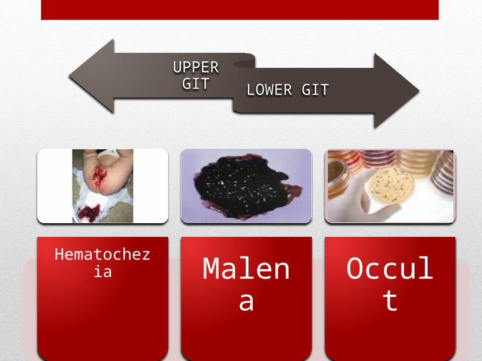
UPPER GIT LOWER GIT
HematocheziaMalena Occult
COMMON CAUSES OF BPR
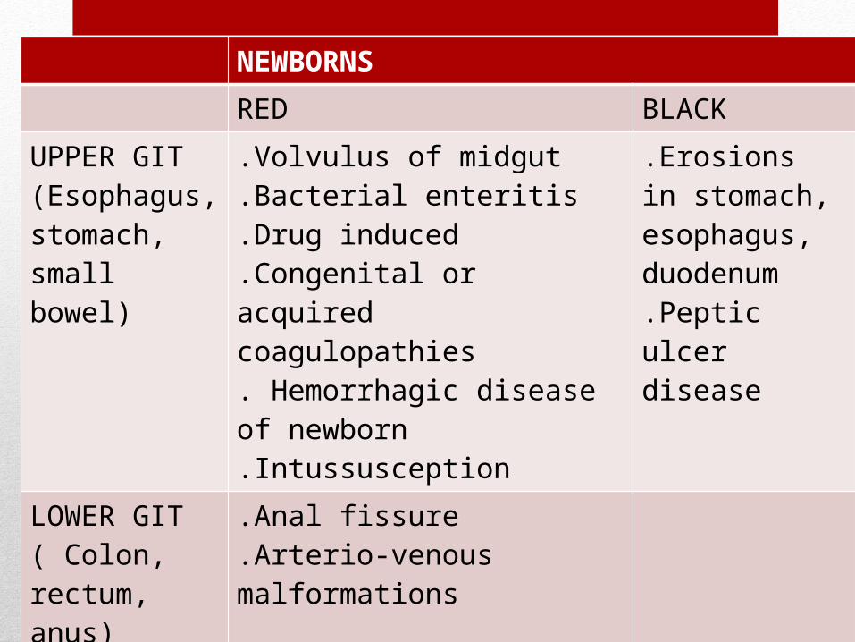
NEWBORNS
RED BLACK
UPPER GIT(Esophagus, stomach, small bowel)
.Volvulus of midgut
.Bacterial enteritis
.Drug induced
.Congenital or acquired coagulopathies. Hemorrhagic disease of newborn.Intussusception
.Erosions in stomach, esophagus, duodenum.Peptic ulcer disease
LOWER GIT( Colon, rectum, anus)
.Anal fissure
.Arterio-venous malformations
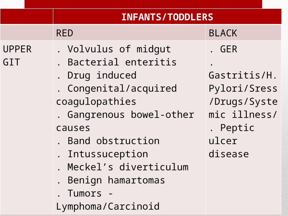
INFANTS/TODDLERS
RED BLACK
UPPER GIT
. Volvulus of midgut
. Bacterial enteritis
. Drug induced
. Congenital/acquired coagulopathies
. Gangrenous bowel-other causes
. Band obstruction
. Intussuception
. Meckel’s diverticulum
. Benign hamartomas
. Tumors - Lymphoma/Carcinoid
. GER
. Gastritis/H.Pylori/Sress/Drugs/Systemic illness/ . Peptic ulcer disease
LOWER GIT
. Rectal polyp
. Anal fissure
. Arterio-venous malformations
. Colonic polyps

OLDER CHILDREN
RED BLACK
UPPER GIT(Esophagus, stomach, small bowel)
. Esophageal varices – Portal HTN
. Gastric varices
. Volvulus of midgut
. Bacterial enteritis
. Drug induced
. Congenital/acquired coagulopathies
. Hemorrhagic disease of newborn
. Intussusception
. Esophageal varices – Portal hypertension. Peptic ulcer disease
LOWER GIT( Colon, rectum, anus)
. Anal fissure
. Inflammatory bowel disease
. Infectious diarrhoea
.Arterio-venous malformations
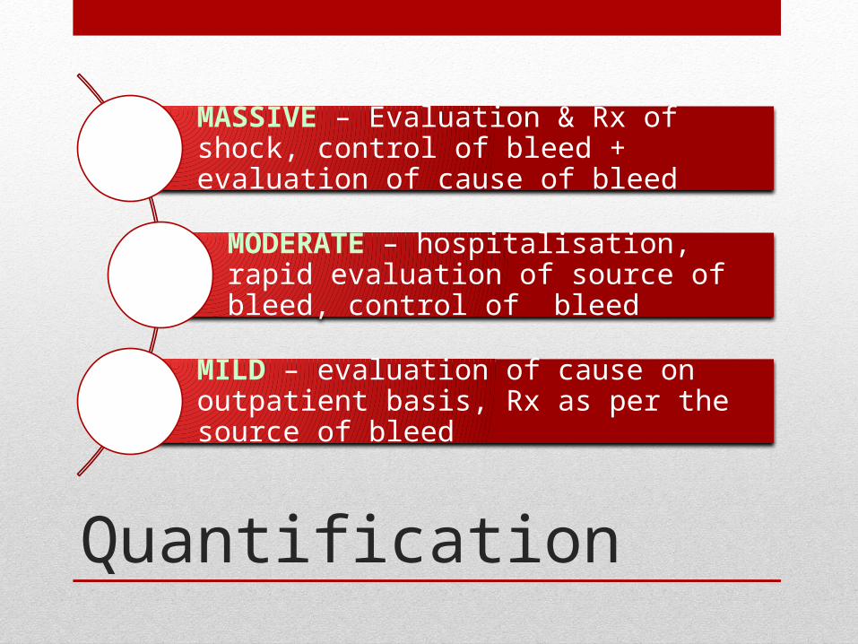
Quantification
MASSIVE – Evaluation & Rx of shock, control of bleed + evaluation of cause of bleed
MODERATE – hospitalisation, rapid evaluation of source of bleed, control of bleed
MILD – evaluation of cause on outpatient basis, Rx as per the source of bleed

Clinical scenarios
Clinical picture Diagnosis
Blood mixed with stools Infective – enteritis/colitis, Hemolytic Uremic syndrome, NEC, Eosinophilic enterocolitis, IBD
Blood streaks on stool Anal fissure/rectal mucosal ulcerationConstipationRectal prolapse
Blood after defecation in drops with normal stools
Rectal polypVascular anomalies of lower GIT
Blood with mucus in stools Infective Intussusception
Frank blood Volvulus of midgutArteriovenous malformationsNEC
Occult bleeding PR Worm infestationAcid peptic disease of upper GITCeliac diseaseIBDPolyposis
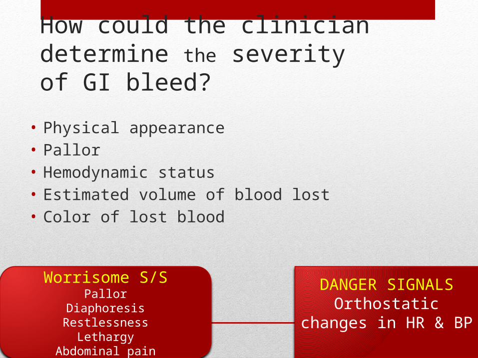
How could the clinician determine the severity of GI bleed?
• Physical appearance• Pallor• Hemodynamic status• Estimated volume of blood lost• Color of lost blood
Worrisome S/SPallor
DiaphoresisRestlessness
LethargyAbdominal pain
DANGER SIGNALSOrthostatic changes in HR
& BP
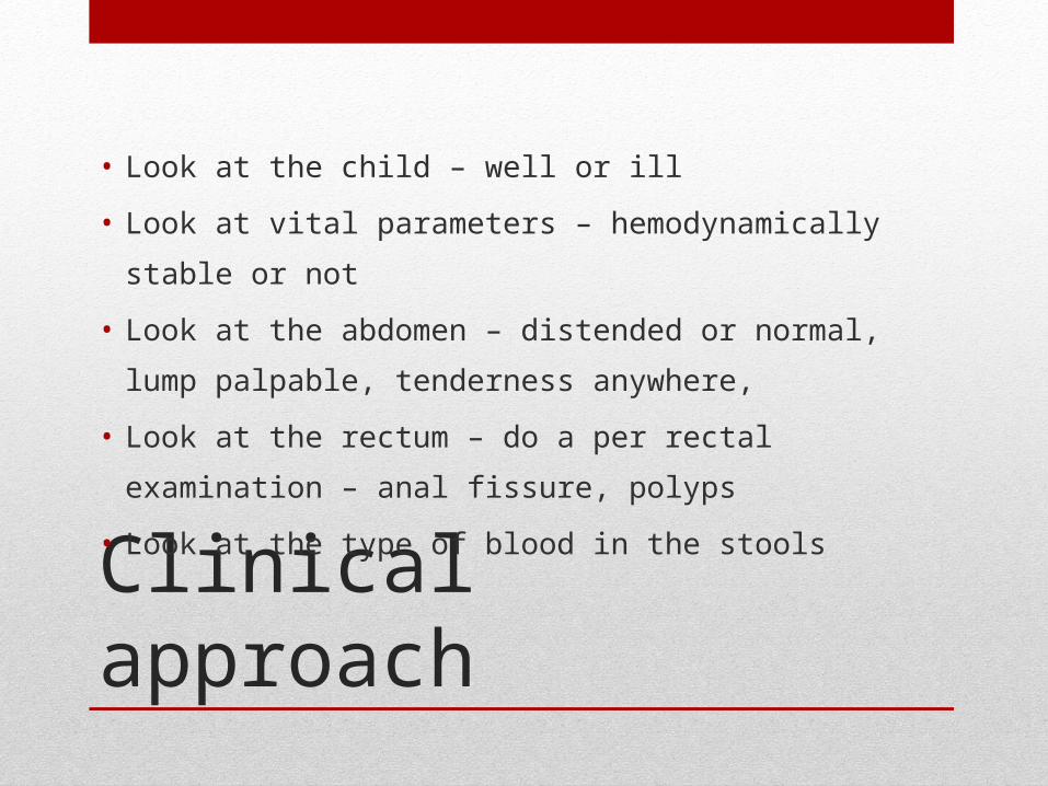
Clinical approach
• Look at the child – well or ill
• Look at vital parameters – hemodynamically stable or not
• Look at the abdomen – distended or normal, lump
palpable, tenderness anywhere,
• Look at the rectum – do a per rectal examination – anal
fissure, polyps
• Look at the type of blood in the stools
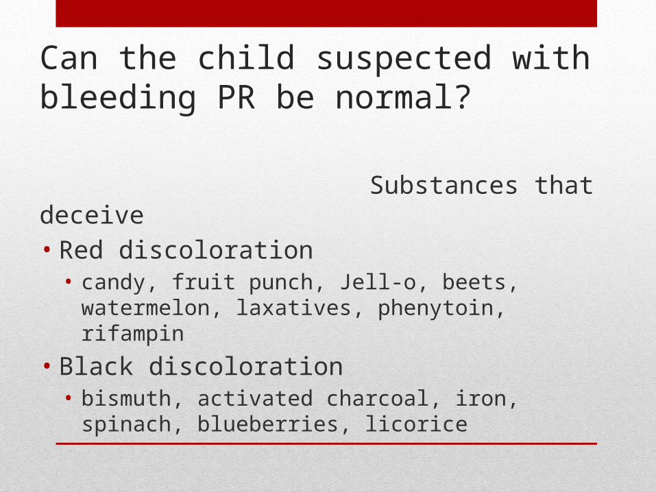
Can the child suspected with bleeding PR be normal?
Substances that deceive• Red discoloration• candy, fruit punch, Jell-o, beets, watermelon, laxatives,
phenytoin, rifampin
• Black discoloration• bismuth, activated charcoal, iron, spinach, blueberries,
licorice

Surgeon’s role