bioimpedance spectroscopy is a unique
TRANSCRIPT
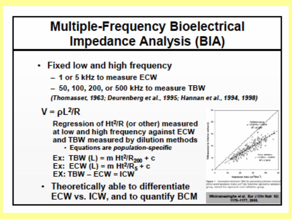


Bioimpedance spectroscopy is a unique bioimpedance approach that differs in underlying basis from the more readily recognized single-frequency bioelectrical impedance analysis in that it does not require the use of statistically derived, population-specific prediction equations. It has the potential advantage of not only measuring total body water, but also offering the unique capacity to differentiate between ECW and ICW and, thus, to provide an estimate of BCM.
BioelectricalImpedanceAnalysis
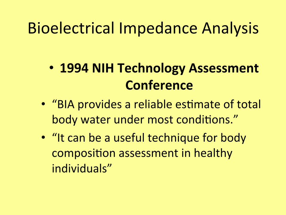
• 1994NIHTechnologyAssessmentConference
• “BIAprovidesareliablees6mateoftotalbodywaterundermostcondi6ons.”
• “Itcanbeausefultechniqueforbodycomposi6onassessmentinhealthyindividuals”

BIA:AdvantagesandLimita6ons
• Advantages– costs($1,000-$15,000)– portable– non-invasive– fast
• Limita6ons– accuracyandprecision– nobeIer/worsethanhydrodensitometry

• Bestusedforepidemiologicstudies
• Choiceofpredic6veequa6onsimportant
• InfluencedbyfluidshiMs• Biggestdrawbackistheneedforappropriatelycalibrated,cross-validatedpredic6veequa6ons(smeage,sex,ethnicity,andhealth)
BIA:AdvantagesandLimita6ons
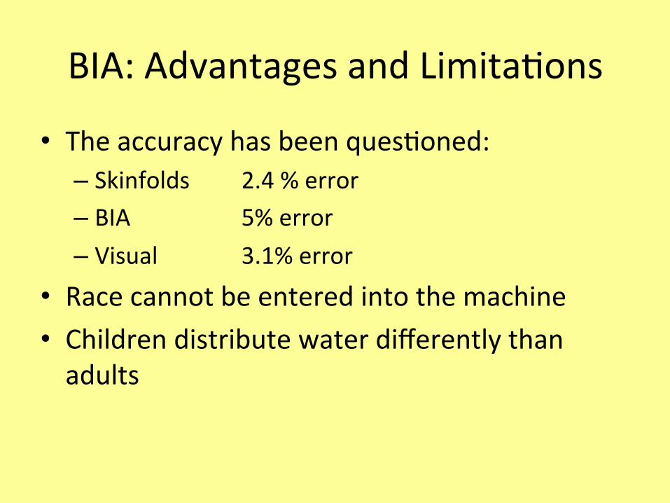
• Theaccuracyhasbeenques6oned:– Skinfolds 2.4%error– BIA 5%error– Visual 3.1%error
• Racecannotbeenteredintothemachine• Childrendistributewaterdifferentlythanadults
BIA:AdvantagesandLimita6ons
Equa6ons
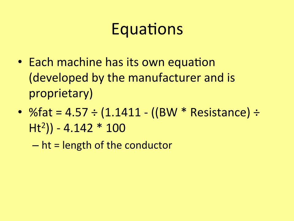
• Eachmachinehasitsownequa6on(developedbythemanufacturerandisproprietary)
• %fat=4.57÷(1.1411-((BW*Resistance)÷Ht2))-4.142*100– ht=lengthoftheconductor
BIA–PhaseangleThe use of the phase angle has become more popular over the last few years because of its high association with clinical results, time of hospitalization and mortality in various diseases. Based on the principles of BIA, which mainly works by measuring body resistance and reactance in order to alternate an electric current, the storage of this current is thought to be able to create a change in phase which is considered to be the ratio between resistance and reactance and which is expressed geometrically as phase angle, being directly calculated as: arc tangent = (Xc / R) x 180° / π.

Graphic derivation diagram of the phase angle and its relation to resistance (R), reactance (Xc), impedance (Z) and frequency of the current applied.

Among all the direct measurements of BIA, the phase angle has proved to be a good predictor of prognosis and mortality regarding hemodialysis, cancer, human immunodeficiency syndrome (HIV), and liver and geriatric diseases. This measurement has attracted strong interest by being a noninvasive, objective and rapid (less than 2 minutes) tool for the determination of nutritional status and risk of patient morbidity, whereas other nutritional screening tools, although also noninvasive, require more time and / or are highly subjective.
Interpre6ng%FatValues
• Allmethodsofmeasuring%fathaveacertainamountofinaccuracy!ThisinaccuracyisdeterminedbytheStandardErrorofEs6mate(SEE).
• TheSEEtellsyoutheamountofdevia6onfromthetrue%fatyoucanexpectfromapar6cularmethod.

• Thereisa67%probabilitythatthetrue%fatiswithin+or-oneSEEfromthemeasuredvalue.
• Example:Measure%fat=20%;SEE=3%unitsofbodyfatThereisa67%probabilitythatthetrue%fatisbetween+or-oneSEEor3%unitsoffatorbetween17-23%.

• Thereisa95%probabilitythatthetrue%fatiswithin+or-twoSEEfromthemeasuredvalue.
• Example:Measured%fat=20%SEE=3%Thereisa95%probabilitythatthetrue%fatiswithin+or-twoSEEor6%ofthemeasuredvalueor14-26%.
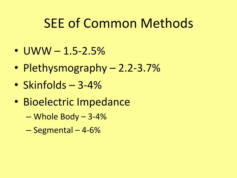
SEEofCommonMethods
• UWW–1.5-2.5%• Plethysmography–2.2-3.7%• Skinfolds–3-4%• BioelectricImpedance
– WholeBody–3-4%– Segmental–4-6%

RiassuntodelleassunzionifaIedaimetodibicompar6mentali

Assump6onsofTwo-ComponentModels
• 1.Thedensityoffatis0.900g/ml• 2.ThedensityofFFMis1.100g/ml• 3.Thedensi6esoffatandFFMarethesameforallindividuals
Assump6ons
• 4.Thedensi6esofthevariouscomponentsofFFMareconstantwithinanindividual
• 5.Theindividualbeingmeasureddiffersfromareferencebody(73.8%water,19.4%protein,6.8%mineral)onlyintheamountoffat

Dual-EnergyX-rayAbsorp6ometry

DualEnergyX-RayAbsorp6ometry(DXA)
• Usesx-raystomeasurethickness,densityandchemicalcomposi6onof6ssue.

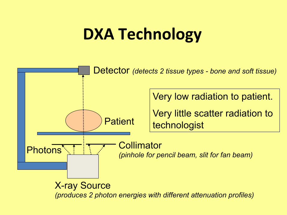
DXATechnology
X-ray Source (produces 2 photon energies with different attenuation profiles)
Photons Collimator (pinhole for pencil beam, slit for fan beam)
Patient
Detector (detects 2 tissue types - bone and soft tissue)
Very low radiation to patient.
Very little scatter radiation to technologist
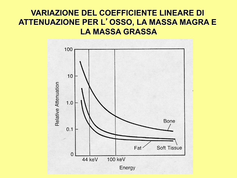
VARIAZIONE DEL COEFFICIENTE LINEARE DI ATTENUAZIONE PER L’OSSO, LA MASSA MAGRA E
LA MASSA GRASSA

• AIenua6onconstantbetweenindividuals.
• Assessestotalbonemineralcontent.Further,eachextra-bonepixelcontainsinforma6onon%fatand%lean6ssue.Areadetermina=onsthattellboththe%fatand%leanmassinasingleregionofinterest.
• Threecomponentmodel– bone,fat,fat-freesoM6ssuemass

Dual-energyXA
• Underlyingprinciple:XraysareaIenuatedbybody6ssue,eachtoadifferentdegreedependingonfrequency(energy).DXAusestwobeamsatdifferentenergies.Ra6ocanaccuratelymeasureaIenua6onofeachcomponent.

DXA• TwodifferentenergylevelX-rays• Lean,fat,andbonemasseachreduce(aIenuate)theX-raysignalinuniqueways
• Computeranalyzesscanpointbypointtodeterminebodycomposi6on
• Method – 20-30/7-10/4-5minutes– Applicabletoyoungandold

TECNICA DUALE DI EMISSIONE RAGGI X
I due diversi l ivel l i di energia per discriminare il tessuto molle da quello osseo, si possono ottenere con due tecniche differenti:
• Energia Pulsata
• Energia Filtrata

TECNICHE DI EMISSIONE RAGGI X
ENERGIA PULSATA Il tubo radiologico (ad anodo fisso) di questa generazione d i s t r u m e n t i v i e n e alimentato con due voltaggi differenti che si alternano per produrre i due livelli di a l ta e bassa energ ia. Tensione tipica:, 100-140 kVp.
ENERGIA FILTRATA Il tubo radiologico (ad anodo fisso o rotante) viene in questo caso alimentato con un solo voltaggio in cui si interpone sul fascio prodotto un filtro di terra rara per produrre i due livelli di diversa energia. Tensione tipica: 84 kVp.

TECNICHE DI EMISSIONE RAGGI X
ENERGIA PULSATA
ENERGIA FILTRATA
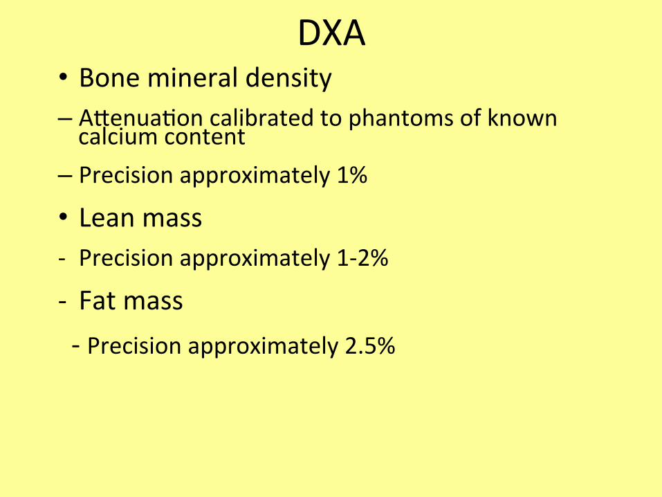
DXA• Bonemineraldensity– AIenua6oncalibratedtophantomsofknowncalciumcontent
– Precisionapproximately1%
• Leanmass- Precisionapproximately1-2%
- Fatmass-Precisionapproximately2.5%
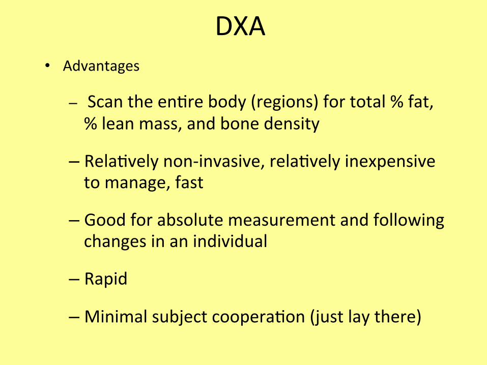
DXA• Advantages
– Scantheen6rebody(regions)fortotal%fat,%leanmass,andbonedensity
– Rela6velynon-invasive,rela6velyinexpensivetomanage,fast
– Goodforabsolutemeasurementandfollowingchangesinanindividual
– Rapid
– Minimalsubjectcoopera6on(justlaythere)
Disadvantage
• Costly(whenbuying)• LimitedAccess

NON SOLO DENSITOMETRIA OSSEA
COMPOSIZIONE CORPOREA Valutazione della massa grassa, magra e ossea settoriale. Campi di applicazione: Medicina Sportiva, Dietologia, Riabilitazione Funzionale, Fisiatria.
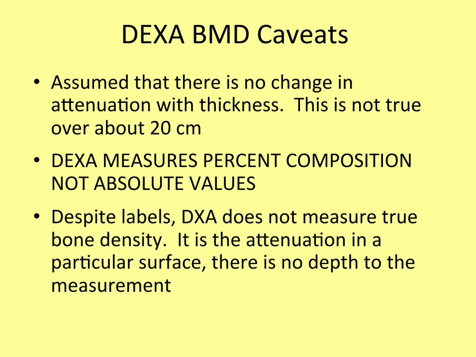
DEXABMDCaveats• AssumedthatthereisnochangeinaIenua6onwiththickness.Thisisnottrueoverabout20cm
• DEXAMEASURESPERCENTCOMPOSITIONNOTABSOLUTEVALUES
• Despitelabels,DXAdoesnotmeasuretruebonedensity.ItistheaIenua6oninapar6cularsurface,thereisnodepthtothemeasurement

• The scanner device incorporates a moving table & a revolving X-ray tube • The table moves the patient back and forth through the revolving X-ray emissions • The X-ray emitter moves (revolves) in a 360o arc around the patient
• Instead of film, the CT scanner collects emitted X-rays via a collector • scintillatior
• Collector transforms X-ray photons into a proportionally strong electric current • The electric current is then converted into an image
• Contrast dyes may be used for image enhancement
Computed Tomography (“CT Scan” or “Cat Scan”)
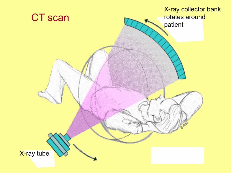
X-ray collector bank rotates around patient
X-ray tube
CT scan

Normal CT scan (abdominal slice)

CT
• Areaofinterestoutlinedontheslice(r=0.94withplanimetryoncadavers)
• Specificdensityforeach6ssueofinterestderivedfromstandards
• Allowstruevolumemeasurementandthustruedensity.

CT
• Excellentcorrela6onbetweenbodyfatmassandcross-sec6onalabdominaladipose6ssuearea– Menr=0.92– Womenr=0.97
• Ninescansrequired• Difficultyisradia6onexposureandcost

CTvsfatinmales

CTvsfatinfemales
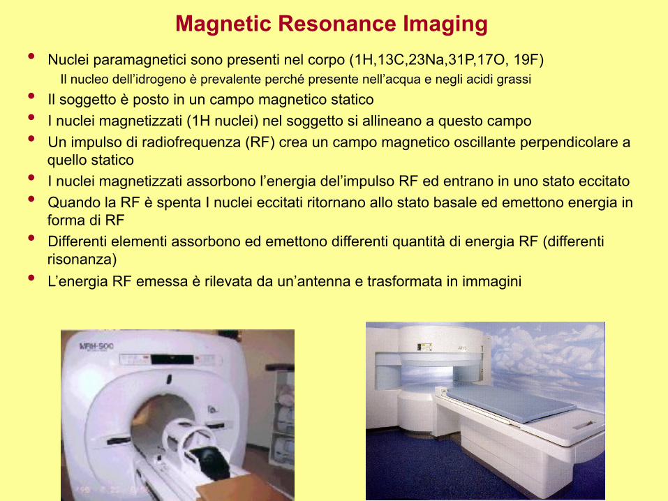
Magnetic Resonance Imaging • Nuclei paramagnetici sono presenti nel corpo (1H,13C,23Na,31P,17O, 19F)
Il nucleo dell’idrogeno è prevalente perché presente nell’acqua e negli acidi grassi • Il soggetto è posto in un campo magnetico statico • I nuclei magnetizzati (1H nuclei) nel soggetto si allineano a questo campo • Un impulso di radiofrequenza (RF) crea un campo magnetico oscillante perpendicolare a
quello statico • I nuclei magnetizzati assorbono l’energia del’impulso RF ed entrano in uno stato eccitato • Quando la RF è spenta I nuclei eccitati ritornano allo stato basale ed emettono energia in
forma di RF • Differenti elementi assorbono ed emettono differenti quantità di energia RF (differenti
risonanza) • L’energia RF emessa è rilevata da un’antenna e trasformata in immagini

Magnetic Resonance Imaging tissues composition & signal intensity
Tissue Signal Intensity T1 Signal Intensity T2
Fat high (whitish) intermediate
Muscle intermediate (gray) intermediate
Hyaline Cartilage intermediate intermediate - low (dull gray)
Ligaments & Tendons low (dark gray) low
Cortical Bone low low
Granulation Tissue intermediate high
Fibrous Tissue low low
Hemorrhage / Edema high - intermediate high
Immature Scar intermediate - low low to high
Mature Scar low low

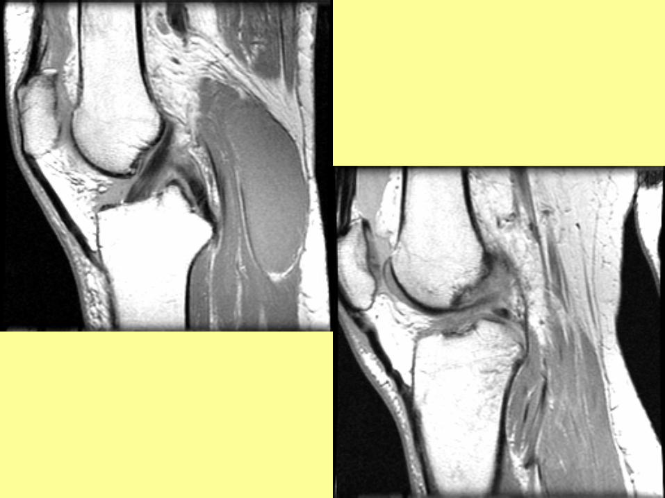

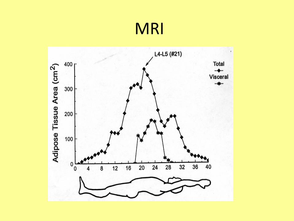
MRI
• SameprincipleasCT• Mayneedasfewas1to4slicesattheL4-L5level(ques6onablebenefit)
• Majordrawbackiscost• Consideredbysometobereferencetechniquesforbodycomposi6on
MRI
NearInfraredRedSpectroscopy(NIRS)
• Basedonthepremisethatthedegreeofinfraredlightabsorp6onisrelatedtothecomposi6onofthesubstancethroughwhichlightpasses
• FatandFat-FreeMassabsorbandreflectlightdifferently
NIR
• Emitinfraredlightatwavelengthsof940-950nmintoabodypart(ie.,biceps)andmeasurestheintensityofthere-emiIedlight
• Morespecificequa6ons/machinesarenecessary
Advantages
• Non-invasive• Safe• Easytoadminister• Fieldtechnique

Disadvantages
• Cost?Isitworthit?• FewAge/GenderSpecificEqua6ons• Accurate?
– Futrex5000 3.1-4.2%– Futrex5000A6.3%– Futrex1000 4.8-6.3%– Sum3 2.4-3.6
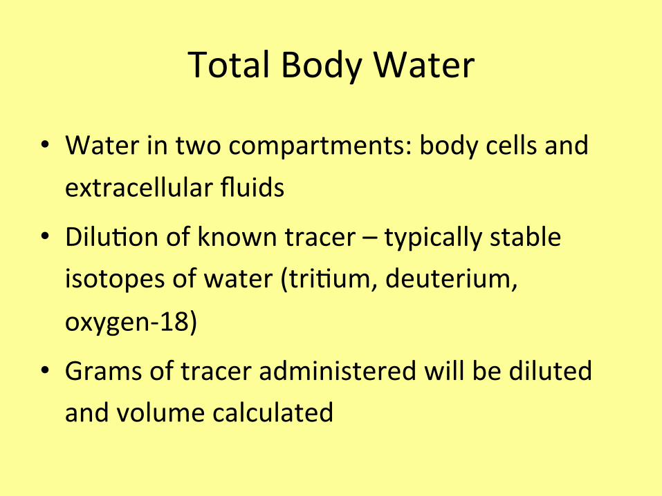
TotalBodyWater
• Waterintwocompartments:bodycellsandextracellularfluids
• Dilu6onofknowntracer–typicallystableisotopesofwater(tri6um,deuterium,oxygen-18)
• Gramsoftraceradministeredwillbedilutedandvolumecalculated
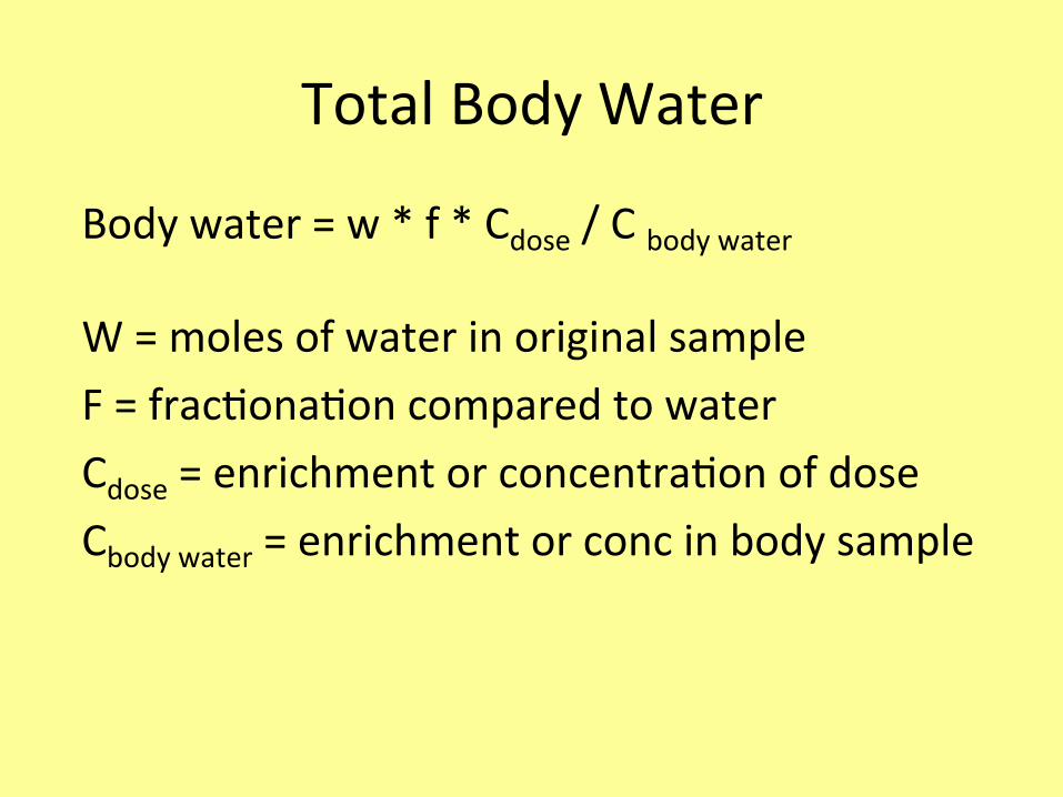
TotalBodyWater
Bodywater=w*f*Cdose/Cbodywater
W=molesofwaterinoriginalsampleF=frac6ona6oncomparedtowaterCdose=enrichmentorconcentra6onofdoseCbodywater=enrichmentorconcinbodysample

TotalBodyWater• Assump6ons
– Traceronlyinbodywater–thelabeledatomscanundergoexchangewithotherorganics.2-5%errorforallisotopes
– Equaldistribu=onoftracertoallcompartments–notusuallyaproblem
– Fastrateofequilibrium–IVtakes2-3hours.Oralupto6ormore
– Notracermetabolism–constantexcre=onanddilu=on.Plateaumethodwithseveraldetermina=onsaZeringes=on

TotalBodyWater
• Fatwillnottakeupwater• Useahydra6onconstanttodeterminefatfreemass
• Problemsencountered:mustassumethehydra6onoffatfreemassequalindifferentindividualsanddiseasestates,measurementerror,instrumenta6onrequired

TotalBodyWater–fatfreemass
• Fatwillnottakeupwater• Useahydra6onconstant(.73)todeterminefatfreemass
• Problemsencountered:mustassumethehydra6onoffatfreemassequalindifferentindividualsanddiseasestates,measurementerror,instrumenta6onrequired
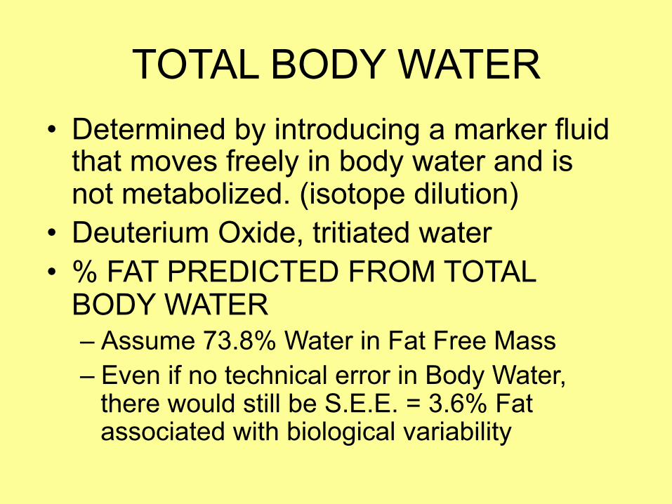
TOTAL BODY WATER • Determined by introducing a marker fluid
that moves freely in body water and is not metabolized. (isotope dilution)
• Deuterium Oxide, tritiated water • % FAT PREDICTED FROM TOTAL
BODY WATER – Assume 73.8% Water in Fat Free Mass – Even if no technical error in Body Water,
there would still be S.E.E. = 3.6% Fat associated with biological variability

Whole-Body Counting
• Scintillation detectors were developed in the early 1950’s.
• They measure the body’s natural potassium as well as other radioactivity in the body.
Whole-Body Counting
• In 1958, Kulwich, Feinstein, and Anderson correlated natural potassium concentration with fat free mass.
• No longer in use

Whole-Body Counting
• There are an estimated 75 counters in the US. • There are more than 180 whole-body counters
worldwide. • Two-thirds of these perform body potassium
measurements in humans.

Whole-Body Counting
• Potassium is naturally distributed in three isotopic states.
• The isotope 40K is radioactive.
Whole-Body Counting
• Gamma rays from 40K are high-energy gammas, many of which exit the body and can be easily detected by external counting
• The smaller the subject, the lower the 40K content and thus the weaker gamma signal.
Whole-Body Counting
• Factors such as age, fitness, or restricted mobility due to surgery or illness do not tend to affect the precision of total body potassium measurements.
• The 40K signal is natural and continuous, therefore the measurement can be interrupted as necessary, until counting is completed.

Whole-Body Counting
The three requirements for 40K whole body-counting include:
• Efficient gamma-ray detectors that can be placed close to the subject.
• Shielding for these detectors to reduce the natural background radiation levels
Whole-Body Counting
• Computer-based instruments that enable identification of the unique gamma rays.

Whole-Body Counting
Precision: • For whole-body counters, precision is in the
range of 2-5% for adults. • In infants and very young children, precision is
only 8-12% for 40 minute sample times.

Whole-Body Counting
• Total cost for an adult whole-body counter is $10,000-15,000.
• Cost of a special shielded room starts at $80,000.
• Start-up costs. • Still in use fortotalbodyprotein,bodycellmass,skeletalmass

Neutron Activation Analysis
• IVNAA measures 11 elements from nuclear reactions.
• Protein, mineral, and fat can be estimated from these elements: – Carbon = Lipid – Nitrogen = Protein – Calcium = Bone

Neutron Activation Analysis
• IVNNA uses a whole-body counter. – It delivers a moderate beam of fast neutrons to the
subject. – Atoms of target elements capture these neutrons. – This creates an unstable isotope.
Neutron Activation Analysis
• Unstable isotopes produce gamma rays when returning to a stable state.
• Gamma rays are measured: – energy level identifies the element – the activity indicates its abundance.

Prompt-Gamma Activation Analysis
• Isotope gets very excited with the added neurons. – Lasts only a fraction of a nanosecond before it
returns to a stable state. – Measured simultaneously as the neutrons are
exposed to the isotope.

Neutron Activation Analysis
Disadvantages: • Radiation exposure. • Must be performed by medical personnel • Cost $30,000 - $300,000.

©1999LippincoIWilliams&Wilkins,Inc.PublishedbyLippincoIWilliams&Wilkins,Inc. 25
Figure7Neutronac=va=onanalysisdetermina=onofbodycomposi=on.Kehayias,Joseph;Valtuena,SilviaCurrentOpinioninClinicalNutri6on&MetabolicCare.2(6):453-463,November1999.
Figure7.C/OandC/Hra6osforfourchemicalcompartments.arbon-to-hydrogen(C/H)andcarbon-to-oxygen(C/O)ra6osforfourchemicalcompartmentsofthebody.C/Hisameasureof'dryness',whereasC/Oisanindexof'fatness'.Measurementerrorsarereducedwhenneutronac6va6onisusedforthesimultaneousmeasurementofra6osofelements.

©1999LippincoIWilliams&Wilkins,Inc.PublishedbyLippincoIWilliams&Wilkins,Inc. 26
Equa6on17Neutronac=va=onanalysisdetermina=onofbodycomposi=on.Kehayias,Joseph;Valtuena,SilviaCurrentOpinioninClinicalNutri6on&MetabolicCare.2(6):453-463,November1999.
Equa6on17

©1999LippincoIWilliams&Wilkins,Inc.PublishedbyLippincoIWilliams&Wilkins,Inc. 27
Figure8Neutronac=va=onanalysisdetermina=onofbodycomposi=on.Kehayias,Joseph;Valtuena,SilviaCurrentOpinioninClinicalNutri6on&MetabolicCare.2(6):453-463,November1999.
Figure8.Theelementalpar66onanalysis(EPA)methodappliedtotheassessmentofskeletalmuscle.Selec6onofelementforskeletalmuscleassessmentbyelementalpar66onanalysis(EPA).Totalbodypotassium(TBK)willrequirealargenumberofcorrec6onsfornon-musclepotassium,whereasphosphorus(TBP)requireshighprecisionmeasurementsofTBPandbone.

©1999LippincoIWilliams&Wilkins,Inc.PublishedbyLippincoIWilliams&Wilkins,Inc. 28
Figure9Neutronac=va=onanalysisdetermina=onofbodycomposi=on.Kehayias,Joseph;Valtuena,SilviaCurrentOpinioninClinicalNutri6on&MetabolicCare.2(6):453-463,November1999.
Figure9.Irradia6onset-upformuscleandproteinassessment.Subjectirradia6onset-upforthefastneutronac6va6onanalysisofphosphorusandnitrogen.Theheight,h,ofthesteppingstoolisadjustedsotheheadisshielded.Awhole-bodydetectorisusedforsubsequentgammaraycoun6ng.

Metodiantropometriciassocia6allacomposizionecorporea
Height–WeightTables
• Developedin1940’sbyINSURANCEcompanies.
• Basedsolelyonmortalitysta6s6cs.
– FaIerpeople=increasedriskofdeath
• Donottakeintoaccountbodycomposi6on!!!

BodyMassIndex
• BMI=Weight(kg)/Height(m)2
• Desirable– Men:21.9–22.4– Women:21.3–22.1– Overweight:25-30– Obese:>30

BodyMassIndex• BMI’sabove27associatedwith↑incidenceofhypertension,diabetes,&CHD.
• S6llusedfrequentlybydoctorsandresearchers.
• Doesnottakebodycomposi6onintoaccounteither!

• Height:5’10”=1.77m
• Weight:221lbs=100.45kg
• BMI=32.09
• THISGUYISOBESE!!!!
BodyMassIndex

WaisttoHipRa6o
• Indica6onofthepaIernofbodyfatdistribu6on
• Indicatorofthehealthrisksofobesity– excesstrunkfat-increasedriskofhypertension,type2diabetes,highcholesterol,CAD,prematuredeath

WaisttoHipRa6o
• Risksincreasewithincreasingra6os– veryhighrisk>0.94youngmenand0.82youngwomen
– veryhighrisk>1.03older(60-69years)menand0.90forolderwomen
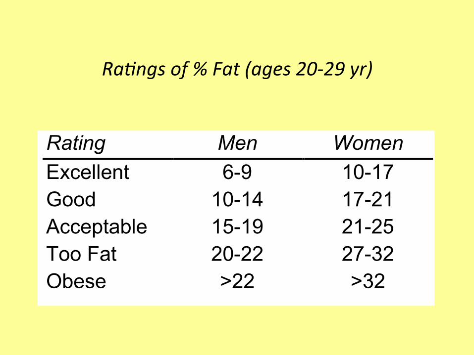
Ra#ngsof%Fat(ages20-29yr)
Rating Men Women Excellent 6-9 10-17 Good 10-14 17-21 Acceptable 15-19 21-25 Too Fat 20-22 27-32 Obese >22 >32
Poten6alUsesonMethodsFoundinLiterature
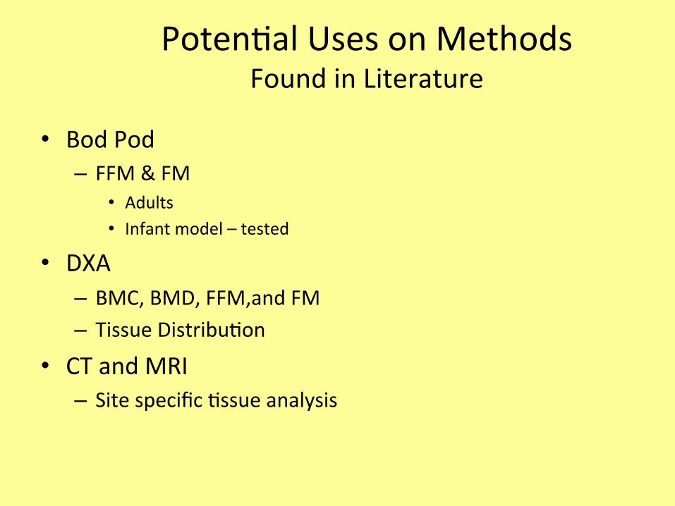
• BodPod– FFM&FM
• Adults• Infantmodel–tested
• DXA– BMC,BMD,FFM,andFM– TissueDistribu6on
• CTandMRI– Sitespecific6ssueanalysis

Poten6alUsesonMethodsFoundinLiterature
• BIA– FFM&FM
• AdultandPediatric– Dialysis– Survival
• Cancer,peritonealdialysis,malnutri6on,obesity– Conges6veHeartfailure
• MFBIAorBIS– FFM,FM,TBW,ECF– Pregnancy,HIV+was6ng

WhichMethodtoUse?
• Dependsoncompartmentofinterest.• Availabilityoftechniques.• Technicaltrainingofstaff.• Condi6onofpa6ent.• Loca6onwhereassessmentwillbedone:
– Laboratory/clinic– Field/remotesite