barrett's esophagus and - hindawi publishing...
TRANSCRIPT
REVIEW
Barrett's esophagus and adenocarcinorna
Z W. LI MD. R. WFNS~I.. MD, FRCP, A.B.R. THOl-tSON. MD. PHO. FRCPC, FACP
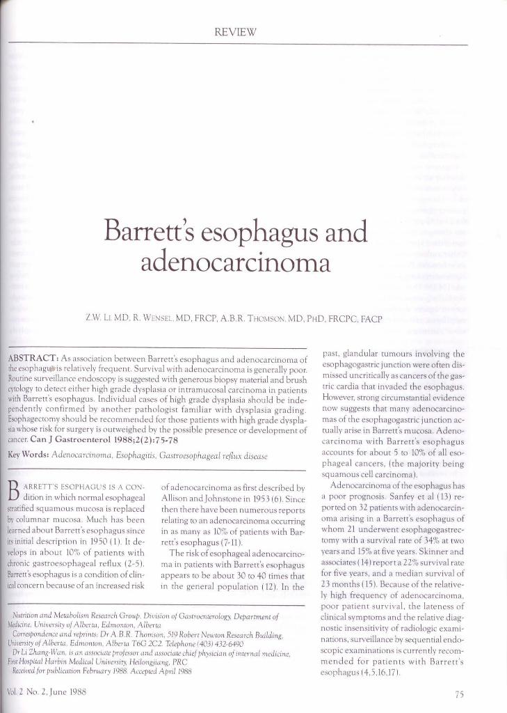
ABSTRACT: As association between Barrett's esophagus and adcnocarc inoma of the esophag141:, is relatively frcqL1cnt. Survival with adenocarcinoma is generally poor. Routine surveillance endoscopy is suggested with generous biopsy material and brush cytology to detect either high grade dysp lasia or intramucosal carcinoma in patien ts
, with Barrett's esophagus. Individual cases of high grade dysplasia should be independently confi rmed by anothe r pathologist familiar with dysplasia grading. Esophagectomy should be recommended for those patients with high grade dysplas1a whose risk for surgery is outweighed by the possible presence or deve lopment of cancer. Can J Gastroenterol 1988;2(2):75-78
Key Words: Adenocamnorna. Esophagi tis. Gascroesophageal reflux disease
B ARRETT"S ESOPHAGUS IS A CON
dition in which normal esophageal stratified squamous mucosa is replaced bv column ar mucosa. Much hns been learned about Barrett's esophagus since its initial description in 1950 (I). It develops in about 10'1(, of patients with chronic gastroesophageal reflux (2-5). Barrett's esophagus is a condition of clin-1cal concern bernuse of an increased ri sk
o f adenocarcinoma as first described by Allison and Johnstone in 1953 (6) . Since then there have been numerous reports relating co an ac.lenocarcinoma occu1Ting in as many as 10% of patients with Bar· rett's esophagus ( 7-11 ).
The risk of esophageal adenocarcino· ma in patients with Barrett's esophagus appears to be about 30 to 40 times that in the general population ( 12). In the
Nucriuon unJ Mecabolism Research Group. Div1s1011 of Guscrocncerology, Deparrm.eni of Medicme, Uni1,ersicy uf Aibena, EJmoncun, Alberw
Corres/>ondence ,md repnncs; Dr A.B.R. Thomson. 519 Hoberc Newron Research Buildmg, Uni1-ersicy of A Iberra, EJmonron. A Iberra T6G 2C2. Teie/>hone ( 403) 432-6490
Dr L1 Zhang-W,m, 1s cm associwe professor ,md assoc1uce cllle/ />hy,iciun oj mcernal medicine, fosr Hospira/ Harbin Medical Uni11crsiry. Heiloniuiang. PRC
Receu:ed for p11blic-ac1011 Febrnary 1988. Accepced April 1988
Vol.2 No. 2.Junc 1988
past, glandular tumours involving the esophagogastric junction were often dismissed uncritically as cancers of the gastric cardia that invaded the esophagus. However, strong circumstantial evidence now suggests that many adenocarcino· mas of the esophagogastric junction ac· cually arise in Barrett's mucosa. Adeno· carcinoma with Barrett's esophagus accoun ts for about 5 to 10% of all eso· phageal cancers, (the majority being squamous cell carcinoma).
AJenocarcinoma of the esophagus has a poor prognosis. San fey et al ( 13) re· ported on 32 patients with adenocarcinoma arising in a Barrett's esophagus of whom 21 underwent esophagogastrec· tomy with a survival rate of 34% at two years and 15% at five years. Skinner and associates ( 14) report a 22% su rvival rate for five years, and a median surviva l of 23 months ( 15 ). Because of the relatively high frequency of adenocarcinoma, poor patient surv ival, rhe lateness of clinical symptoms and the relative diag· nostic insensitivity of radiologic examinations. surveillance by sequential endoscopic examinations is currently recom· mended fo r patients with Barrett's esophagus ( 4.5 .16, 17)
75

LI ~r al
Barrett':, muco:,al change is thought to he secondary ro chronic gastroesophagca l rcnux. Acid insult to the lower c:,ophagus result:, in ulceration with reepilheliali zat1on taking place from the gastric side. ln wch patients the distal ponion of Lhc esophagus becomes lined with columnar mucosa. wilh a potential to undergo dy,plasia and malignant degeneration.
Cigarette smoking and alcohol ingestion may act as co-carcinogens in adenocarci nl,ma of lhe esophagus. San fey er al (13) reported that life histories regarding cigarette smoking were available for 2 3 of 32 patients with adenocarcinoma arising in Barrett's esophagus. Only two of the 23 patien ts had never smoked. Orhcr authors have reported an association between Barrett's adenocarcinoma, cigarette smoking and regular alcoho l intake ( 14.18). Thus, cigarette smoking and alcohol ingestion may be important factors in cancer developing in Barrett"s esophagus, as is also the case with sqw1-mous carcinoma.
CLINICAL FEATURES Adcnocarcinoma in association with
Barrett's esophagus is most common in males in the fifth and six th decade of life who often smoke, drink and have a past history of esophagi tis, hiaral hernia or stricture. Patients with reflux esophagi t is ~econJarv ro scleroderm a, and those with reflux caused by a cardiomyotomy for achalasia, arc also at risk of developing cancer.
In a review of l21 patients with adenocarcinoma arising in a Barrett's esophagus as reported by Siogren and colleagues ( 5), the mean age at diagnosis wns 5 7 years rnnge 2, to 88 years. The male to female ratio was 'i. 5: 1, an cxaggcnncd male predominance over that of Barrett's esophagus in genera l. Oysphagia was the predominam presenting symptom in 87°0. a hiatal hernia was often present in 71 %, and 64°1, of patients had symptoms of reflux esophagi tis for one to 40 years.
The site of adenocarcinoma in relation to the gastroesophageal junction and the squamocolumnar junction is variable, although the incidence appears highest in the distal one-th ird of the esophagus.
76
DIAGNOSIS Surveillance by serial e ndoscopic ex• aminations with esophageal biopsies: Barren' s mucosa is classified as: distinctive specia lized type, cardiac type. fund ic type or dysplastic. Dysplasia is classified as intermediate grade or high grade, and is based on the classification used for gastric dysplasia.
Specia lized columnar epithelium is the most common of the three types of mucosa in adult~ with Barrett's esophagus. In a study of both endoscopic biopsy specimens and esophagecmmy specimens, Hamilton and colleagues ( 19) assessed the relationship between dysplasia in Barrett's esophagus and invasive adenocarcinoma. 0 ( 14 patients with Barrett's esophagus and endoscopic biopsy specimens indicating dysplasia, six pmients had high grade l 43'lo), three had intermediate grade (21'l;,) and five had low grade ( 36'1.,) dysplasia. Distinctive type columnar epithelium was both the most common type of epi thelium noted in the group ( 13 of 14) and the most common type associated with high grade dysplasia (six of six). Of the six patients with h igh grade dysplasia, five underwent esophagectomy and three of these were found to have superficially invasive adenocarcinoma. ln one patient rumour extended only to the muscularis mucosa and in two patients rumour reached the submucosa. The other patient with high grade dysplasia and the eight patients with intermediate or low grade dysp lasia were not yet known to
have developed carcinoma on follow-up. ln a study of the resected specimens
( 19). high grade dysplasia was strongly nssociated w ith adjoining invasive adcnocarc inoma; 84'':, of areas wi th invnsion had high grade dysplasia and 92°{, of areas with high grade dysplasia showed invasion . Intermed iate grade dysplasia was found in six of the areas and low grade in one of the areas adjacent to microscopically in vasive carcinoma. In areas of Barrett's esophagus that did nm adjoin invasive carci noma in the resectcd specimens, h igh grade dysplasia was noted infrequen tly.
Lee (20) has also confirmed previous observations suggesting that high grade dysplasia is closely linked to the presence of adenocarcinoma. He performed a
careful retrospective analysis of a large number of rescctcd specimens from patients with Barrett's esophagus who deve loped adenocarcinoma. The presence of high grade dysplasia was both a sensitive and specific marker for the presence of adjacent invasive rnrcinoma. T he strong association between high grade dysplasia and adenocarcinoma in resecrcd specimens would appear co argue for patients with high grade dysplnsia being considered for elective esophagectomy.
In as much as the prognosis of invasive adenocarcinoma arising in Barrett's esophagus is so poor, identification of a high risk group or ea rl ier identification of the lesion itself might im prove survival. By regu lar endoscopy with adequate biopsy it may be possible to detect early intramucosal carcinomas in certain patients. Lt is fe lt that endoscopic biopsy specimens should be taken with a jumbo biopsy forceps 2 cm apart throughout the entire Barrett's area. [f dysplasia is identified annual examinations arc recom mendcd. If high grade dysplas1a develops csophagectomy should be considered. Esophageal eyto logic exam ination: Brush cytology is an excellent addition to endoscopic biopsies and may provide independent confirmation of the presence of cancer (2 l ). Radiologic examination : The barium swallow has a diagnostic sensitivity for Barrett's esophagus of only 24% (22). A reticular pattern of the esophageal mucosa may be observed with use of double contrast esophagography, hut thb findi ng is neither sensitive nor specific for Barrett"s esophagus. It more probably reflects difficulty· in prospectively diagnosing a mucosa! lesion that usually has only nonspeci fi c radiographic findings. The combination of hiatal hernia, esophageal ulcer and mid-esophageal stricture is h ighly suggestive of Barrett's esophagus. Unfortunately, in retrospective series. no more than 25 to 30% of patients with Barrett's esophagus have been found to have this triad (23.24) Thus, radiological examination of the esophagus is not recommended for detecting or fo llowing Barrett's esophagus. Flow eytometry: Reid e t al (25) used flow cytometry and histology to evaluate 317 biopsy sped mens from 64 con-
CAN J GASTROENTEROL

secutive patients who were in a cancer
surveillance program for Barrett's esophagus, plus three additional patiencs wirh adenocarcinoma in Barrett's esophagus. All patients wi th dysplasia or adenocarcinoma had evidence of genomic instability (aneuploidy) (lr abnormalities of
mucosa! proliferation as detected by flow cytometry. In a small subset of patients with specialized meta p lastic epithelium whose specimens were hiscologically neg
ative or indefinite for dysplasia, the mucosa had ancuplo1d cell populations or proliferative abnormalities tha t were otherwise found only in dys plasia or carcinoma. Further study may prove rh.:it
this subset of patients mcnrs more frequent endoscopic biopsy w rvcillance
because of an increased risk for developing carcinoma.
Thus. flow cytometry is capable of detecting al terations in DNA content o r proliferation or both , that arc present in high frequency in Barrecc's dysplas1a and carcinoma. Because the abnormalities detected by flow cytomecry correlate well with the conventional hisrologic diagno·isof dysplasia and carcinoma, rhc\ may
prove to be a val uab le objective adjunct m the diagnosis of dysplasia and carcinoma 111 Barrett's esophagus.
Acid sulphated mucins: Other mark
ers have recently been proposed ro detect early cancer of chc esophagus including changes in mucin staining to detect acid sulphated mucins. Unfor tunately, recent studies have shown chat acid sulphated mucins arc p resent coo frequently in Barre tt's epithelium with out dysplasia co be of use in defining a subgroup ar increased risk of d ysplasia .
STAGING SYSTEM Rosenberg and co-workers ( 18) report
ed an anaiysis of adcnocarcmoma in Bar
rett's esophagus, utili zing a staging system. They undertook a study of che p ro
gression of changes chat take place in patients with adenocarcinoma arising in
REFERENCES I. Barrett's NR . Chronic pepur ulce r of the
esophagus and esophagi tis Br J Surg 1950;38:175-82
2. Bremner CG, Lynch VP, Ellis FH. Barrett'~ esophagus· Congemtal or
Vol.2 No.2.June 1988
a Barrett's esophagus and developed a staging system based on the extent of the malignancy (as dete rmined by surge ry and by pathologic examinatio n of the tumo ur) . Thb staging syste m is as follows: { I) Carcmoma limi ted co rhe mucosa ( 111-
cluding carcinoma in si tu) nor extending beyond the muscularis mucosa with ncgnrive nodes; (I!) ca rc ino ma limited to the esophageal wall but not extending to the adventitia, with negative nodes; (Ill) a ny of the above with involved regional lymph node~ or full thickness wall penetration of the tumour co the adventiua, without invasion of adjacent organs; anJ (IV J ca rcinoma invading adjacent organ!> or with distant metastases.
The finding of a progression of changes from dysplasia to in situ neoplasia to invasive malignancy suggests mecaplasia as the first step in this process Multiple foci o f malignant change in Barrett's esophagus have been frequently re por
ted.
MANAGEMENT Medical treatment: Patients who have symptomatic or objective evidence of reflux esophagi tis should receive vigorous standard medical treatment including H 2 antagonists, prokinetic agen ts and diet
ary restriction~ Kothari and associates ( 2 7 ) reported that ulcers in five patients with Barrett's, refractory to antacids, healed in e ight t0 16 weeks with cimeridine 1.26 ml/day. Lt is not known if the
risk of dysplasia or carcinoma 1s reduced by such m ed ical therapy
Surgical treatment. Antircflu..x sur• gery: Some investigators have cla imed at least partial regression o f Barrett's
e pithelium after s uccessful antire Ou x procedures (3, IO). Several authors ad
vocate an anti reflu x p rocedure for most pa tients with Barrett's esophagus, b ut
it is not known whether the occurrence of adenocarcinoma can be prevented .
Esophagectomy: A difficult problem
facing doctors who deal with patients
acquired 1 An experimental study of esophagea l mucosa! regeneration in the dog Surgery 1970;6R 175-82
l Goldm::m MC, Beckman RC Barre u \ syndrome: Case report with discussion about concer1, o f pathogenesis.
Barrett's and adenocarcinoma
having Barren 's esophagus b the tim ing
o f a surgical resec tion before invasive adenocarcinoma develops Endoscopic surveil la nce biopsies arc reassuring 1f they arc negative fo r dysplas1a. However, if dysplasia is found on light microscopy, it may be difficult to determine whether the morbidity of a surgical resection is offset by a sufficiently high chance for discovering concomitant early mvas1ve adcnocarcinoma.
Several workers { 17- l9) suggest that the dysplasia-carcinoma sequence most commonly occurs 111 Barrett's mucosa of the d istmctive type. High grade dyspla!>ia in Barrett's mLtcosa is a marker indicating
high p robability of invasive carcinoma and, therefore, the presence of high grade dysplasm in biopsy specimens of Barrett 's mucosa is probably an indication for total csophagccromy in suitable surgica l candidates. A more extensive csophagccromy is required than can be performed usmg the Lewis approach or a left thoracoto my. Rosenberg and others ( 18) CL1rren tly favour a procedLtre that places the esophagogastric anastomosb in the neck When either the stomach or che colon is Ltsed as an esophageal replacement, It is brought up to the neck through a substcrnal tunnel. This operative approach is best for all patients with esophageal cancer. irrespective of the cell type.
Patients foun d to have low grade dysplasia sho uld receive intensive medical treatment for reflux csophagius for up to 12 weeks, at which time esophagoscopy sho uld be repeated ro obtam multiple esophageal biopsy specimens. Patients whose biopsy specimens s how histologic im provement wi ll require intense surveillance (cg, an examination every three co six mo n ths) until at least two consecu tive endoscopic examina
tions reveal no dysplast1 c epithelium Continued intensive treatment and surveillance arc recommended in patients whose repeat biopsies reveal persistent
low grade dysplasia .
Gamoenccrology 1960; W 104-10. 4 Bozymski EM. Herlihy K. Orlando RC
Barrett's esophagus Ann Intern Med 1982;97· IOH
5 Siogren R W. Johnson LF Barrett's esophagus A review. Am J MeJ
77
Li et ul
1983,74. 31 J-20. 6 A llison PR, Johnstone AS. The
oesophagus lined with gastric mucous membrane Thorax 1953:8:87-101.
7 Spechler SJ, Robbins AH. Rubins HB, et al. Ad enocarcinoma and Barrett's esorhagus: An overrated risk? Gasl rocn terology 1984:87 :9 Z 7-3 3
8. Cameron AJ, Ou BJ. Payne WS. Incidence of ad enocarcinoma 111
columnar-lined (Barrett's) esorhagus. N EnglJ Med 1985:)13:857
9 Mes,i:rn RA, HcrmosJA. Robbins AH. et al. Barrett's esorhagus: Cli nical review of 26 ca,es Am J Gastroen terol 1978;69.458-66.
10 Sarr MG. Ha mil ton SR, Marrone GC, ct al Barreu's esophagus: Its rre\'alcncc and association with adcnocarcinoma in patients with symptoms of gastroesophagcal reflux. Am J Surg 1985: 149: 187-92.
I I Sau bier EC. Goui llat C, Samaniego C, ct al. Adcnocarc1noma in columnarli ned Fh1rretr's esophagus. Analysb of U esnrhagcctomics Am J Surg 1985; 150:365-9
12. Spcchlcr SJ, Goyal RK. Barrett 's esophagu,. N Engl) Med 1986;315: 362-71.
13. Sanfcy H, Hamilton SR. Smith RRL,
ct al. Carcinoma arising in Barrett's esophagus. Surg Gynecol Obstel l98'i, 161 :570-4.
14. Skinner DB, Welther BC. Riddell RH, et a l. Barre1t's esoph agus: Comparison of benign and malignant cases. Am J Surg 1983; 198:554-66.
15. Smith RRL. Hamilton SR, BoimonJR, er al. The spectrum of carcinoma arising 111 Barrett's esophagus: A clin1copmhologic stlldy of 26 patients. AmJ Surg Parhol 1984:8:'i63-73.
16. Berardi RS, Devaiah KA. Barrett's esophagus. Surg Gynccol Obstet 1983: 156:521-38.
17. Skinner DB. T he columnar-lined e:,ophagus and ,1Jcnocarc111oma Ann Thorne Surg 1985:40: 321-2. (Edit)
18 Rosenberg JC, Budev H, Edwards RC. cl al. Analysis of adenocarcinoma 111
Barrett's esophagus utilizing a smging sy,tem Cancer 1985;55: 1353-60.
19 H:unilton SR. Smith RRL The relationsh ip between columnar .::pithclial dysplasia and invasive adcnticarcinoma arising in Barrett's esophagus. Am J Clin Pathol 1987;87:301- 12.
20. Lee RG. Dysplasia 111 Barr.::u's csorhagu s. A cli nicopachologic study of
six patien ts. Am J Surg Pmhol 1985;9:845-52.
21. BelladonnaJA, Hajdu Sl. Bains MS, ct al Adenocarc111oma in situ of Barrell', esophagus diagnosed by endoscopic cyrokigy. N Engl J Med 1974:291 :895-6
22. Winters C. Spurling TJ , Chobanian SJ, ct al. Barrett's esophagus. Gastroenterolngy 1987;92: 118-24.
23. Robbins AH, Vincent ME, Saini M. et al. Revised radiologic concepts of chc Barrett's .::sophagus. Gastroi nte:,t Radio! 1978; 3:3 77-81.
24. Levine MS. Kresse! HY, Caroline OF, ct al. Barrett's esophagu s. Reticular pattern of the mucosa. Radiology 1983:147:663-7
25. Reid B. Haggin RC. Rubm CE Rabinov1cch PS. Flarrctt's esophagus: Correlation between flow cytometry and histology in detection of patien1s at n sk fo r adcnocarcinoma Gas1roenterology 1987;93:l- l l.
26. Jass JR Mucin histochemistry of the colum nar epithelium of the oesophagu,. A retrospective scu dy. J Clin Pmhol 1981,34:866-70
27 Kothari T M:rngla JC. Kalra TMS. Barrell\ ulcer and treatment with cimetidine Arch Intern Med 1980; 140:475-7.
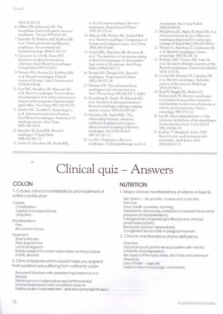
Clinical quiz - Answers COLON 1. Causes, clinical manifestations and treatment of solitary rectal ulcer
Causes Constipation Ergotamine suppositories Idiopathic
Manifestations Pain Blood and mucus
Treatment Stool softeners Stop ergotamine Local analgesics Rare ly, surgery to correct associated rectal prolapse 5-ASA. steroids
2. Clinical features which would make you suspect that a patient was suffering from cathartic colon
Recurrent diarrhea with persistent hypokalemia in a female Melanosls coll in sigmoidoscopy (anthracenes) Narrow featureless colon on barium enema Positive sodium hydroxide test - pink stool (phenolphthalein)
78
NUTRITION 1. Major clinical manifestations of vitamin A toxicity
Skin lesion - dry, pruritic. coarse and scaly skin Hair loss Sore mouth, anorexia . vomiting Headache, d rowsiness, irritability. increased intracranial pressure and papilledema Enlarged liver progressing to fibrosis and cirrhosis Lymphadenopathy Bone pain (painful hyperostosis) Congenital abnormal ity in pregnant women
2. Clinical manifestations of zinc deficiency
Diarrhea Disturbance of central nervous system with mental irritability and depression Skin lesion of the face, limbs. skin folds a nd perineum Alopecia Loss of taste - ageusia Defect in the immunologic mechanisn
CAN J GASTROE1'TEROL

Submit your manuscripts athttp://www.hindawi.com
Stem CellsInternational
Hindawi Publishing Corporationhttp://www.hindawi.com Volume 2014
Hindawi Publishing Corporationhttp://www.hindawi.com Volume 2014
MEDIATORSINFLAMMATION
of
Hindawi Publishing Corporationhttp://www.hindawi.com Volume 2014
Behavioural Neurology
EndocrinologyInternational Journal of
Hindawi Publishing Corporationhttp://www.hindawi.com Volume 2014
Hindawi Publishing Corporationhttp://www.hindawi.com Volume 2014
Disease Markers
Hindawi Publishing Corporationhttp://www.hindawi.com Volume 2014
BioMed Research International
OncologyJournal of
Hindawi Publishing Corporationhttp://www.hindawi.com Volume 2014
Hindawi Publishing Corporationhttp://www.hindawi.com Volume 2014
Oxidative Medicine and Cellular Longevity
Hindawi Publishing Corporationhttp://www.hindawi.com Volume 2014
PPAR Research
The Scientific World JournalHindawi Publishing Corporation http://www.hindawi.com Volume 2014
Immunology ResearchHindawi Publishing Corporationhttp://www.hindawi.com Volume 2014
Journal of
ObesityJournal of
Hindawi Publishing Corporationhttp://www.hindawi.com Volume 2014
Hindawi Publishing Corporationhttp://www.hindawi.com Volume 2014
Computational and Mathematical Methods in Medicine
OphthalmologyJournal of
Hindawi Publishing Corporationhttp://www.hindawi.com Volume 2014
Diabetes ResearchJournal of
Hindawi Publishing Corporationhttp://www.hindawi.com Volume 2014
Hindawi Publishing Corporationhttp://www.hindawi.com Volume 2014
Research and TreatmentAIDS
Hindawi Publishing Corporationhttp://www.hindawi.com Volume 2014
Gastroenterology Research and Practice
Hindawi Publishing Corporationhttp://www.hindawi.com Volume 2014
Parkinson’s Disease
Evidence-Based Complementary and Alternative Medicine
Volume 2014Hindawi Publishing Corporationhttp://www.hindawi.com