author's personal copy - purdue university · author's personal copy biosensors and...
TRANSCRIPT

Autho
r's
pers
onal
co
py
Biosensors and Bioelectronics 22 (2007) 1664–1671
Optical forward-scattering for detection of Listeriamonocytogenes and other Listeria species
Padmapriya P. Banada a, Songling Guo b, Bulent Bayraktar c, Euiwon Bae b,Bartek Rajwa d,e, J. Paul Robinson d,e, E. Daniel Hirleman b, Arun K. Bhunia a,∗
a Molecular Food Microbiology Laboratory, Department of Food Science, Purdue University, West Lafayette, IN 47907, USAb School of Mechanical Engineering, Purdue University, West Lafayette, IN 47907, USA
c School of Electrical and Computer Engineering, Purdue University, West Lafayette, IN 47907, USAd Department of Basic Medical Sciences, Purdue University, West Lafayette, IN 47907, USA
e Weldon School of Biomedical Engineering, Purdue University, West Lafayette, IN 47907, USA
Received 3 April 2006; received in revised form 19 June 2006; accepted 24 July 2006Available online 1 September 2006
Abstract
We demonstrate here the development of a non-invasive optical forward-scattering system, called ‘scatterometer’ for rapid identification ofbacterial colonies. The system is based on the concept that variations in refractive indices and size, relative to the arrangement of cells in bac-terial colonies growing on a semi-solid agar surface will generate different forward-scattering patterns. A 1.2–1.5 mm colony size for a 1 mmlaser beam and brain heart infusion agar as substrate were used as fixed variables. The current study is focused on exploring identification ofListeria monocytogenes and other Listeria species exploiting the known differences in their phenotypic characters. Using diffraction theory,we could model the scattering patterns and explain the appearance of radial spokes and the rings seen in the scattering images of L. mono-cytogenes. Further, we have also demonstrated development of a suitable software for the extraction of the features (scalar values) calculatedfrom images of the scattering patterns using Zernike moment invariants and principal component analysis and were grouped using K-meansclustering. We achieved 91–100% accuracy in detecting different species. It was also observed that substrate variations affect the scatteringpatterns of Listeria. Finally, a database was constructed based on the scattering patterns from 108 different strains belonging to six species ofListeria. The overall system proved to be simple, non-invasive and virtually reagent-less and has the potential for automated user-friendly appli-cation for detection and differentiation of L. monocytogenes and other Listeria species colonies grown on agar plates within 5–10 min analysistime.© 2006 Elsevier B.V. All rights reserved.
Keywords: Light scattering; Listeria monocytogenes; Detection; Identification
1. Introduction
Over the past few years there has been increasing interest innon-invasive light-scattering technologies for studying and/ordetecting bacterial cells (Fratamico et al., 1998; Kaye, 1998;Perkins and Squirrell, 2000). Light-scattering instruments likesurface plasmon resonance (SPR), flow cytometry, etc., haveenabled the study of various biological molecules (Leidberg etal., 1983; Shapiro, 2003) and have been a commercial success.Laser spectroscopic techniques for rapid detection of bacte-
∗ Corresponding author. Tel.: +1 765 494 5443; fax: +1 765 494 7953.E-mail address: [email protected] (A.K. Bhunia).
rial spores have become very popular non-conventional method(Scully et al., 2002; Hybl et al., 2003).
Polarized differential light scattering as a practical means forcharacterization of microorganisms has been investigated forbacterial cells in suspension (Wyatt, 1969; Wyatt and Phillips,1972; Perkins and Squirrell, 2000; Bronk et al., 2001). Fromthe angular variation in the scattered light pattern, informationabout the bacterial size, shape, internal structure and refrac-tive index, has been determined for identification of Salmonellatyphimurium, Escherichia coli (Salzman et al., 1982) andStaphylococcus epidermidis (Stull, 1972). However, there arechallenges associated with bacteria in suspension, such as purityof cultures and arrangement of cells (often in chains or clus-ters). The orientations of and distances between cells change
0956-5663/$ – see front matter © 2006 Elsevier B.V. All rights reserved.doi:10.1016/j.bios.2006.07.028

Autho
r's
pers
onal
co
py
P.P. Banada et al. / Biosensors and Bioelectronics 22 (2007) 1664–1671 1665
with time. However, a colony on a solid surface such as agaris more stable and its optical response could be modeled withscalar diffraction theory.
We used light scattering as a potential non-invasive opti-cal sensor to identify bacterial colonies on agar plates withoutthe need for further sample processing or preparation. A bac-terial colony results from an exponential multiplication of asingle cell on a nutrient agar surface forming in a character-istic dome-shaped structure. The pattern of scattered light isdependent on the properties of the scatterer including refrac-tive index, size, shape and chemical composition of the colony.The optical back-scattering method is widely used for waferinspection and for studying biological cells (Hielscher et al.,1997; Kim et al., 2002), but it did not produce reproducibleresults with bacterial colonies in our investigation (Nebeker etal., 2001). Conversely, optical forward-scattering yielded repro-ducible scattering patterns with colonies of E. coli and alsoListeria species, and thus was used in further experiments (Guo,2004).
Since we observed some unique distinguishable featuresassociated with the patterns of Listeria spp., we focused ourstudies primarily on the species belonging to genus Listeria andinvestigated the scatterometer’s ability to distinguish them.
The genus Listeria is comprised of six species; L. mono-cytogenes, L. innocua, L. welshimeri, L. ivanovii, L. seeligeri,and L. grayi. They are Gram-positive, rod-shaped, measuringabout 0.5–1 �m × 1–2 �m and produce dome-shaped colonies.L. monocytogenes is the only member known to be pathogenicto humans, with a high mortality rate in immuno-suppressedpopulations, and to cause abortion in pregnant women (Vazquez-Boland et al., 2001). L. ivanovii is also pathogenic but infectsruminants.
The conventional detection methods based on biochemical,serological or DNA/RNA characterization involves additionalsteps of sample enrichment in a liquid medium, plating on selec-tive agar media to obtain single colonies, and use of specialreagents for DNA isolation and PCR. All these methods takeabout 3–5 days to get confirmatory results and are labor inten-sive. PCR-based methods though faster is highly dependenton efficient DNA isolation and also is limited by its accu-racy in detecting live bacterial cells, leading to false positiveidentification (Hitchins, 1998; Jaradat et al., 2002). In recentyears, biosensor tools have been used with improved sensitiv-ity but involve multisteps, and some cases these technologiesare unable to differentiate live from dead cells (Ivnitski et al.,1999; Bhunia and Lathrop, 2003; Ligler et al., 2003; Geng etal., 2004).
Thus, the major objectives of this study were: (1) to build ascatterometer based on the principle of forward light scatteringfor bacteria on the solid surface; (2) to investigate the ability ofthis system to differentiate Listeria colonies belonging to differ-ent species grown on the surface of agar plates; (3) modeling ofthe scatter patterns; (4) development of an image analysis tool.Overall, in this paper we propose the development of an efficient,non-invasive, reagent-less, user-friendly biosensing instrumen-tation with automated image analysis tool for rapid identificationof live bacteria.
Fig. 1. Schematic drawing of a scatterometer. Inset: photograph of a typicalplate (BHI) with Listeria colonies.
2. Materials and methods
2.1. Bacterial cultures
The bacterial cultures (108) used in this study are listed inTable 1 (see Supplementary material). They were grown in brainheart infusion (BHI; Difco, Sparks, MD) broth for 18–24 h at37 ◦C for subsequent plating on BHI agar plates. Identity ofthe cultures was confirmed by ribotyping using an automatedRiboprinter® (Qualicon, Willmington, DE). L. monocytogenesATCC 23074 transformed with pNF8, a GFP-encoding plasmidwas obtained from Dr. B.M. Applegate, Purdue University, forconfocal microscopy.
2.2. Preparation of bacterial plates for light-scatteringexperiments
The culture dilutions were plated on the surface of BHI agarplate (100 mm × 15 mm), so as to obtain 20–30 colonies perplate and were incubated at 37 ◦C for 24–36 h or until the diame-ter of the colony reached approximately 1–1.5 mm (Fig. 1, inset).Since the growth rate is different for different species, the sizewas used as a fixed parameter. The thickness of the colony (alongthe optical axis), was typically around 0.1–0.3 cm as measuredfrom the surface profile data obtained by laser triangulationprobe (Microtrak II Laser Displacement Sensor System, MTIinstruments Inc., Albany, NY). A total of 2149 scatter imageswere acquired from 108 different strains belonging to six speciesof Listeria. On an average of 20 images were obtained per strain,from the colonies growing in two or three different plates.
2.3. Scatterometer
The scatterometer system was built on an optical breadboardfor precise and stable positioning of the optical components(Fig. 1) and to provide vibration free base. The system included:a laser diode of 635-nm wavelength; a Petri-plate holder; a detec-
Autho
r's
pers
onal
co
py
1666 P.P. Banada et al. / Biosensors and Bioelectronics 22 (2007) 1664–1671
tion plane; a digital camera to acquire the scattering images. Thelaser, the Petri-plate holder, and the detection plane were placedsequentially on the optical path (Y-axis) of the laser beam. Thedistance from the Petri-plate holder to the laser was 100 mm,and to the detection plane was 300 mm. The laser generates acollimated beam on the order of 1 mm diameter (at the 1/e2
irradiance points) that is directed through the center of the bac-terial colony and through the substrate of bacterial agar medium.While a fraction of the laser beam was fully transmitted throughthe substrate and bacterial colony, another fraction was scat-tered in the forward direction (at relatively small angles to thetransmitted beam). The forward-scattered and transmitted lightformed the scattering patterns of the bacterial colonies on thedetection plane (8 in. × 10 in. white board) and was capturedwith a digital camera.
2.4. Microscopy
L. monocytogenes ATCC 23074 pNF8, a GFP-expressingstrain was used for confocal microscopy. The culture wasgrown on BHI agar containing 50 �g ml−1 erythromycin untilthe colony size was about 0.2 mm. A confocal microscopic(MRC1024, Bio-Rad) image of a whole single colony of GFP-expressing L. monocytogenes excited at 488 nm was taken at10× and analyzed using the confocal assistant (Version 4.02).
Cryo-scanning electron microscopy for the intact colonies ofL. monocytogenes ATCC 19113 and L. innocua F4248 was car-ried out to understand the arrangement of the cells in a colony.For the methods and the pictures please see Supplementarymaterial.
2.5. Modeling of scattering patterns using diffractiontheory
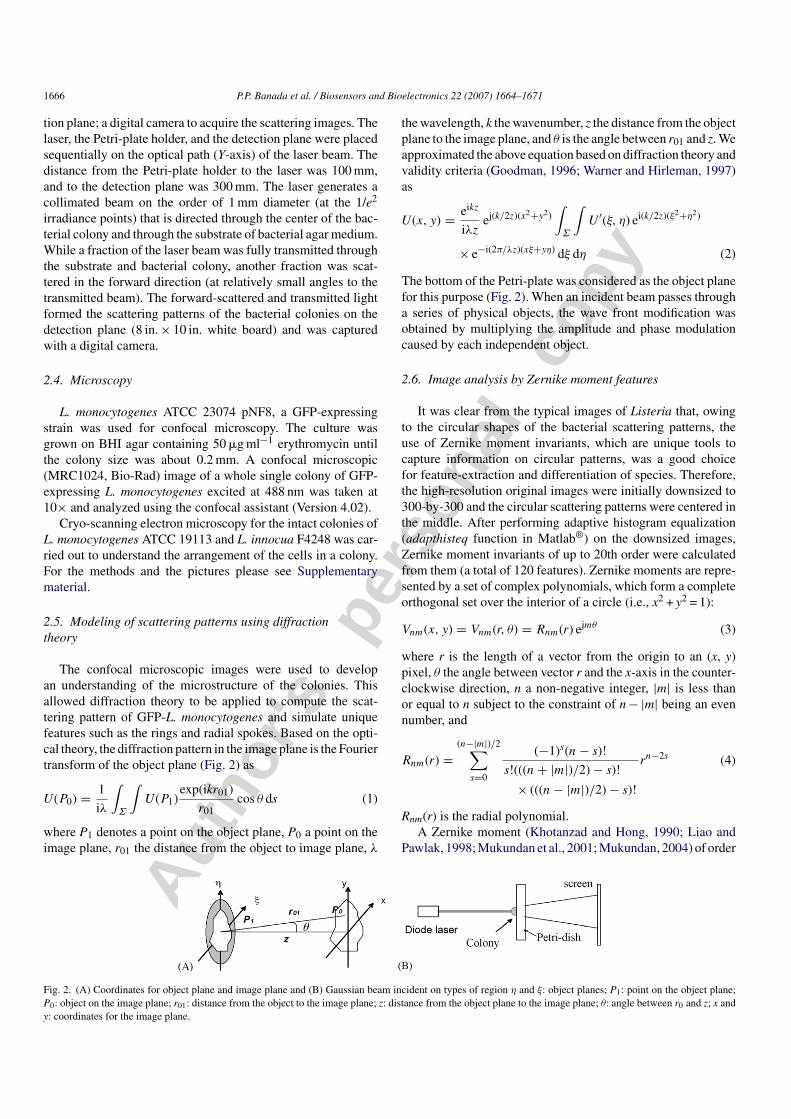
The confocal microscopic images were used to developan understanding of the microstructure of the colonies. Thisallowed diffraction theory to be applied to compute the scat-tering pattern of GFP-L. monocytogenes and simulate uniquefeatures such as the rings and radial spokes. Based on the opti-cal theory, the diffraction pattern in the image plane is the Fouriertransform of the object plane (Fig. 2) as
U(P0) = 1
iλ
∫Σ
∫U(P1)
exp(ikr01)
r01cos θ ds (1)
where P1 denotes a point on the object plane, P0 a point on theimage plane, r01 the distance from the object to image plane, λ
the wavelength, k the wavenumber, z the distance from the objectplane to the image plane, and θ is the angle between r01 and z. Weapproximated the above equation based on diffraction theory andvalidity criteria (Goodman, 1996; Warner and Hirleman, 1997)as
U(x, y) = eikz
iλzej(k/2z)(x2+y2)
∫Σ
∫U ′(ξ, η) ei(k/2z)(ξ2+η2)
× e−i(2π/λz)(xξ+yη) dξ dη (2)
The bottom of the Petri-plate was considered as the object planefor this purpose (Fig. 2). When an incident beam passes througha series of physical objects, the wave front modification wasobtained by multiplying the amplitude and phase modulationcaused by each independent object.
2.6. Image analysis by Zernike moment features
It was clear from the typical images of Listeria that, owingto the circular shapes of the bacterial scattering patterns, theuse of Zernike moment invariants, which are unique tools tocapture information on circular patterns, was a good choicefor feature-extraction and differentiation of species. Therefore,the high-resolution original images were initially downsized to300-by-300 and the circular scattering patterns were centered inthe middle. After performing adaptive histogram equalization(adapthisteq function in Matlab®) on the downsized images,Zernike moment invariants of up to 20th order were calculatedfrom them (a total of 120 features). Zernike moments are repre-sented by a set of complex polynomials, which form a completeorthogonal set over the interior of a circle (i.e., x2 + y2 = 1):
Vnm(x, y) = Vnm(r, θ) = Rnm(r) ejmθ (3)
where r is the length of a vector from the origin to an (x, y)pixel, θ the angle between vector r and the x-axis in the counter-clockwise direction, n a non-negative integer, |m| is less thanor equal to n subject to the constraint of n − |m| being an evennumber, and
Rnm(r) =(n−|m|)/2∑
s=0
(−1)s(n − s)!
s!(((n + |m|)/2) − s)!
× (((n − |m|)/2) − s)!
rn−2s (4)
Rnm(r) is the radial polynomial.A Zernike moment (Khotanzad and Hong, 1990; Liao and
Pawlak, 1998; Mukundan et al., 2001; Mukundan, 2004) of order
Fig. 2. (A) Coordinates for object plane and image plane and (B) Gaussian beam incident on types of region η and ξ: object planes; P1: point on the object plane;P0: object on the image plane; r01: distance from the object to the image plane; z: distance from the object plane to the image plane; θ: angle between r0 and z; x andy: coordinates for the image plane.

Autho
r's
pers
onal
co
py
P.P. Banada et al. / Biosensors and Bioelectronics 22 (2007) 1664–1671 1667
n with repetition m for a digital image function f(x, y) that van-ishes outside the unit circle is defined by (asterisk “*” indicatesconjugate of a complex number):
Znm = n + 1
π
∑x
∑y
f (x, y)V ∗nm(r, θ) dx dy, x2 + y2 ≤ 1
(5)
Under rotation, the orientation angle of a Zernike momentchanges but its magnitude remains unchanged. Therefore, themagnitudes of Zernike moments |Znm| can be used as rotation-invariant features. To compute the Zernike moments of a givenimage, the center of the image is taken as the origin and pixelcoordinates are mapped to the range of the unit circle. Thosepixels falling outside the unit circle are not used.
Our analysis consisted of using PCA as a dimensionalityreduction tool on the Zernike moment invariant features and alsoas an initial grouping of the data. K-means clustering was thenused to reach the final classification rate. Image pre-processing,Zernike moment invariants, and K-means clustering were allperformed using the Matlab® software (The MathWorks Inc.,Natick, MA). Principal component analysis was done usingCytoSpecTM (developed by Valeri Patsekin, Cytometry Labs,Purdue University).
2.7. Effect of different agar media on Listeria scatteringpatterns
L. monocytogenes ATCC 19113 was chosen for growth ondifferent substrates. Nutrient media included brain heart infusionagar (BHIA; Difco); tryptic soy agar (TSA; Difco) and selec-tive (with antimicrobial agents) agar media: Listeria repair agar
(LRA); buffered Listeria enrichment agar (BLEA); polymyxinB–acriflavin–lithium chloride–ceftazidime–aesculin–mannitol(PALCAM) agar. PALCAM contains aesculin (the sugar com-ponent reducing to black pigment) to differentiate bacterialcolonies. We prepared this medium by omitting aesculin whichmay otherwise affect the scattering. The antimicrobial sup-plements acriflavin, cyclohexamide and nalidixic acid wereadded to each medium as recommended by the US Food andDrug Administration (FDA) (Hitchins, 1998). Scattering pat-terns obtained for colonies from different media were thengrouped using the developed software.
3. Results
3.1. Optical forward-scattering analysis of Listeriacolonies on agar plate
The representative scattering patterns of L. monocytogenesATCC 19113, L. ivanovii ATCC19119, L. innocua F4248, L.seeligeri LA15, L. welshimeri ATCC 35897 and L. grayi Lm37are shown in Fig. 3. A total of 17 different strains of L. innocua,12 of L. ivanovii, 5 of L. seeligeri, 3 of L. welshimeri, 2 ofL. grayi, and 69 of L. monocytogenes were tested and theirscattering patterns were collected. For each species, a represen-tative strain was picked and its analysis is demonstrated here.By comparing the scattering patterns of six cultures of six differ-ent species (Fig. 3), each culture could be easily distinguished.The visual analysis of the image revealed unique features; brightcenter spot, concentric rings, radial spokes, and diffusive spotwith various half-angle.
Fig. 3. Scattering-images of representative Listeria species: (A) L. monocytogenes ATCC19113; (B) L. ivanovii ATCC19119; (C) L. innocua F4248; (D) L. seeligeriLA-15; (E) L. welshimeri ATCC35897; (F) L. grayi LM37.
Autho
r's
pers
onal
co
py
1668 P.P. Banada et al. / Biosensors and Bioelectronics 22 (2007) 1664–1671
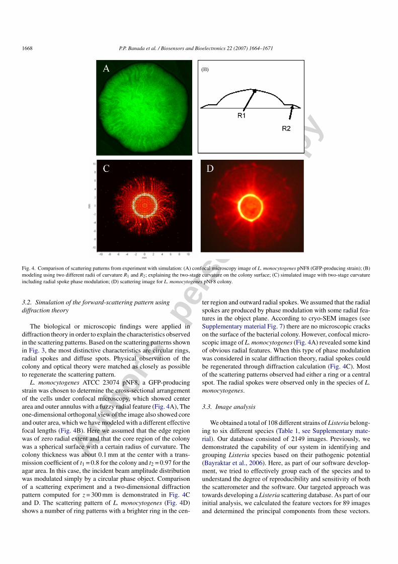
Fig. 4. Comparison of scattering patterns from experiment with simulation: (A) confocal microscopy image of L. monocytogenes pNF8 (GFP-producing strain); (B)modeling using two different radii of curvature R1 and R2; explaining the two-stage curvature on the colony surface; (C) simulated image with two-stage curvatureincluding radial spoke phase modulation; (D) scattering image for L. monocytogenes pNF8 colony.
3.2. Simulation of the forward-scattering pattern usingdiffraction theory
The biological or microscopic findings were applied indiffraction theory in order to explain the characteristics observedin the scattering patterns. Based on the scattering patterns shownin Fig. 3, the most distinctive characteristics are circular rings,radial spokes and diffuse spots. Physical observation of thecolony and optical theory were matched as closely as possibleto regenerate the scattering pattern.
L. monocytogenes ATCC 23074 pNF8, a GFP-producingstrain was chosen to determine the cross-sectional arrangementof the cells under confocal microscopy, which showed centerarea and outer annulus with a fuzzy radial feature (Fig. 4A), Theone-dimensional orthogonal view of the image also showed coreand outer area, which we have modeled with a different effectivefocal lengths (Fig. 4B). Here we assumed that the edge regionwas of zero radial extent and that the core region of the colonywas a spherical surface with a certain radius of curvature. Thecolony thickness was about 0.1 mm at the center with a trans-mission coefficient of t1 = 0.8 for the colony and t2 = 0.97 for theagar area. In this case, the incident beam amplitude distributionwas modulated simply by a circular phase object. Comparisonof a scattering experiment and a two-dimensional diffractionpattern computed for z = 300 mm is demonstrated in Fig. 4Cand D. The scattering pattern of L. monocytogenes (Fig. 4D)shows a number of ring patterns with a brighter ring in the cen-
ter region and outward radial spokes. We assumed that the radialspokes are produced by phase modulation with some radial fea-tures in the object plane. According to cryo-SEM images (seeSupplementary material Fig. 7) there are no microscopic crackson the surface of the bacterial colony. However, confocal micro-scopic image of L. monocytogenes (Fig. 4A) revealed some kindof obvious radial features. When this type of phase modulationwas considered in scalar diffraction theory, radial spokes couldbe regenerated through diffraction calculation (Fig. 4C). Mostof the scattering patterns observed had either a ring or a centralspot. The radial spokes were observed only in the species of L.monocytogenes.
3.3. Image analysis
We obtained a total of 108 different strains of Listeria belong-ing to six different species (Table 1, see Supplementary mate-rial). Our database consisted of 2149 images. Previously, wedemonstrated the capability of our system in identifying andgrouping Listeria species based on their pathogenic potential(Bayraktar et al., 2006). Here, as part of our software develop-ment, we tried to effectively group each of the species and tounderstand the degree of reproducibility and sensitivity of boththe scatterometer and the software. Our targeted approach wastowards developing a Listeria scattering database. As part of ourinitial analysis, we calculated the feature vectors for 89 imagesand determined the principal components from these vectors.
Autho
r's
pers
onal
co
py
P.P. Banada et al. / Biosensors and Bioelectronics 22 (2007) 1664–1671 1669
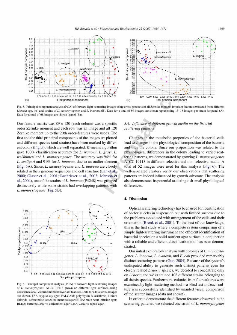
Fig. 5. Principal component analysis (PCA) of forward light-scattering images using cross-products of all Zernike moment invariant features extracted from differentListeria spp. (A) and strains of L. monocytogenes and L. innocua (B). Data for a total of 89 images are shown representing 15–18 images per strain for panel (A).Data for a total of 68 images are shown (panel (B)).
Our feature matrix was 89 × 120 (each column was a specificorder Zernike moment and each row was an image and all 120Zernike moment up to the 20th order-features were used). Thefirst and the third principal components of the images are plottedand different species (and strains) have been marked by differ-ent colors (Fig. 5), which are well separated. K-means algorithmgave 100% classification accuracy for L. ivanovii, L. grayi, L.welshimeri and L. monocytogenes. The accuracy was 94% forL. seeligeri and 91% for L. innocua, due to an outlier element(Fig. 5A). Since, L. monocytogenes and L. innocua are closelyrelated in their genome sequences and cell structure (Lan et al.,2000; Glaser et al., 2001; Buchrieser et al., 2003; Johnson etal., 2004), one of the strains of L. innocua (F4248) was groupeddistinctively while some strains had overlapping patterns withL. monocytogenes (Fig. 5B).
Fig. 6. Principal component analysis (PCA) of forward light-scattering imagesof L. monocytogenes ATCC 19113 grown on different agar surfaces, usingcovariance of all Zernike moment invariant features. Data for a total of 52 imagesare shown. TSA: tryptic soy agar; PALCAM: polymyxin B–acriflavin–lithiumchloride–ceftazimide–aesculin–mannitol agar; BHIA: brain heart infusion agar;BLEA: buffered Listeria enrichment agar; LRA: Listeria repair agar.
3.4. Influence of different growth media on the listerialscattering patterns
Changes in the metabolic properties of the bacterial cellslead to changes in the physiological composition of the bacteriaand thus the colony. Since our proposition was related to thephysiological differences in the colony leading to varied scat-tering patterns, we demonstrated by growing L. monocytogenesATCC 19113 in different selective and non-selective media. Atotal of 52 images were used for this analysis (Fig. 6). Thewell-separated clusters verify our observations that scatteringpatterns are indeed influenced by growth substrate. The analysisalso demonstrates its potential to distinguish small physiologicaldifferences.
4. Discussion
Optical scattering technology has been used for identificationof bacterial cells in suspension but with limited success due tothe problems associated with arrangement of the cells and theirorientation (Bronk et al., 2001). To the best of our knowledge,this is the first study where a complete system comprising of asimple light-scattering instrument and efficient identification ofbacterial species on a solid nutrient agar surface in conjunctionwith a reliable and efficient classification tool has been demon-strated.
Our initial exploratory analysis with colonies of L. monocyto-genes, L. innocua, L. ivanovii, and E. coli provided remarkablydistinct scattering patterns (Guo, 2004). Because of the system’sundisputed ability to generate such distinct patterns even forclosely related Listeria species, we decided to concentrate onlyon Listeria and we examined 108 different strains belonging toall the six species. Furthermore, colonies from four cultures wereexamined by light-scattering method in a blind test and each cul-ture was successfully identified by unaided visual comparisonof the scatter images (data not shown).
In order to demonstrate the different features observed in thescattering patterns, we selected one strain of L. monocytogenes

Autho
r's
pers
onal
co
py
1670 P.P. Banada et al. / Biosensors and Bioelectronics 22 (2007) 1664–1671
as a representative model. Using the confocal image in classicaldiffraction theory, we could generate simulative models of scat-tering patterns by changing the radius of curvature. Diffractiontheory is a very efficient tool for modeling complex biologicalimages. It has been used to modulate color formation in coronas(Gedzelman and Lock, 2003). The airy ring pattern from a circu-lar aperture is a basic characteristic of diffraction phenomena. Ithas been considered for plane wave (Hecht, 2002) and Gaussianbeam propagation (Nourrit et al., 2001). When millions of bac-terial cells form a colony of known size, they form a ‘biologicalaperture’ or ‘biological lens’ with different transmission coeffi-cients for the agar and the colony. However, their physical shapesuch as radius of curvature deviates severely from perfect lenssystem, which is still possible to model with phase modulation.In the confocal microscopy image, there are two different dis-tinct areas—circular core and outer annulus. The dimmer ringsare caused by the overall circular shape of bacterial colony andthe brighter ring in the center believed to be caused by the twodifferent effective focal length which is a function of radii ofcurvature and refractive index. In addition, we observed a ran-domly distributed internal structures pointing in outward radialdirection which believed to contribute to the scattering as shownin the diffraction calculation. From the diffraction theory, whenone-dimensional rectangular aperture is impinged with planewave, the diffracted wave creates a sin c function (sin(x)/x) whichis perpendicular to the longitudinal dimension of the rectangu-lar aperture in the object plane (Hecht, 2002). We observed thesame kind of sin c function intensity modulation in the azimuthaldirection since the internal structures were placed in this direc-tion. When this was modeled in the phase modulation part ofthe integration, we could create radial spokes as in the experi-mental scattering pattern. A similar effect was reported in thefield of telescope optics (Salzman et al., 1982; Richter, 1984) ormodulating optical vortices (Curtis and Grier, 2003). However,this application is believed to be the first attempt to apply thesefunctions to biological scattering.
The ultimate goal of this study is to develop a complete systemwhich could be automated with a built-in scatter image databasefor detection of bacterial colonies growing on semi-solid agar.Therefore, an algorithm was developed where a number of avail-able features were used to adequately represent the patterns inscattering images for distinctive identification. The features arescalar numbers that can be regarded as compact and multiscaleobject descriptors. Appropriate selection of features for efficientpattern recognition was one of the key factors for the overall suc-cess of the process. Features like area, perimeter, and Fourierdescriptors (Kim et al., 2002) were too simplistic for the biolog-ical images. Therefore, more sophisticated features like Zernikemoment invariants were chosen. Moment invariants are scalarvalues that do not change under affine transformations (when theimage is rotated, translated, scaled, and reflected, etc.). Sincemoments are defined in a continuous domain, moment valuesare expected to have discretization errors when used in a discreteimage domain (Teh and Chin, 1986; Liao and Pawlak, 1998). Inour experiments, we chose to use up to the 20th-order Zernikemoments in order to include as many characteristics (both coarseand fine) of the patterns as possible. Recombination in which
some features were combined into a new one for data reductionwas performed using principal component analysis.
In our experiments, there were about 9–20 images per strain,and the original feature vector was made up of 120 Zernikemoment invariants. This is a typical example of the ‘curse ofdimensionality’. Therefore, a stepwise discriminant analysis(SDA) (Huang et al., 2003) would reduce the dimensionalityproblem from the feature-extraction point of view. Addition-ally, more features might improve our differentiation capability.New concepts such as Haralick texture features (Haralick et al.,1973; Haralick and Shapiro, 1992) and discrete radial Cheby-shev moment invariants (Mukundan et al., 2001; Mukundan,2004) which have no discretization errors as Zernike momentsdo, were examined owing to their potential applicability and theresults will be published elsewhere.
Thus, the biosensing method developed in the present studyis rapid, simple, reagent-less, non-invasive, and sensitive forthe identification of bacteria growing on the agar surfaces. Itovercomes the problems related to the detection and differenti-ation of live/dead bacteria which often is a problem with DNAor antibody-based detection systems. The system identifies thebacteria in minutes (5–10 min) compared to the hours (∼4–6 h)required by other rapid detection systems.
5. Conclusions
Here, we have demonstrated for the first time, a fast, reli-able, non-invasive, sensitive and specific method to differentiateListeria colonies of different species with 91–100% accuracyusing a reproducible forward light-scattering scheme and novelimage analysis tool. We are currently in the process of acquir-ing scattering images of several foodborne organisms includingSalmonella enterica, E. coli, Staphylococcus aureus, Enterococ-cus faecalis, and species of Enterobacter, Vibrio, Aeromonas andBacillus, which have shown very distinct patterns in our initialsteps. We envision the scatterometer will have wide spread appli-cation in detection and identification of pathogens not only fromfood but also from clinical, environment, water, or air samples.The observed high specificity, sensitivity and simplicity of theinstrument contribute towards a better identification and classi-fication system.
Acknowledgements
This research was supported through a cooperative agreementwith the Agricultural Research Service of the US Departmentof Agriculture project number 1935-42000-035 and the Centerfor Food Safety and Engineering at Purdue University. We thankDebby Sherman and Jennifer Sturgis for help with microscopy,Brent Nebeker, Ben Buckner and Amanda Lathrop for technicalassistance and consultation.
Appendix A. Supplementary data
Supplementary data associated with this article can be found,in the online version, at doi:10.1016/j.bios.2006.07.028.

Autho
r's
pers
onal
co
py
P.P. Banada et al. / Biosensors and Bioelectronics 22 (2007) 1664–1671 1671
References
Bayraktar, B., Banada, P.P., Hirleman, E.D., Bhunia, A.K., Robinson, J.P.,Rajwa, B., 2006. J. Biomed. Opt. 11 (3), 034006.
Bhunia, A.K., Lathrop, A., 2003. McGraw-Hill Yearbook of Science and Tech-nology. McGraw-Hill, New York.
Bronk, B.V., Li, Z.Z., Czege, J., 2001. J. Appl. Toxicol. 21, 107–113.Buchrieser, C., Rusniok, C., Kunst, F., Cossart, P., Glaser, P., 2003. FEMS
Immunol. Med. Microbiol. 35, 207–213.Curtis, J.E., Grier, D.G., 2003. Opt. Lett. 28, 872–874.Fratamico, P.M., Strobaugh, T.M., Medina, M.B., Gehring, A.G., 1998. Biotech-
nol. Tech. 12, 571–576.Gedzelman, S.D., Lock, J.A., 2003. Appl. Opt. 42, 497–504.Geng, T., Morgan, M.T., Bhunia, A.K., 2004. Appl. Environ. Microbiol. 70,
6138–6146.Glaser, P., Frangeul, L., Buchrieser, C., Rusniok, C., Amend, A., Baquero, F.,
Berche, P., Bloecker, H., Brandt, P., Chakraborty, T., Charbit, A., Chetouani,F., Couve, E., de Daruvar, A., Dehoux, P., Domann, E., Dominguez-Bernal,G., Duchaud, E., Durant, L., Dussurget, O., Entian, K.-D., Fsihi, H., Portillo,F.G.-D., Garrido, P., Gautier, L., Goebel, W., Gomez-Lopez, N., Hain, T.,Hauf, J., Jackson, D., Jones, L.-M., Kaerst, U., Kreft, J., Kuhn, M., Kunst, F.,Kurapkat, G., Madueno, E., Maitournam, A., Vicente, J.M., Ng, E., Nedjari,H., Nordsiek, G., Novella, S., de Pablos, B., Perez-Diaz, J.-C., Purcell, R.,Remmel, B., Rose, M., Schlueter, T., Simoes, N., Tierrez, A., Vazquez-Boland, J.-A., Voss, H., Wehland, J., Cossart, P., 2001. Science 294, 849–852.
Goodman, J.W., 1996. Introduction to Fourier Optics, 3rd ed. Roberts & Com-pany Publishers, Greenwood Village, CO.
Guo, S., 2004. Optical scattering for bacterial colony detection and characteri-zation. M.S. Thesis. Purdue University, West Lafayette, IN.
Haralick, R.M., Shanmuga, K., Dinstein, I., 1973. IEEE Trans. Syst. ManCybern. SMC-3, 610–621.
Haralick, R.M., Shapiro, L.G., 1992. Computer and Robot Vision. Addison-Wesley Publishing Company.
Hecht, E., 2002. Optics. Addison-Wesley, United States.Hielscher, A.H., Eick, A.A., Mourant, J.R., Shen, D., Freyer, J.P., Bigio, I.J.,
1997. Opt. Express 1, 441–453.Hitchins, A.D., 1998. Food and Drug Administration Bacteriological Analytical
Manual, 8th ed. AOAC International, Gaitherburg, MD.Huang, K., Velliste, M., Murphy, R.F., 2003. SPIE 4962, 298–306.Hybl, J.D., Lithgow, G.A., Buckley, S.G., 2003. Appl. Spectrosc. 57, 1207–1215.Ivnitski, D., Abdel-Hamid, I., Atanasov, P., Wilkins, E., 1999. Biosens. Bioelec-
tron. 14, 599–624.Jaradat, Z.W., Schutze, G.E., Bhunia, A.K., 2002. Int. J. Food Microbiol. 76,
1–10.
Johnson, J., Jinneman, K., Stelma, G., Smith, B.G., Lye, D., Messer, J., Ulaszek,J., Evsen, L., Gendel, S., Bennett, R.W., Swaminathan, B., Pruckler, J.,Steigerwalt, A., Kathariou, S., Yildirim, S., Volokhov, D., Rasooly, A.,Chizhikov, V., Wiedmann, M., Fortes, E., Duvall, R.E., Hitchins, A.D., 2004.Appl. Environ. Microbiol. 70, 4256–4266.
Kaye, P.H., 1998. Meas. Sci. Technol. 9, 141–149.Khotanzad, A., Hong, Y.H., 1990. IEEE Trans. Pattern Anal. Mach. Intell. 12,
489–497.Kim, J.H., Ehrman, S.H., Mulholland, G.W., Germer, T.A., 2002. Appl. Optics
41, 5405–5412.Lan, Z., Fiedler, F., Kathariou, S., 2000. J. Bacteriol. 182, 6161–6168.Leidberg, B., Nylander, C., Lundstrom, I., 1983. Sens. Actuators B: Chem. 4,
299–304.Liao, S.X., Pawlak, M., 1998. IEEE Trans. Pattern Anal. Mach. Intell. 20,
1358–1364.Ligler, F.S., Taitt, C.R., Shiver-Lake, L.C., Sapsford, K.E., Shubin, Y., Golden,
J.P., 2003. Anal. Bioanal. Chem. 377, 469–477.Mukundan, R., 2004. Proceedings of the Sixth IASTED Conference on Signal
and Image Processing (SIP2004) Honolulu, pp. 80–84.Mukundan, R., Ong, S.H., Lee, P.A., 2001. International Conference on
Imaging Science Systems and Technology (CISST’01) Las Vegas, pp.23–29.
Nebeker, B.M., Buckner, B., Hirleman, E.D., Lathrop, A., Bhunia, A.K., 2001.Proc. SPIE 4206, 224–234.
Nourrit, V., de Bougrenet de la Tocnaye, J.-L., Chanclou, P., 2001. J. Opt. Soc.Am. A 18, 546–556.
Perkins, E.A., Squirrell, D.J., 2000. Biosens. Bioelectron. 14, 853–859.Richter, J.L., 1984. Appl. Optics 23, 1907–1913.Salzman, G.C., Griffith, J.K., Gregg, C.T., 1982. Appl. Environ. Microbiol. 44,
1081–1085.Scully, M.O., Kattawar, G.W., Lucht, R.P., Opatrny, T., Pilloff, H., Rebane,
A., Sokolov, A.V., Zubairy, M.S., 2002. Proc. Natl. Acad. Sci. U.S.A. 99,10994–11001.
Shapiro, H.M., 2003. Practical Flow Cytometry, 4th ed. Wiley-Liss, Hoboken,NJ.
Stull, V.R., 1972. J. Bacteriol. 109, 1301–1303.Teh, C.H., Chin, R.T., 1986. Comp. Vision Graphics Image Proc. 33, 318–
326.Vazquez-Boland, J.-A., Kuhn, M., Berche, P., Chakraborty, T., Dominguez-
Bernal, G., Goebel, W., Gonzalez-Zorn, B., Wehland, J., Kreft, J., 2001.Clin. Microbiol. Rev. 14, 584–640.
Warner, T.L., Hirleman, E.D., 1997. J. Inst. Environ. Sci. 40, 15–21.Wyatt, P.J., 1969. Nature 221, 1257–1258.Wyatt, P.J., Phillips, D.T., 1972. J. Theor. Biol. 37, 493–501.