application of lissajous overview picture in treadmill gait analysis
TRANSCRIPT

Jpn J Compr Rehabil Sci Vol 6, 2015
33
ABSTRACTOhtsuka K, Saitoh E, Kagaya H, Itoh N, Tanabe S, Matsuda F, Tanikawa H, Yamada J, Aoki T, Kanada Y. Application of Lissajous overview picture in treadmill gait analysis. Jpn J Compr Rehabil Sci 2015; 6: 33-42.Objective: To examine the usefulness of the Lissajous overview picture (LOP) that uses Lissajous figures generated from trajectories of markers in three-dimensional treadmill gait analysis.Methods: Nineteen healthy subjects, two hemiplegic patients, and one patient with coxarthrosis were studied. Three-dimensional treadmill gait analysis was conducted and the LOP was constructed for each healthy subject and patient. Furthermore, three trained physical therapists evaluated the gait of the three patients by visual inspection of video images. The normal grand average LOP, which was obtained by averaging the LOP of all healthy subjects, was compared with the LOP of the three patients.Results: The LOP of the patients revealed the same findings as obtained from visual evaluation of video images: circumduction in the swing phase, decreased toe clearance, hiking, and medial whip in the hemiplegic patients; as well as bilateral Duchenne gait and excessive knee flexion in the patient with coxarthrosis. In addition to the above results, the LOP of the patients showed several findings which could
not be observed by visual inspection of video images only.Conclusion: The LOP allows easy understanding of not only the movement pattern of each limb segment, but also the complete picture of gait, including the posture and symmetry, from the positional relations between the limb segments.Key words: three-dimensional motion analysis, tread-mill walking, Lissajous’ figure, Lissajous overview picture
Introduction
In recent years, many gait studies have been reported which used kinematic, kinetic and physiologic indices measured by a variety of analytical instruments such as three-dimensional motion analysis systems, electromyograms, large force plates, and expiration gas analyzers. Among them, the three-dimensional motion analysis system is superior in analyzing temporal/distance factors and kinematic factors, and is the most widely used instrument in gait analysis. However, a three-dimensional motion analysis system is seldom used in clinical practice because of the following limitations: measurement and analysis are time-consuming; a large dedicated space is required; evaluating patients with poor gait ability is difficult; and the results are difficult to interpret by non-specialists of gait analysis. Therefore, visual assessment of gait remains common in clinical practice. Although visual assessment is easy to perform, intra-rater reliability is an issue. In fact, the reliability varies depending on the disease, assessment items and experience of the raters [1-5]. We have developed a new method of presenting gait information: the Lissajous overview picture (LOP), which uses Lissajous figures generated from the
Japanese Journal of Comprehensive Rehabilitation Science (2015)
Original Article
Application of Lissajous overview picture in treadmill gait analysis
Kei Ohtsuka, RPT, DMSc,1 Eiichi Saitoh, MD, DMSc,2 Hitoshi Kagaya, MD, DMSc,2 Norihide Itoh, RPT, DMSc,1 Shigeo Tanabe, RPT & Eng, PhD,1 Fumihiro Matsuda, RPT,1 Hiroki Tanikawa, RPT,1 Junya Yamada, RPT,3 Takemitsu Aoki, Eng,4 Yoshikiyo Kanada, RPT, DMSc1
1Faculty of Rehabilitation, School of Health Sciences, Fujita Health University, Toyoake, Aichi, Japan2Department of Rehabilitation Medicine I, School of Medicine, Fujita Health University, Toyoake, Aichi, Japan3Department of Rehabilitation, Fujita Health University Hospital, Toyoake, Aichi, Japan4Kissei Comtec Co., Ltd., Matsumoto, Nagano, Japan
Correspondence: Kei Ohtsuka, RPT, DMScFaculty of Rehabilitation, School of Health Sciences, Fujita Health University, 1-98 Dengakugakubo, Kutsukake, Toyoake, Aichi 470-1192, Japan.E-mail: [email protected]: November 18, 2014Conflict of interest: The authors report no conflicts of interest in this study.This study was supported in part by Japan Society for the Promotion of Science KAKENHI Grant Number 21700553.
CW6_AZ098D01_J_欧文.indd 33 2015/03/06 9:18:48

Ohtsuka K et al.: Application of Lissajous overview picture in gait analysis
Jpn J Compr Rehabil Sci Vol 6, 2015
34
trajectories of markers in three-dimensional treadmill gait analysis. The purpose of this study was to construct a reference LOP of normal gait from healthy subjects, and to examine the usefulness of LOP in analyzing pathological gait.
Subjects and Methods
1. Subjects Nineteen healthy subjects (64 ± 4 years of age, 166.3 ± 5.8 cm in height, and 63.9 ± 6.5 kg in weight; mean ± SD) and three patients with gait disturbances who gave written informed consent participated in this study. The characteristics of the patients are shown in Table 1. This study was approved by the ethics committee of Fujita Health University (No. 09-028, 09-055).
2. Methods2.1. Visual inspection using video images Three trained physical therapists evaluated the gait of the three patients with gait disturbances by visual inspection of video images. Their findings were itemized in terms of gait abnormalities and degree of independence. Only consensus findings shared by the three physical therapists were adopted.
2.2. Equipment and method of measurement A KinemaTracer® three-dimensional motion analysis system (Kissei Comtec Co., Ltd., Matsumoto, Japan) was used in this study. The KinemaTracer® is com-posed of a computer for recording and analysis, together with four CCD cameras. The reliability of this system for motion analysis was verified in a previous study [6].
Color markers were attached on 10 landmark sites of the body bilaterally: the acromions, hip joints (one-third of the distance between the greater trochanter and anterior superior iliac spine), knee joints (mid-point of lateral epicondyle of the femur), the lateral malleoli, and the fifth metatarsal heads. After the subjects had become accustomed to walking on the treadmill, data were obtained for 20 sec at a rate of 60 Hz. The treadmill speed was set at 5 km/h for healthy subjects, and either 70% of the comfortable speed for overground walking or the comfortable speed on the treadmill for the patients. The use of a handrail and/or brace was permitted in case 2. The motion trajectory of treadmill walking is considered to have two types of variation. One is “dispersion” of each segmental movement relative to the representative body point, and the other is “drift” of the whole body, which fluctuates antero-posteriorly and laterally. These two types of variation should be interpreted separately. Other than these two elements, high-frequency noise is another technical issue inherent in the KinemaTracer®. In the present study, the drift element and high-frequency element of the raw data were removed by the fast Fournier transform-based smoothing method [7]. The coordinates of markers were adjusted to the position of the center of the corresponding joints. A virtual center of gravity (COG) was also calculated by integrating the coordinates of the 10 markers (joint center points) [8].
2.3. Generation of LOP The Lissajous figure is defined as a figure traced by a point undergoing two simple harmonic oscillations in mutually perpendicular directions. The LOP was
Table 1. Characteristics of the patients.
Sex Age (yr)
Height (cm)
Weight (kg) Diagnosis Affected
sideFIM1-gait
Walking speed2
Visual assessment by physical therapist3
Case 1 M 73 170 56 Cerebral infarction
Left 6 1.4 Medial whip in terminal stance phaseDecrease of toe clearance in swing phase in affected limb
Case 2 M 65 168 74 Subarachnoid hemorrhage
Left 6 1.8 Circumduction in swing phase in affected limbPelvic hiking in swing phase in affected limbTrunk forward lean
Case 3 F 59 131 52 Coxarthrosis Both 6 1.5 Duchenne sign on both sidesExcessive knee flexion in stance phase
1Functional Independence Measure.2Comfortable speed in overground walking (km/h).3Three physical therapists evaluated abnormal gait pattern by visual inspection using video images.
CW6_AZ098D01_J_欧文.indd 34 2015/03/06 9:18:48
Ohtsuka K et al.: Application of Lissajous overview picture in gait analysis
Jpn J Compr Rehabil Sci Vol 6, 2015
35
composed of Lissajous figures generated from the trajectories at the coordinates of 10 markers and the virtual COG, in the horizontal (x-y), sagittal (y-z) and coronal (z-x) planes (x: left/right, y: anterior/posterior, z: superior/inferior). At each marker, the raw data of the three components (x, y and z) for each gait cycle were extracted, normalized by the gait cycle, and averaged. The mean values of the x and y components of the virtual COG were set as 0, and used as a reference for the x and y components of the markers. On the LOP, Lissajous figures from 10 markers plus the virtual COG were presented simultaneously. To adjust for the difference in height among subjects, the data of 19 healthy subjects were normalized by the distance between the hip joint and the floor, and averaged. The LOP obtained from the 19 healthy subjects were averaged and designated the normal grand average LOP. The LOPs obtained from three patients were compared to the normal grand average LOP.
2.4. Visual inspection of video images and LOP Three physical therapists evaluated the three patients by visual inspection of video images first, followed by inspection of the LOP. Only consensus findings shared by the three physical therapists were adopted.
Results
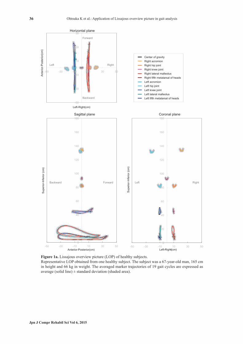
Figure 1a shows representative LOP obtained from one healthy subject. Marker trajectories of 19 gait cycles were averaged and expressed as average (solid line) ± standard deviation (shaded area). Figure 1b shows the normal grand average LOP of the 19 healthy subjects. The LOP had almost the same shape for all individuals. In the horizontal plane, the knee joints, the lateral malleolli and the fifth metatarsal heads showed linear periodic motions (a). In the sagittal plane, the lateral malleolli and the fifth metatarsal heads showed a linear backward motion followed by a convex forward motion (b). The knee joints showed a figure-of-eight motion (c). The movements of the acromions and the hip joints were small, and were aligned on a line perpendicular to the ground passing through the virtual COG (d). In the coronal plane, the acromions, the hip joints and the virtual COG showed butterfly patterns, whereas the knee joints, the lateral malleolli and the fifth metatarsal of heads showed linear vertical periodic motions (e). In all the planes, the left-right trajectories were symmetrical. Case 1: The patient walked with an ankle-foot orthosis (AFO) without using a cane in daily life. Visual inspection of video images detected medial whip in the terminal stance phase and decreased toe clearance in the swing phase in the affected limb. During treadmill gait analysis, he did not use an AFO, cane or handrail. The LOP generated from treadmill
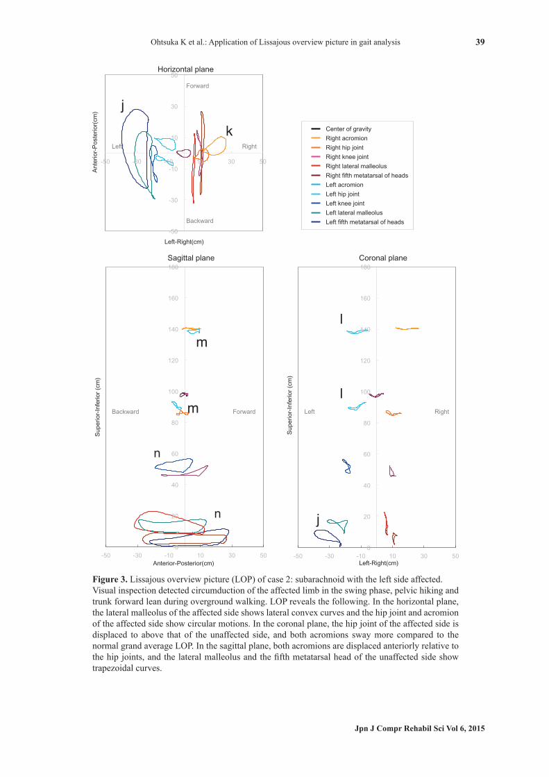
gait analysis showed the following features (Fig. 2). In the horizontal and coronal planes, the lateral malleolus of the affected side (left) showed interior convex curves (f). In the sagittal plane, the lateral malleolus and fifth metatarsal head did not rise in the swing phase (g). In the coronal plane, the hip joint motion of the affected side was displaced to above that of the unaffected side (h). Also, in the horizontal plane, the motions of the lateral malleolus and the fifth metatarsal head of the affected side showed an anterior shift compared to the unaffected side (i). Case 2: The patient walked with a cane and an AFO in daily use. During treadmill gait analysis, he used a handrail and an AFO. Visual inspection of video images detected circumduction of the affected limb in the swing phase, hip hiking, and trunk forward lean during the whole gait cycle. The LOP generated from treadmill gait analysis showed the following features (Fig. 3). In the horizontal plane, the lateral malleolus and the fifth metatarsal head of the affected side showed large lateral convex curves (j). The hip joint and the acromion of the unaffected side showed circular motions, whereas the knee joint, lateral malleolus and the fifth metatarsal head of the unaffected side showed linear periodic motions (k). In the coronal plane, the hip joint motion of the affected side was superior to that of the unaffected side, and both acromions showed greater lateral sway compared to the normal grand average (l). In the sagittal plane, the acromions on both sides showed an anterior shift relative to the hip joints (m). Furthermore, the lateral malleolus and the fifth metatarsal head of the affected side showed trapezoidal curves (n), which differed from the normal grand average. Case 3: The patient did not use a cane or orthosis in daily life. During treadmill gait analysis, he did not use any assistive device. Visual inspection of video images detected the so-called Duchenne gait and excessive flexion of the knee joints during the stance phase on both sides. The LOP generated from treadmill gait analysis showed the following features (Fig. 4). In the coronal plane, the vertical and lateral movements of both hip joints decreased compared to the normal grand average LOP which showed butterfly patterns. The acromions showed linear motions in a medial superior to lateral inferior orientation (o), which also differed from the normal grand average. In the sagittal plane, both knee motions showed a slight anterior shift relative to the COG and lateral malleolus (p). Furthermore, the acromions on both sides showed vertical motions (q). After we showed the LOP findings to the three therapists, they reviewed the video images again. After the review, in addition to their original findings by visual inspection of the video images only, they further identified displacement of the hip joint of the affected side in the swing phase in case 1; excessive rotation of trunk and lateral shift, and excessive lifting of the
CW6_AZ098D01_J_欧文.indd 35 2015/03/06 9:18:48
Ohtsuka K et al.: Application of Lissajous overview picture in gait analysis
Jpn J Compr Rehabil Sci Vol 6, 2015
36
Figure 1a. Lissajous overview picture (LOP) of healthy subjects.Representative LOP obtained from one healthy subject. The subject was a 67-year-old man, 165 cm in height and 66 kg in weight. The averaged marker trajectories of 19 gait cycles are expressed as average (solid line) ± standard deviation (shaded area).
CW6_AZ098D01_J_欧文.indd 36 2015/03/06 9:18:55

Ohtsuka K et al.: Application of Lissajous overview picture in gait analysis
Jpn J Compr Rehabil Sci Vol 6, 2015
37
Figure 1b. Lissajous overview picture (LOP) of healthy subjects.Normal grand average LOP. The normal grand average LOP was constructed from the LOP of 19 healthy subjects (64 ± 4 years, 166.3 ± 5.8 cm, 63.9 ± 6.5 kg).
CW6_AZ098D01_J_欧文.indd 37 2015/03/06 9:18:57

Ohtsuka K et al.: Application of Lissajous overview picture in gait analysis
Jpn J Compr Rehabil Sci Vol 6, 2015
38
Figure 2. Lissajous overview picture (LOP) of case 1: cerebral infarction with the left side affected.Visual inspection of video images detected medial whip in the terminal stance phase and decreased toe clearance in the swing phase in the affected limb. LOP reveals the following. In the horizontal plane, the lateral malleolus of the affected side shows interior convex curves. In the sagittal plane, the lateral malleolus and the fifth metatarsal head of the affected side do not rise in the swing phase. The motions of the lateral malleolus and the fifth metatarsal head of the affected side show an anterior shift compared to the unaffected side. In the coronal plane, the hip joint of the affected side is displaced to above that of the unaffected side.
CW6_AZ098D01_J_欧文.indd 38 2015/03/06 9:18:57
Ohtsuka K et al.: Application of Lissajous overview picture in gait analysis
Jpn J Compr Rehabil Sci Vol 6, 2015
39
Figure 3. Lissajous overview picture (LOP) of case 2: subarachnoid with the left side affected.Visual inspection detected circumduction of the affected limb in the swing phase, pelvic hiking and trunk forward lean during overground walking. LOP reveals the following. In the horizontal plane, the lateral malleolus of the affected side shows lateral convex curves and the hip joint and acromion of the affected side show circular motions. In the coronal plane, the hip joint of the affected side is displaced to above that of the unaffected side, and both acromions sway more compared to the normal grand average LOP. In the sagittal plane, both acromions are displaced anteriorly relative to the hip joints, and the lateral malleolus and the fifth metatarsal head of the unaffected side show trapezoidal curves.
CW6_AZ098D01_J_欧文.indd 39 2015/03/06 9:18:57

Ohtsuka K et al.: Application of Lissajous overview picture in gait analysis
Jpn J Compr Rehabil Sci Vol 6, 2015
40
Figure 4. Lissajous overview picture (LOP) of case 3: coxarthrosis with both sides affected.Visual inspection detected the Duchenne sign on both sides and excessive knee flexion. LOP reveals the following. In the coronal plane, the motions of the hip joints decrease in the lateral and anterior-posterior directions and the acromions move along a linear line in a medial superior to lateral inferior orientation. In the sagittal plane, both knee motions show a slight anterior shift and both acromions show vertical motions.
CW6_AZ098D01_J_欧文.indd 40 2015/03/06 9:18:57
Ohtsuka K et al.: Application of Lissajous overview picture in gait analysis
Jpn J Compr Rehabil Sci Vol 6, 2015
41
lower limb of the affected side in the swing phase in case 2; and trunk forward lean in case 3.
Discussion
We constructed the normal grand average LOP from healthy subjects, and compared the LOP from three patients with pathological gait with the normal grand average. Symmetrical and reproducible LOP were obtained from individuals with normal gait. In the sagittal and horizontal planes, the knee, the lateral malleolus and the fifth metatarsal head moved in an anterior-posterior direction, while the acromion and the hip joint hardly moved. In the coronal plane, the COG, the acromion and the hip joint showed butterfly patterns. These characteristics indicated normal gait pattern. In case 1, the convex movement toward the medial side made by the lateral malleolus of the affected side in the horizontal and coronal planes shown on the LOP corresponds to the medial whip observed by visual inspection of video images. A low track of the lateral malleolus of the affected side in the sagittal plane reflects decreased toe clearance observed by visual inspection. In addition, the trajectory of the hip of the affected side was displaced to above that of the unaffected side in the coronal plane, reflecting hip hiking of the affected side. Furthermore, this patient probably assumed an asymmetric posture because the lower limb of the affected side showed an anterior shift compared to the unaffected side. In case 2, arc-shaped motions toward the lateral side made by the lateral malleolus and the fifth metatarsal head of the affected side in the horizontal and coronal planes of the LOP correspond to the circumduction gait pattern observed by visual inspection of video images. The circular motions of the acromion and the hip joint indicated excessive rotation of the trunk, presumably due to compensation of the circumduction gait. In addition, the trajectory of the hip joint of the affected side was superior to that of the unaffected side, and the distance between the acromion and the hip joint of the affected side became shorter. From these findings, we speculate that the trunk on the affected side is always shortened during walking. The increase in lateral shift of both acromions indicated lateral sway of the trunk, but this may have been due to the use of a handrail in this case. Anterior shift of the acromions relative to the hip joints in the sagittal plane corresponds to the forward lean of the trunk observed by visual inspection. The trapezoidal motions made by the lateral malleolus and the fifth metatarsal head of the unaffected side in the sagittal plane may indicate excessive lifting of the leg to compensate for poor toe clearance in the swing phase. In case 3, bilateral hip joint movements in the vertical and lateral directions were smaller than the grand average LOP, and both acromions swayed in a
medial superior to lateral inferior orientation. These motions suggest that the acromions moved to the lateral side of the trunk with the hip joints as fulcrums, and these findings correspond to the Duchenne gait observed by visual inspection of video images [9]. Also, the knee joint motions were displaced anteriorly relative to the hip joints and the lateral malleoli, reflecting the excessive knee flexion in the stance phase observed by visual inspection. Furthermore, vertical motions of the acromions together with little motion of the hip joints probably reflect the fact that the trunk leans forward during walking. During overground walking, only Lissajous figures in the coronal plane can be obtained. However, when walking on a treadmill, Lissajous figures in the coronal, horizontal and sagittal planes can be generated, because the subject walks within the same space on a treadmill. In addition, treadmill gait analysis is easy to analyze statistically and the results are reproducible because data of multiple steps at a fixed speed can be obtained. Furthermore, treadmill gait has the additional advantages that it can be conducted in a narrow space and can be used to evaluate patients with low gait capability by using handrails and suspension, and is therefore highly suitable for three-dimensional motion analysis. In this study, the LOP identified several abnormalities in three patients with gait disturbance, which could not be observed by visual inspection of the patients’ video images. Visualization of motion patterns by trajectories such as Lissajous figures is accurate and easy to understand compared to verbal descriptions such as “slight” or “moderate”. The Lissajous figures are useful for depicting not only the movement pattern of each limb segment but also the complete picture of gait, including posture and symmetry, by the positional relations between the limb segments. Gait involves periodic motions linked by many joints, and each cycle lasts approximately 1 second in healthy people and approximately 2 seconds even in patients with gait disturbances. Therefore, it is not easy to observe all the motions accurately by visual inspection. Visuali-zation of the movement patterns using trajectories such as the LOP facilitates identification of abnor-malities that may be overlooked by visual inspection of video images, helps accurate understanding of pathologies, and allows easy interpretation even by non-specialists of gait analysis. One of the limitations in this study is that the LOP cannot express temporal factors. Therefore, for clinical application, LOP should not be used alone, but should be used in combination with other temporal and distance factors obtained from three-dimensional motion analysis, as well as analysis of joint angles. We plan to develop the LOP toward clinical application by constructing normal grand average LOP for different age groups and gait speeds, and performing quantitative analysis of movement patterns
CW6_AZ098D01_J_欧文.indd 41 2015/03/06 9:18:58

Ohtsuka K et al.: Application of Lissajous overview picture in gait analysis
Jpn J Compr Rehabil Sci Vol 6, 2015
42
based on the LOP.
References 1 . McGinley JL, Goldie PA, Greenwood KM, Olney SJ.
Accuracy and reliability of observational gait analysis data: judgments of push-off in gait after stroke. Phys Ther 2003; 83: 146-60.
2 . Eastlack ME, Arvidson J, Snyder-Mackler L, Danoff JV, McGarvey CL. Inter-rater reliability of videotaped observational gait-analysis assessment. Phys Ther 1991; 71: 465-72.
3 . Hughes KA, Bell F. Visual assessment of hemiplegic gait following stroke: pilot study. Arch Phys Med Rehabil 1994; 75: 1100-7.
4 . Brunnekreef JJ, van Uden CJ, van Moorsel S, Kooloos JG. Reliability of videotaped observational gait analysis in patients with orthopedic impairments. BMC Musculoskelet Disord 2005; 6: 17.
5 . Krebs DE, Edelstein JE, Fishman S. Reliability of observational kinematic gait analysis. Phys Ther 1985; 65: 1027-33.
6 . Teranishi T, Ohtsuka K, Muraoka Y, Saitoh E, Itoh N, Kanada Y, et al. Accuracy assessment of the three-dimensional treadmill gait analysis equipment. Sogo Rehabilitation 2009; 37: 939-44. Japanese.
7 . Tanabe S, Saitoh E, Ohtsuka K, Teranishi T, Tomita Y, Muraoka Y. Simple method to reduce the effect of patient positioning variation on three-dimensional motion analysis during treadmill gait. Clin Pract 2013; 3: e30: 84-6.
8 . Yamamoto S, Ehara Y. Introduction to Body-Dynamics − Hemiplegic Gait and Ankle Foot Orthosis, 1st ed. Ishiyaku Publishers Inc., 2005. p. 169-70. Japanese.
9 . D’Angelo MG, Berti M, Piccinini L, Romei M, Guglieri M, Bonato S, et al. Gait pattern in Duchenne muscular dystrophy. Gait Posture 2009; 29: 36-41.
CW6_AZ098D01_J_欧文.indd 42 2015/03/06 9:18:58