anti-hepatitis b virus activity of chickweed stellaria media
TRANSCRIPT

Molecules 2012, 17, 8633-8646; doi:10.3390/molecules17078633
molecules ISSN 1420-3049
www.mdpi.com/journal/molecules
Article
Anti-Hepatitis B Virus Activity of Chickweed [Stellaria media (L.) Vill.] Extracts in HepG2.2.15 Cells
Lihua Ma 1,2, Jie Song 1, Yaqin Shi 1, Changmei Wang 1, Bin Chen 1, Donghao Xie 2 and
Xiaobin Jia 1,*
1 Key Laboratory of New Drug Delivery System of Chinese Materia Medica,
Jiangsu Provincial Academy of Chinese Medicine, 100 Shizi Street, Nanjing 210028, China;
E-Mails: [email protected] (L.M.); [email protected] (J.S.);
[email protected] (Y.S.); [email protected] (C.W.); [email protected] (B.C.) 2 School of Pharmacy, Jiangsu University, Zhenjiang, Jiangsu, 212013, China;
E-Mail: [email protected]
* Author to whom correspondence should be addressed; E-Mail: [email protected];
Tel.: +86-25-8563-8672; Fax: +86-25-8563-7809.
Received: 22 May 2012; in revised form: 29 June 2012 / Accepted: 9 July 2012 /
Published: 18 July 2012
Abstract: Stellaria media (Linn.) Villars is a traditional Chinese medicine that has been
used for over 200 years, mainly for the treatment of dermatitis and other skin diseases.
It has also been used as an anti-viral agent. All the fresh chickweed juice samples used in
this study were prepared using macroporous resin and ultrafiltration technology. The
anti-hepatitis B virus (HBV) activity of S. media was evaluated in vitro using the human
HBV-transfected liver cell line HepG2.2.15. The concentrations of hepatitis B surface
antigen (HBsAg) and hepatitis B e antigen (HBeAg) in HepG2.2.15 cell culture medium
were determined by enzyme-linked immunosorbent assay (ELISA) after S. media-n (SM-n)
treatment for 6 or 9 days. HBV DNA was quantified using transcription-mediated
amplification and real-time polymerase chain reaction. In HepG2.2.15 cells, 30 μg/mL
SM-3 effectively suppressed the secretion of HBsAg and HBeAg with inhibition rates of
27.92% and 25.35% after 6 days of treatment, respectively. Consistent with the reduction
in HBV antigens, SM-3 also reduced the level of HBV DNA in a dose-dependent manner.
The characterization and quantitation of the chemical composition of SM-3 showed the
presence of flavonoid C-glycosides, polysaccharides, and protein, which exhibited diverse antiviral activities. In conclusion, our results demonstrate that SM-3 possesses potential
OPEN ACCESS

Molecules 2012, 17 8634
anti-HBV activity in vitro. This is the first report demonstrating the anti-HBV effects of S.
media, which is currently under early development as a potential anti-HBV drug candidate.
Keywords: anti-hepatitis B virus activity; Stellaria media (L.) Vill.; flavonoid;
polysaccharide
1. Introduction
Hepatitis B virus (HBV) infection frequently results in both acute and chronic hepatitis and remains
a major health problem worldwide. According to WHO estimates, over five million cases of acute
hepatitis B infection occur annually and more than 350 million people suffer from chronic HBV
infection [1]. Annually, HBV infection accounts for one million deaths worldwide, mainly due to
cirrhosis, liver failure, and hepatocellular carcinoma [2]. Currently, lamivudine (3TC), entecavir [3],
adefovir, telbivudine [4], IFN-α, and Peg-IFNα-2a [5] have been licensed globally for the treatment of
HBV. Significant side effects of these drugs and inevitable drug resistance have been noted. The use of
nucleoside analogues for the treatment of HBV also has disadvantages, such as the requirement for
long-term therapy and high-drug resistance rate [6,7]. Furthermore, these agents are expensive [8,9].
A significant unmet medical need exists for new safe and efficacious anti-HBV drugs [10]. However,
to develop new anti-HBV agents remains a significant challenge. Given the well-known potency of
Chinese herbs in the treatment of diverse diseases, we were interested in studying their potential
anti-HBV activity.
Stellaria media (L.) Vill., commonly known as chickweed, is a Chinese folk medicine that belongs
to the Caryophyllaceae flowering plant family, which characteristically contain typical C-glycosyl-
flavones [1]. This plant is distributed widely throughout China and contains many polysaccharides,
flavonoids, cyclic peptides as well as other compounds, which exhibit extremely effective
anti-inflammatory and antiviral activity [11]. It has also been used as a folk medicine; for example, the
17th century herbalist John Gerard recommended it as a remedy for mange [12]. Modern herbalists
prescribe it mainly for skin diseases, but also for bronchitis, rheumatic pains, arthritis, and period
pain [13]. A poultice of chickweed can be applied to cuts, burns, and bruises [14]. However, not all
these uses are supported by scientific evidence. In this study, our results show that chickweed that has
undergone membrane ultrafiltration possesses excellent anti-HBV activity. We also report the presence
of large quantities of polyphenol and macromolecular compounds that may contribute to the HBV
inhibitory effect of chickweed. This is the first report describing the anti-HBV activity of S. media,
with great significance in research investigating the underlying mechanism of this activity.
2. Results and Discussion
2.1. Cytotoxic Effects of Different Stellaria Extracts
S. media was collected from the Nanjing Botanical Garden, Memorial, Sun Yat-Sen (China) during
its flowering period. The plant is native to Europe, and is both edible and nutritious; it is used as a raw
leaf vegetable in salads. Prior to this study, no report has investigated whether S. media can inhibit

Molecules 2012, 17 8635
HBV. The HepG2.2.15 cell line was established using a gene plasmid containing two heads and tails
attached to the adw subtype of HBV transfected into human hepatoma cell lines. This cell line has a
consistently high level of expression of HBsAg and HBeAg, and exhibits the biological activity of the
virus particles in vitro [15].
The viability of the HepG2.2.15 cells in the presence of various concentrations of different S. media
extracts was examined using an MTT assay. The results showed that all sample concentrations below
30 μg/mL had no significant toxicity in HepG2.2.15 cells at 9 days (Figure 1). The cytotoxicity of
these samples was examined to determine the treatment concentrations in the following HepG2.2.15
cell culture experiments.
Figure 1. MTT cytotoxicity assay results for the seven Stellaria media samples.
HepG2.2.15 cells were cultured in the presence of SM-1 to SM-7 at various concentrations for 9 days. Cell viability was expressed as a percentage of the blank control. Data were expressed as mean ± SD based on 3 independent experiments. * p < 0.05, vs. blank control.
2.2. Anti-HBV Antigens Secretion Activity in HepG2.2.15 Cells after Stellaria Treatment
The HepG2.2.15 cells were treated with SM-n at various concentrations to determine their
inhibitory effects on the secretion of HBsAg and HBeAg after 9 days. The antigens in the culture
supernatants were quantified using specific ELISA kits. The anti-HBV activity of each sample was
shown by its inhibition of antigen secretion in HepG2.2.15 cells after treatment with the corresponding
sample. 3TC was used for positive cytotoxicity control. The data demonstrated that SM-3 significantly
reduced HBsAg and HBeAg secretion, albeit to a slightly lesser degree (Table 1). Notably, for both
HBsAg and HBeAg, no significant difference was observed between SM-3 and 3TC.
In HepG2.2.15 cells, SM-3 showed no inhibitory effect on cell proliferation at concentrations of
up to 30 μg/mL, as shown by the MTT assay. The 50% cytotoxic concentration was determined to be
38 μg/mL. These results were used to determine the dose range of SM-3 for subsequent experiments.
SM-3 effectively suppressed the secretion of HBV antigens from HBV-transfected HepG2.2.15
cells, achieving 9.83%, 19.79%, 24.05%, and 27.92% inhibition of the secretion of HBsAg, and 6.51%,
18.55%, 21.83%, and 25.35% inhibition for HBeAg, at 1, 3, 10, and 30 μg/mL after 6 days of
treatment (Figure 2). In the same experiment, 100 μg/mL 3TC also suppressed the secretion of both
HBsAg and HBeAg, achieving 29.31% and 28.63% inhibition, respectively (Figure 2). However, the
secretion of HBV antigen at 9 days was not increased compared with those at 6 days, and this study

Molecules 2012, 17 8636
only assayed the secretion of HBV antigens after 6 and 9 days (Figure 2). These data suggest that the
inhibitory activity of SM-3 on the secretion of HBV antigens is similar to that of 3TC.
Table 1. Comparison of the anti-HBV activity demonstrated by different S. media components.
Sample Concentration (μg/mL) HBsAg (%) HBeAg (%) SM-1 30 27.35 11.37 SM-2 30 11.42 5.78 SM-3 30 34.15 27.28 SM-4 30 13.69 4.59 SM-5 10 25.76 9.18 SM-6 10 10.43 5.12 SM-7 30 23.64 12.37 3TC 100 38.65 33.56
HBsAg, hepatitis B surface antigen; HBeAg, hepatitis B e antigen.
Figure 2. Inhibition of HBsAg and HBeAg secretion by SM-3 and 3TC.
SM-3 reduced hepatitis B surface antigen (HBsAg) and hepatitis B e antigen (HBeAg) secretion in a dose-dependent manner in vitro. HepG2.2.15 cells were cultured in the presence of SM-3 at various concentrations (1, 3, 10, and 30 μg/mL) or 100 μg/mL 3TC for 6 and 9 days. In the culture supernatants, HBsAg (A) and HBeAg (B) were quantified using specific enzyme-linked immunosorbent assay (ELISA) kits. The data are presented as mean ± SD of 3 experiments. ** p < 0.01, * p < 0.05 compared with the negative control group.
2.3. Effects of SM-3 on HBV DNA Expression
HBV is a DNA virus and HBV DNA testing is the most accurate indicator for measuring the
contagiousness of hepatitis B. HBV DNA load is an important indicator of the efficacy of the antiviral
treatment selected, and its accurate detection during antiviral therapy is important for guiding clinical
decisions [16]. The cytotoxicity of the samples was examined to determine the treatment
concentrations to be used in the HepG2.2.15 cell culture experiments. According to the analysis of the
pre-test results, only SM-3 was able to significantly reduce HBsAg and HBeAg secretion. To further
confirm the anti-HBV activity of SM-3 in HepG2.2.15 cells, the effect of SM-3 treatment on the level
of HBV DNA was evaluated. Consistent with the inhibitory effects on HBsAg and HBeAg secretion,
30 μg/mL SM-3 treatment led to a statistically significant reduction in the level of extracellular HBV
DNA compared with the negative control after 6 and 9 days (Figure 3).

Molecules 2012, 17 8637
Figure 3. Inhibitory effect of SM-3 on HBV DNA levels in HepG2.2.15 cells.
HepG2.2.15 cells were cultured in the presence of SM-3 at various concentrations or in the presence of 100 μg/mL 3TC for 6 or 9 days. HBV DNA levels were then quantified by fluorescence quantitative PCR. The experiments were performed in triplicate, and data are presented as the mean ± SD of all experiments. ** p < 0.01 vs. blank control.
2.4. Chemical Characterization of SM-3 and Analysis of Total Sugar, Protein, and Total Flavonoid
Content
Under the guidance of the “component structural theory”, we performed a qualitative study on the
chemical composition of SM-3. Total sugar, protein, and total flavonoid content were determined by a
UV spectrophotometer, with the ratio of their content found to be total flavonoid:total sugar:protein =
1:11.86:16.70. This analysis was performed to reveal the chemical composition of SM-3 at the
component level. The primary structure and antiviral activity of the macromolecular compounds,
mainly referring to the polysaccharides were analysed in our laboratory (manuscript in preparation).
The analysis results of SM-3 by HPLC demonstrated that the compounds with UV absorption were
mainly flavonoids (Figure 4).
Figure 4. Chromatogram of SM-3 after elution with ethanol at 330 nm.
To further analyze the flavonoid qualitatively, SM-3 was analysed by LC/MS in positive ion mode.
Data obtained during the LC-MS3 experiments demonstrated that the fragmentation regularity of peak
6 was consistent with vitexin [M+H]+ 433.11 (Figure 5), which has been reported previously [17].
Apart from this, other peaks did not permit the structures to be identified easily; however, the
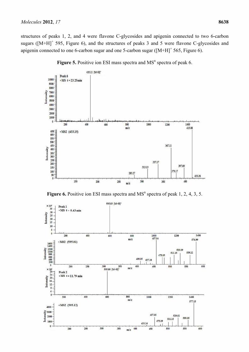
Molecules 2012, 17 8638
structures of peaks 1, 2, and 4 were flavone C-glycosides and apigenin connected to two 6-carbon
sugars ([M+H]+ 595, Figure 6), and the structures of peaks 3 and 5 were flavone C-glycosides and
apigenin connected to one 6-carbon sugar and one 5-carbon sugar ([M+H]+ 565, Figure 6).
Figure 5. Positive ion ESI mass spectra and MSn spectra of peak 6.
Figure 6. Positive ion ESI mass spectra and MSn spectra of peak 1, 2, 4, 3, 5.

Molecules 2012, 17 8639
Figure 6. Cont.
Our results on the regularity of the fragmentation process confirm the findings from previous
analysis [18] and data were consistent with the fragmentation regularity of flavone C-glycosides
(Figure 7). These compounds were identified as isomers according to the above results. However, their
chemical structure could not currently be determined by LC/MS. Therefore, the flavonoids in SM-3
should be further separated, purified, and analysed.
Figure 7. The fragmentation regularity of flavone C-glycosides sugar ring.

Molecules 2012, 17 8640
2.5. Anti-HBV Efficacy Validation of the Components of SM-3
To further verify the anti-HBV activity of SM-3, the sample was treated with macroporous
adsorption resin to divide it into its aqueous and alcohol soluble parts. The chemical analysis of SM-3
showed that it contained two types of compounds: polyphenolic and macromolecular substances.
Samples were obtained by mixing the same amount of two parts of the crude extract. These samples
were named as SM-3-A, SM-3-B, SM-3-C, and SM-3-D. As shown in Figure 8, at 6 days, the
inhibition ratios of the secretion of HBsAg and HBeAg by SM-3-A were 25.29% and 22.63%,
respectively, compared with 24.14% and 22.42% by SM-3-D.
Statistically, the inhibition rates differences were not significantly different between the two
samples, but they were lower than the positive control drug 3TC, with the inhibition of HBsAg and
HBeAg being 34.49% and 27.29%, respectively. After 9 days, the inhibition rate of HBsAg was
slightly increased; however, the inhibition rate of HBeAg showed the opposite trend. Interestingly, the
anti-HBV effect of SM-3-B was very obvious, with a HBeAg inhibitory rate of 28.74%, which was
worthy of further consideration. The data demonstrated that SM-3-A exhibited the best inhibitory
effect of the four mixtures (Figure 8), which provided further validation of the “component structure
theory”. Based on these results, the inhibitory activity of SM-3 appears to warrant further study.
Thus far, no report has been published on the anti-HBV activity of S. media. This study has shown
that SM-3 has significant anti-HBV activity by detecting both antigens and HBV DNA. Large amounts
of polyphenol compounds, such as flavonoids, were found in the SM-3 fraction. It is well known that
polyphenols bind to proteins to form unstable complexes [19]. Therefore, enveloped viruses, such as
HBV, may be affected by polyphenols given that this class of naturally occurring substances which
might interact with the glycoproteins of the viral envelope. Moreover, other macromolecular
compounds have been reported to exhibit an anti-HBV effect, including polysaccharides, which
displayed anti-HBV activity by enhancing immune regulation and acting as antioxidants [20,21].
Together, these findings help to establish a theoretical foundation for the inhibitory effect of SM-3, and
the material basis of its anti-HBV activity may result from an interaction of multiple components being
present in a certain proportion.
3. Experimental
3.1. Compounds and Reagents
Fresh chickweed was collected from-Nanjing Botanical Garden Memorial Sun Yat-Sen, China, and
repeatedly washed in water, with the final wash using distilled water. Species identification was
conducted by Professor D.K. Wu from Nanjing University of Chinese Medicine, Nanjing, China. The
vouchers of the authenticated samples were deposited at the Key Laboratory of New Drug Delivery
System of Chinese Materia Medica, Jiangsu Provincial Academy of Chinese Medicine.
A total of 5 kg of chickweed was homogenised in 5,000 mL of distilled water at 25 °C using a
domestic blender. The total homogenate was filtered through a NITEX nylon mesh filter and the
filtrate was centrifuged at 5,000 ×g for 15 min at 25 °C. The samples were named as SM-1 to SM-7
after different preparation methods (Figure 9). The final filtrate was used directly after being centrifuged
or lyophilised, and was reconstituted as needed into stock solutions of 100 mg/mL in water.

Molecules 2012, 17 8641
Figure 8. Inhibition of HBsAg and HBeAg secretion by SM-3 and 3TC.
A, HBsAg-6 days; B, HBeAg-6 days; C, HBsAg-9 days and D, HBeAg-9 days. SM-3-A, SM-3-B, SM-3-C, and SM-3-D reduced hepatitis B surface antigen (HBsAg) and hepatitis B e antigen (HBeAg) secretion in a dose-dependent manner in vitro. HepG2.2.15 cells were cultured in the presence of SM-3 components at various concentrations (1, 3, 10, and 30 μg/mL) or in the presence of 100 μg/mL 3TC for 6 or 9 days. The secretion of HBsAg was significantly reduced after 6 d, and by 9 days, the level of inhibition was slightly increased (A and C). Comparing the 4 samples, A and D displayed more potential inhibition, and both had no significant difference. The experiments were performed in triplicate and the data are presented as mean ± SD. ** p < 0.01, * p < 0.05 compared with the control group.

Molecules 2012, 17 8642
Figure 9. Flow chart of the preparation of the seven samples by different methods.
3.2. Cell Culture
Confluent cultures of HepG2.2.15 cells (Shanghai Bioleaf Biotech Ceo., Ltd., Shanghai, China)
were treated with various doses of antiviral compounds in MEM (Sigma Chemical Co., St. Louis, MO,
USA) supplemented with 10% fetal bovine serum (Gibco-Invitrogen, Carlsbad, CA, USA) and
500 μg/mL G418 (Sigma Chemical Co.) under 5% CO2 atmosphere at 37 °C. Fresh medium with the
same concentration of compounds was replaced on day 4. Subconfluent monolayer cells of
HepG2.2.15 were detached from the culture dishes by trypsin treatment, centrifuged for 5 min, and
resuspended in fresh media. Cells were seeded in 96-well flat-bottom plates at a density of
5.0 × 10⁴ cells/well and grown in fresh medium. All samples used in this study were dissolved in PBS,
and lamivudine (3TC) was used as the positive control. After plating for 24 h, the confluent
HepG2.2.15 cells were fed with the medium containing the indicated concentration of test samples and
fresh medium with the respective reagents was changed every 3 day. Cell viability was then analysed,
or the culture media and cells were harvested for detection. Cell viability was calculated as follows:
Cell viability (%) = OD (sample)/OD (control) × 100%

Molecules 2012, 17 8643
3.3. Cytotoxicity Assay
The 3-(4,5-dimethylthiazol-2-yl)-2,5-diphenyltetrazolium bromide (MTT, Sigma Chemical Co.)
assay was used to measure the viability of the cultured cells and to further identify the non-toxic
concentrations of different reagents to the culture cells [22]. The HepG2.2.15 cells were treated with
different concentrations (300, 200, 100, 66, 44, 30, 10, and 3 μg/mL) of SM-n (n = 1, 2, 3,……8) for
72 or 144 h. 3TC (100 μg/mL) was used as the positive cytotoxicity control. Incubated medium was
then removed and 100 μL of fresh medium containing MTT (2.5 mg dissolved in 50 μL of dimethyl
sulphoxide [DMSO]) was added to each well. After incubation for 4 h at 37 °C, the culture medium
containing MTT was removed, 100 μL DMSO was added to each well, and the viable cells were
detected by measuring absorbance at 490 nm. Each experiment was performed in triplicate. Cell
viability was expressed as a percentage of the control. Concentrations were considered non-toxic if the
corresponding cell viability was >95%.
3.4. Measurement of HBV Antigens
HepG2.2.15 cells were seeded in 96-well plates at a density of 5.0 × 10⁴/well for the measurement
of HBV antigens, and HBV DNA. After incubation with various concentrations of the samples, or 3TC
for 6 or 9 d, the culture medium was collected, cell debris was removed, and the resulting sample was
stored at −70 °C until further analysis. The levels of HBV surface antigen (HBsAg) and e antigen
(HBeAg) in the supernatant of the HepG2.2.15 cells were determined using the enzyme-linked
immunosorbent assay (ELISA) according to the manufacturer’s protocol (Nanjing Yingke Xinchuang
Biotech Co., Ltd., Nanjing, China). Absorbance was measured at 450/630 nm using a microplate
reader (Varioskan, Thermo Scientific, Vantaa, Finland).
3.5. Quantification of HBV DNA by Quantitative PCR
HepG2.2.15 cells were treated with antiviral agents as described above. For the intracellular assay,
the cells were quantified with a HBV DNA quantitative kit. The primers specific for the detection of
HBV DNA was: P1: 5′-ATCCTGCTGCTATGCCTCATCTT-3′, P2: 5′-CAGTGGGGAAAGCCCTA
CAA-3′, and the sequence of the probe was 5′-TGGCTAGTTTACT AGTGCCATTTTG-3′. The kit
was based on transcription- mediated amplification and a hybridization protection assay [23].
To isolate HBV DNA from the HepG2.2.15 cells, DNA was extracted from culture supernatants,
according to the instructions of the DNA extraction kit used (CAS Array, Shanghai, China). In brief, a
mixture of the cell culture supernatants or amplification standards and 50 μL of the reaction mixture
containing 1.0 μmol/L primer, 0.1 μmol/L fluorescent probe (F Probe), 200 μmol/L DNA, 50 nkat Taq
DNA polymerase, and 1× buffer were placed in a 0.2 mL reaction tube. The reaction tube was heated
to 93 °C for 2 min pre-denaturation, then 93 °C for 45 s and 55 °C for 60 s, during the first 10 cycles,
followed by 30 cycles at 93 °C for 30 s and 55 °C for 45 s. The copy number calibrators of 1 × 10⁵, 1 × 10⁶,
1 × 10⁷, and 1 × 10⁸/L were also amplified. The difference in fluorescence before and after
amplification was plotted on the vertical axis, the copy number was plotted as the abscissa. The
ABI7000 computer software automatically computed the standard curve and the initial copy number of

Molecules 2012, 17 8644
samples, providing quantitative HBV DNA results. The HBV DNA test results were expressed using
qualitative methods (negative: copy number of <106 copies/L; positive: ≥106 copies/L) [24].
3.6. Preliminary Analysis of the Composition of SM-3
The contents of the components in SM-3 were determined by using a UV-spectrophotometer,
including total flavonoids, polysaccharides, and protein. The total content of the polysaccharides was
determined by the phenol-sulphuric acid colorimetric method using glucose as the standard [25].
In addition, protein was quantified according to the Bradford method [26] using bovine serum albumin
as the standard. Lastly, the total content of flavonoids was determined by UV using apigenin as the
standard, and the characterization of their chemical composition was achieved by high-performance
liquid chromatography (HPLC; Agilent 1100, Santa Clara, CA, USA) and liquid chromatography coupled
to mass spectrometry with ion trap mass analyser (LC/MS; Thermo LXQ, ESI, Waltham, MA, USA).
An Altima C18 (250 mm × 4.6 mm, 5 μm) column maintained at 35 °C was used. The solvents used
were (A) 0.1% formic acid in water and (B) methanol. The elution gradient established was as follows:
for 0–3 min, 75%–70% (A); 3–30 min, 70%–55% (A); 30–35 min, 55%–50% (A); and 35–40 min,
50%–75% (A), using a flow rate of 1 mL/min. All online detection was carried out in the diode-array
detector (DAD), using 280 and 330 nm as the preferred wavelengths, and in a mass spectrometer (MS)
connected to the HPLC system via the DAD cell outlet. MS detection was performed using a Thermo
LXQ equipped with an electrospray ionization source and an ion trap mass analyser, which were
controlled by the Chemstation software. The drying gas used was nitrogen at a flow rate of
80 mL/min at 350 °C. The source voltage was −3,500 V, the capillary voltage was −136 V, and
skimmer voltage was −40 V. Spectra were recorded in negative ion mode between 100 and 1,500 m/z.
The MS detector was programmed to perform a series of positive ion scans: A full scan and an MS-MS
scan of the most abundant ion in the first scan, using a normalised collision energy of 1 V.
3.7. Statistical Analyses
All experiments were repeated at least three times, and the results were expressed as mean ± SD.
Statistical significance was determined using the analysis of variance or a rank-sum test. Differences
were considered to be statistically significant at p < 0.05.
4. Conclusions
In conclusion, this study demonstrates that SM-3 possesses potent anti-HBV activity in vitro.
Fraction SM-3 was found to be more effective than other components of S. media at inhibiting the
secretion of HBV antigens. The present findings suggest that S. media may be useful in the
development of novel anti-HBV drugs.
Acknowledgments
This work was supported by the “Significant New Drug Development” Major National Science and
Technology Programs of China. The authors wish to thank Jiangsu Affiliated Hospital on Integration
of Chinese and Western Medicine for providing the PCR analysis instruments for this study.

Molecules 2012, 17 8645
Conflict of Interest
The authors declare no conflict of interest.
References
1. Lavanchy, D. Worldwide epidemiology of HBV infection, disease burden, and vaccine
prevention. J. Clin. Virol. 2005, 34, S1–S3.
2. Dienstag, J.L. Hepatitis B virus infection. N. Engl. J. Med. 2008, 359, 1486–1500.
3. Liaw, Y.F.; Leung, N.; Kao, J.H.; Piratvisuth, T.; Gane, E.; Han, K.H.; Guan, R.; Lau, G.K.K.;
Locarnini, S. Asian-Pacific consensus statement on the management of chronic hepatitis B:
A 2008 update. Hepatol. Int. 2008, 2, 263–283.
4. Liaw, Y.F.; Gane, E.; Leung, N.; Zeuzem, S.; Wang, Y.; Lai, C.L.; Heathcote, E.J.; Manns, M.;
Bzowej, N.; Niu, J. 2-Year GLOBE trial results: Telbivudine is superior to lamivudine in patients
with chronic hepatitis B. Gastroenterology 2009, 136, 486–495.
5. Brunetto, M.R.; Moriconi, F.; Bonino, F.; Lau, G.K.K.; Farci, P.; Yurdaydin, C.; Piratvisuth, T.;
Luo, K.; Wang, Y.; Hadziyannis, S. Hepatitis B virus surface antigen levels: A guide to sustained
response to peginterferon alfa-2a in HBeAg-negative chronic hepatitis B. Hepatology 2009, 49,
1141–1150.
6. Zoulim, F. Mechanism of viral persistence and resistance to nucleoside and nucleotide analogs in
chronic hepatitis B virus infection. Antiviral. Res. 2004, 64, 1–15.
7. Lok, A.S.; Zoulim, F.; Locarnini, S.; Bartholomeusz, A.; Ghany, M.G.; Pawlotsky, J.M.; Liaw, Y.F.;
Mizokami, M.; Kuiken, C. Antiviral drug-resistant HBV: Standardization of nomenclature and
assays and recommendations for management. Hepatology 2007, 46, 254–265.
8. Farrell, G.; Teoh, N. Management of chronic hepatitis B virus infection: A new era of disease
control. Int. Med. J. 2006, 36, 100–113.
9. Leemans, W.; Janssen, H.; de Man, R. Future prospectives for the management of chronic
hepatitis B. World J. Gastroenterol. 2007, 13, 2554–2567.
10. Perrillo, R.P. Current Treatment of Chronic Hepatitis B: Benefits and Limitations; Thieme
Medical Publishers: New York, NY, USA, 2005; pp. 20–28.
11. Hu, Y.M.; Ye, W.C.; Li, Q.; Tian, H.Y.; Wang, H.; Du, H.Y. C-glycosylflavones from Stellaria
media (in Chinese). Chin. J. Nat. Med. 2006, 4, 420–424.
12. Morikawa, T.; Sun, B.; Matsuda, H.; Wu, L.J.; Harima, S.; Yoshikawa, M. Bioactive constituents
from Chinese natural medicines. XIV. New glycosides of β-carboline-type alkaloid, neolignan,
and phenylpropanoid from Stellaria dichotoma L. var. lanceolata and their antiallergic activities.
Chem. Pharm. Bull. 2004, 52, 1194–1199.
13. Slavokhotova, A.A.; Odintsova, T.I.; Rogozhin, E.A.; Musolyamov, A.K.; Andreev, Y.A.;
Grishin, E.V.; Egorov, T.A. Isolation, molecular cloning and antimicrobial activity of novel
defensins from common chickweed (Stellaria media L.) seeds. Biochimie 2011, 93, 450–456.
14. Konesni, M. Mandi’s Herbal Handbook; Mandi Konesni: Victoria, BC, Canada, 2010; p. 45.

Molecules 2012, 17 8646
15. Dang, S.S.; Zhang, Z.G.; Zhang, X.; Song, P.; Bian, J.; Cheng, Y.A. Inhibition on hepatitis B
virus by emodin and astragalus polysaccharides in vitro. J Xi'an Jiaotong University (Medical
Sciences) 2007, 28, 521–525.
16. Li, M.A.; Zhao, G.Z. Comparative study of RQ-PCR and ELQ-PCR in the detection of
HBV-DNA load (in Chinese). J. Chin. Med. Univ. 2005, 34, 4–24.
17. Bai, Y.J.; Lu, J.Q.; Zhang, H.J. Fragmentation Regularity and Analysis on Electrospray Ionization
Mass Spectrometry glycosides of orientin, vitexin and puerarin carbon glycosides. Zhong Cao Yao
2010, 3, 361–364.
18. Song, Z.J.; Liu, X.; Qiu, S.X.; Gu, Y.C.; Ding, L.S. Analysis of flavone-C-glycosides from
Passiflora Incarnate L. by Tandem Mass Spectrometry (in Chinese). J Instrum. Anal. 2008, 27,
72–73.
19. Li, J.; Huang, H.; Zhou, W.; Feng, M.; Zhou, P. Anti-hepatitis B virus activities of Geranium
carolinianum L. extracts and identification of the active components. Biol. Pharm. Bull. 2008, 31,
743–747.
20. Huang, J.; Chen, B.; You, W. Studies on separation of extracellular polysaccharide from
Porphyridium cruentum and its anti-HBV activity in vitro (in Chinese). Chin. J. Mar. Drugs 2005,
24, 18–21.
21. Huang, H.; Gan, X. Advances in studies on anti-hepatitis B virus of natural active polysaccharides
(in Chinese). Chin. Tradit. Herbal Drugs 2006, 37, 1594–1596.
22. Wang, G.F.; Shi, L.P.; Ren, Y.D.; Liu, Q.F.; Liu, H.F.; Zhang, R.J.; Li, Z.; Zhu, F.H.; He, P.L.;
Tang, W. Anti-hepatitis B virus activity of chlorogenic acid, quinic acid and caffeic acid in vivo
and in vitro. Antiviral. Res. 2009, 83, 186–190.
23. Kamisango, K.; Kamogawa, C.; Sumi, M.; Goto, S.; Hirao, A.; Gonzales, F.; Yasuda, K.; Iino, S.
Quantitative detection of hepatitis B virus by transcription-mediated amplification and
hybridization protection assay. J. Clin. Microbiol. 1999, 37, 310–314.
24. Tao, Z.H.; Chen, X.D.; Wang, Z.Y.; Zhou, W.; Yuan, Q. Detection for HBV DNA in plasma by
fluorescence quantitative polymerase chain reaction and its clinical applicability (in Chinese).
J. Clin. Lab. 2002, 5, 282–284.
25. Dubois, M.; Gilles, K.A.; Hamilton, J.K.; Rebers, P.; Smith, F. Colorimetric method for
determination of sugars and related substances. J. Anal. Chem. 1956, 28, 350–356.
26. Bradford, M.M. A rapid and sensitive method for the quantitation of microgram quantities of
protein utilizing the principle of protein-dye binding. Anal. Biochem. 1976, 72, 248–254.
Sample Availability: Samples of the compounds Lamivudine are available from the authors.
© 2012 by the authors; licensee MDPI, Basel, Switzerland. This article is an open access article
distributed under the terms and conditions of the Creative Commons Attribution license
(http://creativecommons.org/licenses/by/3.0/).