tissue stiffness and hypoxia modulate the integrin-linked...
TRANSCRIPT

Microenvironment and Immunology
Tissue Stiffness and Hypoxia Modulate theIntegrin-Linked Kinase ILK to Control BreastCancer Stem-like CellsMei-Fong Pang1,2, Michael J. Siedlik1, Siyang Han2, Melody Stallings-Mann3,Derek C. Radisky3, and Celeste M. Nelson1,2
Abstract
Breast tumors are stiffer and more hypoxic than nonmalig-nant breast tissue. Here we report that stiff and hypoxic micro-environments promote the development of breast cancer stem-like cells (CSC) through modulation of the integrin-linkedkinase ILK. Depleting ILK blocked stiffness and hypoxia-depen-dent acquisition of CSC marker expression and behavior,whereas ectopic expression of ILK stimulated CSC developmentunder softer or normoxic conditions. Stiff microenvironmentsalso promoted tumor formation and metastasis in ovo, wheredepleting ILK significantly abrogated the tumorigenic and met-
astatic potential of invasive breast cancer cells. We furtherfound that the ILK-mediated phenotypes induced by stiff andhypoxic microenvironments are regulated by PI3K/Akt. Anal-ysis of human breast cancer specimens revealed an associationbetween substratum stiffness, ILK, and CSC markers, insofar asILK and CD44 were expressed in cancer cells located in tumorregions predicted to be stiff. Our results define ILK as a keymechanotransducer in modulating breast CSC developmentin response to tissue mechanics and oxygen tension. Cancer Res;76(18); 1–11. �2016 AACR.
IntroductionThe mechanical stiffness of the cellular microenvironment,
dominated by the composition and crosslinking of the extracel-lular matrix (ECM), is a key modulator of cell fate (1, 2). Forexample, human mesenchymal stem cells differentiate downdistinct lineages depending on the stiffness of their underlyingsubstratum (1, 3), and matrix stiffness regulates epithelial plas-ticity by controlling the induction of epithelial–mesenchymaltransition (4). Increased matrix stiffness enhances tumor cellinvasiveness and dissemination (5, 6), and can direct the trans-formation of mammary epithelial cells (7). In contrast, tumorsgrown inmicewith disorganized, compliant ECM architecture areminimally invasive (8). Stiff ECM has been found to promotetumor progression through the induction of signaling pathwaysdownstream of integrins and PI3K (9).
In addition to increased stiffness, invasive breast cancers arefrequently hypoxic (10). Tumor hypoxia correlates with poorprognosis and decreased survival in breast cancer patients(11, 12). Hypoxia can activate signaling pathways that regulatecancer stem-like cells (CSC; ref. 13), a distinct population ofcancer cells with enhanced proliferative and invasive character-
istics (14). Under hypoxic conditions, breast cancer cells expressCSC-associated markers, including CD44, Nanog, CD49f, andALDH (15–19). Hypoxic conditions also promote pluripotencyand viability of CSC populations (20, 21).
Integrin-linked kinase (ILK) is a crucial mediator of signaltransmission from the ECM. ILK interacts with b1-integrin andtransmits extracellular signals from the ECM to regulate cellularactivities including anchorage-dependent growth and survival,migration, invasion, differentiation, and tumor angiogenesis(22). Elevated expression of ILK has been closely associated withhigh-grade human tumors (23, 24) and ILK has been shown toactivate oncogenic pathways to promote tumor progression (25).However, it remains unclear how the expression of ILK and itsdownstream signaling are regulated in the stiff, hypoxic micro-environments common to invasive cancers. Because ILK is acritical adaptor used by cancer cells to sense their surroundingmicroenvironment, we hypothesized that matrix stiffness andhypoxia could affect ILK signaling in breast cancer cells to regulatebreast CSC-associated gene expression and cellular behaviors.Here, we used engineered synthetic substrata to recapitulate themechanical properties of the normal mammary gland as well asthat of breast tumors. We investigated how the mechanicalproperties and oxygen tension in the tumor microenvironmentaffect the formation of breast CSCs. We found that breast CSCmarkers are activated synergistically in response to stiff, hypoxicconditions, and that ILK is an essential regulator of breast CSCs.
Materials and MethodsCell culture and reagents
MDA-MB-231 human breast carcinoma cells and 4T1 murinemammary carcinoma cells were obtained from the ATCC andmaintained in DMEM/F12 or RPMI base medium (respectively)that was supplemented with 10% FBS and 1% gentamycin.Both cell lines were authenticated by short tandem repeat
1Department of Chemical & Biological Engineering, Princeton Univer-sity, Princeton, New Jersey. 2Department of Molecular Biology, Prin-ceton University, Princeton, New Jersey. 3Department of Cancer Biol-ogy, Mayo Clinic Cancer Center, Jacksonville, Florida.
Note: Supplementary data for this article are available at Cancer ResearchOnline (http://cancerres.aacrjournals.org/).
Corresponding Author: Celeste M. Nelson, Princeton University, 303 HoytLaboratory, William Street, Princeton, NJ 08544. Phone: 609-258-8851; Fax:609-258-1247; E-mail: [email protected]
doi: 10.1158/0008-5472.CAN-16-0579
�2016 American Association for Cancer Research.
CancerResearch
www.aacrjournals.org OF1
genotyping, tested for mycoplasma contamination (DDC Med-ical), and were used before passage 20 and within 6 monthsafter resuscitation. To reduce the expression of ILK, cells weretransduced with lentiviral particles carrying short hairpin RNA(shRNA) against ILK (sc-35667-V and sc-35666-V, Santa CruzBiotechnology) or control lentivirus expressing a scrambledshRNA sequence. Stable shRNA-expressing clones were pro-duced according to the manufacturer's instructions and selectedusing puromycin. All cells were maintained in a humidifiedincubator held at 37�C and 5% CO2.
To investigate the effect of matrix stiffness, cells were culturedon synthetic substrata of different compliances conjugated withfibronectin, which were prepared as described previously (4).Cells were seeded at a density of 500,000 cells/cm2 on syntheticsubstrata and cultured for 72 hours at 37�C in either humidifiednormoxic conditions (95% air and 5% CO2) or in a modularincubator chamber (Billups-Rothenberg, Inc.) filled with hypoxicgas (94% N2, 1% O2, and 5% CO2).
Quantitative real-time PCR analysisTotal RNA was extracted using TRIzol reagent according to the
manufacturer's instructions, followed by cDNA synthesis usingthe Verso cDNA synthesis kit (Thermo Scientific). Transcript levelswere measured by quantitative real-time PCR (qRT-PCR) using aBio-RadMiniOpticon instrument and iTaqUniversal SYBRGreenSupermix (Bio-Rad). Amplification was followed by meltingcurve analysis to verify the presence of a single PCR product.Primers specific for GFP, ILK, ITGB1, CD44, Nanog, CD49f,VEGF-A, and 18S rRNA are listed in Supplementary Table S1(Supplementary Information). The expression level of eachmRNA was normalized to that of 18S in the same sample.
Time-lapse imaging and cell trackingTime-lapse imagingwas performedusing aNikon Ti-U inverted
microscope equipped with a stage top incubator maintained at37�C in a 5%CO2, 90% relative humidity atmosphere (PathologyDevices, Inc.). Imageswere acquired every 30minutes using aPlanFluor 10�/0.3 NA air objective (Nikon) and a Hamamatsu Orca-100 camera. Individual GFP-labeled cells were tracked from thealigned image sequences using the "Manual Tracking" plugin inImageJ.
Immunoblotting analysisSamples were lysed in radioimmunoprecipitation assay buffer
(Pierce Biotechnology) supplemented with a protease inhibitorcocktail (Roche). Equal amounts of total protein were separatedby standard electrophoresis using 4–12% gradient NuPage gels(Invitrogen). Proteins were transferred onto nitrocellulose mem-branes, which were then blocked and incubated overnight withprimary antibodies at 4�C. Antibodies used for immunoblottingwere: rabbit anti-b1-integrin (1:1,000; Abcam), rabbit anti-ILK(1:1,000; Abcam), rabbit anti-Nanog (1:1,000; Novus Biologi-cals), mouse anti-CD44 (1:1,000; Novus Biologicals), and rabbitanti-GAPDH(1:1,000; Cell Signaling Technology). Afterwashing,blotswere probedwith horseradish peroxidase (HRP)-conjugatedanti-rabbit or anti-mouse secondary antibodies (1:5,000; CellSignaling Technology) for 1 hour. Blots were incubated with ECLPlus Western Blotting Detection System (GE Healthcare) for 5minutes and signals were detected with a FluorChemE Imager(Cell Biosciences, Inc.).
Soft agar assayApproximately 5,000 cells were resuspended in serum-free
DMEM/F12 medium supplemented with 0.35% agar and platedin a 12-well plate that contained a base layer of DMEM/F12medium supplemented with 0.7% agar. Cells were incubated for14 days and the culture medium was changed twice per week. Atthe end of the experiment, colonies were fixed with 4% PFA,washedwith PBS, and imaged. Colonies were counted if theywerelarger than 10 mm in diameter as measured using ImageJ.
Aldefluor assay and flow cytometryCells were harvested by trypsinization using Accutase
(eBioscience), washed with PBS, and resuspended in flow cyto-metry staining buffer (eBioscience). Cells were filtered through a40-mm cell strainer (Corning) to obtain single-cell suspensions.For intracellular staining, cells werefixed andpermeabilized usingFoxp3 transcription factor staining buffer set (eBioscience) priorto staining. Cells were stained without fixation for cell surfacemarkers. Cells were incubated with the following fluorochrome-conjugated antibodies at 4�C for 45minutes: CD49f (1:200, cloneGoH3; eBioscience), CD44 (1:100, clone IM7; eBioscience), andNanog (1:100, clone eBioMLC-51; eBioscience). Stained cellswere washed twice with flow cytometry buffer (eBioscience). Toassess ALDH activity, the ALDEFLUOR fluorescent reagent sytem(Stem Cell Technologies) was used according to the manufac-turer's instructions. Stained cells were analyzed using an LSRIIflow cytometer (BD Biosciences) and data were analyzed usingFlowJo.
Secondary mammosphere formation assayA single-cell suspension was seeded at a density of 3,000 cells
per well into 12-well ultra-low attachment plates (BD Bio-sciences). Cells were cultured in DMEM/F12 medium containing20 ng/mL EGF and 20 ng/mL basic fibroblast growth factor, andculturemediumwas changed twice perweek. After 7days, primarymammospheres were collected and disassociated enzymaticallywith 0.05% trypsin (Invitrogen) for 5 minutes at 37�C andmechanically by filtering through a 40-mmcell strainer (Corning).Single-cell suspensions were replated onto 12-well ultra-lowattachment plates at a density of 1,000 cells per well. After 14days, secondary mammospheres were transferred to a 12-wellplate. Serum-containingmediumwas added and secondarymam-mospheres were allowed to attach to the bottom surface of theplate. After 12 hours, cells were fixed with 4% PFA and stainedwith 0.05% crystal violet. Samples were imaged, analyzed usingImageJ, and mammospheres were counted if they had a diameterlarger than 50 mm.
Mechanical testing of collagen gelsThe elastic moduli of gels comprising different concentrations
of collagen were estimated from unconfined compression experi-ments, as described previously (26). Briefly, collagen solutionscontaining 500-nm fluorescent beads were allowed to gel for atleast 30 minutes at 37�C within circular polydimethylsiloxane(Sylgard 184) molds. After gelation, the mold was removed andthe cylindrical gel was immersed in PBS. Confocal stacks of the gelwere acquired before and 1 minute after loading a glass coverslipof knownweight to the top of the gel. The thickness of the gel wasestimated from the fluorescence signal of the embedded beadsand, as a first approximation, the measured change in thicknesswas used to estimate the elastic modulus of the gel.
Pang et al.
Cancer Res; 76(18) September 15, 2016 Cancer ResearchOF2

Chicken chorioallantoic membrane assayChicken chorioallantoic membrane (CAM) assays were per-
formed as described previously (27). Cells were transducedwith recombinant adenovirus encoding GFP at an MOI of 100for 24 hours prior to implantation. GFP-transduced cells wereresuspended in neutralized rat tail type I collagen (BD Bio-sciences) at a density of 300,000 cells per 30-mL pellet. Cell-embedded collagen gels were implanted on the CAM of achicken embryo at day 7 of incubation. Tumor formation,angiogenesis, and metastasis were scored in live embryos 5days later using a stereomicroscope. Images were acquiredusing a Nikon digital camera.
Human breast cancer samplesBreast cancer biopsies were derived from waste surgical
material from de-identified patients, and were formalin-fixedand paraffin-embedded, as per approval by the Mayo ClinicInstitutional Review Board. Tissue sections were deparaffinizedby placing them into three changes of xylene and rehydrated ina graded ethanol series. The rehydrated tissue samples wererinsed in water and sections were subjected to heat antigenretrieval as described by the manufacturer (DAKO). Slices wereincubated with each primary antibody for 1 hour at roomtemperature. Sections were then rinsed with Tris-bufferedsaline/Triton-X-100 (TBST) wash buffer, and incubated witheach secondary antibody for 30 minutes. For fluorescent detec-tion, tissue sections were rinsed 3 times for 5 minutes each withPBS containing 1.43 mmol/L 40,6-diamidino-2-phenylindole(DAPI; Thermo Fisher Scientific). Sections were rinsed withPBS and coverslips mounted with anti-fade mounting medium(DAKO). Sections stained for collagen I were rinsed with TBSTwash buffer, and secondary incubation was with DAKO Envi-sion anti-rabbit, HRP for 15 minutes. Tissue sections wererinsed with TBST wash buffer and then incubated in 3,30-diaminobenzidine (DABþ; DAKO), and counterstained withmodified Schmidt hematoxylin. Each antibody and its corre-sponding fluorescent secondary antibody used were: ILK1 (rab-bit polyclonal, Cell Signaling Technology #3862) detected byAlexa594-conjugated donkey anti-rabbit IgG (Invitrogen,#A21207); CD44 (mouse IgG2A, Abcam #ab6124) detectedby Alexa488-conjugated donkey anti-mouse IgG (HþL) (Invi-trogen, #A21202). For DAB staining, collagen I (Abcamab138492) was used.
Whole slide digital images of each breast cancer sample werecaptured with the Aperio Scanscope AT2 slide scanner (colla-gen) and the Aperio Scanscope FL slide scanner (fluorescentimages) using a 20� objective. Using the digitized images, areasof high and low collagen content were selected and circled. Cellnumber in each area was determined by manually counting thenumber of DAPI-positive nuclei. Similarly, double positivecells (488 and 594) were identified and counted in eachselected area. Density of double positive cells was calculatedas the total number of positively stained cells per total numberof cells in each selected area.
Statistical analysisData represent mean � SEM of at least three independent
experiments conducted in triplicate. For CAM assays, data repre-sent mean � SEM with n ¼ 5 chicken embryos per group. Forpatient samples, statistical significancewas testedwith theMann–Whitney U test; for all others, statistical analysis was conducted
using a two-way ANOVA followed by Bonferroni post-test orStudent t test. P < 0.05 was considered to represent a significantdifference between conditions.
ResultsSubstratum stiffness and oxygen tension regulate theexpression of b1-integrin and ILK in invasive breast cancer cells
To investigate how ECM compliance affects breast cancercells, we used synthetic polyacrylamide substrata to mimic thecompliance of the microenvironment in the normal mammarygland (Young modulus, E�130 Pa, "soft") and in breasttumors (E�4020 Pa, "stiff"; ref. 4). On soft substrata, 4T1mouse mammary carcinoma cells (Fig. 1A) and MDA-MB-231human breast carcinoma cells (Supplementary Fig. S1) wererounded in morphology. In contrast, both 4T1 and MDA-MB-231 cells displayed an elongated morphology on stiff substra-ta. Consistent with the expectation that the elongated cellmorphology on stiff substrata was associated with increasedinteractions with the ECM, RT-PCR and immunoblottingrevealed increased expression of b1-integrin and ILK in thecells cultured on stiff substrata at the transcript (Fig. 1B and C)and protein (Fig. 1D) levels.
Hypoxia is prevalent in solid tumors and has been shown toactivate stem cell–like properties in several cancers, includingbreast cancer (28). We found that hypoxia also led to an increasein the transcript and protein levels of b1-integrin (Fig. 1B and D)and ILK (Fig. 1C and D) in both 4T1 and MDA-MB-231 cells(Supplementary Fig. S1). A stiff, hypoxic microenvironmentinduced the highest expression of these markers (Fig. 1B–D andSupplementary Fig. S1). These results suggest that stiff and hyp-oxic microenvironments potentiate signaling downstream of b1-integrin and ILK in breast cancer cells.
The presence of breast CSCs correlates with metastasis andrelapse in breast cancer patients (29). To determine whether themechanical microenvironment and oxygen tension can regulatebreast CSC gene expression, we examined the relative effects ofsubstratum stiffness and hypoxia on the expression of the CSCmarkers, CD44, Nanog, CD49f, and ALDH. We found that sub-stratum stiffness and hypoxia led to synergistic increases in thelevels of each of thesemarkers in both 4T1 andMDA-MB-231 cells(Fig. 1E–H and Supplementary Fig. S1).
ILK has been found to regulate integrin function and cellmotility. We evaluated the effect of stiff substrata on cellmovement using time-lapse imaging, which revealed thatbreast cancer cells cultured on stiff substrata were more motilethan those cultured on soft substrata (Fig. 1I). Together, thesedata suggest that the increased stiffness and decreased oxygentension of the tumor microenvironment enhances integrinsignaling and activates CSC marker expression in invasivebreast cancer cells.
ILK and CD44 are elevated in potentially stiff regionsof human tumors
To evaluate the relationship between stiffness, ILK expres-sion, and CSC marker induction in vivo, we examined theexpression of ILK and CD44 in human breast cancer samples.We compared these to relative collagen content, which ishighly correlated with stiffness in mammary cancers(30, 31). Immunofluorescence analysis revealed that mosttumor cells expressed low levels of ILK and were CD44-negative
Stiffness, Hypoxia, and Cancer Stem Cells
www.aacrjournals.org Cancer Res; 76(18) September 15, 2016 OF3
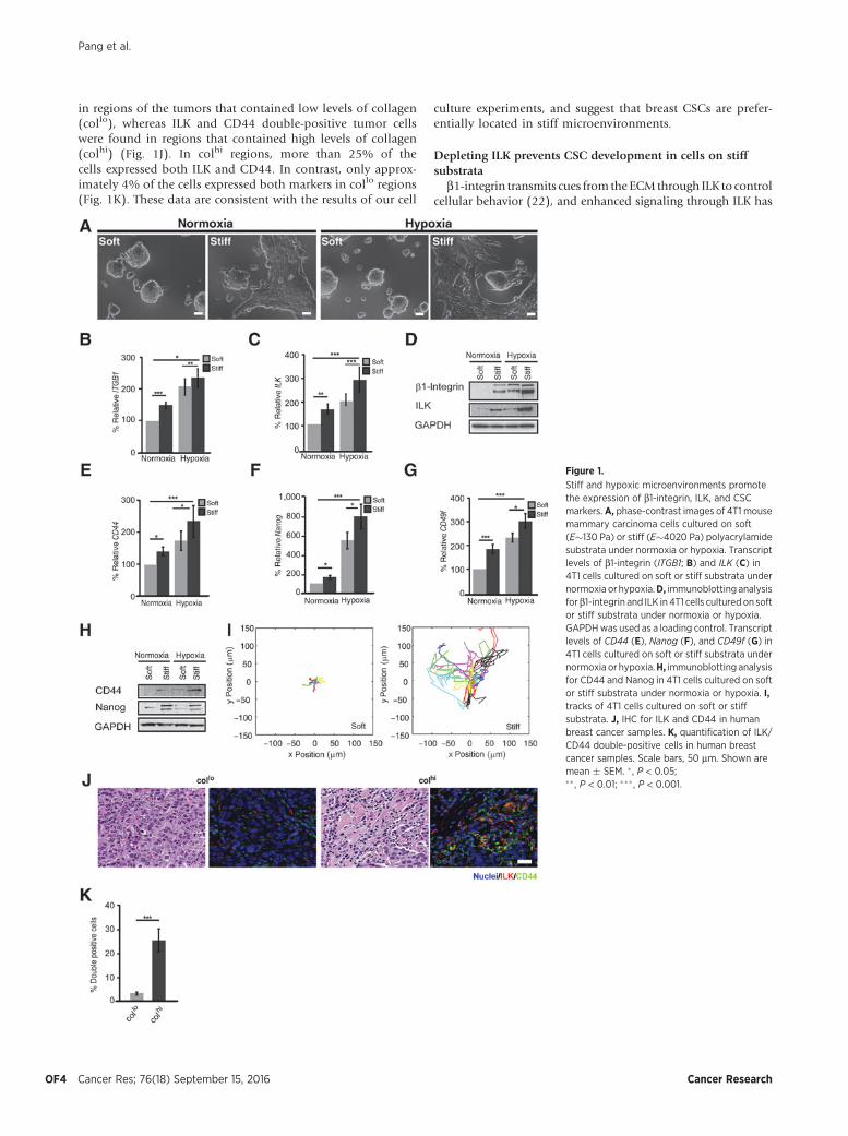
in regions of the tumors that contained low levels of collagen(collo), whereas ILK and CD44 double-positive tumor cellswere found in regions that contained high levels of collagen(colhi) (Fig. 1J). In colhi regions, more than 25% of thecells expressed both ILK and CD44. In contrast, only approx-imately 4% of the cells expressed both markers in collo regions(Fig. 1K). These data are consistent with the results of our cell
culture experiments, and suggest that breast CSCs are prefer-entially located in stiff microenvironments.
Depleting ILK prevents CSC development in cells on stiffsubstrata
b1-integrin transmits cues from the ECM through ILK to controlcellular behavior (22), and enhanced signaling through ILK has
Figure 1.
Stiff and hypoxic microenvironments promotethe expression of b1-integrin, ILK, and CSCmarkers. A, phase-contrast images of 4T1 mousemammary carcinoma cells cultured on soft(E�130 Pa) or stiff (E�4020 Pa) polyacrylamidesubstrata under normoxia or hypoxia. Transcriptlevels of b1-integrin (ITGB1; B) and ILK (C) in4T1 cells cultured on soft or stiff substrata undernormoxia or hypoxia.D, immunoblotting analysisforb1-integrin and ILK in 4T1 cells culturedon softor stiff substrata under normoxia or hypoxia.GAPDHwas used as a loading control. Transcriptlevels of CD44 (E), Nanog (F), and CD49f (G) in4T1 cells cultured on soft or stiff substrata undernormoxia or hypoxia.H, immunoblotting analysisfor CD44 and Nanog in 4T1 cells cultured on softor stiff substrata under normoxia or hypoxia. I,tracks of 4T1 cells cultured on soft or stiffsubstrata. J, IHC for ILK and CD44 in humanbreast cancer samples. K, quantification of ILK/CD44 double-positive cells in human breastcancer samples. Scale bars, 50 mm. Shown aremean � SEM. � , P < 0.05;�� , P < 0.01; ��� , P < 0.001.
Pang et al.
Cancer Res; 76(18) September 15, 2016 Cancer ResearchOF4

Figure 2.
ILK is required for CSC development from invasive breast cancer cells. A, qRT-PCR and immunoblotting analysis for ILK in 4T1 cells stably expressing shRNA againstILK (shILK) or scrambled sequence control (shctrl). B, phase-contrast images of 4T1-shctrl and 4T1-shILK cells cultured on soft or stiff substrata undernormoxia or hypoxia. Transcript levels of ITGB1 (C), CD44 (D), Nanog (E), and CD49f (F) in 4T1-shctrl or 4T1-shILK cells cultured on soft or stiff substrata undernormoxia or hypoxia. Phase-contrast images and quantification of secondary mammospheres (G) and colonies formed in soft agar (H) by 4T1-shctrl and 4T1-shILKcells. Scale bars, 50 mm. Shown are mean � SEM. � , P < 0.05; �� , P < 0.01; ��� , P < 0.001.
Stiffness, Hypoxia, and Cancer Stem Cells
www.aacrjournals.org Cancer Res; 76(18) September 15, 2016 OF5

been implicated in human cancer (23, 24). Matrix stiffness hasbeen shown to increase integrin signaling and promote tumorprogression (9). To define the role of ILK in the CSC response tostiffness and hypoxia, we used short hairpin RNA (shRNA) tostably deplete ILK in 4T1 (4T1-shILK) (Fig. 2A) andMDA-MB-231cells (Supplementary Fig. S2). Depleting ILK led to a significantchange in the morphology of 4T1 cells cultured on stiff substra-tum, as compared with scrambled controls (4T1-shctrl; Fig. 2B).On stiff substrata, 4T1-shILK cells exhibited a rounded morphol-ogy, similar to those on soft substrata, under both normoxia andhypoxia. We also found that knockdown of ILK reduced theexpression of b1-integrin (Fig. 2C) and the CSC markers CD44(Fig. 2D), Nanog (Fig. 2E), and CD49f (Fig. 2F) in 4T1 and MDA-MB-231 cells (Supplementary Fig. S2) under both normoxia andhypoxia. To examine the role of ILK in the regulation of the CSCcharacteristics of anchorage-independent growth and self-renew-al, we performed secondary mammosphere formation and softagar assays. We found that depleting ILK abrogated the ability ofboth 4T1 and MDA-MB-231 cells to form secondary mammo-spheres (Fig. 2G and Supplementary Fig. S2) and colonies in softagar (Fig. 2H; Supplementary Fig. S2). Together, these data suggestthat ILK is necessary for the induction of CSC marker expressionand behavior in breast cancer cells in response to matrix stiffnessand hypoxia.
ILK expression enhances breast CSC developmentTo determine whether ILK is sufficient to induce the develop-
ment of breast CSCs in the absence of a stiff microenvironment,we expressed ILK ectopically using a bicistronic recombinantadenovirus encoding for ILK and GFP (adILK). As a control, weused adenovirus encoding for GFP alone (adGFP). Transductionwith adILK approximately doubled the levels of ILK transcript andprotein in 4T1 (Fig. 3A) and MDA-MB-231 cells (SupplementaryFig. S3), and increased phosphorylation of Akt (pAkt; S437; Fig.3B and Supplementary Fig. S3). qRT-PCR analysis revealed thatectopic expression of ILK increased the transcript levels of b1-integrin (Fig. 3C), CD44 (Fig. 3D), Nanog (Fig. 3E), and CD49f(Fig. 3F) in 4T1 and MDA-MB-231 cells (Supplementary Fig. S3)cultured on soft or stiff substrata under normoxia or hypoxia.Ectopic expression of ILK also enhanced the formation of sec-ondary mammospheres (Fig. 3G and Supplementary Fig. S3) andcolonies in soft agar (Fig. 3H and Supplementary Fig. S3), dem-onstrating that increased expression of ILK can further activateCSCgene expression andbehavior evenwhen cells are cultured onstiff substrata under hypoxic conditions.
ILK signals through PI3K/Akt to regulate CSC developmentPrevious work has suggested that ILK regulates cell survival
through the PI3K/Akt pathway (32), which has also been impli-cated in cancer cell (33, 34) and CSC survival (35, 36), cancer cellproliferation (37, 38), and the CSC phenotype (39). We foundthat knockdown of ILK reduced the phosphorylation of Akt (Fig.4A and Supplementary Fig. S4). Disrupting signaling throughPI3K by treating cells with the selective inhibitor, LY294002 (Fig.4B and Supplementary Fig. S4), also decreased the transcript levelsofb1-integrin (Fig. 4C) and ILK (Fig. 4D) in4T1andMDA-MB-231cells (Supplementary Fig. S4) cultured on soft or stiff substrataunder normoxia or hypoxia. Consistent with the concept that Aktactivation is necessary for the ILK-dependent induction of CSCs inresponse to hypoxia or a stiff microenvironment, the levels ofCD44 (Fig. 4E), Nanog (Fig. 4F), and CD49f (Fig. 4G) decreased
significantly in response to treatment with LY294002 in 4T1 andMDA-MB-231 cells (Supplementary Fig. S4) under all conditions.
Similarly, treatment with LY294002 reduced the formation ofboth secondary mammospheres (Fig. 4H) and colonies in softagar (Fig. 4I). These data suggest that a stiff, hypoxic microenvi-ronment regulates development of CSCs in part by activatingsignaling through ILK and PI3K.
Stiff substratum promotes angiogenesis and dissemination oftumor cells through ILK
To test directly whether stiff substratum promotes tumorigen-esis and tumor cell dissemination, we embedded 4T1 cells incollagen gels of low (collo, 3 mg/mL), medium (colmed, 4.5 mg/mL), or high (colhi, 6 mg/mL) stiffness (Fig. 5A) and then graftedthem onto the CAMs of chicken embryos (40). We found that allcolhi-grafted CAMs formed primary tumors at the graft sites,whereas none of the collo- and only 4 of 6 of the colmed-graftedCAMs developed primary tumors (Fig. 5B). The tumors thatformed in high collagen developed multiple micrometastasesnear blood vessels adjacent to the graft sites (Fig. 5C). To definethe effects of ILK on tumor formation, we performed CAM assaysusing 4T1-shctrl or 4T1-shILK cells embedded in high collagengels. Both 4T1-shctrl and 4T1-shILK cells formed primary tumorson the CAM (Fig. 5D). However, the 4T1-shILK primary tumorswere half the size of the 4T1-shctrl tumors (Fig. 5D). To examinehow ILK affects metastatic potential, we performed qRT-PCRanalysis for GFP in the lungs of the chicken embryos after 5 daysof incubation with GFP-labeled 4T1 cells. We found significantlylower levels of GFP in the lungs of embryos with 4T1-shILK-grafted CAMs than in those with 4T1-shctrl–grafted CAMs (Fig.5E). These data suggest that matrix stiffness and ILK affect tumorformation as well as tumor cell dissemination.
Angiogenesis is a hallmark of cancer that is critical for tumorgrowth and metastasis (41). To investigate how substratumstiffness affects hypoxia-dependent angiogenesis, we examinedthe expression of VEGF-A in 4T1 cells cultured on soft or stiffsubstrata under normoxia or hypoxia. We found that culture onstiff substratum upregulated the expression of VEGF-A (Fig. 5F),suggesting that the mechanical properties of the microenviron-ment can regulate angiogenic signaling. As expected, hypoxiafurther increased the levels of VEGF-A in cells cultured on bothsoft and stiff substrata (Fig. 5F). Depleting ILK significantlyreduced VEGF-A expression in 4T1 cells cultured on soft or stiffsubstrata under normoxia or hypoxia (Fig. 5G). Quantitativeimage analysis revealed that the blood vessel density in the CAMadjacent to 4T1-shILK tumors was significantly lower thanthat adjacent to 4T1-shctrl tumors (Fig. 5H). These data suggestthat loss of ILK impairs the angiogenic potential of breast cancercells. Consistently, we found that inhibiting PI3K with LY294002significantly reduced the levels of VEGF-A (Fig. 5I), whereasectopic expression of ILK increased the levels of VEGF-A(Fig. 5J) under all culture conditions. These data suggest that ILKsignals through the PI3K pathway to regulate VEGF-A expressionin response to matrix stiffness and hypoxia.
DiscussionIt is well appreciated that the distinct physical properties of
the tumor microenvironment can affect cancer cell fate. Tumorstiffness induces integrin clustering and downstream signalingthat control cancer cell proliferation, gene expression, and
Pang et al.
Cancer Res; 76(18) September 15, 2016 Cancer ResearchOF6

Figure 3.
Ectopic expression of ILK induces breast CSCs. A, qRT-PCR and immunoblotting analysis for ILK in 4T1 cells transduced with adGFP or adILK. B, immunoblottinganalysis for phosphorylated and total Akt in 4T1 cells transduced with adGFP or adILK. Transcript levels of ITGB1 (C), CD44 (D), Nanog (E), and CD49f (F) in 4T1 cellstransduced with adGFP or adILK cultured on soft or stiff substrata under normoxia or hypoxia. Phase-contrast images and quantification of secondarymammospheres (G) and colonies formed in soft agar (H) by 4T1 cells transduced with adGFP or adILK. Scale bars, 50 mm. Shown are mean � SEM. � , P < 0.05;�� , P < 0.01; ��� , P < 0.001.
Stiffness, Hypoxia, and Cancer Stem Cells
www.aacrjournals.org Cancer Res; 76(18) September 15, 2016 OF7

Figure 4.
ILK induces breast CSCs by signaling through PI3K. Immunoblotting analysis for phosphorylated and total Akt in 4T1-shctrl and 4T1-shILK cells (A) or 4T1 cells treatedwith or without LY294002 (50 mmol/L; B). Transcript levels of ITGB1 (C), ILK (D), CD44 (E), Nanog (F), and CD49f (G) in 4T1 cells treated with or withoutLY294002 cultured on soft or stiff substrata under normoxia or hypoxia. Phase-contrast images and quantification of secondary mammospheres (H) and coloniesformed in soft agar (I) by 4T1 cells treated with or without LY294002 under normoxia or hypoxia. Scale bars, 50 mm. Shown are mean � SEM. � , P < 0.05;�� , P < 0.01; ��� , P < 0.001.
Pang et al.
Cancer Res; 76(18) September 15, 2016 Cancer ResearchOF8

invasiveness (8, 42, 43). Separately, hypoxia promotes CSCproperties, in part through activation of hypoxia-induciblefactors (HIFs) that regulate the expression of stem cell markers(16, 44). Here, we found that these two features of the tumormicroenvironment, hypoxia and substratum stiffness, worksynergistically through ILK to enhance breast CSC gene expres-sion, tumor growth, and metastasis. Future work using limitingdilution assays in vivo with cells primed by hypoxia, stiffsubstrata, and/or ILK might clarify which subpopulation hasthe highest tumor-initiating potential, but experimentalapproaches that maintain the microenvironmental conditionsof the injected cells need to be developed. Nonetheless, ourfindings are congruent with clinical data showing that hypoxiaand stiffness correlate with a poor prognosis in breast cancer(45, 46).
Also known as tumor-repopulating cells or tumor-initiatingcells, CSCs represent a self-renewing subpopulation of cancer cellsthat promote tumor progression of many solid cancers, includingthose of the breast, colon, and brain (47). While our data suggestthat stiff and hypoxic microenvironments promote the develop-ment of CSCs from breast cancer cells, this relationship is prob-ably not generalizable to all tumor types and stages. Indeed,melanoma tumor-repopulating cells preferentially self-renew onsoft substrata in a Sox2-dependent manner (48). In contrast,glioblastoma tumor-initiating cells are insensitive to matrixmechanics, and enhancing their cytoskeletal contractility causesa loss of their tumor-promoting properties (49). In the breast,mechanosensing and mechanotransduction are altered as a func-tion of aging (50), so the effects of stiffness on CSCs likely alsodepends on age.
Figure 5.
Stiff substratum and ILK signaling promote tumor growth, angiogenesis, and metastasis. A, estimated elastic modulus of low, medium, and high concentrationcollagen gels. Brightfield images and quantification of tumor diameter (B) and number ofmicrometastases (C) formed by4T1 cells embedded in low,medium, or highconcentrations of collagen implanted on CAMs. Scale bars, 50 mm. D, representative images and quantification of tumor diameter on CAMs grafted with4T1-shctrl and 4T1-shILK cells. Scale bars, 25 mm. E, transcript levels of GFP in the lungs of chicken embryos whose CAMs were grafted with 4T1-shctrl or 4T1-shILKcells. Transcript levels ofVEGF-A in 4T1 cells (F) or 4T1-shctrl and 4T1-shILK cells (G) cultured on soft or stiff substrata under normoxia or hypoxia.H, quantification ofthe relative area of the CAM covered with blood vessels 5 days after grafting with 4T1-shctrl or 4T1-shILK cells. Transcript levels of VEGF-A in 4T1 cellstreated with or without LY294002 (I) or transduced with adGFP or adILK (J) and cultured on soft or stiff substrata under normoxia or hypoxia. Shown aremean � SEM. � , P < 0.05; �� , P < 0.01; ��� , P < 0.001.
Stiffness, Hypoxia, and Cancer Stem Cells
www.aacrjournals.org Cancer Res; 76(18) September 15, 2016 OF9
Matrix stiffness clearly activates signaling through b1-integrin,which transmits information about the mechanical properties ofthe microenvironment through the ILK/PI3K/Akt pathway andeventually to CSC-related genes, including the CSC markersexamined here – CD44, Nanog, CD49f, and ALDH. ILK is wellappreciated for its roles in stemness and metastasis (33, 34, 37)and is known to stimulate tumor angiogenesis through VEGF-A(51). Our data suggest a positive feedback loop in which thesignaling activated by ILK induces increased expression ofmechanosensors, including b1-integrin and ILK itself. Hypoxiaenhances activation of this mechanotransduction pathway, andwhile the exact mechanism by which this occurs is not clear,hypoxia has been found to upregulate the expression of ILK inprostate cancer (52) and colorectal cancer cells (53) in a HIF1a-dependent manner (54). Recent studies indicate that HIF1a candrive breast cancer metastasis through ECM stiffening and colla-gen fiber alignment (55). It is possible that hypoxia promotesECM remodeling and, concomitantly, elevates the levels of ILK toprime breast cancer cells to bemore sensitive tomechanically stiffmicroenvironments. Our results clearly indicate that loss of ILKabrogates the mechanosensing capability of tumor cells, preventsthe development of CSCs, and blocks tumor growth and dissem-ination. These data suggest that ILK acts as a critical mechan-osensor that signals through the PI3K/Akt pathway tomediate theformation of breast CSCs under stiff and hypoxic conditions. Thestiff, hypoxic regions of a tumor might, therefore, be responsiblefor inducing both CSC gene expression and behavior in tumorcells residingwithin, aswell as angiogenesis to facilitate tumor celldissemination. Therapies targeted at microenvironment-inducedsignaling would need to address both physical properties todisrupt the formation and metastatic spread of breast CSCs.
Disclosure of Potential Conflicts of InterestNo potential conflicts of interest were disclosed.
Authors' ContributionsConception and design: M.-F. Pang, D.C. Radisky, C.M. NelsonDevelopment of methodology: M.-F. Pang, S. HanAcquisition of data (provided animals, acquired and managed patients,provided facilities, etc.): M.-F. Pang, M.J. Siedlik, S. Han, M. Stallings-MannAnalysis and interpretation of data (e.g., statistical analysis, biostatistics,computational analysis): M.-F. Pang, M.J. Siedlik, S. Han, M. Stallings-MannWriting, review, and/or revision of the manuscript: M.-F. Pang, M.J. Siedlik,D.C. Radisky, C.M. NelsonStudy supervision: C.M. Nelson
AcknowledgmentsWe thank Christina J. DeCoste and John J. Grady for their technical support
with flow cytometry.
Grant SupportThis work was supported in part by grants from the NIH (GM083997,
HL110335, HL118532, HL120142), the NSF (CMMI-1435853), the David &Lucile Packard Foundation, the Alfred P. Sloan Foundation, the Camille &Henry Dreyfus Foundation, and the Burroughs Wellcome Fund (C.M. Nelson),and by a grant from the NIH (CA187692 to C.M. Nelson and D.C. Radisky).M.-F. Pang was supported in part by postdoctoral fellowships from theSwedish Society for Medical Research (SSMF) and the New Jersey Commis-sion on Cancer Research (NJCCR). M. J. Siedlik was supported in part by theNSF Graduate Research Fellowship program.
The costs of publication of this articlewere defrayed inpart by the payment ofpage charges. This article must therefore be hereby marked advertisement inaccordance with 18 U.S.C. Section 1734 solely to indicate this fact.
Received March 1, 2016; revised July 14, 2016; accepted July 15, 2016;published OnlineFirst August 8, 2016.
References1. Engler AJ, Sen S, Sweeney HL, Discher DE. Matrix elasticity directs stem cell
lineage specification. Cell 2006;126:677–89.2. Butcher DT, Alliston T, Weaver VM. A tense situation: forcing tumour
progression. Nat Rev Cancer 2009;9:108–22.3. Lui C, Lee K, Nelson CM. Matrix compliance and RhoA direct the differ-
entiation of mammary progenitor cells. Biomech Model Mechanobiol2012;11:1241–9.
4. Lee K, Chen QK, Lui C, Cichon MA, Radisky DC, Nelson CM. Matrixcompliance regulates Rac1b localization, NADPH oxidase assembly, andepithelial-mesenchymal transition. Mol Biol Cell 2012;23:4097–108.
5. Boyd NF, Rommens JM, Vogt K, Lee V, Hopper JL, Yaffe MJ, et al. Mam-mographic breast density as an intermediate phenotype for breast cancer.Lancet Oncol 2005;6:798–808.
6. Li T, Sun L, Miller N, Nicklee T, Woo J, Hulse-Smith L, et al. The associationof measured breast tissue characteristics with mammographic density andother risk factors for breast cancer. Cancer Epidemiol Biomarkers Prev2005;14:343–9.
7. Paszek MJ, Zahir N, Johnson KR, Lakins JN, Rozenberg GI, Gefen A, et al.Tensional homeostasis and the malignant phenotype. Cancer Cell2005;8:241–54.
8. Goetz JG, Minguet S, Navarro-Lerida I, Lazcano JJ, Samaniego R,Calvo E, et al. Biomechanical remodeling of the microenvironmentby stromal caveolin-1 favors tumor invasion and metastasis. Cell2011;146:148–63.
9. Levental KR, Yu H, Kass L, Lakins JN, Egeblad M, Erler JT, et al. Matrixcrosslinking forces tumor progression by enhancing integrin signaling. Cell2009;139:891–906.
10. Axelson H, Fredlund E, Ovenberger M, Landberg G, Pahlman S. Hypoxia-induced dedifferentiation of tumor cells–a mechanism behind heteroge-neity and aggressiveness of solid tumors. Semin Cell Dev Biol 2005;16:554–63.
11. Bos R, Zhong H, Hanrahan CF, Mommers EC, Semenza GL, Pinedo HM,et al. Levels of hypoxia-inducible factor-1 alpha during breast carcinogen-esis. J Natl Cancer Inst 2001;93:309–14.
12. Zhong H, De Marzo AM, Laughner E, Lim M, Hilton DA, Zagzag D, et al.Overexpression of hypoxia-inducible factor 1alpha in common humancancers and their metastases. Cancer Res 1999;59:5830–5.
13. Miller KD. E2100: a phase III trial of paclitaxel versus paclitaxel/bevaci-zumab for metastatic breast cancer. Clin Breast Cancer 2003;3:421–2.
14. Valent P, Bonnet D, De Maria R, Lapidot T, Copland M, Melo JV, et al.Cancer stem cell definitions and terminology: the devil is in the details. NatRev Cancer 2012;12:767–75.
15. Xing F, Okuda H, Watabe M, Kobayashi A, Pai SK, Liu W, et al. Hypoxia-induced Jagged2 promotes breast cancer metastasis and self-renewal ofcancer stem-like cells. Oncogene 2011;30:4075–86.
16. Mathieu J, Zhang Z, ZhouW,WangAJ,Heddleston JM, PinnaCM, et al. HIFinduces human embryonic stem cell markers in cancer cells. Cancer Res2011;71:4640–52.
17. Louie E,Nik S, Chen JS, SchmidtM, SongB, PacsonC, et al. Identificationofa stem-like cell population by exposingmetastatic breast cancer cell lines torepetitive cycles of hypoxia and reoxygenation. Breast Cancer Res 2010;12:R94.
18. Al-Hajj M, Wicha MS, Benito-Hernandez A, Morrison SJ, Clarke MF.Prospective identification of tumorigenic breast cancer cells. Proc NatlAcad Sci U S A 2003;100:3983–8.
19. Velasco-VelazquezMA,HomsiN,De La FuenteM, Pestell RG. Breast cancerstem cells. Int J Biochem Cell Biol 2012;44:573–7.
20. Koh MY, Powis G. Passing the baton: the HIF switch. Trends Biochem Sci2012;37:364–72.
21. Yoshida Y, Takahashi K, Okita K, Ichisaka T, Yamanaka S. Hypoxiaenhances the generation of induced pluripotent stem cells. Cell Stem Cell2009;5:237–41.
Pang et al.
Cancer Res; 76(18) September 15, 2016 Cancer ResearchOF10
22. Hannigan G, Troussard AA, Dedhar S. Integrin-linked kinase: a cancertherapeutic target unique among its ILK. Nat Rev Cancer 2005;5:51–63.
23. Graff JR, Deddens JA, Konicek BW, Colligan BM, Hurst BM, Carter HW,et al. Integrin-linked kinase expression increaseswith prostate tumor grade.Clin Cancer Res 2001;7:1987–91.
24. Ito R,OueN, Zhu X, Yoshida K,NakayamaH, Yokozaki H, et al. Expressionof integrin-linked kinase is closely correlated with invasion and metastasisof gastric carcinoma. Virchows Arch 2003;442:118–23.
25. Chan J, Ko FC, Yeung YS, Ng IO, Yam JW. Integrin-linked kinase over-expression and its oncogenic role in promoting tumorigenicity of hepa-tocellular carcinoma. PLoS One 2011;6:e16984.
26. Varner VD, Gleghorn JP, Miller E, Radisky DC, Nelson CM. Mechanicallypatterning the embryonic airway epithelium. Proc Natl Acad Sci U S A2015;112:9230–5.
27. Palmer TD, Lewis J, Zijlstra A. Quantitative analysis of cancer metastasisusing an avian embryo model. J Vis Exp 2011;51:e2815.
28. Favaro E, Lord S, Harris AL, Buffa FM. Gene expression and hypoxia inbreast cancer. Genome Med 2011;3:55.
29. Velasco-Velazquez MA, Popov VM, Lisanti MP, Pestell RG. The role ofbreast cancer stem cells in metastasis and therapeutic implications. Am JPathol 2011;179:2–11.
30. Cox TR, Erler JT. Remodeling and homeostasis of the extracellularmatrix: implications for fibrotic diseases and cancer. Dis Model Mech2011;4:165–78.
31. Fenner J, Stacer AC, Winterroth F, Johnson TD, Luker KE, Luker GD.Macroscopic stiffness of breast tumors predicts metastasis. Sci Rep 2014;4:5512.
32. Nho RS, Xia H, Kahm J, Kleidon J, Diebold D, Henke CA. Role of integrin-linked kinase in regulating phosphorylation of Akt and fibroblast survivalin type I collagen matrices through a beta1 integrin viability signalingpathway. J Biol Chem 2005;280:26630–9.
33. Garcia-Regalado A, Vargas M, Garcia-Carranca A, Arechaga-Ocampo E,Gonzalez-De la Rosa CH. Activation of Akt pathway by transcription-independent mechanisms of retinoic acid promotes survival and invasionin lung cancer cells. Mol Cancer 2013;12:44.
34. Clark AS, West K, Streicher S, Dennis PA. Constitutive and inducible Aktactivity promotes resistance to chemotherapy, trastuzumab, or tamoxifenin breast cancer cells. Mol Cancer Ther 2002;1:707–17.
35. Hambardzumyan D, Becher OJ, Rosenblum MK, Pandolfi PP, Manova-Todorova K, Holland EC. PI3K pathway regulates survival of cancer stemcells residing in the perivascular niche following radiation in medullo-blastoma in vivo. Genes Dev 2008;22:436–48.
36. Dubrovska A, Kim S, Salamone RJ, Walker JR, Maira SM, Garcia-EcheverriaC, et al. The role of PTEN/Akt/PI3K signaling in the maintenance andviability of prostate cancer stem-like cell populations. Proc Natl Acad SciU S A 2009;106:268–73.
37. Yang XL, Lin FJ, Guo YJ, Shao ZM, Ou ZL. Gemcitabine resistance in breastcancer cells regulated by PI3K/AKT-mediated cellular proliferation exertsnegative feedback via the MEK/MAPK and mTOR pathways. Onco TargetsTher 2014;7:1033–42.
38. Li X, KongX,Wang Y, YangQ. BRCC2 inhibits breast cancer cell growth andmetastasis in vitro and in vivo via downregulatingAKTpathway.CellDeathDis 2013;4:e757.
39. BleauAM,HambardzumyanD,Ozawa T, Fomchenko EI,Huse JT, BrennanCW, et al. PTEN/PI3K/Akt pathway regulates the side population pheno-
type and ABCG2 activity in glioma tumor stem-like cells. Cell Stem Cell2009;4:226–35.
40. Lokman NA, Elder AS, Ricciardelli C, Oehler MK. Chick chorioallantoicmembrane (CAM) assay as an in vivo model to study the effect of newlyidentified molecules on ovarian cancer invasion and metastasis. Int J MolSci 2012;13:9959–70.
41. Weis SM, Cheresh DA. Tumor angiogenesis: molecular pathways andtherapeutic targets. Nat Med 2011;17:1359–70.
42. Provenzano PP, InmanDR, Eliceiri KW, Knittel JG, Yan L, Rueden CT, et al.Collagen density promotes mammary tumor initiation and progression.BMC Med 2008;6:11.
43. Paszek MJ, Weaver VM. The tension mounts: mechanics meets mor-phogenesis and malignancy. J Mammary Gland Biol Neoplasia 2004;9:325–42.
44. Xiang L, Gilkes DM, Hu H, Takano N, Luo W, Lu H, et al. Hypoxia-inducible factor 1 mediates TAZ expression and nuclear localization toinduce the breast cancer stem cell phenotype. Oncotarget 2014;5:12509–27.
45. Schindl M, Schoppmann SF, Samonigg H, Hausmaninger H, Kwasny W,Gnant M, et al. Overexpression of hypoxia-inducible factor 1alpha isassociated with an unfavorable prognosis in lymph node-positive breastcancer. Clin Cancer Res 2002;8:1831–7.
46. Wei SC, Fattet L, Tsai JH, Guo Y, Pai VH, Majeski HE, et al. Matrix stiffnessdrives epithelial-mesenchymal transition and tumourmetastasis through aTWIST1-G3BP2 mechanotransduction pathway. Nat Cell Biol 2015;17:678–88.
47. Reya T,Morrison SJ, ClarkeMF,Weissman IL. Stem cells, cancer, and cancerstem cells. Nature 2001;414:105–11.
48. Tan Y, Tajik A, Chen J, Jia Q, Chowdhury F, Wang L, et al. Matrix softnessregulates plasticity of tumour-repopulating cells via H3K9 demethylationand Sox2 expression. Nat Commun 2014;5:4619.
49. Wong SY,Ulrich TA,Deleyrolle LP,MacKay JL, Lin JM,Martuscello RT, et al.Constitutive activation of myosin-dependent contractility sensitizes glio-ma tumor-initiating cells tomechanical inputs and reduces tissue invasion.Cancer Res 2015;75:1113–22.
50. Pelissier FA, Garbe JC, Ananthanarayanan B, Miyano M, Lin C, Jokela T,et al. Age-related dysfunction in mechanotransduction impairs differ-entiation of human mammary epithelial progenitors. Cell Rep 2014;7:1926–39.
51. Tan C, Cruet-Hennequart S, Troussard A, Fazli L, Costello P, Sutton K, et al.Regulation of tumor angiogenesis by integrin-linked kinase (ILK). CancerCell 2004;5:79–90.
52. ChouCC, ChuangHC, Salunke SB, Kulp SK, ChenCS. A novel HIF-1alpha-integrin-linked kinase regulatory loop that facilitates hypoxia-inducedHIF-1alpha expression and epithelial-mesenchymal transition in cancercells. Oncotarget 2015;6:8271–85.
53. Xiao L, Yue X, Ming X, Xu L, Ding M, Xu J, et al. The integrin-linked kinasegene up-regulated by hypoxia plays its pro-survival role in colorectal cancercells. J Recept Signal Transduct Res 2014;34:64–72.
54. Abboud ER, Coffelt SB, Figueroa YG, Zwezdaryk KJ, Nelson AB, SullivanDE, et al. Integrin-linked kinase: a hypoxia-induced anti-apoptotic factorexploited by cancer cells. Int J Oncol 2007;30:113–22.
55. Gilkes DM, Bajpai S, Chaturvedi P, Wirtz D, Semenza GL. Hypoxia-inducible factor 1 (HIF-1) promotes extracellularmatrix remodeling underhypoxic conditions by inducing P4HA1, P4HA2, and PLOD2 expression infibroblasts. J Biol Chem 2013;288:10819–29.
www.aacrjournals.org Cancer Res; 76(18) September 15, 2016 OF11
Stiffness, Hypoxia, and Cancer Stem Cells