anesthesia for noncardiac surgery in adults with ... · pdf fileanesthesia for noncardiac...
TRANSCRIPT

� CLINICAL CONCEPTS AND COMMENTARY
Bruno Riou, M.D., Ph.D., Editor
Anesthesiology 2009; 111:432–40 Copyright © 2009, the American Society of Anesthesiologists, Inc. Lippincott Williams & Wilkins, Inc.
Anesthesia for Noncardiac Surgery in Adults withCongenital Heart DiseaseMaxime Cannesson, M.D.,* Michael G. Earing, M.D.,† Vincent Collange, M.D.,* Judy R. Kersten, M.D., F.A.C.C.‡
CONGENITAL heart defects are the most common groupof birth defects, occurring in approximately 8 in 1,000live births.1 Excluding bicuspid aortic valves, the major-ity of untreated patients born with congenital heart dis-ease die in infancy or childhood, and only 15–25% sur-vive into adulthood.2 Advances in prenatal diagnosis,interventional cardiology, pediatric cardiac surgery, an-esthesia, and critical care have resulted in survival ofapproximately 90% of these children to adulthood. Now,for the first time in history, estimates suggest that moreadults than children are living with congenital heartdisease (CHD) in the United States.3– 4 Many of thesepatients will require additional palliative or curativecardiac surgery and noncardiac surgery at some timeduring adulthood. Adults with CHD demonstrate spe-cific and complex anatomy and physiology. Periopera-tive morbidity and mortality are increased in adults withrepaired or palliated CHD; however, no major studyfocusing on this topic has been performed.5 Few guide-lines are available to direct the management of thesechallenging patients. Nevertheless, a task force of theAmerican College of Cardiology recommended that adultpatients with moderate to severe CHD undergoing non-cardiac surgery should be referred to an adult CHD centerto obtain appropriate consultation with expert cardiolo-gists and anesthesiologists.6
The purpose of this article is to provide an overview ofthe long-term consequences and the preoperative andintraoperative implications of CHD for the anesthesiolo-
gist involved in the care of adults with CHD undergoingnoncardiac surgery.
Epidemiology of CHD
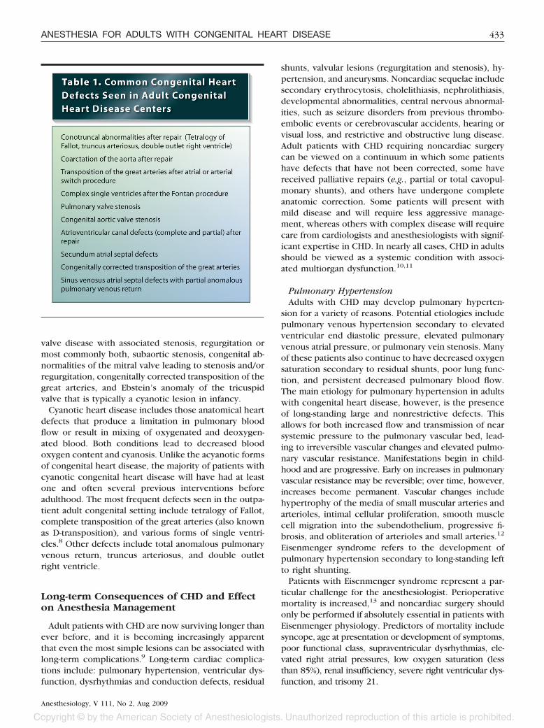
About 25% of adults with CHD have a mild form of thedisease that has allowed them to survive into adulthoodwithout surgical or interventional cardiac catheteriza-tion. The most common lesions in this category includemild aortic valve stenosis (usually in setting of bicuspidaortic valve), small restrictive ventricular septal defects,atrial septal defects, mild pulmonary valve stenosis, mi-tral valve prolapse, and isolated congenitally correctedtransposition of the great arteries (table 1). The vastmajority of adults with CHD seen in the outpatient set-ting, however, are patients who have had previous sur-gical or catheter-based intervention (table 1).
CHD lesions can be functionally classified into thosethat produce left to right shunts (acyanotic) and thosethat produce cyanosis (right to left shunting). A left toright shunt exists when oxygenated blood from the leftatrium, left ventricle, or aorta transits to the right atrium,right ventricle, or the pulmonary artery. Thus, the lungsreceive all the deoxygenated blood from the systemicvenous return (effective pulmonary blood flow; amountof deoxygenated blood that is carried to lungs to beoxygenated) plus the volume of fully oxygenated bloodthat is shunted through the defect. This results in volumeoverload of one or more cardiovascular chambers orstructures depending on the location of the defect. If thedefect is large and nonrestrictive, there is both increasedflow and transmission of near systemic pressure to thepulmonary vascular bed. Over time, this can lead toirreversible changes in the pulmonary vascular bed, lead-ing to increased pulmonary vascular resistance and asso-ciated pulmonary artery hypertension. If the pulmonaryartery pressure is at systemic levels, there may be re-versed (right to left) or bidirectional shunting at the levelof the defect (Eisenmenger syndrome).
Atrial and ventricular septal defects are among themost common congenital abnormalities, constituting25% of all adult CHD.7 Other lesions also considered inthe acyanotic group that are commonly seen in busyadult congenital outpatient clinics include but are notlimited to coarctation of the aorta, congenital aortic
* Assistant Professor of Anesthesiology, Departments of Anesthesiology andIntensive Care, Hospices Civils de Lyon, Louis Pradel Hospital, Claude Ber-nard Lyon 1 University. † Assistant Professor of Pediatrics, Division of Cardi-ology, Children’s Hospital of Wisconsin Herma Heart Center. ‡ Professor andVice Chair of Anesthesiology, Departments of Anesthesiology and Pharmacologyand Toxicology, Medical College of Wisconsin.
Received from the Departments of Anesthesiology and Intensive Care,Hospices Civils de Lyon, Louis Pradel Hospital, Claude Bernard Lyon 1University, ERI 22, Lyon, France, the Division of Cardiology, Children’sHospital of Wisconsin Herma Heart Center, Milwaukee, Wisconsin, and theDepartments of Anesthesiology and Pharmacology and Toxicology, MedicalCollege of Wisconsin, Milwaukee, Wisconsin. Submitted for publication Oc-tober 14, 2008. Accepted for publication April 24, 2009. Support was pro-vided solely from institutional and/or departmental sources. The figures andtables in this article were prepared by Dimitri Karetnikov, 7 Tennyson Drive,Plainsboro, New Jersey 08536.
Address correspondence to Dr. Cannesson: Service d’Anesthesie Reani-mation, Hopital Cardiologique Louis Pradel, 200 avenue du Doyen Lepine,69500 Bron, France. [email protected]. This article may beaccessed for personal use at no charge through the Journal Web site, www.anesthesiology.org.
Anesthesiology, V 111, No 2, Aug 2009 432
valve disease with associated stenosis, regurgitation ormost commonly both, subaortic stenosis, congenital ab-normalities of the mitral valve leading to stenosis and/orregurgitation, congenitally corrected transposition of thegreat arteries, and Ebstein’s anomaly of the tricuspidvalve that is typically a cyanotic lesion in infancy.
Cyanotic heart disease includes those anatomical heartdefects that produce a limitation in pulmonary bloodflow or result in mixing of oxygenated and deoxygen-ated blood. Both conditions lead to decreased bloodoxygen content and cyanosis. Unlike the acyanotic formsof congenital heart disease, the majority of patients withcyanotic congenital heart disease will have had at leastone and often several previous interventions beforeadulthood. The most frequent defects seen in the outpa-tient adult congenital setting include tetralogy of Fallot,complete transposition of the great arteries (also knownas D-transposition), and various forms of single ventri-cles.8 Other defects include total anomalous pulmonaryvenous return, truncus arteriosus, and double outletright ventricle.
Long-term Consequences of CHD and Effecton Anesthesia Management
Adult patients with CHD are now surviving longer thanever before, and it is becoming increasingly apparentthat even the most simple lesions can be associated withlong-term complications.9 Long-term cardiac complica-tions include: pulmonary hypertension, ventricular dys-function, dysrhythmias and conduction defects, residual
shunts, valvular lesions (regurgitation and stenosis), hy-pertension, and aneurysms. Noncardiac sequelae includesecondary erythrocytosis, cholelithiasis, nephrolithiasis,developmental abnormalities, central nervous abnormal-ities, such as seizure disorders from previous thrombo-embolic events or cerebrovascular accidents, hearing orvisual loss, and restrictive and obstructive lung disease.Adult patients with CHD requiring noncardiac surgerycan be viewed on a continuum in which some patientshave defects that have not been corrected, some havereceived palliative repairs (e.g., partial or total cavopul-monary shunts), and others have undergone completeanatomic correction. Some patients will present withmild disease and will require less aggressive manage-ment, whereas others with complex disease will requirecare from cardiologists and anesthesiologists with signif-icant expertise in CHD. In nearly all cases, CHD in adultsshould be viewed as a systemic condition with associ-ated multiorgan dysfunction.10,11
Pulmonary HypertensionAdults with CHD may develop pulmonary hyperten-
sion for a variety of reasons. Potential etiologies includepulmonary venous hypertension secondary to elevatedventricular end diastolic pressure, elevated pulmonaryvenous atrial pressure, or pulmonary vein stenosis. Manyof these patients also continue to have decreased oxygensaturation secondary to residual shunts, poor lung func-tion, and persistent decreased pulmonary blood flow.The main etiology for pulmonary hypertension in adultswith congenital heart disease, however, is the presenceof long-standing large and nonrestrictive defects. Thisallows for both increased flow and transmission of nearsystemic pressure to the pulmonary vascular bed, lead-ing to irreversible vascular changes and elevated pulmo-nary vascular resistance. Manifestations begin in child-hood and are progressive. Early on increases in pulmonaryvascular resistance may be reversible; over time, however,increases become permanent. Vascular changes includehypertrophy of the media of small muscular arteries andarterioles, intimal cellular proliferation, smooth musclecell migration into the subendothelium, progressive fi-brosis, and obliteration of arterioles and small arteries.12
Eisenmenger syndrome refers to the development ofpulmonary hypertension secondary to long-standing leftto right shunting.
Patients with Eisenmenger syndrome represent a par-ticular challenge for the anesthesiologist. Perioperativemortality is increased,13 and noncardiac surgery shouldonly be performed if absolutely essential in patients withEisenmenger physiology. Predictors of mortality includesyncope, age at presentation or development of symptoms,poor functional class, supraventricular dysrhythmias, ele-vated right atrial pressures, low oxygen saturation (lessthan 85%), renal insufficiency, severe right ventricular dys-function, and trisomy 21.
433ANESTHESIA FOR ADULTS WITH CONGENITAL HEART DISEASE
Anesthesiology, V 111, No 2, Aug 2009

A primary goal of anesthetic management in patientswith pulmonary hypertension is to minimize increases inpulmonary vascular resistance and to maintain systemicvascular resistance (fig. 1). Abrupt increases in pulmo-nary vascular resistance may precipitate either acuteright ventricular failure and decreased cardiac output inpatients without intracardiac shunting or oxygen desatu-ration followed by decreased cardiac output in patientswith intracardiac shunting. In both cases, severe brady-cardia may occur with progression to cardiac arrest. Pre-vention and treatment of pulmonary hypertensive crisisincludes hyperventilation (with 1.0 fractional inspired ox-ygen concentration), correction of acidosis, avoidance ofsympathetic nervous system stimulation, maintenance ofnormothermia, minimization of intrathoracic pressure,and use of inotropic support. Inhaled nitric oxide maybe useful to treat sudden increases in pulmonary vascu-lar resistance, and this drug should probably be availablein the operating room for use in high-risk patients. Re-gional anesthesia may be an acceptable alternative togeneral anesthesia for patients undergoing peripheralprocedures. However, spinal or epidural anesthesia mayproduce unacceptable decreases in systemic vascular resis-tance in patients with unrestrictive intracardiac shunts, andthis action could exacerbate right to left shunting. Con-versely, general anesthesia allows for optimal control of
ventilation and may be preferable in patients undergoinghigh-risk surgery.
Bleeding and Thrombosis RiskMost patients with Eisenmenger syndrome will be se-
verely cyanotic, defined as an oxygen saturation of nomore than 85%. Cyanotic patients are at high risk forboth perioperative bleeding and thrombosis, even dur-ing minor surgery. Both are secondary to abnormalitiesin platelet number and function as well as complexabnormalities in the coagulation system. Thrombocyto-penia is felt to be secondary to shortened platelet sur-vival due to peripheral consumption. The abnormalitiesin the coagulation pathways are less understood. Pa-tients with severe cyanosis have low levels of circulatingvitamin K–dependent clotting factors, factor V, and vonWillebrand factor. This leads to an elevated InternationalNormalized Ratio and a prolonged activated partial throm-boplastin time. These patients, however, do not have anelevated bleeding time, likely secondary to increased bloodviscosity and decreased flow. Further contributing to thebleeding risk is the presence of arteriolar dilation andincreased tissue vascularity. This is felt to be secondaryto increased release of endothelium-derived nitric oxideand prostaglandins in response to increased wall sheerstress in the setting of increased blood viscosity.
Fig. 1. Factors that influence the distribution of blood flow between the systemic and pulmonary circulations are shown.
434 CANNESSON ET AL.
Anesthesiology, V 111, No 2, Aug 2009

Although patients with cyanosis have an increased riskfor bleeding, this is not protective against thrombosis. Inthe setting of cyanosis, these patients develop secondaryerythrocystosis. Secondary erythrocytosis develops as acompensatory response to chronic hypoxia and resultsfrom overproduction of erythropoietin. This results inincreased whole blood viscosity resulting from increasedred blood cell mass and decreased plasma volume. Theend result is decreased flow in the small arterioles andcapillaries. This is further exacerbated in the setting ofiron deficiency and dehydration. Iron-deficient red bloodcells are less deformable and have been found to be oneof the strongest independent predictors of thrombosis inthe setting of Eisenmenger syndrome.14
In the perioperative setting, preoperative fasting mightexacerbate symptoms of hyperviscosity and increase therisk of cerebrovascular thrombosis. Thus, adequate hy-dration with intravenous fluids must be maintained, par-ticularly in fasting patients. Although euvolemic phlebot-omy is no longer a routine practice, preoperative phlebotomyto improve surgical hemostasis may be useful when hemato-crit levels exceed 65%.8
Careful preoperative assessment of the coagulationsystem is essential, and replacement of coagulation fac-tors and platelets should be considered in patients un-dergoing moderate or major surgery. In addition, irondeficiency should be corrected preoperatively if the pro-cedure is not urgent. It is important to note, however,that in the setting of secondary erythrocytosis with in-creased hemogloblin and decreased plasma volume,standard techniques to measure International Normal-ized Ratio and activated partial thromboplastin time maybe unreliable. Because of these changes, the concentra-tion of citrate in the sampling tube must also be adjusted.In most centers, the anticoagulant in the tube can beadjusted by the following formula: anticoagulant in sam-pling tube (3.8% citrate) in ml � {(100–hematocrit)/100} � 0.02 for a draw of 10 ml of whole blood.
Heart FailureRight-sided and left-sided heart failure are common
complications of both corrected and uncorrected CHD.Increases in atrial natriuretic peptide, renin, aldosterone,and norepinephrine have been observed in adults withCHD many years after surgical correction, even in asymp-tomatic patients. Abnormal cardiac autonomic nervous sys-tem regulation and altered hemodynamics contribute tothe development of heart failure in these patients. Man-agement of left ventricular failure with diuretics, digoxin, an-giotensin-converting enzyme inhibitors, and �-blockers is sim-ilar to other forms of acquired heart failure, and treatmentshould be optimized in the perioperative period.5 Incontrast to left ventricular failure, there are no evidence-based guidelines for the management of heart failure inpatients with a systemic right ventricle (congenitallycorrected transposition of the great arteries, Mustard, or
Senning repairs of transposition of the great arteries, andsingle ventricles).15 Further clinical trials are warranted.
DysrhythmiasAtrial and ventricular dysrhythmias are common in
adults with CHD. Dysrhythmias arise in patients whohave undergone previous curative or palliative surgery asa primary consequence of the underlying congenitaldefect or secondary to surgical repair.16,17 For example,supraventricular dysrhythmias occur in 20–45% of patientswith previous atrial surgery (late atrial septal defect clo-sure,18 Mustard, Senning, or Fontan procedures) or in thosewith atrial distension. The most common form of tachy-arrhythmia observed is intraatrial reentrant tachycardiaoriginating from the right atrium. Atrial tachyarrhyth-mias are often resistant to pharmacological treatmentand can result in rapid hemodynamic deterioration. Ven-tricular dysrhythmias are most frequently encounteredin patients who have significantly decreased right or leftventricular function. Other risk factors include previousventriculotomy, earlier surgical era, or older age at initialsurgery. Patients who were repaired late are exposed tolonger periods of cyanosis, volume overload, and pres-sure overload. As a result, they have increased myocar-dial fibrosis and associated slowing of conduction andan increased risk for dysrhythmias. Acute hypoxemia canprovoke ventricular dysrhythmias because subendocardialmyocardial perfusion is already impaired in hypertrophiedmyocardium. Some patients will require a permanent pa-cemaker to treat bradycardia secondary to postoperativeatrioventricular block. The management of patients withpacemakers and intracardiac defibrillators has been re-viewed elsewhere.19
Anesthetic Management
Preoperative EvaluationThe preoperative evaluation of patients with CHD un-
dergoing noncardiac surgery should use a multidisci-plinary approach that includes the participation of anes-thesiologists, cardiologists, intensivists, and surgeons.Guidelines of the task force for the organization of de-livery systems for adults with CHD recommend thatpatients with moderate or complex CHD should be man-aged in a regional adult congenital heart disease cen-ter.3,6 Perioperative risk is substantially increased inadults with CHD, particularly in those with poor func-tional class, pulmonary hypertension, congestive heartfailure, and cyanosis.20 Major surgery and proceduresthat involve one lung ventilation or changes in position(e.g., prone, Trendelenburg) could produce importanthemodynamic effects that contribute to increasing risk.Therefore, the anesthesiologist should be familiar withthe patient’s specific anatomy and physiology as deter-mined from echocardiographic and cardiac catheteriza-
435ANESTHESIA FOR ADULTS WITH CONGENITAL HEART DISEASE
Anesthesiology, V 111, No 2, Aug 2009

tion results. This knowledge is useful to anticipate intra-operative events that may precipitate acute changes inthe magnitude or direction of intracardiac shunts ormodulate flow through systemic to pulmonary shunts. Ifrecent examination results are not available, a preoper-ative echocardiogram may be indicated.20
PremedicationMany adults with CHD undergoing noncardiac surgery
have undergone previous cardiac surgery and are famil-iar with anesthesia. Some may present with anxiety,physical limitations, and associated anomalies or syn-dromes (the most common is trisomy 21). Adults withCHD are more likely to be living with their parents andto develop a variety of psychosocial issues.20 Conse-quently, psychological preparation of patients for sur-gery is important. Premedication with anxiolytics andhypnotics must be undertaken very cautiously becausehypoventilation and hypercapnia may produce deleteri-ous increases in pulmonary vascular resistance, particu-larly, in patients with underlying pulmonary hyperten-sion or systemic to pulmonary shunts. However, patientswith chronic hypoxemia retain a normal ventilatory re-sponse to hypercarbia as well as to opioid analgesics.
Endocarditis ProphylaxisThe American Heart Association has recently pub-
lished updated guidelines for the prevention of infectiveendocarditis.21 After reviewing the literature over thelast 40 yr, an expert panel found that very few cases ofendocarditis could have been prevented with antibioticprophylaxis. As result, only patients with cardiac condi-tions associated with the highest risk for adverse out-comes should continue following antibiotic prophylaxisbefore surgery: patients with previous endocarditis, un-repaired cyanotic CHD, including palliative shunts andconduits; completely repaired congenital heart defectswith prosthetic material or device, whether placed bysurgery or by catheter intervention, during the first 6months after the procedure; repaired CHD with residualdefects at the site or adjacent to the site of a prostheticpatch or prosthetic device (which inhibit endothelializa-tion). Except for the conditions listed above, antibioticprophylaxis is no longer recommended for other formsof CHD.
Intraoperative Management
Adults with CHD who have undergone complete ana-tomic repair and have no evidence of late functionaldeterioration can be managed by using conventionalapproaches. In contrast, patients with more complexCHD and moderate to major surgery will require specificintraoperative management.
MonitoringIn addition to direct examination of the patient, stan-
dard conventional noninvasive monitoring includingpulse oximetry, electrocardiogram, arterial blood pres-sure, capnography and temperature are used in all pa-tients. Pulse oximetry is perhaps uniquely important inthe management of CHD. For example, decreases inarterial saturation can signify increases in pulmonaryvascular resistance, increases in right to left shunting, ordecreases in pulmonary blood flow through systemic topulmonary shunts. In contrast, increases in left to rightshunting may not be detected by pulse oximetry andarterial oxygen saturation may be maintained even ifsystemic cardiac output is severely compromised. Thecapnogram is altered, and end tidal carbon dioxide con-centrations underestimate PaCO2 in the case of right toleft shunting.
Knowledge of the anatomy and physiology of specificpalliative repairs is important for choosing appropriatemonitoring. For example, congenital defects that areassociated with inadequate pulmonary blood flow (e.g.,pulmonary valve atresia or univentricular heart) are pal-liated with systemic to pulmonary shunts. In patientswith a classic Blalock-Taussig shunt (end to side anasto-mosis of the subclavian and pulmonary arteries) arterialpressure and SpO2 cannot be measured on the ipsilateralside. A Glenn shunt or bidirectional cavopulmonary anas-tomosis consists of an end to side anastomosis of the di-vided superior vena cava to pulmonary artery. Total cavo-pulmonary connection (Fontan circulation) is establishedwhen the pulmonary and systemic circulations are totallyseparated by diverting all the systemic venous return tothe pulmonary artery, usually without interposition of asubpulmonic ventricle. These alterations in intracardiacanatomy complicate the placement of central venouscatheters in palliated adults, and the anatomical varia-tions must be considered when interpreting values ob-tained from central venous monitoring. For example, inpatients with a Fontan circulation, central venous pres-sure reflects mean pulmonary artery pressure. In pa-tients with an intraatrial baffle (e.g., Mustard or Senningprocedure), pulmonary artery catheter placement maybe difficult or impossible. Vascular access may also bedifficult because many of these patients have alreadyundergone previous vessel catheterization. Invasive arte-rial pressure monitoring can be essential in managingpatients with Eisenmenger syndrome, intracardiac orsystemic to pulmonary shunts undergoing major surgerywho are also sensitive to sudden changes in preload, andsystemic and pulmonary vascular resistance. Finally,transesophageal echocardiography might be useful inadults with CHD undergoing noncardiac surgery to mon-itor intravascular volume status and ventricular function.In the presence of complex CHD, transesophageal echo-cardiography should be performed by an individual fa-miliar with CHD.
436 CANNESSON ET AL.
Anesthesiology, V 111, No 2, Aug 2009

Anesthetic TechniqueThere are no evidence-based recommendations to
guide the anesthetic management of patients with CHDundergoing noncardiac surgery. Given the large scope ofabnormalities encompassed by CHD,22,23 it is also impos-sible to propose a single approach for anesthetic man-agement that would address every possible defect. How-ever, a major objective of intraoperative management isto promote tissue oxygen delivery by preventing arterialdesaturation, maintaining a balance between pulmonaryand systemic flows, and by optimizing hematocrit. Man-
agement strategies for specific defects are outlined intable 2.
Anesthetic AgentsThere are few studies that have evaluated the hemo-
dynamic effects of anesthetic agents in adult patientswith CHD. Most intravenous agents depress myocardialcontractility and decrease systemic vascular resistance,and these actions could have an adverse effect on tissueoxygen delivery during induction of anesthesia. Alterna-tively, some evidence suggests that etomidate may pro-
Anatomy - Physiology Potential Issues
Specific Anesthetic Management
Atri
al se
ptal
defe
ctVe
ntri
cula
r sep
tal
defe
ctCo
arct
atio
nof
the
aort
aAo
rtic
sten
osis
L-(c
onge
nita
llyco
rrec
ted)
tran
spos
ition
of
the
grea
t art
erie
sL-R shunt Small to moderate size defects well tolerated
Atrial fibrillation (increased risk if repaired after age 40)Risk of paradoxical emboliLarge defects lead to arrhythmias, exercise intolerance, and rarely PHT (occurs in less than 5% of patients)
De-air intravenous lines
Anatomy - Physiology Potential Issues
Specific Anesthetic Management
L-R shuntMay be associated with other defects
Unrepaired:Large defect risk of PHT (50% by age 2)Small to moderate size defects, risk for endocarditis,sub-pulmonic obstruction, subaortic obstruction, and aortic regurgitationRight ventricular failure
Repaired:Complete heart block in some patients (rare)Persistent PHTDysrhythmia
Manage L-R shuntMaintain pulmonary blood flow if R-L shunt presentIncreased risk of postopera-tive pulmonary infection
Manage pacemaker
LV pressure overload and hypertrophyAortic branch collateralsAssociated with bicuspid aortic valve (50-80%)Endothelial dysfunction (diffuse aortopathy)
Blood pressure gradient between upper and lower limbsRisk of bleeding if thoracic surgeryLV hypertrophy and LV diastolic dysfunctionSystemic hypertensionAneurysms of ascending aorta and descending aortaPremature coronary artery diseaseIntracranial aneurysms (10%)
Inaccurate blood pressure (left arm) if previous subclavian angioplastyPostoperative hypertensionAvoid tachycardia, hypotension
LV pressure overload and hypertrophy
Unrepaired:Pulmonary edemaPHTMyocardial ischemiaSyncopePost stenotic dilatation
Repaired:Aortic regurgitationLV diastolic dysfunction
Avoid tachycardia, hypotensionAvoid factors that increase myocardial oxygen consumption
LV is the subpulmonic ventricleRV is the systemic ventricle
Unrepaired:Complete heart blockArrhythmias (atrial and ventricular)Anatomic right ventricular failureSystemic AV valve regurgitation
Repaired:Same
Pacemaker managementExternal pacing availableManage dysrhythmiasManage heart failure
Atri
al se
ptal
defe
ctVe
ntri
cula
r sep
tal
defe
ctCo
arct
atio
nof
the
aort
aAo
rtic
ste
nosi
s
L-(c
onge
nita
llyco
rrec
ted)
tran
spos
ition
of
the
grea
t art
erie
s
CongenitalHeart
Disease
Table 2. Proposed Management Strategies for Specific Defects
(continued)
437ANESTHESIA FOR ADULTS WITH CONGENITAL HEART DISEASE
Anesthesiology, V 111, No 2, Aug 2009

vide hemodynamic stability in the setting of CHD17 sim-ilarly to other settings of impaired cardiac function. Thepotential favorable effects of ketamine in this populationhave not been adequately investigated. Ketamine hasbeen suggested to increase pulmonary vascular resis-tance in adults without CHD.24 However, this agentproduces beneficial effects in children with CHD andsevere pulmonary hypertension undergoing sevofluraneanesthesia25 by maintaining systemic vascular resistanceand ventricular performance, without increasing pulmo-nary vascular resistance. As in the case of intravenous
anesthetic agents, the choice of a specific volatile anes-thetic agent to be used should be based on the patient’sphysiology and the overall goal of balancing pulmonaryand systemic blood flow.
Intracardiac and Systemic to Pulmonary ShuntsShunting has an important effect on anesthetic man-
agement. All intravenous lines must be meticulouslydeaired to decrease the risk of systemic air embolizationin patients with intracardiac shunts. The hemodynamiceffects of ventilation strategies, positioning, pharmaco-
Tetr
alog
y of
Fal
lot
Uni
vent
ricu
lar h
eart
D-t
rans
posi
tion
of th
e gr
eat a
rter
ies
Anatomy - Physiology Potential Issues
Specific Anesthetic Management
ASD = atrial septal defect; AV = atrioventricular; L-R = left to right; LV = left ventricle; PDA = patent ductus arteriosus; PHT = pulmonary hypertension; R-L = right to left; RV = right ventricle; VSD = ventricular septal defect.
Pulmonic stenosis (valvular, subvalvular, and/or supravalvular)
RV hypertrophyOverriding aortic rootVSDCyanosis
Unrepaired: Rare, mean age of death 25 yr of ageR-L shuntCyanosisPalliated: Blalock-Taussig shunt Chronic left ventricular volume overload Cyanosis if shunt is too small Pulmonary hypertension
Repaired:Sinus and AV node dysfunctionDysrhythmia: atrial and ventricularAscending aortic aneurysmResidual pulmonary regurgitation or stenosisResidual VSDLeft ventricular dysfunctionPersistent pulmonary hypertension from previousshuntsRV failure from chronic pulmonary insufficiency
Avoid tachycardia, hypovolemia, increased contractility
Maintain pulmonary blood flowMaintain sytemic blood pressure
Detect and manage dysrhythmiasManage pacemakerExternal pacing available
Anatomy - Physiology Potential Issues
Specific Anesthetic Management
Pulmonary artery arises from LVAorta arises from RVPossible associated lesions: VSD, ASD, PDA,
Pulmonary stenosis, coarctation of the aorta
Abnormal coronary artery anatomy
Unrepaired: Associated with VSD or ASD or PDARepaired: Senning or MustardAtrial dysrhythmiaSinus node dysfunction (by age 40, 50% have pacemaker)Systemic ventricle dysfunctionResidual atrial or ventricular level shuntsRepaired: Arterial switchMyocardial ischemia (narrowed, occluded coronary arteries, endothelial dysfunction)Ascending aortic aneurysm
Maintain pulmonary blood flowManage dysrhythmiaManage heart failure
Detailed preoperative evaluation, both functional and coronary imaging study
Double inlet atrioventricular connectionsAbsence of one atrioventricular connectionSingle well-developed ventricle
Unrepaired: Rare Dysrhythmias Congestive heart failure Bidirectional shunting Cyanosis PHTRepaired: Blalock-Taussig shunt, Glenn shunt, or Fontan Dysrhythmias Heart failure Hepatic dysfunction ThromboemboliRestrictive lung disease
Manage dysrhythmiasMaintain pulmonary blood flow
Manage dysrhythmiasMaintain low pulmonary vascular resistanceMaintain adequate preloadReplace coagulation factors
Tetr
alog
y of
Fal
lot
Uni
vent
ricu
lar h
eart
D-t
rans
posi
tion
of th
e gr
eat a
rter
ies
CongenitalHeart
Disease
Table 2. continued
438 CANNESSON ET AL.
Anesthesiology, V 111, No 2, Aug 2009

logical agents, and blood loss must all be considered toappropriately balance pulmonary and systemic bloodflow in patients with intracardiac or systemic to pulmo-nary shunts (figs. 1 and 2). Ventilation with high airwaypressures can compromise venous return, increase pul-monary vascular resistance, and exacerbate right to leftshunting in patients with cyanotic heart disease. Inade-quate anesthesia and sympathetic nervous system stim-ulation might increase systemic vascular resistance, ex-acerbate left to right shunting, and reduce systemiccardiac output in a patient with a large atrial septaldefect. Trendelenburg position can increase central ve-nous (superior vena cava) pressure and cause cerebral
hypoperfusion in a patient with a Glenn shunt or Fontan.Systemic hypotension can also result in a decrease inpulmonary blood flow, with subsequent arterial desatu-ration, in a patient with a systemic to pulmonary artery
shunt (Blalock-Taussig shunt, or central shunt). Theseare a few examples of the complex physiology that mustbe considered in managing adults with CHD for noncar-diac surgery.
Univentricular HeartSingle ventricle anatomy and physiology is probably,
along with Eisenmenger syndrome, the most challengingCHD for the anesthesiologist to manage. The Fontanprocedure was first described in 1971 and was appliedinitially to patients with tricuspid atresia. Today, indica-tions for this procedure have been extended to all formsof single ventricles. Since its initial description, at least10 different variations have been performed. In all forms,this operation bypasses the right ventricle, leaving pas-sive, nonpulsatile flow from both inferior and superiorvena cava to the pulmonary artery.26 Any factor thatincreases pulmonary vascular resistance will decreasepulmonary blood flow and cause arterial desaturation.Patients with a Fontan circulation frequently presentwith cardiac and noncardiac complications that includesupraventricular dysrhythmias, restrictive lung disease,thromboembolic complications,27 and hepatic dysfunc-tion. Both procoagulant and anticoagulant effects28 areobserved in patients with a Fontan as a result of liverdysfunction and/or factor loss in patients with protein-losing enteropathy, and these abnormalities substantially
Fig. 2. Schematic representations of nor-mal heart, Blalock Taussig shunt, andFontan circulation. SVC � superior venacava; IVC � inferior vena cava; RA � rightatrium; RV � right ventricle; PA � pulmo-nary artery; LPA � left pulmonary artery;RPA � right pulmonary artery; LV � leftventricle; LA � left atrium; Ao � aorta.
439ANESTHESIA FOR ADULTS WITH CONGENITAL HEART DISEASE
Anesthesiology, V 111, No 2, Aug 2009

increase the risk for intraoperative bleeding. Patientswith a Fontan circulation should maintain an arterialsaturation above 90 to 95%. Arterial saturation below90% in these patients should be considered abnormaland should provoke further evaluation for the pres-ence of venovenous collaterals, arteriovenous malfor-mations, or a residual shunt.
Postoperative Management
Patients presenting with severe CHD and/or high risksurgery should be managed if possible in a postoperativeintensive care unit experienced with caring for adultswith congenital heart disease. The major risks during thepostoperative period are similar to the risks describedabove. These risks include bleeding, dysrhythmias, andthromboembolic events. In cases of pulmonary hyper-tension, oral pulmonary vasodilatators such as sildenafiland inhaled nitric oxide may be beneficial.
Conclusions
The number of adult patients with CHD is rapidlyincreasing, and these patients will be presenting withgreater frequency for noncardiac surgery. The cardiovas-cular anatomy and physiology of CHD are complex andrequire specific knowledge of the defect and its anes-thetic implications. Adults with moderate or severe CHDrequiring noncardiac surgery are at high risk, particularlythose with poor functional class, pulmonary hyperten-sion, congestive heart failure, and cyanosis; and thesepatients should receive care in a regional adult CHDcenter with multidisciplinary collaboration among anes-thesiologists, cardiologists, surgeons, and intensivists.There are no evidence-based guidelines for the periop-erative management of adults with CHD. Large-scale clin-ical trials are required to elucidate the optimal anestheticmanagement of these challenging patients.
References
1. Perloff JK, Warnes CA: Challenges posed by adults with repaired congenitalheart disease. Circulation 2001; 103:2637–43
2. Mitchell SC, Korones SB, Berendes HW: Congenital heart disease in 56,109births. Incidence and natural history. Circulation 1971; 43:323–32
3. Warnes CA, Liberthson R, Danielson GK, Dore A, Harris L, Hoffman JI,Somerville J, Williams RG, Webb GD: Task force 1: The changing profile ofcongenital heart disease in adult life. J Am Coll Cardiol 2001; 37:1170–5
4. Deanfield J, Thaulow E, Warnes C, Webb G, Kolbel F, Hoffman A, Sorenson K,Kaemmer H, Thilen U, Bink-Boelkens M, Iserin L, Daliento L, Silove E, Redington A,Vouhe P, Priori S, Alonso MA, Blanc JJ, Budaj A, Cowie M, Deckers J, FernandezBurgos E, Lekakis J, Lindahl B, Mazzotta G, Morais J, Oto A, Smiseth O, Trappe HJ,Klein W, Blomstrom-Lundqvist C, de Backer G, Hradec J, Parkhomenko A, PresbiteroP, Torbicki A: Management of grown up congenital heart disease. Eur Heart J 2003;24:1035–84
5. Fleisher LA, Beckman JA, Brown KA, Calkins H, Chaikof E, Fleischmann KE,Freeman WK, Froehlich JB, Kasper EK, Kersten JR, Riegel B, Robb JF, Smith SCJr, Jacobs AK, Adams CD, Anderson JL, Antman EM, Buller CE, Creager MA,Ettinger SM, Faxon DP, Fuster V, Halperin JL, Hiratzka LF, Hunt SA, Lytle BW,Nishimura R, Ornato JP, Page RL, Tarkington LG, Yancy CW: ACC/AHA 2007guidelines on perioperative cardiovascular evaluation and care for noncardiac
surgery: A report of the American College of Cardiology/American Heart Asso-ciation Task Force on Practice Guidelines (Writing Committee to Revise the 2002Guidelines on Perioperative Cardiovascular Evaluation for Noncardiac Surgery):Developed in collaboration with the American Society of Echocardiography,American Society of Nuclear Cardiology, Heart Rhythm Society, Society of Car-diovascular Anesthesiologists, Society for Cardiovascular Angiography and Inter-ventions, Society for Vascular Medicine and Biology, and Society for VascularSurgery. Circulation 2007; 116:e418–99
6. Landzberg MJ, Murphy DJ Jr, Davidson WR Jr, Jarcho JA, Krumholz HM,Mayer JE Jr, Mee RB, Sahn DJ, Van Hare GF, Webb GD, Williams RG: Task force4: Organization of delivery systems for adults with congenital heart disease. J AmColl Cardiol 2001; 37:1187–93
7. Marelli AJ, Mackie AS, Ionescu-Ittu R, Rahme E, Pilote L: Congenital heartdisease in the general population: Changing prevalence and age distribution.Circulation 2007; 115:163–72
8. Khairy P, Poirier N, Mercier LA: Univentricular heart. Circulation 2007;115:800–12
9. Earing MG, Connolly HM, Dearani JA, Ammash NM, Grogan M, Warnes CA:Long-term follow-up of patients after surgical treatment for isolated pulmonaryvalve stenosis. Mayo Clin Proc 2005; 80:871–6
10. Khairy P, Landzberg MJ: Adult congenital heart disease: Toward prospec-tive risk assessment of a multisystemic condition. Circulation 2008; 117:2311–2
11. Dimopoulos K, Diller GP, Koltsida E, Pijuan-Domenech A, PapadopoulouSA, Babu-Narayan SV, Salukhe TV, Piepoli MF, Poole-Wilson PA, Best N, FrancisDP, Gatzoulis MA: Prevalence, predictors, and prognostic value of renal dysfunc-tion in adults With congenital heart disease. Circulation 2008; 117:2320–8
12. Heath D, Edwards JE: The pathology of hypertensive pulmonary vasculardisease: A description of six grades of structural changes in the pulmonaryarteries with special reference to congenital cardiac septal defects. Circulation18:533–47
13. Ammash NM, Connolly HM, Abel MD, Warnes CA: Noncardiac surgery inEisenmenger syndrome. J Am Coll Cardiol 1999; 33:222–7
14. Ammash N, Warnes CA: Cerebrovascular events in adult patients withcyanotic congenital heart disease. J Am Coll Cardiol 1996; 28:768–72
15. Shaddy RE, Webb G: Applying heart failure guidelines to adult congenitalheart disease patients. Expert Rev Cardiovasc Ther 2008; 6:165–74
16. Triedman JK: Arrhythmias in adults with congenital heart disease. Heart2002; 87:383–9
17. Andropoulos DB, Stayer SA, Skjonsby BS, East DL, McKenzie ED, FraserCD: Anesthetic and perioperative outcome of teenagers and adults with congen-ital heart disease. J Cardiothorac Vasc Anesth 2002; 2002:731–6
18. Gatzoulis MA, Freeman MA, Siu SC, Webb GD, Harris L: Atrial arrhythmiaafter surgical closure of atrial septal defects in adults. N Engl J Med 1999;340:839–46
19. Practice advisory for the perioperative management of patients with car-diac rhythm management devices: Pacemakers and implantable cardioverter-defibrillators: A report by the American Society of Anesthesiologists Task Forceon Perioperative Management of Patients with Cardiac Rhythm ManagementDevices. ANESTHESIOLOGY 2005; 103:186–98
20. Foster E, Graham TP, Driscoll DJ, Reid GJ, Reiss JG, Russell IA, Sermer M,Siu SC, Uzark K, Williams RG, Webb GD: Task force 2: Special health care needsof adults with congenital heart disease. J Am Coll Cardiol 2001; 37:1176–83
21. Wilson W, Taubert KA, Gewitz M, Lockhart PB, Baddour LM, Levison M,Bolger A, Cabell CH, Takahashi M, Baltimore RS, Newburger JW, Strom BL, TaniLY, Gerber M, Bonow RO, Pallasch T, Shulman ST, Rowley AH, Burns JC, FerrieriP, Gardner T, Goff D, Durack DT: Prevention of infective endocarditis: Guidelinesfrom the American Heart Association: A guideline from the American HeartAssociation Rheumatic Fever, Endocarditis, and Kawasaki Disease Committee,Council on Cardiovascular Disease in the Young, and the Council on ClinicalCardiology, Council on Cardiovascular Surgery and Anesthesia, and the Quality ofCare and Outcomes Research Interdisciplinary Working Group. Circulation 2007;116:1736–54
22. Brickner ME, Hillis LD, Lange RA: Congenital heart disease in adults.Second of two parts. N Engl J Med 2000; 342:334–42
23. Brickner ME, Hillis LD, Lange RA: Congenital heart disease in adults. Firstof two parts. N Engl J Med 2000; 342:256–63
24. Tweed WA, Minuck M, Myrnin D: Circulatory responses to ketamineanesthesia. ANESTHESIOLOGY 1972; 37:613–9
25. Williams GD, Philip BM, Chu LF, Boltz MG, Kamra K, Terwey H, HammerGB, Perry SB, Feinstein JA, Ramamoorthy C: Ketamine does not increase pulmo-nary vascular resistance in children with pulmonary hypertension undergoingsevoflurane anesthesia and spontaneous ventilation. Anesth Analg 2007; 105:1578–84
26. Hosking MP, Beynen FM: The modified Fontan procedure: Physiology andanesthetic implications. J Cardiothorac Vasc Anesth 1992; 6:465–75
27. Balaji S, Gewillig M, Bull C, de Leval MR, Deanfield JE: Arrhythmias afterthe Fontan procedure. Comparison of total cavopulmonary connection andatriopulmonary connection. Circulation 1991; 84:III162–7
28. van Nieuwenhuizen RC, Peters M, Lubbers LJ, Trip MD, Tijssen JG, MulderBJ: Abnormalities in liver function and coagulation profile following the Fontanprocedure. Heart 1999; 82:40–6
440 CANNESSON ET AL.
Anesthesiology, V 111, No 2, Aug 2009