an env-likeprotein encoded . by a drosophila that...
TRANSCRIPT

An Env- l ikeprote in encoded . by a Drosophila retroelement: evidence that gypsy is an infect ious retrovirus
Sun U. Song, a Tatiana Gerasimova, ~ Maryellen Kurkulos, 1'2 Jef D. Boeke, 2'3 and Victor G. Corces 1'3
~Department of Biology, The Johns Hopkins University, Baltimore, Maryland 21218 USA; 2Department of Molecular Biology and Genetics, Johns Hopkins University School of Medicine, Baltimore, Maryland 21205 USA
The gypsy element of Drosophila differs from most LTR retrotransposons in containing a third open reading frame that resembles retroviral env genes. The protein encoded by ORF3 is glycosylated and processed, like all retroviral envelope proteins. The protein is expressed at high levels in fly strains in which gypsy elements are active. In these strains the protein is found primarily in viral particles. When larvae of fly strains in which gypsy is normally inactive are exposed to sucrose gradient fractions containing these particles, a high level of gypsy insertion activity is observed in their progeny. Thus, gypsy has the expected properties of an insect retrovirus.
[Key Words: Drosophila; mutagenesis; transposable element; insect retrovirus]
Received May 6, 1994; revised version accepted July 11, 1994.
The gypsy element of Drosophila melanogaster has been classified traditionally as a long terminal repeat (LTR) retrotransposon; however, it is one of a small group of LTR retrotransposons from insects that are unusual in that they contain three open reading frames (ORFs). In these retroelements, the first two ORFs correspond to retroviral gag and poi, whereas ORF3 is of unknown function but corresponds in size and genomic location to retroviral env (Fig. 1). All elements with three ORFs de- scribed so far are from insects, either Drosophila or the lepidopteran Trichoplusia ni. Recent results have shown that in two elements encoding an ORF3, gypsy and tom, a subgenomic mRNA similar in structure to retroviral env mRNAs can be found (P61isson et al. 1994; Tanda et al. 1994). In the case of gypsy, this transcript is observed only in certain strains in which gypsy transpositional activity is high (P41isson et al. 1994). The primary se- quences of the encoded ORF3 proteins of these elements are quite variable and show no obvious similarity to ret- roviral Env proteins. However, retroviral Env proteins are themselves very variable in primary sequence. Like retroviral Env proteins, the proteins encoded by "retro- transposon" ORF3s contain a putative transmembrane domain near their carboxyl terminus, multiple putative N-glycosylation sites, and putative protease cleavage sites (resembling the cleavage sites in a variety of retro- viral Env proteins) at conserved positions (Fig. 1 }. These features of gypsy and the other ORF3-containing insect retrotransposons have prompted the suggestion that these elements may represent endogenous insect retro-
3Corresponding authors.
viruses (Boeke 1988; Boeke and Corces 1989; Coffin 1993).
To address the question of whether gypsy represents a virus or a transposon more directly, we have turned to the use of Drosophila strains that show genetic instabil- ities associated with high-frequency insertion by gypsy elements. These strains are characterized by a large number of full-length gypsy elements in the euchroma- tin, appearance of gypsy insertion mutations at high fre- quency, and the presence of both large amounts of gypsy full-length RNA and spliced ORF3 mRNA (P61isson et al. 1994; N. Prud'homme, M. Gans, M. Masson, C. Ter- zian, and A. Bucheton, in prep.).
Mobilization of gypsy can be detected by a sensitive assay using the female-sterile mutation ovo D1. This dominant mutation has no effect on males but prevents development of the female germ line. gypsy insertion into the ovo DI allele results in its conversion into a re- cessive ovo allele (M6vel-Ninio et al. 1989). Thus, het- erozygous ovoD~/+ females are sterile, whereas females in which gypsy insertions into ovo D~ occurred during germ-line development are able to produce progeny. When OVO D1 males are crossed to females of gypsy-active backgrounds, a high frequency of ovo D1 reversion is ob- served; many of these reversion events are caused by gypsy or copia insertion (M6vel-Ninio et al. 1989). No such reversion is observed when ovo D~ males are crossed to females of the SS line, in which gypsy is inactive (Kim et al. 1994). The characteristic of high-frequency gypsy transposition is conferred by the f lamenco gene on the X chromosome; females of the y v f ma l flare strain must be homozygous for the X chromosome for high-fre-
2046 GENES & DEVELOPMENT 8:2046-2057 �9 1994 by Cold Spring Harbor Laboratory Press ISSN 0890-9369/94 $5.00
Cold Spring Harbor Laboratory Press on September 9, 2018 - Published by genesdev.cshlp.orgDownloaded from

Drosophila gypsy virus particles
might be responsible for the subsequent gypsy inser- tions. We find expression of gypsy ORF3 protein prod- ucts in the ovaries of these flies and show that these proteins have Env-like characteristics. Furthermore, we provide direct evidence for infectious gypsy virus parti- cles in these tissues.
Figure 1. ORF3 of certain insect retroelements encodes a pro- tein resembling retroviral env. Overall structure of gypsy DNA (Marlor et al. 1986) is indicated at top. ORF3s have been de- scribed from a handful of insect LTR retroelements; the con- served aspects of the structures of their predicted proteins are indicated to scale in the lower panel. (Forks) Putative N-linked glycosylation sites [only those amino-terminal to the proposed transmembrane (TM) domains are indicated}; (stippled boxes) TMs; lvertical lines with tetrapeptide motifs) putative dibasic cleavage sites. All elements are from D. melanogaster except as indicated. Note that all features are well-conserved between the D. melanogaster and D. virilis gypsy elements, despite their 20% amino acid sequence divergence (Mizrokhi and Mazo 1990). TM domains were predicted by Kyte-Doolittle hydropa- thy plots using DNA Strider 1.2 (Marck 1988).
quency gypsy insertion to occur in their progeny (N. Prud'homme, M. Gans, M. Masson, C. Terzian, and A. Bucheton, in prep.). Thus, there is a maternal effect of the f l am gene that determines gypsy mobilization. This suggests that a critical f lam-control led step must occur in the ovaries of the mother to determine whether gypsy insertion will occur in the subsequent generation. ORF3 protein is expressed in the ovarian follicle cells of these strains (P41isson et al. 1994), suggesting that virus assem- bly might be occurring in this tissue. Preliminary sup- port for the idea of an infectious gypsy virus came from the studies of Kim et al. (1994), who have shown that microinjection of crude egg plasm from active strains into inactive strain embryos (or feeding of pupal extracts to larvae) results in detection of subsequent gypsy inser- tions in the progeny of the recipients using the ovo ~ reversion assay. Because these experiments made use of extremely crude preparations, we have examined whether an infectious particle with the characteristics of a retrovirus is present in the ovaries of flare females and
R e s u l t s
Env-l ike proteins encoded by gypsy ORF3 are expressed in ovaries of flam f emales
gypsy encodes a subgenomic mRNA for ORF3 of 2.2 kb; the sequence of this mRNA predicts a protein of 54 kD (P41isson et al. 1994). The ORF3 protein contains a pu- tative signal peptidase cleavage site as well as a dibasic cleavage site resembling those in retroviral Env protein precursors. Cleavage at these sites would result in a sur- face protein of 32 kD and a trans-membrane protein of 20 kD (Fig. 1); these values do not take into account unpre- dictable variations that may result from glycosylation. To investigate the nature of gypsy ORF3 proteins, mono- clonal antibodies were raised against a t rpE-ORF3 fusion protein (see Materials and methods). Two monoclonal antibodies (mAb), 7B3 and 8E7, were isolated, which re- acted specifically with the ORF3 portion of the fusion protein. These antibodies were used to probe immuno- blots of SDS-polyacrylamide gels on which various ovary extracts or in vitro translation products were sep- arated. Both antibodies react specifically with the ORF3 portion of fusion proteins (Materials and methods), as well as an in vitro translation product expressed from a T7-ORF3 expression construct (Fig. 2C). The in vitro- translated protein migrates with an apparent molecular mass of 60 kD.
Using either antibody, ORF3-specific protein bands of 66 and 54 kD are observed in the y v f m a l f lare (referred to from this point on simply as flare) ovary extracts (Fig. 2A). In addition, antibody 7B3 reacts with a protein of 28 kD. These protein bands are absent in Ore-R and f l am / + heterozygotes and, thus, represent native gypsy ORF3 proteins. The presence of multiple ORF3-specific species suggests that post-translational modifications and prote- olytic cleavages may have occurred.
gypsy insertion activity, as measured by the ovo D1 re- version assay, is known to be influenced by the age at which the females are mated to the ovo D1 males (P41is- son et al. 1994; N. Prud'homme, M. Gans, M. Masson, C. Terzian, and A. Bucheton, in prep.). The expression of ORF3 protein peaks at the same age (i.e., in 1- to 3-day- old females) as does gypsy activity in the ovo DI reversion assay, and is absent in strains heterozygous for the flare X chromosome (Fig. 2B). Thus, the pattern of expression of ORF3 mRNA and protein parallels that of gypsy in- sertion activity.
If ORF3 encodes an envelope protein, it is expected to be a glycoprotein, as is the case for retroviral Env pro- teins (see Fig. 1). This hypothesis was tested by immu- noblotting of ovary extracts with and without treatment with endoglycosidase F (EndoF) and comparison to in
GENES & DEVELOPMENT 2047
Cold Spring Harbor Laboratory Press on September 9, 2018 - Published by genesdev.cshlp.orgDownloaded from

Song et al.
vitro-synthesized ORF3 protein. As can be seen from Fig- ure 2C, EndoF treatment reduces the apparent molecular mass of the 66-kD species such that it comigrates wi th ORF3 protein translated in vitro.
Figure 2. ORF3 glycoprotein is expressed and processed in flare stratus. Ovary extracts were run on PAGE and immuno- blotted with antibody 7B3. flare-Specific bands are indicated with an arrowhead. (A) Specific expression of ORF3 in flare strains�9 Approximately 70 Ixg of protein was run per lane. {Lane 1) Ore-R {flare +); {lane 2) flare~ + heterozygote; (lane 3)flare homozygote; 12% PAGE. (B) Time course of ORF3 protein ex- pression. ~Ovaries were extracted from flies of various ages: {Lane 1) 2-3 days; (lane 2) 4-5 days; {lane 3) 6-7 days; (lane 4) 8-9 days; 15% PAGE. (C)ORF3 protein is glycosylated. Ovary extracts were run without pretreatment {lane 1) or after EndoF treatment {lane 3); also included in lane 2 is in vitro-translated ORF3 protein; 12% PAGE.
ORF3 proteins are associated with virus-like particles
Extracts of 3- to 5-day-old flare and control Oregon R (Ore-R) (flam +) females were prepared and subjected to sucrose density gradient analysis. Figure 3 shows the re- sult of reverse transcriptase {RT) assays using poly(A)- oligo(dT) primer template and immunoblo t t ing analyses done on the gradient fractions. In both strains three peaks of RT activity were observed, but the second peak is larger in the flare extract, gypsy-specific ORF3 pro- teins were observed primari ly in the flarn gradient where they were readily detected wi th both mAbs in the second peak, in which the proteins were abundant. A smal l amount of protein cross-reacting wi th ORF3 antibodies was seen in the first peak of both strains.
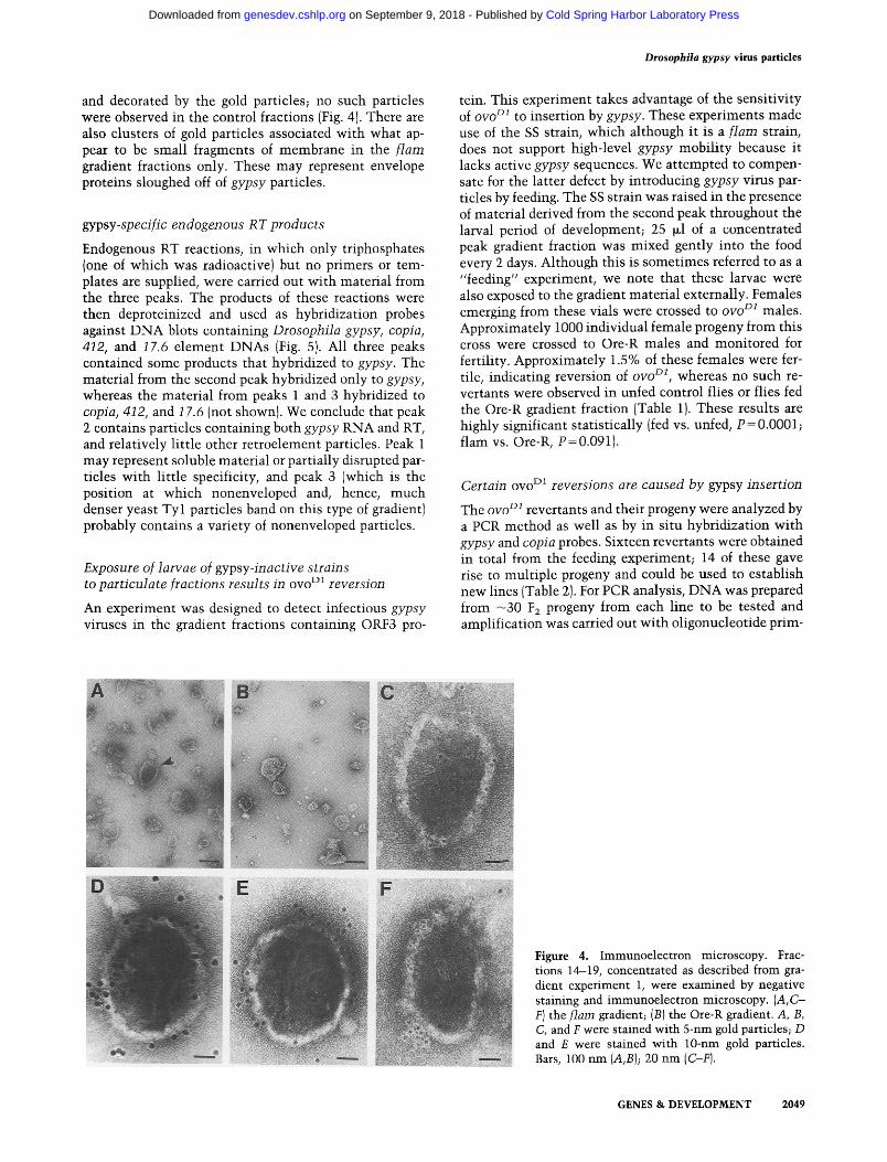
Peak 2 fractions from both the tiara and Ore-R gradi- ents were concentrated and examined by immunoelec- tron microscopy using mAbs 7B3 and 8E7 and colloidal gold-labeled goat anti-mouse IgG. Virus-like particles of irregular shape and - 1 0 0 n m in diameter were observed
~; ; e e 10 12 14 16 18 20 = 24 26 28 3~ Fraction number
Figure 3. Sucrose density gradient analysis of gypsy particles. RT profiles from two independent viral particle preparation experiments {gradient experiments 2 and 1, respectively) are presented (A,B}. {O] flam; (11) Ore-R. The input extracts con- tained equal amounts of protein. The indicated fractions of gradient 2 were immunoblotted with the 7B3 antibody (C,D); similar results were ob- tained with the 8E7 antibody {not shown}. Lanes A and B in these panels contain ovary extract from flare flies and in vitro-translated ORF3 protein, respectively�9 Samples A and B were frozen and thawed once between the time gels C and D were run; this may account for the larger amount of apparent degradation products m lane A of panel D.
B it ~'~10
v 2 4 6 8 10 12 14 16 18 20 22 24 26 28 30
Fraction number
2048 GENES & DEVELOPMENT
Cold Spring Harbor Laboratory Press on September 9, 2018 - Published by genesdev.cshlp.orgDownloaded from
Drosophila gypsy virus particles
and decorated by the gold particles; no such particles were observed in the control fractions (Fig. 4). There are also clusters of gold particles associated with what ap- pear to be small fragments of membrane in the f lam gradient fractions only. These may represent envelope proteins sloughed off of gypsy particles.
gypsy-specific endogenous RT products
Endogenous RT reactions, in which only triphosphates (one of which was radioactive) but no primers or tem- plates are supplied, were carried out with material from the three peaks. The products of these reactions were then deproteinized and used as hybridization probes against DNA blots containing Drosophila gypsy, copia, 412, and 17.6 element DNAs (Fig. 5). All three peaks contained some products that hybridized to gypsy. The material from the second peak hybridized only to gypsy, whereas the material from peaks 1 and 3 hybridized to copia, 412, and 17.6 (not shown). We conclude that peak 2 contains particles containing both gypsy RNA and RT, and relatively little other retroelement particles. Peak 1 may represent soluble material or partially disrupted par- ticles with little specificity, and peak 3 (which is the position at which nonenveloped and, hence, much denser yeast Tyl particles band on this type of gradient) probably contains a variety of nonenveloped particles.
Exposure of larvae of gypsy-inactive strains to particulate fractions results in ovo TM reversion
An experiment was designed to detect infectious gypsy viruses in the gradient fractions containing ORF3 pro-
tein. This experiment takes advantage of the sensitivity of ovo D1 to insertion by gypsy. These experiments made use of the SS strain, which although it is a flare strain, does not support high-level gypsy mobility because it lacks active gypsy sequences. We attempted to compen- sate for the latter defect by introducing gypsy virus par- ticles by feeding. The SS strain was raised in the presence of material derived from the second peak throughout the larval period of development; 25 ~1 of a concentrated peak gradient fraction was mixed gently into the food every 2 days. Although this is sometimes referred to as a "feeding" experiment, we note that these larvae were also exposed to the gradient material externally. Females emerging from these vials were crossed to ovo D1 males. Approximately 1000 individual female progeny from this cross were crossed to Ore-R males and monitored for fertility. Approximately 1.5% of these females were fer- tile, indicating reversion of ovo DI, whereas no such re- vertants were observed in unfed control flies or flies fed the Ore-R gradient fraction (Table 1). These results are highly significant statistically (fed vs. unfed, P = 0.0001; flam vs. Ore-R, P=0.091).
Certain ovo TM reversions are caused by gypsy insertion
The ovo D1 revertants and their progeny were analyzed by a PCR method as well as by in situ hybridization with gypsy and copia probes. Sixteen revertants were obtained in total from the feeding experiment; 14 of these gave rise to multiple progeny and could be used to establish new lines (Table 2). For PCR analysis, DNA was prepared from - 3 0 F2 progeny from each line to be tested and amplification was carried out with oligonucleotide prim-
Figure 4. Immunoelectron microscopy. Frac- tions 14-19, concentrated as described from gra- dient experiment 1, were examined by negative staining and immunoelectron microscopy. (A,C- F) the flare gradient; (B) the Ore-R gradient. A, B, C, and F were stained with 5-nm gold particles; D and E were stained with 10-nm gold particles. Bars, 100 nm (A,B); 20 nm (C-F).
GENES & DEVELOPMENT 2049
Cold Spring Harbor Laboratory Press on September 9, 2018 - Published by genesdev.cshlp.orgDownloaded from

Song et al.
Figure 5. gypsy-specific RT products. (A) Plasmids containing the indicated Drosophila retrotransposons were cleaved with different restriction enzymes and the fragments were separated electrophoretically. The DNA was then blotted to nitrocellulose. (B) The product of an endogenous reaction using material from fractions 15-17 of the flare gradient 1 were separated from unincorported nucle- otides by Sephadex GS0 chromatography and used as a hybridization probe.
ers derived from the ovo locus or the gypsy terminus {Fig. 6). As expected, all samples gave rise to a wild-type ovo band, indicating at least one unrearranged ovo locus. Four of the revertants gave rise to PCR products whose size was consistent wi th that of previously isolated gypsy insert ions in ovo (M6vel-Ninio et al. 1989). Three of these PCR products, as well as the wild-type ovo PCR product, were sequenced and were shown to represent gypsy insertions into ovo (Fig. 6). All three sequenced insertions were different, and none of these insertion sites were exact matches to the previously proposed gypsy target site consensus sequence; instead, they con- formed to the more general consensus sequence YRYRYR [(Y)pyrimidine; (R)purine].
The remaining revertants were analyzed by in situ hy- bridization wi th gypsy and copia probes. Three addi- tional l ines were shown to contain gypsy insertions in 4E, the cytological location of ovo. The remaining seven lines all showed hybridizat ion of the copia probe to 4E (Fig. 7). Insertion of copia into ovo is not completely unexpected because it was observed in a previous study in which the y v f real flare strain was crossed to ovo D1 (M6vel-Ninio et al. 1989). Thus, all 14 lines tested con- tained an insert ion of either a gypsy or a copia element at the ovo locus, at least at the resolution of in situ hybrid- ization. In addition to the gypsy elements inserted at 4E, additional sites of new gypsy insertion were detected in many of the revertant lines.
Table 1. Feeding of particulate fractions results in ovo TM reversion
Treatment a
Total F o F 1 Fertile Reversion females females F 1 frequency studied tested females (%)b
flarn fraction 31 1048 16 1.53 Ore-R fraction 18 451 0 <0.2 Control unfed 33 1194 0 <0.1
aSS larvae were fed {or not fed) with the indicated fraction. bThe literature value for reversion of ovo TM is 0.01% {M4vel- Ninio et al. 1989}.
Unusual properties of ovo TM rever tant progeny
The F:z progeny of ovo D1 revertants show a number of unusual properties (Table 2). The phenotype of the Fz males is expected to be 50% white-eyed and 50% red- eyed because of the heterozygous w h i t e (w) muta t ion expected to be present in the SS mother. However, the observed result was that wi th in any one line, males were either all white eyed (6 lines; Table 2) or all red eyed (8 lines). This behavior differs from that of crosses in which gypsy was introduced directly by crossing flare females x ovo m males. In this case, individual revertant lines gave rise to a mixture of y e l l o w + and y e l l o w male progeny.
The unusual segregation of the w marker in the male progeny of these crosses is difficult to explain. There are two classes of such lines: those that give only white-eyed male progeny and those that give rise to only red-eyed male progeny. In principle, the former class could be ex- plained by a mitot ic recombinat ion event that resulted in homozygosis of the X chromosome, result ing in ho- mozygosity for ovo + and w. However, this cannot ex- plain our results because some of these lines show evi- dence of gypsy or copia insert ion at 4E, which would not have been selected for were the ovo reversion already accounted for by homozygosis. Sex ratios of the progeny were not unusual in any of these lines; however, reces- sive lethali ty was associated wi th three of the X chro- mosomes that we studied (data not shown).
Several of the lines derived by feeding of particles showed both somatic and germ-line instabil i t ies in the F2 generation and beyond. Apparent somatic muta t ions wi th phenotypes corresponding to w and Lobe were ob- served. Also, new mutat ions were observed in the sub- sequent generations wi th a variety of mutan t phenotypes (Table 2).
gypsy insert ions in SS strain after exposure of larvae to part iculate fractions
In a second feeding experiment, SS larvae that were fed particulate fractions were s imply mated to unexposed SS flies. The progeny larvae from this cross were examined
2050 GENES & DEVELOPMENT
Cold Spring Harbor Laboratory Press on September 9, 2018 - Published by genesdev.cshlp.orgDownloaded from
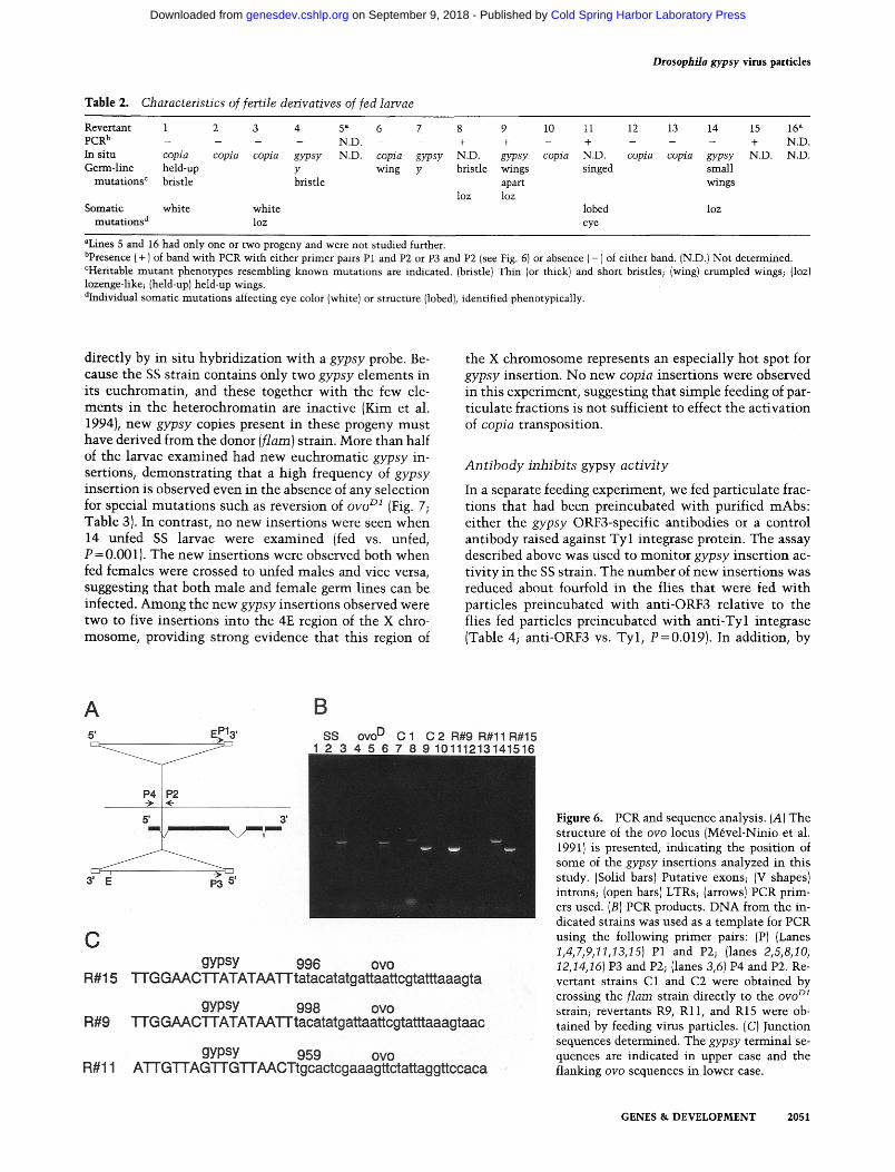
Table 2. Characteristics of fertile derivatives of fed larvae
Drosophila gypsy virus partic les
Revertant 1 2 3 4 5 ~ 6 7 8 9 10 11 12 13 14 15 16 ~ PCR b . . . . N.D. - - + + - + - - - + N.D. In situ copia copia copia gypsy N.D. copia gypsy N.D. gypsy copia N.D. cop ia copia gypsy N.D. N.D. Germ-l ine held-up y wing y bristle wings singed small
mu ta t ions r bristle bristle apart wings loz loz
Somat ic whi te whi te lobed loz muta t ions d loz eye
aLines 5 and 16 had only one or two progeny and were no t studied further. bpresence ( + ) of band wi th PCR wi th ei ther pr imer pairs P1 and P2 or P3 and P2 (see Fig. 6) or absence ( - ) of either band. (N.D.} N o t determined. r m u t a n t phenotypes resembling k n o w n muta t ions are indicated. {bristle) Thin (or thick) and short bristles; {wing) c rumpled wings; (loz) lozenge-like; (held-up) held-up wings. aIndividual somat ic mu ta t ions affecting eye color (white) or s t ructure (lobed), identified phenotypical ly .
directly by in situ hybridization wi th a gypsy probe. Be- cause the SS strain contains only two gypsy elements in its euchromatin, and these together wi th the few ele- ments in the heterochromat in are inactive (Kim et al. 1994), new gypsy copies present in these progeny mus t have derived from the donor (flare) strain. More than half of the larvae examined had new euchromatic gypsy in- sertions, demonstrat ing that a high frequency of gypsy insertion is observed even in the absence of any selection for special muta t ions such as reversion of ovo T M (Fig. 7; Table 3). In contrast, no new insertions were seen when 14 unfed SS larvae were examined (fed vs. unfed, P = 0.001). The new insertions were observed both when fed females were crossed to unfed males and vice versa, suggesting that both male and female germ lines can be infected. Among the new gypsy insertions observed were two to five insertions into the 4E region of the X chro- mosome, providing strong evidence that this region of
the X chromosome represents an especially hot spot for gypsy insertion. No new copia insertions were observed in this experiment, suggesting that simple feeding of par- ticulate fractions is not sufficient to effect the activation of copia transposition.
Ant ibody inhibits gypsy activi ty
In a separate feeding experiment, we fed particulate frac- tions that had been preincubated wi th purified mAbs: either the gypsy ORF3-specific antibodies or a control antibody raised against Tyl integrase protein. The assay described above was used to moni tor gypsy insertion ac- tivity in the SS strain. The number of new insertions was reduced about fourfold in the flies that were fed wi th particles preincubated with anti-ORF3 relative to the flies fed particles preincubated wi th ant i -Tyl integrase (Table 4; anti-ORF3 vs. Tyl , P=0.019). In addition, by
Figure 6. PCR and sequence analysis. (A) The structure of the ovo locus {M6vel-Ninio et al. 1991} is presented, indicating the position of some of the gypsy insertions analyzed in this study. (Solid bars) Putative exons; (V shapes) introns; (open bars) LTRs; (arrows) PCR prim- ers used. (B) PCR products. DNA from the in- dicated strains was used as a template for PCR using the following primer pairs: (P) {Lanes 1,4,7,9,11,13,15) P1 and P2; {lanes 2,5,8,10, 12,14,16} P3 and P2; (lanes 3,6) P4 and P2. Re- vertant strains C1 and C2 were obtained by crossing the flare strain directly to the ovo TM
strain; revertants R9, Rll, and R15 were ob- tained by feeding virus particles. (C) Junction sequences determined. The gypsy terminal se- quences are indicated in upper case and the flanking ovo sequences in lower case.
GENES & D E V E L O P M E N T 2051
Cold Spring Harbor Laboratory Press on September 9, 2018 - Published by genesdev.cshlp.orgDownloaded from

Song et al.
Figure 7. In situ hybridization. {A) copia in situ hybridization on ovo TM revertant 2; (B) gypsy in situ hybridization on SS feed- ant 5-7; (C) copia in situ hybridization on the SS strain; (D) gypsy in situ hybridiza- tion on SS. See Table 3 for additional data on SS feedants.
the O V O m l reversion assay, there was also a fourfold in- hibition of ovo ml reversion conferred by pret reatment wi th anti-ORF3 antibody (Table 4; anti-ORF3 vs. Tyl , P = 0.045). Thus, the anti-ORF3 antibodies appear to ex- ert a partially protective effect against the gypsy virus particles.
D i s c u s s i o n
Evidence that ORF3 is env- l ike
Our experiments show that ORF3-containing fractions also contain a gypsy-speci f ic RT activity, which is capa- ble of producing gypsy-speci f ic cDNA species. These re- suits suggest that gypsy RNA and gypsy primer tRNA (or partially synthesized gypsy cDNAs) cosediment wi th RT in the sucrose gradient. Furthermore, we observe virus- like particles by immunoelec t ron microscopy of the same fractions using an anti-ORF3 antibody. Nei ther the activity nor the virus-like particles (VLPs) are observed in a control gradient prepared from a flare + strain. These observations provide strong evidence for a mature gypsy virus particle that contains ORF3 protein. The peak po- sition of these gypsy particles in the sucrose gradients (fraction 16) is very different from that of control Tyl particles from yeast, which peak at fraction 25 under these conditions. This may be because the gypsy parti- cles contain a membranous envelope that would greatly reduce their density relative to Ty l particles, which lack an envelope. In support of this notion, a second peak of gypsy-specif ic RT activity is found at fractions 25-28; these fractions lack ORF3 protein completely, copia and 412 RTs are also found in these fractions; neither copia nor 412 have an ORF3 and, thus, are more like yeast Tyl .
We suspect that this peak of gypsy activity represents either partially assembled gypsy particles or partially de- graded particles that lack an envelope. A previous study
Table 3. New gypsy and copia copies in derivatives of SS strain that were fed gradient fractions
New gypsy New copia Line copies (no.) copies (no.)
1-2 0 2-1 1 2-2 0 3-1 0 4-1 1 5-1 2 5-2 1 5-3 3 5-6 0 5-7 2 8-1 0 8-2 0 8-3 2 8-4 2 8-5 4 8-6 0 8-7 3 8-8 0
10-1 2
{including 4E insertion)
(including 4E insertion)
(including 4E insertion) (including 4E insertion)
{including 4E insertion)
(including 4E insertion)
New gypsy and copia copies were detected by in situ hybridiza- tion. Progeny, e.g., 2-1 and 2-2, are siblings. For lines 1-2 and 10-1, a fed male was crossed to an unfed female. For all other lines, a fed female was crossed to an unfed male. The 4E inser- tions in 8-3, 8-4, and 8-7 may not be independent. In a control experiment, 14 unfed SS larvae were examined and 0/14 had new copies of gypsy.
2052 GENES & DEVELOPMENT
Cold Spring Harbor Laboratory Press on September 9, 2018 - Published by genesdev.cshlp.orgDownloaded from

Drosophila gypsy virus particles
Table 4. Ant ibody inhibition
Antibody
anti-Tyl anti-ORF3 ~ integrase b
Feeding of SS strains, assay by in situ hybridization number of new gypsy copies observed/ number of strains checked by in situ (%)
ovo 01 reversion assay c number of fertile F1 females/ F 1 females tested (%)
6/33 22/33 (18) (67)
2/1030 9/930 (0.19) (0.97)
~Mixture of 45 ~zg of 7B3 and 45 ~g of 8E7. b90 [~g of 8B11 (Eichinger and Boeke 1988). CPerformed as described for Table 1.
reported tentative evidence for g y p s y virus (or virus-like) particle production in tissue culture cells. A variety of virion types were found in extracellular fluid and were associated with some g y p s y RNA, but no evidence was presented to identify definitively the g y p s y particles (Sy- omin et al. 1993). It would be interesting to test the supematants of such tissue culture cell lines for g y p s y insertion activity by the ovo D1 reversion assay.
We have shown that ORF3 protein is N-glycosylated by treatment of protein extracts with EndoF and ob- served a shift in the electrophoretic mobility of the ORF3 protein to a mobility very similar to that of in vitro-translated ORF3 protein. Because retroviral Env proteins are also N-glycosylated, this result provides fur- ther support for the conclusion that ORF3 has an env- like function. N-linked glycosylation signals are con- served in the ORF3s of g y p s y (both D. m e l a n o g a s t e r and D. virilis), 17.6, 297, t om , and TED. Furthermore, the TED retroelement ORF3 protein has also been shown to be glycosylated in insect cells (P. Friesen, pers. comm.). Thus N-glycosylation is apparently a conserved feature of this class of retroelement ORF3 proteins. Further- more, the ORF3 protein appears to be cleaved into at least two smaller products that may correspond to ret- roviral surface and transmembrane proteins.
I n f e c t i v i t y
Supporting evidence for a virus intermediate in the g y p s y
insertion process comes from experiments in which the fractions containing the virus particles were introduced into the SS strain, which lacks active g y p s y copies. These particles were introduced into the flies by expos- ing larvae to the fractions. Whether the g y p s y particles enter the larvae by ingestion, by adsorption to external larval surfaces, or by some other larval surface is not clear. However, such exposure resulted in a high fre- quency of g y p s y insertion into the ovo D1 locus, as mon- itored by the reversion assay. Similar results to these were obtained by Kim et al. (1994), who microinjected whole embryo extracts of f l a m strains into SS embryos and exposed SS larvae to whole pupal extracts of f lare strains. In our experiments, additional copies of g y p s y
were observed to have been inserted in these ovo D1 re- vertant lines as well, suggesting high g y p s y activity in the established lines, and their progeny showed evidence of both germ-line and somatic mutations at high fre- quencies. Kim et al. (1994) made no mention of either additional insertions or continuing genetic instability in their ovo DI revertant lines; therefore, it is possible that by using purified particle fractions, much higher levels of g y p s y are introduced than when the virus is transmitted by a cross.
In subsequent experiments in which SS larvae were exposed to particles, no selection for ovo ~ reversion was carried out, and the larvae exposed to particles were sim- ply crossed to unexposed SS flies. The F1 progeny of these flies also had a very high frequency of unselected g y p s y insertions. The latter experiment is important be- cause it shows that there is no need for special selections to observe high-frequency g y p s y infection/insertion. Re- markably, a large fraction of these flies carried a g y p s y
insertion at 4E, the cytological location of ovo. By preincubating the particles with anti-ORF3 anti-
bodies before feeding, we observed a nearly fou~old re- duction in the number of new insertion events resulting from feeding, consistent with ORF3 protein being re- quired for infection and subsequent insertion of gypsy .
A h y b r i d dy sgenes i s - l i k e p h e n o m e n o n
The progeny of the ovo D1 revertants showed a number of unexpected and, in some cases, difficult-to-explain prop- erties. Many of these properties are reminiscent of the behavior of the progeny of hybrid dysgenic crosses. The first is the appearance of visible mutants in subsequent generations, as if genetic instability mediated by these viruses or other entities in the fractions to which the flies were exposed can be maintained over several gen- erations. This is somewhat surprising because both ovo D1 strain and Ore-R fail to give rise to ovo + rever- tants at high frequency (N. Prud'homme, M. Gans, M. Masson, C. Terzian, and A. Bucheton, in prep.). If a f l a m mutation is necessary for g y p s y mobilization, it is not obvious why additional g y p s y insertions are seen in sub- sequent generations. Either the requirement for f l a m
GENES & DEVELOPMENT 2053
Cold Spring Harbor Laboratory Press on September 9, 2018 - Published by genesdev.cshlp.orgDownloaded from

Song et al.
phenotype can be overridden by the very high gypsy vi- rus load obtained by these flies, or perhaps preformed gypsy viruses can be physically transmitted to subse- quent generations. The latter could also explain, at least theoretically, the appearance of somatic mutation in the subsequent generations.
A number of the ovo D~ revertant lines showed evi- dence for copia insertions at band 4E, the known cyto- genetic location of ovo. None of the parental strains in this experiment (i.e., ovo m~, Ore-R, SS, or flam) carries a copia insertion at this position. Although we have not proven that the copia insertions are directly responsible for this class of ovo D~ reversion event, it does seem likely that this is the case. There are numerous addi- tional examples among those we have examined by in situ hybridization of copia inserted at positions not cor- responding to those of any of the parental lines. This result suggests (but does not prove) that copia transpo- sition is somehow elevated in these lines. There are many reports in the literature suggesting that hybrid dys- genic lines show mobility of elements other than the transposon responsible for the dysgenesis (e.g., in P-M dysgenic strains; Gerasimova et al. 1984a, b; Lewis and Brookfield 1987), but these conclusions remain contro- versial and do not appear to represent a completely gen- eral phenomenon (Woodruff et al. 1987; Eggleston et al. 1988; Engels 1989), and the mechanism of mobility in these cases (i.e., transposition or homology dependent) is certainly unproven. In the case of copia mobilization by injection of gypsy viruses, two possible mechanisms could be imagined: (1) copia retrotransposition could be mobilized in trans by gypsy elements, that is, trans-cap- sidation, or (2) high-frequency gypsy infection/integra- tion causes a DNA damage response to which copia (and perhaps other transposable elements) are responsive. The first hypothesis seems unlikely given that copia and gypsy are molecular archetypes of the two major sub- classes of the LTR retrotransposons and, hence, are about as dissimilar in sequence and structure as two LTR retrotransposons can be. Furthermore, much experimen- tation has shown that molecular hybrids between even rather closely related LTR retrotransposons are inactive for transposition. There is evidence that LTR retrotrans- posons in a variety of host organisms respond to global cellular regulatory pathways, including DNA damage pathways (Strand and McDonald 1985; Rolfe et al. 1986; McEntee and Bradshaw 1988). Thus, the latter explana- tion seems more plausible.
A number of reports in the literature implicate high- frequency gypsy insertion as correlated with genetic in- stabilities reminiscent of hybrid dysgenesis. These in- clude the strains Uc (unstable chromosome), tuh (tumor- ous head), flare (used in this study), and MS {mutator strain). The Uc strain is characterized by a high fre- quency of chromosome rearrangements, new gypsy-in- duced germ line and somatic insertions, and the gradual appearance of sterility (Lim 1980; Lira et al. 1983; B.H. Judd, pers. comm.). Another stock that gave rise to many gypsy-induced insertion mutations bears a tuh ~;3 muta- tion; this stock is also unstable with regard to the high-
frequency gypsy insertion phenotype (Kuhn 1970; D. Kuhn, pers. comm.). The y v f m a l flare strain is known to give a high rate of gypsy-induced ovo DI reversion, as well as giving a high frequency of cut mutations (M6vel- Ninio et al. 1989). The MS strain is characterized by a high rate of gypsy- and hobo-induced mutations, espe- cially cut and forked mutations, and also shows somatic insertions (Kim et al. 1990, 1994; Kim and Belyaeva 1991).
Conclusions
Kim et al. (1994) have performed experiments in which crude embryo or pupal extracts derived from the MSN1 strain (an SS strain carrying a gypsy element derived from MS) were microinjected into embryos or fed to lar- vae of SS strains, respectively. Using the ovo D1 reversion assay, they provided evidence that the MSN1 strain ex- tracts contained a trans-acting factor that stimulated gypsy insertion. Although these results strongly support the notion that gypsy is an infectious retrovirus, they failed to prove it. In the experiments by Kim et al., as well as in our ovo ml reversion experiment, it is formally possible that what is being transmitted to the progeny is not a viral particle but a positive activator of transposi- tion. Although the recipient SS strain used in these ex- periments lacks active gypsy copies, the ovo DI strain to which it was crossed may well contain active gypsy el- ements, and these experiments cannot rule out the pos- sibility that these ovo DI strain-derived elements were activated in trans by the material from the MSN1 (or flam) donor strains. However, in our second feeding ex- periment, in which only the inactive SS strain was used, we have ruled out any role of the gypsy elements in the ovo D1 strain. Final proof of gypsy infectivity will require the mobilization of genetically marked gypsy elements.
Could gypsy be both a retrotransposon and a retrovi- rus? The fact that the flare mutation specifically affects ORF3 expression raises the possibility that gypsy is a facultative retrovirus, expressing an infectious form only under special circumstances, and that under separate conditions it might transpose by an intracellular path- way that requires only gag and pol. However, at this point this must remain only a speculation, for the exist- ing data are equally consistent with all gypsy "transpo- sition" being mediated entirely by an intercellular infec- tion process. The finding that gypsy is probably an in- fectious retrovirus of Drosophila raises many new questions that will be the subject of future investiga- tions. The availability of a genetically tractable system with infectious retroviruses will be especially useful for tackling such problems. What is the receptor for the ORF3 protein, and what is the route by which virus par- ticles enter the larva, and eventually the germ line of the progeny? What is the host range of the gypsy virus? Its presence in sibling species of D. melanogaster (Mizrokhi and Mazo 1990) suggests that at least the genus Droso- phila will be susceptible. The development of marked gypsy elements bearing phenotypic markers will be a powerful tool for answering such questions, and also
2054 GENES & DEVELOPMENT
Cold Spring Harbor Laboratory Press on September 9, 2018 - Published by genesdev.cshlp.orgDownloaded from

should a l low the d e v e l o p m e n t of a n e w micro in jec t ion- i ndependen t germ-l ine t r anformat ion strategy for Droso- phila.
M a t er i a l s a nd m e t h o d s
Strains and genetic crosses
The strains MG#3 (flam/FM3)(N. Prud'homme, M. Gans, M. Masson, C. Terzian, and A. Bucheton, in prep.), ovo TM v (Mdvel- Ninio et al. 1989), and SS (w flare) (Kim et al. 1990) were pro- vided by A. Bucheton (CNRS, Gifsur Yvette, France). These strains are maintained on standard Drosophila medium; all ge- netic experiments were carried out at 25~
Generation of mAbs
The mAbs 7B3 and 8E7 were generated by using the following protocol. The 1612 bp StyI-XhoI gypsy fragment was inserted into pATH3 (Koerner et al. 1991) for TrpE-ORF3 fusion protein production. Cells containing the construction were induced with ~-indole acrylic acid and an 89-kD TrpE-ORF3 fusion pro- tein was isolated. BALB/c mice (5-6 months old) were immu- nized with the fusion protein. Mice received an initial injection of 100 mg of protein emulsified 1:1 with Freund's complete adjuvant. After 2 weeks, the mice were given three boosts of 100 mg of protein in Freund's incomplete adjuvant at 2-week inter- vals. Six days after the final boost, serum samples were tested by immunoblotting. Mice giving good serum responses were boosted with 100 mg of protein with Freund's incomplete adju- vant 4 days before the fusion. Spleen cells were fused in the presence of PEG 4000 (GIBCO) to sp2/0 myeloma cells, using standard protocols (Harlow and Lane 1988). Hybridoma super- natants were screened 1-2 weeks later on immunoblot strips containing either TrpE protein or the fusion protein. Antibodies 7B3 and 8E7 react only with the fusion protein. Positive pre- clones were cloned by limiting dilution. Ascites were produced as described (Harlow and Lane 1988).
Immunoblotting
Ovaries from 3- to 5-day-old females flies were isolated in buffer [0.1 M NaC1, 0.01 M Tris-HC1 (pH 7.4), 0.001 M EDTA, 0.001 M PMSF] and transferred into SPS lysis buffer [2.5% SDS, 60 mM Tris-HC1 (pH 7.4), 0.005% bromophenol blue, 10% glycerol) for homogenization. After homogenization, proteins were boiled for 10 min, spun for 5 min, and stored at -20~ Gradient fractions (100 ~xl) were precipitated by 10% TCA and resus- pended in sample buffer. Samples for immunoblotting were pre- pared as above.
Proteins were run on 12% or 15% polyacrylamide gels and electroblotted onto nitrocellulose. Immunoblots were blocked for 0.5-1 hr with 5% powdered milk in PBST [150 mM NaC1, 10 mM phosphate (pH 7.0), 0.3% Tween 20]. Primary antibodies (hybridoma culture supernatant diluted l:10)were added in 1% milk in PBST and incubated for 1.5 hr at room temperature. Blots were washed for 1 hr in PBST and incubated with perox- idase-conjugated secondary antibodies (Sigma) for 1.5 hr in PBST at a dilution of 1:10,000. The washing procedure was re- peated, and the blots were subjected to ECL Western blotting protocols (Amersham).
Homogenization of flies and sucrose density gradient analysis
Two grams of 3- to 5-day-old female flies were homogenized in
Drosophila gypsy virus particles
buffer [0.1 M NaC1, 0.01 M Tris-HC1 (pH 7.4}, 0.001 M EDTA, 0.001 M PMSF] using a Dounce homogenizer at 4~ This ho- mogenate was spun down at 10K rpm for 10 min. The superna- tant was transferred to a new 15-ml tube and spun down again at 10K rpm for 5 min to get rid of debris. The total protein content of flare and control extracts was assayed by OD2s o and found to be identical. The final supernatants (-4.5 ml) were loaded onto 20-70% linear sucrose gradients prepared and frac- tionated as described (Braiterman et al. 1994). In gradient exper- iment 1, no protease inhibitor cocktails were added, whereas in experiment 2, lx PIC1 and PIC2 were added to the extracts as described (Braiterman et al. 1994). Five-microliter aliquots of the fractions were used for exogenous and endogenous RT as- says, and 100-~1 aliquots were precipitated by TCA and used for immunoblot analysis.
RT assays
Exogenous RT assays using a poly{A)-oligo(dT) primer template were performed in 42 mM Tris-HC1 (pH 8.2), 16.7 mM DTT, 50 mM NaC1, 5 mM MgC12, 0.5 mM MnC12, 0.042% NP-40, 8.3 ~M TTP, 4.17 ~g/ml of oligo(dT), and 8.33 txg/ml of poly(A). [R-B2p]TTP (0.25 ~Ci ) was added per 25 ~1 of reaction cocktail and 5 ~xl of fraction, and incubated at room temperature for 1 hr. Incorporation was assayed as described (Goff et al. 1981) . The washed DEAE paper was analyzed directly with ImageQuant software using a Molecular Dynamics PhosphorImager. The en- dogenous RT reaction uses internal primer templates; it was performed in an assay cocktail containing 50 mM Tris-HC1 (pH 8), 20 mM DTT, 60 mM NaC1, 6 mM MnCI~, 5 mM MgCI~, 0.1% NP-40, 2 mM dATP, and 600 mM each of dGTP, dTTP, and dCTP. In 65 ~1 of the cocktail, 5-~1 aliquots of sucrose gradient fractions and 100 ~Ci of [e~-g2P]dATP were added and incubated for 3 hr at room temperature. After incubation, 10 ml of 1 mM dATP was added and incubated for an additional 30 min. The final product was phenol/chloroform-extracted and precipitated in ethanol. DNA purified on a Sephadex G50 column was used as a probe for the DNA blotting analysis.
Electron microscopy
Aliquots (100 }xl) of fractions 14-20 were pooled together and run on 20-70% step sucrose gradients to concentrate the virus particles. Centrifugation was for 1.5 hr at 25,000 rpm at 4~ in a Beckman SW28 rotor. Thirty microliters of concentrated virus particles were adsorbed to a collodion grid for 5 min at room temperature. The grids were rinsed on three drops of PBS for 1 min each and then incubated on a drop of a mixture of 7B3 and 8E7 (as ascites fluid at a dilution of 1:200 in PBS} for 30 min at 37~ and then for 30 rain at room temperature. After the pri- mary antibody incubations, the grids were washed on three drops of PBS and incubated on a drop of gold-labeled goat anti- mouse IgG (Amersham) at a concentration of 1:30. After sec- ondary antibody incubation, the grids were rinsed on two drops of PBS and then on two drops of H20. The grids were fixed in 2.5% glutaraldehyde for 5 min at room temperature and then rinsed in H20 three times for 1 min each. After washing, the grids were stained negatively in a solution of 1% uranyl acetate and 0.2% tylose on two drops for 45 sec each. Electron micros- copy was performed on a Zeiss transmission electron micro- scope (model TEM 10A).
DNA manipulations
Genomic DNA was isolated from 20-30 adult flies using the following method. Flies were homogenized in 400 ~xl of buffer
GENES & DEVELOPMENT 2055
Cold Spring Harbor Laboratory Press on September 9, 2018 - Published by genesdev.cshlp.orgDownloaded from

Song et al.
[0.1 M Tris-HC1 (pH 9.0), 0.1 M EDTA, 1% SDS, 1% DEPC] with a plastic rod in an Eppendorf tube. This homogenate was incu- bated for 30 min at 70~ and then 54 ml of 8 M potassium acetate was added and left on ice for 30 min. After spinning for 15 min at 4~ the supernatant was transferred carefully to a fresh Eppendorf tube. DNA was then precipitated by adding a 0.5-volume of isopropanol at room temperature and spun down for 5 min. The DNA pellet was washed carefully with 70% ethanol, respun, dried, and resuspended in 30-50 ml of H20. About 20 ng of genomic DNA was used as template for PCR. PCR was done as follows: 94~ for 1 min and 20 sec, 65~ for 2 min, and 72~ for 3 min for 40 cycles. HotTub polymerase and reaction buffers (Amersham) were used with 200 ~M dXTPs, 1 ~M each primer, and 1% glycerol included in the reaction.
DNA cycle sequencing using oligonucleotide primer JB715, Taq polymerase, and fluorescent dideoxy terminators was car- ried out directly on PCR products isolated from agarose gels.
The oligonucleotides used were P 1, 5'-CAACATGACCGAG- GACGGTCATAAAC-3'; P2, 5'-CTCCCGCTCTGCGGGCTT- CTCTTT-3'; P3, 5'-CTTTGCCGAAAATATGCAATG-3'; P4, 5'-CGGCTTTTTCAGCGGCTAACC-3'; and JB715, 5'-TAAA- ACGGAGGTGGCGG-3'.
For in situ hybridization, biotinylated probes of gypsy and copia DNA were used and hybridizations were performed as described (Lira 1993).
Exposure of larvae to gradient fractions
For infectivity experiments in which larvae were exposed to particles, particles were concentrated by step gradient centrifu- gation. Particles were concentrated from fractions 14-18 by pooling the fractions and pelleting the particles through 20 ml of 20% sucrose at 25,000 rpm in an SW28 rotor for 90 min onto a 20-ml 70% sucrose cushion. The concentrated particles were collected as a single 400-lxl interface fraction. For feeding exper- iments, 25 Ixl of this concentrate or unconcentrated fraction 16 was added to standard fly food by gentle mixing every other day for 10 days.
For the antibody inhibition experiment, the indicated anti- bodies were purified from ascites on protein G using a mAb- TrapGII kit (Pharmacia)before use. Concentrated particles (125 ixl) were preincubated with antibodies (90 ~g) overnight at 4~ and fed as described above.
Methods for obtaining new lines from feeding experiments
SS flies were fed sucrose gradient fractions containing particles five times every 2 days. Females emerging from these vials were crossed with ovo TM v males (for reversion experiments, see Ta- ble 2) or to unfed SS males for direct observation of polytene chromosomes of progeny larvae without selection (Tables 3 and 4). The Fl female progeny from the former cross were mated to Ore-R males to check their fertility. The F2 progeny from this cross were subjected to PCR and in situ hybridization. Individ- ual progeny F 2 females were also crossed with FM4 males. In- dividuals from this cross were mated again with FM4 males to test for lethals in the ovo TM revertant chromosome (a lethal would produce FM4 males only). These manipulations are dia- gramed in Figure 8.
Deglycosidation with endoglycosidase F
Ovary protein preparation was made as explained above in Western analysis. These proteins were denatured in denaturing buffer {0.5% SDS, 1% B-mercaptoethanol) by boiling for 10 min at 100~ This protein extract was spun down, and the super-
In s i tu hybridization assay OVO D l reve rs iOn assay
S ~f Isolate revertant ovo ~ fertile females
Isolate individual larvae and carry out in situ hybridization ~. ovo am v/w X (,.)'Ore R
x
.C. aoa, s,s J. In situ hybridization
ovo ~ v/FM4
Isolation of mutations with visible phenotypes
Figure 8. Scheme for in situ hybridization assay and for gen- erating ovo TM revertant lines, ovo D~R indicates ovo TM revertant.
natant was taken to a new tube. After adding deglycosidation reaction buffer [50 mM Na phosphate [pH 7.5}, 1% NP40], 10 units of PNGase F {peptide/N-glycosidase F; New England Bi- olabs) enzyme was added. Incubation was for 5 hr at 37~
In vitro transcription~translation
A 1.5-kb DNA fragment, corresponding to the spliced ORF3 mRNA, was amplified using primers 1475 (5'-AGTTAAGTTA- GAAAAGCATGTTCACCCTCATGATGTTCATACCCTTG- 3') and 1463 (5'-ACGAAGCAATACATTGTTAGTTGT-3'). These PCR fragments were cloned directly into the TA cloning vector {pCRII, Invitrogen Corp.). The orientation of the insert was determined by restriction enzyme digestion and sequenc- ing. The plasmid pTA-env has gypsy ORF3 under the control of the bacteriophage T7 promoter. Coupled in vitro T7 transcrip- tion-translation was done with the TnT-coupled reticulocyte lysate system (Promega) by following their standard protocol. For the production of nonradiolabeled protein, both amino acid mixture (-Met) and amino acid mixture (-Leu) were used in the reaction.
Statistical analyses
P values were calculated by a contingency table analysis using Statview II {Abacus Concepts). The reported values are continu- ity-corrected P values.
A c k n o w l e d g m e n t s
We are especially grateful to Alain P61isson, Alexander Kim, Nicole Prud'homme, and Alain Bucheton for generously provid- ing Drosophila strains as well as information about their exper- iments before publication. We thank Mike Dellannoy for assis- tance with immunoelectron microscopy. This work was sup- ported by a Human Frontiers Grant to J.D.B. and National Institutes of Health grant GM35463 and American Cancer So- ciety grant DB-7F to V.G.C.
The publication costs of this article were defrayed in part by payment of page charges. This article must therefore be hereby
2056 GENES & DEVELOPMENT
Cold Spring Harbor Laboratory Press on September 9, 2018 - Published by genesdev.cshlp.orgDownloaded from

Drosophila gypsy virus particles
marked "advertisement" in accordance with 18 USC section 1734 solely to indicate this fact.
R e f e r e n c e s
Boeke, J.D. 1988. getrotransposons. In RNA genetics. Volume II, retroviruses, viroids, and RNA recombination (ed. E. Do- mingo, J.J. Holland, and P. Ahlquist) pp. 59-103. CRC Press, Boca Raton, FL.
Boeke, J.D. and V.G. Corces. 1989. Transcription and reverse transcription in retrotransposons. Annu. Rev. Microbiol. 43: 403-433.
Braiterman, L.T., G.M. Monokian, D.J. Eichinger, S.L. Merbs, A. Gabriel, and J.D. Boeke. 1994. In-frame linker insertion mu- tagenesis of yeast transposon Tyl: Phenotypic analysis. Gene 139: 19-26.
Coffin, J.M. 1993. Reverse transcriptase and evolution. In Re- verse transcriptase. (ed. S. Goff and A. Skalka) pp. 445-479. Cold Spring Harbor Laboratory Press, Cold Spring Harbor, New York.
Eggleston, W.B., D.M. Johnson-Schlitz, and W.R. Engels. 1988. P-M hybrid dysgenesis does not mobilize other transposable element families in D. melanogaster. Nature 331: 368-371.
Eichinger, D.J. and J.D. Boeke. 1988. The DNA intermediate in yeast Tyl element transposition copurifies with virus-like particles: Cell-free Tyl transposition. Cell 54: 955-966.
Engels, W.R. 1989. P elements in Drosophila melanogaster. In Mobile DNA (ed. D.E. Berg and M.M. Howe), pp. 437-484. American Society for Microbiology, Washington, D.C.
Gerasimova, T.I., L.V. Matyuyina, Y.V. Ilyin, and G.P. Georgiev. 1984a. Simultaneous transposition of different mobile elements: Relation to multiple mutagenesis in Drosophila melanogaster. Mol. & Gen. Genet. 194: 517- 522.
Gerasimova, T.I., L.J. Mizrokhi, and G.P. Georgiev. 1984b. Transposition bursts in genetically unstable Drosophila melanogaster. Nature 309: 714-716.
Goff, S., P. Traktman, and D. Baltimore. 1981. Isolation and properties of Moloney murine leukemia virus mutants: Use of a rapid assay for release of virion reverse transcriptase. J. Virol. 38: 239-248.
Harlow, E. and D. Lane. 1988. Antibodies: A laboratory man- ual. Cold Spring Harbor Laboratory, Cold Spring Harbor, New York.
Kim, A.I. and E.S. Belyaeva. 1991. Transposition of mobile ele- ments gypsy (mdg4) and hobo in germ-line and somatic cells of a genetically unstable mutator strain of Drosophila mel- anogaster. Mol. & Gen. Genet. 229: 437-444.
Kim, A.I., E.S. Belyaeva, and M.M. Aslanian. 1990. Autonomous transposition of gypsy mobile elements and genetic instabil- ity in Drosophila melanogaster. Mol. & Gen. Genet. 224: 303-308.
Kim, A.I., C. Terzian, P. Santamaria, A. P41isson, N. Prud- 'homme, and A. Bucheton. 1994. Retroviruses in inverte- brates: The gypsy retrotransposon is apparently an infec- tious retrovirus of Drosophila melanogaster. Proc. Natl. Acad. Sci. 91: 1285-1289.
Koemer, T.J., J.E. Hill, A.M. Myers, and A. Tzagoloff. 1991. High expression vectors with multiple cloning sites for construc- tion of trpE fusion genes: pATH vectors. Methods Enzymol. 33: 477-490.
Kuhn, D. 1970. Another case of mass mutation. Drosophila Inf. Serv. 45: 127.
Lewis, A.P. and J.F.Y. Brookfield. 1987. Movement of Droso- phila melanogaster transposable elements other than P ele-
ments in a P-M hybrid dysgenic cross. Mol. & Gen. Genet. 208: 506-510.
Lim, J.K. 1980. Site-specific intrachromosomal rearrangements in Drosophila melanogaster: Cytogenetic evidence for trans- posable elements. Cold Spring Harbor Symp. Quant. Biol. 45: 553-560.
1993. In situ hybridization with biotinylated DNA. Drosophila Inf. Serv. 72: 73-77.
Lim, J.K., M.J. Simmons, J.D. Raymond, N.M. Cox, R.F. Doll, and T.P. Culbert. 1983. Homologue destabilization by a pu- tative transposable element in Drosophila melanogaster. Proc. Natl. Acad. Sci. 80: 6624--6627.
Marck, C. 1988. "DNA Strider": A "C" program for the fast analysis of DNA and protein sequences on the Apple Macin- tosh family of computers. Nucleic Acids Res. 16: 1829- 1836.
Marlor, R.L., S.M. Parkhurst, and V.G. Corces. 1986. The Droso- phila melanogaster gypsy transposable element encodes pu- tative gene products homologous to retroviral proteins. Mol. Cell. Biol. 6: 1129-1134.
McEntee, K. and V. Bradshaw. 1988. Effects of DNA damage on transcription and transposition of Ty retrotransposons in yeast. In Banbury Report 30. Eukaryotic transposable ele- ments as mutagenic agents {ed. M.E. Lambert, J.F. McDon- ald, and I.B. Weinstein), pp. 245-253, Cold Spring Harbor Laboratory, Cold Spring Harbor, New York.
M4vel-Ninio, M., C.M. Mariol, and M. Gans. 1989. Mobiliza- tion of the gypsy and copia retrotransposon in Drosophila melanogaster induces reversion of the ovo D dominant fe- male-sterile mutations: Molecular analysis of revertant alle- les. EMBO J. 8: 1549-1558.
M4vel-Ninio, M., R. Terracol, and F.C. Kafatos. 1991. The ovo gene of Drosophila encodes a zinc finger protein required for female germ line development. EMBO J. 10: 2259-2266.
Mizrokhi, L.J. and A.M. Mazo. 1990. Cloning and analysis of the mobile element gypsy from D. virilis. Nucleic Acids Res. 19: 913-916.
P41isson, A., S.U. Song, N. Prud'homme, P. Smith, A. Bucheton, and V.G. Corces. 1994. gypsy transposition correlates with the production of a retroviral envelope-like protein under the tissue-specific control of the Drosophila flamenco gene. EMBO J. (in press).
Rolfe, M., A. Spanos, and G. Banks. 1986. Induction of yeast Ty element transcription by ultraviolet light. Nature 319: 339- 340.
Strand, D.J. and J.F. McDonald. 1985. copia is transcriptionally responsive to environmental stress. Nucleic Acids Res. 13: 4401-4410.
Syomin, B.V., V. Konstantin, K.V. Kandror, A.B. Semakin, V.L. Tsuprun, and A.S. Stepanov. 1993. Presence of the gypsy (mdg4) retrotransposon in extracellular virus-like particles. FEBS Lett. 323: 285-288.
Tanda, S., J.L. Mullor, and V.G. Corces. 1994. The Drosophila tom retrotransposon encodes an envelope protein. Mol. Cell. Biol. 14: 5392-5401.
Woodruff, R.C., J.L. Blount, and J.N. Thompson Jr. 1987. Hybrid dysgenesis in D. melanogaster is not a general release mech- anism for DNA transpositions. Science 237: 1206-1208.
GENES & DEVELOPMENT 2057
Cold Spring Harbor Laboratory Press on September 9, 2018 - Published by genesdev.cshlp.orgDownloaded from

10.1101/gad.8.17.2046Access the most recent version at doi: 8:1994, Genes Dev.
S U Song, T Gerasimova, M Kurkulos, et al. that gypsy is an infectious retrovirus.An env-like protein encoded by a Drosophila retroelement: evidence
References
http://genesdev.cshlp.org/content/8/17/2046.full.html#ref-list-1
This article cites 26 articles, 6 of which can be accessed free at:
License
ServiceEmail Alerting
click here.right corner of the article or
Receive free email alerts when new articles cite this article - sign up in the box at the top
Copyright © Cold Spring Harbor Laboratory Press
Cold Spring Harbor Laboratory Press on September 9, 2018 - Published by genesdev.cshlp.orgDownloaded from