acute exercise does not modify brain activity and memory
TRANSCRIPT

RESEARCH ARTICLE
Acute exercise does not modify brain activity
and memory performance in APP/PS1 mice
Angelica Miki Stein1,2,3, Victor Munive1,2, Ana M. Fernandez1,2, Angel Nuñez4,
Ignacio Torres Aleman1,2*
1 Cajal Institute, Madrid, Spain, 2 Ciberned, Madrid, Spain, 3 Universidade Estadual Paulista, São Paulo,
Brazil, 4 School of Medicine, Autonoma University of Madrid. Madrid, Spain
Abstract
Age is the main risk factor for Alzheimer´s disease (AD). With an increasingly aging popula-
tion, development of affordable screening techniques to determine cognitive status will help
identify population-at-risk for further follow-up. Because physical exercise is known to modu-
late cognitive performance, we used it as a functional test of cognitive health. Mice were
submitted to treadmill running at moderate speed for 30 min, and their brain activity was
monitored before and after exercise using electrocorticogram (ECG) recordings. After exer-
cise, normal, but not APP/PS1 mice, a well established AD model, showed significantly
increased ECG theta rhythm. At the same time normal, but not AD mice, showed signifi-
cantly enhanced performance in a spatial memory test after exercise. Therefore, we postu-
late that a running bout coupled to pre- and post-exercise brain activity recordings will help
identify individuals with cognitive alterations, by determining the presence or absence of
exercise-specific changes in brain activity. Work in humans using a bout of moderate exer-
cise plus electroencephalography, a clinically affordable procedure, is warranted.
Introduction
With an increasingly larger proportion of aged people in modern societies, cognitive loss in
the aging population is becoming a paramount problem for public health agencies. Hence, an
urgent need for early identification of individuals at risk of developing cognitive loss is widely
recognized [1]. Current clinical practice to determine the presence of cognitive problems relies
entirely on psychometric tests that are reliable but laborious, and require well-trained person-
nel [2, 3]. Furthermore, these tests are useful to detect early signs of cognitive disturbances,
already when deficits are present [3]. Intense focus on this problem has resulted in the appear-
ance of new diagnostic techniques based on biochemical parameters in cerebro-spinal fluid
(CSF) and in brain imaging. The first approach is problematic because CSF samples are not
easy to obtain. The second one is expensive and requires state-of-the-art facilities. In both
cases, highly trained professionals are a must. On top of that, any of these procedures have yet
been implemented for early diagnosis, prior to the appearance of cognitive disturbances.
As exemplified by cardiovascular screening programs, preventive screening procedures of
the general population are an optimal way to detect populations-at-risk, but difficult to support
PLOS ONE | https://doi.org/10.1371/journal.pone.0178247 May 22, 2017 1 / 11
a1111111111
a1111111111
a1111111111
a1111111111
a1111111111
OPENACCESS
Citation: Miki Stein A, Munive V, Fernandez AM,
Nuñez A, Torres Aleman I (2017) Acute exercise
does not modify brain activity and memory
performance in APP/PS1 mice. PLoS ONE 12(5):
e0178247. https://doi.org/10.1371/journal.
pone.0178247
Editor: Sergio Pellis, University of Lethbridge,
CANADA
Received: March 22, 2017
Accepted: May 10, 2017
Published: May 22, 2017
Copyright: © 2017 Miki Stein et al. This is an open
access article distributed under the terms of the
Creative Commons Attribution License, which
permits unrestricted use, distribution, and
reproduction in any medium, provided the original
author and source are credited.
Data Availability Statement: All relevant data are
within the paper and its Supporting Information
file.
Funding: AMS was supported by Capes PDSE
99999.010743/2014-06 and Fapesp 2013/19729-
0, Universidade Estadual Paulista (Brazil). VM is a
recipient of an FPI fellowship. We are thankful to L.
Guinea for excellent technical support. This work
was funded by grants SAF2013-40710-R/
SAF2016-76462 AEI/FEDER, and by Ciberned.

with available diagnostic procedures even by public health systems of developed countries.
Therefore, development of affordable and more objective tests would make much more feasi-
ble this goal. From this perspective, for the present study we chose clinical procedures of wide
use such as a bout of exercise (for cardiovascular screening) and electroencephalographic
recording (for neurological testing) and combined them to develop an easy-to-implement pro-
cedure. As a single bout of exercise has been shown to improve cognition both in humans [4,
5], and rodents [6], and physical fitness helps predict cognitive loss [7, 8], we reasoned that
cognitive status could be determined using exercise as an stimulus.
At the same time, exercise acutely modulates brain activity in humans [9], and rodents [10].
Brain activity can be monitored by a number of different techniques, but electroencephalogra-
phy (EEG) is probably the most extended and used. For many years, EEG recordings have
been extensively applied to AD patients with the idea of using this technique as a diagnostic
tool. Three major effects of AD on EEG have been observed: slowing of the EEG, reduced com-
plexity of the EEG signals, and perturbations in EEG synchronization [11, 12]. A major disad-
vantage of this technique for AD diagnosis is that the EEG changes observed in AD patients
are shared by other pathologies such as different types of dementia, mild cognitive impairment
[13], and encephalopathy [14]. Moreover, EEG recordings have been performed in AD
patients because AD is associated with an elevated risk for seizures. It is known that people
with AD are 10 times more likely to develop epilepsy than the age-matched general population
(for a review see [15]. However, only 1.5% of AD patients develop seizures. With the idea of
gaining additional support to the use of EEG as a diagnostic procedure in AD, and based on
these prior observations, we speculated that the pro-cognitive actions of acute exercise could
be lost in AD patients and could be reflected in specific alterations in the EEG pattern. For this
reason, and as an initial proof-of-concept experiment, we combined exercise with electrocorti-
cographic recordings in control and APP/PS1 mice to determine possible differences between
the two groups. The results show that an exercise-based test aids to identify cognitive deterio-
ration using electrical activity recordings of the brain as a read-out.
Materials and methods
Animals
Male adult C57BL/6J mice (19–23 g, 4–6 months old; Harlan Laboratories, Spain) and in-bred
APP/PS1 mice of the same age and genetic background [16] were housed in standard cages
(48 × 26 cm2) with 5 animals per cage. Mice were kept in a room with controlled temperature
(22˚C) under a 12-12h light-dark cycle; fed with a pellet rodent diet and water ad libitum. All
experimental protocols were performed during the light cycle. Animal procedures followed
European guidelines (2010/63/EU) and were approved by the local Bioethics Committee
(Madrid Government). The presence of brain amyloid deposits was confirmed by immunocy-
tochemistry in APP/PS1 mice (S1A Fig).
Electrocorticogram recordings in freely moving animals
Adult WT and APP/PS1 mice were anesthetized with isofluorane (2–3% for induction) mixed
with O2 (0.5–1 L/min) and placed in a stereotaxic device. The skin was cut along midline and
a craniotomy was made (0.5 mm diameter) on the parietal cortex (AP: -2, L: 4: V: 1 mm, from
the bone surface). A stainless steel macro-electrode of<0.5 MOhms was placed without dis-
rupting the meninges to register the electrical cortical activity (ECG), using a DSI Implantable
Telemetry device (Data Sciences International). After surgery to implant the transmitter
device, mice remain in their cages a minimum of 4 days to recover. Animals were then placed
in the treadmill chamber 15 or 30 minutes for familiarization. Animals were recorded after
Exercise and cognition
PLOS ONE | https://doi.org/10.1371/journal.pone.0178247 May 22, 2017 2 / 11
Competing interests: The authors have declared
that no competing interests exist.

familiarization. ECG baseline was registered during 5 minutes (pre-running control period)
and another 5 minutes immediately after running. Animals run 15 minutes the first two days
and 30 minutes the following 2 days. Signals were stored in a PC using DSI software and fil-
tered off-line between 0.3–50 Hz with Spike 2 software (Cambridge Electronic Design, Cam-
bridge, UK). ECG segments of 5 minutes were analyzed by Spike 2 software, using the Fast
Fourier Transform algorithm to obtain the power spectra. The mean power density was calcu-
lated for 5 different frequency bands that constitute the global EEG: delta band (0.3–4 Hz),
theta band (4–8 Hz), alpha band (8–12 Hz), beta band (12–30 Hz) and gamma band (30–50
Hz). The total power of the five frequency bands were considered 100%, and the percentage of
each frequency band was calculated.
Recordings in anesthetized animals
Experiments were performed on 6 urethane anesthetized (1.6 g/kg ip) adult WT mice. Animals
were placed in a Kopf stereotaxic device in which surgical procedures and recordings were per-
formed. Supplemental doses of anesthetic were given to maintain areflexia. Local anesthetic
(lidocaine 1%) was applied to all skin incisions and pressure points. An incision was made
exposing the skull, and small holes were drilled in the skull over the parietal cortex (coordinate
as above) and over the CA1 hippocampal area (AP: -1.5, L: 2: V: 2 mm). ECG and hippocampal
field potential (HFP) were recorded with tungsten macroelectrodes (<1 MOhms World Preci-
sion Instruments, Sarasota, FL). Recordings were filtered (0.3–50 Hz), amplified via an AC
preamplifier (DAM80; World Precision Instruments), and fed into a personal computer (sam-
ple rate 200 Hz) for off-line analysis with Spike 2 software. The mean power density was calcu-
lated from 5 minutes of spontaneous activity or when theta rhythm was evoked by sensory
stimulation (stroking the fur on the animal’s back). The percentage of theta rhythm was calcu-
lated in the ECG and HFP. Cross-correlations between the theta rhythm recorded in the ECG
and the HFP were calculated for 5 minutes of spontaneous activity or during sensory stimula-
tion. Previously, recordings were digital filtered between 4–8 HZ (theta frequency band) to iso-
late the theta rhythm.
Treadmill running
Before submitting animals to treadmill running, they were handled daily and familiarized with
the apparatus (Letica, Italy) to minimize novelty stress. The electrical shock system that
encourages the animals to run was disconnected to avoid pain stress. The exercise group ran
for 15 and 30 min in separate days and at a moderate speed: the first two minutes speed was
gradually increased to reach a final steady speed of 9 m/min. The control group remained for
the same time in the treadmill without running. We chose this mild intensity exercise regime
for two reasons: 1) to avoid changes in stress hormones that could interfere with post-exercise
behavioral assessment and 2) to avoid fatigue as the protocol is intended to be translated into
clinical testing. Additional groups of animals were used for behavioral testing (see below).
Y-maze
Working memory was assessed by recording spontaneous exploring behaviour in a Y-maze
[17]. The maze was made of black-painted wood and each arm was 25 cm long, 14 cm high, 5
cm wide and positioned at equal angles. Before treadmill running, and after the animals had
remained in the treadmill apparatus for ~30 minutes to adapt to the novel environment, they
were placed at the end of one of the arms of the maze and allowed to move freely during a 5
min session with one of the arms randomly blocked. Thereafter they were allowed to recover
for 90 minutes before running for 30 minutes in the treadmill. After running, they were placed
Exercise and cognition
PLOS ONE | https://doi.org/10.1371/journal.pone.0178247 May 22, 2017 3 / 11

again in the maze with all three arms opened (see S1B Fig). Number of entries in the “new”
arm were scored and compared to those to the “old” arms. The whole session was recorded by
video and analyzed later using Ethovision. Arm entry was considered to be completed when
the hind paws of the mouse were completely placed inside the arm. An alternative, shorter,
inter-trial test of 60 min was also used in another group of animals to determine whether sed-
entary mice could learn a less demanding procedure.
Blood lactate. Lactate was measured in blood before and after running to determine exer-
cise intensity. Blood was collected from the tail vein using a small puncture with a surgical
knife. First drop of blood was discarded and the second one was used to determine lactate lev-
els using a blood lactate analyzer and reagent strips by Lactate Plus1 (Tanner, Fuller, Ross,
2012). Because the ECG device inserted on the head of the animals may cause physical alter-
ations, we determine lactate both in sham operated and ECG-operated mice; lactate values
were found to be similar and pooled together.
Aβ immunocytochemistry
Immunocytochemical assays were run as described before [16]. Animals were deeply anesthe-
tized with pentobarbital (50 mg/kg) and perfused transcardially with 4% paraformaldehyde in
0.1 M phosphate buffer, pH 7.4 (PB). Coronal 50-μm-thick brain sections were cut in a vibra-
tome and collected in PBS. Sections were incubated with 100% methanol and 0.03% H2O2 to
eliminate endogenous peroxidase followed by incubation overnight at 4˚C with primary anti-
body in PB- 1% bovine albumin- 1% Triton X-100. For immunocytochemistry of Aß plaques,
a pre-treatment of 70% formic acid was used before incubation with anti-human Aß antibody
(1:50, Dako clone 6F/3D). After several washes in PB, sections were incubated with a biotin-
coupled secondary antibody (1:500, Pierce) followed by ABC amplification system (1:250,
Pierce) using diaminobenzidine as chromogen. Sections were dehydrated and mounted with
DEPEX. Omission of primary antibody was used as control. Panoramic pictures were obtained
with a Leica (Germany) microscope using the stitching tool.
Statistics
Statistical analysis was performed using GraphPad Prism 5 software (San Diego, CA, USA)
and SPSS. All results are shown as mean ± s.e.m. After normal distribution was confirmed
using the Saphiro-Wilk test, we used the paired Student’s t-test when comparing pre- and
post-exercise data, the t-test for independent variables when comparing the two experimental
groups, and one-way analysis of variance followed by Bonferroni´s test when comparing mul-
tiple groups. Probability values <0.05 were considered significant.
Results
Exercise modifies electrocorticogram activity and improves cognitive
function in control but not AD mice
Each animal run in independent sessions and in separate days for 15 and for 30 minutes, at
moderate speed in the treadmill. For each running session, electrocorticogram recordings
(ECG) were carried out in the parietal cortex in freely moving conditions before and after exer-
cise. As shown in Fig 1, control, but not AD mice showed a significant increase in the theta fre-
quency band of the ECG after exercise.
WT mice increased theta frequencies after 15 min or 30 min of exercise (13.8% in control
to 22.7%, p<0.001 vs. pre-exercise; or to 18.8%, p<0.001, vs. pre-exercise, respectively). No
statistical differences were observed between the percentage of frequency bands after 15 or 30
Exercise and cognition
PLOS ONE | https://doi.org/10.1371/journal.pone.0178247 May 22, 2017 4 / 11
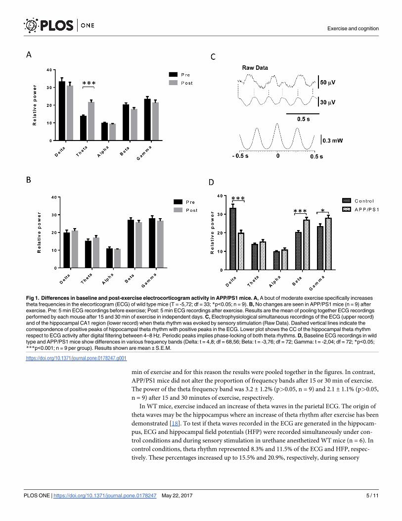
min of exercise and for this reason the results were pooled together in the figures. In contrast,
APP/PS1 mice did not alter the proportion of frequency bands after 15 or 30 min of exercise.
The power of the theta frequency band was 3.2 ± 1.2% (p>0.05, n = 9) and 2.1 ± 1.1% (p>0.05,
n = 9) after 15 and 30 minutes of exercise, respectively.
In WT mice, exercise induced an increase of theta waves in the parietal ECG. The origin of
theta waves may be the hippocampus where an increase of theta rhythm after exercise has been
demonstrated [18]. To test if theta waves recorded in the ECG are generated in the hippocam-
pus, ECG and hippocampal field potentials (HFP) were recorded simultaneously under con-
trol conditions and during sensory stimulation in urethane anesthetized WT mice (n = 6). In
control conditions, theta rhythm represented 8.3% and 11.5% of the ECG and HFP, respec-
tively. These percentages increased up to 15.5% and 20.9%, respectively, during sensory
Fig 1. Differences in baseline and post-exercise electrocorticogram activity in APP/PS1 mice. A, A bout of moderate exercise specifically increases
theta frequencies in the elecorticogram (ECG) of wild type mice (T = -5,72; df = 33; *p<0.05; n = 9). B, No changes are seen in APP/PS1 mice (n = 9) after
exercise. Pre: 5 min ECG recordings before exercise; Post: 5 min ECG recordings after exercise. Results are the mean of pooling together ECG recordings
performed by each mouse after 15 and 30 min of exercise in independent days. C, Electrophysiological simultaneous recordings of the ECG (upper record)
and of the hippocampal CA1 region (lower record) when theta rhythm was evoked by sensory stimulation (Raw Data). Dashed vertical lines indicate the
correspondence of positive peaks of hippocampal theta rhythm with positive peaks in the ECG. Lower plot shows the CC of the hippocampal theta rhythm
respect to ECG activity after digital filtering between 4–8 Hz. Periodic peaks implies phase-locking of both theta rhythms. D, Baseline ECG recordings in wild
type and APP/PS1 mice show differences in various frequency bands (Delta: t = 4,8; df = 68,56; Beta: t = -3,76; df = 72; Gamma: t = -2,04; df = 72; *p<0.05;
***p<0.001; n = 9 per group). Results shown are mean ± S.E.M.
https://doi.org/10.1371/journal.pone.0178247.g001
Exercise and cognition
PLOS ONE | https://doi.org/10.1371/journal.pone.0178247 May 22, 2017 5 / 11

stimulation of the animal’s back. Raw data showed theta activity in the HFP during sensory
stimulation while the ECG showed theta oscillations between larger delta waves evoked by the
anesthetic (Fig 1C, Raw Data). Moreover, cross-correlation (CC) of theta rhythm recorded in
the ECG and HFP showed that theta rhythms during sensory stimulation were in phase
because the CC showed periodic peaks around the zero reference (Fig 1C, lower plot). Thus,
the fact that both theta rhythms changed similarly in both conditions strongly suggests that the
theta rhythm recorded in the parietal cortex reflect, at least in part, the hippocampal theta
rhythm.
In control conditions (before exercise), WT mice (n = 9) showed a 34.4% of delta waves,
13.9% of theta, 9.7% of alpha, 19.4% of beta and 22.6% of gamma frequency bands. However,
APP/PS1 mice (n = 9) showed a significant decrease of the percentage of delta waves (19.5%;
p<0.001) and an increase of beta (26.9%; p<0.001) and gamma (27.9%; p = 0.04) frequencies.
Theta and alpha frequencies did not change in APP/PS1 mice (15.0% and 10.7%, respectively)
respect to values of WT animals, suggesting that the cortical activity is faster in APP/PS1 mice
at rest (Fig 1D).
Interestingly, as reported in human patients [19], blood lactate levels in APP/PS1 mice were
higher at rest, but did not increase after running, whereas in control mice a moderate increase
was seen (Table 1).
We submitted another group of exercised mice (30 minutes) to a Y maze, a test that mea-
sures spatial working memory [20]. The sedentary group remained in the treadmill without
running, for the same time. Under the relatively prolonged inter-trial conditions used in this
test, control exercised mice entered the novel arm significantly more times and spent more
time in it than the sedentary group, indicating that exercise enhanced working memory (Fig
2). Importantly, both sedentary and exercised mice were able to learn in the Y maze to the
same extent when using a shorter inter-trial time (S1C Fig). However, while both control and
APP/PS1 mice behaved similarly in the acquisition phase of the Y maze (Fig 2A), after exercise
the latter did not improve their performance in the Y maze (Fig 2B and 2C).
Discussion
In this proof-of-concept study we have taken advantage of the cognitive-promoting actions of
exercise [21, 22], its association to cognitive fitness [7, 8], and its effects on brain activity [23],
to show that normal mice improve cognitive skills in the Y maze after exercise. In parallel,
mice showed exercise-induced changes in the ECG pattern, specifically in the theta wave. Con-
versely, APP/PS1 mice did not improve memory skills after exercise and did not show changes
in theta after exercise. Collectively, these data indicate that exercise-induced increases in theta
-an ECG frequency associated to attention [24], and memory [25], result in improved memory
in healthy mice, but not in APP/PS1 mice, a well-established model of AD-like amyloidosis
and cognitive deterioration.
Most studies on the effect of exercise on the EEG have reported increased activity in the
alpha frequency band, which may reflect a state of decreased cortical activation in comparison
with states with high cognitive activity that increase beta and gamma and reduce alpha
Table 1. Blood lactate changes in wild type (WT) controls and AD (APP/PS1) mice after 15 and 30 minutes of moderate running exercise.
Pre-exercise
(mean ± sd)
15 min
(mean ± sd)
30 min
(mean ± sd)
WT 1.92 ± 0.93 3.28 ± 0.86 2.77 ± 1.43
APP/PS1 2.61 ± 0.24 2.94 ± 0.25 2.72 ± 0.26
https://doi.org/10.1371/journal.pone.0178247.t001
Exercise and cognition
PLOS ONE | https://doi.org/10.1371/journal.pone.0178247 May 22, 2017 6 / 11

frequency bands [26, 27]. However, new evidence suggests that regular physical activity can
impact cortical function and facilitate plasticity. Aerobic exercise has the capacity to induce
short-term neuroplasticity within the human motor cortex, as assessed through cortical cir-
cuits evoked by transcranial magnetic stimulation [28]. Our findings agree with these results
indicating that the hippocampal theta rhythm -involved in rodents in many plasticity processes
such as LTP [29], increases after exercise. APP/PS1 mice did not increase the power of the
theta frequency band after exercise, suggesting that plasticity processes may be reduced.
Accordingly, the EEG of AD patients shows an increase in slow frequencies (11–12).
Exercise is nowadays one of the most promoted approaches for prevention and treatment
of Alzheimer´s disease [30]. In all cases, based on pre-clinical studies, chronic exercise of vari-
ous intensities has been considered the appropriate regime. However, our results extend its
utility as a potential diagnostic tool when used acutely. We consider that this wide utility of
exercise is based on its ample neuroprotective actions, including increased brain perfusion,
neurotrophic input and metabolic fitness [31–36]. All these beneficial actions, necessary to
preserve normal levels of brain activity, may be partially diminished during aging, leading to
Fig 2. Y maze performance after exercise. A, Both control and APP/PS1 mice performed similar in the acquisition phase of the Y maze before exercise,
showing a similar rate of entries in each of the three arms of the maze. B, However, while control mice significantly improved their performance after exercise
in the Y maze, as determined by increased preference for the novel arm, APP/PS1 did not. C, Improved performance was corroborated by increased time
spent in the novel arm only in control mice. Sed: sedentary; Exe: exercised (n = 6 per group; one-way ANOVA (exercise x genotype interaction): F = 28.78,
df = 17; ***p<0.0001 control vs APP/PS1; post-hoc t-test: *p<0.05 vs sedentary).
https://doi.org/10.1371/journal.pone.0178247.g002
Exercise and cognition
PLOS ONE | https://doi.org/10.1371/journal.pone.0178247 May 22, 2017 7 / 11

gradual cognitive deterioration. We postulate that a bout of exercise unveils this gradual loss of
exercise neuroprotection before cognitive deterioration develops. This is reflected in an abnor-
mal pattern of brain activity recorded in the ECG in response to exercise; namely, no changes
in theta activity.
Brain activity patterns previously reported using ECG in AD mice had shown general
abnormalities [15] together with progressive changes along aging in all spectral frequencies
[37], or specifically in theta and delta bands [38]. Other reports also indicate the existence of
specific changes in theta and gamma activities [39–42], or in the whole ECG pattern [43].
These varied observations are probably due to the different types of AD mouse models used.
However, collectively these data suggest that AD animal models exhibit altered cortical excit-
ability and hippocampal dysrhythmicity. Our findings in APP/PS1 animals show cortical
hyperexcitability because they exhibited a decrease of delta power and an increase in faster fre-
quency bands at rest. These results are in agreement with the presence of seizure activity in
AD animal models [44] and with the already mentioned fact that people with AD are more
likely to develop epilepsy [45]. Our observations add to the growing potential translability of
rodent studies to humans using electrophysiological recordings [46].
Indeed, with the idea of translating these observations to the clinical practice, our results
indicate the feasibility of an easy-to-carry-out test to determine cognitive health in the general
population based on accessible diagnostic tools. Similar translation studies from rodents [47]
to humans [48] has proven successful for exercise as a protective measure against AD. There-
fore, a simple diagnostic procedure derived from our observations and others [49], would con-
sist in submitting test subjects to moderate exercise in conjunction with recording EEG
activity before and after exercise. Individuals not showing changes after exercise in EEG activ-
ity will be categorized as “at-risk” and should undergo further testing. On-going studies in
healthy volunteers and cognitively deteriorated subjects will help clarify the possible transla-
tion of this test to the clinic.
Supporting information
S1 Fig. A, Aβ immunostaining of control (WT) and APP/PS1 (AD) show the presence of
small deposits (Aβ plaques) only in the latter. B, Time line of experimental procedure for Y
maze plus exercise used in the experiments shown in Fig 2. C, Both sedentary (white bars) and
exercised (grey bars) wild type mice learn the Y maze task as indicated by increased entries to
the novel arm of the maze when using a shorter (60 min) inter-trial time (F = 15.774; df = 30;���p<0.001; n = 9 per group).
(JPG)
Author Contributions
Conceptualization: ITA.
Data curation: AMS VM AN.
Formal analysis: AMS VM AN.
Funding acquisition: AMS AN ITA.
Investigation: AMS VM AN AMF.
Methodology: AN ITA.
Project administration: ITA.
Exercise and cognition
PLOS ONE | https://doi.org/10.1371/journal.pone.0178247 May 22, 2017 8 / 11
Resources: AN ITA.
Software: AN.
Supervision: AN ITA.
Validation: AMS VM AN AMF ITA.
Visualization: AMS VM AN AMF ITA.
Writing – original draft: ITA.
Writing – review & editing: AMS VM AN AMF ITA.
References1. Shah H, Albanese E, Duggan C, Rudan I, Langa KM, Carrillo MC, et al. Research priorities to reduce
the global burden of dementia by 2025. Lancet Neurol. 2016; 15(12):1285–94. https://doi.org/10.1016/
S1474-4422(16)30235-6 PMID: 27751558
2. Dubois B, Hampel H, Feldman HH, Scheltens P, Aisen P, Andrieu S, et al. Preclinical Alzheimer’s dis-
ease: Definition, natural history, and diagnostic criteria. Alzheimers Dement. 2016; 12(3):292–323.
https://doi.org/10.1016/j.jalz.2016.02.002 PMID: 27012484
3. Scheltens P, Blennow K, Breteler MM, de Strooper B, Frisoni GB, Salloway S, et al. Alzheimer’s dis-
ease. Lancet. 2016; 388(10043):505–17. https://doi.org/10.1016/S0140-6736(15)01124-1 PMID:
26921134
4. Winter B, Breitenstein C, Mooren FC, Voelker K, Fobker M, Lechtermann A, et al. High impact running
improves learning. Neurobiol Learn Mem. 2007; 87(4):597–609. https://doi.org/10.1016/j.nlm.2006.11.
003 PMID: 17185007
5. Kamijo K, Hayashi Y, Sakai T, Yahiro T, Tanaka K, Nishihira Y. Acute effects of aerobic exercise on
cognitive function in older adults. J Gerontol B Psychol Sci Soc Sci. 2009; 64(3):356–63. https://doi.org/
10.1093/geronb/gbp030 PMID: 19363089
6. Siette J, Reichelt AC, Westbrook RF. A bout of voluntary running enhances context conditioned fear, its
extinction, and its reconsolidation. Learn Mem. 2014; 21(2):73–81. PubMed Central PMCID:
PMCPMC3895230. https://doi.org/10.1101/lm.032557.113 PMID: 24429425
7. Muller J, Chan K, Myers JN. Association Between Exercise Capacity and Late Onset of Dementia, Alz-
heimer Disease, and Cognitive Impairment. Mayo Clin Proc. 2017; 92(2):211–7. https://doi.org/10.
1016/j.mayocp.2016.10.020 PMID: 28082018
8. Boyle PA, Buchman AS, Wilson RS, Leurgans SE, Bennett DA. Association of muscle strength with the
risk of Alzheimer disease and the rate of cognitive decline in community-dwelling older persons. Arch
Neurol. 2009; 66(11):1339–44. PubMed Central PMCID: PMCPMC2838435. https://doi.org/10.1001/
archneurol.2009.240 PMID: 19901164
9. Enders H, Cortese F, Maurer C, Baltich J, Protzner AB, Nigg BM. Changes in cortical activity measured
with EEG during a high-intensity cycling exercise. J Neurophysiol. 2016; 115(1):379–88. PubMed Cen-
tral PMCID: PMCPMC4760484. https://doi.org/10.1152/jn.00497.2015 PMID: 26538604
10. Vissing J, Andersen M, Diemer NH. Exercise-induced changes in local cerebral glucose utilization in
the rat. J Cereb Blood Flow Metab. 1996; 16(4):729–36. https://doi.org/10.1097/00004647-199607000-
00025 PMID: 8964814
11. Jeong J. EEG dynamics in patients with Alzheimer’s disease. Clin Neurophysiol. 2004; 115(7):1490–
505. https://doi.org/10.1016/j.clinph.2004.01.001 PMID: 15203050
12. van der HK, Vein AA, Reijntjes RH, Westendorp RG, Bollen EL, van Buchem MA, et al. EEG correlates
in the spectrum of cognitive decline. Clin Neurophysiol. 2007; 118(9):1931–9. https://doi.org/10.1016/j.
clinph.2007.05.070 PMID: 17604688
13. Fonseca LC, Tedrus GM, Letro GH, Bossoni AS. Dementia, mild cognitive impairment and quantitative
EEG in patients with Parkinson’s disease. Clin EEG Neurosci. 2009; 40(3):168–72. https://doi.org/10.
1177/155005940904000309 PMID: 19715179
14. Faigle R, Sutter R, Kaplan PW. Electroencephalography of encephalopathy in patients with endocrine
and metabolic disorders. J Clin Neurophysiol. 2013; 30(5):505–16. PubMed Central PMCID:
PMCPMC3826953. https://doi.org/10.1097/WNP.0b013e3182a73db9 PMID: 24084183
15. Born HA, Kim JY, Savjani RR, Das P, Dabaghian YA, Guo Q, et al. Genetic suppression of transgenic
APP rescues Hypersynchronous network activity in a mouse model of Alzeimer’s disease. J Neurosci.
Exercise and cognition
PLOS ONE | https://doi.org/10.1371/journal.pone.0178247 May 22, 2017 9 / 11

2014; 34(11):3826–40. PubMed Central PMCID: PMCPMC3951689. https://doi.org/10.1523/
JNEUROSCI.5171-13.2014 PMID: 24623762
16. Fernandez AM, Jimenez S, Mecha M, Davila D, Guaza C, Vitorica J, et al. Regulation of the phospha-
tase calcineurin by insulin-like growth factor I unveils a key role of astrocytes in Alzheimer’s pathology.
Mol Psychiatry. 2012; 17(7):705–18. https://doi.org/10.1038/mp.2011.128 PMID: 22005929
17. Sarter M, Bodewitz G, Stephens DN. Attenuation of scopolamine-induced impairment of spontaneous
alteration behaviour by antagonist but not inverse agonist and agonist beta-carbolines. Psychopharma-
cology (Berl). 1988; 94(4):491–5.
18. Rivas J, Gaztelu JM, Garcia-Austt E. Changes in hippocampal cell discharge patterns and theta rhythm
spectral properties as a function of walking velocity in the guinea pig. Exp Brain Res. 1996; 108(1):113–
8. PMID: 8721159
19. Mancuso M, Filosto M, Bosetti F, Ceravolo R, Rocchi A, Tognoni G, et al. Decreased platelet cyto-
chrome c oxidase activity is accompanied by increased blood lactate concentration during exercise in
patients with Alzheimer disease. Exp Neurol. 2003; 182(2):421–6. PMID: 12895452
20. Paul C-M, Magda G, Abel S. Spatial memory: Theoretical basis and comparative review on experimen-
tal methods in rodents. Behavioural Brain Research. 2009; 203(2):151–64. https://doi.org/10.1016/j.
bbr.2009.05.022 PMID: 19467271
21. Byun K, Hyodo K, Suwabe K, Ochi G, Sakairi Y, Kato M, et al. Positive effect of acute mild exercise on
executive function via arousal-related prefrontal activations: an fNIRS study. Neuroimage. 2014;
98:336–45. https://doi.org/10.1016/j.neuroimage.2014.04.067 PMID: 24799137
22. Dimitrova J, Hogan M, Khader P, O’Hora D, Kilmartin L, Walsh JC, et al. Comparing the effects of an
acute bout of physical exercise with an acute bout of interactive mental and physical exercise on electro-
physiology and executive functioning in younger and older adults. Aging Clin Exp Res. 2016.
23. Brummer V, Schneider S, Abel T, Vogt T, Struder HK. Brain cortical activity is influenced by exercise
mode and intensity 1. Med Sci Sports Exerc. 2011; 43(10):1863–72. https://doi.org/10.1249/MSS.
0b013e3182172a6f PMID: 21364475
24. Sellers KK, Yu C, Zhou ZC, Stitt I, Li Y, Radtke-Schuller S, et al. Oscillatory Dynamics in the Frontopar-
ietal Attention Network during Sustained Attention in the Ferret. Cell Rep. 2016; 16(11):2864–74.
PubMed Central PMCID: PMCPMC5024719. https://doi.org/10.1016/j.celrep.2016.08.055 PMID:
27626658
25. Klimesch W. EEG alpha and theta oscillations reflect cognitive and memory performance: a review and
analysis. Brain Res Brain Res Rev. 1999; 29(2–3):169–95. PMID: 10209231
26. Steriade M, Llinas RR. The functional states of the thalamus and the associated neuronal interplay.
Physiol Rev. 1988; 68(3):649–742. PMID: 2839857
27. Crabbe JB, Dishman RK. Brain electrocortical activity during and after exercise: a quantitative synthe-
sis. Psychophysiology. 2004; 41(4):563–74. https://doi.org/10.1111/j.1469-8986.2004.00176.x PMID:
15189479
28. Lulic T, El-Sayes J, Fassett HJ, Nelson AJ. Physical activity levels determine exercise-induced changes
in brain excitability. PLoS One. 2017; 12(3):e0173672. PubMed Central PMCID: PMCPMC5344515.
https://doi.org/10.1371/journal.pone.0173672 PMID: 28278300
29. Vertes RP. Hippocampal theta rhythm: a tag for short-term memory. Hippocampus. 2005; 15(7):923–
35. https://doi.org/10.1002/hipo.20118 PMID: 16149083
30. Forbes D, Thiessen EJ, Blake CM, Forbes SC, Forbes S. Exercise programs for people with dementia.
Cochrane Database Syst Rev. 2013; 12:CD006489.
31. Patten AR, Sickmann H, Hryciw BN, Kucharsky T, Parton R, Kernick A, et al. Long-term exercise is
needed to enhance synaptic plasticity in the hippocampus 1. Learn Mem. 2013; 20(11):642–7. https://
doi.org/10.1101/lm.030635.113 PMID: 24131795
32. Pedersen BK, Pedersen M, Krabbe KS, Bruunsgaard H, Matthews VB, Febbraio MA. Role of exercise-
induced brain-derived neurotrophic factor production in the regulation of energy homeostasis in mam-
mals. Exp Physiol. 2009; 94(12):1153–60. https://doi.org/10.1113/expphysiol.2009.048561 PMID:
19748969
33. Ropelle ER, Flores MB, Cintra DE, Rocha GZ, Pauli JR, Morari J, et al. IL-6 and IL-10 Anti-Inflammatory
Activity Links Exercise to Hypothalamic Insulin and Leptin Sensitivity through IKK+ and ER Stress Inhi-
bition. PLoS Biol. 2010; 8(8):e1000465. https://doi.org/10.1371/journal.pbio.1000465 PMID: 20808781
34. Stranahan AM, Lee K, Becker KG, Zhang Y, Maudsley S, Martin B, et al. Hippocampal gene expression
patterns underlying the enhancement of memory by running in aged mice. Neurobiol Aging. 2008.
35. Takimoto M, Hamada T. Acute exercise increases brain region-specific expression of MCT1, MCT2,
MCT4, GLUT1, and COX IV proteins. J Appl Physiol (1985). 2014; 116(9):1238–50.
Exercise and cognition
PLOS ONE | https://doi.org/10.1371/journal.pone.0178247 May 22, 2017 10 / 11

36. Trejo JL, Carro E, Torres-Aleman I. Circulating insulin-like growth factor I mediates exercise-induced
increases in the number of new neurons in the adult hippocampus. J Neurosci. 2001; 21(5):1628–34.
PMID: 11222653
37. Jyoti A, Plano A, Riedel G, Platt B. Progressive age-related changes in sleep and EEG profiles in the
PLB1Triple mouse model of Alzheimer’s disease. Neurobiol Aging. 2015; 36(10):2768–84. https://doi.
org/10.1016/j.neurobiolaging.2015.07.001 PMID: 26239174
38. Jyoti A, Plano A, Riedel G, Platt B. EEG, activity, and sleep architecture in a transgenic AbetaPPswe/
PSEN1A246E Alzheimer’s disease mouse. J Alzheimers Dis. 2010; 22(3):873–87. https://doi.org/10.
3233/JAD-2010-100879 PMID: 20858963
39. Siwek ME, Muller R, Henseler C, Trog A, Lundt A, Wormuth C, et al. Altered theta oscillations and aber-
rant cortical excitatory activity in the 5XFAD model of Alzheimer’s disease. Neural Plast. 2015;
2015:781731. PubMed Central PMCID: PMCPMC4398951. https://doi.org/10.1155/2015/781731
PMID: 25922768
40. Ittner AA, Gladbach A, Bertz J, Suh LS, Ittner LM. p38 MAP kinase-mediated NMDA receptor-depen-
dent suppression of hippocampal hypersynchronicity in a mouse model of Alzheimer’s disease. Acta
Neuropathol Commun. 2014; 2:149. PubMed Central PMCID: PMCPMC4212118. https://doi.org/10.
1186/s40478-014-0149-z PMID: 25331068
41. Goutagny R, Gu N, Cavanagh C, Jackson J, Chabot JG, Quirion R, et al. Alterations in hippocampal
network oscillations and theta-gamma coupling arise before Abeta overproduction in a mouse model of
Alzheimer’s disease. Eur J Neurosci. 2013; 37(12):1896–902. https://doi.org/10.1111/ejn.12233 PMID:
23773058
42. Rubio SE, Vega-Flores G, Martinez A, Bosch C, Perez-Mediavilla A, del Rio J, et al. Accelerated aging
of the GABAergic septohippocampal pathway and decreased hippocampal rhythms in a mouse model
of Alzheimer’s disease. FASEB J. 2012; 26(11):4458–67. https://doi.org/10.1096/fj.12-208413 PMID:
22835830
43. Schneider F, Baldauf K, Wetzel W, Reymann KG. Behavioral and EEG changes in male 5xFAD mice.
Physiol Behav. 2014; 135:25–33. https://doi.org/10.1016/j.physbeh.2014.05.041 PMID: 24907698
44. Palop JJ, Chin J, Roberson ED, Wang J, Thwin MT, Bien-Ly N, et al. Aberrant excitatory neuronal activ-
ity and compensatory remodeling of inhibitory hippocampal circuits in mouse models of Alzheimer’s dis-
ease. Neuron. 2007; 55(5):697–711. https://doi.org/10.1016/j.neuron.2007.07.025 PMID: 17785178
45. Born HA. Seizures in Alzheimer’s disease. Neuroscience. 2015; 286:251–63. https://doi.org/10.1016/j.
neuroscience.2014.11.051 PMID: 25484360
46. Platt B, Welch A, Riedel G. FDG–PET imaging, EEG and sleep phenotypes as translational biomarkers
for research in Alzheimer’s disease. Biochemical Society Transactions. 2011; 39(4):874–80. https://doi.
org/10.1042/BST0390874 PMID: 21787316
47. Huttenrauch M, Brauss A, Kurdakova A, Borgers H, Klinker F, Liebetanz D, et al. Physical activity
delays hippocampal neurodegeneration and rescues memory deficits in an Alzheimer disease mouse
model. Transl Psychiatry. 2016; 6:e800. https://doi.org/10.1038/tp.2016.65 PMID: 27138799
48. Hoffmann K, Sobol NA, Frederiksen KS, Beyer N, Vogel A, Vestergaard K, et al. Moderate-to-High
Intensity Physical Exercise in Patients with Alzheimer’s Disease: A Randomized Controlled Trial. J Alz-
heimers Dis. 2015; 50(2):443–53.
49. Hyodo K, Dan I, Kyutoku Y, Suwabe K, Byun K, Ochi G, et al. The association between aerobic fitness
and cognitive function in older men mediated by frontal lateralization 1. Neuroimage. 2016; 125:291–
300. https://doi.org/10.1016/j.neuroimage.2015.09.062 PMID: 26439424
Exercise and cognition
PLOS ONE | https://doi.org/10.1371/journal.pone.0178247 May 22, 2017 11 / 11