the effect of acute aerobic exercise on the consolidation
TRANSCRIPT

The Effect of Acute Aerobic Exercise on the Consolidation of Motor Memories
by
Sarah Holman
A thesis
presented to the University of Waterloo
in fulfillment of the
thesis requirement for the degree of
Master of Science
in
Kinesiology
Waterloo, Ontario, Canada, 2019
© Sarah Holman 2019

ii
Author’s Declaration
I hereby declare that I am the sole author of this thesis. This is a true copy of the thesis, including
any required final revisions, as accepted by my examiners.
I understand that my thesis may be made electronically available to the public.

iii
Abstract
Previous research has shown that acute aerobic exercise performed prior to motor training
can assist with motor skill acquisition through enhancement of motor cortical plasticity.
Recently, studies using high intensity interval training performed post-motor training have found
improvements in the retention of the motor skill. This suggests that exercise performed post-
motor training may enhance motor memory consolidation, although the mechanisms of this are
unclear. We hypothesized that acute continuous moderate intensity exercise performed post-
motor training would also assist with motor skill retention and that this behavioural change
would be positively correlated with neural markers of cortical plasticity. Thirty-three participants
were randomly assigned to one of two groups: exercise (EXE) or control (CON). During the first
visit, participants completed a motor training session of a bimanual wrist flexion task using wrist
movements to control the cursor position on a computer screen to reach a target. Movement to
the target was cued, allowing for the generation of a cortical movement-related potential (MRP).
MRP modulations represent changes in the excitability of the brain prior to movement which are
associated with task performance. After motor training, EXE performed a session of continuous
moderate intensity exercise on a recumbent bike for 20 minutes (70% of heart rate reserve
(HRR)). CON read for the same amount of time. Both groups completed two post-training tests
after the exercise or rest: one 10 minutes after the exercise/rest session (post-training test 1), and
one once heart rate returned to resting level in EXE (post-training test 2) or 30 minutes after rest
in CON. Participants returned to the lab 1 day and 7 days later to complete retention and transfer
tests of the task. MRPs were measured using electroencephalography (EEG) to investigate neural
markers related to motor performance and exercise during the first visit. To assess behavioural
changes measures of speed and accuracy were collected at all timepoints as response time (RT)

iv
and root mean square (RMS) of the difference in the actual from the ideal trajectory of the
cursor.
Results show that EXE had a smaller change in accuracy scores compared to CON at
both retention timepoints, however this group difference was only significant at the 7 day
retention. There were no significant differences in speed between the groups at the retention tests
and no significant differences in accuracy or speed between the groups at the transfer tests.
Consistent with previous motor training studies our MRP data shows an increase in amplitude
from early to late training, however this was only significant at CZ and not FCZ. MRP amplitude
was not significantly increased after exercise at the post-training test 2. Correlational analysis
revealed a significant correlation between the change in the MRP amplitude sum from early to
late training and the change in RT from the last 5 trials of post-training 2 to retention 1. This was
only observed in EXE and when both groups were pooled together. Our results suggest that post-
motor training exercise helps to retain the accuracy of the skill after motor training. Additionally,
there may be a relationship between excitability increases during training and performance of the
skill at retention which may be enhanced with post-motor training exercise. These results inform
motor learning paradigms and future studies with other populations including older adults and
neurorehabilitation patients.

v
Acknowledgements
I would not have been able to complete this thesis without the following people.
First, Dr. Richard Staines, thank you for your constant support and mentorship. You are an
incredible supervisor and I am so grateful to have studied under your guidance for the past 2
years. To my committee members, Dr. Laura Middleton and Dr. Sean Meehan, thank you for
providing me with different perspectives. Your questions and suggestions helped to make my
project and this document something I am proud of.
To my lab mates and peers, thank you for sharing your advice and expertise. I am lucky to have
been surrounded by such intelligent, supportive, and encouraging individuals.
To my family, thank you for always believing in me. I appreciate everything each of you has
done to support me throughout my education. I could not ask for better parents or siblings. To
Zen, you live up to your name, thank you for keeping me grounded.

vi
Table of Contents
List of Figures............................................................................................................................. viii
List of Tables ................................................................................................................................ ix
List of Abbreviations .................................................................................................................... x
1.0 Introduction ............................................................................................................................. 1
2.0 Literature Review ................................................................................................................... 2
2.1 MECHANISMS OF NEUROPLASTICITY .......................................................................................................... 2 2.1.1 Synaptic plasticity. ........................................................................................................................ 2 2.1.2 Priming. ......................................................................................................................................... 4 2.1.3 Encoding motor memories. ........................................................................................................... 5 2.1.4 Consolidating motor memories. .................................................................................................... 5 2.1.5 Plasticity-related compounds. ....................................................................................................... 7
2.2 ASSESSING PLASTICITY .............................................................................................................................. 9 2.2.1 Movement-related cortical potentials (MRP). .............................................................................. 9 2.2.2 Contingent negative variation. .................................................................................................... 10 2.2.3 Assessing change in MRPs. ......................................................................................................... 11 2.2.4 Alternative analysis of EEG data. ............................................................................................... 11 2.2.5 Limitations of EEG. ..................................................................................................................... 12
2.3 MOTOR LEARNING .................................................................................................................................... 13 2.3.1 Motor skill acquisition and retention. ......................................................................................... 13 2.3.2 Behavioural changes associated with motor learning. ............................................................... 15 2.3.3 Cognition in motor learning........................................................................................................ 15 2.3.4 Neural networks of motor learning. ............................................................................................ 16 2.3.5 Neural markers of motor learning. ............................................................................................. 17 2.3.6 Bimanual motor training (BMT). ................................................................................................ 19
2.4 EXERCISE .................................................................................................................................................. 21 2.4.1 Acute and chronic effects of exercise. ......................................................................................... 21 2.4.2 The effects of acute exercise on cortical plasticity...................................................................... 22 2.4.3 The effects of acute exercise on motor learning and retention. .................................................. 24 2.4.4 The correlation of neural markers and behavioural changes. .................................................... 26
3.0 Rationale, Objectives, and Hypotheses ............................................................................... 29
3.1 RATIONALE ............................................................................................................................................... 29
3.2 OBJECTIVES & HYPOTHESES .................................................................................................................... 30
4.0 Methods.................................................................................................................................. 32
4.1 PARTICIPANTS........................................................................................................................................... 32
4.2 PROCEDURES. ........................................................................................................................................... 32
4.3 EXERCISE. ................................................................................................................................................. 34
4.4 MOTOR TASK. ........................................................................................................................................... 35
4.5 DATA ACQUISITION. .................................................................................................................................. 38

vii
4.6 DATA ANALYSIS. ...................................................................................................................................... 39
5.0 Proposed Hypotheses Testing .............................................................................................. 42
6.0 Results .................................................................................................................................... 44
6.1 PARTICIPANT CHARACTERISTICS .............................................................................................................. 44
6.2 SUPPLEMENTARY DATA ........................................................................................................................... 45
6.3 EXERCISE DATA........................................................................................................................................ 46
6.4 BEHAVIOURAL DATA ................................................................................................................................ 49
6.5 NEUROPHYSIOLOGICAL DATA .................................................................................................................. 53
6.6 CORRELATIONAL DATA ............................................................................................................................ 56
7.0 Discussion .............................................................................................................................. 58
7.1 PARTICIPANT CHARACTERISTICS .............................................................................................................. 59
7.2 SUPPLEMENTARY DATA ........................................................................................................................... 59
7.3 EXERCISE DATA........................................................................................................................................ 60
7.4 BEHAVIOURAL DATA ................................................................................................................................ 60
7.5 NEUROPHYSIOLOGICAL DATA .................................................................................................................. 64
7.6 CORRELATIONAL DATA ............................................................................................................................ 66
8.0 Limitations ............................................................................................................................. 69
9.0 Conclusions and Future Directions ..................................................................................... 70
10.0 References ............................................................................................................................ 72

viii
List of Figures
Figure 1 34
Figure 2 37
Figure 3 38
Figure 4 38
Figure 5 50
Figure 6 51
Figure 7 52
Figure 8 53
Figure 9 54
Figure 10 54
Figure 11 55
Figure 12 55
Figure 13 57

ix
List of Tables
Table 1 45
Table 2 48

x
List of Abbreviations
AMPA Alpha-amino-3-hydroxy-5-methyl-4-isoxazolepropionic acid
BDNF Brain derived neurotropic factor
BMI Body mass index
BMT Bimanual motor training
BOLD Blood-oxygen-level dependent
BP Bereitschaftspotential
Ca2+ Calcium
CMC Corticomuscular coherence
CNV Contingent negative variation
CON Control group
cTBS Continuous theta burst stimulation
DA Dopamine
DLPFC Dorsolateral-prefrontal cortex
ECR Extensor carpi radialis
EEG Electroencephalography
EHQ Edinburgh Handedness Questionnaire
E-LTP Early long term potentiation
EMG Electromyography
EPI Epinephrine
EPSP Excitatory postsynaptic potential
ERD Event-related desynchronization
EXE Exercise group
FCR Flexor carpi radialis
fMRI Functional magnetic resonance imaging
GABA Gamma-aminobutyric acid
GABAA / GABAB Subtype of gamma-aminobutyric acid
GAQ Get Active Questionnaire
HR Heart rate
HRR Heart rate reserve
ICF Intracortical facilitation
IGF-1 Insulin-like growth factor 1
IPAQ International Physical Activity Questionnaire
iTBS Intermittent theta burst stimulation
LICI
L-LTP
Long-interval intracortical inhibition
Late long term potentiation
LTD Long term depression
LTP Long term potentiation
M1 Primary motor cortex
MEP Motor evoked potential
MET Metabolic equivalent
mRNA Messenger ribonucleic acid
MRP Movement-related cortical potential
Mg2+ Magnesium
Na+ Sodium

xi
NE Norepinephrine
NMDA N-methyl-D-aspartate
O2 Oxygen
PANAS Positive and Negative Affect Schedule
PAS Paired associative stimulation
PFC Prefrontal cortex
PMC Premotor cortex
RAP Re-afferent potential
RMS Root mean square
RPE Rate of perceived exertion
RPM Revolutions per minute
RT Response time
rTMS Repetitive transcranial magnetic stimulation
S1 Primary somatosensory cortex
SICI Short-interval intracortical inhibition
SMA Supplementary motor area
SMHSQ St. Mary’s Hospital Sleep Questionnaire
SSS Stanford Sleepiness Scale
tDCS Transcranial direct current stimulation (tDCS)
TMS Transcranial magnetic stimulation
VEGF Vascular endothelial growth factor

1
1.0 Introduction
The ability of humans to learn new motor skills is vital to our survival. Motor skills allow
us to interact with the external environment, live independently, and engage in recreational
activities. Across the average human’s lifespan there are scenarios in which new motor skills
need to be learned. Research has helped to inform motor learning paradigms including strategies
to enhance motor learning before, during, and after motor training. The time period after practice
is important for the consolidation of the motor memory, which is the encoded sequence of
muscle activations, and depending on what occurs during this time, retention of the motor skill
can be enhanced or diminished. One such strategy shown to enhance the consolidation of motor
memories is an acute session of high intensity interval training (Roig, Skriver, Lundbye-Jensen,
Kiens, & Nielsen, 2012). The purpose of this study was to examine whether continuous moderate
intensity post-motor training exercise would assist with the retention of a bimanual motor skill.
A secondary objective of the study was to investigate the neurophysiological changes in the brain
during and post-motor training in an attempt to correlate the hypothesized behavioural
improvement with neural markers of motor learning and consolidation.

2
2.0 Literature Review
2.1 Mechanisms of Neuroplasticity
2.1.1 Synaptic plasticity. Neuroplasticity is the ability of the brain to adapt in response
to stimuli. There are various forms of neuroplasticity, one form commonly studied is synaptic
plasticity, in which the adaptation occurs at the level of the synapse. This process is thought to be
an underlying mechanism of many types of learning and memory, including that of motor skills.
Two types of synaptic plasticity are long term potentiation (LTP) and long term depression
(LTD). LTP was first discovered in the rabbit hippocampus and is the strengthening of the
synaptic connections through high-frequency signals (Lømo, 1966). LTD is the weakening of
synaptic connections through low frequency signals or through the temporal unpairing of a
stimulus with an action potential (Ito & Kano, 1982). These processes are input-specific,
meaning that they only affect the synapses that are activated and not all synapses on the involved
neurons. Other properties of LTP and LTD include associativity, and cooperativity. Associativity
describes the phenomenon that although a weak stimulation in isolation will not induce LTP,
when a weak stimulation is paired with a strong stimulation from a different presynaptic neuron,
both of the synapses will be involved in LTP. Cooperativity involves a similar process; however,
it occurs when multiple weak stimuli converge onto a single postsynaptic cell. The sum of these
weak stimuli produce a stimulus strong enough to induce LTP in the postsynaptic cell.
LTP and LTD occur as a result of ionotropic glutamate receptor activity: N-methyl-D-
aspartate (NMDA), and alpha-amino-3-hydroxy-5-methyl-4-isoxazolepropionic acid (AMPA). In
LTP, both the pre- and postsynaptic neurons must be active (depolarized). This depolarization is
the result of glutamate being released in the synaptic cleft. Glutamate binds to both NMDA and
AMPA receptors on the postsynaptic cell. When this occurs, AMPA opens and allows sodium
3
(Na+) into the cell, causing excitatory postsynaptic potentials (EPSPs) reflective of
depolarization in the postsynaptic cell. This depolarization removes the magnesium (Mg2+) block
on the NMDA receptor pore on the postsynaptic cell, allowing the flow of ions in. Calcium
(Ca2+) and Na+ enter the postsynaptic cell through the NMDA receptor and activate a second-
messenger pathway that causes the addition of more AMPA receptors into the postsynaptic
membrane, allowing more glutamate to bind to these newly available receptors. This results in a
larger postsynaptic response the next time glutamate is released into the synapse. This process is
called early LTP (E-LTP). With enough stimulation, late LTP (L-LTP) results in the addition of
a new dendritic spine which requires genetic modifications and protein synthesis signalled
through second-messenger systems. The transition from E-LTP to L-LTP is thought to occur as a
result of a tagging process, termed the synaptic tagging hypothesis (Frey & Morris, 1998).
Specifically, initial changes within the cell “tag” a synapse to eventually undergo L-LTP
(Redondo & Morris, 2011). Researchers have suggested that increases in levels of
neurotransmitters post-exercise may assist this tagging process (Thomas, Beck, et al., 2016).
LTD is the opposing process of LTP, resulting in the removal of AMPA receptors from the
postsynaptic cell and the reduction of dendritic spines.
Depending on the duration and intensity of the stimuli and the environment that the
subject occupies, LTP and LTD can last from hours to months. In humans, LTP and LTD are
difficult to explore directly however, there is evidence that interventions such as transcranial
magnetic stimulation (TMS) cause LTP and LTD-like plasticity in the primary motor cortex
(M1) (Huang, Chen, Rothwell, & Wen, 2007; Ziemann, Iliac ́, Pauli, Meintzschel, & Ruge,
2004). LTP and LTD-like plasticity are also thought to occur in response to motor training
(Bütefisch et al., 2000; Nudo, 2003) and can be enhanced through different exercise intensities

4
(Mang, Snow, Campbell, Ross, & Boyd, 2014; McDonnell, Buckley, Opie, Ridding, & Semmler,
2013; Singh, Neva, & Staines, 2014).
2.1.2 Priming. Priming refers to any action that increases the potential for neuroplasticity
to occur. Exercise is thought to prime the cortex for synaptic plasticity, the specific mechanisms
of which are not fully understood. It has been proposed that exercise acts to lower the threshold
for the induction of LTP by increasing the availability of LTP-related compounds (Farmer et al.,
2004). For example, acute exercise increases levels of peripheral brain derived neurotropic factor
(BDNF) (Skriver et al., 2014; Winter et al., 2007). BDNF is a neurotrophin that supports growth
of new synapses. As described below, BDNF and other compounds are involved in the various
stages of LTP and may assist LTP through the described mechanisms. Increases in BDNF may
also increase the likelihood for LTP to occur by decreasing inhibition in M1. This reduction of
inhibition is observed in TMS studies that have shown that a single session of moderate intensity
exercise results in decreases in short-interval intracortical inhibition (SICI) (Singh, Duncan,
Neva, & Staines, 2014; Singh, Neva, et al., 2014; Smith, Goldsworthy, Garside, Wood, &
Ridding, 2014). Cortical inhibition is mediated by gamma-aminobutyric acid (GABA), an
inhibitory neurotransmitter. A subtype of GABA receptors, GABAA receptors, are thought to
mediate SICI (Sanger, Garg, & Chen, 2001). Animal research has discovered that in the
hippocampus, BDNF reduces the amount of postsynaptic GABAA receptors, which causes a
reduction in the amplitude of postsynaptic inhibitory currents (Brünig, Penschuck, Berninger,
Benson, & Fritschy, 2001). Therefore, one mechanism through which exercise may prime M1
for LTP-like plasticity is by increasing BDNF levels which reduces the presentation of
postsynaptic GABAA receptors, thereby reducing intracortical inhibition. While most of the
BDNF levels measured in humans post-exercise are peripheral, there is evidence to suggest that

5
peripheral BDNF is positively correlated with brain BDNF levels (Klein et al., 2011). As well,
after exercise the increase in peripheral BDNF is likely driven by an increase in brain BDNF
(Rasmussen et al., 2009).
2.1.3 Encoding motor memories. Exercise performed prior to motor training has been
shown to benefit motor acquisition (Statton, Encarnacion, Celnik, & Bastian, 2015). This is
thought to be achieved through priming encoding mechanisms. Encoding is the initial formation
of the motor memory, which mechanistically aligns with a short-term change at the synapse, E-
LTP. Research examining the effects of cortical stimulation on motor memory has shown that
TMS enhances the encoding of motor memories through LTP-like mechanisms (Butefisch,
Khurana, Kopylev, & Cohen, 2004). Animal studies have found associations between a decrease
in the threshold for synaptic plasticity to occur and improvements in memory encoding
(Stackman et al., 2002). This suggests that exercise performed before motor training may prime
the central nervous system for the encoding of the motor memory. It is also possible that exercise
performed before motor acquisition can assist with the consolidation of motor memories. Based
on past studies, only high intensity and not moderate intensity exercise performed pre-motor
training has a beneficial effect on both acquisition and retention scores (Mang et al., 2014; Roig
et al., 2012; Snow et al., 2016; Statton et al., 2015). This suggests that high intensity exercise
may increase LTP-related compounds (described below) long enough to affect consolidation
mechanisms (Roig et al., 2016; Statton et al., 2015).
2.1.4 Consolidating motor memories. On the other hand, when exercise is performed
after motor practice, it assists with the consolidation of motor memories (Roig et al., 2016, 2012;
Thomas et al., 2016; Thomas, Beck, et al., 2016; Thomas, Johnsen, et al., 2016). Consolidation is
when the encoded sequence is solidified and stabilized. It has been proposed that declarative

6
memories, memories of facts and events, are consolidated at two levels: synaptic consolidation
and systems consolidation (Dudai, 2012). Synaptic consolidation is a quick process, taking
minutes to hours, and lasting hours. L-LTP is thought to be the mechanism behind consolidation
at the synaptic level. Systems consolidation is a longer process, taking between days to years,
involving a shift in the storage of a memory trace in the brain. Although these concepts were first
proposed for declarative memory, it is possible that these forms of consolidation are also present
in procedural memory of skills and tasks, including motor memories. It is likely that the
procedural processes have the same mechanisms, just in different areas of the brain (Brashers-
Krug, Shadmehr, & Bizzi, 1996) such as M1 (Muellbacher et al., 2002). In animals, some
research has suggested that the formation of new dendritic spines (L-LTP) can occur in the
contralateral M1 after just one motor training session (Xu et al., 2009), however other
researchers have only found synaptogenesis in M1 after multiple days of motor training sessions
(Kleim et al., 2004). In humans, increases in corticospinal excitability in M1 are positively
associated with improvements in retention, leading researchers to suggest that corticospinal
excitability acts as a physiological sign of consolidation (Tunovic, Press, & Robertson, 2014).
Correlations between corticospinal excitability and LTP suggest that LTP may be a mechanism
of consolidation (Cantarero, Tang, O’Malley, Salas, & Celnik, 2013; Ziemann et al., 2004).
Exercise has recently been shown to enhance corticospinal excitability indicating that it may
assist motor memory consolidation through these mechanisms (Ostadan et al., 2016). M1 is not
the only cortical area that is involved in the consolidation of motor memories. It is likely that
consolidation involves the whole sensorimotor network, including motor planning regions such
as the dorsal premotor cortex (Meehan et al., 2013), and sometimes cognitive areas including the
dorsolateral-prefrontal cortex (DLPFC) (Kantak, Sullivan, Fisher, Knowlton, & Winstein, 2010,
7
2011). Evidence for systems consolidation occurring for motor memories includes brain imaging
studies suggesting that after a motor skill is acquired, the cortex is reorganized and the motor
memory trace is stored in different areas of the brain (Shadmehr & Holcomb, 1997).
2.1.5 Plasticity-related compounds. A few biomarkers related to exercise that have
previously been studied and associated with LTP include BDNF, vascular endothelial growth
factor (VEGF), insulin-like growth factor 1 (IGF-1), lactate, epinephrine (EPI), norepinephrine
(NE), and dopamine (DA). Although the exact mechanisms of BDNF are not completely
understood, it is believed that BDNF promotes the induction of LTP. Researchers have suggested
that BDNF facilitates E-LTP (Lu, Christian, & Lu, 2008) based on research showing that BDNF
assists with the movement of synaptic vesicles to the synaptic cleft (Pozzo-Miller et al., 1999). In
L-LTP, BDNF affects signalling mechanisms leading to gene transcription (Ying et al., 2002).
Other studies have provided evidence that BDNF can also support LTP through other
mechanisms which researchers suggest may be specific to the brain region (Panja & Bramham,
2014). VEGF is thought to promote LTP through increasing the intracellular release of Ca2+ and
activating pathways required for L-LTP (Kim et al., 2008). IGF-1 is a protein that indirectly
assists with synaptic plasticity by complementing and modulating the effects of BDNF in the
process of LTP (Cotman, Berchtold, & Christie, 2007). Lactate is another compound involved as
it acts as an energy substrate for LTP (Izumi, Katsuki, & Zorumski, 1997) and increases levels of
BDNF (Schiffer et al., 2011). EPI, NE, and DA are catecholamine neurotransmitters. EPI
promotes the induction and increases the duration of LTP, suggesting that it assists in the
conversion of E-LTP to L-LTP (Korol & Gold, 2008). NE promotes LTP through a process
termed metaplasticity. Metaplasticity is a change at the synapse that occurs before the induction
of LTP/LTD making the threshold for LTP/LTD higher or lower (Abraham & Bear, 1996). NE

8
achieves this by increasing translation of messenger ribonucleic acid (mRNA) for AMPA
receptor subunits (Maity, Rah, Sonenberg, Gkogkas, & Nguyen, 2015). The effect of DA on LTP
is largely related to DA receptors. When DA binds to receptors on the synaptic cells, it activates
second-messenger system pathways that promote protein synthesis and genetic modifications
necessary for L-LTP (Frey & Morris, 1998). DA receptors can also modulate activity of NMDA
receptors through secondary messengers and pathways in the cell (Kotecha et al., 2002). Overall,
the presence of catecholamines facilitates cortical plasticity, however this relationship is not
linear, but an inverted u-shape (Korol & Gold, 2008; Monte-Silva, Liebetanz, Grundey, Paulus,
& Nitsche, 2010; Thirugnanasambandam, Grundey, Paulus, & Nitsche, 2011).
Winter et al. (2007) measured some of these biomarkers when investigating the effect of
acute high and moderate intensity aerobic exercise on vocabulary learning. Behaviourally, high
intensity exercise (blood lactate level > 10 mmol), led to a faster learning score compared to the
moderate intensity exercise (≤ 2 mmol) or rest. The high intensity exercise group had the largest
increases in BDNF, lactate, and some of the catecholamines (EPI, NE). However, moderate
intensity exercise still resulted in some increases in catecholamines. There was no correlation
between vocabulary learning and NE or lactate. However, in the high intensity exercise condition
increases in EPI were correlated with retention scores and increases in BDNF were correlated
with better acquisition. This provides some insight into how acute aerobic exercise affects BDNF
and the catecholamines, and how this relationship may assist cognitive tasks such as learning and
memory. Motor learning relies on different networks and areas of the brain. In a study examining
the effects of aerobic exercise on motor memory, Skriver et al. (2014) found that the group that
performed high intensity exercise prior to a visuomotor tracking task performed significantly
better than the control group at the 24 hour and 7 day retention. Exercise increased all of the

9
biomarkers, except for VEGF. A correlational analysis between performance of the skill and
concentration of biomarkers revealed that NE and lactate were positively associated with
acquisition rates and BDNF was positively associated with retention 1 hour and 7 days after
practice. NE was also positively associated with 7 day retention scores, and lactate was
positively associated with retention scores at 1 hour, 24 hours, and 7 days. This suggests that
exercise improves motor memory consolidation through the elevation of LTP-related
compounds.
In summary, exercise increases LTP-related compounds and reduces intracortical
inhibition before LTP induction. The difference between exercise performed prior to motor
training and exercise performed after motor training seems to be in the phase of plasticity it will
prime. Pre-motor training exercise will increase the availability of LTP-related compounds to
enhance the encoding of motor memories. Post-motor training exercise will increase the
availability of LTP-related compounds to consolidate motor memories (Roig et al., 2016).
2.2 Assessing Plasticity
2.2.1 Movement-related cortical potentials (MRP). One way to assess plasticity is by
examining changes in the electrical activity of the brain through the use of
electroencephalography (EEG). One of the benefits to using EEG is that it has a high temporal
resolution. EEG uses electrodes placed on the scalp to measure cortical voltage changes. In this
case, synaptic plasticity is not being measured directly, but rather indirectly through
accompanying changes in voltage caused by postsynaptic potentials. Such changes can be
observed in MRPs (Kornhuber & Deecke, 1965). MRPs are slow negative shifts of electrical
activity that are associated with the generation of movement. MRPs occur 2 s before movement
onset and have amplitudes of 5-30 μV. Since MRPs have low frequencies of 0-5 Hz, the analysis
10
of EEG data must therefore filter out higher frequencies and average many MRP traces together
to account for biological noise (Luck, 2005; Wright, Holmes, & Smith, 2011).
In the past, MRPs have been used to study motor learning (Smith & Staines, 2006). Using
EEG to measure changes to MRPs serves as an alternative marker of learning to complement
behavioural performance measures. The EEG data is time-locked the onset of movement,
providing a temporal window through which MRPs can be examined. MRPs can be divided into
smaller components: the early component, late component, and the re-afferent potential (RAP).
Each component represents a different aspect of movement planning and execution. The early
component is a slow negativity that largely represents activity from the premotor and
supplementary motor areas, thus it is referred to as the motor preparation stage. There are some
differences in the areas contributing to this activity depending on whether the movement is self-
paced or cued. In the self-paced MRP, this early component is also known as the
Bereitschaftspotential (BP), and can be further divided into two early phases. In the BP, the
dominant contributing area is the supplementary motor area (SMA) and in the early component
of the cued-MRP the dominant contributing area is the premotor cortex (PMC) (Smith & Staines,
2012). The late component, representing the motor execution stage, is a sharp negativity that
occurs as a result of output from the M1 via the corticospinal tract. The RAP is a positive
deflection that symbolizes activity in the primary somatosensory cortex (S1) which represents
feedback from the muscle spindles.
2.2.2 Contingent negative variation. The MRP is not the only negative slow wave
potential to occur prior to movements. First discovered by Walter, Cooper, Aldridge, McCallum
and Winter (1964), using a reaction time task, the contingent negative variation (CNV) is also
associated with movement. The slow negativity of the CNV represents movement planning,

11
similar to the MRP, but unlike the MRP, is representative of attending to an imperative stimulus
(Brunia & van Boxtel, 2001). Temporally, the CNV and MRP overlap, however their cortical
contributions and thus their functions differ (Brunia & van Boxtel, 2001). While the MRP is
time-locked to movement onset, the CNV is time-locked to the imperative stimulus.
2.2.3 Assessing change in MRPs. Changes in MRPs are quantified through the
measurement of amplitude and latency. Amplitude provides a measure of the amount of cortical
activity and thus is thought to represent the effort required to plan the movement (Lang,
Beisteiner, Lindinger, & Deecke, 1992; Wright et al., 2011). The latency of the MRP indicates
the length of time it takes for the MRP to start and finish and is related to the amount of time
needed to plan and prepare the movement (Wright et al., 2011). There are various factors that
can affect the above components of MRPs such as hand dominance, an individual’s skill level,
learning a motor task, and exercise, as described in later sections (Shibasaki & Hallett, 2006;
Smith & Staines, 2010; Tarkka & Hallett, 1990; Thacker, Middleton, McIlroy, & Staines, 2014;
Wright et al., 2011).
2.2.4 Alternative analysis of EEG data. An alternative way to assess plasticity using
EEG is through spectral analysis. An EEG waveform is made up of brain waves with different
frequencies. Different brain waves are dominant during certain mental states or tasks that the
individual is engaged in. Using spectral analysis, the composition of the EEG waveform can be
studied. Two types of brain waves are typically studied with movement-related tasks: alpha
waves and beta waves. Alpha waves have a band frequency between 7.5-12.5 Hz and are
dominant in an awake resting state of the brain. Beta waves have a band frequency between 12.5-
30 Hz and are dominant during cognitive tasks. During movement, event-related
desynchronization (ERD) occurs, which is a decrease in amplitude of beta and alpha waves. This

12
decrease is indicative of increased brain activity and excitability in sensorimotor areas (Neuper
& Pfurtscheller, 2001). Previous research summarized by Dal Maso, Desormeau, Boudrias, and
Roig (2018) demonstrate that alpha- and beta-band ERD are modulated during and post-motor
training tasks. Further, research has suggested that beta-band ERD may be a neural marker for
the consolidation of motor memories (Pollok, Latz, Krause, Butz, & Schnitzler, 2014).
Coherence analysis assesses the functional connectivity of brain areas. This is beneficial when
studying a network of brain regions such as the neural networks of motor learning discussed in
the next section. Additionally, coherence analysis can be used to determine synchrony between
activity in the two hemispheres. Using this approach, Andres et al. (1999) found that during
bimanual motor training (BMT) the functional connectivity of sensorimotor areas of the right
and left hemispheres increases. EEG can also be used in combination with electromyography
(EMG) to assess corticomuscular coherence (CMC). CMC is a measure of the relationship
between brain and muscle activity. After motor training, the amplitude of CMC increases (Perez,
Lundbye-Jensen, & Nielsen, 2006). In summary, these techniques can be used to study motor
learning. Some of the above have recently been employed to measure the effects of post-motor
training exercise on the consolidation of motor memories (Dal Maso et al., 2018). The results of
this study are discussed below. The current study adds to this literature by analyzing modulations
of MRP components.
2.2.5 Limitations of EEG. EEG is limited in that only the brain activity at the cortical
level is detected. Therefore, the contributions of subcortical structures to the detected EEG signal
are not able to be directly determined. Additionally, the spatial resolution of EEG is limited.
Consequently, researchers cannot differentiate between areas of the brain that are active and
contributing to the overall measured brain activity with specificity.
13
2.3 Motor Learning
2.3.1 Motor skill acquisition and retention. Motor learning refers to a permanent
change in a movement response that is the result of practice or experience (Schmidt & Lee,
2011). This process results in the formation of a motor memory, which is the encoded sequence
of muscle activations, causing plastic changes in the brain referred to as the motor engram
(Eichenbaum, 2016). Motor learning is assessed by measuring the acquisition, retention, and
transfer of motor skills. Acquisition refers to the process whereby the learner is introduced to the
task and integrates cognitive and motor aspects of the task to improve their performance
(Newell, 1991). Retention of a motor skill refers to the ability to perform the motor skill at a later
time point. Long-term retention tests are thus more indicative of motor learning, as permanent
changes must be assessed not hours post-training, but rather, days, weeks, or months later
(Schmidt & Bjork, 1992). Transfer of the motor skill is the ability to perform the skill in a new
task or context (Müssgens & Ullén, 2015). Both retention and transfer are important in
neurorehabilitation protocols since the goal of such is for the individual to be able to perform the
skill months and years into the future, and to be able to perform it in diverse every-day settings
(Krakauer, 2006).
Different types of practice, such as constant vs. variable practice and blocked vs. random
practice employed during acquisition can affect retention and transfer of the skill. Constant
practice is when the learner repeatedly performs the skill without any task modifications. For
example, reaching to the same target, at the same speed, from the same starting position.
Variable practice is practice of the same skill with slight modifications such as reaching to
different targets or using different speeds or starting positions. Variable practice can be
scheduled in blocks or randomly. For example, blocked practice consists of moving to target A

14
for 20 trials and then target B for 20 trials. Random practice consists of moving to randomized
targets each trial, for example target A for trial 1, target B for trial 2, etc. Motor learning research
has shown that variable practice leads to better retention and transfer of the motor skill than
constant practice (Müssgens & Ullén, 2015; Schmidt & Bjork, 1992). But variable practice must
be scheduled randomly to have this beneficial effect (Schmidt, 1975; Sherwood, 1996; Wulf,
1991). Random variable practice is thought to benefit motor learning through contextual
interference. When different tasks or variations of a skill are completed in succession the tasks
interfere with one another. While the short-term performance may be negatively affected, this
greatly improves retention and transfer of the task as it requires the individual to reconstruct each
motor plan every time thus resulting in a more robust motor memory than if completing the same
task over and over (Müssgens & Ullén, 2015; Wulf, 1991). This reconstruction requires
cognitive engagement such as increased attention and use of problem-solving skills. It is not
surprising then that constant and (random) variable practice seem to rely on different areas of the
sensorimotor network to consolidate the motor skill. Variable practice dominantly relies on the
DLPFC, and constant practice relies on M1 (Kantak et al., 2010). Retention and transfer
performance are poor when 1-Hz repetitive TMS (rTMS) is applied to the DLPFC after variable
practice. This behavioural decrement is not observed when 1-Hz rTMS is applied to M1 after
variable practice. The reverse is true for constant practice – 1-Hz rTMS applied to DLPFC has
no effect on retention and transfer scores, and 1-Hz rTMS applied to M1 negatively affects
retention and transfer scores (Kantak et al., 2010, 2011). This difference in areas of the brain that
are involved in the consolidation of the motor skill for random variable and constant practice
may contribute to why random variable practice leads to better retention and transfer of the skill.

15
2.3.2 Behavioural changes associated with motor learning. Fast and slow learning and
online and offline learning are terms used to describe behavioural learning processes. Fast
learning refers to the quick, initial improvements in the task that happen over the first training
session(s). This time period of fast learning is variable depending on the difficulty of the motor
task – it could be within the first training session for easier tasks or over many training sessions
for more difficult tasks (Dayan & Cohen, 2011). Slow learning refers to the performance
improvements of smaller increments that are seen over a longer period of time (Doyon & Benali,
2005). Online and offline learning refer to the changes in skill performance that occur during and
after the training sessions respectively. Offline learning is the result of the neural processes of
consolidation, and it allows the learner to transition from the fast to the slow learning phase
(Dayan & Cohen, 2011; Doyon & Benali, 2005; Karni et al., 1998). Offline learning can be
enhanced or diminished based on what occurs during the period immediately following motor
practice. For example, sleep enhances offline motor learning (King, Hoedlmoser, Hirschauer,
Dolfen, & Albouy, 2017) and introducing an entirely new motor skill after motor training
diminishes offline learning (Brashers-Krug et al., 1996; Cantarero et al., 2013).
2.3.3 Cognition in motor learning. Motor learning requires cognitive input such as
attention, task-switching, inhibition of response, information processing of visuospatial cues, and
working memory. These are specifically important during the early phases of motor learning
when the task is initially introduced, and learners need to devote more cognitive resources to be
able to complete the novel task. Increased activity in frontal cortical areas such as the prefrontal
cortex (PFC) and pre-SMA represent this higher cognitive load during early motor learning
(Jueptner et al., 1997; Nachev, Kennard, & Husain, 2008; Pascual-Leone, Wassermann,
Grafman, & Hallett, 1996; Robertson, Tormos, Maeda, & Pascual-Leone, 2001).
16
2.3.4 Neural networks of motor learning. There are two sensorimotor networks of brain
structures thought to be involved in fast and slow phases of motor learning, the storage of motor
memories, and the sites of the motor engram. The two networks are the cortico-basal ganglia-
thalamo-cortical (cortico-striatal) and the cortico-cerebello-thalamo-cortical (cortico-cerebellar)
systems. A model initially proposed by Doyon and Ungerleider (2002) suggested that both
systems are involved in the fast learning phase of motor sequence learning, when participants are
required to learn a sequence of movements, and motor adaptation, when participants adapt their
movement to external perturbations. In the slow learning phase of motor sequence learning and
motor adaptation, different networks are responsible for the consolidation and reactivation of the
motor memory. The cortico-striatal network is involved in the slow learning phase of motor
sequence learning, and the cortico-cerebellar network is involved in the slow learning phase of a
motor adaptation skill. Consequently, motor memories of motor sequence skills are represented
in the cortico-striatal network, and motor memories of motor adaptation skills are represented in
the cortico-cerebellar network (Doyon et al., 2002; Doyon, Penhune, & Ungerleider, 2003). In
2005, Doyon & Benali re-assessed the literature and proposed that the hippocampus and frontal
association areas were also involved in fast learning of both motor sequence learning and motor
adaptation. More recently, Penhune and Steele (2012) proposed a model stating that instead of
activity in one region indicating fast or slow learning, the contribution of each loop to the phase
of motor learning depends on the motor task. For example, motor sequences with a higher
cognitive load rely on frontal cortical areas and striatal input. In sum, motor learning relies on
many cortical and subcortical structures that summate to form a neural network of motor
learning. The activity of each structure and importantly the interaction between these structures
varies depending on the task and phase of learning.

17
2.3.5 Neural markers of motor learning. Associated with the fast and slow learning
phases are changes in brain activity with each stage. The underlying mechanisms of these
changes in brain activity likely involve LTP processes occurring in various brain regions across
the sensorimotor networks.
During a single motor training session, there is increased functional connectivity between
many sensorimotor and cognitive areas including: M1-PMC-SMA, as well as PFC-PMC (Sun,
Miller, Rao, & D’Esposito, 2007). This increase in functional connectivity between these regions
allows communication from different areas to prepare, plan, and execute a new movement. In
addition to increased functional connectivity, there are increases and decreases of activity in
different brain areas across the sensorimotor network. When first introduced to a task there are
increases in the blood-oxygen-level dependent (BOLD) activity of cognitive and motor areas
(such as M1) relative to rest. Within one session, as the skill is practiced over a short period of
time, this BOLD signal increases in the PMC, SMA, parietal areas, basal ganglia, and
cerebellum, and decreases in the PFC, M1, and preSMA (Dayan & Cohen, 2011). The BOLD
signal, assessed using functional magnetic resonance imaging (fMRI), is not a direct measure of
brain activity however, it has been correlated with activity from postsynaptic potentials
(Logothetis, 2008). EEG studies, which also capture activity from postsynaptic potentials, have
found increases in brain activity during a single session of motor training in motor preparatory
areas. This is represented as an increase in amplitude of the MRPs, specifically, the early
component of the MRP which is related to the preparation and planning of movement (Smith &
Staines, 2006, 2010, 2012). These increases in amplitude indicate an increased excitability,
which is likely a result of the combination of a reduction in GABA-mediated intracortical
inhibition and increase in EPSPs. This combination of disinhibition and excitation makes the

18
induction of LTP more likely to occur as it brings the membrane potential closer to the threshold
for action potentials to occur. Previous research has hypothesized the disinhibition of
intracortical connections as one of the mechanisms underlying the behavioural fast learning
phase (Karni et al., 1998). In support of this, Floyer-Lea, Wylezinska, Kincses, and Matthews
(2006) found that within a 30 minute motor training session the concentration levels of GABA
decreased in the contralateral M1 and S1. This reduction of GABA concentration was
accompanied with improvements in performance across the single training session. It is likely
that this mechanism assists with the encoding of the motor memory during fast learning, but not
the consolidation of the motor memory associated with the transition to slow learning because
the GABA levels start to return to baseline soon after the motor training session.
With practice over multiple training sessions, the movement is sequenced together and
the representation of the movement in M1 expands to cover more cortical space, paralleling with
the behavioural slow learning phase (Karni et al., 1998; Nudo et al., 1996; Nudo, 2003). When
this occurs, the individual is able to perform the task with little input from the preparatory and
planning regions such as the SMA (Aizawa, Inase, Mushiake, Shima, & Tanji, 1991) and the
cerebellum (Doyon et al., 2002; Galea et al., 2011). Instead, there is a shift to more posterior
areas of the cortex that are activated during the motor task such as M1 and S1, and a subcortical
area, the dorsolateral striatum (Dayan & Cohen, 2011). BOLD activity of M1 is increased
relative to when the skill was first being learned as a result of the expansion of the cortical
representation (Karni et al., 1995, 1998). The relative decrease in brain activity from cognitive
and motor preparatory areas is the basis behind the neural efficiency theory which postulates
trained individuals (experts) are able to successfully complete the task using the least amount of
energy. There is evidence for this occurring in fMRI (Jäncke, Shah, & Peters, 2000) and EEG

19
studies (Wright et al., 2011). In EEG studies this is represented as a decrease in amplitude and
shorter latency of MRPs (Wright et al., 2011). The decrease in amplitude of MRPs suggests less
synaptic activity is required to produce the motor skill, and the shorter latency of MRPs suggests
that less time is needed to plan and prepare the motor skill. Decreases in the amplitude of the
MRPs are specifically seen in the electrodes overlaying the SMA and somatosensory areas,
suggesting that less planning is needed for these movements (Del Percio et al., 2008). Evidence
for neural efficiency has also been found in EEG studies using brain wave analysis. Specifically,
experts had lower amplitudes of low- and high-frequency alpha ERD (Del Percio et al., 2010).
Such changes in the characteristics of MRPs and changes in alpha waves would likely not occur
over a single training session, but rather over the long-term training of the motor skill. However,
some studies have found decreases in the amplitude of MRPs during a single motor training
session. One study summarized by Wright et al. (2011) found that during the first half of the
training session the amplitude of the BP increased, which correlated with improvements in the
speed of the button-pressing task (Taylor, 1978). Once the performance plateaued, the BP
amplitude did not continue to increase but stayed constant or decreased in amplitude. This was
interpreted as an improvement in neural efficiency once the individual had achieved a high
performance level. One possibility for neural efficiency to occur within one training session is
that the task may be fairly simple and easy to learn.
2.3.6 Bimanual motor training (BMT). Many of the studies referenced above have
employed unimanual tasks such as reaching and grasping, or sequences of finger movements and
have found that unimanual motor training can induce changes in M1 (ex: Karni et al., 1998).
Previous motor learning studies in our research group have employed BMT tasks. BMT is a
more complex motor training protocol that uses both upper limbs simultaneously. There are two

20
types of movements associated with BMT: inphase movement, when both wrists move in flexion
or in extension, and antiphase movement, when the wrists complete opposite movements. BMT
tasks are of interest because bilateral movements have been found to increase activity in the
afflicted hemisphere of stroke patients (Staines, McIlroy, Graham, & Black, 2001). Thus, BMT
may be beneficial in clinical settings. BMT leads to increases in the amplitude of the early
component of the cued MRP, a modulation that is related to a behavioural decrease in response
time (RT) and increase in successful trials (Smith & Staines, 2006). In the context of these
studies, only inphase BMT (not antiphase or unimanual training) caused this increase in
amplitude in the early MRP (Smith & Staines, 2010). Similarly, only after inphase BMT does the
spatial representation of the extensor carpi radialis (ECR) muscle in M1 expand (Neva, Legon, &
Staines, 2012). This expansion of M1 after inphase BMT is not a long-term change in M1 that
occurs over many training sessions (Karni et al., 1998; Nudo et al., 1996; Nudo, 2003) but rather,
it is a short-term change that is likely reflective of early LTP-like mechanisms and a disinhibition
of already present intracortical connections (Jacobs & Donoghue, 1991; Stinear & Byblow,
2002). All this considered, inphase BMT is an effective way to induce short-term changes in
excitability, specifically in preparatory motor areas, which could induce LTP processes.
There is some evidence to suggest that the combination of BMT and TMS techniques,
such as intermittent theta burst stimulation (iTBS), have additive effects leading to increased
amplitude and spatial extent of the motor evoked potential (MEP) (Neva, Singh, Vesia, &
Staines, 2014). In-phase BMT also may reduce the interhemispheric inhibition between the right
M1 and left M1, and continuous TBS (cTBS) to the right M1 could increase the excitatory
connections between the hemispheres, leading to enhanced cortical excitability (Neva, Vesia,
Singh, & Staines, 2014). This suggests that other interventions that also affect cortical

21
excitability, such as aerobic exercise, may be used in combination with BMT to complement its’
effects. Although the BMT and TMS interventions above did not investigate the effects on the
consolidation of motor memories and retention of the motor skill, the prolonged increase in the
ECR spatial map suggests that it may have assisted with consolidation mechanisms.
2.4 Exercise
2.4.1 Acute and chronic effects of exercise. It is well-known that aerobic exercise has
many benefits for the brain (Hillman, Erickson, & Kramer, 2008). Chronic exercise leads to
more long-term changes such as increases in brain volume (Erickson et al., 2011; Hillman et al.,
2008), and acute exercise causes more short-term changes such as increases in cortical
excitability (Singh & Staines, 2015). The effects of acute and chronic exercise are not unrelated
as they rely on similar mechanisms, with chronic exercise leading to a cumulative effect of such
mechanisms (Roig, Nordbrandt, Geertsen, & Nielsen, 2013). For example after acute exercise
peripheral BDNF levels increase (Skriver et al., 2014; Winter et al., 2007), and after long-term
exercise interventions, resting peripheral BDNF levels increase (Huang, Larsen, Ried-Larsen,
Møller, & Andersen, 2014). As well, acute and chronic exercise can lead to increased M1
excitability (Cirillo, Lavender, Ridding, & Semmler, 2009; Singh, Neva, et al., 2014; Singh &
Staines, 2015). The molecular processes of acute and chronic exercise can interact and
complement one another. Studies have shown that exercise-trained animals have an enhanced
(hippocampal) BDNF response to acute exercise (Berchtold, Chinn, Chou, Kesslak, & Cotman,
2005) and individuals with high physical activity levels have a larger cortical excitability
response to acute exercise (Lulic, El-Sayes, Fassett, & Nelson, 2017).
Behaviourally, acute and chronic exercise also lead to similar effects. For example, both
acute and chronic exercise can improve short and long-term cognitive function (Kramer &

22
Erickson, 2007; Tomporowski, 2003). However, some aspects of cognition are differently
affected by chronic and acute exercise. Acute exercise has a positive effect on short and long-
term memory, while chronic exercise has slight improvements on short-term memory and no
effect on long-term memory (Roig et al., 2013). Broadly, both chronic and acute exercise can
enhance motor skill acquisition and retention (Etnier, Romero, & Traustadottir, 2001; Quaney et
al., 2009; Roig et al., 2012; Statton et al., 2015). While chronic exercise seems to have general
enhancement effects, the effects of acute exercise on motor learning depend on when the
exercise takes place relative to the motor learning (Roig et al., 2016). In summary, acute and
chronic exercise both benefit brain function. Unsurprisingly, the combination of both chronic and
acute exercise sessions seems to yield the most benefits to not just motor learning, but many
brain functions. The next sections discuss acute exercise more specifically since the current study
investigated the effects of acute exercise on the consolidation of motor memories.
2.4.2 The effects of acute exercise on cortical plasticity. Using TMS, researchers have
examined how acute exercise can modulate the excitability of the cortex. Singh, Duncan, et al.
(2014) had participants complete moderate intensity cycling (65-70% maximal heart rate (HR))
and compared activity in M1 before and after exercise. TMS was applied to the ECR motor
hotspot and was used to measure input-output curves, SICI, long-interval intracortical inhibition
(LICI), and intracortical facilitation (ICF). While no differences for the input-output curves or
LICI were observed, after exercise there were increases in ICF and decreases in SICI. SICI and
LICI are thought to be mediated by different types of GABA receptors (GABAA and GABAB
respectively), which may explain why one decreased significantly and the other, although it
follows a similar trend, did not. ICF is mediated by glutamate and NMDA receptors. Increases in
ICF and decreases in SICI typically need to occur prior to LTP. Thus, acute exercise is thought

23
to prime M1 for LTP-like plasticity in this way. Smith, Goldsworthy, Garside, Wood, and
Ridding, (2014) also found this decrease in SICI 15 minutes after both low-moderate intensity
exercise (40% predicted HRR), and moderate-high intensity exercise (80% predicted HRR). This
suggests that exercise promotes future cortical modulations by reducing intracortical inhibition.
When LTP-like plasticity is experimentally induced through paired associative stimulation (PAS)
after moderate intensity exercise, there are greater increases in M1 excitability than when PAS is
preceded by a period of rest (Singh, Neva, et al., 2014). This is also accompanied by a reduction
in SICI, suggesting that acute moderate intensity exercise selectively reduces GABAA-mediated
inhibition to prime the brain for LTP. This same priming effect of moderate intensity exercise is
not observed in LTD-like plasticity (McDonnell et al., 2013; Singh, Duncan, & Staines, 2016).
When LTD-like plasticity is experimentally induced through cTBS after moderate intensity
exercise, no reduction in MEP amplitude occurs (McDonnell et al., 2013; Singh, Duncan, et al.,
2016). Therefore, moderate intensity exercise does not prime LTD-like plasticity, it opposes the
induction of LTD-like plasticity, making it less likely to occur. This could be the result of
glutamatergic activity demonstrated through increased ICF, or exercise-related increases in LTP-
related compounds which typically oppose LTD.
Exercise does not selectively alter M1 cortical excitability. Other areas in the
sensorimotor network are affected and contribute to this change. Results from Thacker et al.
(2014) suggest that exercise enhances activity in the motor planning regions of the sensorimotor
network, specifically the SMA. Using EEG, Thacker et al. (2014) examined how exercise
modulated components of the BP. Participants completed ballistic wrist extension movements
before and at two time points after a session of moderate intensity exercise. They found that
exercise resulted in an earlier onset of the early BP component at the post measure when HR

24
returned to baseline compared to the pre-exercise measure. This finding suggests that exercise
affects the motor planning regions of the sensorimotor network, and further that these exercise-
induced modulations are not an arousal effect since HR had returned to baseline when these
changes were observed. The cerebellum is another structure in the sensorimotor network that is
modulated post-exercise. Similar to the post-exercise reduction in inhibition in M1 observed in
the above studies, Mang et al. (2016) found a decrease in cerebellar inhibition post high intensity
exercise which enhanced MEP amplitudes. Evidence also suggests that the functional
connectivity of resting-state sensorimotor networks are affected by exercise (Rajab et al., 2014).
Using fMRI, researchers examined an increase in resting-state functional connectivity between
M1, S1, secondary somatosensory areas, and the thalamus after a session of moderate intensity
exercise.
2.4.3 The effects of acute exercise on motor learning and retention. The role of
exercise is to not to cause LTP, but to prime LTP, increasing the potential for it to occur.
Behavioural studies have shown that moderate intensity exercise performed prior to motor
training can enhance the acquisition but not the retention of a motor skill (Snow et al., 2016;
Statton et al., 2015). This is true for different types of motor tasks, as well as if the exercise
intensity is prescribed based on HR (Statton et al., 2015) or peak oxygen (O2) uptake (Snow et
al., 2016). Thus, pre-motor training exercise primes neural mechanisms responsible for encoding
the motor memory. When exercise is performed after motor training, improvements in the
retention of motor skills are observed which suggests that post-motor training exercise primes
consolidation mechanisms (Roig et al., 2012; Thomas, Flindtgaard, et al., 2016; Thomas, Beck,
et al., 2016; Thomas, Johnsen, et al., 2016). Pre-motor training high intensity exercise can also
assist with consolidation in addition to enhancing acquisition (Mang, Snow, Wadden, Campbell,

25
& Boyd, 2016; Mang, Snow, Campbell, Ross, & Boyd, 2014; Stavrinos & Coxon, 2017; Winter
et al., 2007). In this case it affects retention as well as acquisition likely because high intensity
exercise has longer-lasting effects that temporally overlap with consolidation mechanisms.
One particular study that has provided insight into this relationship regarding the timing
of exercise and motor learning was conducted by Roig et al. (2012). Researchers compared
whether acute high intensity exercise influenced motor acquisition and retention, and also
whether the timing of exercise changed this relationship. Participants were divided into groups
that completed only motor training (control), exercise prior to motor training (PRE), or exercise
post-motor training (POST). Exercise was prescribed based on graded exercise tests. The motor
task required participants to track a sine wave curve on a computer screen by flexing and
extending their wrist to move a single handle which corresponded with vertical cursor
movement. One baseline test and three retention tests were performed (1 hour, 24 hours, and 7
days post). Results revealed that both exercise groups had better accuracy in the 24 hour and 7
day retention tests, but the POST group had a better performance in the 7 day retention test
compared to the PRE group. These results suggest that aerobic exercise performed after motor
training may enhance motor memory consolidation.
The timing and intensity of the exercise can affect retention of the motor skill, but the
type of exercise does not (Thomas et al., 2016; Thomas, Beck, et al., 2016; Thomas, Johnsen, et
al., 2016). Exercise performed in close temporal proximity to motor acquisition of a unimanual
visuomotor tracing task has a better effect on retention performance than exercise completed
with a large temporal delay from motor acquisition (Thomas, Beck, et al., 2016). This suggests
that there is an optimal temporal window with which exercise post-motor training can interact
with consolidation mechanisms. Exercise performed at higher intensities (90% maximal power

26
output) leads to better performance on retention than exercise performed at low intensities (45%
maximal power output) (Thomas, Johnsen, et al., 2016). However, low intensity exercise can still
provide some retention benefit over no exercise. Thus, there may be a dose-response relationship
between exercise intensity and retention scores. This relationship between exercise intensity and
retention performance needs to be further explored to inform motor training protocols.
Exercising at a moderate or low intensity may still be beneficial for populations who cannot
participate in high intensity exercise, such as neurorehabilitation patients. The type of exercise
does not seem to affect motor memory consolidation as strength training, circuit training, and
indoor floor hockey all result in improved retention scores one day later, with no significant
differences between them (Thomas et al., 2016). However, participants did not complete a 7 day
retention test, therefore it is unknown whether the type of exercise would have different effects at
a longer-term retention.
2.4.4 The correlation of neural markers and behavioural changes. The research
outlined above leads to questions about the mechanisms behind exercise-related improvements in
behaviour and the relationship between such neural and behavioural outcomes. Few studies have
investigated the relationship between the neural modulations and behavioural changes associated
with the combination of exercise and motor learning (Dal Maso, Desormeau, Boudrias, & Roig,
2018; Mang et al., 2014; Singh, Neva, & Staines, 2016; Stavrinos & Coxon, 2017). The findings
of these studies have not shown conclusive correlations, possibly due to a small sample size that
is not able to capture the effects. Despite this, there are some significant findings and correlations
that informed the current study. Singh, Neva, et al. (2016) found that exercise performed before
motor training increased cortical excitability more so than training or exercise alone. However,
no behavioural improvements in the motor task were observed. Instead, their results
27
demonstrated that exercise and motor training alone both contribute to excitability changes in the
cortex. The effects of exercise are more global in M1, and the effects of training are specific to
the involved muscles. Specifically, motor training alone led to a significant increase in the
number of active sites of the ECR representation, while exercise alone did not. However, the
combination of exercise and training significantly increased the central zone MEP amplitude.
Mang, Snow, Campbell, Ross and Boyd (2014) did not find any correlations between exercise-
enhanced responses to PAS and exercise-related improvements in motor retention. Additionally,
increases in peripheral BDNF did not correlate with enhanced responses to PAS or behavioural
improvements. Similarly, even though exercise significantly modified neural and behavioural
measures, Stavrinos and Coxon (2017) did not find a correlation between exercise-related
reductions of SICI and offline improvements. However, when all participants (exercise and
control groups) were considered together, they did find a significant correlation between changes
in SICI and offline learning. This suggests that a larger sample size is necessary to be able to
observe this correlation. Nevertheless, there has been one recent study that has observed a
correlation between neural modulations and behavioural improvements (Dal Maso et al., 2018).
This study stemmed from the previous behavioural studies of Roig and colleagues as it
investigated the cortical modulations underlying improvements in retention associated with post-
motor training exercise. EEG was used to assess modulations in alpha and beta ERD, functional
connectivity, and CMC. The motor training consisted of a unimanual motor task that required
participants to use grip force to move a cursor up and down on the screen to reach 12 targets.
They replicated their past behavioural results showing that the exercise group had a better
retention of the skill 24 hours later. The exercise group also had increased functional
connectivity between sensorimotor areas, some increased beta-band CMC, and a decrease in

28
beta-band ERD in the contralateral sensorimotor area. However, the only neural modulation that
correlated with enhanced retention was the decrease in beta-band ERD. Beta-band ERD is
thought to represent neural activity related to planning and execution of movements. Thus, a
decrease in beta-band ERD suggests that exercise assists motor learning by making the neural
networks more efficient. That is, less neural activity is needed to perform the task. Beta band
ERD, like SICI, is mediated by GABA receptors. Thus, this supports the idea that exercise can
suppress inhibition which may prime the cortex for LTP-like plasticity.
In sum, high intensity post-motor training exercise provides a benefit to the retention of
the motor skill. While the neural markers of this behavioural improvement have yet to be
determined, it is likely that the neurophysiological mechanisms of exercise are the same
regardless of the timing of exercise relative to motor training. To further investigate the effect of
post-motor training exercise on the consolidation of motor memories and inform motor learning
paradigms the next steps should include manipulating motor training and exercise variables,
identifying neural markers, and investigating correlations to better understand the mechanisms
behind the behavioural changes.

29
3.0 Rationale, Objectives, and Hypotheses
3.1 Rationale
Previous research has suggested that acute aerobic exercise may assist in the
consolidation of motor memories (Roig, Skriver, Lundbye-Jensen, Kiens, & Nielsen, 2012;
Thomas et al., 2016; Thomas, Beck, et al., 2016; Thomas, Johnsen, et al., 2016). The timing of
exercise relative to motor training seems to play a role in this relationship as studies have found
that the long-term retention of the motor skill is improved when exercise is performed
immediately after motor training (Lundbye-Jensen, Skriver, Nielsen, & Roig, 2017; Roig et al.,
2016, 2012; Thomas, Beck, et al., 2016). This suggests that exercise performed post-motor
training may improve retention scores by priming consolidation mechanisms. The intensity of
exercise is also related to the long-term retention of the motor skill as it has been shown that high
intensity interval training leads to superior retention scores compared to low intensity interval
training (Thomas, Johnsen, et al., 2016). The effects of a continuous moderate intensity exercise
have not yet been examined. This study investigated the effects of acute continuous moderate
intensity exercise on the consolidation of motor memories. Since there is limited research that
has investigated the cortical modulations underlying the behavioural changes of this relationship
this study also examined neurophysiological modulations by measuring MRPs. Initial
correlational studies suggested some associations between behavioural performance and beta-
band ERD (Dal Maso et al., 2018). Further electrophysiological research is needed in healthy
young adults to investigate how aerobic exercise can assist motor memory consolidation. These
results inform motor learning paradigms and future studies with other populations including
older adults and neurorehabilitation patients.
30
3.2 Objectives & Hypotheses
The objectives of the study were as follows:
1. Examine whether a session of continuous moderate intensity aerobic exercise performed after
motor training would affect the consolidation of the motor skill as measured by performance
at retention measures.
Main hypothesis: EXE would have a significantly lower change in RMS and RT scores
compared to CON from the last 5 trials of post-training 2 to the retention measures. This was
predicted from previous behavioural results (Kantak et al., 2010, 2011; Lundbye-Jensen et al.,
2017; Roig et al., 2012).
In addition to our main hypothesis we assumed that the motor training would improve
RMS and RT scores. Therefore, we predicted that both EXE and CON would have significant
improvements in RMS and RT at late training and post-training measures and that at these time
points there would be no significant difference between the groups. Past research has indicated
that performance differences between groups do not emerge immediately after the intervention
(Lundbye-Jensen et al., 2017; Roig et al., 2012).
2. Investigate the influence of motor training and aerobic exercise on cortical activity relating to
motor preparation and execution (MRPs).
Main hypothesis: EXE would have a larger amplitude increase of the early MRP
component at the post-training measure compared to CON. This was predicted from recent
research that suggests there are changes in motor preparatory regions post-exercise (Dal Maso et
al., 2018).
In addition to our main hypothesis we predicted that both groups would have a larger
amplitude of the early MRP component at the late training and post-training measure compared

31
to the early training measure. This was based on studies that have demonstrated that inphase
BMT alone causes an increase in the amplitude of the early component (Smith & Staines, 2010;
Smith & Staines, 2006).
3. Investigate the relationship between behavioural measures and MRP changes.
Hypothesis: There would be a correlation between increases in the amplitude of the early
MRP component and changes in the RMS and RT score from the last 5 trials of post-training 2 to
retention. This hypothesis was based on recent findings that demonstrate a correlation between
cortical activity in motor preparatory areas and motor skill retention (Dal Maso et al., 2018).

32
4.0 Methods
4.1 Participants.
Participants were healthy young adults between the ages of 18-35, recruited from the
University of Waterloo community. There were 34 participants recruited to participate in the
study and 33 participants included in the study with 17 participants in EXE and 16 participants in
CON. Exclusion criteria assessed by the University of Waterloo Health History Questionnaire
consisted of the following: injury that makes exercise uncomfortable, any medications affecting
the central nervous system, history of seizure/epilepsy, any neurological injury/disease, and
recent history of concussion. Additionally, any individual accumulating less than 600 metabolic
equivalents (METs) per week as assessed by the International Physical Activity Questionnaire
(IPAQ) was excluded from the study. The Get Active Questionnaire (GAQ) was used to
determine if the individual could safely participate in exercise. Informed written consent was
obtained before beginning the study and participants were allowed to withdraw from the study at
any point for any reason. Once the data was collected, individual identifiers were removed from
the data to ensure participant anonymity. Upon completion of the study participants were
compensated for their time.
4.2 Procedures.
Prior to collection, participants completed the Positive and Negative Affect Schedule
(PANAS) scale to assess their current affect, the Stanford Sleepiness Scale (SSS) to determine
their current level of sleepiness, and the Edinburgh Handedness Questionnaire (EHQ) to evaluate
handedness. Affect and sleepiness may contribute to arousal levels which could influence
learning and performance of the motor task. Handedness is not likely to affect the task since it is
bimanual, however it would provide some explanation should one hand be dominant in the task.

33
Participants were also asked to provide an estimate of their weight and height so body mass
index (BMI) could be calculated. Once completed, participants were randomly assigned to one of
two groups: CON or EXE. Participants were given instructions for the task and were oriented
and familiarized with the custom-made BMT device used previously in our lab (Neva, Singh,
Vesia, & Staines, 2014; Singh et al., 2016; Smith & Staines, 2006, 2010, 2012). After this, as
illustrated in Figure 1, participants performed the baseline test and then completed the motor
training. The CON group performed the motor training and then rested for a period of 30
minutes. During the rest period they read while they were in a seated position on the cycle
ergometer. After motor training the EXE group performed a session of aerobic exercise for 30
minutes. Both groups then completed a second PANAS scale to assess any mood differences
post-exercise or rest. Two post-training tests were completed, one 10 minutes after the
rest/exercise session (post-training test 1), and one once HR had returned to baseline in the EXE
group and 30 minutes after rest in the CON group (post-training test 2) (Singh, Duncan, et al.,
2014; Singh, Neva, et al., 2016). This second post-training test was to account for fatigue and
arousal effects that may have affected performance in the first post-training test (Roig et al.,
2016). During the time between the post-training test 1 and post-training test 2 both groups were
provided with reading material. Participants returned to the lab 1 day and 7 days later to
complete retention and transfer tests. Two participants returned 2 days and 8 days later due to
campus closure. Prior to completing the retention and transfer tests participants completed the
PANAS scale and the SSS. On visit 2 they also completed the St. Mary’s Hospital Sleep
Questionnaire (SMHSQ). The SMHSQ provided an indication of the quality and quantity of
sleep the night after the experimental session. Sleep is important to measure as it can affect the
consolidation of motor memories (King et al., 2017). All three visits for each participant were
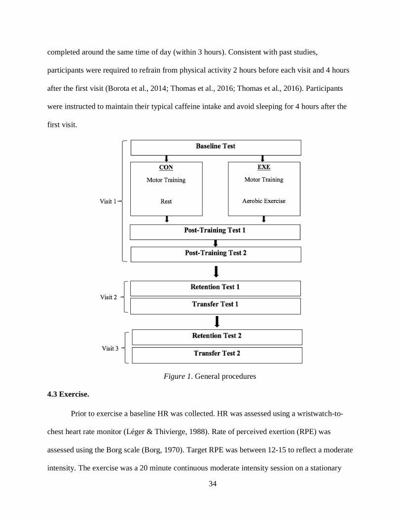
34
completed around the same time of day (within 3 hours). Consistent with past studies,
participants were required to refrain from physical activity 2 hours before each visit and 4 hours
after the first visit (Borota et al., 2014; Thomas et al., 2016; Thomas et al., 2016). Participants
were instructed to maintain their typical caffeine intake and avoid sleeping for 4 hours after the
first visit.
4.3 Exercise.
Prior to exercise a baseline HR was collected. HR was assessed using a wristwatch-to-
chest heart rate monitor (Léger & Thivierge, 1988). Rate of perceived exertion (RPE) was
assessed using the Borg scale (Borg, 1970). Target RPE was between 12-15 to reflect a moderate
intensity. The exercise was a 20 minute continuous moderate intensity session on a stationary
Figure 1. General procedures

35
recumbent bike. During exercise arms were resting on the handle bars and participants were
instructed to keep their arms relaxed to avoid fatiguing the muscles involved in the motor
training task. Cycling was chosen based on past literature that has demonstrated that lower body
aerobic exercise can modulate excitability in the motor cortical representations for the upper
body (Singh, Duncan, et al., 2014). Participants completed a 5-minute self-paced warm-up to get
their HR up to a moderate intensity level, which was calculated as 70% of their HRR based on
their age-predicted HR max and resting HR (i.e., 70% HRR = 0.7(HRmax – HRrest) + HRrest).
Once they reached this HR at the end of the 5 minute warm-up, they exercised at this intensity
for 20 minutes. HR was monitored continuously throughout the session and RPE was reported
every 5 minutes. Participants kept a pace of 55-65 revolutions per minute (RPM) and adjusted
the bike’s resistance level to maintain target HR (Singh, Neva, et al., 2016; Thacker et al., 2014).
After 20 minutes at this intensity participants completed a 5-minute cool-down after which HR
and RPE measures were taken again. The intensity and duration of exercise were selected to be
less intense than high intensity interval training and more intense than a low intensity interval or
continuous exercise bout. This intensity was meant to challenge participants but not exhaust
them. Continuous moderate intensity exercise has previously been shown to induce cortical
modulations (Singh et al., 2014, 2016; Thacker et al., 2014). Additionally, no known studies
have examined the effects of post-motor training continuous moderate intensity exercise on
retention of motor skills. Addressing this gap in the literature is important because high intensity
interval training is not appropriate for all populations.
4.4 Motor Task.
The motor training task was a modified version of the BMT task that has been validated
by past motor learning studies from our lab that have examined behavioural and cortical
36
modulations (Neva, Singh, Vesia, & Staines, 2014; Singh et al., 2016; Smith & Staines, 2006,
2010, 2012). The details of the seating arrangement, training apparatus, and task specifics are
outlined in Singh et al. (2016). A figure of the set-up is illustrated in Figure 2. The task required
participants to hold two handles, one in each hand, of the custom-made BMT device. The
handles were connected to potentiometers to measure their movement. A customized LabVIEW
program allowed for the movement of the handles to correspond to movement of the cursor on a
screen. Flexion/extension of the right wrist moved the cursor vertically and flexion/extension of
the left wrist moved the cursor horizontally. The subject’s view of the screen during the motor
training is shown in Figure 3. During a single trial participants first saw the cursor (black circle)
and moved it to the start position (x) in the bottom right corner. When this occurred, the cursor
disappeared and 1 of 3 possible targets (30, 45, and 60 from the y-axis) appeared in the top
left corner. After a 2 s delay the cursor reappeared which was the cue for participants to move
the cursor to the specified target location. This ensured the measurement of the early cued-MRP
component that starts approximately 2 s before movement. The participant then had 2 s to move
the cursor to the target. After this feedback was given in the form of RT. This was not a true RT,
but rather was calculated as the time between when the trial started and when the cursor reached
the target. Participants started the next trial when they were ready. The movement from the start
position to the targets required participants to complete inphase BMT as used by Singh et al.
(2016) shown in Figure 4. This was specifically selected because of past research that has
demonstrated that inphase BMT enhances the amplitude of the cued-MRP early component
(Smith and Staines, 2010) and can also increase cortical excitability of M1 (Singh, Neva, et al.,
2016) suggesting the beginning of early plasticity processes. Participants were told the objective

37
of the task was to flex both wrists simultaneously to move the cursor onto the target and to
perform the task as quickly and as accurately as possible within the 2 s timeframe.
The motor training was a random variable practice structure consistent with previous
studies (Kantak et al., 2010, 2011). Participants completed 180 trials of training of which 50%
were the test target (45), 25% were a target located 30 from the y-axis, and the remaining 25%
were a target located 60 from the y-axis. This equated to 90 trials of the test target, and 45 trials
each of the other two targets. The 180 trials were completed in a randomized order. Before
training participants completed 5 familiarization trials and 5 baseline trials to the test target.
Post-training test 1 and retention test 1 and 2 were identical to the baseline test. Post-training test
2 consisted of a larger amount of trials (50 trials) to the test target to ensure enough EEG data
was collected to be able to average an MRP trace. Transfer test 1 and 2 were 5 trials of a new
target (approximately 37.5 and 52.5 from the y-axis). The transfer test was included as an
alternative measure of learning. During the baseline, post-training, retention, and transfer tests no
feedback of RT was given.
Figure 2. Experimental set-up (Neva et al., 2012)

38
4.5 Data acquisition.
EEG was only collected during the training and post-training test 2 at visit 1. EMG was
collected on all days at all timepoints. EMG electrodes were placed on the muscle bellies of the
flexor carpi radialis (FCR) of each arm, and the ground electrodes were placed on the clavicles
after proper skin preparation according to the SENIAM recommendations (Hermens, Freriks,
Disselhorst-Klug, & Rau, 2000). The EMG data was amplified (x1000), band pass filtered (10-
500 Hz), and digitized at a rate of 1000 Hz.
EEG was recorded from a 32 channel cap (Quik-Cap, Neuroscan, Compumedics, NC,
USA) according to the International 10-20 system. Ten electrodes recorded electrical activity
over frontal and sensorimotor areas (FZ, F3, F4, FC3, FCZ, FC4, C3, CZ, C4, PZ). FP1 was also
collected to determine blinks. Reference electrodes were placed on the left and right mastoid
processes. Impedances of all electrodes were maintained below 5 kΩ. Data was amplified,
Figure 4. Inphase movement (Neva et al., 2012)
Figure 3. Consecutive view of screen during a single acquisition trial (Singh et al., 2016)

39
filtered (DC-200 Hz, 6dB octave roll-off) and digitized (1000 Hz, SynAmps2, Scan 4.5,
Compumedics Neuroscan, Charlotte, NC) before being stored for off-line analysis.
Behavioural data was collected in the form of RT and the trajectory of the cursor. Event
codes from the customized LabVIEW program indicated the start of the trial, when the target
appeared, cue to move, start of cursor movement, and the end of the trial. Voltage data from the
potentiometers and cursor location were recorded in the customized LabVIEW program. This
data was collected at a sample rate of 40 Hz.
4.6 Data analysis.
The EEG data was analyzed in Neuroscan (Compumedics Neuroscan, NC, USA)
software. The customized LabVIEW program sent event codes to the continuous EEG file.
MRPs were extracted from the EEG data by averaging artifact-free baseline-corrected individual
epochs that were time-locked to the onset of cursor movement and extended from 2 s before to
500 ms after the onset of cursor movement. Averaged MRP traces representing cortical activity
at early training, late training, and post-training 2 were created for each participant. The first 60
trials of training were used to represent early training, and the last 60 trials of training were used
to represent late training. All 50 of the post-training 2 trials were used to represent post-training
2. An average of 6 epochs were removed from each average trace due to artifacts. From each
averaged MRP trace an area report was used to sum all data points of the trace together from the
time window of -1250 to 0. This time was selected to include cortical activity during planning
and execution of the skill. This sum provided a measure of the amplitude of the early MRP. MRP
amplitudes were maximal over the midline electrodes so statistical analysis was run on the data
from FCZ and CZ.

40
To assess accuracy, a customized LabVIEW program compared the actual trajectory of
the cursor to the ideal trajectory of the cursor. The program computed an ‘ideal’ trajectory by
creating a straight line from the start position to the target over the time period of 2 s. This ‘ideal’
trajectory was then compared point-by-point to the actual trajectory resulting in directional error
values in the form of pixel co-ordinates. These error values were squared and summed, and the
RMS value was calculated as the square root of this sum. Therefore, the magnitude of the RMS
value reflects the amplitude of the difference between the actual and the ‘ideal’ trajectory and the
units of the RMS values are arbitrary.
EMG was initially included in the study to be able to study the components of a true RT
(i.e., reaction time and movement time). However, since the BMT task is not a true reaction time
task due to the predictability of the cue, the EMG data was not analyzed for reaction time,
instead time to target was used as a RT measure. This RT was calculated as the time between the
cue to move and when the cursor reached the target. If the participant did not reach the target
their RT was recorded as 2000 ms which was the maximum amount of time participants had to
reach the target.
For the behavioural data (RMS and RT), at each timepoint (baseline, early training, late
training, post-training 1, post-training 2, retention 1, retention 2) 5 trials to the test target were
averaged to represent performance. The baseline, post-training 1, retention tests all only
consisted of 5 trials to the test target. The first 5 trials and the last 5 trials to the test target in the
motor training session were averaged to represent early training and late training performance
respectively. For post-training 2, the first 5 trials were averaged to represent performance. The
last 5 trials of post-training 2 were averaged and used to normalize the retention and transfer

41
scores to their performance at the end of visit 1. For the transfer tests, the 5 trials to the new
targets were averaged.

42
5.0 Proposed Hypotheses Testing
Our main objective was to examine whether a session of continuous moderate intensity
aerobic exercise performed after motor training would affect the consolidation of the motor skill
as measured by performance at retention measures. To analyze this data a mixed-model Analysis
of Variance (ANOVA) for each behavioural measure was used with time (retention 1, retention
2) as the within-subjects factor and group (EXE/CON) as the between-subjects factor. To test our
main hypothesis that EXE would have a significantly lower change in RMS and RT scores
compared to CON from the last 5 trials of post-training 2 to the retention measures pre-planned
contrasts were used to compare groups at the retention time points. To normalize the RMS and
RT scores the last 5 trials of each participant’s post-training 2 test was subtracted from their
scores at retention 1, and retention 2. To test our assumption that there would be significant
improvements in RMS and RT at late training and post-training measures and no differences
between the groups at these time points, a mixed-model ANOVA for each behavioural measure
was used with time (baseline, early training, late training, post-training 1, post-training 2) as the
within-subjects factor and group (EXE/CON) as the between-subjects factor. Our second
objective was to investigate the influence of motor training and aerobic exercise on cortical
activity relating to motor preparation and execution. To test our hypothesis that EXE would have
a larger amplitude of the early MRP component at the post-training measure compared to CON,
and our prediction that both EXE and CON would have a larger amplitude of the early MRP
component at the late training measure compared to the early training measure we used a mixed-
model ANOVA. The within-subjects factor was time (early training, late training, and post-
training 2) and the between-subjects factor was group (EXE/CON). Pre-planned contrasts were
used to compare early and late time points. For all ANOVA tests and assumptions of normality,

43
homogeneity of variance, and sphericity, SAS University Edition was used. To investigate
whether there was a relationship between behavioural measures and MRP changes we examined
the correlation between change in the amplitude of the early component and change in RMS and
RT from the last 5 trials of post-training 2 to retention. Correlational analysis were conducted
using the Pearson correlation. A t value was calculated from the coefficient and a two-tailed t test
was run to test for significance.

44
6.0 Results
6.1 Participant Characteristics
A total of 34 participants were recruited for the study. Of these 34 participants, one was
excluded for accumulating less than 600 METs per week. Therefore, the total number of
participants collected was 33 with 17 participants in EXE and 16 participants in CON (Table 1).
Of the 33 participants, 19 were female and 14 were male (EXE 10F, 7M; CON 9F, 7M). The age
range of participants was 19-26 and the average age (± standard deviation) was 21.93 ± 2.13.
The average BMI score in EXE was 24.64 ± 3.39 and in CON was 22.76 ± 2.01. All participants
were deemed safe to participate in exercise from the GAQ. According to the EHQ, 3 participants
were lefthanded, one was dominant in an equal number of tasks for both hands, and the rest were
righthanded. From the IPAQ a total of 23 participants were categorized as being highly
physically active which equates to vigorous-intensity activity on at least 3 days per week and
accumulating at least 1500 MET-minutes per week, or 7 or more days of any combination of
walking, moderate or vigorous intensity activities accumulating at least 3000 MET-minutes per
week. Ten participants were categorized as being moderately physically active which equates to
3 or more days of vigorous intensity activity of at least 20 minutes per day, or 5 or more days of
moderate intensity activity and/or walking at least 30 minutes per day, or 5 or more days of any
combination of walking, moderate intensity or vigorous intensity activities achieving a minimum
of at least 600 MET minutes per week. There were 13 highly active participants in EXE and 10
highly active participants in CON. The average MET-minutes per week was 3017.12 ± 1627.19.
A two-tailed t test revealed that the average MET-minutes per week in the EXE (3263.05 ±
1817.26) and in CON (2755.81 ± 1408.73) were not significantly different (t31 = 0.89, p = 0.37).

45
Participant Group Gender Age IPAQ
Category
MET-
minutes/week
Handedness BMI
1 EXE F 21 Moderate 2412 R 21.8
2 EXE M 20 High 4980 L 23.9
3 CON M 26 Moderate 1065 R 20.5
4 CON F 21 High 4709 R 20.1
5 EXE M 26 High 3147 R 22.4
6 CON F 23 High 2121 L 21.6
7 CON M 21 High 1710 R 25.8
8 EXE F 23 High 4990 R 24.3
9 EXE F 23 Moderate 1048.5 R 22.5
10 CON F 23 High 2594 R 23.3
11 EXE F 25 High 3393 R 21.6
12 CON M 23 Moderate 1704 R 24.1
13 EXE M 19 Moderate 1012 R 29.3
14 CON M 24 Moderate 2373 R 26.2
15 EXE M 19 High 2388 R 27
16 EXE F 24 High 4868 R 21.9
17 CON M 22 High 3660 R 20.2
18 CON F 21 High 3485 R 22.7
19 EXE M 20 High 1759 L/R 35
20 EXE F 20 High 2982 R 23.2
21 CON M 21 Moderate 1400 R 23.7
22 EXE M 19 High 3772.5 R 24.3
23 CON F 22 Moderate 893 R 25.1
24 CON F 20 High 3006 R 20.5
25 CON F 21 High 2600 R 20.8
26 EXE F 19 High 2764 L 23.9
27 EXE F 22 High 8412 R 26.3
28 CON F 19 High 6125 R 22.7
29 EXE M 24 Moderate 1475 R 23.7
30 EXE F 22 High 3333 R 22
31 CON F 24 High 4212 R 24.2
32 CON M 26 Moderate 2436 R 22.7
33 EXE F 21 High 2736 R 25.8
6.2 Supplementary Data
Affect scores from the PANAS scale were compared using a mixed-model ANOVA. The
within-subjects factor was time (pre-intervention, post-intervention, day 2, day 3) and the
between-subjects factor was group (EXE/CON). Results revealed a significant interaction effect
between group and time (F3,93 = 11.88, p <0.01). Post-hoc comparisons using the Tukey-Kramer
test indicated that EXE pre-intervention mean score (39.97 ± 3.34) was significantly different
Table 1
Participant Characteristics

46
than CON pre-intervention mean score (37.56 ± 3.29) as well as CON post-intervention (36.81 ±
3.45), and CON day 2 mean score (37.81 ± 3.37). EXE also had post-intervention mean score
(41.73 ± 3.50) that was significantly different from all of the CON scores (pre-intervention, post-
intervention, day 2, day 3: 38.37 ± 4.35) and EXE day 2 (38.64 ± 4.43) and day 3 (38.20 ± 3.22)
scores. The only score that was not significantly different from EXE post-intervention score was
EXE pre-intervention score. There were no significant main effects of time (F3,93 = 2.28, p =
0.08) or group (F1,31 = 3.06, p = 0.09).
On the SSS all participants rated their degree of sleepiness between a 1-3 except for one
participant who during the first visit rated their sleepiness as a 4 (1 = feeling active, vital, alert,
or wide awake; 2 = functioning at high levels, but not at peak; able to concentrate; 3 = awake,
but relaxed; responsive but not fully alert; 4 = somewhat foggy, let down). According to the SSS
a score above 3 is considered ‘sleepy’ (Berry & Wagner, 2014). The mode of all SSS scores was
2. Statistical analyses were not carried out on this data.
Sleep data collected from the SMHSQ was not scored or statistically analyzed but was
collected to help explain any outliers. On average participants had 7.25 ± 1.15 hours of sleep the
night of visit 1. The amount of sleep ranged from 4.6-9.5 hours. All participants rated their sleep
as ‘fairly well’, ‘well’, or ‘very well’ except for one participant who rated their sleep as ‘fairly
badly’.
6.3 Exercise Data
EXE had a resting HR of 66.29 ± 6.19 bpm and CON had a resting HR of 63.75 ± 9.78
bpm. The average target HR (70% of HRR) for EXE was 158.64 ± 2.37 bpm. The average HR
for EXE during the 20 minute exercise was 156.01 ± 3.82 bpm. The average HR for CON during
the rest session was 65 ± 9.73 bpm. Across all participants in EXE the HRR percentage reached

47
during exercise ranged from 62.40-74.81% with the average HRR percentage at 67.91% ± 3.82.
The average RPE score was 14.13 ± 1.05. Participants in EXE maintained an average RPM of
61.01 ± 2.45 and all individual averages were within the required range of 55-65. After exercise
during the post-training test 1 the average HR for EXE was 91.82 ± 7.10 bpm and for CON was
63.88 ± 8.80 bpm. For post-training test 2, it took EXE an average of 30.35 ± 10.71 minutes for
HR to return to near resting levels. Due to time constraints one participant completed post-
training test 2 before their HR was back to resting levels (indicated by the * in Table 2). CON
completed post-training test 2 an average of 30.25 ± 1 minutes after the rest session. At post-
training test 2 the average HR for EXE was 71.47 ± 7.68 bpm and for CON was 65.29 ± 10.31
bpm.

48
Participant Group Rest
HR
Target
HR
Exercise/Rest
Session AVG
HR
AVG % of
HRR during
Exercise
AVG
RPE
AVG
RPM
HR
Post1
HR
Post2
Rest/exercise
to Post2
(min)
1 EXE 69 160 158.5 68.85 13.5 59 93 70 50
2 EXE 62 158 154.75 67.21 15.25 62.75 93 68 38
3 CON 62 154 63 - - - 60 64 30
4 CON 52 155 52.5 - - - 51 50 30
5 EXE 62 154 150.5 67.045 16 61.25 91 64 51
6 CON 66 158 66.5 - - - 62 67 30
7 CON 53 155 55.5 - - - 55 53 30
8 EXE 73 160 153.25 64.72 13 56.75 94 78 19
9 EXE 74 160 154.75 65.65 14 57.5 89 78 21
10 CON 63 157 67.75 - - - 63 65 30
11 EXE 74 159 163 73.55 13.125 62.25 88 79 18
12 CON 56 155 58.75 - - - 61 61 30
13 EXE 66 160 154 65.19 15 63 87 68 38
14 CON 65 157 59.75 - - - 67 62 34
15 EXE 70 162 160.5 69.08 14.75 62.25 105 75 29
16 EXE 60 155 161.75 74.82 12.75 61.25 97 66 31
17 CON 52 154 53.5 - - - 53 52 30
18 CON 54 155 57.25 - - - 58 58 30
19 EXE 67 160 150 62.41 14.75 62.25 93 72 18
20 EXE 70 161 158 67.69 15.125 57.25 93 74 30
21 CON 74 161 76.5 - - - 69 74 30
22 EXE 66 160 158.75 68.70 11.75 59.75 102 88* 31
23 CON 68 159 73.5 - - - 69 72 30
24 CON 72 162 75.5 - - - 66 71 30
25 CON 88 166 86.75 - - - 89 88 30
26 EXE 62 159 153.75 66.01 14.5 64.75 89 62 26
27 EXE 72 160 156 66.67 14 62.25 94 77 27
28 CON 73 163 73.75 - - - 67 73 30
29 EXE 70 158 158.5 70.24 14.25 63.25 91 75 35
30 EXE 52 154 151.5 68.15 14.625 58.25 71 57 19
31 CON 61 154 67 - - - 59 62 30
32 CON 61 154 59 - - - 64 57 30
33 EXE 58 157 154.75 68.62 14 63.5 91 64 35
Table 2
HR, RPE, and RPM

49
6.4 Behavioural Data
A mixed-model ANOVA on the RMS retention data (Figure 5) with time (retention 1,
retention 2) as the within-subjects factor and group (EXE/CON) as the between-subjects factor
revealed a significant main effect of time (F1,31 = 3.85, p = 0.05). Post-hoc comparisons using the
Tukey-Kramer test revealed no significant differences between timepoints. There was no main
effect of group (F1,31 = 1.56, p = 0.22) or interaction effect between group and time (F1,31 = 0.10,
p = 0.75). Planned contrasts revealed there was no significant difference between the groups at
retention 1 (F1,31 = 3.19, p = 0.08). However, the groups were significantly different at retention
2 (F1,31 = 5.01, p = 0.03) and when the retention timepoints were pooled together (F1,31 = 8.10, p
< 0.01). To represent the size of this difference a percent change from the last 5 trials of post-
training 2 was calculated as the difference between the two groups’ normalized retention scores
at each time point divided by the average of all the last 5 trials of post-training 2. At retention 1
this was equivalent to a 10.47% change from the last 5 trials of post-training 2, at retention 2 this
was a 13.13% change, and when the retention timepoints were pooled together this was a 11.80%
change.
The transfer test data was also normalized to the last 5 trials of post-training 2 to provide
a reference of performance at the end of visit 1. To analyze the transfer test data a two-tailed t
test was run to compare groups at transfer 1 and transfer 2. Results revealed no significant
difference between the groups at transfer 1 (t31 = -0.23, p = 0.81) or at transfer 2 (t31 = 1.36, p =
0.18).

50
A mixed-model ANOVA on the RMS acquisition and post-training measures (Figure 6)
with time (baseline, early training, late training, post-training 1, post-training 2) as the within-
subjects factor and group (EXE/CON) as the between-subjects factor revealed a significant main
effect of time (F4,124 = 98.29, p <0.01). Post-hoc comparisons using the Tukey-Kramer test
revealed that all timepoints were significantly different from one another except for post-training
1 and post-training 2, and late-training and post-training 2. There was no significant main effect
of group (F1,31 = 0.05, p = 0.81) and no significant interaction between group and time (F4, 124 =
1.07, p = 0.37).
Figure 5. Change in RMS from the last 5 trials of post-training 2 to retention and transfer. Data was normalized
to show each group’s performance as the difference from the 5 last trials of post-training 2. A lower score is
representative of a better performance. Error bars represent standard error of the mean. * indicates significant
difference p <0.05.
0
20
40
60
80
100
120
Retention 1 Retention 2 Transfer 1 Transfer 2
No
rmal
ized
RM
S (
arb
itra
ry u
nit
s)
Exercise Control

51
A mixed-model ANOVA on the RT retention data (Figure 7) with time (retention 1,
retention 2) as the within-subjects factor and group (EXE/CON) as the between-subjects factor
revealed no significant main effects of time (F1,31 = 0.46, p = 0.50), group (F1,31 = 0.01, p = 0.90),
and no significant interaction between group and time (F1,31 = 2.93, p = 0.09). Planned contrasts
revealed no significant differences between the groups at retention 1 (F1,31 = 1.12, p = 0.29),
retention 2 (F1,31 = 1.85, p = 0.18), or when the retention timepoints were pooled together (F1,93 =
0.05, p = 0.83). A two-tailed t test was run on the normalized RT transfer data to compare
groups at transfer 1 and transfer 2. Results revealed no significant difference between the groups
at transfer 1 (t31 = -0.60, p = 0.55) or transfer 2 (t31 = -0.26, p = 0.79).
Figure 6. RMS scores for baseline, training, and post-training tests. A lower score is representative of a better
performance. Error bars represent standard error of the mean.
0
50
100
150
200
250
300
350
400
450
Baseline Early Training Late Training Post 1 Post 2
RM
S (ar
bit
rary
un
its)
Exercise Control

52
A mixed-model ANOVA on the RT acquisition and post-training measures (Figure 8)
with time (baseline, early training, late training, post-training 1, post-training 2) as the within-
subjects factor and group (EXE/CON) as the between-subjects factor revealed a main effect of
time (F4,124 = 115.9, p <0.01). Post-hoc comparisons using the Tukey-Kramer test revealed that
baseline and early training were not significantly different from each other, but they were
significantly different from every other timepoint. Late training, post-training 1 and post-training
2 were not significantly different from each other. There was no significant main effect of group
(F1,31 = 0.14, p = 0.71) and no significant interaction between group and time (F4,124 = 0.18, p =
0.94).
Figure 7. Change in RT from late training to post/retention/transfer. Data was normalized to show each group’s
performance as the difference from their late training score. A lower score is representative of a better performance.
Error bars represent standard error of the mean.
0
50
100
150
200
250
300
350
400
450
Retention 1 Retention 2 Transfer 1 Transfer 2
Norm
aliz
ed R
T (
ms)
Exercise Control

53
6.5 Neurophysiological Data
A mixed-model ANOVA with the within-subjects factor as time (early training, late
training, and post-training 2) and the between-subjects factor as group (EXE/CON) was run on
the data from each electrode (FCZ and CZ). Results from the FCZ electrode (Figure 9) revealed
a main effect of time (F2,62 = 10.33, p < 0.01). Post-hoc comparisons using the Tukey-Kramer
test revealed that post-training 2 was significantly different than early training and late training.
There were no main effects of group (F1,31 = 0.06, p = 0.80) and no interaction between group
and time (F2,62 = 2.52, p = 0.08). The planned contrast between early and late training was not
significant (F1,62 = 3.19, p = 0.07). Grand average traces of EXE and CON are displayed in
Figure 10a and 10b respectively. Results from the CZ electrode (Figure 11) also showed a main
effect of time (F2,62 = 3.71, p = 0.03). Post-hoc comparisons using the Tukey-Kramer test
revealed that post-training 2 was significantly different than late training. There were no main
effects of group (F1,31 = 0.23, p = 0.63) and no interaction between group and time (F2,62 = 0.92,
Figure 8. RT scores for baseline, training, and post-training tests. A lower score is representative of a better
performance. Error bars represent standard error of the mean.
0
200
400
600
800
1000
1200
1400
1600
1800
2000
Baseline Early Training Late Training Post 1 Post 2
RT
(m
s)
Exercise Control

54
p = 0.40). The planned contrast between early and late training was significant (F1,62 = 4.68, p =
0.03). Grand average traces of EXE and CON are displayed in Figure 12a and 12b respectively.
Figure 9. Sum of all individual data points from MRP trace recorded at FCZ. Error bars represent standard error of the mean.
Figure 10. Grand-average MRP trace of EXE (A) and CON (B) recorded from electrode FCZ. Blue represents
early training, green represents late training, and red represents post-training test 2. Data is time-locked to cursor
movement; 0 represents the start of cursor movement.
A
B
Early Training
Late Training
Post-Training Test 2
-4500
-4000
-3500
-3000
-2500
-2000
-1500
-1000
-500
0
500
1000
Early Training Late Training Post 2
Su
m o
f M
RP
(μV
)
Exercise Control

55
Figure 11. Sum of all individual data points from MRP trace recorded at CZ. Error bars represent standard error of
the mean. * indicates significant difference p <0.05.
Figure 12. Grand-average MRP trace of EXE (A) and CON (B) recorded from electrode CZ. Blue represents
early training, green represents late training, and red represents post-training test 2. Data is time-locked to cursor
movement; 0 represents the start of cursor movement.
A
B
Early Training
Late Training
Post-Training Test 2
-5000
-4500
-4000
-3500
-3000
-2500
-2000
-1500
-1000
-500
0
Early Training Late Training Post 2
Su
m o
f M
RP
(μ
V)
Exercise Control

56
6.6 Correlational Data
Initially we predicted that in both groups there would be a larger MRP amplitude at late
training and post-training 2 than early training. Our results show an increase in the MRP
amplitude from early to late training however the MRP amplitude at post-training 2 was not
significantly different than early training at the CZ electrode, and was significantly smaller than
the early training at the FCZ electrode. Given these results we examined the relationship between
the change in the sum of the MRP amplitude from early to late training and the change in RMS
and RT from the last 5 trials of post-training 2 to the retention time points. This allowed us to
examine whether changes in the MRP amplitudes during motor training were associated with
performance at retention scores. In other words, the degree to which the neural representation of
skill acquisition predicted participants’ ability to consolidate (or retain) the skill. A Pearson
correlation and significance test was run on each comparison. To test for significance a t value
was calculated from the coefficient and a two-tailed t test was conducted. There was a positive
linear relationship in EXE between the change in MRP amplitude sum recorded from early to
late training and the change in RT from the last 5 trials of post-training 2 to retention 1. This
correlation was significant at both FCZ (r = 0.77, p < 0.01) (Figure 13) and CZ (r = 0.59, p =
0.01). Additionally, when this relationship was examined with both EXE and CON pooled
together, there was a significant correlation at FCZ (r = 0.45, p < 0.01) and at CZ, this
correlation approached significance (r = 0.32, p = 0.06). We also examined the relationship
between the change in the sum of the MRP amplitude from early to late training and the change
in RMS and RT from baseline to late training. This allowed us to determine whether behavioural
improvements during training were associated with changes in the MRP amplitudes during the
motor training. We did not find any significant correlations between these variables.
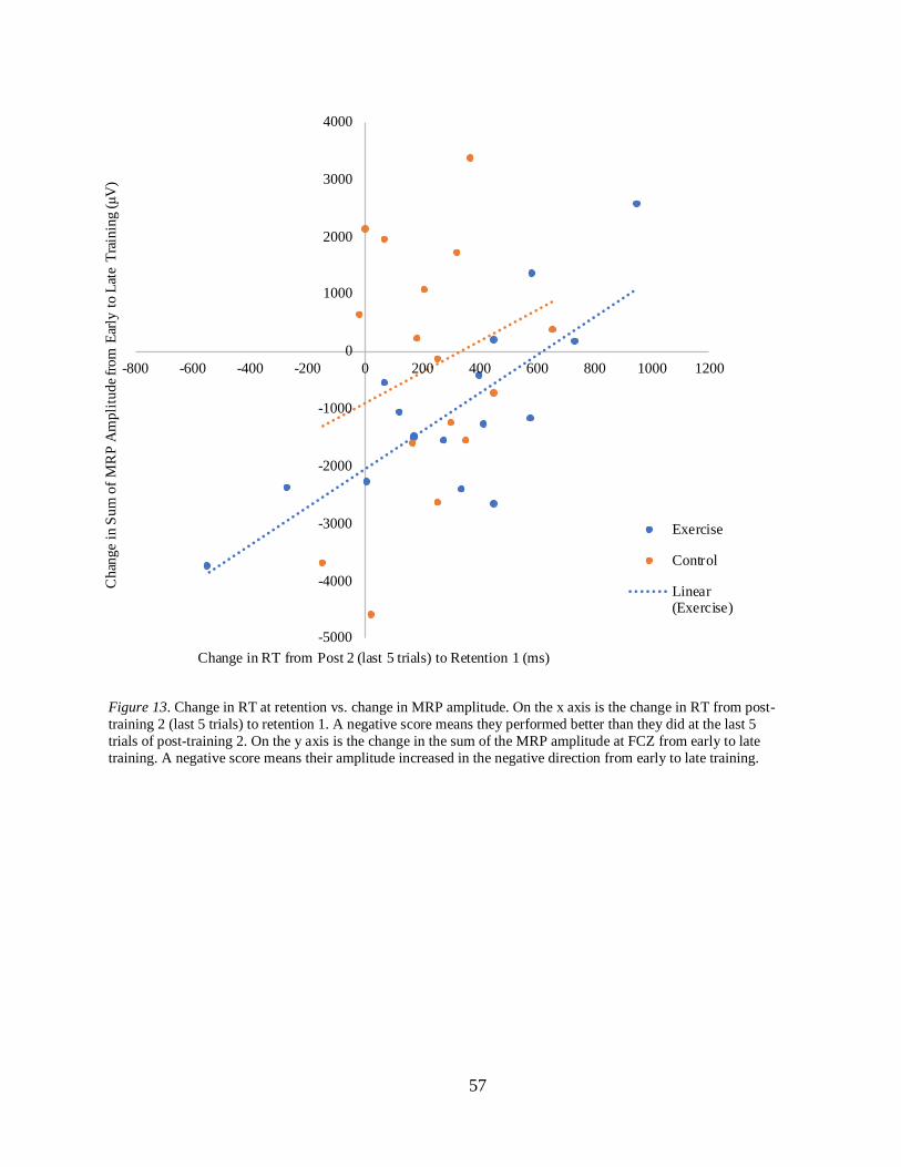
57
Figure 13. Change in RT at retention vs. change in MRP amplitude. On the x axis is the change in RT from post-
training 2 (last 5 trials) to retention 1. A negative score means they performed better than they did at the last 5
trials of post-training 2. On the y axis is the change in the sum of the MRP amplitude at FCZ from early to late
training. A negative score means their amplitude increased in the negative direction from early to late training.
-5000
-4000
-3000
-2000
-1000
0
1000
2000
3000
4000
-800 -600 -400 -200 0 200 400 600 800 1000 1200
Ch
ange
in S
um
of
MR
P A
mpli
tud
e fr
om
Ear
ly t
o L
ate
Tra
inin
g (μ
V)
Change in RT from Post 2 (last 5 trials) to Retention 1 (ms)
Exercise
Control
Linear(Exercise)

58
7.0 Discussion
The results of our study are generally consistent with the previous studies that have
shown that post-motor training high intensity interval training helps to retain performance of a
unimanual motor skill, suggesting the timing of this exercise enhances consolidation mechanisms
(Roig et al., 2012; Thomas, Flindtgaard, et al., 2016; Thomas, Beck, et al., 2016; Thomas,
Johnsen, et al., 2016). Our study adds to the literature in the following ways. First, we show that
continuous moderate intensity exercise also provides a benefit to the retention of a motor skill.
Second, we show that exercise is able to help retain a more complex, bimanual motor task.
Finally, we attempt to correlate neural modulations with behavioural performance at retention
tests. Our results provide some evidence that exercise enhances the relationship between
increases in excitability during training and performance of the skill at retention.
The main objective of our study was to examine whether a session of continuous
moderate intensity aerobic exercise performed after motor training would affect motor memory
consolidation as measured by performance at retention measures. Our results support the main
hypothesis that EXE would have a significantly lower change in RMS scores compared to CON
from the last 5 trials of post-training 2 to retention. However, EXE did not have a significantly
lower change in RT score than CON from the last 5 trials of post-training 2 to retention. In
addition, our results support our prediction that both EXE and CON would have significant
improvements in RMS and RT at late training and post-training measures and that at these time
points there would be no significant difference between the groups. Our second objective was to
investigate the influence of motor training and aerobic exercise on cortical activity relating to
MRPs. Our main hypothesis that EXE would have a larger amplitude increase of the early MRP
component at the post-training measure compared to CON was not supported. We also predicted
59
that both groups would have a larger amplitude of the early MRP component at the late training
measure compared to the early training measure. This was observed however it was only
supported with statistical significance at the CZ electrode. Finally, we wanted to investigate the
relationship between the neural markers of skill acquisition and the consolidation of the skill
measured through retention tests. We predicted there would be a correlation between increases in
the amplitude of the early MRP component and changes in the RMS and RT score from the last 5
trials of post-training 2 to retention. There was a significant correlation found between the
change in MRP amplitude sum from early to late training and the change in RT from the last 5
trials of post-training 2 to retention 1. This was found in EXE, and when EXE and CON were
pooled together, but not in CON alone which provides some support for this final hypothesis.
7.1 Participant Characteristics
The two groups had similar descriptive characteristics, BMI, MET-minutes per week, and
resting HR values, giving some indication that the groups were comparable. Initially we had a
high cut-off for the IPAQ scale that excluded individuals in the highly physically active
category. However, due to the variability around the IPAQ categorization we choose not to
include this. Therefore, the only IPAQ exclusion criteria was accumulating less than 600 MET-
minutes per week which was maintained to exclude individuals who have a very low level of
physical activity and may not be able to complete 20 minutes of moderate intensity exercise.
7.2 Supplementary Data
The supplementary data collected from the SSS and SMHSQ does not suggest there
would be differences between the groups. However, data from PANAS shows a significant
difference between the groups at the pre-intervention (before exercise). EXE had a higher mood
score, meaning they were in a more positive mood. Additionally, EXE had a post-intervention

60
(after exercise) score that was significantly higher than every other score in both groups except
for the EXE pre-intervention score. This suggests that EXE had a more positive mood prior to
beginning the study, and that this mood was maintained after completing motor training and
exercise. There are no known studies that have shown that mood affects motor acquisition or
motor memory consolidation. However, mood can contribute to cortical arousal and cognition
which may impact how well the task is performed (Mitchell & Phillips, 2007). Our data show
that behaviourally the groups were no different during the motor training session. Therefore, it is
unlikely that the differences in mood affected motor acquisition or consolidation.
7.3 Exercise Data
Participants in the exercise group were required to complete 20 minutes of continuous
moderate intensity exercise on a recumbent bike. Both the average percentage of HRR reached
during exercise and the average RPE during exercise reflected the targets set for moderate
intensity. All participants maintained RPM within the set range, so we are confident that this
variable was controlled for. At post-training 1, HR was still elevated in EXE, but at post-training
2, HR was back to near-resting levels. Our estimation that it would take approximately 30
minutes after exercise for HR to get back to resting was correct as both groups had the same
average amount of time from exercise to post-training 2. The standard deviation was larger in the
EXE group which reflects the variability in the time it took for HR to go down from person to
person.
7.4 Behavioural Data
Performance of the task during acquisition, retention, and transfer was assessed through
measures of accuracy (RMS) and speed (RT). We hypothesized that exercise would have the
same benefit for both RMS and RT performance because performance of speed and accuracy in
61
the task are largely related. As individuals improve at locating the target with the cursor they
should be able to quicken their movement to the target. The object of the task was to
simultaneously flex both wrists to move the cursor diagonally from the bottom right of the screen
to the target in the top left within the 2 s timeframe. During the first few trials participants may
have prioritized speed to be able to reach the cursor within the 2 s. However, once this was
achieved participants likely prioritized accuracy. Therefore, the primary measure of performance
was RMS scores.
The ANOVA with our RMS retention data showed a main effect of time but the post-hoc
did not reveal any significant differences between retention 1 and retention 2. As well there was
no main effect of group or interaction between group and time. Our hypothesis tests revealed that
the retention of the skill in CON was significantly worse than EXE at retention 2 and when the
retention time points were pooled together. This significant difference between groups was not
observed at retention 1. These results suggest that the benefit of exercise post-motor training is
that it helps to consolidate a motor memory so that is resistant to degradation over time. At
retention 1 CON does not retain the skill as well as EXE, however there is no significant
difference between the groups at this timepoint. It seems likely that the motor memory formed in
CON is still strong 1 day after motor training and therefore our hypothesis test comparing the
groups at this timepoint did not reach significance. As a longer period of time goes by, this motor
memory may degrade at a faster pace in CON than in EXE. This is likely why at retention 2, one
week post-motor training, the retention score of CON is significantly worse than EXE.
The transfer tests were not consistent with the retention test results. The groups are not
significantly different from each other at transfer 1 or transfer 2. However, EXE appears to
62
improve from transfer 1 to transfer 2, and CON appears to get worse. This suggests that there
may be a learning effect in EXE from transfer 1 to transfer 2 that is not present in CON.
The above results suggest that post-motor training moderate intensity exercise assists
with retaining the accuracy of the motor skill. While the neural mechanisms of this behavioural
benefit cannot be determined from this study, previous studies suggest that exercise increases the
availability of LTP-related compounds (Skriver et al., 2014) and assists with the induction of
LTP by reducing cortical inhibition and increasing excitation (Singh, Neva, et al., 2014). Studies
with similar motor training structures to ours have suggested that BMT can induce early LTP-
like plasticity (Neva et al., 2012). Post-motor training exercise may then enhance these BMT-
induced plasticity mechanisms by increasing the availability of LTP-related compounds
throughout consolidation. Examples of these LTP-related compounds include BDNF,
catecholamine neurotransmitters, and lactate. These compounds assist with tagging synapses to
undergo the transition from E-LTP to L-LTP. It could be through these mechanisms that exercise
enhances motor memory consolidation resulting in a robust motor memory that is resistant to
degradation over time.
There were no differences between the groups at baseline, training, or post-training RMS
scores (Figure 6). This suggests that both EXE and CON had similar performance levels at these
timepoints and that the motor training improved all participants’ RMS performance from their
baseline scores to early training and to late training timepoints (Figure 6). This confirms that
participants were able to improve their accuracy during the acquisition of the skill. As predicted,
there were no differences between the two post-training tests, and between late training and post-
training 2. We did not expect there to be a difference from late training to the post-training tests
or between the post-training tests as previous research has demonstrated that performance

63
differences between groups do not emerge immediately after the intervention (Lundbye-Jensen et
al., 2017; Roig et al., 2012). Consolidation has not yet occurred at the time of the post-training
tests as post-training 1 occurs 50 minutes after training, and the post-training 2 occurs roughly an
hour and 20 minutes after training. Therefore, if post-motor training exercise enhances the
consolidation of the motor memory, a difference in performance between the groups at these
timepoints is not to be expected.
The results of our analyses on the RT retention data were not consistent with the RMS
retention data. There were no main effects of time or group and no interaction. Our hypothesis
was not supported as there were no significant differences between the groups at the retention
tests. There were also no significant differences between the groups at the transfer tests.
Consistent with the RMS acquisition and post-training data there were no differences
between the groups’ RT scores at baseline, training, or post-training (Figure 8). Both groups
improved their RT from baseline and early training to late training. Their performance at late
training then stayed consistent at the post-training tests. These findings confirm that participants
improved their speed over the acquisition of the skill, and that there were no immediate
performance benefits as predicted.
These results show that by the end of the motor training all participants had improved
their performance in both speed and accuracy, however, at the retention tests, speed and accuracy
of the skill were not equally retained. When the speed-accuracy data is plotted, there is a positive
linear relationship during training and at the retention timepoints. That is, participants who had
lower RMS scores also had lower RT scores. Therefore, it does not appear that this finding is a
result of a speed-accuracy trade-off. It could be that a few higher RT scores in the data that may
have increased the variability in EXE at the retention tests, especially at retention 1. This likely

64
had some contribution to the lack of effect observed in speed but the main reason for not seeing a
significant difference in speed between the groups at the retention timepoints is likely due to the
design of the task. A timeframe of 2 s was given to complete the task in order to increase the
difficulty of the task and to ensure that the planned movement was performed relatively quickly.
If they did not reach the target within the 2 s timeframe their RT was recorded as 2000 ms.
Therefore, the range of the RT scores may have been too narrow to see a significant difference
between training and retention.
7.5 Neurophysiological Data
Our main hypothesis for our second objective was not supported as EXE did not have a
larger amplitude increase at the post-training measure compared to CON. In fact, our results
showed that the MRP amplitudes decreased for both groups at the post-training measure. At the
FCZ electrode the main effect of time revealed that the amplitude at post-training 2 was
significantly smaller than early and late training. At CZ the main effect of time showed that post-
training 2 was significantly smaller than late training, and that there was no difference between
early training and post-training 2. We did not observe an effect of exercise on the cued-MRP.
This is inconsistent with Thacker et al. (2014) who found an earlier onset of the BP component
post-exercise when HR returned to resting. Had we observed an earlier onset of the cued-MRP it
would have been reflected in a larger sum of the MRP amplitude. However, differences in the
study objectives and tasks likely contributed to the contrasting results. Our study had a motor
learning component as our objective was to examine the consolidation of motor memories.
Thacker et al. (2014) examined the effects of exercise on motor cortical areas. Considering these
objectives, our task was more complex, requiring participants to complete cued bimanual
movements to control a cursor on screen, whereas Thacker et al. (2014) had participants

65
complete self-paced unimanual wrist extension movements. Cued and self-paced MRPs have
slightly different cortical generators and the complexity of our task likely required additional
cortical input from other brain areas. Therefore, it may not have been as easily enhanced through
exercise. There are many factors that could have prevented us from observing the increased
excitability in the motor preparatory areas post-exercise. Changes in the position of the electrode
and changes in the impedance of electrodes are possible but unlikely as the cap was kept on
during exercise and was not adjusted at any timepoint. Sweat or movement of the gel during
exercise may have changed the impedance of the electrodes however impedance was checked in
EXE and CON prior to beginning post-training test 2. Although our main hypothesis for our
neurophysiological data was not supported, it does not mean that exercise does not enhance
consolidation mechanisms. This was the first study to measure MRP modulations in attempt to
examine the effects of post-motor training exercise on the consolidation of motor memories.
Perhaps measurement of MRP modulations is not the best way to examine this relationship.
Instead, analysis of the spectral components may be better suited to assess markers of
consolidation such as the work by Dal Maso et al. (2018).
In addition to our main hypothesis we predicted from previous research that there would
be an increase in the MRP amplitude from early to late training in both groups (Smith & Staines,
2010; Smith & Staines, 2006). This increase in amplitude is thought to be a neural marker for
skill acquisition. The hypothesis test contrasting early and late MRP amplitudes across all
participants showed that the late training MRP amplitude was significantly greater than the early
training MRP amplitude when measured from the CZ electrode. This relationship between early
and late training MRP amplitudes was also observed at FCZ, however the magnitude of this was
much smaller and therefore did not reach significance. As the early MRP is largely generated
66
from activity in premotor and supplementary motor areas, we would expect the maximal MRP
amplitude to be captured by FCZ. It is possible that anatomical variability as well as variability
in the EEG cap placement may have contributed to slightly higher amplitude recordings at CZ
rather than FCZ. Additionally, differences in baseline activity and arousal levels across
participants could have resulted in varied initial MRP amplitudes and therefore different changes
in MRP amplitudes from early to late training. Despite only being significant at CZ, these results
provide evidence that the motor training caused some neural modulations. Our hypothesis that
both groups would have a larger amplitude of the early MRP component at the late training
compared to the early training was partially supported. An increase in amplitude from early to
late training is reflective of increased excitability. As discussed above, this increased excitability
is not necessarily a marker of LTP but is likely caused by a reduction in GABA-mediated
inhibition and an increase in EPSPs, which in turn increases the likelihood of LTP occurring.
Though we cannot conclude that LTP was occurring during motor training, it is possible that
motor training started the induction of LTP in both EXE and CON, and this LTP may have been
enhanced and prolonged by exercise in EXE.
7.6 Correlational Data
Our correlational analysis revealed a significant correlation between the change in the
MRP amplitude sum from early to late training and the change in RT from the last 5 trials of
post-training 2 to retention 1. Specifically, the greater the MRP amplitude increase, the better the
change in RT score from the last 5 trials of post-training 2 to retention 1. This correlation was
only observed in EXE and when both groups were pooled together. This provides some evidence
that there is an underlying relationship between increases in the MRP amplitude during training
and the ability to maintain performance of the skill. Since this relationship was only observed in
67
EXE and when EXE and CON were pooled together, it suggests that this relationship may be
present in both groups, and it is strengthened when exercise helps to consolidate these neural
changes to translate them to performance of the skill.
There was no observed relationship between the change in the sum of the MRP
amplitude from early to late training and the change in RMS and RT from baseline to late
training. Thus, the behavioural improvements during training were not associated with changes
in the MRP amplitudes during the training. This contrasts a finding from a previous study that
correlated increases in the amplitude of the BP with improvements in the speed of the task during
training and when behavioural performance plateaued, the BP amplitude stopped increasing
(Taylor, 1978). Task differences likely contributed to our contrasting results. Taylor (1978)
required participants to learn a single series button press with 6 buttons and perform it as fast as
they could every 20 s. Participants in this task would have prioritized speed and would have
improved relatively quickly. Our task was more complex and involved a random variable
practice structure to 3 different targets. Random variable practice structure has been shown to
improve the retention of the skill but can be of detriment to performance during training. It is
also possible that the increase in amplitude from early to late training was not large enough to
assess this relationship. Perhaps participants would have had a larger magnitude of behavioural
improvement from baseline to late training and a larger amplitude change from early to late
training if the same target had been presented each time. A longer training period or a larger
sample size may be needed to observe a significant correlation between improvements in our
task during training and amplitude increases from early to late training.
These results lead to the question of whether exercise performed prior to and after
training would be of benefit for the retention of the motor skill. There is some evidence that

68
exercise performed prior to motor training improves the acquisition of the skill (Snow et al.,
2016; Statton et al., 2015). Our data provides some evidence that individuals with larger neural
modulations as a result of training are better able to retain the speed of the skill. Therefore, if
pre-motor training exercise enhanced these neural modulations during training then it could be
that exercising before and after motor training would further improve the retention of the skill.
Other movement-related neural markers that could be used to examine this relationship include
beta-band ERD. Measuring beta-band ERD may provide additional insight into changes in
movement preparation over motor training as well as post-exercise. Previous studies have shown
that beta-band ERD is altered by motor training structure and movement parameters (Park, Kim,
& Chung, 2013; Tzagarakis, Ince, Leuthold, & Pellizzer, 2010), and post-motor training exercise
(Dal Maso et al., 2018).

69
8.0 Limitations
Only collecting EEG during the first visit limited our ability to correlate neural activity at
retention with behavioural measures at these timepoints. However, comparing EEG
measurements across different days would be challenging as there are many variables that
contribute to the amplitude of the recorded MRP. For example, baseline neural activity,
impedance, and position of the electrodes. Day-to-day individuals may have different arousal
levels thus we would not have been able to say with confidence that the measurements were
comparable. If we were able to control for all of these variables, we would have expected to see
larger MRP amplitudes relative to early training for participants in EXE who were able to retain
their performance. Since CON did not retain their performance as well as EXE, we would have
expected CON to have a smaller MRP amplitude than EXE. Due to this limitation of EEG we
only attempted to correlate retention scores with neural activity during the initial consolidation
period after motor training and exercise.
We did not account for fitness levels besides excluding individuals with low physical
activity levels. While physical activity levels and fitness are correlated, they are not synonymous.
The IPAQ was used to get an idea of participant’s physical activity levels based on their METs
per week. However, as with any self-report measure, the IPAQ is subjective and is not as reliable
as an objective measurement of fitness.
70
9.0 Conclusions and Future Directions
Our main finding from this study is that moderate intensity exercise performed post-
motor training assists with the retention of the motor skill. This is an important finding for sports
and rehabilitation applications as it suggests that exercise could be used as an adjunct to motor
practice. Specific recommendations for coaches and clinicians include structuring practice
sessions to first involve practicing a specific technical skill and completing aerobic exercise after
this practice. Our results suggest that this may promote consolidation mechanisms to prevent the
motor memory from degrading over practice sessions that occur days apart. One recent study had
stroke patients complete high intensity interval training post-motor training and found a benefit
to motor skill retention (Nepveu et al., 2017). While some neurorehabilitation patients may be
able to participate in high intensity exercise, not all may be deemed safe to do so. Previous
research has suggested that low intensity exercise provides a benefit, but high intensity exercise
provides a greater benefit (Thomas, Johnsen, et al., 2016). If there is this proposed dose-response
relationship between exercise intensity level and retention benefit, moderate intensity exercise
should provide benefits that are greater than low intensity but less than high intensity.
Neurorehabilitation patients who are not able to safely participate in high intensity exercise may
start with low intensity exercise, and eventually use moderate intensity exercise to progress to be
able to participate in high intensity exercise to get the maximal benefit.
In the future, examining the influence of fitness levels on this relationship between post-
motor training exercise on the consolidation of motor memories would be beneficial. To do this a
graded exercise test could be used to determine fitness levels. By testing individuals of low,
moderate, and high fitness levels we could investigate the interaction between acute and chronic

71
exercise to see if post-training exercise equally enhances consolidation for individuals of all
fitness levels.
Previous research provides evidence that high intensity interval training can help to retain
motor skill performance, likely by enhancing motor memory consolidation. Our results
demonstrate that moderate continuous exercise also shows this benefit. Currently, the results do
not allow researchers to conclude that one intensity provides more benefit than the other. High
intensities seem to be more beneficial than low intensities, however, it is not known if the
intensity employed in this study is equivalent or worse at retaining motor skill performance than
high intensity interval training. To further investigate the relationship between exercise intensity
and motor memory consolidation, different intensities should be tested and directly compared.
Finally, future research should look to include individuals in special populations
including older adults and neurorehabilitation patients. Currently there is a limited amount of
research examining the relationship between exercise and motor learning in older adults, and
most of this research examines the effect of chronic exercise (Hübner & Voelcker-Rehage,
2017). Additionally, in stroke patients, bimanual movements can increase activity between
hemispheres (Staines et al., 2001). Considering our results, it is possible that exercising after
BMT may enhance this effect. This is a future avenue that should be explored.

72
10.0 References
Abraham, W. C., & Bear, M. F. (1996). Metaplasticity: The plasticity of synaptic plasticity.
Trends in Neurosciences. https://doi.org/10.1016/S0166-2236(96)80018-X
Aizawa, H., Inase, M., Mushiake, H., Shima, K., & Tanji, J. (1991). Reorganization of activity in
the supplementary motor area associated with motor learning and functional recovery.
Experimental Brain Research. https://doi.org/10.1007/BF00230980
Andres, F. G., Mima, T., Schulman, A. E., Dichgans, J., Hallett, M., & Gerloff, C. (1999).
Functional coupling of human cortical sensorimotor areas during bimanual skill acquisition.
Brain. https://doi.org/10.1093/brain/122.5.855
Berchtold, N. C., Chinn, G., Chou, M., Kesslak, J. P., & Cotman, C. W. (2005). Exercise primes
a molecular memory for brain-derived neurotrophic factor protein induction in the rat
hippocampus. Neuroscience. https://doi.org/10.1016/j.neuroscience.2005.03.026
Berry, R. B., & Wagner, M. H. (2014). Sleep Medicine Pearls. Sleep Medicine Pearls.
https://doi.org/10.1016/C2012-0-03542-9
Borg, G. (1970). Perceived exertion as an indicator of somatic stress. Scandinavian Journal of
Rehabilitation Medicine, 2(2), 92–98. https://doi.org/S/N
Borota, D., Murray, E., Keceli, G., Chang, A., Watabe, J. M., Ly, M., … Yassa, M. A. (2014).
Post-study caffeine administration enhances memory consolidation in humans. Nature
Neuroscience. https://doi.org/10.1038/nn.3623
Brashers-Krug, T., Shadmehr, R., & Bizzi, E. (1996). Consolidation in human motor memory.
Nature, 382(6588), 252–255. https://doi.org/10.1038/382252a0
Brunia, C. H., & van Boxtel, G. J. (2001). Wait and see. International Journal of
Psychophysiology, 43(1), 59–75. https://doi.org/10.1016/S0167-8760(01)00179-9

73
Brünig, I., Penschuck, S., Berninger, B., Benson, J., & Fritschy, J. M. (2001). BDNF reduces
miniature inhibitory postsynaptic currents by rapid downregulation of GABAA receptor
surface expression. European Journal of Neuroscience. https://doi.org/10.1046/j.0953-
816X.2001.01506.x
Bütefisch, C. M., Davis, B. C., Wise, S. P., Sawaki, L., Kopylev, L., Classen, J., & Cohen, L. G.
(2000). Mechanisms of use-dependent plasticity in the human motor cortex. Proceedings of
the National Academy of Sciences of the United States of America, 97(7), 3661–3665.
https://doi.org/10.1073/pnas.050350297
Butefisch, C. M., Khurana, V., Kopylev, L., & Cohen, L. G. (2004). Enhancing Encoding of a
Motor Memory in the Primary Motor Cortex By Cortical Stimulation. Journal of
Neurophysiology. https://doi.org/10.1152/jn.01038.2003
Cantarero, G., Tang, B., O’Malley, R., Salas, R., & Celnik, P. (2013). Motor Learning
Interference Is Proportional to Occlusion of LTP-Like Plasticity. Journal of Neuroscience.
https://doi.org/10.1523/JNEUROSCI.4706-12.2013
Cirillo, J., Lavender, A. P., Ridding, M. C., & Semmler, J. G. (2009). Motor cortex plasticity
induced by paired associative stimulation is enhanced in physically active individuals.
Journal of Physiology, 587(24), 5831–5842. https://doi.org/10.1113/jphysiol.2009.181834
Cotman, C. W., Berchtold, N. C., & Christie, L. A. (2007). Exercise builds brain health: key
roles of growth factor cascades and inflammation. Trends in Neurosciences.
https://doi.org/10.1016/j.tins.2007.06.011
Cunnington, R., Iansek, R., Bradshaw, J., & Phillips, J. (1996). Movement-related potentials
associated with movement preparation and motor imagery. Experimental Brain Research,
111(3). https://doi.org/10.1007/BF00228732

74
Dal Maso, F., Desormeau, B., Boudrias, M. H., & Roig, M. (2018). Acute cardiovascular
exercise promotes functional changes in cortico-motor networks during the early stages of
motor memory consolidation. NeuroImage, 174, 380–392.
https://doi.org/10.1016/j.neuroimage.2018.03.029
Dayan, E., & Cohen, L. G. (2011). Neuroplasticity subserving motor skill learning. Neuron.
https://doi.org/10.1016/j.neuron.2011.10.008
Del Percio, C., Rossini, P. M., Marzano, N., Iacoboni, M., Infarinato, F., Aschieri, P., … Eusebi,
F. (2008). Is there a “neural efficiency” in athletes? A high-resolution EEG study.
NeuroImage. https://doi.org/10.1016/j.neuroimage.2008.05.061
Doyon, J., Song, A. W., Karni, A., Lalonde, F., Adams, M. M., & Ungerleider, L. G. (2002).
Experience-dependent changes in cerebellar contributions to motor sequence learning.
Proceedings of the National Academy of Sciences. https://doi.org/10.1073/pnas.022615199
Doyon, J., & Ungerleider, L. G. (2002). Functional anatomy of motor skill learning. In L. R.
Squire & D. L. Schacter (Eds.), Neuropsychology of memory (3rd ed., pp. 225–238). The
Guilford Press.
Doyon, Julien, & Benali, H. (2005). Reorganization and plasticity in the adult brain during
learning of motor skills. Current Opinion in Neurobiology.
https://doi.org/10.1016/j.conb.2005.03.004
Doyon, Julien, Penhune, V., & Ungerleider, L. G. (2003). Distinct contribution of the cortico-
striatal and cortico-cerebellar systems to motor skill learning. Neuropsychologia.
https://doi.org/10.1016/S0028-3932(02)00158-6
Dudai, Y. (2012). The Restless Engram: Consolidations Never End. Annual Review of
Neuroscience. https://doi.org/10.1146/annurev-neuro-062111-150500

75
Eichenbaum, H. (2016). Still searching for the engram. Learning and Behavior.
https://doi.org/10.3758/s13420-016-0218-1
Erickson, K. I., Voss, M. W., Prakash, R. S., Basak, C., Szabo, A., Chaddock, L., … Kramer, A.
F. (2011). Exercise training increases size of hippocampus and improves memory.
Proceedings of the National Academy of Sciences, 108(7), 3017–3022.
https://doi.org/10.1073/pnas.1015950108
Etnier, J. L., Romero, D. H., & Traustadottir, T. (2001). Acquisition and Retention of Motor
Skills as a Function of Age and Aerobic Fitness. Journal of Aging and Physical Activity.
Farmer, J., Zhao, X., Van Praag, H., Wodtke, K., Gage, F. H., & Christie, B. R. (2004). Effects
of voluntary exercise on synaptic plasticity and gene expression in the dentate gyrus of adult
male sprague-dawley rats in vivo. Neuroscience.
https://doi.org/10.1016/j.neuroscience.2003.09.029
Floyer-Lea, A., Wylezinska, M., Kincses, T., & Matthews, P. M. (2006). Rapid Modulation of
GABA Concentration in Human Sensorimotor Cortex During Motor Learning. Journal of
Neurophysiology. https://doi.org/10.1152/jn.00346.2005
Frey, U., & Morris, R. G. M. (1998). Synaptic tagging: Implications for late maintenance of
hippocampal long- term potentiation. Trends in Neurosciences.
https://doi.org/10.1016/S0166-2236(97)01189-2
Galea, J. M., Vazquez, A., Pasricha, N., Orban De Xivry, J. J., & Celnik, P. (2011). Dissociating
the roles of the cerebellum and motor cortex during adaptive learning: The motor cortex
retains what the cerebellum learns. Cerebral Cortex, 21(8), 1761–1770.
https://doi.org/10.1093/cercor/bhq246
Hermens, H. J., Freriks, B., Disselhorst-Klug, C., & Rau, G. (2000). Development of
76
recommendations for SEMG sensors and sensor placement procedures. Journal of
Electromyography and Kinesiology, 10(5), 361–374. https://doi.org/10.1016/S1050-
6411(00)00027-4
Hillman, C. H., Erickson, K. I., & Kramer, A. F. (2008). Be smart, exercise your heart: exercise
effects on brain and cognition. Nat Rev Neurosci., 9(1), 58–65.
https://doi.org/10.1038/nrn2298
Huang, T., Larsen, K. T., Ried-Larsen, M., Møller, N. C., & Andersen, L. B. (2014). The effects
of physical activity and exercise on brain-derived neurotrophic factor in healthy humans: A
review. Scandinavian Journal of Medicine & Science in Sports.
https://doi.org/10.1111/sms.12069
Huang, Y.-Z., Chen, R. S., Rothwell, J. C., & Wen, H. Y. (2007). The after-effect of human theta
burst stimulation is NMDA receptor dependent. Clinical Neurophysiology, 118(5), 1028–
1032. https://doi.org/DOI 10.1016/j.clinph.2007.01.021
Hübner, L., & Voelcker-Rehage, C. (2017). Does physical activity benefit motor performance
and learning of upper extremity tasks in older adults? – A systematic review. European
Review of Aging and Physical Activity. https://doi.org/10.1186/s11556-017-0181-7
Ito, M., & Kano, M. (1982). Long-lasting depression of parallel fiber-Purkinje cell transmission
induced by conjunctive stimulation of parallel fibers and climbing fibers in the cerebellar
cortex. Neuroscience Letters, 33(3), 253–258. https://doi.org/10.1016/0304-3940(82)90380-
9
Izumi, Y., Katsuki, H., & Zorumski, C. F. (1997). Monocarboxylates (pyruvate and lactate) as
alternative energy substrates for the induction of long-term potentiation in rat hippocampal
slices. Neuroscience Letters. https://doi.org/10.1016/S0304-3940(97)00567-3

77
Jacobs, K., & Donoghue, J. (1991). Reshaping the cortical motor map by unmasking latent
intracortical connections. Science. https://doi.org/10.1126/science.2000496
Jäncke, L., Shah, N. J., & Peters, M. (2000). Cortical activations in primary and secondary motor
areas for complex bimanual movements in professional pianists. Cognitive Brain Research.
https://doi.org/10.1016/S0926-6410(00)00028-8
Jueptner, M., Stephan, K. M., Frith, C. D., Brooks, D. J., Frackowiak, R. S. J., & Passingham, R.
E. (1997). Anatomy of Motor Learning. I. Frontal Cortex and Attention to Action. Journal
of Neurophysiology. https://doi.org/10.1152/jn.1997.77.3.1313
Kamijo, K., O’Leary, K. C., Pontifex, M. B., Themanson, J. R., & Hillman, C. H. (2010). The
relation of aerobic fitness to neuroelectric indices of cognitive and motor task preparation.
Psychophysiology, 47(5), 814–821. https://doi.org/10.1111/j.1469-8986.2010.00992.x
Kantak, S. S., Sullivan, K. J., Fisher, B. E., Knowlton, B. J., & Winstein, C. J. (2010). Neural
substrates of motor memory consolidation depend on practice structure. Nature
Neuroscience. https://doi.org/10.1038/nn.2596
Kantak, S. S., Sullivan, K. J., Fisher, B. E., Knowlton, B. J., & Winstein, C. J. (2011). Transfer
of motor learning engages specific neural substrates during motor memory consolidation
dependent on the practice structure. Journal of Motor Behavior.
https://doi.org/10.1080/00222895.2011.632657
Karni, A., Meyer, G., Jezzard, P., Adams, M. M., Turner, R., & Ungerleider, L. G. (1995).
Functional MRI evidence for adult motor cortex plasticity during motor skill learning.
Nature. https://doi.org/10.1038/377155a0
Karni, A., Meyer, G., Rey-Hipolito, C., Jezzard, P., Adams, M. M., Turner, R., & Ungerleider, L.
G. (1998). The acquisition of skilled motor performance: Fast and slow experience-driven

78
changes in primary motor cortex. Proceedings of the National Academy of Sciences, 95(3),
861–868. https://doi.org/10.1073/pnas.95.3.861
Kim, B. W., Choi, M., Kim, Y.-S., Park, H., Lee, H.-R., Yun, C.-O., … Son, H. (2008). Vascular
endothelial growth factor (VEGF) signaling regulates hippocampal neurons by elevation of
intracellular calcium and activation of calcium/calmodulin protein kinase II and mammalian
target of rapamycin. Cellular Signalling, 20(4), 714–725.
https://doi.org/https://doi.org/10.1016/j.cellsig.2007.12.009
King, B. R., Hoedlmoser, K., Hirschauer, F., Dolfen, N., & Albouy, G. (2017). Sleeping on the
motor engram: The multifaceted nature of sleep-related motor memory consolidation.
Neuroscience and Biobehavioral Reviews. https://doi.org/10.1016/j.neubiorev.2017.04.026
Kleim, J. A., Hogg, T. M., VandenBerg, P. M., Cooper, N. R., Bruneau, R., & Remple, M.
(2004). Cortical Synaptogenesis and Motor Map Reorganization Occur during Late, But
Not Early, Phase of Motor Skill Learning. Journal of Neuroscience.
https://doi.org/10.1523/JNEUROSCI.3440-03.2004
Klein, A. B., Williamson, R., Santini, M. A., Clemmensen, C., Ettrup, A., Rios, M., … Aznar, S.
(2011). Blood BDNF concentrations reflect brain-tissue BDNF levels across species.
International Journal of Neuropsychopharmacology.
https://doi.org/10.1017/S1461145710000738
Kornhuber, H. H., & Deecke, L. (1965). Hirnpotentialänderungen bei Willkürbewegungen und
passiven Bewegungen des Menschen: Bereitschaftspotential und reafferente Potentiale.
Pflügers Archiv Für Die Gesamte Physiologie Des Menschen Und Der Tiere, 284(1), 1–17.
https://doi.org/10.1007/BF00412364
Korol, D. L., & Gold, P. E. (2008). Epinephrine converts long-term potentiation from transient to
79
durable form in awake rats. Hippocampus. https://doi.org/10.1002/hipo.20372
Kotecha, S. A., Oak, J. N., Jackson, M. F., Perez, Y., Orser, B. A., Van Tol, H. H. M., &
MacDonald, J. F. (2002). A D2 class dopamine receptor transactivates a receptor tyrosine
kinase to inhibit NMDA receptor transmission. Neuron, 35(6), 1111–1122.
https://doi.org/10.1016/S0896-6273(02)00859-0
Krakauer, J. W. (2006). Motor learning: Its relevance to stroke recovery and neurorehabilitation.
Current Opinion in Neurology. https://doi.org/10.1097/01.wco.0000200544.29915.cc
Kramer, A. F., & Erickson, K. I. (2007). Capitalizing on cortical plasticity: influence of physical
activity on cognition and brain function. Trends in Cognitive Sciences.
https://doi.org/10.1016/j.tics.2007.06.009
Lang, W., Beisteiner, R., Lindinger, G., & Deecke, L. (1992). Changes of cortical activity when
executing learned motor sequences. Experimental Brain Research, 89(2), 435–440.
https://doi.org/10.1007/BF00228259
Léger, L., & Thivierge, M. (1988). Heart Rate Monitors: Validity, Stability, and Functionality.
The Physician and Sportsmedicine, 16(5), 143–151.
https://doi.org/10.1080/00913847.1988.11709511
Logothetis, N. K. (2008). What we can do and what we cannot do with fMRI. Nature.
https://doi.org/10.1038/nature06976
Lømo, T. (1966). Frequency potentiation of excitatory synaptic activity in the dentate area of the
hippocampal formation. Acta Physiol. Scand, 227, 128. https://doi.org/citeulike-article-
id:3133405
Lu, Y., Christian, K., & Lu, B. (2008). BDNF: A key regulator for protein synthesis-dependent
LTP and long-term memory? Neurobiology of Learning and Memory.

80
https://doi.org/10.1016/j.nlm.2007.08.018
Luck, S. J. (2005). An Introduction to the Event-Related Potential Technique. Monographs of the
Society for Research in Child Development. https://doi.org/10.1118/1.4736938
Lulic, T., El-Sayes, J., Fassett, H. J., & Nelson, A. J. (2017). Physical activity levels determine
exerciseinduced changes in brain excitability. PLoS ONE, 12(3).
https://doi.org/10.1371/journal.pone.0173672
Lundbye-Jensen, J., Skriver, K., Nielsen, J. B., & Roig, M. (2017). Acute Exercise Improves
Motor Memory Consolidation in Preadolescent Children. Frontiers in Human
Neuroscience, 11. https://doi.org/10.3389/fnhum.2017.00182
Maity, S., Rah, S., Sonenberg, N., Gkogkas, C. G., & Nguyen, P. V. (2015). Norepinephrine
triggers metaplasticity of LTP by increasing translation of specific mRNAs. Learning and
Memory. https://doi.org/10.1101/lm.039222.115
Mang, C. S. S., Snow, N. J. J., Wadden, K. P. P., Campbell, K. L. L., & Boyd, L. A. A. (2016).
High-Intensity Aerobic Exercise Enhances Motor Memory Retrieval. Medicine and Science
in Sports and Exercise, (28), 2477–2486. https://doi.org/10.1249/MSS.0000000000001040
Mang, C. S., Snow, N. J., Campbell, K. L., Ross, C. J. D., & Boyd, L. A. (2014). A single bout
of high-intensity aerobic exercise facilitates response to paired associative stimulation and
promotes sequence-specific implicit motor learning. Journal of Applied Physiology,
117(11), 1325–1336. https://doi.org/10.1152/japplphysiol.00498.2014
Mang, Cameron S., Brown, K. E., Neva, J. L., Snow, N. J., Campbell, K. L., & Boyd, L. A.
(2016). Promoting Motor Cortical Plasticity with Acute Aerobic Exercise: A Role for
Cerebellar Circuits. Neural Plasticity. https://doi.org/10.1155/2016/6797928
McDonnell, M. N., Buckley, J. D., Opie, G. M., Ridding, M. C., & Semmler, J. G. (2013). A
81
single bout of aerobic exercise promotes motor cortical neuroplasticity. Journal of Applied
Physiology, 114(9), 1174–1182. https://doi.org/10.1152/japplphysiol.01378.2012
Meehan, S. K., Zabukovec, J. R., Dao, E., Cheung, K. L., Linsdell, M. a, & Boyd, L. a. (2013).
One hertz repetitive transcranial magnetic stimulation over dorsal premotor cortex enhances
offline motor memory consolidation for sequence-specific implicit learning. The European
Journal of Neuroscience. https://doi.org/10.1111/ejn.12291
Mitchell, R. L. C., & Phillips, L. H. (2007). The psychological, neurochemical and functional
neuroanatomical mediators of the effects of positive and negative mood on executive
functions. Neuropsychologia. https://doi.org/10.1016/j.neuropsychologia.2006.06.030
Monte-Silva, K., Liebetanz, D., Grundey, J., Paulus, W., & Nitsche, M. a. (2010). Dosage-
dependent non-linear effect of L-dopa on human motor cortex plasticity. The Journal of
Physiology, 588, 3415–3424. https://doi.org/10.1113/jphysiol.2010.190181
Muellbacher, W., Ziemann, U., Wissel, J., Dang, N., Kofler, M., Facchini, S., … Hallett, M.
(2002). Early consolidation in human primary motor cortex. Nature, 415(6872), 640–644.
https://doi.org/10.1038/nature712
Müssgens, D. M., & Ullén, F. (2015). Transfer in Motor Sequence Learning: Effects of Practice
Schedule and Sequence Context. Frontiers in Human Neuroscience.
https://doi.org/10.3389/fnhum.2015.00642
Nachev, P., Kennard, C., & Husain, M. (2008). Functional role of the supplementary and pre-
supplementary motor areas. Nature Reviews Neuroscience. https://doi.org/10.1038/nrn2478
Nepveu, J.-F., Thiel, A., Tang, A., Fung, J., Lundbye-Jensen, J., Boyd, L. A., & Roig, M. (2017).
A Single Bout of High-Intensity Interval Training Improves Motor Skill Retention in
Individuals With Stroke. Neurorehabilitation and Neural Repair.

82
https://doi.org/10.1177/1545968317718269
Neuper, C., & Pfurtscheller, G. (2001). Event-related dynamics of cortical rhythms: Frequency-
specific features and functional correlates. In International Journal of Psychophysiology.
https://doi.org/10.1016/S0167-8760(01)00178-7
Neva, J. L., Legon, W., & Staines, W. R. (2012). Primary motor cortex excitability is modulated
with bimanual training. Neuroscience Letters, 514(2), 147–151.
https://doi.org/10.1016/j.neulet.2012.02.075
Neva, J. L., Singh, A. M., Vesia, M., & Staines, W. R. (2014). Selective modulation of left
primary motor cortex excitability after continuous theta burst stimulation to right primary
motor cortex and bimanual training. Behavioural Brain Research, 269, 138–146.
https://doi.org/10.1016/j.bbr.2014.04.041
Neva, J. L., Vesia, M., Singh, A. M., & Staines, W. R. (2014). Modulation of left primary motor
cortex excitability after bimanual training and intermittent theta burst stimulation to left
dorsal premotor cortex. Behavioural Brain Research, 261, 289–296.
https://doi.org/10.1016/j.bbr.2013.12.029
Newell, K. M. (1991). Motor Skill Acquisition. Annual Review of Psychology.
https://doi.org/10.1146/annurev.ps.42.020191.001241
Nudo, R J, Milliken, G. W., Jenkins, W. M., & Merzenich, M. M. (1996). Use-dependent
alterations of movement representations in primary motor cortex of adult squirrel monkeys.
The Journal of Neuroscience : The Official Journal of the Society for Neuroscience.
https://doi.org/8551360
Nudo, Randolph J. (2003). Adaptive plasticity in motor cortex: Implications for rehabilitation
after brain injury. Journal of Rehabilitation Medicine, 35(SUPPL. 41), 7–10.

83
https://doi.org/10.1080/16501960310010070
Ostadan, F., Centeno, C., Daloze, J. F., Frenn, M., Lundbye-Jensen, J., & Roig, M. (2016).
Changes in corticospinal excitability during consolidation predict acute exercise-induced
off-line gains in procedural memory. Neurobiology of Learning and Memory, 136, 196–
203. https://doi.org/10.1016/j.nlm.2016.10.009
Panja, D., & Bramham, C. R. (2014). BDNF mechanisms in late LTP formation: A synthesis and
breakdown. Neuropharmacology. https://doi.org/10.1016/j.neuropharm.2013.06.024
Park, H., Kim, J. S., & Chung, C. K. (2013). Differential Beta-Band Event-Related
Desynchronization during Categorical Action Sequence Planning. PLoS ONE.
https://doi.org/10.1371/journal.pone.0059544
Pascual-Leone, A., Wassermann, E. M., Grafman, J., & Hallett, M. (1996). The role of the
dorsolateral prefrontal cortex in implicit procedural learning. Experimental Brain Research.
https://doi.org/10.1007/BF00230427
Penhune, V. B., & Steele, C. J. (2012). Parallel contributions of cerebellar, striatal and M1
mechanisms to motor sequence learning. Behavioural Brain Research.
https://doi.org/10.1016/j.bbr.2011.09.044
Percio, C. Del, Infarinato, F., Iacoboni, M., Marzano, N., Soricelli, A., Aschieri, P., … Babiloni,
C. (2010). Movement-related desynchronization of alpha rhythms is lower in athletes than
non-athletes: A high-resolution EEG study. Clinical Neurophysiology.
https://doi.org/10.1016/j.clinph.2009.12.004
Perez, M. A., Lundbye-Jensen, J., & Nielsen, J. B. (2006). Changes in corticospinal drive to
spinal motoneurones following visuo-motor skill learning in humans. Journal of
Physiology, 573(3), 843–855. https://doi.org/10.1113/jphysiol.2006.105361

84
Pollok, B., Latz, D., Krause, V., Butz, M., & Schnitzler, A. (2014). Changes of motor-cortical
oscillations associated with motor learning. Neuroscience, 275, 47–53.
https://doi.org/10.1016/j.neuroscience.2014.06.008
Pozzo-Miller, L. D., Gottschalk, W., Zhang, L., McDermott, K., Du, J., Gopalakrishnan, R., …
Lu, B. (1999). Impairments in high-frequency transmission, synaptic vesicle docking, and
synaptic protein distribution in the hippocampus of BDNF knockout mice. The Journal of
Neuroscience : The Official Journal of the Society for Neuroscience.
https://doi.org/10.1523/JNEUROSCI.19-12-04972.1999
Quaney, B. M., Boyd, L. A., McDowd, J. M., Zahner, L. H., Jianghua He, Mayo, M. S., &
MacKo, R. F. (2009). Aerobic exercise improves cognition and motor function poststroke.
Neurorehabilitation and Neural Repair. https://doi.org/10.1177/1545968309338193
Rajab, A. S., Crane, D. E., Middleton, L. E., Robertson, A. D., Hampson, M., & MacIntosh, B. J.
(2014). A single session of exercise increases connectivity in sensorimotor-related brain
networks: a resting-state fMRI study in young healthy adults. Frontiers in Human
Neuroscience, 8. https://doi.org/10.3389/fnhum.2014.00625
Rasmussen, P., Brassard, P., Adser, H., Pedersen, M. V., Leick, L., Hart, E., … Pilegaard, H.
(2009). Evidence for a release of brain-derived neurotrophic factor from the brain during
exercise. Experimental Physiology. https://doi.org/10.1113/expphysiol.2009.048512
Redondo, R. L., & Morris, R. G. M. (2011). Making memories last: The synaptic tagging and
capture hypothesis. Nature Reviews Neuroscience. https://doi.org/10.1038/nrn2963
Robertson, E. M., Tormos, J. M., Maeda, F., & Pascual-Leone, A. (2001). The role of the
dorsolateral prefrontal cortex during sequence learning is specific for spatial information.
Cerebral Cortex. https://doi.org/10.1093/cercor/11.7.628

85
Roig, M., Nordbrandt, S., Geertsen, S. S., & Nielsen, J. B. (2013). The effects of cardiovascular
exercise on human memory: A review with meta-analysis. Neuroscience and Biobehavioral
Reviews. https://doi.org/10.1016/j.neubiorev.2013.06.012
Roig, M., Skriver, K., Lundbye-Jensen, J., Kiens, B., & Nielsen, J. B. (2012). A Single Bout of
Exercise Improves Motor Memory. PLoS ONE, 7(9).
https://doi.org/10.1371/journal.pone.0044594
Roig, M., Thomas, R., Mang, C. S., Snow, N. J., Ostadan, F., Boyd, L. A., & Lundbye-Jensen, J.
(2016). Time-Dependent Effects of Cardiovascular Exercise on Memory. Exercise and
Sport Sciences Reviews. https://doi.org/10.1249/JES.0000000000000078
Sanger, T. D., Garg, R. R., & Chen, R. (2001). Interactions between two different inhibitory
systems in the human motor cortex. The Journal of Physiology. https://doi.org/PHY_11174
[pii]
Schiffer, T., Schulte, S., Sperlich, B., Achtzehn, S., Fricke, H., & Strüder, H. K. (2011). Lactate
infusion at rest increases BDNF blood concentration in humans. Neuroscience Letters.
https://doi.org/10.1016/j.neulet.2010.11.035
Schmidt, R. (1975). A Schema Theory of Discrete Motor Skill Learning. Psychological Review.
https://doi.org/10.1037/h0076770
Schmidt, R. A., & Bjork, R. A. (1992). New Conceptualizations of Practice: Common Principles
in Three Paradigms Suggest New Concepts for Training. Psychological Science.
https://doi.org/10.1111/j.1467-9280.1992.tb00029.x
Schmidt, R., & Lee, T. (2011). Motor Control and Learning: A Behavioral Emphasis. Human
Kinetics. https://doi.org/10.1016/0021-9290(88)90286-2
Shadmehr, R., & Holcomb, H. H. (1997). Neural correlates of motor memory consolidation.
86
Science (New York, N.Y.), 277(5327), 821–825.
https://doi.org/10.1126/science.277.5327.821
Sherwood, D. E. (1996). The benefits of random variable practice for spatial accuracy and error
detection in a rapid aiming task. Research Quarterly for Exercise and Sport.
https://doi.org/10.1080/02701367.1996.10607923
Shibasaki, H., & Hallett, M. (2006). What is the Bereitschaftspotential? Clinical
Neurophysiology. https://doi.org/10.1016/j.clinph.2006.04.025
Singh, A. M., Duncan, R. E., Neva, J. L., & Staines, W. R. (2014). Aerobic exercise modulates
intracortical inhibition and facilitation in a nonexercised upper limb muscle. BMC Sports
Science, Medicine and Rehabilitation, 6(1). https://doi.org/10.1186/2052-1847-6-23
Singh, A. M., Duncan, R. E., & Staines, W. R. (2016). Aerobic exercise abolishes cTBS-induced
suppression of motor cortical excitability. Neuroscience Letters, 633, 215–219.
https://doi.org/10.1016/j.neulet.2016.09.027
Singh, A. M., Neva, J. L., & Staines, W. R. (2014). Acute exercise enhances the response to
paired associative stimulation-induced plasticity in the primary motor cortex. Experimental
Brain Research, 232(11), 3675–3685. https://doi.org/10.1007/s00221-014-4049-z
Singh, A. M., Neva, J. L., & Staines, W. R. (2016). Aerobic exercise enhances neural correlates
of motor skill learning. Behavioural Brain Research, 301, 19–26.
https://doi.org/10.1016/j.bbr.2015.12.020
Singh, A. M., & Staines, W. R. (2015). The effects of acute aerobic exercise on the primary
motor cortex. Journal of Motor Behavior. https://doi.org/10.1080/00222895.2014.983450
Skriver, K., Roig, M., Lundbye-Jensen, J., Pingel, J., Helge, J. W., Kiens, B., & Nielsen, J. B.
(2014). Acute exercise improves motor memory: Exploring potential biomarkers.

87
Neurobiology of Learning and Memory, 116, 46–58.
https://doi.org/10.1016/j.nlm.2014.08.004
Smith, A. E., Goldsworthy, M. R., Garside, T., Wood, F. M., & Ridding, M. C. (2014). The
influence of a single bout of aerobic exercise on short-interval intracortical excitability.
Experimental Brain Research, 232(6), 1875–1882. https://doi.org/10.1007/s00221-014-
3879-z
Smith, A. L., & Staines, W. R. (2010). Cortical and behavioral adaptations in response to short-
term inphase versus antiphase bimanual movement training. Experimental Brain Research,
205(4), 465–477. https://doi.org/10.1007/s00221-010-2381-5
Smith, A. L., & Staines, W. R. (2006). Cortical adaptations and motor performance
improvements associated with short-term bimanual training. Brain Research, 1071(1), 165–
174. https://doi.org/10.1016/j.brainres.2005.11.084
Smith, A. L., & Staines, W. R. (2012). Externally cued inphase bimanual training enhances
preparatory premotor activity. Clinical Neurophysiology, 123(9), 1846–1857.
https://doi.org/10.1016/j.clinph.2012.02.060
Snow, N. J., Mang, C. S., Roig, M., McDonnell, M. N., Campbell, K. L., & Boyd, L. A. (2016).
The effect of an acute bout of moderate-intensity aerobic exercise on motor learning of a
continuous tracking task. PLoS ONE, 11(2). https://doi.org/10.1371/journal.pone.0150039
Stackman, R. W., Hammond, R. S., Linardatos, E., Gerlach, A., Maylie, J., Adelman, J. P., &
Tzounopoulos, T. (2002). Small conductance Ca2+-activated K+ channels modulate
synaptic plasticity and memory encoding. Journal of Neuroscience.
https://doi.org/22/23/10163 [pii]\
Staines, W. R., McIlroy, W. E., Graham, S. J., & Black, S. E. (2001). Bilateral movement
88
enhances ipsilesional cortical activity in acute stroke: A pilot functional MRI study.
Neurology. https://doi.org/10.1212/WNL.56.3.401
Statton, M. A., Encarnacion, M., Celnik, P., & Bastian, A. J. (2015). A single bout of moderate
aerobic exercise improves motor skill acquisition. PLoS ONE, 10(10).
https://doi.org/10.1371/journal.pone.0141393
Stavrinos, E. L., & Coxon, J. P. (2017). High-intensity interval exercise promotes motor cortex
disinhibition and early motor skill consolidation. Journal of Cognitive Neuroscience.
https://doi.org/10.1162/jocn_a_01078
Stinear, J. W., & Byblow, W. D. (2002). Disinhibition in the human motor cortex is enhanced by
synchronous upper limb movements. Journal of Physiology.
https://doi.org/10.1113/jphysiol.2002.023986
Sun, F. T., Miller, L. M., Rao, A. A., & D’Esposito, M. (2007). Functional connectivity of
cortical networks involved in bimanual motor sequence learning. Cerebral Cortex.
https://doi.org/10.1093/cercor/bhl033
Tarkka, I. M., & Hallett, M. (1990). Cortical topography of premotor and motor potentials
preceding self-paced, voluntary movement of dominant and non-dominant hands.
Electroencephalography and Clinical Neurophysiology, 75(2), 36–43.
https://doi.org/10.1016/0013-4694(90)90150-I
Taylor, M. J. (1978). Bereitschaftspotential during the acquisition of a skilled motor task.
Electroencephalography and Clinical Neurophysiology. https://doi.org/10.1016/0013-
4694(78)90157-8
Thacker, J. S., Middleton, L. E., McIlroy, W. E., & Staines, W. R. (2014). The influence of an
acute bout of aerobic exercise on cortical contributions to motor preparation and execution.

89
Physiological Reports, 2(10). https://doi.org/10.14814/phy2.12178
Thirugnanasambandam, N., Grundey, J., Paulus, W., & Nitsche, M. A. (2011). Dose-Dependent
Nonlinear Effect of L-DOPA on Paired Associative Stimulation-Induced Neuroplasticity in
Humans. Journal of Neuroscience, 31(14), 5294–5299.
https://doi.org/10.1523/JNEUROSCI.6258-10.2011
Thomas, R., Flindtgaard, M., Skriver, K., Geertsen, S. S., Christiansen, L., Korsgaard Johnsen,
L., … Lundbye-Jensen, J. (2016). Acute exercise and motor memory consolidation: Does
exercise type play a role? Scandinavian Journal of Medicine & Science in Sports.
https://doi.org/10.1111/sms.12791
Thomas, R., Beck, M. M., Lind, R. R., Korsgaard Johnsen, L., Geertsen, S. S., Christiansen, L.,
… Lundbye-Jensen, J. (2016). Acute Exercise and Motor Memory Consolidation: The Role
of Exercise Timing. Neural Plasticity, 2016. https://doi.org/10.1155/2016/6205452
Thomas, R., Johnsen, L. K., Geertsen, S. S., Christiansen, L., Ritz, C., Roig, M., & Lundbye-
Jensen, J. (2016). Acute exercise and motor memory consolidation: The role of exercise
intensity. PLoS ONE, 11(7). https://doi.org/10.1371/journal.pone.0159589
Tomporowski, P. D. (2003). Effects of acute bouts of exercise on cognition. Acta Psychologica.
https://doi.org/10.1016/S0001-6918(02)00134-8
Tunovic, S., Press, D. Z., & Robertson, E. M. (2014). A Physiological Signal That Prevents
Motor Skill Improvements during Consolidation. Journal of Neuroscience.
https://doi.org/10.1523/JNEUROSCI.3497-13.2014
Tzagarakis, C., Ince, N. F., Leuthold, A. C., & Pellizzer, G. (2010). Beta-Band Activity during
Motor Planning Reflects Response Uncertainty. Journal of Neuroscience.
https://doi.org/10.1523/jneurosci.6026-09.2010

90
Walter, W. G., Cooper, R., Aldridge, V. J., McCallum, W. C., & Winter, W. G. (1964).
Contingent negative variation: An electrical sign of sensorimotor association and
expectancy in the human brain. Nature, 203, 380–384. https://doi.org/10.1038/203380a0
Winter, B., Breitenstein, C., Mooren, F. C., Voelker, K., Fobker, M., Lechtermann, A., …
Knecht, S. (2007). High impact running improves learning. Neurobiology of Learning and
Memory, 87(4), 597–609. https://doi.org/10.1016/j.nlm.2006.11.003
Wright, D. J., Holmes, P. S., & Smith, D. (2011). Using the movement-related cortical potential
to study motor skill learning. Journal of Motor Behavior.
https://doi.org/10.1080/00222895.2011.557751
Wulf, G. (1991). The Effect of Type of Practice on Motor Learning in Children. Applied
Cognitive Psychology, 5, 123–134.
Xu, T., Yu, X., Perlik, A. J., Tobin, W. F., Zweig, J. A., Tennant, K., … Zuo, Y. (2009). Rapid
formation and selective stabilization of synapses for enduring motor memories. Nature.
https://doi.org/10.1038/nature08389
Ying, S., Futter, M., Rosenblum, K., Webber, M. J., Hunt, S. P., Bliss, T. V. P., & Bramham, C.
R. (2002). Brain-Derived Neurotrophic Factor Induces Long-Term Potentiation in Intact
Adult Hippocampus : Requirement for ERK Activation Coupled to CREB and Upregulation
of Arc Synthesis. The Journal of Neuroscience. https://doi.org/22/5/1532 [pii]
Ziemann, U., Iliac ́, T., Pauli, C., Meintzschel, F., & Ruge, D. (2004). Learning Modifies
Subsequent Induction of Long-Term Potentiation-Like and Long-Term Depression-Like
Plasticity in Human Motor Cortex. Journal of Neuroscience.
https://doi.org/10.1523/JNEUROSCI.5016-03.2004