acute brain attack - the filipino doctor acute brain attack.pdf · ich w/ acute hydrocephalus 13...
TRANSCRIPT

ACUTE BRAIN ATTACK(1998)
ST. LUKE’S MEDICAL CENTER - INSTITUTE FOR NEUROSCIENCES

St. Luke’s Medical Center - Institute for Neurosciences Institute for Neurosciences, St. Luke's Medical Center279 E. Rodriguez Sr. Blvd., Quezon City Tel. No.: 723-0101
1

CPM 3RD EDITION MANAGEMENT OF ACuTE BRAIN ATTACk
2
1
Infarction
Stable?
2
Y
N
Ictus >24 H?
3
Y
N
4
Regular Room
6
Ictus <24 Hor enrolled in
a study
5
unstable(w/ fluctuating or deteriorating
neurologic status)?
7
Stroke unit
Not needingsurgery?
8
Y
N
9
Stroke unit
10
Intubated or Needing Surgery1. Massive Cerebellar infarction2. Massive MCA Hemispheral
11
Direct toOR or ICu
FIGuRE 1
• Pending relative's consent
Algorithm for the Management of Ischemic Stroke

MANAGEMENT OF ACuTE BRAIN ATTACk CPM 3RD EDITION
3
1
Spontaneous ICH or
Ruptured AVM
Stable?
2
Y
N
Ictus >24 H?
3
Y
N
4
Regular Room
6
Ictus <24 Hor enrolled in
a study
5
unstable(w/ fluctuating or deteriorating
neurologic status)?
7
Stroke unit
Not needingsurgery?
8
Y
N
9
1. Hematoma <50 mL2. Cerebellar hge w/
intact sensorium3. ICH w/ IV extension
12
1. Cerebellar hge w/ deteriorating sensorium
2. Intracerebral lobar hge >50 mL w/ deteriorating sensorium
3. ICH w/ acute hydrocephalus
13
Direct toOR or ICu
10
Stroke unit
11
Intubated or Needing
Surgery
Algorithm for the Management of Hemorrhagic Stroke
FIGuRE 2A
• Pending relative's consent
CPM 3RD EDITION MANAGEMENT OF ACuTE BRAIN ATTACk
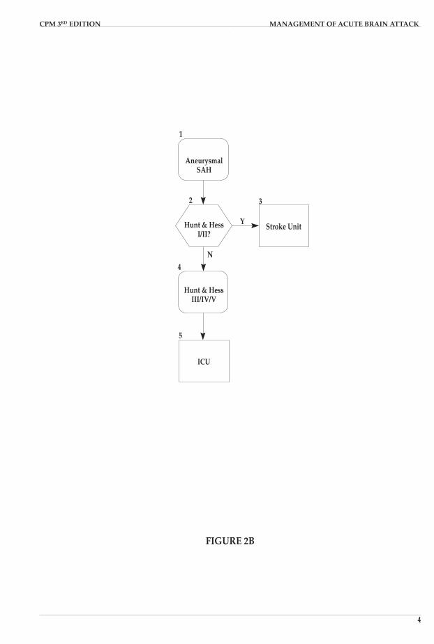
4
1
Aneurysmal SAH
Hunt & Hess
I/II?
2
Y
N
3
Stroke unit
4
Hunt & HessIII/IV/V
5
ICu
FIGuRE 2B

MANAGEMENT OF ACuTE BRAIN ATTACk CPM 3RD EDITION
5
Guidelines for Emergency Management of Acute Brain Attack (First 48 Hours since Ictus) - ER Protocol WHAT TO DO DuRING THE FIRST HOuR AT THE ER
0 to 15 minutes (simultaneously)
A. Brief but meaningful history and physical / neurologic examination
1. Include existing medical problems like DM, HPN, heart disease and renal disease
2. Important symptoms in making a diagnosis of Acute Brain Attack
• Alteration in consciousness • Aphasia • Dysarthria • Facial weakness or asymmetry • Incoordination, weakness, paralysis or sensory
loss of one or more limbs (usually one half of the body)
• Ataxia, poor balance or clumsiness, or difficulty walking
• Visual loss: Monocular, binocular or partial loss of field
• Vertigo, double vision, unilateral hearing loss, nausea, vomiting, headache, photophobia or phonophobia
3. Timing of onset of attack: The first SIX HOuRS is the GOLDEN PERIOD
4. Initial neurologic assessment (to be done by ER/Medical/Neurology Resident)
• Determine level of consciousness • Presence of seizure activity • Glasgow coma scale • Pupils: Size, equality, reactivity • Funduscopy • Limb movements, spontaneous and evoked • Meningeal signs • Diaz Scoring Form • NIH Stroke Scale
B. Emergency Management (ABC of emergency care) 1. Airway control, intubate as necessary 2. Monitor vital signs 3. IVF therapy: Plain NSS 1 liter 4. Initial/baseline diagnostic tests: a. Complete blood count b. PT/PTT c. Serum electrolytes d. Creatinine e. Capillary blood sugar f. 12 lead ECG to rule out AMI and arrhythmias g. Chest x-ray 5. Management of elevated ICP if present 1. Elevate head of bed to 20-30o
2. Hyperventilation (maintain pCO2 25-30 mmHg) 3. Mannitol 20% (0.8 - 1.5 g/kg IV) over 20
minutes 4. Surgical intervention (continuous drainage of
CSF) warranted if increased intracranial pressure is secondary to hydrocephalus
6. Management of hypertension a. Common causes of elevated blood pressure
should be checked: Full bladder, pain, underlying hypertension, physiological response to brain hypoxia or increased intracranial pressure
b. Parenteral drugs may be warranted in the following conditions:
a. Hemorrhagic transformation b. Acute myocardial infarction c. Left ventricular failure d. Renal failure secondary to accelerated
hypertension e. Aortic dissection c. Avoid the use of sublingual nifedipine which can
result in precipitous decline in blood pressure d. Administration of antihypertensives warranted
if: For Ischemic Stroke i. DBP >140 mmHg on 2 readings 5 minutes
apart ® start continuous IV infusion of an antihypertensive agent
ii. SBP >220 mmHg or DBP 121-140 mmHg or MAP >130 mmHg on 2 readings 20 minutes apart ® may give easily titratable antihypertensive
iii. SBP between 185-220 mmHg or DBP between 105-120 mmHg ® emergency therapy should be deferred except if with:
• Left ventricular failure • Aortic dissection • Acute myocardial ischemia • Renal failure secondary to accelerated HPN • Hemorrhagic transformation iv. SBP <185 mmHg or DBP <105 mmHg →
antihypertensive therapy is usually not indicated.
For Intracerebral Hemorrhage i. Goal of treatment should be to lower the MAP
to between 100-130 or no more than 25% of initial MAP (within minutes to 2 hours).
ii. Then toward 160/100 mmHg within 2-6 hours iii. Treatment is recommended if SBP >180 mmHg
or MAP >130 mmHg. iv. Same agents may be used as above for Ischemic
stroke. v. If BP is persistently >180/120, then oral agents
should be started (loop diuretics, beta-blockers, ACE inhibitors, alpha 2-agonists or calcium antagonists.)
vi. If ICP monitor is in place, maintain CPP at 70-100 mmHg. (CPP = ABP - ICP) where CPP = cerebral perfusion pressure; ABP = arterial blood pressure; ICP = intracranial pressure.
For Subarachnoid Hemorrhage i. Before clipping, HPN is lowered to decrease risk

CPM 3RD EDITION MANAGEMENT OF ACuTE BRAIN ATTACk
6
Intravenous Antihypertensives used in Hypertensive Emergencies
Onset of Duration Special Drug Dose Action of Action Adverse Effects Indications
Nicardipine 5-15 5-10 1-4 h Tachycardia, Most HPN emerg. except HCl mg/h IV min HA, flushing, Acute HF; caution with local phlebitis coronary ischemia
Hydralazine 10-20 10-20 3-8 h Tachycardia, Eclampsia HCl mg IV min flushing, HA, 10-50 20 - 30 vomiting, mg IM min increased angina
NTG 5-100 2-5 3-5 min HA, vomiting Coronary µg/min min methemoglobine- ischemia as IV mia, tolerance infusion with prolonged use
HA = headache HF = heart failure NTG = nitroglycerine
ALERT THE BRAIN ATTACk TEAM!Initially, this shall be done by hospital paging.Each member of the BAT will then be paged individually through his or her pager.
The following will be paged or notified immediately of a Brain Attack Situation: 1. Neurology resident 2. Neurosurgery resident 3. Senior medical resident neurology rotator 4. CT scan technician (local 5420 or 5408)
of rebleeding. ii. If BP remains elevated prior to clipping of the
ruptured aneurysm, treatment is recommended as per the guideline for intracerebral hemorrhage.
iii. After clipping of the aneurysm, elevated BP is usually not treated. BP is maintained more than 140 mmHg either by induced hypervolemia and hypertension to decrease ischemia from vasospasm.
7. Seizures should be adequately and promptly treated
a. Partial seizures can be treated with carbamazepine 200 mg BID or phenytoin 300 mg/day
b. Status epilepticus can be treated with either lorazepam (1 - 2 mg IV) or diazepam (5 - 10 mg IV) and loading with phenytoin 15-20 mg/kg IV, maintained at 300 mg/day
8. Control of agitation a. Midazolam 5-7.5 mg IV or b. Chlorpromazine (Thorazine) 25 mg + diphen-
hydramine (Benadryl) 50 mg admixed in 1 syringe and given intramuscularly (with BP precautions)
9. Fever should be treated aggressively with antipyretics
or hypothermic devices to lower body temperature. a. Maintain core temperature between 36-37.50C b. Cooling measures (antipyretics, tepid sponge
bath, cooling blanket) should be instituted if temperature >37.5oC.
10. Hyperglycemia or hypoglycemia should be controlled.
16 to 30 minutes
A. Assessment by Neurology/Neurosurgery resident 1. Is it a brain attack or not? 2. Transport patient to CT scan accompanied by
Neurology intern/resident 3. Decision to admit to regular room, Stroke unit, ICu
or CCu (see Flowchart) 4. ER resident accomplishes order sheet with Neurology
resident 5. ER nurse coordinates with admitting section 6. Consultation-Liaison Psychiatry Fellow assessment
of patient and relatives
By the end of the hour, the following should have been made:
MANAGEMENT OF ACuTE BRAIN ATTACk CPM 3RD EDITION
7
A. Definite diagnosis and classification of Acute Brain Attack and disposition (see Algorithm):
1. Non-Cardioembolic Infarction a. Acute stable stroke <24 hours: Admit to Stroke
unit b. Acute stable stroke >24 hours: Admit to regular
room c. Acute stable or unstable stroke enrolled in a trial
(interventional or therapeutic): Admit to Stroke unit
d. Acute unstable stroke not needing surgery: admit to Stroke unit
e. Acute unstable stroke needing immediate surgery:
i. Massive cerebellar infarction: Bring to OR immediately after securing relatives' consent or admit to ICu pending relatives' consent
ii. Massive MCA infarction (Malignant Infarction): Bring to OR immediately after securing relatives' consent or admit to ICu pending relatives' consent
2. Cardioembolic Infarction: Admit to CCu or Stroke unit
3. Spontaneous Intracerebral Hemorrhage (ICH) a. Acute stable stroke <24 hours: Admit to Stroke
unit b. Acute stable stroke >24 hours: Admit to regular
room c. Acute unstable stroke not needing immediate
surgery but a candidate for surgery, hence, needs close observation: Admit to Stroke unit
i. Intracerebral hematoma less than 50 mL ii. Intracerebral hematoma with intact
sensorium iii. Intracerebral hematoma with intraventricular
extension d. Acute unstable stroke needing emergency
surgery: Bring to OR after securing relatives' consent or to ICu pending relatives' consent
i. Cerebellar hematoma with deteriorating sensorium
ii. Intracerebral lobar hematoma more than 50 mL with deteriorating sensorium
4. Aneurysmal Subarachnoid Hemorrhage (SAH) a. Hunt and Hess I and II: Admit to Stroke unit b. Hunt and Hess III, IV and V: Admit to ICu c. Early angiography may be warranted depending
on the attending physician 5. Ruptured Arteriovenous Malformation (AVM): Same
disposition as Intracerebral Hemorrhage 6. Transient Ischemic Attacks a. Recurrent TIAs: Admit to Stroke unit b. Single TIA: Admit to regular roomB. Admission order must be completed (See Section II for
the admitting orders for the particular type of stroke)C. Family assessment by the Consultation Liaison Fellow
must be done according to their reaction to the patient's illness.
D. Proper endorsement to attending physician in case no referral to Neuroscience is made.

CPM 3RD EDITION MANAGEMENT OF ACuTE BRAIN ATTACk
8
Profiles of the Various Types of Acute Brain Attack
INTRACEREBRAL HEMORRHAGE
A. Definition Spontaneous intracerebral hemorrhage (ICH) refers to
bleeding from an arterial source directly into the brain substance in the absence of immediately preceding trauma. Primary ICH is not directly caused by another disease, while secondary forms are related to acquired or congenital conditions.
B. Etiologies of ICH (kaufmanns 1991) 1. Hypertensive 2. Non-hypertensive
Congenital Vascular Drug- Post- Anomalies Related Infectious
i. Aneurysms i. Sympatho- aneurysm mimetics ii. AVMs ii. Anti- coagulants iii. Fibrinolytics Coagulopathy Postoperative Neonatal i. Intracranial intraven- ii. Carotid tricular iii. Cardiac
Tumors Post-stroke Others i. Arterial infarction ii. Venous occlusion Vasculopathy, Delayed vasculitis post-traumatic i. Cerebral i. Parenchymal amyloid angiopathy ii. Moya-moya ii. Aneurysmal iii. Vasculitis iii. Postoperative
C. Distribution of Hypertensive ICH (kasei, et al 1992) Putaminal 35-50% Lobar/subcortical white matter 30% Cerebellum 16% Thalamus 10-15% Pontine 5-12%
D. Signs and Symptoms Typical: Abrupt onset, alteration of consciousness
rapidly progressing to stupor and coma, with focal neurologic signs occasionally accompa-nied by headache and vomiting.
Supratentorial hematomas 1. Seizures develop in about half of lobar hematomas. 2. Specific clinical findings are related to size and
location of hematoma. 3. Caudate and thalamic hemorrhages may extend into
the ventricles and produce hydrocephalus. 4. Watch out for trantentorial herniation.
Infratentorial hematomas 1. Cerebellar a. Dizz iness , pupi l la ry and oculomotor
abnormalities, dysarthria, cerebellar and pyramidal tract signs
b. Produces hydrocephalus in 75% of cases c. Clinical triad: Appendicular ataxia, ipsilateral
gaze palsy and peripheral facial weakness; 75% of cases have at least two
d. Watch out for tonsillar herniation 2. Pontine a. Changes in vital signs, posturing, ocular bobbing,
pinpoint pupils, vertigo, autonomic dysfunction, locked-in
E. Clinical Course 1. Highest case-fatality rate of all strokes 2. Neurologic deterioration may be related to
appearance of new signs or increased severity of preexisting deficits
3. Causes of neurologic deterioration: Cerebral edema, ICH progression or rebleeding, hydrocephalus
4. Risk of deterioration was highest on the first day and progressively decreases thereafter
5. Large hematoma size and marked mass effect on initial scan are predictors of deterioration
F. Diagnostic Studies Laboratory tests Screening for hematologic and clotting abnormalities,
infection, vasculitis, drugs CT Scan 1. Should be done in emergent situations 2. Very sensitive, acute blood shows up as increased
density 3. Can detect hematomas as small as few millimeters
in diameter 4. Gives information on location, size, mass effect,
intraventricular extension and hydrocephalus 5. Perform with intravenous contrast enhancement
in patients less than 40 years old without history of hypertension, continued neurologic worsening, with history of neoplasm, vasculitis, bacterial endocarditis, or atypical location or appearance of hematoma
6. Subacute blood appears isodense to brain
MRI 1. Estimation of age of bleed 2. Acute blood: Isointense on T1 & T2, use gradient
echo

MANAGEMENT OF ACuTE BRAIN ATTACk CPM 3RD EDITION
9
3. Later changes on MR depends on state of RBC degradation
4. useful in evaluating for underlying pathologies
Angiogram 1. Perform if vascular anomaly is suspected 2. Positive in 50% of young patients with ICH
G. Medical Treatment Goal: Prevention of complications and careful
management of blood pressure 1. Maintain SBP 140-160 and DBP 90-100 mmHg (MAP
around 115) a. use intravenous nicardipine for better titration
of BP b. Sustained hypertension may alter cerebral
autoregulation, promote progression of bleed and increase edema
c. Hypotension may result in cerebral hypoperfusion especially in the setting of increased intracranial pressure (ICP)
2. Manage increased ICP accordingly (see guidelines on increased ICP management)
3. Consider prophylactic use of anticonvulsants a. There is higher incidence of seizures in ICH
especially in lobar hematomas b. Role of prophylactic anticonvulsants in deep
hemorrhages is unclear. It is justified to withhold anticonvulsants until clinically indicated.
4. Prevent ion and t reatment of respiratory complications
5. Prevention and treatment of infections 6. Maintenance of adequate nutrition. 7. Early rehabilitation once stable, bed sore precautions,
DVT prophylaxis (Ted hose stockings or compression boots)
H. Surgical Treatment Role depends on size, location and extent of hematoma 1. Consider ventriculostomy for ICP monitoring and
drainage (in cases of intraventricular extension) 2. Surgical evacuation Indications a. Secondary deterioration i. Suggest reversibility of deficit since function
survived initial insult ii. Temporal lobe hematoma may be life-
threatening iii. Surgery not recommended if bleed is
massive with irreversible neurologic deficits, markedly depressed level of consciousness and immediate loss of brain stem function
b. Cerebellar hemorrhage, especially if greater than 3 cm in diameter
i. Accessible ii. May prevent rapid and i rreversible
deterioration c. Diagnostic uncertainty i. Exploratory operation • in suspicious but undiagnosed cases • in setting of recurrent hemorrhages with
progressive deficit
d. Improvement of functional recovery i. Likely best in moderately-sized bleeds
evacuated early ii. Patients who are alert with bleed <2 cm in
diameter should be managed medically iii. Patients with clots >85 cc almost always
succumb
Open evacuation vs aspirationEarly vs late
ANEuRYSMAL SuBARACHNOID HEMORRHAGE
A. Introduction 1. Common and often devastating occurrence 2. Despite considerable advances in diagnostic
techniques, surgical and anesthetic techniques, and perioperative management, the outcome for patients with SAH remain poor, with overall mortality rates of 25% and significant morbidity of approximately 50% of survivors.
B. Epidemiology 1. Population-based incidence rates for SAH vary from
6-16 per 100,000 with the highest rates reported in Finland and Japan.
2. unlike other types of stroke, the incidence of SAH has not declined over time.
3. Incidence of SAH increases with age (mean age of approximately 50 years) and is higher in women than in men.
4. Population-based mortality rates for SAH have progressively declined since 1970.
5. Survival rate after SAH has improved during this time, but differences between community rates and hospital rates are apparent, with a difference of approximately 20% in mortality at 1 year after SAH.
C. Risk Factors 1. Putative risk factors: Age, gender and race 2. Smoking is a consistent and strong risk factor 3. use of alcohol or binge drinking may also be a risk
factor for SAH
D. Natural History of Ruptured Aneurysms 1. Case-fatality rate of approximately 70% for persons
who rebled 2. Rebleeding was maximal (4%) on the first days after
SAH and then constant at a rate of 1% to 2% per day over the subsequent 4 weeks
3. Risk of rebleeding with conservative therapy is between 20% and 30% for the first month after hemorrhage and then stabilizes at a rate of approximately 3% per year
4. Risk factors for acute rebleeding a. Interval from hemorrhage to admission and
treatment, initial BP and neurological status on admission have been related to recurrent hemorrhage in the first 2 weeks after SAH
b. Gender, age, prior medical condition, shape and direction of aneurysm, early interval to

CPM 3RD EDITION MANAGEMENT OF ACuTE BRAIN ATTACk
10
angiography, variation in BP, hydrocephalus, intraventricular blood and use of ventricular drains
c. Rebleeding in the late phase after SAH (more than 1 month) has been related to aneurysm location and size, and persistent elevated blood pressure
E. Clinical Manifestations Presenting symptoms 1. Typical: Abrupt onset of a (usually) severe headache
of atypical quality 2. May or may not be associated with a brief loss
of consciousness, nausea and/or vomiting, focal neurological deficits (including cranial nerve palsies) or stiff neck)
F. Diagnosis Non-contrast CT 1. Cornerstone of SAH diagnosis 2. If performed within 24 hours of the ictus, high-
density clot in the subarachnoid space can be demonstrated in 92% of cases
3. Diagnostic sensitivity of CT scanning progressively declines after the first day, however, and diagnostic lumbar puncture should be performed if the initial CT scan is negative
Selective catheter cerebral angiography 1. Currently the standard for diagnosing cerebral
aneurysm as the cause of SAH 2. Approximately 20-25% of cerebral angiograms
performed for SAH will not indicate a source of bleeding
3. Repeat angiography after approximately 1 week will disclose a previously unrecognized aneurysm in an additional 1% to 2% of cases
4. MRA or infusion CT angiography is recommended when conventional angiography cannot be per-formed
Transcranial Doppler ultrasonography 1. Commonly used for non-invasive diagnosis
and follow-up of cerebral vasospasm, although cerebral angiography may be required for definitive diagnosis
G. Prevention of Rebleeding after SAH 1. Regulated bed rest or antihypertensive therapy alone
is not recommended to prevent rebleeding after SAH, although both are frequently included in the overall treatment of patient with SAH.
2. Antifibrinolytic therapy to prevent rebleeding is rec-ommended in certain clinical situations, e.g., patients with a low risk of vasospasm and/or a beneficial effect of delaying surgery. However, antifibrinolytic therapy has been associated with a higher rate of cerebral ischemic resulting in no benefit in terms of overall outcome. Future studies are recommended to determine whether a combination of antifibrinolytic therapy with other treatments to reduce vasospasm will be beneficial.
H. Surgical Treatment of Ruptured Aneurysms 1. Surgical clipping is strongly recommended to reduce
the rate of rebleeding after aneurysmal SAH. 2. Although early surgery reduces the risk of rebleeding
after SAH, older studies showed that overall outcome is not different than that for delayed surgery. Early surgery is recommended for the good-graded patient with an uncomplicated aneurysm. For other clinical situations, either early or delayed surgery is recommended, depending on the specific clinical situation. Early referral to specialized centers is sternly recommended.
3. Wrapping or coated aneurysm, or incompletely clipped aneurysm probably has an increased risk of re-hemorrhage. Complete surgical obliteration of the aneurysm is recommended whenever possible.
I. Cerebral Vasospasm after SAH 1. Cerebral vasospasm is the delayed narrowing of
large-capacitance arteries at the base of the brain after SAH often associated with radiographic or cerebral blood flow, evidence of diminished perfusion in the distal territory of the affected artery.
2. Onset 3 to 5 days after the hemorrhage, maximal narrowing at 5 to 14 days and gradual resolution over 2 to 4 weeks
3. In 50% of cases, it is manifested by the occurrence of a delayed neurological ischemic deficit which may resolve or progress to cerebral infarction.
4. Progression to cerebral infarction occurs in approximately 50% of symptomatic cases.
J. Treatment of Vasospasm 1. Hypertension/Hypervolemia/Hemodilution a. Recommended for prevention and treatment of
ischemic complication from vasospasm b. The aneurysm should be clipped when possible,
and patients receiving this therapy should be closely monitored in an intensive care setting for hemodynamic function.
c. Initiation of H/H/H therapy is associated with significant risks, including cardiac failure, electrolyte abnormalities, cerebral edema, bleeding abnormalities and rupture of an unsecured aneurysm.
2. Oral nimodipine is strongly recommended to reduce poor outcome related to vasospasm.
3. Transluminal angioplasty is recommended for treatment of vasospasm in patients whose conventional therapy has failed.
k. Other Complications Associated with SAH 1. Acute (obstructive) hydrocephalus a. Acute hydrocephalus after SAH complicates
approximately 20% of cases. Ventriculostomy is recommended although it may be associated with increased rebleeding and infection.
b. The etiology of acute ventriculomegaly after SAH is usually obstructive hydrocephalus caused by intraventricular blood, the incidence of acute hy-drocephalus in SAH parallels clinical grade with a greater frequency among poor-grade patients.

MANAGEMENT OF ACuTE BRAIN ATTACk CPM 3RD EDITION
11
2. Chronic (communicating) hydrocephalus a. Ventriculomegaly occurred in more than 60% of
patients by 30 days after SAH b. Temporary or permanent CSF diversion is
recommended in symptomatic patients 3. Hyponatremia a. Incidence of hyponatremia following SAH ranges
from 10% to 34% b. usually develops several days after the
hemorrhage and often parallels the time course of vasospasm
c. More common in patients with poor clinical grade and hydrocephalus, and may be an independent risk factor for poor outcome
d. It is strongly recommended that management of hyponatremia after SAH emphasizes the avoidance of volume contraction; management should include intravascular administration of isotonic fluids.
e. It is recommended that volume status in certain patients with recent SAH be assessed by monitoring central venous pressure, pulmonary capillary wedge pressure, fluid balance and body weight, although these parameters have not been tested in clinical trials. Trend indicating volume contraction should be corrected by increasing the volume of fluids administered.
f. It is recommended that hypotonic fluids be avoided as they may contribute to hyponatremia; fluid restriction should not be instituted to treat hyponatremia.
4. Seizures a. A large number of seizure-like episodes are
associated with aneurysmal rupture and have an incidence of about 25% although seizure incidence as high as 90% has been reported.
b. It is unclear whether these episodes are truly epileptic in origin or reflect a release phenomenon associated with a sudden rise in intracranial pressure.
c. Because of the potential risk of rebleeding with a seizure, the administration of prophylactic an-ticonvulsant is recommended in the immediate post-hemorrhage period.
d. The long-term use of anticonvulsants is not routinely recommended for patients with no seizure episodes and should be considered only for patients with risk factors such as prior seizure, hematoma, infarct or middle cerebral artery aneurysms.
Grading Scale for Subarachnoid Hemorrhage
Hunt and Hess Scale
Grade Neurological Status 1 Asymptomatic 2 Severe headache or meningismus; no neurological deficit (except cranial nerve
palsy) 3 Drowsy; minimal neurological deficit 4 Stuporous; moderate to severe hemiparesis 5 Deep coma; decerebrate posturing
L. Treatment Protocols 1. Emergency Evaluation and Care 2. Hunt & Hess grades 1 or 2 a. Frequent neurological assessments b. Strict bed rest c. Prophylactic measures for DVT d. Oral nimodipine therapy should be initiated e. Angiography: Timing depends on the interval
between admission and planned surgery 3. Hunt & Hess grades 3 to 5 a. Admit to ICu b. Isotonic or hypertonic intravenous fluids should
be administered c. Central intravenous access (with the ability to
measure either the CVP or pulmonary artery pressure) is desirable in most of these patients
d. If obtunded, an endotracheal intubation for airway protection should be performed if necessary
e. If initial or subsequent CT scan shows significant hydrocephalus and the patient is lethargic or has a decreasing level of consciousness, a ventriculos-tomy is usually performed.
4. Patients with Intracerebral Hemorrhage a. May be considered for emergency evacuation or
the intracerebral clot b. Clipping of the aneurysm can often be
accomplished during removal of the clot c. Patients who are obtunded and/or have a
significant lateralizing deficit but who are otherwise neurologically stable may undergo cerebral angiography before surgical removal of the blood clot.
M. Postoperative Care 1. Sequential CT scans may be necessary to differ- entiate neurological deterioration caused by va- sospasm, hydrocephalus or cerebral edema 2. Rehabilitation program
TRANSIENT ISCHEMIC ATTACk
A. Introduction 1. TIAs are temporary focal or retinal deficits caused
by vascular disease that clear completely with time in less than 24 hours.
2. Most TIAs are much shorter, the majority clearing within 1 hour. In the Cooperative Study Group of Transient Ischemic Attacks, the median duration of carotid distribution was 14 minutes and that of vertebrobasilar TIAs was 8 minutes.
3. TIAs are a syndrome of diverse causes. Proper diagnosis is crucial in choosing the appropriate therapy to minimize stroke.
B. Manifestations 1. The symptoms of TIAs are protean and depend on
the vascular territory involved. When the carotid artery is involved, the symptoms reflect ischemia to the ipsilateral eye or brain. Visual disturbances such as graying, fogging or blurring of vision,

CPM 3RD EDITION MANAGEMENT OF ACuTE BRAIN ATTACk
12
hemispheric weakness or numbness of the contra-lateral face or limbs occur. Language difficulties, cognitive dysfunction and behavioral changes may also occur.
2. Vertebrobasilar TIAs include dizziness, ataxia, vertigo, dysarthria, diplopia, and unilateral or bilateral motor or sensory symptoms.
3. The time course of the episodes is also important. TIAs are generally less than 1 hour. The onset is sudden.
C. Evaluation of patients with TIAs (SLMC guidelines) Initial Evaluation 1. CBC 2. ESR 3. urinalysis 4. RBS, FBS, BuN, creatinine, cholesterol, TG, LDL,
VLDL, HDL 5. PT with INR, PTT, VDRL 6. ECG 7. Plain Cranial CT SCAN 8. Carotid and Transcranial Doppler studies 9. CXR
Secondary evaluation ( to resolve diagnostic uncertainty)
1. Trans-thoracic or trans-esophageal echo 2. MRI 3. Antiphospholipid antibodies 4. Cerebral angiogram 5. 24-hour holter 6. 2D-Echo
Optional 1. Ambulatory ECG 2. Screen for prothrombotic states: Protein C&S, serum
electrophoresis, antithrombin III 3. CSF exam 4. Thallium scan
D. Management Risk Reduction a. Hypertension*, smoking, heart disease, oral
contraceptive b. Alcohol, estrogen use, blood lipids, diabetes, physical
activity *Hypertension = BP systolic 140 mmHg and diastolic
of 90 mmHg
Medical treatment options 1. Aspirin: 80-325 mg/day 2. Ticlopidine: 250 mg/tab BID 3. Clopidogrel: 75 mg/day 4. Dipyridamole: 200 mg BID alone or in combination
with aspirin 5. Cilostazol: 100 mg BID
ACuTE ISCHEMIC STROkE
A. Emergent Evaluation 1. Non-contrast cranial CT scan 2. ECG 3. Chest x-ray 4. Hematologic studies a. CBC b. Platelet count c. PT/PTT d. ESR e. Fibrinogen level 5. Serum electrolytes 6. Blood glucose (Hemoglucotest) 7. BuN, creatinine
B. Emergent Supportive Care 1. Stroke patients who have depressed levels of
consciousness should have airway support and ventilatory assistance.
2. Supplemental oxygen should be given only to hypoxic patients.
3. Avoid using drugs that can lower the blood pressure rapidly. May give rapid-acting antihypertensive medications for MAP >130 mmHg or SBP >220 mmHg.
4. Control either hyperglycemia or hypoglycemia after stroke.
C. General Early Supportive Care 1. Early referral for rehabilitation and physical
therapy 2. For immobilized patients, consider giving
prophylactic low molecular weight heparin
D. Treatment of Acute Neurological Complications 1. Corticosteroids are not recommended for the
management of cerebral edema and increased intracranial pressure after ischemic stroke.
2. Osmotherapy and hyperventilation are recommended for patients whose condition is deteriorating secondary to increased intracranial pressure, including those with herniation syndrome.
3. There is general agreement to recommend surgical decompression and evacuation of larger cerebellar infarctions that compress the brain stem.
4. Administration of anticonvulsants to prevent recurrent seizures is recommended. Prophylactic administration of anticonvulsants to patients with recent stroke who have not had seizures is not recommended.
5. There is general agreement that surgical intervention including continuous drainage of CSF can be used to treat increased intracranial pressure secondary to hydrocephalus.
E. Acute Treatment of Ischemic Stroke with Anti-Thrombotic or Antiplatelet Aggregating Drugs

MANAGEMENT OF ACuTE BRAIN ATTACk CPM 3RD EDITION
13
>200 + 2
TOTAL SCORE Interpretation: Score >7 = >90% probability of bleed Score <7 = Probably infarct
2. National Institute of Health Stroke Scale (NIHSS) 1.a.Level of Consciousness:
0 Alert 1 Not alert, only needs minor stimulation 2 Not alert, obtunded 3 unresponsive
1.b. Ask patient the month and their age: 0 Answers both correctly 1 Answers one correctly 2 Both incorrect
1.c. Ask patient to open and close eyes and raise arm
0 Obeys both correctly 1 Obeys one correctly 2 Both incorrect
2. Best gaze (only horizontal eye movement): 0 Normal 1 Partial gaze palsy 2 Total gaze palsy
3. Visual Field testing: 0 No visual field loss 1 Partial hemianopia 2 Complete hemianopia 3 Bilateral hemianopia (blind including cortical
blindness)
4. Facial Paresis (Ask patient to show teeth or raise eyebrows and
close eyes tightly):
0 Normal symmetrical movement 1 Minor paralysis (flattened nasolabial fold,
asymmetry on smiling) 2 Partial paralysis (total or near total paralysis of
lower face) Complete paralysis of one or both sides (absence of facial movement in the upper and
lower face)
5. Motor Function - Arm (right and left): 0 Normal (extends arms 90 (or 45) degrees for 10
seconds without drift) 1 Drift before 10 seconds 2 Falls before 10 seconds 3 No effort against gravity 4 No movement
6. Motor Function - Leg (right and left):
1. Heparin a. use of unfractionated heparin in acute ischemic
stroke is not recommended. b. use heparin in patients with recent cardioembolic
stroke is up to the physician's preference. c. Low molecular weight heparin (e.g., Fraxiparine)
may be used within 48 hours of stroke ictus. 2. Thrombolytic Therapy a. rTPA may be used within 3 hours of ictus in
carefully selected patients. 3. Antiplatelet Therapy a. Aspirin (ASA) at doses of 100-325 mg may be
given within 48 hours of acute ischemic stroke.
F. Cytoprotective Therapies in Acute Ischemic Stroke 1. use of neuroprotective agents remains to be a
matter of preference of the treating physician. 2. Neuroprotective agents proven to be beneficial
for acute ischemic stroke includes citicoline and piracetam.
G. Secondary Prophylaxis in Ischemic Stroke 1. use of ASA, ticlopidine, clopidogrel, cilostazol, or
dipyridamole have been proven in clinical trials to benefit stroke patients for the prevention of another future event.
Appendix
1. Diaz Stroke Scale Name: Age/Sex: Date/Time of stroke ictus: Date/Time Neurologic scoring done: (Please encircle the appropriate score) A. Vomiting + 4 B. Level of Consciousness unarousable + 4 Drowsy - arousable + 2 Awake 0 C. Fever + 3 D. Respiratory Pattern Ataxic or apneustic (rapid irregular) + 3 Hyperventilation (rapid regular) + 3 Cheyne-Stokes (slow irregular) + 1 Normal or regular 0 E. upper GI Bleeding + 3 F. Neurologic deficit maximal at onset + 2 G. Headache + 2 H. Nuchal rigidity + 2 I. Diastolic Blood Pressure (mmHg) <90 - 2 91-99 0 >100 + 2 J. Systolic Blood Pressure (mmHg) <150 - 2 151-169 - 1 170-180 0 181-199 + 1

CPM 3RD EDITION MANAGEMENT OF ACuTE BRAIN ATTACk
14
0 Normal (hold leg 30 degrees position for 5 seconds)
1 Drift before 5 seconds 2 Falls before 5 seconds 3 No effort against gravity 4 No movement
7. Lim Ataxia: 0 No ataxia 1 Present in one lim 2 Present in two limbs 8. Sensory (use pinprick to test arms, legs, trunk and face
- compare side to side):
0 Normal 1 Mild to moderate decrease in sensation 2 Severe to total sensory loss
9. Best Language (describe picture, name items, read sentences)
0 No aphasia 1 Mild to moderate aphasia 2 Severe aphasia 3 Mute or global aphasia 10. Dysarthria (read several words): 0 Normal articulation 1 Mild 2 Severe
11. Extinction and inattention: 0 Normal 1 Inattention or extinction to bilateral simultaneous
stimulation in one of the sensory modalities 2 Severe hemi-inattention or hemi-inattention
to more than one modality
3. Barthel Activity of Daily Living (ADL) Index ITEMS
BOWELS 0 incontinent 1 occasional accident 2 continent
BLADDER 0 incontinent/catheterized & unable to manage
1 occasional accident 2 continent
GROOMING 0 needs help 1 independent for face/hair/teeth/
shaving
TOILET uSE 0 dependent 1 needs some help 2 independent
FEEDING 0 dependent 1 needs help e.g. cutting, spreading
butter 2 independent in all actions
TRANSFER 0 unable (BED-CHAIR) 1 major help, can sit 2 minor help (verbal or physical) 3 independent
WALkING 0 unable 1 independent in wheelchair 2 walks with help of person (verbal/
physical) 3 independent (may use aid)
DRESSING 0 dependent 1 needs help, but does half 2 independent (including buttons/zips/laces
STAIRS 0 unable 1 needs help (verbal/physical) 2 independent
BATHING 0 dependent 1 independent
TOTAL
4. Modified Rankin Scale Definition Grade
No symptoms 0 Minor symptoms which do not interfere 1 with lifestyle; able to carry out all usual duties of daily living
Minor handicap - symptoms which lead 2 to some restriction in lifestyle but do not interfere with the patient's capacity to look after himself; unable to carry out some previous activities, but able to look after own affairs without assistance
Moderate handicap - symptoms 3 which significantly restrict lifestyle and prevent totally independent existence; requiring some help, but able to walk without assistance
Moderately severe handicap - symptoms 4 which clearly prevent independent existence (not needing constant attention); unable to walk without assistance and unable to attend to own bodily needs without assistance
Severe handicap - totally dependent 5 requiring constant attention night and day; bedridden, incontinent, requiring constant nursing care and attention

MANAGEMENT OF ACuTE BRAIN ATTACk CPM 3RD EDITION
15
Functionally independent - Grades 0, 1, 2 Functionally dependent - Grades 3, 4, 5
5. Hunt and Hess Scale for SAH Hunt and Hess Scale
Grade Neurological Status 1 Asymptomatic 2 Severe headache or meningismus; No neurological deficit (except cranial nerve
palsy) 3 Drowsy; minimal neurological deficit 4 Stuporous; moderate to severe hemiparesis 5 Deep coma; decerebrate posturing
6. Glasgow Coma Scale Best Motor Response
6 Obeys commands 5 Localizes to pain 4 Withdraws to pain 3 Abnormal flexion 2 Abnormal extension 1 No movement
Best Verbal Response 5 Oriented and appropriate 4 Confused conversation 3 Inappropriate 2 Incomprehensible sounds 1 No sounds
Eye Opening 4 Spontaneous 3 To speech 2 To pain 1 No Eye Opening
References: 1. Adams HP Jr, Brott FG, Crowell RM, et al. Guidelines for the
Management of Patients within Acute Ischemic Stroke. A statement for Healthcare Professional from a Special Writing Group of the Stroke Council, American Heart Association. Stroke 1994; 25(9):190-1914.
2. Adams HP Jr, Brott TG, Furlan AJ et al. Guidelines for Thrombolytic Therapy for Acute Stroke: A Supplement to the Guidelines for the Management of Treatment of Acute Ischemic Stroke. A statement for Healthcare Professional from a Special Writing Group of the Stroke Council, American Heart Association. Stroke 1996;27(9):1711-1718.
3. American Academy of Neurology Quality Standards Subcommittee. Practice Parameter-Stroke Prevention with Non-valvular Atrial Fibrillation. Neurology 1998; 51;671-673.
4. Barnett HJM, Mohr JP, Stein BM, Yatsu FM (eds). Stroke - Pathophysiology, Diagnosis, and Management, 2nd Edition. Churchill Livingstone, New York, 1992.
5. Borderick JP. Guidelines for Medical Care and Treatment of Blood Pressure in Patient with Acute Stroke. Proceedings of the National Symposium on Rapid Identification and Treatment of Acute Stroke. December 1996.
6. Caplan L. Intracerebral Hemorrhage Revisited. Neurology 1988;38:624-7.
7. Chinese Aspirin Stroke Trial. A randomized placebo-controlled of early aspirin use in 20,000 patients with acute ischemic stroke. CAST (Chinese Acute Stroke Trial) collaborative Group. Lancet 1997;349:1641-9.
8. Clark WM, Warachi SJ, Pettigrew LC et al. A randomized Dose-response Trial of Citicoline in Acute Ischemic Stroke Patients. Neurology 1997;49:671-678.
9. Culebras A, kase CS, Masden JC et al. Practice Guidelines for the use of Imaging in Transient Ischemic Attacks and Acute Stroke. A Report of the Stroke Council of the American Heart Association. Stroke 1997;28(7):1480-1497.
10. Davis, Anthony, et al., Anticoagulation Prescribing Guidelines for General Practice. 1995.
11. De Deyn PP, Reuck JD, Deberdt W et al. Treatment of Acute Ischemic Stroke with Piracetam. Members of the Piracetam in Acute Stroke Study (PASS) Group. Stroke 1997;28:2347-52.
12. Feinberg WM. Guidelines for the Management of Transient Ischemic Attacks. From the Ad hoc Committee on Guidelines for the Management of TIAs of the Stroke Council, American Heart Association. Stroke 1994;25(6):1320-35.
13. Indredavid B, Sierdahl SA, Bakke F et al. Stroke unit Treatment, Long-term Effects. Stroke 1997;28:1861-1866.
14. Mayberg MR, Batjer HH, Dacey R, et al. Guidelines for the Management of Aneurysmal Subarachnoid Hemorrhage. A statement for Healthcare Professional from a Special Writing Group of the Stroke Council, American Heart Association. Stroke 1994;25(11):2315-2328.
15. Mayer SA, Sacco RL, Shi T, Mohr JP. Neurologic Deterioration in Noncomatose Patients with Supratentorial Intracerebral Hemorrhage. Neurology 1994;44:1379-84.
16. PNA Stroke Council. Practice Guidelines on Management of Acute Ischemic Stroke. Philippine Journal of Neurology. 1997;3:69-91.
17. Ronning OM, Guldvog B. Stroke unit Versus General Medical wards, II: Neurological Deficits and Activities of Daily Living. A Quasi-randomized Controlled Trial. Stroke 1998;29:586-590.
18. Sacco RL, Wolf PA, Bharucha NE, et al. Subarachnoid and Intracerebral Hemorrhage: Natural History, Prognosis, and Precursive Factors in the Framingham Study. Neurology 1984;34:847-54.
19. Stroke unit Trialists' Collaboration. How do stroke units Improve Patients Outcomes? A Collaborative Systemic Review of the Randomized Trials. Stroke 1997;28:2139-2144.
20. The International Stroke Trial (IST): A Randomized Trial of Aspirin, Subcutaneous Heparin Both or Neither Among 19,435 Patients with Acute Ischemic Stroke. IST Collaborative Group. Lancet 1997;349:1569-81.
21. The Sixth Report of the Joint National Committee on Prevention, Detection, Evaluation, and Treatment of High Blood Pressure. NIH. November 1997.
22. Van Swieten JC et al. Interobserver Agreement for the Assessment of Handicap In Stroke Patients. Stroke 1988;19:604-607.
23. Wade DT & Hewer RL. Functional abilities After Stroke: Measurement, Natural History and Prognosis. Journal of Neurol Neurosurg & Psych 1987;50:177-182.
24. Weir CJ, Murray GD, Dykes AG, Lees kR. Is Hyperglycemia an Independent Predictor of Poor Outcome After Acute Stroke? Results of a Long-term Follow-up Study. BMJ 1997;314:1306-36.
25. Welch kMA, Caplan LR, Reis DJ, Siesjo Bk, Weir B (eds). Primer on Cerebrovascular Diseases. Academic Press, San Diego, 1997.