acute disseminated encephalomyelitis, multiphasic - brain
TRANSCRIPT
Brain (2000), 123, 2407–2422
Acute disseminated encephalomyelitis, multiphasicdisseminated encephalomyelitis and multiplesclerosis in childrenR. C. Dale,1 C. de Sousa,1 W. K. Chong,2 T. C. S. Cox,2 B. Harding3 and B. G. R. Neville1,4
Departments of 1Neurology, 2Radiology and Correspondence to: Dr Russell Dale, Department of3Histopathology, Great Ormond Street Hospital for Neurology, Great Ormond Street Hospital for Children,Children and 4Institute of Child Health, University College London WC1 3JH, UKLondon, London, UK E-mail: [email protected]
SummaryForty-eight children with disseminated demyelination ofthe CNS, 28 with acute disseminated encephalomyelitis(ADEM), seven with multiphasic disseminatedencephalomyelitis (MDEM) and 13 with multiple sclerosiswere studied for a mean follow-up period of 5.64 years.The presentation findings of the ADEM/MDEM groupwere compared with those of the multiple sclerosis group.The following findings were more commonly seen inADEM/MDEM presentation compared with the multiplesclerosis presentations: predemyelinating infectiousdisease (74 versus 38%, P < 0.05); polysymptomaticpresentation (91 versus 38%, P < 0.002); pyramidal signs(71 versus 23%, P < 0.01); encephalopathy (69 versus15%, P < 0.002); and bilateral optic neuritis (23 versus8%, not significant). Seizures occurred only in the ADEM/MDEM group (17 versus 0%, not significant). Unilateraloptic neuritis occurred only in the multiple sclerosispatients (23 versus 0%, P < 0.01). There were nodifferences in the frequencies of transverse myelitis,brainstem involvement, cerebellar signs and sensorydisturbance between the two groups. ADEM/MDEMpatients were more likely to have blood leucocytosis (64versus 22%, P < 0.05), CSF lymphocytosis (64 versus42%, not significant) and CSF protein elevation (60 versus
Keywords: demyelination; multiple sclerosis; children; neuroimaging
Abbreviations: ADEM � acute disseminated encephalomyelitis; MDEM � multiphasic disseminated encephalomyelitis;VEP � visual evoked potential
IntroductionPost-infectious CNS disease is common in acute paediatricpractice. Most commonly, an acute cerebellar syndromefollows varicella in 1 in 1000 cases. Other infection-specificprocesses include sensorineural deafness following mumpsand Sydenham’s chorea associated with streptococcalinfections.
Other inflammatory diseases of the CNS are more difficult
© Oxford University Press 2000
33%, not significant). Patients presenting with multiplesclerosis were more likely to have intrathecal synthesis ofoligoclonal bands on presentation (64 versus 29%, notsignificant). MRI showed that subcortical white matterlesions were almost universal in both groups, thoughperiventricular lesions were more common in multiplesclerosis (92 versus 44%, P < 0.01). By contrast, in ADEM/MDEM there was absolute and relative periventricularsparing in 56 and 78% of patients, respectively. Follow-up MRI revealed complete or partial lesion resolution in90% and no new lesions in the ADEM/MDEM group. Allof the multiple sclerosis patients had new lesions on repeatMRI (five during relapse and six during asymptomaticconvalescent phases). The outcome in the ADEM patientswas mixed; 57% of patients made a complete recovery.The mean follow-up for the 35 ADEM/MDEM patientswas 5.78 years (range 1.0–15.4 years). Eight of the 13multiple sclerosis patients relapsed within the first year;11 had a relapsing–remitting course, one a primaryprogressive course and one a secondary progressivecourse. These differences in the presentation of ADEM/MDEM compared with multiple sclerosis may help in theprognosis given to families regarding the possibility oflater development of multiple sclerosis.
to categorize. Inflammation occurring at an isolated CNS sitemay occur, such as isolated transverse myelitis or opticneuritis. In these cases, the majority of these children havea single event of inflammation, though both of thesepresentations may be the first episode of a relapsinginflammatory disease such as multiple sclerosis.
When dissemination of inflammatory disease at multiple
2408 R. C. Dale et al.
Tab
le1A
Cli
nica
lfe
atur
esof
AD
EM
and
MD
EM
pati
ents
Age
Clin
ical
Enc
epha
l-Py
ram
idal
Bra
inst
emC
ereb
ella
rV
isua
lSe
nsor
yM
yelit
isFo
llow
-up
Dia
gnos
isIm
pair
men
t(y
ears
),op
athy
(yea
rs)
sex
(A)
AD
EM
patie
nts
3,M
A�
��
�–
––
2.4
AD
EM
Non
e3,
MA
��
��
�–
–3.
8A
DE
MN
one
3,F
A�
��
��
(BO
N)
––
13.3
AD
EM
↓V
A,
↓IQ
3,M
A�
��
�–
––
7.4
AD
EM
Beh
avio
ur,
mot
or3,
MA
–�
��
––
–12
.6A
DE
MN
one
4,M
A�
��
��
––
6.6
AD
EM
Scol
iosi
s4,
MA
��
��
––
–2.
3A
DE
MM
otor
4,M
A�
��
�–
––
1.4
AD
EM
Non
e4,
FA
�–
––
––
–9.
3A
DE
ME
pile
psy,
mot
orbe
havi
our,
↓IQ
4,M
A�
�–
–�
(BO
N)
––
15.4
AD
EM
↓IQ
5,F
A�
��
��
––
8.7
AD
EM
↓IQ
,be
havi
our
5,M
A�
��
�–
––
3.0
AD
EM
Beh
avio
ur5,
FA
�–
–�
––
�1.
4A
DE
MN
one
5,M
A–
�–
––
––
1.5
AD
EM
Non
e6,
FA
–�
––
�(B
ON
)–
–1.
5A
DE
MN
one
8,F
A–
�–
�–
�–
2.3
AD
EM
Non
e9,
MA
�–
–�
––
�4.
3A
DE
MM
otor
9,F
A�
––
–�
(BO
N)
––
1.0
AD
EM
Non
e10
,M
A�
��
––
–�
5.4
AD
EM
Non
e10
,F
A�
–�
–�
–�
15.0
AD
EM
Para
pleg
ia,
↓V
A,
↓IQ
10,
MA
–�
––
–�
–2.
8A
DE
MN
one
11,
MA
��
��
––
–7.
3A
DE
MN
one
11,
FA
––
�–
––
�2.
7A
DE
MPa
rapl
egia
,ne
urop
athi
cbl
adde
r
ADEM, MDEM and multiple sclerosis in children 2409
Tab
le1
Con
tinu
ed
Age
Clin
ical
Enc
epha
l-Py
ram
idal
Bra
inst
emC
ereb
ella
rV
isua
lSe
nsor
yM
yelit
isFo
llow
-up
Dia
gnos
isIm
pair
men
t(y
ears
),op
athy
(yea
rs)
sex
12,
FA
��
–�
–�
�14
.3A
DE
MPa
raes
thes
ia13
,F
A�
��
�–
�–
9.3
AD
EM
Non
e13
,M
A�
––
–�
–�
1.3
AD
EM
Non
e13
,F
A–
�–
––
�–
1.5
AD
EM
Para
esth
esia
15,
FA
��
––
�(B
ON
)�
–7.
3A
DE
M↓↓
VA
(B)
MD
EM
patie
nts
4,F
A–
��
––
––
1.5
MD
EM
Non
eR
(0.1
5)–
�–
–�
(UO
N)
––
4,M
A�
��
�–
––
14.4
MD
EM
Non
eR
(0.1
)–
��
�–
––
6,M
A–
�–
–�
(BO
N)
––
10.0
MD
EM
Non
eR
(0.1
5)�
�–
––
––
R(2
.5)
––
––
�(B
ON
)–
–7,
MA
�–
––
�(B
ON
)–
–3.
0M
DE
MN
one
R(0
.15)
�–
�–
––
–8,
FA
––
––
––
�3.
5M
DE
MPa
rapl
egia
,ne
urop
athi
cbl
adde
rR
(0.1
5)–
–�
––
––
12,
FA
�–
––
�(B
ON
)–
–2.
2M
DE
MN
one
R(0
.3)
�–
––
––
–R
(1.0
)�
––
–�
(BO
N)
––
15,
MA
–�
�–
––
–2.
5M
DE
MN
one
R(0
.1)
–�
�–
––
–
F�
fem
ale;
M�
mal
e;A
�ac
ute;
BO
N�
bila
tera
lop
ticne
uriti
s;V
A�
visu
alac
uity
;R
�re
laps
ing
(yea
rsto
rela
pse)
;B
ON
�bi
late
ral
optic
neur
itis;
UO
N�
unila
tera
lop
ticne
uriti
s.

2410 R. C. Dale et al.
sites within the CNS is monophasic, the disease is termed‘acute disseminated encephalomyelitis’ (ADEM). It has beendemonstrated that relapses may occur immediately afterADEM. If these relapses are thought to represent part of thesame acute monophasic immune process, the term‘multiphasic disseminated encephalomyelitis’ (MDEM) isused. If, however, relapses occur that are disseminated withrespect to site and time, which support a chronic immuneprocess, multiple sclerosis is diagnosed.
In children, ADEM (and MDEM) is diagnosed morecommonly than multiple sclerosis. In adults the oppositeis true.
When a child presents with a first episode of disseminatedCNS inflammation, the most important issue on recovery isthe potential risk of relapse and, therefore, the prognosis. Inview of the variable length of time between presentation andrelapse in multiple sclerosis, this can be adequately examinedonly by prolonged follow-up. A follow-up study ofdisseminated demyelination disease in children wasperformed. The children who remained monophasic (ADEM/MDEM) were compared with those who had developedclinically definite multiple sclerosis (from now on referredto as multiple sclerosis) to see whether differences exist atdisease presentation.
Patients and methodsAll children presenting to Great Ormond Street Hospital,London, between 1985 and 1999 with one or more episodeof disseminated CNS demyelination were included in thestudy. Children were excluded if they had a precedingneurological abnormality, isolated optic neuritis or isolatedtransverse myelitis, infective encephalomyelitis, cerebralvasculitis, systemic lupus erythematosus or other intracranialpathologies. Clinical and investigation findings on diseasepresentation were accumulated from patient notes. MRIs ofthe brain and spinal cord were reviewed by two paediatricneuroradiologists (W.K.C. and T.C.S.C.), who were blindedto the clinical findings. The images were assessed for lesionsite, size, characteristics and symmetry. Between April andJune 2000, a follow-up study was performed. After gainingapproval from the research ethics committee of Great OrmondStreet Hospital, the GP (general practitioner) of each patientwas traced by using regional health authority registers.Written consent was obtained from each GP to re-contact thefamilies of patients. The patient was contacted first by letter(for written consent) and subsequently by telephone. Atelephone questionnaire was used to determine residualdisability and the incidence of further neurological disease.Only three patients, who had moved abroad, were lost tofollow-up. Using this method of tracing patients, the meanfollow-up was extended from 2.36 to 5.64 years [5.78 yearsfor the ADEM/MDEM group, 5.3 years for the clinicallydefinite multiple sclerosis (using Poser criteria) group].
The monophasic group (ADEM/MDEM) was comparedwith the multiple sclerosis group to see if there were
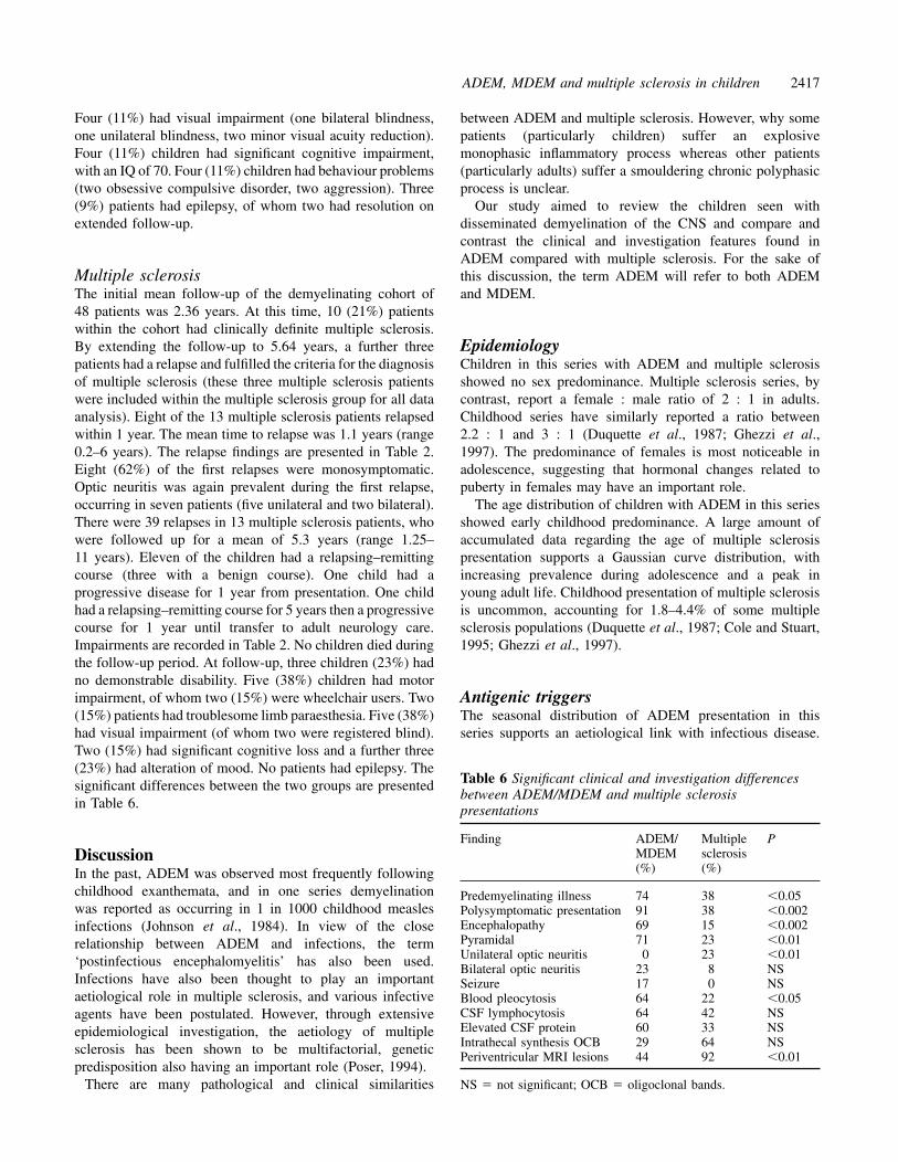
Fig. 1 Seasonal presentation of ADEM/MDEM.
differences in clinical and investigation findings at diseasepresentation. Statistical differentiation was made bycalculating the standard error of the difference between thetwo groups.
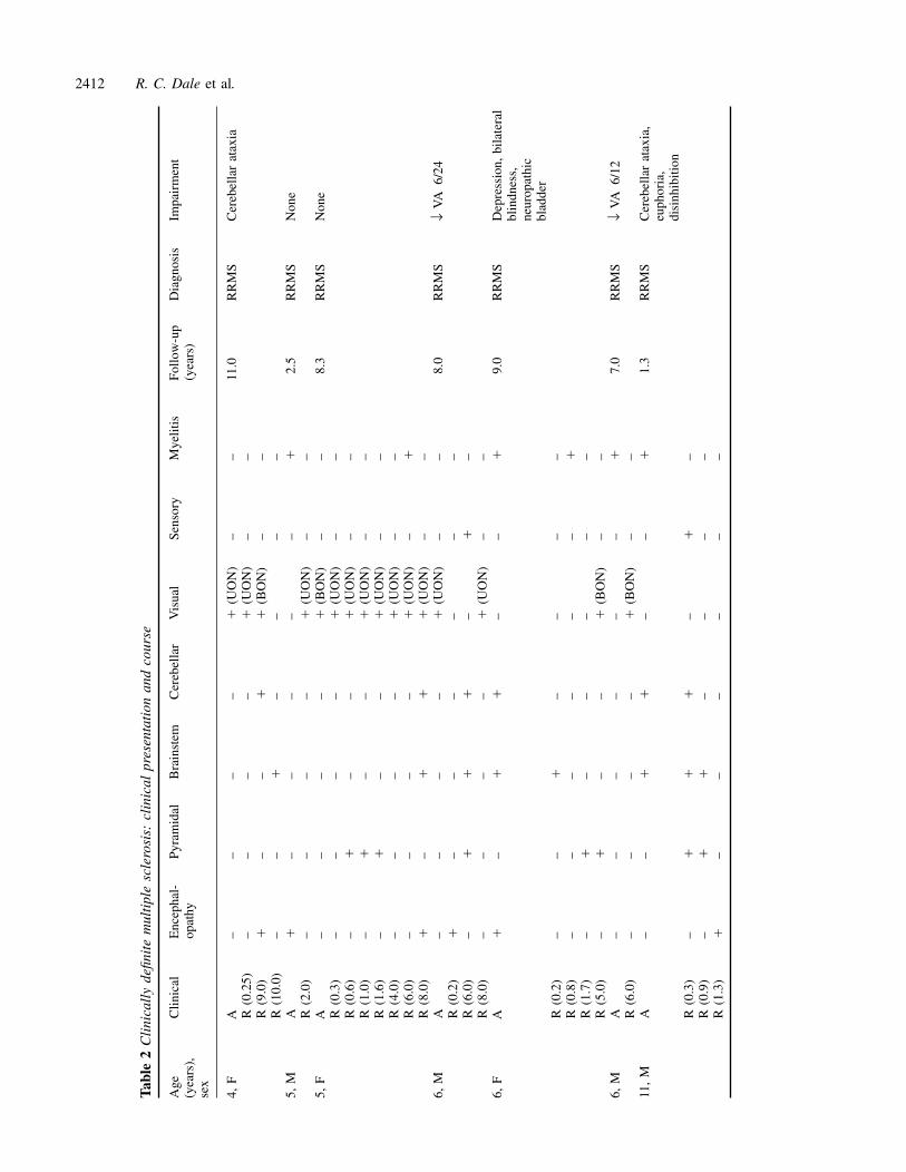
ResultsForty-eight children had evidence of disseminatedinflammatory CNS disease. Twenty-eight children fulfilledcriteria for the diagnosis of ADEM, seven for the diagnosisof MDEM and 13 for the diagnosis of multiple sclerosis.
Age, sex and family historyADEM/MDEMSixteen female and 19 male patients with ADEM/MDEMwere included. Forty-six per cent of the patients were between3 and 5 years of age (Table 1). No patients presented before3 years of age. There was no family history of multiplesclerosis in any patient in the ADEM/MDEM group.
Multiple sclerosisOf the 13 patients with multiple sclerosis, six were femaleand seven male. Age of presentation was between 4 and15 years, only three patients (23%) presenting under 5 yearsof age. One child had a second-degree relative with multiplesclerosis.
Clinical findingsADEM/MDEMThe presentation of the ADEM/MDEM patients showed aseasonal distribution (Fig. 1), being more prevalent in thewinter months. Twenty-six patients (74%) had a precedingillness in the month before presentation. The mean latencybetween predemyelinating illness and the onset ofneurological signs was 13.0 days (range 2–31 days). Thepreceding illnesses were described as upper respiratory tractinfection (9 patients); influenza (3); tonsillitis (4); lowerrespiratory tract infection (3); vaccination (2); gastroenteritis
ADEM, MDEM and multiple sclerosis in children 2411
(2); varicella (1); and fever of unknown origin (2). Tenchildren had serological evidence of specific triggers:streptococcus (3); mycoplasma (1); influenza B (1);enterovirus (1); Epstein–Barr virus (1); varicella (1); mumpsrubella immunization (1); and BCG vaccine (1).
Neurological presentation varied from an acute explosiveonset, with a maximum neurological deficit attained within1 day, to more indolent progression with maximum deficitat 31 days (mean 7.1 days). Symptoms and signs of systemicdisease were common, headache being present in 58%, feverin 43% and meningism in 31%. Seizures occurred in sixpatients (17%); they were associated with fever in fourpatients.
The neurological signs are presented in Table 1. Apolysymptomatic presentation (having more than one clinicalfinding) occurred in 32 (91%) of the children. Alteration inmental state and level of consciousness (encephalopathy)occurred in 24 (69%). Four patients needed ventilation dueto severely impaired consciousness. Pyramidal motor signsoccurred in 25 patients (71%), of whom 16 had symmetricalupper motor neurone weakness and nine asymmetricalweakness.
Cranial neuropathies occurred in 18 patients (51%) (facialweakness, ophthalmoplegia, dysarthria and dysphagia indescending order of frequency). No patient had internuclearophthalmoplegia. Visual impairment occurred in 13 patients(37%), of whom eight (23%) fulfilled the criteria for clinicalbilateral optic neuritis. No patient had unilateral optic neuritis.Impairment in visual acuity was often severe in the opticneuritis group, median visual acuity being 2/60. Seventeenpatients (49%) had cerebellar findings and eight (23%) hadclinical myelitis causing acute flaccid paraparesis and urinaryretention. Sensory disturbance occurred in six children (17%).Infrequent clinical findings included cogwheel rigidity andtitubation (1 patient), athetosis (1) and polyuria/polydipsia (1).
Seven children had relapses in the months after presentation(MDEM). Relapses occurred no more than 8 weeks afterstopping steroid treatment. One child relapsed within a weekof a routine BCG vaccination. The MDEM group wassubsequently relapse-free for a mean follow-up period of5.3 years (range 1.5–14.4 years).
Multiple sclerosisTwo-thirds of the multiple sclerosis patients presented duringthe six winter months (October to March). On presentation,five (38%) of the children had had an infection in the monthpreceding neurological onset. The mean latency betweenpredemyelinating illness and the onset of neurological signswas 9 days (range 1–21 days). The preceding illness wasupper respiratory tract infection (3), influenza (1) and feverof unknown origin (1). No microbiological agents wereincriminated in any patient. Maximum deficit occurred amean of 9 days after neurological onset (range 1–60 days).Headache occurred in 23%, fever in 15% and meningism in8% of the patients. Seizures occurred in no patients. The
clinical presentation findings and subsequent relapses arepresented in Table 2. Eight (62%) of the patients hada monosymptomatic presentation and five (38%) had apolysymptomatic presentation. The involvement ofpresentation sites, in descending order of frequency, was asfollows: optic neuritis 31% (three unilateral, one bilateral);transverse myelitis 31%; brainstem 23%; cerebellar 23%;pyramidal 23% (all hemiplegia); sensory 15%;encephalopathy 15%. Of the brainstem signs, internuclearophthalmoplegia occurred in two patients.
Investigation findingsADEM/MDEMThere was frequent investigation evidence of inflammationin the ADEM/MDEM group. Elevation of the white cellcount in the blood (33 patients tested) occurred in 64% ofpatients (mean 14.7 � 109/l, range 6.7–37.3 � 109/l).Neutrophils were abundant, being elevated in 52% of patients.By contrast, the lymphocyte count was elevated in none, and52% of patients had lymphopenia. The C-reactive proteinconcentration and erythrocyte sedimentation rate wereelevated in 35 and 46% of patients, respectively. Six patientshad T-cell subpopulation measurements during the acutephase: one had an activated CD3 population and one had adecreased CD4 count.
CSF evidence of inflammation (either pleocytosis or raisedprotein) occurred in 75% of 33 patients tested. LymphocyteCSF pleocytosis occurred in 64% (mean 51 cells/mm3, range0–270) and elevated CSF protein concentration occurred in60% (mean 0.69 g/dl, range 0.1–3.3).
Oligoclonal bands were measured concomitantly in CSFand serum on presentation in 21 patients. Six patients (29%)had evidence of intrathecal synthesis of oligoclonal bands,five (24%) had oligoclonal presence in both CSF and serumand 10 (47%) had no oligoclonal bands in serum or CSF.The six monophasic patients with intrathecal synthesis ofoligoclonal bands were followed up for a mean of 6.5 relapse-free years (range 2.2–13.3 years).
Multiple sclerosisTwo children of nine tested (22%) had an elevated peripheralleucocyte (neutrophil) count on presentation. No patient hadlymphopenia. Erythrocyte sedimentation rate was elevated infive of seven children (71%) tested in the acute phase, butresolved during convalescence. Twelve children had CSFexamination during presentation. CSF evidence ofinflammation (elevated cell count or elevated protein)occurred in 58%. Five children (42%) had CSF lymphocytosis(mean 18 cells/mm3, range 0–130 cells/mm3). Four children(33%) had CSF protein elevation (mean 0.38 g/dl, range 0.2–0.99 g/dl).
Oligoclonal bands were measured in 11 patients duringthe acute phase. Intrathecal synthesis of oligoclonal bands
2412 R. C. Dale et al.
Tab
le2
Cli
nica
lly
defin
ite
mul
tipl
esc
lero
sis:
clin
ical
pres
enta
tion
and
cour
se
Age
Clin
ical
Enc
epha
l-Py
ram
idal
Bra
inst
emC
ereb
ella
rV
isua
lSe
nsor
yM
yelit
isFo
llow
-up
Dia
gnos
isIm
pair
men
t(y
ears
),op
athy
(yea
rs)
sex
4,F
A–
––
–�
(UO
N)
––
11.0
RR
MS
Cer
ebel
lar
atax
iaR
(0.2
5)–
––
–�
(UO
N)
––
R(9
.0)
�–
–�
�(B
ON
)–
–R
(10.
0)–
–�
––
––
5,M
A�
––
––
–�
2.5
RR
MS
Non
eR
(2.0
)–
––
–�
(UO
N)
––
5,F
A–
––
–�
(BO
N)
––
8.3
RR
MS
Non
eR
(0.3
)–
––
–�
(UO
N)
––
R(0
.6)
–�
––
�(U
ON
)–
–R
(1.0
)–
�–
–�
(UO
N)
––
R(1
.6)
–�
––
�(U
ON
)–
–R
(4.0
)–
––
–�
(UO
N)
––
R(6
.0)
––
––
�(U
ON
)–
�R
(8.0
)�
–�
��
(UO
N)
––
6,M
A–
––
–�
(UO
N)
––
8.0
RR
MS
↓V
A6/
24R
(0.2
)�
––
––
––
R(6
.0)
–�
��
–�
–R
(8.0
)–
––
–�
(UO
N)
––
6,F
A�
–�
�–
–�
9.0
RR
MS
Dep
ress
ion,
bila
tera
lbl
indn
ess,
neur
opat
hic
blad
der
R(0
.2)
––
�–
––
–R
(0.8
)–
––
––
–�
R(1
.7)
–�
––
––
–R
(5.0
)–
�–
–�
(BO
N)
––
6,M
A–
––
––
–�
7.0
RR
MS
↓V
A6/
12R
(6.0
)–
––
–�
(BO
N)
––
11,
MA
––
��
––
�1.
3R
RM
SC
ereb
ella
rat
axia
,eu
phor
ia,
disi
nhib
ition
R(0
.3)
–�
��
–�
–R
(0.9
)–
��
––
––
R(1
.3)
�–
––
––
–
ADEM, MDEM and multiple sclerosis in children 2413
Tab
le2
Con
tinu
ed
Age
Clin
ical
Enc
epha
l-Py
ram
idal
Bra
inst
emC
ereb
ella
rV
isua
lSe
nsor
yM
yelit
isFo
llow
-up
Dia
gnos
isIm
pair
men
t(y
ears
),op
athy
(yea
rs)
sex
11,
MA
–�
–�
––
–4.
0R
RM
SPa
rapl
egia
,pa
raes
thes
ia,
labi
lem
ood
R(0
.3)
––
––
–�
–R
(0.6
)–
�–
�–
––
R(0
.75)
––
–�
–�
–R
(2.0
)–
–�
––
�–
R(3
.0)
––
––
––
�11
,M
A–
–�
––
––
6.0
RPM
S↓
IQ,
unila
tera
lbl
indn
ess,
trem
orhe
mip
legi
a,pa
raes
thes
iaR
(0.7
5)–
––
�–
�–
R(0
.8)
––
��
––
–R
(3.0
)–
–�
––
––
R(4
.0)
�–
––
�(U
ON
)–
–P
(5.0
)–
�–
––
�–
14,
FA
–�
––
–�
–6.
1R
RM
SU
nila
tera
lbl
indn
ess,
trem
orR
(1.4
)–
––
–�
(UO
N)
––
R(1
.9)
––
––
�(B
ON
)–
–R
(2.5
)–
––
––
–�
R(3
.0)
––
––
––
�14
,F
A–
�–
––
––
1.9
RR
MS
Tre
mor
R(1
.2)
–�
–�
–�
–14
,F
A–
––
––
�–
2.0
PPM
S↓
IQ,
para
pleg
ia,
inte
rnuc
lear
opht
halm
ia,
neur
opat
hic
blad
der
P(0
.3)
–�
��
�(B
ON
)�
–15
,M
A–
––
–�
(UO
N)
––
1.4
RR
MS
Non
eR
(1.0
)�
––
–�
(UO
N)
––
F�
fem
ale;
M�
mal
e;A
�ac
ute;
R�
rela
psin
g(y
ears
tore
laps
e);
P�
prog
ress
ive;
UO
N�
unila
tera
lop
ticne
uriti
s;B
ON
�bi
late
ral
optic
neur
itis;
RR
MS
�re
laps
ing–
rem
ittin
gm
ultip
lesc
lero
sis;
RPM
S�
rela
psin
gpr
ogre
ssiv
eM
S;PP
MS
�pr
imar
ypr
ogre
ssiv
em
ultip
lesc
lero
sis;
VA
�vi
sual
acui
ty.

2414 R. C. Dale et al.
(CSF positive, serum negative) occurred in seven of 11patients (64%) during the acute phase, though repeat testingduring convalescence or on relapse showed that nine of the11 (82%) had evidence of intrathecal synthesis at some stageof their multiple sclerosis course. The remaining patients hadno oligoclonal bands in their serum or CSF.
NeurophysiologyADEM/MDEMEEG was performed in 21 patients. Only one patient wasnormal; all others demonstrated excessive slow-wave activity(asymmetrical in 13, symmetrical in seven). Only one EEGdemonstrated epileptiform discharges. Visual evokedpotentials (VEPs) were studied in 21 patients. Eleven patientshad normal VEPs, whereas 10 showed attenuation,degradation and delay of the waveform (seven bilaterally,three unilaterally). All eight patients with bilateral opticneuritis had abnormal VEPs. Of 13 patients tested withoutclinical visual impairment, only two had abnormal VEPs.
No patients had abnormal peripheral nerve conductionstudies (six tested).
Multiple sclerosisEEG was performed in seven patients. Five had an abnormalEEG, showing excessive slow-wave activity (threeasymmetrical, two symmetrical). No EEG containedepileptiform discharges. The VEP was studied acutely innine patients; it was abnormal in seven patients, demonstratingattenuation, degradation and delay of the waveform. All fourpatients with optic neuritis had an abnormal VEP (threeunilateral, one bilateral). However, three of five patientswithout clinical visual impairment had an abnormal VEP.
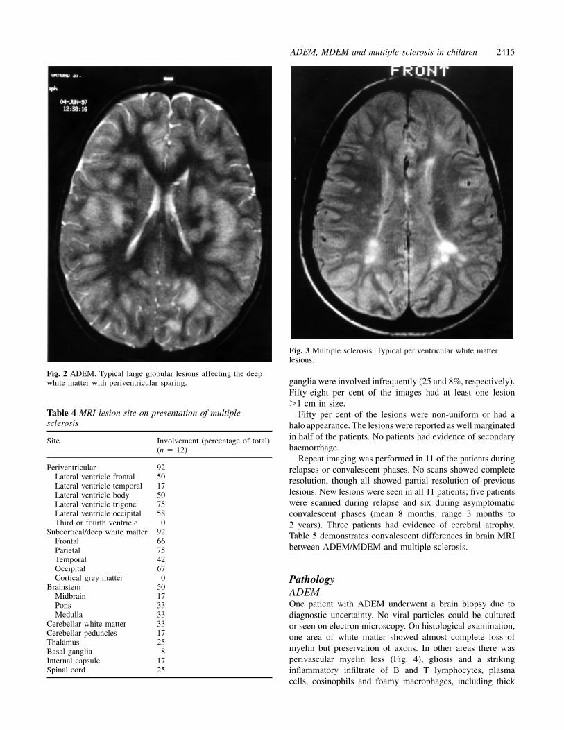
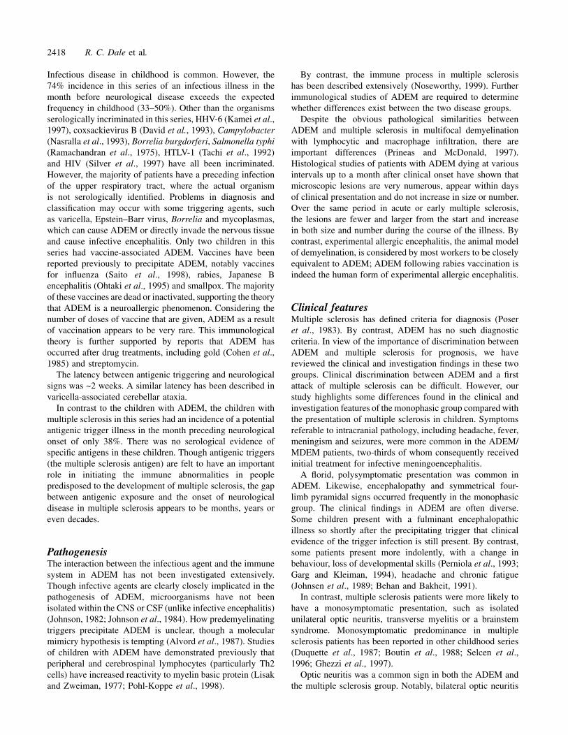
NeuroimagingADEM/MDEMMRI brain scans were performed in all 35 children, and 32of the images were available for review. Seventeen of thechildren had preceding CT scans. Only 59% of the CT scanshad been reported as abnormal, whereas all MRI brain scansdemonstrated disseminated CNS lesions. MRI FLAIR (fluid-attenuated inversion recovery) sequence and T2-weightedimages demonstrated the abnormalities more readily than T1-weighted images. The lesion site prevalence is presented inTable 3. In the ADEM group, the lesions were predominantlyin the white matter (Fig. 2). Absolute and relative sparing ofthe periventricular white matter occurred in 56 and 78% ofimages, respectively. Involvement of the deep and subcorticalwhite matter was nearly universal. Twelve per cent of imageshad a cortical grey matter lesion. The thalami and basalganglia were involved in 41 and 28%, respectively. Thesupratentorial white matter lesions were universallyasymmetrical, whereas the thalamic and basal ganglia lesions
Table 3 MRI lesion site in ADEM/MDEM patients
Site Involvement (percentage of total)(n � 32)
Periventricular white matter 44Lateral ventricle frontal 16Lateral ventricle body 16Lateral ventricle trigone 16Lateral ventricle temporal 16Lateral ventricle occipital 16Third ventricle 16Fourth ventricle 0
Subcortical/deep white matter 91Subcortical frontal 66Subcortical temporal 34Subcortical parietal 85Subcortical occipital 53
Cortical grey matter 12Cortical frontal 12Cortical temporal 3Cortical parietal 9Cortical occipital 6
Brainstem 56Midbrain 25Pons 38Medulla 28
Cerebellar white matter 31Cerebellar peduncles 9Thalamus 41Basal ganglia 28Internal capsule 28Spinal cord 28
were symmetrical in 46 and 100%, respectively. The majorityof the lesions were large (82% of the scans had at least onelesion measuring �1 cm), had poorly defined margins andwere uniform in character (only 22% of the scans had oneor more lesions which were non-uniform). One scan hadevidence of secondary haemorrhage.
Imaging was performed during convalescence follow-up in 19 ADEM/MDEM patients (mean 1.5 years afterdemyelination, range 2 months to 9 years). Seven patients(37%) had complete lesion resolution, 10 (53%) had partiallesion resolution and two (10%) were unchanged. There wereno new lesions on convalescent imaging in this group.
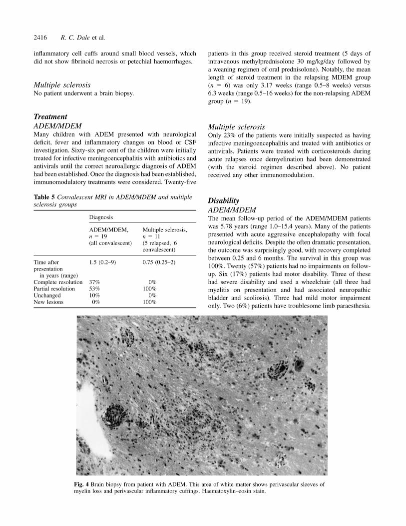
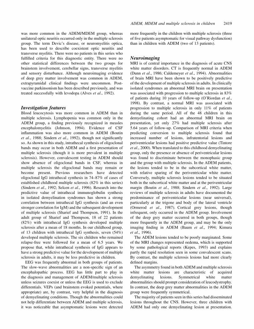
Multiple sclerosisMRI brain scans were performed in all children; 12 ofthe scans were available for review. T2-weighted imagingdemonstrated the lesions best, though T1-weighted imagingdemonstrated the older lesions adequately on convalescentimaging. The lesion sites at presentation are presented inTable 4. Ninety-two per cent of patients had periventricularlesions predominantly at the trigone or posterior lateralventricle (Fig. 3). Absolute and relative sparing of theperiventricular white matter occurred in 8 and 17% of images,respectively. The lesions were also prevalent throughout thesubcortical white matter (92% of images). No patients hadany cortical grey matter involvement. The thalami and basal
ADEM, MDEM and multiple sclerosis in children 2415
Fig. 2 ADEM. Typical large globular lesions affecting the deepwhite matter with periventricular sparing.
Table 4 MRI lesion site on presentation of multiplesclerosis
Site Involvement (percentage of total)(n � 12)
Periventricular 92Lateral ventricle frontal 50Lateral ventricle temporal 17Lateral ventricle body 50Lateral ventricle trigone 75Lateral ventricle occipital 58Third or fourth ventricle 0
Subcortical/deep white matter 92Frontal 66Parietal 75Temporal 42Occipital 67Cortical grey matter 0
Brainstem 50Midbrain 17Pons 33Medulla 33
Cerebellar white matter 33Cerebellar peduncles 17Thalamus 25Basal ganglia 8Internal capsule 17Spinal cord 25
Fig. 3 Multiple sclerosis. Typical periventricular white matterlesions.
ganglia were involved infrequently (25 and 8%, respectively).Fifty-eight per cent of the images had at least one lesion�1 cm in size.
Fifty per cent of the lesions were non-uniform or had ahalo appearance. The lesions were reported as well marginatedin half of the patients. No patients had evidence of secondaryhaemorrhage.
Repeat imaging was performed in 11 of the patients duringrelapses or convalescent phases. No scans showed completeresolution, though all showed partial resolution of previouslesions. New lesions were seen in all 11 patients; five patientswere scanned during relapse and six during asymptomaticconvalescent phases (mean 8 months, range 3 months to2 years). Three patients had evidence of cerebral atrophy.Table 5 demonstrates convalescent differences in brain MRIbetween ADEM/MDEM and multiple sclerosis.
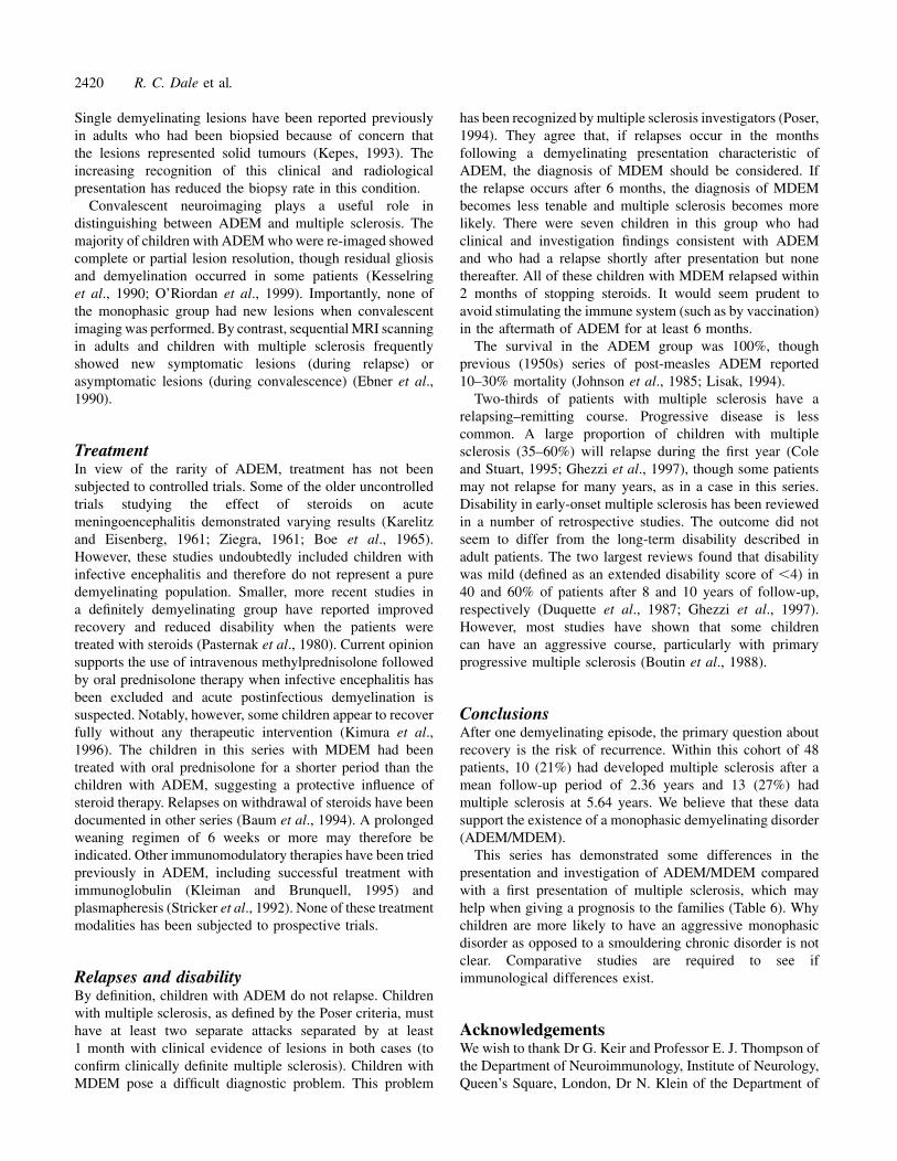
PathologyADEMOne patient with ADEM underwent a brain biopsy due todiagnostic uncertainty. No viral particles could be culturedor seen on electron microscopy. On histological examination,one area of white matter showed almost complete loss ofmyelin but preservation of axons. In other areas there wasperivascular myelin loss (Fig. 4), gliosis and a strikinginflammatory infiltrate of B and T lymphocytes, plasmacells, eosinophils and foamy macrophages, including thick
2416 R. C. Dale et al.
inflammatory cell cuffs around small blood vessels, whichdid not show fibrinoid necrosis or petechial haemorrhages.
Multiple sclerosisNo patient underwent a brain biopsy.
TreatmentADEM/MDEMMany children with ADEM presented with neurologicaldeficit, fever and inflammatory changes on blood or CSFinvestigation. Sixty-six per cent of the children were initiallytreated for infective meningoencephalitis with antibiotics andantivirals until the correct neuroallergic diagnosis of ADEMhad been established. Once the diagnosis had been established,immunomodulatory treatments were considered. Twenty-five
Table 5 Convalescent MRI in ADEM/MDEM and multiplesclerosis groups
Diagnosis
ADEM/MDEM, Multiple sclerosis,n � 19 n � 11(all convalescent) (5 relapsed, 6
convalescent)
Time after 1.5 (0.2–9) 0.75 (0.25–2)presentation
in years (range)Complete resolution 37% 0%Partial resolution 53% 100%Unchanged 10% 0%New lesions 0% 100%
Fig. 4 Brain biopsy from patient with ADEM. This area of white matter shows perivascular sleeves ofmyelin loss and perivascular inflammatory cuffings. Haematoxylin–eosin stain.
patients in this group received steroid treatment (5 days ofintravenous methylprednisolone 30 mg/kg/day followed bya weaning regimen of oral prednisolone). Notably, the meanlength of steroid treatment in the relapsing MDEM group(n � 6) was only 3.17 weeks (range 0.5–8 weeks) versus6.3 weeks (range 0.5–16 weeks) for the non-relapsing ADEMgroup (n � 19).
Multiple sclerosisOnly 23% of the patients were initially suspected as havinginfective meningoencephalitis and treated with antibiotics orantivirals. Patients were treated with corticosteroids duringacute relapses once demyelination had been demonstrated(with the steroid regimen described above). No patientreceived any other immunomodulation.
DisabilityADEM/MDEMThe mean follow-up period of the ADEM/MDEM patientswas 5.78 years (range 1.0–15.4 years). Many of the patientspresented with acute aggressive encephalopathy with focalneurological deficits. Despite the often dramatic presentation,the outcome was surprisingly good, with recovery completedbetween 0.25 and 6 months. The survival in this group was100%. Twenty (57%) patients had no impairments on follow-up. Six (17%) patients had motor disability. Three of thesehad severe disability and used a wheelchair (all three hadmyelitis on presentation and had associated neuropathicbladder and scoliosis). Three had mild motor impairmentonly. Two (6%) patients have troublesome limb paraesthesia.
ADEM, MDEM and multiple sclerosis in children 2417
Four (11%) had visual impairment (one bilateral blindness,one unilateral blindness, two minor visual acuity reduction).Four (11%) children had significant cognitive impairment,with an IQ of 70. Four (11%) children had behaviour problems(two obsessive compulsive disorder, two aggression). Three(9%) patients had epilepsy, of whom two had resolution onextended follow-up.
Multiple sclerosisThe initial mean follow-up of the demyelinating cohort of48 patients was 2.36 years. At this time, 10 (21%) patientswithin the cohort had clinically definite multiple sclerosis.By extending the follow-up to 5.64 years, a further threepatients had a relapse and fulfilled the criteria for the diagnosisof multiple sclerosis (these three multiple sclerosis patientswere included within the multiple sclerosis group for all dataanalysis). Eight of the 13 multiple sclerosis patients relapsedwithin 1 year. The mean time to relapse was 1.1 years (range0.2–6 years). The relapse findings are presented in Table 2.Eight (62%) of the first relapses were monosymptomatic.Optic neuritis was again prevalent during the first relapse,occurring in seven patients (five unilateral and two bilateral).There were 39 relapses in 13 multiple sclerosis patients, whowere followed up for a mean of 5.3 years (range 1.25–11 years). Eleven of the children had a relapsing–remittingcourse (three with a benign course). One child had aprogressive disease for 1 year from presentation. One childhad a relapsing–remitting course for 5 years then a progressivecourse for 1 year until transfer to adult neurology care.Impairments are recorded in Table 2. No children died duringthe follow-up period. At follow-up, three children (23%) hadno demonstrable disability. Five (38%) children had motorimpairment, of whom two (15%) were wheelchair users. Two(15%) patients had troublesome limb paraesthesia. Five (38%)had visual impairment (of whom two were registered blind).Two (15%) had significant cognitive loss and a further three(23%) had alteration of mood. No patients had epilepsy. Thesignificant differences between the two groups are presentedin Table 6.
DiscussionIn the past, ADEM was observed most frequently followingchildhood exanthemata, and in one series demyelinationwas reported as occurring in 1 in 1000 childhood measlesinfections (Johnson et al., 1984). In view of the closerelationship between ADEM and infections, the term‘postinfectious encephalomyelitis’ has also been used.Infections have also been thought to play an importantaetiological role in multiple sclerosis, and various infectiveagents have been postulated. However, through extensiveepidemiological investigation, the aetiology of multiplesclerosis has been shown to be multifactorial, geneticpredisposition also having an important role (Poser, 1994).
There are many pathological and clinical similarities
between ADEM and multiple sclerosis. However, why somepatients (particularly children) suffer an explosivemonophasic inflammatory process whereas other patients(particularly adults) suffer a smouldering chronic polyphasicprocess is unclear.
Our study aimed to review the children seen withdisseminated demyelination of the CNS and compare andcontrast the clinical and investigation features found inADEM compared with multiple sclerosis. For the sake ofthis discussion, the term ADEM will refer to both ADEMand MDEM.
EpidemiologyChildren in this series with ADEM and multiple sclerosisshowed no sex predominance. Multiple sclerosis series, bycontrast, report a female : male ratio of 2 : 1 in adults.Childhood series have similarly reported a ratio between2.2 : 1 and 3 : 1 (Duquette et al., 1987; Ghezzi et al.,1997). The predominance of females is most noticeable inadolescence, suggesting that hormonal changes related topuberty in females may have an important role.
The age distribution of children with ADEM in this seriesshowed early childhood predominance. A large amount ofaccumulated data regarding the age of multiple sclerosispresentation supports a Gaussian curve distribution, withincreasing prevalence during adolescence and a peak inyoung adult life. Childhood presentation of multiple sclerosisis uncommon, accounting for 1.8–4.4% of some multiplesclerosis populations (Duquette et al., 1987; Cole and Stuart,1995; Ghezzi et al., 1997).
Antigenic triggersThe seasonal distribution of ADEM presentation in thisseries supports an aetiological link with infectious disease.
Table 6 Significant clinical and investigation differencesbetween ADEM/MDEM and multiple sclerosispresentations
Finding ADEM/ Multiple PMDEM sclerosis(%) (%)
Predemyelinating illness 74 38 �0.05Polysymptomatic presentation 91 38 �0.002Encephalopathy 69 15 �0.002Pyramidal 71 23 �0.01Unilateral optic neuritis 0 23 �0.01Bilateral optic neuritis 23 8 NSSeizure 17 0 NSBlood pleocytosis 64 22 �0.05CSF lymphocytosis 64 42 NSElevated CSF protein 60 33 NSIntrathecal synthesis OCB 29 64 NSPeriventricular MRI lesions 44 92 �0.01
NS � not significant; OCB � oligoclonal bands.
2418 R. C. Dale et al.
Infectious disease in childhood is common. However, the74% incidence in this series of an infectious illness in themonth before neurological disease exceeds the expectedfrequency in childhood (33–50%). Other than the organismsserologically incriminated in this series, HHV-6 (Kamei et al.,1997), coxsackievirus B (David et al., 1993), Campylobacter(Nasralla et al., 1993), Borrelia burgdorferi, Salmonella typhi(Ramachandran et al., 1975), HTLV-1 (Tachi et al., 1992)and HIV (Silver et al., 1997) have all been incriminated.However, the majority of patients have a preceding infectionof the upper respiratory tract, where the actual organismis not serologically identified. Problems in diagnosis andclassification may occur with some triggering agents, suchas varicella, Epstein–Barr virus, Borrelia and mycoplasmas,which can cause ADEM or directly invade the nervous tissueand cause infective encephalitis. Only two children in thisseries had vaccine-associated ADEM. Vaccines have beenreported previously to precipitate ADEM, notably vaccinesfor influenza (Saito et al., 1998), rabies, Japanese Bencephalitis (Ohtaki et al., 1995) and smallpox. The majorityof these vaccines are dead or inactivated, supporting the theorythat ADEM is a neuroallergic phenomenon. Considering thenumber of doses of vaccine that are given, ADEM as a resultof vaccination appears to be very rare. This immunologicaltheory is further supported by reports that ADEM hasoccurred after drug treatments, including gold (Cohen et al.,1985) and streptomycin.
The latency between antigenic triggering and neurologicalsigns was ~2 weeks. A similar latency has been described invaricella-associated cerebellar ataxia.
In contrast to the children with ADEM, the children withmultiple sclerosis in this series had an incidence of a potentialantigenic trigger illness in the month preceding neurologicalonset of only 38%. There was no serological evidence ofspecific antigens in these children. Though antigenic triggers(the multiple sclerosis antigen) are felt to have an importantrole in initiating the immune abnormalities in peoplepredisposed to the development of multiple sclerosis, the gapbetween antigenic exposure and the onset of neurologicaldisease in multiple sclerosis appears to be months, years oreven decades.
PathogenesisThe interaction between the infectious agent and the immunesystem in ADEM has not been investigated extensively.Though infective agents are clearly closely implicated in thepathogenesis of ADEM, microorganisms have not beenisolated within the CNS or CSF (unlike infective encephalitis)(Johnson, 1982; Johnson et al., 1984). How predemyelinatingtriggers precipitate ADEM is unclear, though a molecularmimicry hypothesis is tempting (Alvord et al., 1987). Studiesof children with ADEM have demonstrated previously thatperipheral and cerebrospinal lymphocytes (particularly Th2cells) have increased reactivity to myelin basic protein (Lisakand Zweiman, 1977; Pohl-Koppe et al., 1998).
By contrast, the immune process in multiple sclerosishas been described extensively (Noseworthy, 1999). Furtherimmunological studies of ADEM are required to determinewhether differences exist between the two disease groups.
Despite the obvious pathological similarities betweenADEM and multiple sclerosis in multifocal demyelinationwith lymphocytic and macrophage infiltration, there areimportant differences (Prineas and McDonald, 1997).Histological studies of patients with ADEM dying at variousintervals up to a month after clinical onset have shown thatmicroscopic lesions are very numerous, appear within daysof clinical presentation and do not increase in size or number.Over the same period in acute or early multiple sclerosis,the lesions are fewer and larger from the start and increasein both size and number during the course of the illness. Bycontrast, experimental allergic encephalitis, the animal modelof demyelination, is considered by most workers to be closelyequivalent to ADEM; ADEM following rabies vaccination isindeed the human form of experimental allergic encephalitis.
Clinical featuresMultiple sclerosis has defined criteria for diagnosis (Poseret al., 1983). By contrast, ADEM has no such diagnosticcriteria. In view of the importance of discrimination betweenADEM and multiple sclerosis for prognosis, we havereviewed the clinical and investigation findings in these twogroups. Clinical discrimination between ADEM and a firstattack of multiple sclerosis can be difficult. However, ourstudy highlights some differences found in the clinical andinvestigation features of the monophasic group compared withthe presentation of multiple sclerosis in children. Symptomsreferable to intracranial pathology, including headache, fever,meningism and seizures, were more common in the ADEM/MDEM patients, two-thirds of whom consequently receivedinitial treatment for infective meningoencephalitis.
A florid, polysymptomatic presentation was common inADEM. Likewise, encephalopathy and symmetrical four-limb pyramidal signs occurred frequently in the monophasicgroup. The clinical findings in ADEM are often diverse.Some children present with a fulminant encephalopathicillness so shortly after the precipitating trigger that clinicalevidence of the trigger infection is still present. By contrast,some patients present more indolently, with a change inbehaviour, loss of developmental skills (Perniola et al., 1993;Garg and Kleiman, 1994), headache and chronic fatigue(Johnsen et al., 1989; Behan and Bakheit, 1991).
In contrast, multiple sclerosis patients were more likely tohave a monosymptomatic presentation, such as isolatedunilateral optic neuritis, transverse myelitis or a brainstemsyndrome. Monosymptomatic predominance in multiplesclerosis patients has been reported in other childhood series(Duquette et al., 1987; Boutin et al., 1988; Selcen et al.,1996; Ghezzi et al., 1997).
Optic neuritis was a common sign in both the ADEM andthe multiple sclerosis group. Notably, bilateral optic neuritis
ADEM, MDEM and multiple sclerosis in children 2419
was more common in the ADEM/MDEM group, whereasunilateral optic neuritis occurred only in the multiple sclerosisgroup. The term Devic’s disease, or neuromyelitis optica,has been used to describe coexistent optic neuritis andtransverse myelitis. There were no patients in this series whofulfilled criteria for this diagnostic entity. There were noother statistical differences between the two groups forbrainstem involvement, cerebellar signs, transverse myelitisand sensory disturbance. Although neuroimaging evidenceof deep grey matter involvement was common in ADEM,extrapyramidal clinical findings were uncommon. Post-vaccine parkinsonism has been described previously, and wastreated successfully with levodopa (Alves et al., 1992).
Investigation featuresBlood leucocytosis was more common in ADEM than inmultiple sclerosis. Lymphopenia was common only in theADEM group, a finding previously recognized in measlesencephalomyelitis (Johnson, 1994). Evidence of CSFinflammation was also more common in ADEM (Boutinet al., 1988; Sindern et al., 1992), though not significantlyso. As shown in this study, intrathecal synthesis of oligoclonalbands may occur in both ADEM and a first presentation ofmultiple sclerosis (though it is more prevalent in multiplesclerosis). However, convalescent testing in ADEM shouldshow absence of oligoclonal bands in CSF, whereas inmultiple sclerosis the oligoclonal bands may remain orbecome present. Previous researchers have detectedoligoclonal IgG intrathecal synthesis in 74–87% of cases ofestablished childhood multiple sclerosis (82% in this series)(Sindern et al., 1992; Selcen et al., 1996). Research into thepredictive value of intrathecal immunoglobulin synthesisin isolated demyelination syndromes has shown a strongcorrelation between intrathecal IgG synthesis (and an evenstronger correlation for IgM) and the subsequent developmentof multiple sclerosis (Sharief and Thompson, 1991). In theadult group of Sharief and Thompson, 18 of 22 patients(82%) with intrathecal IgG synthesis developed multiplesclerosis after a mean of 18 months. In our childhood group,of 13 children with intrathecal IgG synthesis, seven (54%)developed multiple sclerosis. The six children who remainedrelapse-free were followed for a mean of 6.5 years. Wepropose that, while intrathecal synthesis of IgG appears tohave a strong predictive value for the development of multiplesclerosis in adults, it may be less predictive in children.
EEG was frequently abnormal in both groups of patients.The slow-wave abnormalities are a non-specific sign of anencephalopathic process. EEG has little part to play inthe diagnosis and management of ADEM/multiple sclerosisunless seizures coexist or unless the EEG is used to excludedifferentials. VEPs (and brainstem evoked potentials, whereappropriate) are, by contrast, very helpful in the diagnosisof demyelinating conditions. Though the abnormalities couldnot help differentiate between ADEM and multiple sclerosis,it was noticeable that asymptomatic lesions were detected
more frequently in the children with multiple sclerosis (threeof five patients asymptomatic for visual pathway dysfunction)than in children with ADEM (two of 13 patients).
NeuroimagingMRI is of central importance in the diagnosis of acute CNSwhite matter disorders. CT is frequently normal in ADEM(Dunn et al., 1986; Caldemeyer et al., 1994). Abnormalitiesof brain MRI have been shown to be positively predictiveof the development of multiple sclerosis in adults. In clinicallyisolated syndromes an abnormal MRI brain on presentationwas associated with progression to multiple sclerosis in 83%of patients during 10 years of follow-up (O’Riordan et al.,1998). By contrast, a normal MRI was associated withprogression to multiple sclerosis in only 11% of patientsduring the same period. All of the 48 children in thisdemyelinating cohort had an abnormal MRI brain onpresentation, yet only 27% had multiple sclerosis after5.64 years of follow-up. Comparison of MRI criteria whenpredicting conversion to multiple sclerosis found thatincreased number of lesions, infratentorial lesions andperiventricular lesions had positive predictive value (Tintoreet al., 2000). When translated to this childhood demyelinatinggroup, only the presence or absence of periventricular lesionswas found to discriminate between the monophasic groupand the group with multiple sclerosis. In the ADEM patients,the lesions tended to be in the subcortical white matter,with relative sparing of the periventricular white matter.Conversely, multiple sclerosis lesions tended to be situatedboth in the subcortical white matter and at the periventricularmargin (Boutin et al., 1988; Sindern et al., 1992). Largereviews of multiple sclerosis in adults have documented thepredominance of periventricular lesions (near universal),particularly at the trigone and body of the lateral ventricle(Ormerod et al., 1987). Cortical grey lesions, thoughinfrequent, only occurred in the ADEM group. Involvementof the deep grey matter occurred in both groups, thoughmore frequently in the ADEM group, and may be the onlyimaging finding in ADEM (Baum et al., 1994; Kimuraet al., 1996).
The ADEM lesions tended to be poorly marginated. Someof the MRI changes represented oedema, which is supportedby some pathological reports (Kepes, 1993) and explainspartly the rapid resolution seen in some convalescent scans.By contrast, the multiple sclerosis lesions had more clearlydefined margins.
The asymmetry found in both ADEM and multiple sclerosiswhite matter lesions are characteristic of acquireddemyelinating lesions. Symmetrical white matterabnormalities should prompt consideration of leucodystrophy.In contrast, the deep grey matter abnormalities in the ADEMgroup were frequently symmetrical.
The majority of patients seen in this series had disseminatedlesions throughout the CNS. However, three children withADEM had only one demyelinating lesion at presentation.
2420 R. C. Dale et al.
Single demyelinating lesions have been reported previouslyin adults who had been biopsied because of concern thatthe lesions represented solid tumours (Kepes, 1993). Theincreasing recognition of this clinical and radiologicalpresentation has reduced the biopsy rate in this condition.
Convalescent neuroimaging plays a useful role indistinguishing between ADEM and multiple sclerosis. Themajority of children with ADEM who were re-imaged showedcomplete or partial lesion resolution, though residual gliosisand demyelination occurred in some patients (Kesselringet al., 1990; O’Riordan et al., 1999). Importantly, none ofthe monophasic group had new lesions when convalescentimaging was performed. By contrast, sequential MRI scanningin adults and children with multiple sclerosis frequentlyshowed new symptomatic lesions (during relapse) orasymptomatic lesions (during convalescence) (Ebner et al.,1990).
TreatmentIn view of the rarity of ADEM, treatment has not beensubjected to controlled trials. Some of the older uncontrolledtrials studying the effect of steroids on acutemeningoencephalitis demonstrated varying results (Karelitzand Eisenberg, 1961; Ziegra, 1961; Boe et al., 1965).However, these studies undoubtedly included children withinfective encephalitis and therefore do not represent a puredemyelinating population. Smaller, more recent studies ina definitely demyelinating group have reported improvedrecovery and reduced disability when the patients weretreated with steroids (Pasternak et al., 1980). Current opinionsupports the use of intravenous methylprednisolone followedby oral prednisolone therapy when infective encephalitis hasbeen excluded and acute postinfectious demyelination issuspected. Notably, however, some children appear to recoverfully without any therapeutic intervention (Kimura et al.,1996). The children in this series with MDEM had beentreated with oral prednisolone for a shorter period than thechildren with ADEM, suggesting a protective influence ofsteroid therapy. Relapses on withdrawal of steroids have beendocumented in other series (Baum et al., 1994). A prolongedweaning regimen of 6 weeks or more may therefore beindicated. Other immunomodulatory therapies have been triedpreviously in ADEM, including successful treatment withimmunoglobulin (Kleiman and Brunquell, 1995) andplasmapheresis (Stricker et al., 1992). None of these treatmentmodalities has been subjected to prospective trials.
Relapses and disabilityBy definition, children with ADEM do not relapse. Childrenwith multiple sclerosis, as defined by the Poser criteria, musthave at least two separate attacks separated by at least1 month with clinical evidence of lesions in both cases (toconfirm clinically definite multiple sclerosis). Children withMDEM pose a difficult diagnostic problem. This problem
has been recognized by multiple sclerosis investigators (Poser,1994). They agree that, if relapses occur in the monthsfollowing a demyelinating presentation characteristic ofADEM, the diagnosis of MDEM should be considered. Ifthe relapse occurs after 6 months, the diagnosis of MDEMbecomes less tenable and multiple sclerosis becomes morelikely. There were seven children in this group who hadclinical and investigation findings consistent with ADEMand who had a relapse shortly after presentation but nonethereafter. All of these children with MDEM relapsed within2 months of stopping steroids. It would seem prudent toavoid stimulating the immune system (such as by vaccination)in the aftermath of ADEM for at least 6 months.
The survival in the ADEM group was 100%, thoughprevious (1950s) series of post-measles ADEM reported10–30% mortality (Johnson et al., 1985; Lisak, 1994).
Two-thirds of patients with multiple sclerosis have arelapsing–remitting course. Progressive disease is lesscommon. A large proportion of children with multiplesclerosis (35–60%) will relapse during the first year (Coleand Stuart, 1995; Ghezzi et al., 1997), though some patientsmay not relapse for many years, as in a case in this series.Disability in early-onset multiple sclerosis has been reviewedin a number of retrospective studies. The outcome did notseem to differ from the long-term disability described inadult patients. The two largest reviews found that disabilitywas mild (defined as an extended disability score of �4) in40 and 60% of patients after 8 and 10 years of follow-up,respectively (Duquette et al., 1987; Ghezzi et al., 1997).However, most studies have shown that some childrencan have an aggressive course, particularly with primaryprogressive multiple sclerosis (Boutin et al., 1988).
ConclusionsAfter one demyelinating episode, the primary question aboutrecovery is the risk of recurrence. Within this cohort of 48patients, 10 (21%) had developed multiple sclerosis after amean follow-up period of 2.36 years and 13 (27%) hadmultiple sclerosis at 5.64 years. We believe that these datasupport the existence of a monophasic demyelinating disorder(ADEM/MDEM).
This series has demonstrated some differences in thepresentation and investigation of ADEM/MDEM comparedwith a first presentation of multiple sclerosis, which mayhelp when giving a prognosis to the families (Table 6). Whychildren are more likely to have an aggressive monophasicdisorder as opposed to a smouldering chronic disorder is notclear. Comparative studies are required to see ifimmunological differences exist.
AcknowledgementsWe wish to thank Dr G. Keir and Professor E. J. Thompson ofthe Department of Neuroimmunology, Institute of Neurology,Queen’s Square, London, Dr N. Klein of the Department of
ADEM, MDEM and multiple sclerosis in children 2421
immunology, Great Ormond Street Hospital, London and theparents, patients and GPs involved in the follow-up study.
ReferencesAlves RS, Barbosa ER, Scaff M. Postvaccinal parkinsonism. MovDisord 1992; 7: 178–80.
Alvord EC Jr, Jahnke U, Fischer EH, Kies MW, Driscoll BF,Compston DA. The multiple causes of multiple sclerosis: theimportance of age of infections in childhood. J Child Neurol 1987;2: 313–21.
Baum PA, Barkovich AJ, Koch TK, Berg BO. Deep gray matterinvolvement in children with acute disseminated encephalomyelitis.AJNR Am J Neuroradiol 1994; 15: 1275–83.
Behan PO, Bakheit AM. Clinical spectrum of postviral fatiguesyndrome. [Review]. Br Med Bull 1991; 47: 793–808.
Boe J, Solberg CO, Saeter T. Corticosteroid treatment for acutemeningoencephalitis: a retrospective study of 346 cases. Br Med J1965; 1: 1094–5.
Boutin B, Esquivel E, Mayer M, Chaumet S, Ponsot G, Arthuis M.Multiple sclerosis in children: report of clinical and paraclinicalfeatures of 19 cases. Neuropediatrics 1988; 19: 118–23.
Caldemeyer KS, Smith RR, Harris TM, Edwards MK. MRI in acutedisseminated encephalomyelitis. Neuroradiology 1994; 36: 216–20.
Cohen M, Day CP, Day JL. Acute disseminated encephalomyelitisas a complication of treatment with gold. Br Med J (Clin Res Ed)1985; 290: 1179–80.
Cole GF, Stuart CA. A long perspective on childhood multiplesclerosis. Dev Med Child Neurol 1995; 37: 661–6.
David P, Baleriaux D, Bank WO, Amrom D, De Temmerman D,Babusiaux C, et al. MRI of acute disseminated encephalomyelitisafter coxsackie B infection. J Neuroradiol 1993; 20: 258–65.
Dun V, Bale JF Jr, Zimmerman RA, Perdue Z, Bell WE. MRI inchildren with postinfectious disseminated encephalomyelitis. MagnReson Imaging 1986; 4: 25–32.
Duquette P, Murray TJ, Pleines J, Ebers GC, Sadovnick D, WeldonP, et al. Multiple sclerosis in childhood: clinical profile in 125patients. J Pediatr 1987; 111: 359–63.
Ebner F, Millner MM, Justich E. Multiple sclerosis in children:value of serial MR studies to monitor patients. AJNR Am JNeuroradiol 1990; 11: 1023–7.
Garg BP, Kleiman MB. Acute disseminated encephalomyelitispresenting as a neurodegenerative disease in infancy. Pediatr Neurol1994; 11: 57–8.
Ghezzi A, Deplano V, Faroni J, Grasso MG, Liguori M, MarrosuG, et al. Multiple sclerosis in childhood: clinical features of 149cases. Mult Scler 1997; 3: 43–6.
Johnsen SD, Sidell AD, Bird CR. Subtle encephalomyelitis inchildren: a variant of acute disseminated encephalomyelitis. J ChildNeurol 1989; 4: 214–7.
Johnson RT. Viral infections of the nervous system. New York:Raven Press; 1982.
Johnson RT. The virology of demyelinating diseases. [Review].Ann Neurol 1994; 36 Suppl: S54–60.
Johnson RT, Griffin DE, Hirsch RL, Wolinsky JS, Roedenbeck S,Lindo de Soriano I, et al. Measles encephalomyelitis—clinical andimmunological studies. N Engl J Med 1984; 310: 137–41.
Johnson RT, Griffin DE, Gendelman HE. Postinfectiousencephalomyelitis. Semin Neurol 1985; 5: 180–90.
Kamei A, Ichinohe S, Onuma R, Hiraga S, Fujiwara T. Acutedisseminated demyelination due to primary human herpesvirus-6infection. [Review]. Eur J Pediatr 1997 Sep; 156: 709–12.
Karelitz S, Eisenberg M. Measles encephalitis. Evaluation oftreatment with adrenocorticotropin and adrenal corticosteroids.Pediatrics 1961; 27: 811–8.
Kepes JJ. Large focal tumor-like demyelinating lesions of thebrain: intermediate entity between multiple sclerosis and acutedisseminated encephalomyelitis? A study of 31 patients. Ann Neurol1993; 33: 18–27.
Kesselring J, Miller DH, Robb SA, Kendall BE, Moseley IF,Kingsley D, et al. Acute disseminated encephalomyelitis. MRIfindings and the distinction from multiple sclerosis. Brain 1990;113: 291–302.
Kimura S, Nezu A, Ohtsuki N, Kobayashi T, Osaka H, Uehara S.Serial magnetic resonance imaging in children with postinfectiousencephalitis. Brain Dev 1996; 18: 461–5.
Kleiman M, Brunquell P. Acute disseminated encephalomyelitis.Response to intravenous immunoglobulin. J Child Neurol 1995; 10:481–3.
Lisak RP. Immune mediated parainfectious encephalomyelitis. In:McKendall RR, Stroop WG, editors. Handbook of neurovirology.New York: Marcel Dekker; 1994. p.173–86.
Lisak RP, Zweiman B. In vitro cell-mediated immunity ofcerebrospinal-fluid lymphocytes to myelin basic protein in primarydemyelinating diseases. N Engl J Med 1977; 297: 850–3.
Nasralla CA, Pay N, Goodpasture HC, Lin JJ, Svoboda WB.Postinfectious encephalopathy in a child following Campylobacterjejuni enteritis. AJNR Am J Neuroradiol 993; 14: 444–8.
Noseworthy JH. Progress in determining the causes and treatmentof multiple sclerosis. [Review]. Nature 1999; 399 (6738 Suppl):A40–7.
Ohtaki E, Matsuishi T, Hirano Y, Maekawa K. Acute disseminatedencephalomyelitis after treatment with Japanese B encephalitisvaccine. J Neurol Neurosurg Psychiatry 1995; 59: 316–7.
O’Riordan JI, Thompson AJ, Kingsley DP, MacManus DG, KendallBE, Rudge P, et al. The prognostic value of brain MRI in clinicallyisolated syndromes of the CNS. Brain 1998; 121: 495–503.
O’Riordan JI, Gomez-Anson B, Moseley IF, Miller DH. Long termMRI follow-up of patients with post infectious encephalomyelitis:evidence for a monophasic disease. J Neurol Sci 1999; 167: 132–6.
Ormerod IEC, Miller DH, McDonald WI, du Boulay EP, Rudge P,Kendall BE, et al. The role of NMR imaging in the assessment ofmultiple sclerosis and isolated neurological lesions. Brain 1987;110: 1579–616.
2422 R. C. Dale et al.
Pasternak JF, De Vivo DC, Prensky AL. Steroid-responsiveencephalomyelitis in childhood. Neurology 1980; 30: 481–6.
Perniola T, Margari L, Buttiglione M, Andreula C, Simone IL,Santostasi R. A case of Landau–Kleffner syndrome secondary toinflammatory demyelinating disease. Epilepsia 1993; 34: 551–6.
Pohl-Koppe A, Burchett SK, Thiele EA, Hafler DA. Myelin basicprotein reactive Th2 T cells are found in acute disseminatedencephalomyelitis. J Neuroimmunol 1998; 91: 19–27.
Poser CM. The epidemiology of multiple sclerosis: a generaloverview. [Review]. Ann Neurol 1994; 36 Suppl 2: S180–93.
Poser CM, Paty DW, Scheinberg L, McDonald WI, Davis FA, EbersGC, et al. New diagnostic criteria for multiple sclerosis: guidelinesfor research protocols. Ann Neurol 1983; 13: 227–31.
Prineas JW, McDonald WI. Demyelinating diseases. In: GrahamDI, Lantos PL, editors. Greenfield’s neuropathology. 6th ed. London:Arnold; 1997. p. 813–96.
Ramachandran S, Wickremesinghe HR, Perera MV. Acutedisseminated encephalomyelitis in typhoid fever. Br Med J 1975;1: 494–5.
Saito H, Endo M, Takase S, Itahara K. Acute disseminatedencephalomyelitis after influenza vaccination. Arch Neurol 1998;37: 564–6.
Selcen D, Anlar B, Renda Y. Multiple sclerosis in childhood: reportof 16 cases. Eur Neurol 1996; 36: 79–84.
Sharief MK, Thompson EJ. The predictive value of intrathecalimmunoglobulin synthesis and magnetic resonance imaging in acuteisolated syndromes for subsequent development of multiple sclerosis.Ann Neurol 1991; 29: 147–51.
Silver B, McAvoy K, Mikesell S, Smith TW. Fulminatingencephalopathy with perivenular demyelination and vacuolarmyelopathy as the initial presentation of human immunodeficiencyvirus infection. Arch Neurol 1997; 54: 647–50.
Sindern E, Haas J, Stark E, Wurster U. Early onset MS under theage of 16: clinical and paraclinical features. Acta Neurol Scand1992; 86: 280–4.
Stricker RB, Miller RG, Kiprov DD. Role of plasmapheresisin acute disseminated (postinfectious) encephalomyelitis. J ClinApheresis 1992; 7: 173–9.
Tachi N, Watanabe T, Wakai S, Sato T, Chiba S. Acute disseminatedencephalomyelitis following HTLV-1 associated myelopathy [letter].J Neurol Sci 1992; 110: 234–5.
Tintore M, Rovira A, Martinez MJ, Rio J, Diaz-Villoslada P, BrievaL, et al. Isolated demyelinating syndromes: comparison of differentMR imaging criteria to predict conversion to clinically definitemultiple sclerosis. AJNR Am J Neuroradiol 2000; 21: 702–6.
Ziegra SR. Corticosteroid treatment for measles encephalitis. JPediatr 1961; 59: 322–3.
Received February 12, 2000. Revised July 16, 2000.Accepted August 18, 2000