a transcriptional enhancer and an interferon-responsive
TRANSCRIPT
Vol. 6, No. 10MOLECULAR AND CELLULAR BIOLOGY, Oct. 1986, p. 3550-35540270-7306/86/103550-05$02.00/0Copyright © 1986, American Society for Microbiology
A Transcriptional Enhancer and an Interferon-Responsive Sequencein Major Histocompatibility Complex Class I GenesJONATHAN VOGEL,* MICHEL KRESS, GEORGE KHOURY, AND GILBERT JAY
Laboratory of Molecular Virology, National Cancer Institute, Bethesda, Maryland 20892
Received 7 February 1986/Accepted 17 May 1986
The major histocompatibility complex class I antigens play an indispensable role in cell-cell interactions.Perturbation of their expression has been shown to have deleterious physiological consequences, including theescape of transformed cells from immune detection. In an attempt to understand how class I genes areregulated, we dissected the Ld gene to identify potential control regions. By using a test vector containing thesinian virus 40 early promoter placed upstream of the bacterial chloramphenicol acetyltransferase (cat) gene,we demonstrated the presence of a transcriptional enhancer within the 5'-flanking region. The sequence isfunctional in both orientations and has been mapped within 350 base pairs upstream of the Ii transcriptionalstart site. Although human adenovirus 12 can suppress endogenous class I genes, it cannot down-regulate theactivity of the transiently transfected cat gene which has been placed under the control of the Ld enhancer andpromoter. Our results suggested that if the human adenovirus 12-induced function regulates the expression ofclass I genes by a trans mechanism, then its target site must not be within 1.9 kilobases of the 5'-flanking region.Treatment of cells with interferon increases the accumulation of class I transcripts. Expression of the cat geneunder the control of the Ld enhancer and promoter also can be up-regulated by interferon. Our study showsthat the target sequence required for this enhancement resides, at least in part, within the same 350-base pairsegment which contains the transcriptional enhancer.
The expression of class I genes which map to the H-2region of the murine major histocompatibility complexserves to assure proper cell-cell interaction and the integrityof the mouse (13, 16). Because the products of these class Igenes (designated K, D, and L) are required for the presen-tation of tumor cells and virus-infected cells to the immunesystem (32), an understanding of how their expression isregulated is important.The H-2 class I genes are activated early during embryonic
development and continue to be expressed throughout life(2, 4, 30). Although virtually all adult tissues express thesegenes, the level of expression differs (11). The liver, kidney,lung, thymus, and spleen have been found to accumulatehigh levels of class I transcripts, but the brain and muscleshave greatly reduced levels (1). In addition, different class Igenes are expressed at markedly different levels in the samecell type (17). Interestingly, impressive numbers of primarytumors has now been observed to have a greatly reducedlevel of total class I antigens. What determines the observeddifferences in class I gene expression in the above cases hasnot been defined.
In tissue culture, the expression of class I genes can bemodulated in different ways. Treatment with interferonresults in increased expression (6, 20); transformation byhuman adenovirus 12 (Adl2) causes decreased expression(24). Since suppression of class I genes in transformed cellsincreases tumorigenicity (28) and derepression abrogatestumorigenicity (12), a molecular definition of the regulatoryelements associated with the expression of class I antigens isparticularly relevant.Because the expression of certain mammalian genes has
been found to be under the control of transcriptional enhanc-ers (25), we sought to determine whether a similar controlelement exists for class I genes. A clone of the complete Ldgene (designated pLd) (5) containing 1.9 kilobases (kb) of 5'-
* Corresponding author.
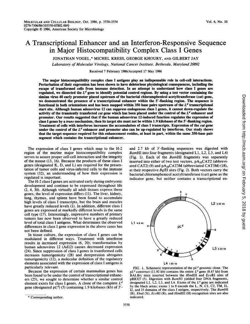
and 2.7 kb of 3'-flanking sequences was digested withBamHI into four fragments (designated Li, L2, L3, and L4)(Fig. 1). Each of the BamHI fragments was separatelyinserted into either of two test vectors, pA1OCAT2 (abbrevi-ated CAT2) (9) and pA1oCAT3M (abbreviated CAT3M) (18),at their respective BgII sites (Fig. 2). Both vectors carry thebacterial chloramphenicol acetyltransferase (cat) gene as theindicator gene, but neither contains a transcriptional en-
B
Li 4.8 kb
L2 3.05 kb
B
L3 2.22 kb
L4 1.85 kb
FIG. 1. Schematic representation of the pLd genomic clone. ThepLd construct (11.92 kb) contains the entire Ld gene (8.67 kb) fromBALB/c mice inserted between the HindIII and EcoRI sites ofpBR327 (5). Digestion with BamHI yielded four DNA fragments,designated Ll, L2, L3, and L4. Exons of the Ld gene are indicatedby the black areas; exons 1 to 8 encode the L, N, Cl, C2, TM, I1,12, and 13 domains of the class I antigen, respectively. The BamHI(B), XbaI (X), EcoRI (R), and HindlIl (H) recognition sites are alsoindicated.
3550
on February 5, 2018 by guest
http://mcb.asm
.org/D
ownloaded from

VOL. 6, 1986
\<B
\%_ deleted I
pCAT2-L1 (+)dl
pCAT3M-L1 (+)dI
FIG. 2. Construction and schematic representation of the various recombinant plasmids. The pCAT2-Ll(+) construct (10.5 kb) wasderived by the insertion of the Li fragment from pLd, in the correct orientation, at the BgIII site of CAT2 (9). The pCAT2-L1(-) construct(10.5 kb) contains the Li fragment in the opposite orientation. Conversion of the SalI site of pCAT2-Ll(+) to an XbaI site, followed bydigestion with XbaI and religation, gave rise to pCAT2-Ll(+)dl (6.0 kb). The pCAT3M-Ll(+) construct (9.2 kb) contains the Li fragment inthe correct orientation at the BgIII site of CAT3M (18). The pCAT3M-Ll(-) (9.2 kb) has the Li fragment in the opposite orientation.Digestion of pCAT3M-Ll(+) with XbaI and religation yielded pCAT3M-Ll(+)dl (4.7 kb). The BamHI (B), XbaI (X), BgIII (G), and SalI (S)recognition sites are indicated.
hancer. Although CAT2 contains the simian virus 40 (SV40)early promoter 5' to the CAT sequence (9), CAT3M does not(18). Transcriptional termination in each case is assured bythe inclusion of the SV40 poly(A) site located downstream ofthe indicator gene.
Individual constructs carrying different regions of the Ldgene were separately transfected into mouse L cells by thecalcium phosphate technique (10). At 48 h after transfection,the cells were harvested and the cell extracts were tested forCAT activity by their ability to convert [14C]chloramphen-icol into mono- and diacetylated forms as determined bythin-layer chromatography (9). In this assay, the controlCAT2 vector, which contains a transcriptional promoter butno enhancer, showed only a very low level of enzymeactivity (Fig. 3A). The construct carrying the Li fragment[designated CAT2-L1(+)] in an orientation relative to the catgene that is identical to the orientation in the Ld gene gave amarked enhancement in enzyme activity (Fig. 3A). Insertionof the L2, L3, and L4 BamHI fragments, in either orienta-tion, at the BglII site of CAT2, however, gave no detectableCAT activity (data not shown). These results demonstrated
the presence of a transcriptional control element locatedspecifically on the 5' side of the Ld gene which can activatea heterologous test gene containing the SV40 promoter.Consistent with the role of this control element as a tran-scriptional enhancer rather than a strong promoter is thefinding that the construct carrying the same Li fragmentinserted in the opposite orientation [designated CAT2-Ll(-)] also showed an elevation in CAT activity over thecontrol CAT2 vector (Fig. 3A). The fourfold difference inCAT activity observed between CAT2-Ll(+) and CAT2-Li(-) may be explained by the fact that the Li fragment is4.8 kb long, and as a consequence the enhancer sequence, inthe opposite orientation, could be much farther from theSV40 promoter than if it were in the correct orientation. Theefficiency of enhancer elements is often affected by theirdistance from the promoter.Although the promoter of the Ld gene has not been
functionally mapped, it is thought to be in the Li fragmentwhich ends on the 3' side approximately 14 base pairs (bp)downstream from the transcriptional start site (5, 15). To testthis possibility, constructs containing the Li fragment in-
NOTES 3551
on February 5, 2018 by guest
http://mcb.asm
.org/D
ownloaded from

MOL. CELL. BIOL.
serted in the CAT3M vector in either orientation weretransfected into L cells and tested for CAT activity (Fig. 3B).Since CAT3M has neither an enhancer nor a promoter, it hasno background CAT activity. The CAT3M-L1(+) constructshowed a high level of CAT activity, thus confirming thepresence of the Ld promoter within the Li fragment. Whenthis fragment was placed in the opposing orientation[CAT3M-L1(-)], no CAT activity was detected despite thepresence of an enhancer element.
Since the L gene is only one member of a family of class Igenes (26, 31), it was of interest to determine whether asecond class I gene also has a similar regulatory region in its5'-flanking region. A 2.8-kb fragment was obtained from theKb gene (21) by combined digestion with BamHI and NruI.After the conversion of the NruI blunt end to a BgIII stickyend, this fragment was inserted at the BglII site of CAT3M.This fragment was derived from a region of the Kb gene thatis analogous to the Li fragment of the Ld gene, and thefragment ends on the 3' side approximately 50 bp down-stream from the RNA start site. Upon transfection into Lcells, the construct containing the Kb fragment in the correctorientation [CAT3M-K(+)] expressed a high level of CATactivity, but the one with the insert in the opposing orienta-tion [CAT3M-K(-)] did not (Fig. 3C). The results areanalogous and comparable to those obtained with the Lifragment, suggesting the presence of a similar regulatorysequence in two distinct class I genes.To further localize the control region within the Li frag-
ment, a deletion was made in the CAT3M-Ll(+) constructby digestion with XbaI. Upon religation, the resulting con-struct [CAT3M-Ll(+)dl] was shortened by 4.5 kb and con-tained only 350 bp of 5'-flanking sequences from the Ld gene.When equivalent amounts of DNA were transfected into Lcells, CAT3M-Ll(+)dl was at least as efficient as CAT3M-Ll(+) in inducing CAT activity (Fig. 4). The higher level ofactivity observed with CAT3M-Ll(+)dl, when comparedwith CAT3M-Ll(+), was accounted for by the fact that the
C4 Cs
4U U
!
L) U u uL u u
A B C
FIG. 3. Identification of a transcriptional enhancer in the 5'-flanking region of both the Ld and Kb genes. Equivalent amounts (10,ug) of plasmid DNA were introduced into mouse L cells by thecalcium phosphate technique (10). Approximately 48 h posttransfec-tion, cell extracts were prepared and CAT activity was determined(9). All CAT assays were obtained with a cell extract concentrationwhich yields enzyme activity in the linear range. Panels: A, activa-tion of the cat gene by the insertion of the Li fragment from the Ldgene, in both orientations, in the CAT2 test vector which containsthe SV40 early promoter; B, activation of the cat gene by theinsertion of the Li fragment, in both orientations, in the CAT3M testvector which lacks a promoter; C, activation of the cat gene by theinsertion of a 2.8-kb 5'-flanking fragment from the Kb gene, in bothorientations [designated K(+) and K(-)], in the CAT3M test vector.
CAT3M CAT3M Lli CA,T3M - L 1 - di
5 1 5 m 1 R i9
A B C
FIG. 4. Localization of the transcriptional enhancer to a 326-bpsegment immediately 5' to the LI gene. Comparison of CAT activityinduced in L cells transfected with CAT3M-L1(+) containing 1.9 kbof 5'-flanking sequence from the Ld gene (B) and CAT3M-L1(+)dlcontaining only 350 bp of 5'-flanking sequence (C). Both of theseconstructs were derived from the same CAT3M parental vector (A).The amount (in micrograms) of plasmid DNA transfected is indi-cated below each lane.
deleted construct was only about half the size of the parentalconstruct; therefore, on a molar basis, twice as many deletedmolecules were transfected into L cells. The results obtainedwere consistent with an enhancer element in the 350-bpsegment located immediately upstream of the Ld gene;sequences as much as 1.6 kb further upstream most likelyhave neither stimulatory nor inhibitory effects. WhenCAT3M-Ll(+)dl was compared directly with SV2CAT,which contains the cat gene under the control of the SV40regulatory elements, the Ld enhancer-promoter was quitecomparable to the SV40 enhancer (data not shown).Because the expression of class I genes is suppressed in
cells transformed by human Adl2 (24, 28), it is conceivablethat this activity may be mediated in trans through the sameregulatory region. To test this possibility, we transfectedSV2CAT and CAT3M-Ll(+) in parallel into C3AT1 cells(12, 28), an established Adl2-transformed cell line whichexpresses class I transcripts at less than 5% of the level ofcontrol cells (Fig. 5). Although not as efficient as SV2CAT,the CAT3M-Ll(+) construct was definitely active in theAdl2-transformed cells. Similar results were also obtainedwith CAT3M-Ll(+)dl (data not shown). This suggests that if
SV2CAT CAT3M LI:
5 25 5 25
A BFIG. 5. Comparison of the transcriptional regulatory regions of
the SV40 early genes and the Ld gene in Adl2-transformed cells.Comparison of CAT activity induced in C3AT1 cells transfectedwith either SV2CAT containing the early SV40 enhancer andpromoter (A) or CAT3M-L1(+) containing the LI enhancer andpromoter (B). The amount (in micrograms) of plasmid DNAtransfected is indicated below each lane.
3552 NOTES
on February 5, 2018 by guest
http://mcb.asm
.org/D
ownloaded from

SV2CAT CAT3M-L1flW L-L1(+)2 L-L1f+)dII transfected with either CAT3M-L1(+) or CAT3M-Ll(+)dl.In both types of cells, the steady-state levels of CAT activityalso increased by approximately fourfold upon treatmentwith interferon (Fig. 6C and D). The extent of inductionobserved with the transfected test gene carrying the Ldregulatory se uence was similar to that observed with the
;-> ; endogenous L gene as determined by Northern blot analysis* 0* w * * * * (12). Although this correlation may be coincidental, the
v 1 9s*** T T results do suggest that interferon treatment increases thetranscription of class I genes and that the target sequence forrecognition must lie, at least in part, within the immediate
A B C D 5'-flanking region of the gene. This study, however, does notexclude the possibility that mRNA stabilization also plays a
eG. 6. Effect ofinterferon(LeN) onthe'-flanking reionofthe role in the overall increase in the accumulation of class Igene. Mouse cx,,-interferon (Lee BioMolecular, San Diego, rncit pntetetwt nefrnif.), at a concentration of 2,000 U/ml, was added 24 h after transcripts upon treatment with interferon.isfection and incubated for an additional 24 h, after which time There is overwhelming evidence for the differential ex-s were harvested. The percent conversion of [14C]chloramphen- pression of the same class I gene in different tissues (3), theto acetylated forms was determined by liquid scintillation differential expression of different class I genes in the same
nting. Panels A and B: CAT assays assessing the effects of tissue (17), and the differential activation of class I genesrferon on C3AT1 cells transiently transfected with either during development (4). In addition, there is convincing2CAT (A) or CAT3M-L1(+) (B). Panels C and D: CAT assays evidence that the expression of class I genes can be modu-bssing the effects of interferon on L-cell lines L-L1(+)2 (C) and lated by exogenous factors like mitogens, immunomodula-1(+)dll (D), stably transfected with CAT3M-Ll(+) and tors, and infectious agents (19, 22). Such alterations in the173M-Ll(+)dl, respectively. level of class I antigens on the cell surface have been shown
to have significant physiological consequences which affect12 suppresses class I gene expression through a trans- the integrity of the animal (14, 28, 29). All these findingsing factor, the target sequence for recognition cannot point to the importance of defining regulatory regions thatide in full within 1.9 kb of the 5' region of the Ld gene. act as targets for the modulation of class I gene expression.-se results are in agreement with our previous finding that In the present study, we defined a transcriptional controlauthentic Ld gene with ample flanking sequences is also region within a 350-bp segment located immediately up-)ressed when stably transfected into Adl2-transformed stream of the Ld gene. This region contains a transcriptionalIs (28). enhancer, in addition to a promoter; it can augment the'he expression of class I antigens is superinduced by expression of a test gene when placed upstream, in eitherrferon (20). Although the accumulation of class I tran- orientation, relative to the heterologous SV40 early pro-ipts increases upon treatment with interferon (6, 12), it is moter. A comparable region has also been found in the Kbyet clear whether this enhancement is entirely the result gene, suggesting that at least two H-2 class-I genes share thein increased rate of transcription or of a combination of location of an essential regulatory element. We also demon-reased transcription and stabilization of mRNA (7, 8). strated that the combination of class I enhancer and pro-rn though interferon is species specific, human class I moter is as efficient as the combination of SV40 enhancertes, when stably transfected into murine cells, can be and promoter in driving the expression of the cat gene in)erinduced by treatment with mouse interferon (23). This mouse L cells.gests that interferon acts by inducing a trans-acting factor Although the early 1A gene of human Adl2 can act,ich does not discriminate between human and mouse directly or indirectly, to suppress class I gene expressionss I genes. (24), our results were consistent with the suggestion that theCo identify the target sequence within a class I gene early lA-induced function cannot act in trans to down-ognized by this hypothetical trans-acting factor(s), we regulate class I genes within a 1.9-kb segment on the 5' sideisfected CAT3M-L1(+) in parallel with control SV2CAT of the Ld gene. In contrast, the observed ability of interferon) C3AT1 cells. The CAT RNAs from these two DNA to increase class I gene expression was due, at least in part,structs were expected to be identical except for their to the presence of a target sequence within a 350-bp segmentreme 5' nontranslated regions; the CAT3M-L1(+) tran- in the 5'-flanking region of the Ld gene. Interestingly, aipts contained 7 nucleotides derived from the Ld gene relatively conserved sequence observed among several in-ich were not present in the SV2CAT transcript. At 24 h terferon-inducible genes (class I, class II, and metallo--r transfection, half of the cultures were treated with thionein II) (8) was present within this 350-bp segment that00 U of murine a,3-interferon per ml (12). The cells were we identified.vested at 48 h posttransfection and tested for the pres- The present study is an attempt to define the structural-e of CAT activity. Although treatment with interferon did basis for the regulation of the expression of class I genes. Wealter the level of CAT activity in cells transfected with previously have suggested that the expression of H-2 class I
itrol SV2CAT (Fig. 6A), there was a fourfold increase in genes may be regulated by DNA methylation (27) and that aivity when CAT3M-L1(+) was used (Fig. 6B). Similar majority of the potential methylation sites are located withinults were obtained with CAT3M-Ll(+)dl, which contains the 5'-flanking regulatory region we identified. These studiesy 350 bp of 5'-flanking sequences. Comparable results may lead to an understanding of how the expression of thesere observed when L cells were used as recipients (data genes can be modulated in different tissues, during develop-shown). ment, and in a variety of disease states.Co avoid the argument that transient transfection assays In a recent study, Kimura et al. analyzed in detail thenot reflect steady-state events in the cell, we repeated the 5'-flanking region of the Kb gene (15). The results from thatrferon treatment on L cells which had been stably study are in general agreement with our findings with the Ld
IFN:
F:Ld ICalitrancell,icolcoulinte:SV2asseL-LCAI
Ad]actiresiThetheexpcellT
intescri
notof a
inciEvegensupsugwhi
clasI
rectrarintcconext]scriwhi
afte2,04harencnotconactiresionl'weI
notTI
do i
inte
NOTES 3553VOL. 6, 1986
on February 5, 2018 by guest
http://mcb.asm
.org/D
ownloaded from

MOL. CELL. BIOL.
gene. As with the Ld gene, the Kb enhancer is also located ina comparable region on the 5' side of the gene. In contrast toour findings, however, Kimura et al. suggested that the Kbpromoter, but not the enhancer, is down-regulated in Adl2-transformed cells (15). Our data do not suggest the presenceof a cis element within a 1.9-kb segment on the 5' side of theLd gene that is regulated by the Adl2 function in trans. Ourfindings, but not those of Kimura et al. (15), are in agreementwith the studies in which the entire class I gene containingample flanking sequences was stably transfected and ex-pressed in Adl2-transformed cells (28). Our results alsoshow the presence of an interferon-responsive site at the 5'side of the Ld gene.
We thank R. A. Flavell and J. G. Seidman for kindly providingthe Kb and Ld genes, respectively. We also thank Yves Barra,Hiroaki Hayashi, Steve Hinrichs, Kay Reynolds, Laurence Rubin,Kenichi Tanaka, and Masato Tanaka for helpful suggestions.
This work was supported in part by a cooperative grant (INT-NC-8404) from the National Science Foundation and the Centre Na-tional de la Recherche Scientifique.
LITERATURE CITED1. Bieberich, C., G. Scangos, K. Tanaka, and G. Jay. 1986.
Regulated expression of a murine class I gene in transgenicmice. Mol. Cell. Biol. 6:1339-1342.
2. Buc-Caron, M. H., H. Condamine, and F. Jacob. 1978. Thepresence of F9 antigen on the surface of mouse embryonic cellsuntil day 8 of embryogenesis. J. Embryol. Exp. Morphol.47:149-169.
3. Cosman, D., M. Kress, G. Khoury, and G. Jay. 1982. Tissue-specific expression of an unusual H-2 (class I)-related gene.Proc. Natl. Acad. Sci. USA 79:4947-4951.
4. Edidin, M. 1964. Transplantation antigen levels in the earlymouse embryos. Transplantation 2:627-637.
5. Evans, G. A., D. H. Margulies, R. E. Camerini-Otero, K. Ozato,and J. G. Seidman. 1982. Structure and expression of a mousemajor histocompatibility antigen gene, H-2Ld. Proc. Natl. Acad.Sci. USA 79:1994-1998.
6. Fellous, M., U. Nir, D. Wallach, G. Merlin, M. Rubinstein, andM. Revel. 1982. Interferon-dependent induction of mRNA forthe major histocompatibility antigens in human fibroblasts andlymphoid cells. Proc. Natl. Acad. Sci. USA 79:3082-3086.
7. Friedman, R. L., S. P. Manly, M. McMahon, I. M. Kerr, andG. R. Stark. 1984. Transcriptional and posttranscriptional reg-ulation of interferon-induced gene expression in human cells.Cell 38:745-755.
8. Friedman, R. L., and G. R. Stark. 1985. a-Interferon-inducedtranscription of HLA and metallothionein genes containinghomologous upstream sequences. Nature (London) 314:637-639.
9. Gorman, C. M., L. F. Moffat, and B. H. Howard. 1982. Recom-binant genomes which express chloramphenicol acetyltrans-ferase in mammalian cells. Mol. Cell. Biol. 2:1044-1051.
10. Graham, F. L., and A. J. van der Eb. 1973. A new technique forthe assay of infectivity of human adenovirus 5 DNA. Virology52:456-467.
11. Graziano, K. D., and M. Edidin. 1971. Serological quantitationof histocompatibility-2 antigens and the determination of H-2 inadult and fetal organs, p. 251-256. In A. Lengerova and M.Vojtiskova (ed.), Proceedings of the symposium on im-munogenetics of the H-2 system. S. Karger, Basel.
12. Hayashi, H., K. Tanaka, F. Jay, G. Khoury, and G. Jay. 1985.Modulation of the tumorigenicity of human adenovirus 12-transformed cells by interferon. Cell 43:263-267.
13. Hood, L., M. Steinmetz, and B. Malissen. 1983. Genes of themajor histocompatibility complex of the mouse. Annu. Rev.Immunol. 1:529-568.
14. Hui, K., F. Grosveld, and H. Festenstein. 1984. Rejection of
transplantable AKR leukaemia cells following MHC DNA-mediated cell transformation. Nature (London) 311:750-752.
15. Kimura, A., A. Israel, 0. LeBail, and P. Kourilsky. 1986.Detailed analysis of the mouse H-2Kb promoter: enhancer-likesequences and their role in the regulation of class I geneexpression. Cell 44:261-272.
16. Klein, J., F. Figueroa, and Z. A. Nagy. 1983. Genetics of themajor histocompatibility complex: the final act. Annu. Rev.Immunol. 1:119-142.
17. Kress, M., W.-Y. Liu, E. Jay, G. Khoury, and G. Jay. 1983.Comparison of class I (H-2) gene sequences: derivation ofunique probes for members of this multigenic family. J. Biol.Chem. 258:13929-13936.
18. Laimins, L. A., P. Gruss, R. Pozzatti, and G. Khoury. 1984.Characterization of enhancer elements in the long terminalrepeat of Moloney murine sarcoma virus. J. Virol. 49:183-189.
19. Lindahl, P., I. Gresser, P. Leary, and M. Lovey. 1976. Interferontreatment of mice: enhanced expression of histocompatibilityantigens on lymphoid cells. Proc. Natl. Acad. Sci. USA73:1284-1287.
20. Lindahl, P., P. Leary, and I. Gresser. 1974. Enhancement of theexpression of histocompatibility antigens of mouse lymphoidcells by interferon in vitro. Eur. J. Immunol. 4:779-784.
21. Mellor, A. L., L. Golden, E. Weiss, H. Bullman, J. Hurst, E.Simpson, R. F. L. James, A. R. M. Townsend, P. M. Taylor, W.Schmidt, J. Ferluga, L. Leben, M. Santamaria, G. Atfield, H.Festenstein, and R. A. Flavell. 1982. Expression of murineH-2Kb histocompatibility antigen in cells transformed withcloned H-2 genes. Nature (London) 298:529-534.
22. Meruelo, D., S. H. Numelstein, P. P. Jones, M. Lieberman, andH. 0. McDevitt. 1978. Increased synthesis and expression of H2antigens on thymocytes as a result of radiation leukemia virusinfection: a possible mechanism for H-2 linked control ofvirus-induced neoplasia. J. Exp. Med. 147:470487.
23. Rosa, F., P. P. LeBouteiller, A. Abadie, Z. Mishall, F. A.Lemonnier, D. Bourrel, M. Damotte, J. Kalil, B. Jordan, and M.Fellous. 1983. HLA class I gene integrated into murine cells areinducible by interferon. Eur. J. Immunol. 13:495499.
24. Schrier, P. I., R. Bernards, R. T. M. J. Vaessen, A. Houweling,and A. J. van der Eb. 1983. Expression of class I majorhistocompatibility antigens switched off by highly oncogenicadenovirus 12 in transformed rat cells. Nature (London)305:771-775.
25. Serfling, E., M. Jasin, and W. Schaffner. 1985. Enhancers andeukaryotic gene transcription. Trends Genet. 1:224-230.
26. Steinmetz, M., A. Winoto, K. Minard, and L. Hood. 1982.Clusters of genes encoding mouse transplantation antigens. Cell28:489-498.
27. Tanaka, K., E. Appella, and G. Jay. 1983. Developmentalactivation of the H-2K gene is correlated with an increase inDNA methylation. Cell 35:457465.
28. Tanaka, K., K. J. Isselbacher, G. Khoury, and G. Jay. 1985.Reversal of oncogenesis by the expression of a major histocom-patibility complex class I gene. Science 228:26-30.
29. Wallich, 1R., N. Bulbuc, G. J. Hammerling, S. Katzav, S. Segal,and M. Feldman. 1985. Abrogation of metastatic properties oftumor cells by de novo expression of H-2K antigens followinggene transfection. Nature 315:301-305.
30. Webb, C. G., W. E. Gall, and G. M. Edelnan. 1977. Synthesisand distribution of H-2 antigens in preimplantation of mouseembryos. J. Exp. Med. 146:923-932.
31. Weiss, E. H., L. Golden, K. Fahrner, A. L. Mellor, J. J. Devlin,H. Bullman, H. Tiddens, H. Bud, and R. A. Flavell. 1984.Organization and evolution of the class I gene family in themajor histocompatibility complex of the C57BL/10 mouse.Nature (London) 310:650-655.
32. Zinkernagel, R. M., and P. C. Doherty. 1979. MHC-restrictedcytotoxic T cells: studies on the biological role of polymorphicmajor transplantation antigens determining T-cell restriction-specificity, function, and responsiveness. Adv. Immunol. 27:51-177.
3554 NOTES
on February 5, 2018 by guest
http://mcb.asm
.org/D
ownloaded from