wind1 promotes shoot regeneration through transcriptional ... · wind1 promotes shoot regeneration...
TRANSCRIPT

RESEARCH ARTICLE
WIND1 Promotes Shoot Regeneration through Transcriptional Activation of ENHANCER OF SHOOT REGENERATION1 in Arabidopsis Akira Iwasea, Hirofumi Harashimaa, Momoko Ikeuchia, Bart Rymena, Mariko Ohnumaa, Shinichiro Komakia,1, Kengo Morohashib,2, Tetsuya Kuratac,3, Masaru Nakatad,4, Masaru Ohme-Takagid,e, Erich Grotewoldb, and Keiko Sugimotoa,5
aRIKEN Center for Sustainable Resource Science, Yokohama 230-0045, Japan. bCenter for Applied Plant Sciences and Department of Molecular Genetics, The Ohio State University, Columbus, Ohio, USA. cGraduate School of Biological Sciences, Nara Institute of Science and Technology, Ikoma 630-0192, Japan. dNational Institute of Advanced Industrial Science and Technology, Tsukuba 305-8562, Japan. eGraduate School of Science and Engineering, Saitama University, Saitama 338-8570, Japan. 1Current address: Department of Developmental Biology, University of Hamburg, Ohnhorststr. 18 - 22609 Hamburg, Germany. 2Current address: Department of Applied Biological Science, Faculty of Science and Technology, Tokyo University of Science, Noda 278-8510, Japan. 3Current address: Graduate School of Life Sciences, Tohoku University, Sendai 980-8577, Japan. 4Current address: Division of Crop Development, Central Region Agricultural Research Center, NARO; Joetsu 943-0193, Japan. 5Address correspondence to [email protected]. Short title: WIND1 activates ESR1 expression One-sentence summary: WIND1 directly activates the expression of another AP2/ERF transcription factor, ENHANCER OF SHOOT REGENERATION1 (ESR1), to promote callus formation and subsequent shoot regeneration. The author responsible for distribution of materials integral to the findings presented in this article in accordance with the policy described in the Instructions for Authors (www.plantcell.org) is: Keiko Sugimoto ([email protected]). ABSTRACT Many plant species display remarkable developmental plasticity and regenerate new organs after injury. Local signals produced by wounding are thought to trigger organ regeneration but molecular mechanisms underlying this control remain largely unknown. We previously identified an AP2/ERF transcription factor WOUND INDUCED DEDIFFERENTIATION 1 (WIND1) as a central regulator of wound-induced cellular reprogramming in plants. In this study, we demonstrate that WIND1 promotes callus formation and shoot regeneration by up-regulating the expression of the ENHANCER OF SHOOT REGENERATION1 (ESR1) gene, which encodes another AP2/ERF transcription factor in Arabidopsis. The esr1 mutants are defective in callus formation and shoot regeneration and conversely, its overexpression promotes both of these processes, indicating that ESR1 functions as a critical driver of cellular reprogramming. Our data show that WIND1 directly binds the Vascular system-specific and Wound-REsponsive cis-element (VWRE)-like motifs within the ESR1 promoter and activates its expression. The expression of ESR1 is strongly reduced in WIND1-SRDX dominant repressors and ectopic overexpression of ESR1 bypasses defects in callus formation and shoot regeneration in WIND1-SRDX plants, supporting the notion that ESR1 acts downstream of WIND1. Together, our findings uncover a key molecular pathway that links wound signaling to shoot regeneration in plants.
Plant Cell Advance Publication. Published on December 23, 2016, doi:10.1105/tpc.16.00623
©2016 American Society of Plant Biologists. All Rights Reserved

2
INTRODUCTION
Many multicellular organisms regenerate their bodies after injury, and this regenerative capacity is
vital for their survival after partial loss of their bodies. Plants, in particular, maintain high
developmental plasticity during post-embryonic development and display diverse forms of
regeneration (Ikeuchi et al., 2016). One common example of plant regeneration is de novo
organogenesis, i.e., the formation of new organs such as shoots and roots, from cut sites. This mode
of regeneration has been widely used in agriculture as a tool, for instance, for propagation of elite
cultivars and genetic engineering (Thorpe, 2007). As in animals, plant regeneration is initiated by at
least two cellular mechanisms. One is by the reactivation of relatively undifferentiated cells existing
in the somatic tissue and the other is by the reprogramming of mature somatic cells (Birnbaum and
Sánchez Alvarado, 2008; Tanaka and Reddien, 2011; Ikeuchi et al., 2016). In some cases, these
initiating cells directly regenerate new organs, but in other cases they first develop callus, a mass of
dividing cells, from which new organs form (Hicks, 1994).
Molecular mechanisms underlying plant organ regeneration have been studied mostly in
vitro where the balance between two plant hormones, auxin and cytokinin, determines the
developmental fate of regenerating organs. Generally, a high ratio of auxin to cytokinin favors root
regeneration, while a low ratio of auxin to cytokinin stimulates shoot regeneration (Skoog and
Miller, 1957). Intermediate levels of auxin and cytokinin promote callus formation (Skoog and
Miller, 1957). A protocol routinely used for Arabidopsis explants involves first incubation of a
tissue fragment on auxin- and cytokinin-containing callus inducing medium (CIM) to produce
callus, and subsequent transfer to cytokinin-rich shoot inducing medium (SIM) and auxin-rich root
inducing medium (RIM) to promote shoot and root regeneration, respectively (Valvekens et al.,
1988). Accumulating evidence suggests that callus on CIM primarily derives from relatively
undifferentiated pericycle cells through a genetic programme underlying auxin-induced lateral root
development (Che et al., 2007; Atta et al., 2009; Sugimoto et al., 2010). Accordingly, many
regulators of lateral root development, including ABERRANT LATERAL ROOT4 (ALF4),
AUXIN RESPONSE FACTOR7 (ARF7), ARF19, LATERAL ORGAN BOUNDARIES
DOMAIN16 (LBD16), LBD17, LBD18 and LBD29, are required for callus formation on CIM
(Sugimoto et al., 2010, Fan et al., 2012, Ikeuchi et al., 2013). A recent study has demonstrated that
additional regulators, PLETHORA3 (PLT3), PLT5 and PLT7, are also needed to make
CIM-induced callus pluripotent (Kareem et al, 2015). Key players acting downstream of PLT3,
PLT5 and PLT7 to confer pluripotency are PLT1 and PLT2, which are also known for their role in
root meristem development (Aida et al., 2004; Galinha et al., 2007). PLT3, PLT5 and PLT7, in
addition, induce CUP SHAPED COTYLEDON1 (CUC1) and CUC2, important regulators of shoot
meristem development during embryogenesis (Aida et al., 1997; Aida et al., 1999), presumably to
3
introduce the potential to form shoots in the callus (Kareem et al, 2015). While CUC1 and CUC2 do
not show an organised pattern of expression in CIM-induced callus, some root meristem regulators,
such as WUSCHEL-related homeobox5 (WOX5) and SCARECROW (SCR), display expression
patterns similar to those observed in the root meristem (Gordon et al., 2007; Atta et al., 2009;
Sugimoto et al, 2010). Thus, CIM-induced callus appears to represent a pluripotent cell mass that
has characteristics more similar to root meristems (Ikeuchi et al., 2013).
Given that CIM-induced callus possesses root meristem-like properties, regenerating
roots after transfer to RIM might be relatively straightforward, requiring further establishment of
root meristem identity and execution of root developmental programme by an auxin-induced
transcriptional cascade (Ozawa et al., 1998; Che et al., 2002; Ikeuchi et al., 2016). By contrast,
shoot regeneration on SIM ought to be more complex as it requires the conversion of root meristem
fate into shoot meristem fate. What is central for the shoot meristem initiation is the activation of
the key shoot stem cell regulator WUSCHEL (WUS) by cytokinin, which facilitates the partitioning
of CIM-induced callus into WUS-expressing domains and CUC2-expressing domains (Che et al.,
2006; Gordon et al., 2007; Chatfield et al., 2013). A cluster of CUC2-expressing cells continues to
proliferate to form promeristems, in which polarized expression of the auxin transporter
PIN-FORMED1 (PIN1) and another meristem regulator SHOOT MERISTEMLESS (STM) further
organises the formation of functional shoot meristem. Previous studies have also identified several
other regulators, such as ENHANCER OF SHOOT REGENERATION1/DORNRÖSCHEN
(ESR1/DRN), ESR2/DRN-LIKE (DRNL) and RAP2.6L, that contribute to shoot regeneration in
vitro (Banno et al., 2001; Kirch et al., 2003; Che et al, 2006). An early study showed that
overexpression of ESR1 promotes shoot regeneration without or at low doses of exogenous
cytokinin (Banno et al., 2001). The esr1-1/drn-2 loss-of-function mutant, referred to as esr1-1
hereafter, however, does not display strong defects in shoot regeneration when cultured on CIM and
SIM (Matsuo et al., 2011). By contrast, loss-of-function mutations in ESR2 and RAP2.6L cause
clear defects in in vitro shoot regeneration (Matsuo et al., 2011; Che et al., 2006), suggesting that
they play more profound roles.
We recently showed that wound stress provides another important cue for shoot
regeneration, since intact plants cultured on CIM and SIM hardly regenerate shoots without
wounding (Iwase et al. 2015). Wounding provokes various physiological responses, including rapid
induction of reactive oxygen species, Ca2+ waves, and the production of stress-responsive hormones
(Miller et al., 2009; Mousavi et al., 2013), but whether these early physiological responses direct
cells for reprogramming is not established. A set of key regulators that are rapidly activated in
response to wounding and have pivotal roles in wound-induced callus formation are a subfamily of
AP2/ERF transcription factors, WOUND INDUCED DEDIFFERENTIATION1 (WIND1, a.k.a

4
RAP2.4), WIND2, WIND3, and WIND4 (Iwase et al., 2011a; Iwase et al., 2011b). All WIND genes
are induced by wounding and overexpression of each of them promotes callus formation (Iwase et
al., 2011a; Iwase et al., 2011b). Importantly, WIND1 substitutes the early wound response and
confers pluripotency, since plants overexpressing WIND1 regenerate shoots on SIM without wound
stress (Iwase et al., 2015). Conversely, dominant repression of WIND1 in WIND1-SRDX explants
strongly blocks shoot regeneration, suggesting that WIND proteins function as key regulators of
cellular reprogramming in response to wound stress (Iwase et al., 2015; Ikeuchi et al., 2016).
In this study, we set out to investigate how wound signaling links to regeneration at the
molecular level. WIND proteins are likely to play key roles in this regulation and identification of
genes directly targeted by WIND1 should help unveil how WIND1-mediated signaling controls the
transcription of key regulators in regeneration. We provide both in vivo and in vitro evidence that
WIND1 directly binds the ESR1 promoter and activates its expression. We also show that ESR1
functions downstream of WIND1 and facilitates both callus formation and shoot regeneration in
response to wound stress. Our results thus uncover a key transcriptional mechanism that directly
links the wound response to organ regeneration in plants.
RESULTS
Wound stress activates ESR1 expression in a WIND1-dependent manner
Our previous microarray data showed that ESR1 expression is strongly up-regulated in callus
overexpressing WIND1 under the control of the cauliflower mosaic virus 35S promoter (Iwase et al.,
2011a). This is interesting, since ESR1 expression is restricted to the shoot apical meristem and leaf
primordia during normal development (Kirch et al., 2003) and its overexpression confers increased
shoot regenerative capacity in tissue culture (Banno et al., 2001). To validate this observation, we
compared the level of ESR1 expression between 14-day-old wild-type (WT) seedlings and
35S:WIND1 callus. As shown in Supplemental Figure 1, our quantitative reverse transcription PCR
(RT-qPCR) analysis confirmed very low abundance of ESR1 transcripts in WT seedlings and a
strong increase in 35S:WIND1 callus.
Given that the expression of the WIND1 gene is strongly activated by wounding (Iwase et
al., 2011a), we then tested whether ESR1 is also up-regulated in response to wound stress. Our
time-course expression analysis using leaf explants dissected from 14-day-old WT seedlings
revealed that WIND1 mRNA levels start to increase within 30 min after wounding and peak at 1 h
(Figure 1A). Similarly, the level of ESR1 transcripts starts to increase within 30 min after wounding
and peaks by 3 h (Figure 1A). ESR1 expression is barely detectable in WT root and hypocotyl
explants but we also detected an increase in the ESR1 expression in these organs after wounding
(Supplemental Figures 2A and 2B). Importantly, wound-induced ESR1 activation is strongly

5
suppressed in WIND1-SRDX explants (Figure 1A), suggesting that WIND1 is involved in ESR1
activation.
We have previously reported that the WIND1 induction by wounding is localized to
wound sites (Iwase et al., 2011a). To explore the wound-induced expression of the ESR1 gene in
planta, we examined the pattern of its promoter activity using ProDRN/ESR1:GUS lines, referred to
hereafter as ProESR1:GUS, in which the expression of the b-glucuronidase (GUS) gene is driven by
the promoter of ESR1 (Kirch et al., 2003). As expected, the promoter activity of ESR1 is not
detected in intact leaf explants, but its activity is induced locally at wound sites (Figure 1B). We
detected similar patterns of ESR1 promoter activity in wounded roots and hypocotyls (Supplemental
Figures 2C and 2D). We also introduced the ProESR1:GUS construct into the WIND1-SRDX plants
and found that WIND1 is required for the activation of the ESR1 promoter at wound sites (Figure
1B). Transverse sections of petioles close to wound sites showed that ESR1 promoter activity is
often detected within the vasculature, i.e., in xylem parenchyma and procambium cells, but is also
found in non-vascular cells, such as mesophyll, that have started to undergo cell division
presumably to develop callus (Figure 1C). Liu et al. (2014) recently showed that wounding induces
auxin accumulation at wound sites of Arabidopsis leaves. To uncouple the effect of wounding from
auxin accumulation, we tested whether inhibition of auxin transport by N-1-naphthylphthalamic
acid (NPA) interferes with ESR1 activation at wound sites. As shown in Supplemental Figure 2E, 1
µM NPA does not block ESR1 expression at wound sites, suggesting that local auxin transport does
not contribute to ESR1 activation at wound sites.
To further corroborate these observations, we generated ProESR1:ESR1-GFP transgenic
plants (ESR1-GFP) in which we drove the expression of the ESR1-GFP fusion protein by the ESR1
promoter. We introduced this construct into the esr1-2/drn1 mutant, referred to hereafter as esr1-2
(Chandler et al., 2007; Matsuo et al., 2011), and confirmed that ESR1-GFP fusion proteins are
functional by the complementation of cotyledon phenotypes in esr1-2 (Supplemental Figure 3A).
As shown in Figure 1D, we detect clear accumulation of ESR1-GFP fusion proteins within the
nuclei of cells close to wound sites. These observations demonstrate that the expression of the ESR1
gene is induced locally at wound sites and WIND1 is required for its activation.
WIND1 directly binds the ESR1 promoter and activates its expression
Having established that WIND1 is required for the induction of the ESR1 gene, we subsequently
investigated whether WIND1 directly binds the ESR1 promoter in vivo. Using antibodies against
GFP proteins, we immunoprecipitated WIND1-GFP proteins from root explants of
ProWIND1:WIND1-GFP plants (Iwase et al., 2011a) and tested whether the chromatin of the ESR1
gene co-purified with WIND1-GFP. Since we detected strong WIND1 expression in wound-induced

6
callus (Iwase et al., 2011a), we used root explants cultured on Murashige-Skoog (MS) medium for
10 days, at which time they developed large callus at wound sites. As shown in Figure 2A, our
chromatin immunoprecipitation coupled by quantitative PCR (ChIP-qPCR) analysis detected a
strong enrichment of the ESR1 promoter sequence using a pair of primers designed around 500-bp
upstream of the translational start site. Using a particle bombardment-mediated transient expression
assay, we subsequently investigated whether WIND1 could activate the ESR1 promoter in
Arabidopsis MM2d culture cells. We fused a 1,000-bp ESR1 promoter to the luciferase reporter gene
and judged the promoter activation by the relative enzymatic activity of luciferase. As shown in
Supplemental Figure 4A, ectopically expressed WIND1 displays strong transactivation activity for
the 1,000-bp ESR1 promoter, demonstrating that WIND1 can activate ESR1 in vivo. We also
performed the transactivation assay using the ESR1 promoter truncated to 500, 250, 200, 150 and 100
bp from the translational start site and found that the 150-bp sequence upstream of the translational
start site is sufficient for the ESR1 induction by WIND1.
To further characterize WIND1’s binding to the ESR1 promoter, we tested their direct
interaction in vitro by an electrophoresis mobility shift assays (EMSA). We expressed the WIND1
protein fused with Maltose Binding Protein (MBP) and 6xHis-tag (His6) in E coli, and purified the
MBP-WIND1-His6 protein using the His-tag by affinity chromatography. We used the dehydration
responsive element (DRE, core sequence TACCGACAT) as a positive control, since WIND1 was
previously shown to bind the DRE sequence (Lin et al., 2008). Our EMSA indeed showed that the
MBP-WIND1-His6 protein strongly binds the DRE sequence in vitro, causing the DRE probe band
to shift in the gel (Supplemental Figure 4B). Using this experimental setup, we also found that the
MBP-WIND1-His6 protein, but not the MBP-GFP-His6 protein, binds the 513-bp sequence of the
ESR1 promoter (Supplemental Figure 4C). To further narrow down WIND1’s binding site within
the ESR1 promoter, we tested WIND1’s binding to eleven ~50-bp DNA probes, designated as R1 to
R11, that cover the -513 to -64 bp of the ESR1 promoter with a 10-bp overlap between each probe.
As shown in Figure 2B and Supplemental Figure 4D, we found reproducible binding of
MBP-WIND1-His6 protein specifically to the probe R10 that covers the -153- to -104-bp sequence
of the ESR1 promoter.
The results from our transactivation assay and EMSA consistently suggest that the 150-bp
ESR1 promoter is sufficient for WIND1’s binding and activation of the ESR1 gene. Interestingly, the
R10 sequence where WIND1 likely binds within the 150-bp ESR1 promoter contains two Vascular
system-specific and Wound-REsponsive cis-element (VWRE)-like motifs (core sequence
AAATTT) previously implicated in wound-induced activation of gene expression in tobacco
(Sasaki et al., 2002; Sasaki et al., 2006). We therefore asked whether these motifs are required for
the activation of the ESR1 promoter by mutating either one or both of the VWRE-like motifs. Our

7
transactivation activity assay revealed that the m1 and m4 mutations, abolishing both VWRE-like
motifs, strongly hinder ESR1 induction by WIND1 whereas the m2 or m3 mutation, abolishing only
the first or second VWRE-like motif, results in partial reduction in ESR1 activation (Figure 2C). By
contrast, the m5 mutation, introduced outside the two VWRE-like motifs, does not alter ESR1
activation by WIND1 (Figure 2C). These results strongly suggest that WIND1 directly binds the
two VWRE-like motifs within the ESR1 promoter to activate its expression.
ESR1 promotes callus formation at wound sites
Given that WIND1 is required for callus induction at wound sites (Iwase et al., 2011a), we asked
whether ESR1 also participates in wound-induced callus formation. WT leaf explants cultured on
MS medium develop a large mass of callus cells at wound sites (Figures 3A and 3B). By contrast,
callus formation is severely compromised in esr1-2 leaf explants (Figures 3A and 3B). To further
investigate the involvement of ESR1, we generated the ESR1-SRDX dominant repressor line in
which we drove the expression of ESR1-SRDX chimeric proteins under the ESR1 promoter. We
confirmed the cotyledon formation defects, previously described for esr1-2 mutants (Chandler et al.,
2007), in the ESR1-SRDX plants. As expected, we also found clear defects in callus formation from
leaf explants, demonstrating the requirement of ESR1 for wound-induced callus formation (Figures
3A and 3B). In addition, we found that ESR1-GFP plants develop larger callus at wound sites
(Figures 3A and 3B). Our RT-qPCR analysis showed that, compared to the WT, the level of ESR1
expression is at least 1.5-fold higher in ESR1-GFP plants after wounding (Supplemental Figure 3B),
suggesting that the increased level of ESR1 expression promotes callus formation.
Our results so far are consistent with the hypothesis that WIND1 activates ESR1
expression to promote callus formation at wound sites. To validate this further, we generated double
transgenic lines between WIND1-SRDX and esr1/drn-D gain-of-function mutants, referred to
hereafter as esr1-D, in which the expression of ESR1 is ectopically activated by the 35S promoter
(Kirch et al., 2003), and tested whether ectopic expression of ESR1 in WIND1-SRDX plants was
sufficient to rescue defects in wound-induced callus formation. As shown in Figures 4A and 4B,
WIND1-SRDX leaf explants, cultured on MS medium, display strong defects in callus formation,
producing only a very small mass of cells at wound sites. Introduction of the esr1-D
gain-of-function mutation into the WIND1-SRDX plants complements most of these callus
formation defects, since leaf explants from WIND1-SRDX esr1-D double transgenic plants develop
WT-like callus (Figures 4A 4B). In addition, we examined whether the esr1-2 mutation blocked
callus formation induced by WIND1 overexpression. As shown in Figure 4C, 35S:WIND1 T1 plants
display weak, intermediate, and strong callus formation, which we previously classified as type I,
type II, and type-III plants, respectively (Iwase et al., 2011a). As expected, WIND1 overexpression

8
in esr1-2 mutants causes milder callus phenotypes, producing ~10% of WT-like T1 plants without
any visible callus formation. These results therefore support that ESR1 functions downstream of
WIND1 and promotes callus formation at wound sites.
We previously reported that WIND1 promotes callus formation via the B-type
ARABIDOPSIS RESPONSE REGULATOR (ARR)-mediated cytokinin signalling pathway (Iwase
et al., 2011a). To test the functional relationship between ESR1 and cytokinin signalling, we
evaluated ESR1 expression in arr1 arr12 double mutants defective in B-type ARR signalling
(Mason et al., 2005). Interestingly, our RT-qPCR analysis revealed that wound-induced activation of
ESR1 is compromised in arr1 arr12 plants, while the esr1-2 mutation does not interfere with the
expression of a cytokinin-responsive ARR5 gene (Argyros et al., 2008) (Figures 4D and 4E).
Together, these results suggest that ESR1 functions downstream of B-type ARR-mediated cytokinin
signalling.
ESR1 promotes shoot regeneration at wound sites.
When Arabidopsis WT explants are cultured on MS medium without any exogenous plant
hormones, they develop callus and often roots, but they hardly regenerate shoots from wound sites
(Figure 5A). Unexpectedly, we noticed that leaf explants from ESR1-GFP plants regenerate shoots
at wound sites (Figure 5A). Further investigation of this phenotype revealed that only 1 out of 646
(0.2%) leaf explants from WT plants regenerate shoots, whereas 31 out of 170 (18.2%) leaf
explants from ESR1-GFP plants develop one or sometimes multiple shoots at wound sites (Figure
5B). Intriguingly, this phenotype is not limited to leaf explants and we also observed shoot
regeneration from various other organs such as cotyledons and inflorescence stems (Figure 5A).
Similarly, root explants from WT plants develop callus at wound sites but they never regenerate
shoots under our culture condition (Figures 5A and 5B). By contrast, 89 out of 634 (14.0%)
explants from ESR1-GFP plants develop shoots at wound sites (Figures 5A and 5B).
To further explore the causal relationship between ESR1 expression and shoot
regeneration, we generated LexA-VP16-estrogen (XVE)-ESR1 transgenic plants in which we
induced ESR1 expression by the application of 17b-estradiol (Figures 5C and 5D). Our RT-qPCR
analysis confirmed that 0.1 to 10 µM 17b-estradiol induces ESR1 expression in a dose-dependent
manner (Figure 5D). As expected, leaf explants from WT plants hardly develop shoots at wound
sites in the presence of 17b-estradiol. In sharp contrast, leaf explants from up to 35% of XVE-ESR1
plants reproducibly regenerate shoots from wound sites, when cultured in the presence of 0.1 to 10
µM 17b-estradiol (Figures 5C and 5D). Interestingly, XVE:ESR1 plants regenerate shoots only upon
wounding and when they are grown without wound stress, they develop callus (Figures 5C).

9
Nevertheless, these XVE:ESR1-derived calli are capable of regenerating shoots when transferred to
SIM, indicating that they retain the potential to develop shoots (Supplemental Figure 5A). Together,
these results demonstrate that the increased dosage of ESR1 strongly promotes shoot regeneration at
wound sites.
The WIND1-ESR1 pathway is required for shoot regeneration in vitro
Having uncovered a clear enhancement of wound-induced shoot regeneration by ESR1, we
reexamined the requirement of ESR1 for in vitro shoot regeneration. We incubated root explants of
WT, esr1-1, and esr1-2 seedlings for 4 days on CIM and then transferred them to SIM to induce
shoot regeneration. As shown in Figures 6A and 6B, both esr1-1 and esr1-2 display clear defects in
shoot regeneration, producing fewer shoots compared to WT explants. Chandler et al. (2007)
reported that the esr1-1 allele, carrying a dSpm transposon insertion immediately after the start
codon, shows weaker cotyledon phenotypes than the esr1-2 allele, carrying the dSpm insertion in
the central AP2 domain. We observed a similar trend for shoot regeneration phenotypes since
esr1-1, but not esr1-2, explants occasionally develop shoots. In addition, we detected severe defects
in shoot regeneration in ESR1-SRDX root explants (Figures 6A and 6B), confirming the
requirement of ESR1 in in vitro shoot regeneration. Interestingly, we noticed that shoot
regeneration is significantly enhanced in ESR1-GFP plants, forming nearly twice as many shoots
from each root explant compared to WT (Figures 6A and 6B). By contrast, esr1-2, ESR1-SRDX,
and ESR1-GFP root explants develop callus comparable to WT on CIM (Supplemental Figure 5B),
suggesting that ESR1 does not play major roles in hormone-induced callus formation in vitro.
To investigate how ESR1 promotes shoot regeneration in vitro, we first examined ESR1
expression in explants cultured on CIM and SIM. Interestingly, incubation of petiole or root
explants with kinetin and 2,4-D, cytokinin, and auxin in CIM, strongly stimulates ESR1 promoter
activity at wound sites, as shown by the enhanced GUS staining in ProESR1:GUS plants (Figure 7A).
Importantly, the application of either kinetin or 2,4-D alone does not activate the ESR1 promoter in
both petiole and root explants (Figure 7A), implying that cytokinin and auxin have synergistic
effects on ESR1 activation. Our RT-qPCR analyses also confirmed that ESR1 expression is
activated in root explants cultured for 4 days on CIM, and in addition showed that its expression is
enhanced, by more than 3-fold, after transfer to SIM (Figure 7B). Matsuo et al. (2011) previously
showed that ESR2 is not expressed in Arabidopsis explants incubated on CIM and its expression is
detected only after they start to form shoots on SIM. Our RT-qPCR analysis confirmed these results
and further showed that the late activation of ESR2 expression is dependent on ESR1 (Figure 7B).
Similarly, the expression of key shoot regulators such as CUC1, WUS, STM, and RAP2.6L, is also
increased after transfer to SIM (Gordon et al., 2007; Che et al., 2006; Chatfield et al., 2013) and

10
their expression requires ESR1 to different degrees (Figure 7B). We also confirmed the previously
reported up-regulation of PLT1, 2, 3, 5, 7 and CUC2 in explants cultured on CIM (Kareem et al.,
2015) but found that none of their activation requires functional ESR1 (Figure 7B).
We previously demonstrated that WIND1-SRDX explants cultured on CIM and SIM are
defective in shoot regeneration (Iwase et al., 2015, Figures 8A and 8B). Consistently, the WIND1
promoter is active in pericycle cells as well as callus cells derived from pericycle cells in root
explants cultured on CIM and SIM (Iwase et al., 2011a, Figure 8C). To test whether ESR1 also acts
downstream of WIND1 in this context, we examined the promoter activity of ESR1 in
WIND1-SRDX root explants. As reported previously (Matsuo et al., 2011), ESR1 promoter activity
is visible within callus of ProESR1:GUS root explants cultured on CIM and SIM (Figure 8C). By
contrast, the ESR1 promoter activity is strongly suppressed in ProESR1:GUS WIND1-SRDX plants
(Figure 8C), indicating the requirement of WIND1 for the activation of the ESR1 promoter. We also
introduced the esr1-D construct into WIND1-SRDX plants and examined whether ectopic activation
of ESR1 is sufficient to rescue the shoot regeneration phenotype in WIND1-SRDX explants. As
expected, plants expressing both WIND1-SRDX and esr1-D show similar or slightly higher levels of
shoot regeneration compared to the WT (Figures 8A and 8B). These results thus demonstrate that
ESR1 functions downstream of WIND1 and promotes shoot regeneration in vitro.
The WIND1-ESR1 pathway is not required for de novo root regeneration at wound sites.
Liu et al. (2014) recently showed that Arabidopsis leaf explants cultured on B5 medium are capable
of developing new roots from wound sites. We found that Arabidopsis leaf explants cultured on MS
medium also regenerate roots in the absence of a supply of exogenous phytohormones
(Supplemental Figure 6A). Using this system, we asked whether WIND1 and ESR1 are also
involved in root regeneration at wound sites. We typically observe >40% of WT leaf explants
regenerating roots from wound sites (Supplemental Figure 6B). Interestingly, both WIND1-SRDX
and ESR1-SRDX plants are capable of forming similar numbers of roots (Supplemental Figures 6A
and 6B), suggesting that the WIND1-ESR1 pathway is not required for de novo root regeneration at
wound sites.
DISCUSSION
In this study we demonstrate that a wound-induced reprogramming regulator WIND1 directly
activates ESR1 expression to promote callus formation and subsequent shoot regeneration at wound
sites. Our data show that the level of ESR1 is a key determinant of shoot fate, since mild
overexpression of ESR1 induces shoot formation at wound sites. WIND1 is also expressed in
phytohormone-induced callus cells in explants cultured on CIM and SIM, and it is required for

11
ESR1 activation and shoot regeneration in in vitro conditions. Our findings therefore uncovered an
important transcriptional cascade underlying shoot regeneration in plants.
WIND1 as a molecular link between wound stress and regeneration
We previously showed that WIND1 promotes the reacquisition of competency for shoot
regeneration (Iwase et al., 2015), but the precise molecular mechanisms underlying this control
were not known. In this study, we demonstrate that a central role of WIND1 is to activate the
expression of ESR1 to promote callus formation and shoot regeneration. Our data show that WIND1
first activates ESR1 expression, and culturing explants on auxin and cytokinin permits stronger
ESR1 expression (Figure 9). It is interesting to note that the overexpression of ThWIND1-L, a
WIND1 homolog from salt cress Thellungiella halophila, also induces ESR1 expression in
Arabidopsis (Zhou et al., 2012), implying that WIND1-mediated ESR1 activation might be
conserved at least among Brassicaceae plants.
Our in vivo and in vitro data show that WIND1 directly binds the promoter of ESR1 using
the two VWRE-like motifs (Figures 2B and 2C). The 14-bp VWRE motif (GAAAAGAAAATTTC)
was first identified within the promoter of a wound-inducible peroxidase gene, tpoxN1, in Nicotiana
tabacum (tobacco; Sasaki et al., 2006). A later study showed that two tobacco Wound-Responsive
AP2/ERF family transcription factors WRAF1 and WRAF2 bind the VWRE motif to induce the
tpoxN1 gene after wounding and the core sequence (AAATTT) within the VWRE motif is essential
for their binding (Sasaki et al., 2007). Interestingly, both WRAF1 and WRAF2 are induced within 30
min after wounding, preceding the accumulation of tpoxN1 transcripts after 1 h. We detect similar
transcriptional changes for WIND1, starting within 30 min after wounding, followed by the peak
accumulation of ESR1 transcripts after 1 h. The closest homolog of WRAF1 and WRAF2 in
Arabidopsis is RAP2.6 (At1g43160) and its expression is also induced very rapidly after wounding
(Kilian et al., 2007; Iwase et al., 2011a). It is thus plausible that one of the earliest wound-induced
transcriptional changes is mediated by a set of wound-inducible AP2/ERF transcription factors and
they activate target genes through binding the core VWRE motif within the target promoters. The
physiological roles of RAP2.6 in the wound response have not been investigated so far but it will be
interesting to test whether RAP2.6 can also activate ESR1 by using the VWRE-like motifs. In
addition to ESR1, we found 228 other genes in Arabidopsis that carry two closely located
VWRE-like motifs within the 1-kb promoter region, ~10% of which are induced more than 2-fold
by WIND1 overexpression (Iwase and Rymen, unpublished results). These genes are putative
targets of WIND1 and future studies should investigate their functional relationships to WIND1. We
should note that the core VWRE sequence does not resemble other GC-rich sequences such as DRE
(Yamaguchi-Shinozaki and Shinozaki, 1994) and the GCC box (core sequence TAAGAGCCGCC)

12
(Ohme-Takagi and Shinshi, 1995), recognized by other AP2/ERF transcription factors. While
WRAF1 and WRAF2 do not recognize these GC-rich motifs, WIND1 recognizes both DRE and the
GCC box at least in vitro (Lin et al., 2008 and this study). It will be important to examine whether
WIND1 binds both of these motifs in vivo and, if so, whether it activates different sets of genes, for
instance, in a context-dependent manner.
We provide genetic evidence that WIND1 and ESR1 are not required for root
regeneration from Arabidopsis leaf explants, at least under the condition used in this study. These
results are in agreement with the finding that this form of root regeneration is driven by auxin
accumulation at wound sites, which promotes the establishment of root cell fate through the
activation of WUSCHEL RELATED HOMEOBOX11 (WOX11) and WOX12 (Liu et al., 2014).
Intriguingly, Liu et al. (2014) showed that auxin-mediated callus formation (and subsequent root
regeneration) derives primarily from leaf procambium cells, while our study suggests that
wound-induced callus formation may originate from various cell types, including xylem
parenchyma and mesophyll cells (Figure 1C). How these callus cells from different cellular origins
contribute to shoot regeneration remains to be verified, but these observations together suggest that
regeneration of roots and shoots from Arabidopsis leaf explants is operated by at least two distinct
molecular mechanisms.
Activation of ESR1 expression by auxin and cytokinin
How exogenously supplied auxin enhances ESR1 expression is not currently clear. Previous studies
have shown that the auxin-inducible transcription factor AUXIN RESPONSE FACTOR
5/MONOPTEROS (ARF5/MP) binds the ESR1/DRN promoter and activates its expression during
embryonic development (Cole et al., 2009). The ESR1/DRN promoter possesses two canonical
Auxin responsive elements (AuxREs) where ARF5/MP binds in vivo, and mutations in these
sequences alter auxin-induced expression of ESR1/DRN in developing embryos (Cole et al., 2009).
Interestingly, the expression of ARF5/MP is strongly up-regulated in explants cultured on CIM and
SIM (Che et al., 2006), raising the possibility that ARF5/MP might participate in auxin-induced
ESR1 expression in cultured explants. Alternatively, a recent study by Fan et al. (2012) has shown
that ARF7 and ARF19 mediate auxin-induced callus formation by up-regulating the expression of
LBD16, LBD17, LBD18, and LBD29. It is thus possible that these ARFs bind the ESR1 promoter
and activate its expression in parallel. We also do not rule out the possibility that ESR1 acts
downstream of the ARF-LBD pathway and future studies should clarify how these auxin-responsive
transcriptional regulators directly or indirectly regulate ESR1 expression.
Our results show that cytokinin activates ESR1 expression at least partially through ARR1
and ARR12 (Figure 4D), demonstrating that ESR1 acts downstream of the B-type ARR-mediated

13
pathway. How wound stress activates the B-type ARR pathway in a WIND-dependent manner is not
currently known, but our data suggest that WIND1 may activate ESR1 both directly and indirectly
via enhancing cytokinin signalling. In addition, Kareem et al. (2015) found that PLT3, PLT5, and
PLT7 are the key regulators of in vitro shoot regeneration acting downstream of both auxin and
cytokinin signalling. We showed that the expression of these PLT genes is not altered in esr1-2
mutants (Figure 7B), suggesting that they do not act downstream of ESR1. Instead, PLTs may
function upstream of, or in parallel to, ESR1 and it will be interesting to investigate these functional
relationships in future studies.
Role of ESR1 in callus formation and shoot regeneration
Our data suggest that ESR1 is induced immediately after wounding (Figure 1) and promotes callus
formation at wound sites (Figure 3). We also show that the level of ESR1 expression is one key
limiting factor for shoot regeneration, since mild overexpression of ESR1 dramatically improves
shoot regeneration from wound sites (Figure 5). Intriguingly, XVE:ESR1 plants regenerate shoots
only upon wounding and they develop callus without wound stress (Figure 5C). Banno et al. (2001)
also reported callus formation in 35S:ESR1 plants and suggested that constitutive overexpression of
ESR1 interferes with differentiation into shoot cells. Based on our observations, we hypothesize that
ESR1 activation alone is not sufficient to fully establish the shoot fate and additional
wound-induced events, such as induction and/or accumulation of some other signals, are also
needed to confer shoot fate at wound sites.
Our data show that ESR1 is not essential for hormone-induced callus formation but that it
plays pivotal roles in shoot regeneration in vitro (Supplemental Figure 5B, Figure 6). The cause for
the apparent discrepancy between our results and a previous report (Matsuo et al., 2011) is not clear,
but one possibility is that we employ slightly different culture conditions, such as long-day light
condition as opposed to the continuous light conditions employed in Matsuo et al. (2011), and WT
root explants appear to produce more shoots in our conditions. Matsuo and Banno (2008) reported
that overexpression of ESR1-SRDX chimeric proteins blocks shoot regeneration, suggesting that
ESR1, potentially together with other redundant transcriptional regulators, promotes shoot
regeneration. Our observation further highlights the functional importance of ESR1, since loss of
ESR1 in esr1 mutants or ESR1-SRDX expression by its own promoter is sufficient to cause severe
regeneration defects (Figure 6). Since esr1 mutant calli turn green and develop some green foci
(Figure 6), they might be able to develop shoot promeristems and/or shoot primordia, although they
are severely impaired in shoot outgrowth. Shoot regeneration defects in esr1 mutants are
accompanied by strong inhibition of key shoot meristem regulators, such as WUS and STM (Figure
7), further substantiating that these mutants fail to complete shoot regeneration. Given that the

14
levels of PLT3, PTL5, and PLT7 expression are comparable between WT and esr1-2 mutants
(Figure 7), esr1-2 calli likely retain reasonable levels of pluripotency, but they cannot progress
through the shoot program without functional ESR1. A previous overexpression study suggested
that ESR1 directly activates CUC1 in in vitro shoot regeneration (Matsuo et al., 2009). Our data
show that ESR1 is required for CUC1 expression (Figure 7), supporting the notion that ESR1
functions upstream of CUC1. Kareem et al. (2015) showed that PLT3, PLT5, and PLT7 are required
for CUC1 expression in vitro, but plt3 plt5 plt7 mutants still have some residual CUC1 expression
on SIM. It is thus possible that these two pathways, governed by PLTs and ESR1, regulate CUC1
expression in parallel.
Our microarray data show that ESR2, a close homolog of Arabidopsis ESR1, is also
up-regulated in 35S:WIND1 callus (Iwase et al., 2011a), implying that WIND1 may also target
ESR2 to promote shoot regeneration. Intriguingly, however, we do not detect any significant
elevation of ESR2 expression after wound stress in any of the tissues examined, suggesting that
wounding (or WIND1) does not directly induce ESR2 expression. Our data show that ESR2
expression is strongly dependent on ESR1 in in vitro conditions (Figure 7). Thus, the high levels of
ESR1 in 35S:WIND1 callus may contribute to ESR2 induction. Matsuo et al. (2011) showed that
ESR2 is expressed much later than ESR1 on SIM, and ESR1 indirectly activates ESR2 expression.
These results also agree with the view that ESR2 acts downstream of ESR1 in in vitro shoot
regeneration.
Together, this study has unveiled how a wound-induced transcriptional pathway integrates with
signals mediated by externally supplied auxin and cytokinin to specify the developmental fate of
regenerating organs. In nature, only a subset of plant species are capable of regenerating shoots
from cut sites in the absence of exogenous hormones (Ikeuchi et al., 2016). It will be therefore
interesting to explore whether the shoot regenerative potential of various plant species correlates
with the inducibility of ESR1 after wounding. As WIND1 is likely to activate other developmental
regulators, identifying additional downstream targets of WIND1 should further advance our
understanding of how wound stress promotes regeneration at wound sites. Interestingly, many
regulators acting during regeneration, including ESR1, are epigenetically silenced by
Polycomb-mediated histone modification, but they are rapidly induced after wounding (Ikeuchi et
al., 2015a; Ikeuchi et al., 2015b; Iwase et al., unpublished results). Exploring how wound stress lifts
the epigenetic repression of these regulators and allows their transcriptional induction will be
another exciting challenge in future studies.
METHODS
Plant materials, growth condition and transformation

15
All plants used in this study were in the Col-0 background. The ProESR1/DRN:GUS, esr1-1/drn-2,
esr1-2/drn-1, esr1/drn-D, ProWIND1:GUS, 35S:WIND1, ProWIND1:WIND1-GFP,
ProWIND1:WIND1-SRDX, and arr1 arr12 plants were described previously (Kirch et al., 2003; Iwase
et al., 2011a; Mason et al., 2005). Plants were grown at 22ºC under long-day conditions with 16 h of
white light (100 µmol m-2 s-1) and 8 h of darkness. For plant transformation, T-DNA vectors
carrying an appropriate construct were introduced into Agrobacterium tumefaciens strain GV3101
by electroporation and the resultant Agrobacterium was infiltrated into Arabidopsis by the floral dip
method (Clough and Bent, 1998).
Callus formation and regeneration assay
To induce callus or de novo root formation from petioles, first and second rosette leaves were cut
with microscissors (Natsume Seisakusho, MB-50-15) and their explants were incubated on
phytohormone-free MS medium supplemented with 1% sucrose and 0.6% Gellan gum (Gelzan,
Sigma). To induce callus and shoot regeneration in vitro, root explants were first cultured on callus
inducing medium (CIM) to induce callus and then transferred to shoot inducing medium (SIM) to
induce shoots (Valvekens et al., 1988). Wound-induced callus phenotypes were recorded at 8 days
after wounding and CIM-induced callus phenotypes were recorded at 4 days after incubation on
CIM. The projected area of callus was quantified by ImageJ.
Microscopy
GUS staining was performed as previously described (Kertbundit et al., 1991) and stained samples
were observed using a Leica M165 C stereomicroscope. GFP signal was detected using a Leica
TCS SP5 II confocal laser microscope. For Technovit sectioning, cut petioles of ProESR1:GUS
plants were fixed in a FAA solution (formalin : acetic acid : 70% ethanol, 1 : 1 : 18), dehydrated
through a graded ethanol series, and embedded in Technovit 7100 (Heraeus Kulzer). Sections of 4
µm thickness were prepared with RM2135 (Leica), counter-stained by safranin O, and observed
under a Olympus BX51 microscope.
RNA isolation and RT-qPCR analysis
Total RNA was isolated with an RNeasy Plant Mini Kit (Qiagen) according to the manufacturer’s
instructions and their cDNA was synthesized using a PrimeScript RT reagent Kit with gDNA
Eraser (TaKaRa). RT-qPCR analysis was performed using an Mx3000P qPCR system (Agilent
Technologies) and Thunderbird SYBR qPCR mix (Toyobo). Three biological replicates were used
for each treatment. The protein phosphatase 2A subunit A3 (PP2AA3) gene was used as a reference
(Czechowski et al., 2005). A list of primers used for RT-qPCR is provided in Supplemental

16
Table 1.
Plasmid construction
To construct the ProESR1:ESR1-GFP and ProESR1:ESR1-SRDX vectors, genomic fragments
containing the 2,000-bp promoter sequence and ESR1 coding sequence were amplified by PCR and
cloned into the pGFP_NOSG and pSRDX_NOSG vectors, respectively (Yoshida et al., 2013). The
resulting ProESR1:ESR1-GFP and ProESR1:ESR1-SRDX fragments were subcloned into the pBCKH
vector by Gateway LR Clonase II (Life technologies) for plant transformation (Mitsuda et al., 2006).
To construct the ProESR1:LUC reporter vector, the 1,000-bp promoter sequence of ESR1 was
amplified by PCR and cloned into the pGEM-T Easy vector (Promega). The ProESR1:LUC vector
with truncated ESR1 promoter was generated using primers that respectively amplify the 500-, 250-,
150- and 100-bp sequence from the translational start site. The ProESR1:LUC vector with mutations
in R10 were generated by introducing the respective mutations into the PCR primers. The sequence
of a firefly luciferase (L-LUC) gene was PCR amplified from the GAL4GCC-LUC vector (Ohta et al.,
2001) and inserted into the pGEM-T Easy vector together with a NOS terminator. To construct the
pMGWA-WIND1 and pMGWA-GFP vectors, the coding sequence of WIND1 and GFP genes were
PCR amplified without stop codons and cloned into the pDONR221 vector using Gateway BP
Clonase II. The resultant plasmids were subsequently cloned into the pMGWA vector (Busso et al.,
2005) using LR Clonase II (Life Technologies). To construct the pER8-ESR1 vector, the
PCR-amplified coding sequence of ESR1 was cloned into the pER8 vector (Zuo et al., 2000). A list
of primers used for PCR amplification is provided in Supplemental Table 1.
Transient expression assay
The Pro35S:WIND1 (Iwase et al., 2011a) and Pro35S:SG (Ohta et al., 2001) vectors were used as an
effector and control, respectively. The ProESR1:L-LUC vector was used as a reporter, and the pPTRL
vector, driving the expression of a luciferase gene from Renilla (R-LUC) by the 35S promoter
(Fujimoto et al., 2000), was used as an internal control. Particle bombardment was carried out using
Biolistic PDS-1000/He™ system (Bio-Rad) and luciferase assays were carried out using the
Dual-Luciferase™ Reporter Assay System (Promega) as previously reported (Hiratsu et al., 2002).
Arabidopsis MM2d cultured cells (Menges and Murray, 2002) were used as host cells and luciferase
activities were quantified using the Mithras LB940 Microplate Luminometer (Berthold
Technologies).
Chromatin immunoprecipitation
The chromatin immunoprecipitation experiment was performed, following a previously reported

17
protocol (Gendrel et al., 2005) with several modifications. Roots of 30-day-old Arabidopsis plants
harboring ProWIND1:WIND1-GFP (Iwase et al., 2011a) were cut and their 5-mm explants were
incubated on MS medium for 10 days to generate wound-induced callus. Approximately 1 g of
fresh root explants was used as a starting material and WIND1-GFP proteins were
immunoprecipitated using antibodies against GFP (Abcam, ab290). Sterile-filtered rabbit serum
(Equitech-Bio, SR30) was used as a negative control.
Electrophoresis mobility shift assay
To express the MBP (Maltose binding protein)-WIND1-His6 and MBP-GFP-His6 proteins, E. coli
SoluBL21 cells (AMS Biotechnology) were transformed with pMGWA-WIND1 and pMGWA-GFP
vectors. The resulting E. coli cells were grown at 37ºC in LB medium containing 100 mg/l
ampicillin until OD600 reached 0.6. The production of fusion proteins was induced at 18ºC by
adding 0.3 mM IPTG overnight. Cells were harvested by centrifugation and cell pellets were stored
at -30ºC until use. The cell pellets were resuspended in EMSA binding buffer (10 mM Tris-HCl (pH
7.5), 50 mM KCl, 1 mM DTT) and lysed by sonication (Digital Sonifier® 450D, BRANSON). After
the addition of Triton X-100 to 0.2% (w/v), the cell slurry was incubated for 20 min at 4ºC and
clarified by centrifugation. The supernatant was passed through a column packed with Amylose
resins (NEB) and eluted with EMSA binding buffer containing 10 mM maltose.
To generate the DNA probe, the 513-bp sequence of the ESR1 promoter was amplified
with the set of biotinylated primers (Eurofins Genomics) listed in Supplemental Table 2 using the
ESR1 promoter as template. The biotinylated PCR products were purified with the QIAquick® Gel
Extraction Kit (Qiagen). Chemiluminescent EMSA was performed with a LightShift®
Chemiluminescent EMSA Kit (Thermo Scientific) according to the manufacturer's instructions. The
~50-bp oligo DNA probes were produced by annealing the complementary oligonucleotides listed
in Supplemental Table 2. The double-stranded oligonucleotides were end-labeled with [γ-32P]ATP
(PerkinElmer) using T4 Polynucleotide Kinase (NEB). The DNA binding reaction was performed
for 20 min at room temperature in EMSA binding buffer containing 50 ng/µl Poly (dI•dC) (Sigma)
and 0.05% (w/v) NP-40. These reactions were resolved on 5% polyacrylamide/TBE gels (Bio-rad)
in half-strength TBE buffer (Bio-rad) and the gels were dried with HydroTech™ Gel Drying
System (Bio-rad). Radioactive probes were detected using a Typhoon™ FLA-7000 system (GE
Healthcare).
Accession numbers
Sequence data from this article can be found in the Arabidopsis Genome Initiative under the
following accession numbers: WIND1 (At1g78080), ESR1 (At1g12980), ESR2 (At1g24590), ARR1

18
(At3g16857), ARR5 (At3g48100), ARR12 (At2g25180), CUC1 (At3g15170), CUC2 (At5g53950),
WUS (At2g17950), STM (At1g62360), PLT1 (At3g20840), PLT2 (At1g51190), PLT3 (At5g10510),
PLT5 (At5g57390), PLT7 (At5g65510), RAP2.6L (At5g13330), and PP2AA3 (At 1g13320).
Supplemental Data
Supplemental Figure 1. ESR1 is up-regulated in callus induced by WIND1 overexpression.
Supplemental Figure 2. Wounding activates the ESR1 expression in roots and hypocotyls.
Supplemental Figure 3. Characterisation of ProESR1:ESR1-GFP plants.
Supplemental Figure 4. WIND1 directly binds the ESR1 promoter in vitro.
Supplemental Figure 5. ESR1 overexpression induces callus with competency for shoot
regeneration.
Supplemental Figure 6. WIND1 and ESR1 are not required for root regeneration from leaf
explants.
Supplemental Table 1. A list of PCR primers used in this study.
Supplemental Table 2. A list of oligonucleotides used in EMSA.
ACKNOWLEDGEMENTS
We are grateful to the members of Sugimoto’s lab for discussions and comments on the manuscript.
We thank Mariko Mouri, Chika Ikeda, Yasuko Yatomi, Mieko Ito and Noriko Doi for their technical
assistance. This work was supported by a grant from the Scientific Technique Research Promotion
Program for Agriculture, Forestry, Fisheries and Food Industry and grants from the Ministry of
Education, Culture, Sports and Technology of Japan to AI (15K18565, 15KK0265), MI
(15K18564), and to KS (26291064, 15H05961). MI is supported by the RIKEN Special
Postdoctoral Researcher Programme and the Naito Foundation.
AUTHOR CONTRIBUTIONS
A.I. and K.S conceived the research and designed the experiments. A.I., H.H., M.I., M.O., and B.R.
performed the experiments. M.N. and M. O-T. provided the pGFP_NOSG vector, and S.K provided
the pMGWA-GFP vector. T.K., K.M., and E.G. helped with the ChIP experiment. A.I. and K.S
wrote the manuscript with input from all co-authors.
REFERENCES
Aida, M., Ishida, T., Fukaki, H., Fujisawa, H. and Tasaka, M. (1997). Genes involved in organ
separation in Arabidopsis: an analysis of the cup-shaped cotyledon mutant. Plant Cell 9, 841–857.

19
Aida, M., Ishida, T. and Tasaka, M. (1999). Shoot apical meristem and cotyledon formation
during Arabidopsis embryogenesis: interaction among the CUP-SHAPED COTYLEDON and
SHOOT MERISTEMLESS genes. Development 126, 1563–1570.
Aida, M., Beis, D., Heidstra, R., Willemsen, V. and Blilou, I. Galinha, C., Nussaume, L., Noh,
Y.S., Amasino, R., and Scheres, B. (2004). The PLETHORA genes mediate patterning of the
Arabidopsis root stem cell niche. Cell 119: 109–120.
Argyros, R.D, Mathews, D.E., Chiang Y.H., Palmer, C.M., Thibault, D.M, Etheridge, N.,
Argyros, D.A., Mason, M.G., Kieber, J.J., and Schallera, G.E. (2008). Type B response
regulators of Arabidopsis play key roles in cytokinin signaling and plant development. Plant Cell
20: 2102–2116.
Atta, R., Laurens, L., Boucheron-Dubuisson, E., Guivarc’h, A., Carnero, E., Giraudat-Pautot,
V., Rech, P., and Chriqui, D. (2009). Pluripotency of Arabidopsis xylem pericycle underlies shoot
regeneration from root and hypocotyl explants grown in vitro. Plant J. 57: 626–644.
Banno, H., Ikeda, Y., Niu, Q. and Chua, N. (2001). Overexpression of Arabidopsis ESR1 Induces
Initiation of Shoot Regeneration. Plant Cell 13: 2609–2618.
Birnbaum, K. D. and Sánchez Alvarado, A. (2008). Slicing across kingdoms: regeneration in
plants and animals. Cell 132: 697–710.
Busso, D., Delagoutte-Busso, B., and Moras, D. (2005). Construction of a set Gateway-based
destination vectors for high-throughput cloning and expression screening in Escherichia coli. Anal.
Biochem. 343: 313–321.
Chandler, J. W., Cole, M., Flier, A., Grewe, B. and Werr, W. (2007). The AP2 transcription
factors DORNROSCHEN and DORNROSCHEN-LIKE redundantly control Arabidopsis embryo
patterning via interaction with PHAVOLUTA. Development 134: 1653–1662.
Chatfield, S.P., Capron, R., Severino, A., Penttila, P.-A., Alfred, S., Nahal, H., and Provart,
N.J. (2013). Incipient stem cell niche conversion in tissue culture: using a systems approach to
probe early events in WUSCHEL-dependent conversion of lateral root primordia into shoot
meristems. Plant J. 73: 798–813.
Che, P., Gingerich, D.J., Lall, S., and Howell, S.H. (2002). Global and hormone-induced gene
expression changes during shoot development in Arabidopsis. Plant Cell 14: 2771–2785.

20
Che, P., Lall, S., Nettleton, D., and Howell, S. (2006). Gene expression programs during shoot,
root, and callus development in Arabidopsis tissue culture. Plant Physiol. 141: 620–637.
Che, P., Lall, S., and Howell, S.H. (2007). Developmental steps in acquiring competence for shoot
development in Arabidopsis tissue culture. Planta, 226: 1183–1194.
Clough, S.J. and Bent, A.F. (1998). Floral dip: a simplified method for Agrobacterium-mediated
transformation of Arabidopsis thaliana. Plant J. 16: 735–743.
Cole, M., Chandler, J., Weijers, D., Jacobs, B., Comelli, P., and Werr, W. (2009).
DORNROSCHEN is a direct target of the auxin response factor MONOPTEROS in the Arabidopsis
embryo. Development 136: 1643–1651.
Czechowski, T., Stitt, M., Altmann, T. and Udvardi, M.K. (2005). Genome-wide identification
and testing of superior reference genes for transcript normalization. Plant Physiol. 139: 5–17.
Fan, M., Xu, C., Xu, K., and Hu, Y. (2012). Lateral organ boundaries domain transcription factors
direct callus formation in Arabidopsis regeneration. Cell Res. 22: 1169–1180.
Fujimoto, S.Y., Ohta, M., Usui, A, Shinshi, H. and Ohme-Takagi, M. (2000). Arabidopsis
ethylene-responsive element binding factors act as transcriptional activators or repressors of GCC
box-mediated gene expression. Plant Cell 12: 393–404.
Galinha, C., Hofhuis, H., Luijten, M., Willemsen, V., Blilou, I., Heidstra, R. and Scheres, B.
(2007). PLETHORA proteins as dose-dependent master regulators of Arabidopsis root development.
Nature 449: 1053–1057.
Gendrel, A.-V., Lippman, Z., Martienssen, R. and Colot, V. (2005). Profiling histone
modification patterns in plants using genomic tiling microarrays. Nat. Methods 2: 213–218.
Gordon, S.P., Heisler, M.G., Reddy, G.V., Ohno, C., Das, P., and Meyerowitz, E.M. (2007).
Pattern formation during de novo assembly of the Arabidopsis shoot meristem. Development 134:
3539–3548.
Hicks, G. S. (1994). Shoot induction and organogenesis in vitro: A developmental perspective. In
Vitr. Cell. Dev. Biol. 30:10–15.

21
Hiratsu, K., Ohta, M., Matsui, K. and Ohme-Takagi, M. (2002). The SUPERMAN protein is an
active repressor whose carboxy-terminal repression domain is required for the development of
normal flowers. FEBS Lett. 514: 351–354.
Ikeuchi, M., Sugimoto, K. and Iwase, A. (2013). Plant callus: mechanisms of induction and
repression. Plant Cell 25, 3159–3173.
Ikeuchi, M., Iwase, A., Rymen, B., Harashima, H. Shibata, M., Ohnuma, M., Breuer, C.,
Morao, A. K., de Lucas, M., de Veylder, L., Goodrich, J., Brady, S.M., Roudier, F., and
Sugimoto, K. (2015a). PRC2 represses dedifferentiation of mature somatic cells in Arabidopsis.
Nat. Plants 1: 15089.
Ikeuchi, M., Iwase, A., and Sugimoto, K. (2015b). Control of plant cell differentiation by histone
modification and DNA methylation. Curr. Opin. Plant Biol. 28: 60–67.
Ikeuchi, M., Ogawa, Y., Iwase, A. and Sugimoto, K. (2016). Plant regeneration: cellular origins
and molecular mechanisms. Development 143: 1442–1451.
Iwase, A., Mitsuda, N., Koyama, T., Hiratsu, K., Kojima, M., Arai, T., Inoue, Y., Seki, M.,
Sakakibara, H., Sugimoto, K., et al. (2011a). The AP2/ERF transcription factor WIND1 controls
cell dedifferentiation in Arabidopsis. Curr. Biol. 21: 508–514.
Iwase, A., Ohme-Takagi, M. and Sugimoto, K. (2011b). WIND1: A key molecular switch for
plant cell dedifferentiation. Plant Signal. Behav. 6: 1943–1945.
Iwase, A., Mita, K., Nonaka, S., Ikeuchi, M., Koizuka, C., Ohnuma, M., Ezura, H., Imamura,
J. and Sugimoto, K. (2015). WIND1-based acquisition of regeneration competency in Arabidopsis
and rapeseed. J. Plant Res. 128: 389–397.
Kareem, A., Durgaprasad, K., Sugimoto, K., Du, Y., Pulianmackal, A.J., Trivedi, Z.B.,
Abhayadev, P.V., Pinon, V., Meyerowitz, E.M., Scheres, B., and Prasad, K. (2015).
PLETHORA genes control regeneration by a two-step mechanism. Curr. Biol. 25: 1017–1030.
Kertbundit, S., De Greve, H., Deboeck, F., Van Montagu, M. and Hernalsteens, J.P. (1991). In
vivo random beta-glucuronidase gene fusions in Arabidopsis thaliana. Proc. Natl. Acad. Sci. U. S.
A. 88: 5212–5216.

22
Kilian, J., Whitehead, D., Horak, J., Wanke, D., Weinl, S., Batistic, O., D’Angelo, C.,
Bornberg-Bauer, E., Kudla, J., and Harter, K. (2007). The AtGenExpress global stress
expression data set: protocols, evaluation and model data analysis of UV-B light, drought and cold
stress responses. Plant J. 50: 347–63.
Kirch, T., Simon, R., Grünewald M., and Werr, W. (2003). The DORNRÖSCHEN /
ENHANCER OF SHOOT REGENERATION1 gene of arabidopsis acts in the control of meristem
cell fate and lateral organ development. Plant Cell 15: 694–705.
Lin, R.-C., Park, H.-J. and Wang, H.-Y. (2008). Role of Arabidopsis RAP2.4 in regulating light-
and ethylene-mediated developmental processes and drought stress tolerance. Mol. Plant 1: 42–57.
Liu, J., Sheng, L., Xu, Y., Li, J., Yang, Z., Huang, H. and Xu, L. (2014). WOX11 and 12 are
involved in the first-step cell fate transition during de novo root organogenesis in Arabidopsis. Plant
Cell 7: 1–14.
Mason, M.G., Mathews, D.E., Argyros, D.A., Maxwell, B.B., Kieber, J.J., Alonso, J.M., Ecker,
J.R. and Schaller, G.E. (2005). Multiple type-B response regulators mediate cytokinin signal
transduction in Arabidopsis. Plant Cell 17: 3007–3018.
Matsuo, N. and Banno H. (2008). The Arabidopsis transcription factor ESR1 induces in vitro
shoot regeneration through transcriptional activation. Plant Physiol Biochem. 46: 1045-1050.
Matsuo, N., Mase, H., Makino, M., Takahashi, H. and Banno, H. (2009). Identification of
ENHANCER OF SHOOT REGENERATION 1-upregulated genes during in vitro shoot
regeneration. Plant Biotechnol. 26, 385–393.
Matsuo, N., Makino, M. and Banno, H. (2011). Arabidopsis ENHANCER OF SHOOT
REGENERATION (ESR)1 and ESR2 regulate in vitro shoot regeneration and their expressions are
differentially regulated. Plant Sci. 181: 39–46.
Menges, M. and Murray, J.A. (2002). Synchronous Arabidopsis suspension cultures for analysis
of cell-cycle gene activity. Plant J. 30: 203–212.
Miller, G., Schlauch, K., Tam, R., Cortes, D., Torres, M.A., Shulaev, V., Dangl, J.L. and
Mittler, R. (2009). The plant NADPH oxidase RBOHD mediates rapid systemic signaling in
response to diverse stimuli. Sci. Signal. 2: ra45.

23
Mitsuda, N., Hiratsu, K., Todaka, D., Nakashima, K., Yamaguchi-Shinozaki, K. and
Ohme-Takagi, M. (2006). Efficient production of male and female sterile plants by expression of a
chimeric repressor in Arabidopsis and rice. Plant Biotechnol. J. 4: 325–332.
Mousavi, S.A.R., Chauvin, A., Pascaud, F., Kellenberger, S. and Farmer, E.E. (2013).
GLUTAMATE RECEPTOR-LIKE genes mediate leaf-to-leaf wound signalling. Nature 500: 422–
426.
Ohme-Takagi, M. and Shinshi, H. (1995). Ethylene-inducible DNA binding proteins that interact
with an ethylene-responsive element. Plant Cell 7: 173–182.
Ohta, M., Matsui, K., Hiratsu, K., Shinshi, H. and Ohme-Takagi, M. (2001). Repression
domains of class II ERF transcriptional repressors share an essential motif for active repression.
Plant Cell 13: 1959–1968.
Ozawa, S., Yasutani, I., Fukuda, H., Komamine, A., and Sugiyama, M. (1998). Organogenic
responses in tissue culture of srd mutants of Arabidopsis thaliana. Development 125: 135–142.
Sasaki, K., Hiraga, S., Ito, H., Seo, S., Matsui, H. and Ohashi, Y. (2002). A wound-inducible
tobacco peroxidase gene expresses preferentially in the vascular system. Plant Cell Physiol. 43:
108–117.
Sasaki, K., Ito, H., Mitsuhara, I., Hiraga, S., Seo, S., Matsui, H. and Ohashi, Y. (2006). A novel
wound-responsive cis-element, VWRE, of the vascular system-specific expression of a tobacco
peroxidase gene, tpoxN1. Plant Mol. Biol. 62: 753–768.
Sasaki, K., Mitsuhara, I., Seo, S., Ito, H., Matsui, H. and Ohashi, Y. (2007). Two novel
AP2/ERF domain proteins interact with cis-element VWRE for wound-induced expression of the
Tobacco tpoxN1 gene. Plant J. 50: 1079–1092.
Skoog, F. and Miller, C.O. (1957). Chemical regulation of growth and organ formation in plant
tissues cultured in vitro. Symp. Soc. Exp. Biol. 11: 118–130.
Sugimoto, K., Jiao, Y., and Meyerowitz, E.M. (2010). Arabidopsis regeneration from multiple
tissues occurs via a root development pathway. Dev. Cell 18: 463–471.
Tanaka, E.M. and Reddien, P.W. (2011). The cellular basis for animal regeneration. Dev. Cell 21:
172–185.

24
Thorpe, T.A. (2007). History of plant tissue culture. Mol. Biotechnol. 37: 169–180.
Valvekens, D., Van Montagu, M. and Van Lijsebettens, M. (1988). Agrobacterium
tumefaciens-mediated transformation of Arabidopsis thaliana root explants by using kanamycin
selection. Proc. Natl. Acad. Sci. U. S. A. 85: 5536–5540.
Yamaguchi-Shinozaki, K. and Shinozaki, K. (1994). A novel cis-acting element in an
Arabidopsis gene is involved in responsiveness to drought, low-temperature, or high-salt stress.
Plant Cell 6: 251–264.
Yoshida, K., Sakamoto, S., Kawai, T., Kobayashi, Y., Sato, K., Ichinose, Y., Yaoi, K.,
Akiyoshi-Endo, M., Sato, H., Takamizo, T., Ohme-Takagi, M., and Mitsuda N. (2013).
Engineering the Oryza sativa cell wall with rice NAC transcription factors regulating secondary
wall formation. Front. Plant Sci. 4: 383.
Zhou, C., Guo, J., Feng, Z., Cui, X. and Zhu, J. (2012). Molecular characterization of a novel
AP2 transcription factor ThWIND1-L from Thellungiella halophila. Plant Cell, Tissue Organ Cult.
110: 423-433.
Zuo, J., Niu, Q.-W., and Chua, N.-H. (2000). An estrogen receptor-based transactivator XVE
mediates highly inducible gene expression in transgenic plants. Plant J. 24: 265–273.

D WT ESR1-GFP
0 h
72 h
* * *
* *
* * * * *
*
*
* * * * *
xy
mp
p
c
ph
C
B
0 h
48 h
ProESR1:GUS ProESR1:GUS
WIND1-SRDX
Rela
tive e
xpre
ssio
n
(ES
R1
/PP
2A
A3
) R
ela
tive e
xpre
ssio
n
(WIN
D1
/PP
2A
A3
)
0 4 8 12 Time after wounding (h)
5
0
10
0.02
0
0.01
WT
WIND1-SRDX
A
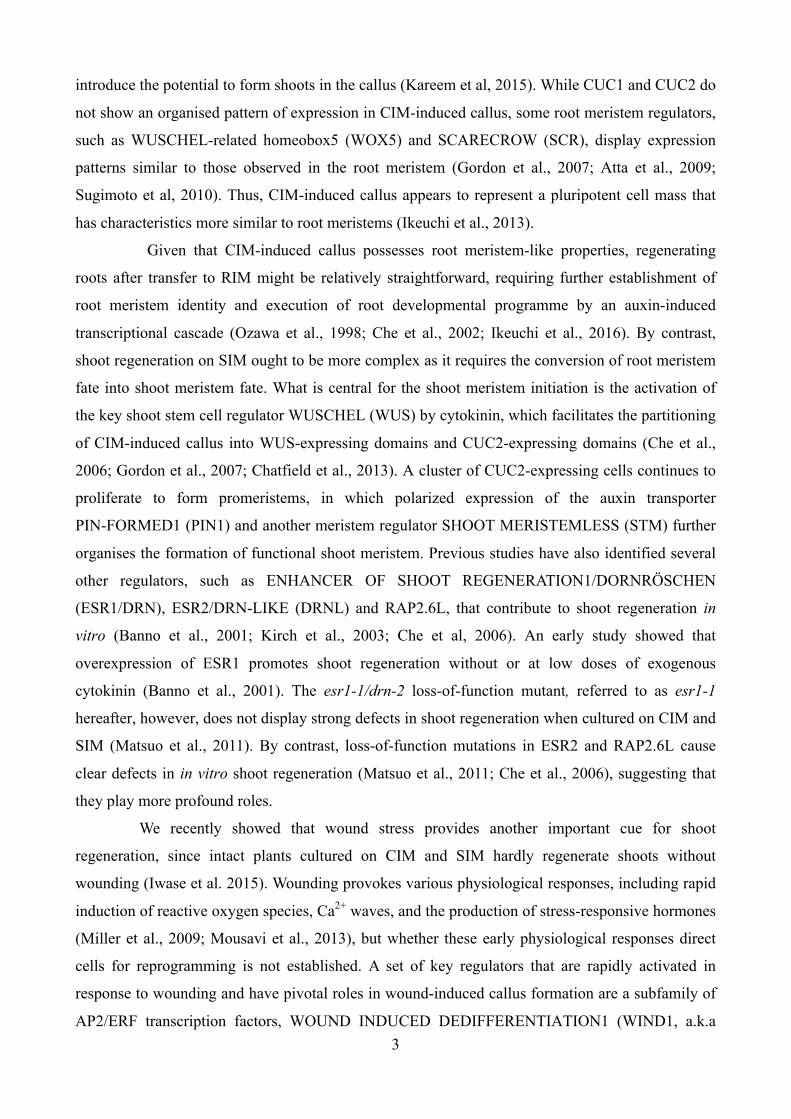
Figure 1. Wounding activates ESR1 expression in a WIND1-dependent manner.
(A) RT-qPCR analysis of WIND1 (upper panel) and ESR1 (lower panel) expression after wounding. First and second
leaves of 14-day-old WT seedlings were cut and leaf explants were cultured on phytohormone-free MS medium.
WIND1 expression peaks at 1 hour (h) after wounding, and ESR1 expression peaks at 3 h. The induction of ESR1 is
strongly suppressed in ProWIND1:WIND-SRDX (WIND1-SRDX) plants. Expression levels are normalized against those
of PP2AA3. Data are mean ± SE (n = 3, biological replicates). (B) Induction of the ESR1 promoter activity at the
wound sites of leaf petioles. Leaf explants of ProESR1:GUS and ProESR1:GUS WIND1-SRDX plants were cultured on
MS medium. ESR1 activation is compromised in ProESR1:GUS WIND1-SRDX plants. Dashed lines mark wound sites.
Representative images of petioles at 0 and 48 h after wounding are shown. (C) Transverse section of ProESR1:GUS
petioles close to wound sites at 48 h after wounding. GUS staining is found in several cell types, xylem parenchyma
cells, procambium cells (left panel) and mesophyll cells (right panel), that have started to undergo cell division.
Sections were counter-stained by safranin O. Asterisks mark new division planes. mp, mesophyll cells; xy, xylem
cells; pc, procambium cells; ph, phloem cells. (D) Nuclear accumulation of ESR1-GFP fusion proteins within the
epidermal cells near wound sites at 72 h after wounding. Note that wound stress produces strong green auto-
fluorescence in both WT and ProESR1:ESR1-GFP (ESR1-GFP) plants, but these signals are found mostly at the cut
edge or cytoplasm. Scale bars = 300 µm in (B), (D) and 50 µm in (C).

A
B
C -2000 -1000 -2500 -1500 -500 +1 +500 +1000
P1 P2 P3 P4 P5
Control
GFP
P1 P2 P3 P4 P5
Rela
tive e
nrichm
ent
[Effecter]
Control
WIND1 NOS
35S:WIND1
35SΩ NOS
35SΩ
m1:
WT:
m2:
m3:
m4:
VWRE-like
m5:
ProESR1:L-LUC
-150 +1 L-LUC [Reporter]
R10
35S:WIND1-m1
35S:WIND1-WT
Relative LUC activity
(L-LUC / R-LUC)
Control-WT
0 5 10 20 25 15
35S:WIND1-m2
35S:WIND1-m3
35S:WIND1-m4
35S:WIND1-m5
ESR1
5
10
0
15
20
ATG
+
1
-64 -
513
R1 R2
R3 R4
R5 R6
R7 R8
R9
R10
R11
*
caaacttcacaaaatttaattaaatttttataccatttatgtaccta caaacttcacCTTGCttaattaaGCAAGtataccatttatgtaccta caaacttcacCTTGCttaattaaatttttataccatttatgtaccta caaacttcacaaaatttaattaaGCAAGtataccatttatgtaccta
caaaAttAaAaaaatttaattaaatttttataccatttatgtaccta caaacttcacGTTGACAGTAAGTGAACCtataccatttatgtaccta
VWRE-like
ATG
ATG
R1 R2 R3 R4 R5 R6 R7 R8 R9 R10 R11
Figure 2. WIND1 directly binds the ESR1 promoter and activates its expression.
(A) Chromatin immunoprecipitation of WIND1-GFP fusion proteins on the ESR1 locus. Quantitative PCR analysis,
using P1-P5 primers designed within the promoter and coding sequence of ESR1, shows the strongest enrichment of
WIND1-GFP using P3 primers designed around -500 bp upstream of the translational start site. The black box
represents the coding sequence of ESR1 and +1 ATG indicates the translational start site. Black lines mark the relative
distance from the translational start site. Data are normalized against input DNA and shown as a relative enrichment
of DNA immunoprecipitated with rabbit serum (control). Data are mean ± SE (n = 3, technical replicates). (B)
Electrophoresis Mobility Shift Assays (EMSAs) of MBP-WIND1-His6 protein’s binding to the ESR1 promoter in
vitro. Upper panel shows the position of ~50-bp DNA probes, designated as R1 to R11, that cover -513 to -64-bp
nucleotides of the ESR1 promoter. Arrowheads and asterisks show free and shifted DNA probes, respectively. Dashed
lines separate results obtained in three different experiments. Note that the band shifts only with the R10 probe,
indicating that MBP-WIND1-His6 binds -153 to -104-bp upstream of the translational start site. (C) WIND1-induced
transient activation of the ESR1 expression in Arabidopsis culture cells. Upper panel shows the effector constructs,
control and 35S:WIND1, and the reporter construct, ProESR1:L-LUC. For the effector constructs, gray arrows mark
35SΩ, cauliflower mosaic virus 35S promoter with the tobacco mosaic virus omega translation amplification sequence,
and gray boxes mark NOS, Agrobacterium nopaline synthase transcriptional terminator. White box marks the WIND1
coding sequence. For the reporter construct, black bar represents the 150-bp promoter sequence of ESR1 with R10
sequence marked with white box. The gray box represents the coding sequence of L-LUC, encoding a firefly
luciferase gene, and +1 ATG indicates the translational start site. Middle panel shows WT R10 sequence with two
VWRE-like motifs marked in blue and m1 to m5 mutations marked in red. Bottom panel shows the ESR1 promoter
activity as judged by the L-LUC activity relative to R-LUC, Renilla luciferase. Co-bombardment of 35S:WIND1 and
ProESR1:L-LUC activates the ESR1 promoter. Note that abolishing each of the two VWRE-like motifs by m2 or m3
mutation results in reduced ESR1 induction and abolishing both motifs by m1 or m4 mutation has additive effects,
indicating that both motifs are required for activation of ESR1 by WIND1. Data are mean ± SE (n = 6, technical
replicates).

0.0
0.5
1.0
1.5
2.0
2.5
x
0.0
0.5
1.0
1.5
2.0
2.5
WT esr1-2 ESR1-SRDX ESR1-GFP
A
Figure 3. ESR1 promotes callus formation at wound sites.
(A) Callus formation at wound sites of WT, esr1-2, ProESR1:ESR1-SRDX (ESR1-SRDX), and ESR1-GFP leaf
explants. Leaf explants were cultured on phytohormone-free MS medium and callus phenotypes were scored
at 8 days after wounding. Box plots represent the distribution of projected callus area (n = 12 per genotype).
Statistical significance against WT was determined by a Student's t-test (***p < 0.001, **p < 0.01, *p < 0.1).
(B) Callus generated at wound sites of WT, esr1-2, ESR1-SRDX, and ESR1-GFP leaf explants.
Representative images at 8 days after wounding are shown. Scale bars = 500 µm in (B).
B
0
0.5
1.0
1.5
2.5
2.0
WT esr1-2 ESR1-
SRDX
ESR1-
GFP
Rela
tive c
allu
s a
rea
***
*
**
WT esr1-2
ESR1-SRDX ESR1-GFP

0.0
0.5
1.0
1.5
2.0
2.5
x
0.0
0.5
1.0
1.5
2.0
2.5
WT WIND1-SRDX esr1-D WIND1-SRDX essr1-D
B
A
0
0.5
1.0
1.5
2.5
2.0
Rela
tive c
allu
s a
rea
WT esr1-D WIND1-
SRDX
WIND1-
SRDX
esr1-D
***
*
WT WIND1-SRDX
esr1-D WIND1-SRDX
esr1-D
C
0 20 40 60 80 100
(%) Appearance frequency
Type I Type II Type III Wild type-like
35S:WIND1 / esr1-2
35S:WIND1 / WT
D
WT arr1,12
0
0.01
Rela
tive e
xpre
ssio
n
(ES
R1
/PP
2A
A3
)
WT esr1-2
1
0
2
3
Rela
tive e
xpre
ssio
n
(AR
R5
/PP
2A
A3
)
E
Figure 4. ESR1 functions downstream of WIND1 in wound-induced callus formation.
(A) Callus formation at wound sites of WT, ProWIND1:WIND1-SRDX (WIND1-SRDX), esr1-D, and WIND1-SRDX
esr1-D leaf explants. Leaf explants were cultured on phytohormone-free MS medium and callus phenotypes were
scored at 8 days after wounding. Box plots represent the distribution of projected callus area (n = 12 per genotype).
Statistical significance against WT was determined by a Student's t-test (***p < 0.001, *p < 0.1). (B) Callus
generated at wound sites of WT, WIND1-SRDX, esr1-D, and WIND1-SRDX esr1-D leaf explants. Note that
ectopic induction of ESR1 rescues the callus formation deficiency in WIND1-SRDX explants. Representative
images at 8 days after wounding are shown. (C) The esr1-2 mutation partly suppresses WIND1-induced callus
formation in T1 seedlings grown on MS medium. Phenotypic severity was scored according to Figure 2 in Iwase
et al. (2011). T1 plants showing weak, intermediate, and strong callus formation are classified as type I, type II,
and type-III plants, respectively. (n = 104 for WT; n = 43 for esr1-2). (D) The arr1 arr12 mutation partially
suppresses the ESR1 expression after wounding. (E) The esr1-2 mutation does not suppress the ARR5 expression
after wounding. First and second leaves of 14-day-old WT, arr1 arr12, and esr1-2 seedlings were cut and leaf
explants were cultured on phytohormone-free MS medium. Expression levels are normalized against those of the
PP2AA3 gene. Data are mean ± SE (n = 3, biological replicates). Scale bars = 250 µm in (B).

ESR1-GFP WT
Stem
Leaf
Cotyledon
Root
*
*
*
*
*
*
Leaf Root
Shoo
t re
genera
tio
n (
%)
A
B
WT ESR1-GFP WT ESR1-GFP
D
0 0.1 0.5 5 10 0 0.1 0.5 5 10
WT XVE-ESR1
(µM)
Rela
tive e
xpre
ssio
n
(ES
R1
/PP
2A
A3
) S
hoo
t re
genera
tio
n (
%)
40
30
20
10
0
8
6
4
2
0
10
12
20
12
8
4
0
16
20
12
8
4
0
16
***
***
* *
***
***
Figure 5. ESR1 promotes shoot regeneration at wound sites.
(A) Induction of shoot regeneration at wound sites of ESR1-GFP explants. WT and ESR1-GFP explants were
cultured on phytohormone-free MS medium for 50 days (leaves), 30 days (cotyledons and inflorescence stems), and
40 days (roots). Dashed lines represent wound sites and asterisks mark regenerating shoots. (B) Quantitative
analysis of shoot regeneration at wound sites. Leaf and root explants were cultured on phytohormone-free MS
medium for 55 days. Data are shown as frequency (%) of explants regenerating shoots (n = 646 for WT leaves, 170
for ESR1-GFP leaves, 576 for WT roots and 634 for ESR1-GFP roots). Statistical significance was determined by a
proportion test (***p < 0.001). (C) Induction of shoot regeneration at wound sites of XVE-ESR1 leaf explants. (Top
panel) XVE-ESR1 leaf explants were cultured on phytohormone-free MS medium in the absence (–) or presence (+)
of 10 µM 17b-estradiol (ED). (Bottom panel) Unwounded XVE-ESR1 plants develop callus in the presence of 10
µM ED. The dashed line represents wound sites and an asterisk marks regenerating shoots. An arrowhead marks
callus developing from hypocotyls in intact XVE-ESR1 plants. (D) The frequency of shoot regeneration positively
correlates with the level of ESR1 expression in XVE-ESR1 plants. ESR1 expression and shoot regeneration were
quantified at 6 and 16 days, respectively, after the application of 0.1 to 10 µM ED. Expression levels are normalized
against those of PP2AA3. Expression data are mean ± SE (n = 3, biological replicates). Shoot regeneration is
quantified as the frequency (%) of explants regenerating shoots (n = 50 per ED concentration). Statistical
significance was determined by a proportion test (***p < 0.001, *p < 0.1). Scale bars = 1 mm in (A) and (C).
ED (-) ED (+) C *
*

WT esr1-2
ESR1-
SRDX
ESR1-
GFP
A S
hoo
t re
genera
tio
n (
%)
B
4
2
0
6
8
10
WT esr1-2 ESR1-
SRDX
ESR1-
GFP
*** ***
***
esr1-1
esr1-1
***
Figure 6. ESR1 promotes shoot regeneration in vitro.
(A) Shoot regeneration of WT, esr1-1, esr1-2, ESR1-SRDX, and ESR1-GFP root explants in vitro. Root explants
were cultured on CIM for 4 days and transferred to SIM. Representative images of root explants cultured on
SIM for 18 days are shown. (B) Quantitative analysis of shoot regeneration phenotypes. Regeneration
phenotypes are scored as the number of regenerating shoots per explant. Data are mean ± SE (n ≥ 30 per
genotype). Statistical significance was determined by a Student’s t-test (***p < 0.001). Scale bars = 5 mm in
(A).

0 h
B5 kinetin 2,4-D kinetin + 2,4-D
48 h
MS
A
CUC1 CUC2
WUS STM
ESR1 ESR2 1.2
0
0.8
0.9
1
2
0
3
5
10
0
15
0.6
0.3
0
90
60
30
0
Rela
tive g
ene e
xpre
ssio
n
MS CIM SIM SIM
0 4 7 16
WT esr1-2
1
2
4
0
3
5
0.4
PLT3
1
2
0
3 PLT5
1
2
0
PLT7
1
2
0
PLT1
4
8
0
12
PLT2
2
4
8
0
6
10 1.2
0.8
0.4
0
RAP2.6L
MS CIM SIM SIM
0 4 7 16
MS CIM SIM SIM
0 4 7 16
MS CIM SIM SIM
0 4 7 16 (d)
B
Figure 7. ESR1 is required for the transcriptional activation of shoot regeneration regulators in vitro.
(A) Incubation of leaf (top panel) and root (bottom panel) explants on B5 medium containing both kinetin and
2,4-D, strongly enhances the ESR1 promoter activity at wound sites. ProESR1:GUS explants were freshly
prepared on MS medium and transferred to B5 medium with or without 0.1 mg/L kinetin and 0.5 mg/L 2,4-D.
Representative images at 0 and 48 h after incubation are shown. Note that kinetin or 2,4-D alone does not
activate the ESR1 promoter in both leaf and root explants. (B) RT-qPCR analysis of ESR1 expression and
other shoot regeneration regulators in WT and esr1-2 root explants cultured on CIM and SIM. Total RNA was
extracted from root explants freshly prepared on MS medium (MS 0), cultured on CIM for 4 days (CIM 4),
cultured on CIM for 4 days and subsequently on SIM for 7 or 16 days (SIM 7 or SIM 16). Expression levels
are normalized against those of PP2AA3. Data are mean ± SE (n = 3, biological replicates). Scale bars = 500
µm in (A).
Roots
Leaves

WT esr1-D WIND1-SRDX
WIND1-SRDX
esr1-D
Shoo
t re
genera
tio
n
4
2
0
6
8
10
WT esr1-D WIND1-SRDX WIND1-SRDX
esr1-D
A
B
Figure 8. ESR1 functions downstream of WIND1 in in vitro shoot regeneration.
(A) Shoot regeneration of WT, WIND1-SRDX, esr1-D and WIND1-SRDX esr1-D root explants in vitro. Root
explants were cultured on CIM for 4 days and transferred to SIM. Representative images of root explants
cultured on SIM for 21 days are shown. (B) Quantitative analysis of shoot regeneration phenotypes.
Regeneration phenotypes are scored as a number of regenerating shoots per explant. Data are mean ± SE (n
≥ 50 per genotype). Statistical significance was determined by a Student’s t-test (***p < 0.001). Note that
ectopic induction of ESR1 is sufficient to rescue the shoot regeneration phenotypes in WIND1-SRDX
explants. (C) Induction of WIND1 and ESR1 promoter activities in callus developing from root explants on
SIM. Root explants from ProWIND1:GUS, ProESR1:GUS, and ProESR1:GUS WIND1-SRDX plants were
cultured on CIM for 4 days and transferred to SIM. Note that the promoter activity of ESR1 is strongly
reduced by dominant repression of WIND1. Representative images at 1 day after transfer to SIM are shown.
Scale bars = 500 µm in (A) and 100 µm in (C).
C ProWIND1:GUS ProESR1:GUS
WIND1-SRDX
ProESR1:GUS
***
*** ***

ESR1
WIND1
CUC1 RAP2.6L WUS STM
shoot regeneration
wounding
ESR2
Figure 9. A schematic model describing how the WIND1-ESR1 pathway promotes shoot regeneration inArabidopsis.Local wound stress induces WIND1 expression at wound sites. WIND1 directly binds the ESR1 promoter andactivates its expression. WIND1 also activates ESR1 indirectly through enhancing the B-type ARR-mediatedcytokinin signalling. ESR1 expression is synergistically enhanced by exogenously supplied auxin and cytokinin tofurther boost shoot regeneration in vitro. ESR1 is required for the up-regulation of key shoot regulators such asCUC1, RAP2.6L, ESR2, WUS and STM to promote shoot regeneration.
ARR1 ARR12
auxin cytokinin

DOI 10.1105/tpc.16.00623; originally published online December 23, 2016;Plant Cell
and Keiko SugimotoKomaki, Kengo Morohashi, Tetsuya Kurata, Masaru Nakata, Masaru Ohme-Takagi, Erich Grotewold
Akira Iwase, Hirofumi Harashima, Momoko Ikeuchi, Bart Rymen, Mariko Ohnuma, ShinichiroSHOOT REGENERATION1 in Arabidopsis
WIND1 promotes shoot regeneration through transcriptional activation of ENHANCER OF
This information is current as of September 17, 2018
Supplemental Data /content/suppl/2017/02/06/tpc.16.00623.DC2.html /content/suppl/2016/12/23/tpc.16.00623.DC1.html
Permissions X
https://www.copyright.com/ccc/openurl.do?sid=pd_hw1532298X&issn=1532298X&WT.mc_id=pd_hw1532298
eTOCs http://www.plantcell.org/cgi/alerts/ctmain
Sign up for eTOCs at:
CiteTrack Alerts http://www.plantcell.org/cgi/alerts/ctmain
Sign up for CiteTrack Alerts at:
Subscription Information http://www.aspb.org/publications/subscriptions.cfm
is available at:Plant Physiology and The Plant CellSubscription Information for
ADVANCING THE SCIENCE OF PLANT BIOLOGY © American Society of Plant Biologists