a simple strategy to fabricate electrochemical sensor … rajabhat university, phitsanulok 65000,...
TRANSCRIPT
NU. International Journal of Science 2015; 12(2) : 29 – 38 29
A simple strategy to fabricate electrochemical sensor based on nickel
nanoparticles modified glassy carbon electrode for the determination of
glucose in urine
Kulwadee Pinwattana* and Rapiphun Janmanee
Department of Chemistry, Faculty of Science and Technology,
Pibulsongkram Rajabhat University, Phitsanulok 65000, Thailand
*Corresponding author, E-mail: [email protected]
ABSTRACT
A non-enzymatic electrochemical sensor has been successfully fabricated using
nickel nanoparticles as catalyst. Nickel nanoparticles were electrochemically deposited on a
glassy carbon electrode surface by amperometric technique. Nickel nanoparticles modified
glassy carbon electrode exhibited high catalytic activity for oxidation of glucose. The
morphology of nickel nanoparticles was characterized by scanning electron microscopy
(SEM). Electrochemical behaviors of glucose were investigated by cyclic voltammetry and
chronoamperometry in alkaline aqueous solution. At the optimum conditions, the calibration
curve for glucose determination was linear in the range of 0.05 – 5.00 mM and 5.00 – 17.5 mM
and a low detection limit (S/N = 3) of 36 µM. Furthermore, the proposed sensor was
successfully applied to analyze glucose level in human urine samples when compared with
clinical laboratory measurements. The glucose sensor showed a strong potential as a sensitive
non-enzymatic and may be used in the future for the clinical diagnostic tool.
Keywords: glucose sensor, nickel nanoparticles, electrodeposition, non-enzymatic
INTRODUCTION
Glucose determination has gained great attention as a result of its wide
application in many areas such as biotechnology, food industry, and clinical
diagnostics (Rahman et al. , 2010; Fang et al.,2011; Zhu et al. , 2012; Wang et al. ,
2013; Guo et al. , 2015). Especially, glucose plays an important analyte in clinical
diagnostics because high levels of glucose in the blood (over 6 mM) will increase the
risk of diabetes. Diabetes is a serious health care problem, which is a familiar disease
and one of the main causes of disability and death in the world. According to the
World Health Organization, almost 350 million people are living with diabetes in the
world and in 2030, it is estimated that diabetes will be the 7th leading cause of death.
(Shaw et al., 2010; alwan, 2011; Baghayeri et al., 2016) Blood glucose level has been
measured as an effective clinical biomarker for diabetes risk. Nevertheless, drawing
blood from fingertip or vein force causes a painful prick. On the other hand, urine is
another instructive body liquid, which has been a useful indicator for diagnosis. The
glucose level in urine is as well a good pointer for preliminary screening of patients
with high level diabetes or having renal glycosuria (Kim et al., 2014; Fan et al., 2015;
Baghayeri et al., 2016) However, blood or urine has the complex matrices, therefore,
glucose measurement is an analytical challenge.
30 NU. International Journal of Science 2015; 12(2) : 29 – 38
Electrochemical glucose sensors are generally categorized into two
classifications: enzymatic (Ali et al., 2013; Razmi and Mohammad-Rezaei, 2013;
Kong et al., 2014; Su et al., 2014) and non-enzymatic (Lin et al., 2013; Guo et al.,
2014; Li et al., 2014; Wang et al., 2014). Enzymatic glucose sensors are based on the
immobilization of glucose oxidase on various substrates and these enzymatic glucose
sensor have been widely developed due to their excellent selectivity and high
sensitivity. However, the drawback of these enzymatic sensors is their lack of long-
term stability because the glucose oxidase can be easily affected by pH, temperature,
ionic detergents, humidity and other types of matrix and also enzyme is expensive.
Non-enzymatic glucose sensor has attracted considerable attention to overcome the
problems of using enzyme-based glucose sensors.
Various metal nanoparticles have been investigated for electrocatalytic
oxidation of glucose in non-enzymatic glucose sensor. Previous studies have focused
on the noble metals such as gold (Au), platinum (Pt), palladium (Pd), and silver (Ag)
(Xiao et al., 2009; Lorestani et al., 2015; Thanh et al., 2016). However, these metals
are very costly. Therefore, nickel nanoparticles are of particular interest, owing to their
excellent electrocatalytic activity, low cost and environmental friendly. Nickel based
non-enzymatic glucose sensors have shown an outstanding electrocatalytic activity
toward glucose oxidation, which is mediated by Ni(II)/Ni(III) redox couples in
alkaline aqueous solution (Zhao et al., 2007; Niu et al., 2013; Shervedani et al., 2014).
It is well known that nanomaterials exhibit special properties compared to the bulky
materials. Thus, nickel nanoparticles are an attractive electrocatalyst for glucose
oxidation.
In the present work, a simple method was developed to fabricate a non-
enzymatic glucose sensor. Nickel nanoparticles were electrochemically deposited on
the surface of glassy carbon electrode by one step strategy. The morphology of the
nickel nanoparticles modified electrode was characterized with scanning electron
microscopy (SEM). The electrochemical and electrocatalytic properties of the nickel
nanoparticles modified glassy carbon electrode toward the oxidation of glucose were
discussed. In addition, the detecting performance of the proposed glucose sensor was
evaluated and optimized by chronoamperometry. The results demonstrated that this
approach have been shown high catalytic activity for glucose oxidation. Moreover, the
proposed glucose sensor can be successfully applied in human urine.
MATERIALS AND METHODS
Chemicals and reagents
Glucose and nickel nitrate hexahydrate were purchased from Sigma-Aldrich
Co. Sodium hydroxide was purchased from RCI Labscan Ltd. Sodium acetate
trihydrate was purchased from Baker Analyzed. Acetic acid was purchased from
Merck. All chemical reagents were of analytical grade and all solutions were prepared
with deionized water.
Instrumentation
Electrochemical measurements were performed with a Digi-Ivy model
DY2113 potentiostat. A conventional three-electrode system was used, containing an
Ag/AgCl electrode as the reference electrode, a Pt wire as the counter electrode, and
NU. International Journal of Science 2015; 12(2) : 29 – 38 31
the modified glassy carbon electrode as the working electrode. The surface
morphological characterization of nickel nanoparticles was examined by means of
scanning electrode microscope (SEM; LEO, model 1400VP) at an accelerating
voltage of 20 kV.
Preparation of nickel nanoparticles modified glassy carbon electrode Glassy carbon electrode was polished before each experiment with 1.0 and
0.3 µm alumina powder, respectively. Then, the polished electrode was rinsed thoroughly with deionized water and was ultrasonicated in ethanol and deionized water for 10 min each to remove organic and other contaminants.
To prepare the nickel nanoparticles modified glassy carbon electrode, the electrodeposition of nickel nanoparticles on the electrode surface was performed in 0.1 M acetate buffer solution (pH 4.0) containing 5 mM nickel nitrate at -1.1 V for 250 s under the unstirring conditions. After that, the modified electrode was scanned by cyclic voltammetry from 0.1 to 0.8 V in 0.1 M NaOH until a steady state voltammogram was obtained. The final resulting modified electrode was denoted by Ni NPs/GCE
Electrochemical measurement
All electrochemical experiments were performed with a three-electrode
system at room temperature of 25 °C. All potentials were measured relative to the
Ag/AgCl reference electrode. Cyclic voltammetric scans were performed at the
potential range from 0 to 0.8 V at scan rate of 0.1 V/s. The chronoamperometric
measurements were performed at 0.54 V.
Real sample analysis
The urine samples were collected from a normal healthy female volunteer.
The utilization of the Ni NPs/GCE for glucose determination in urine sample analysis
was also investigated by direct analysis. Urine samples were diluted 10 times with 0.1 M
NaOH to reduce the matrix effect of urine samples.
RESULTS AND DISCUSSION
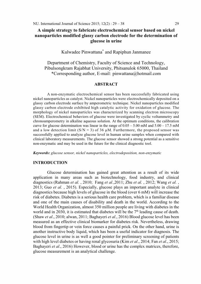
Characterization of nickel nanoparticles modified glassy carbon electrode For the modified electrode, nickel nanoparticles were electrochemically
deposited on the glassy carbon electrode surface by chronoamperometry. The nickel
nanoparticles were investigated using SEM. The surface morphology of the
unmodified glassy carbon substrate and the electrodeposited nickel nanoparticles on
glassy carbon substrate are shown in Figure 1. The result in the SEM image indicated
that nickel nanoparticles were electrodeposited on the glassy carbon substrate surface
and were well distributed on the surface with diameters in the range of 60 – 150 nm.
32 NU. International Journal of Science 2015; 12(2) : 29 – 38
Figure 1 Scanning electron microscope image of the unmodified glassy carbon
substrate (A) and the electrodeposited nickel nanoparticles on glassy
carbon substrate (B).
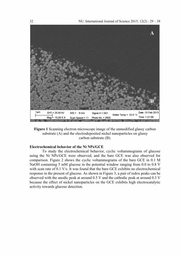
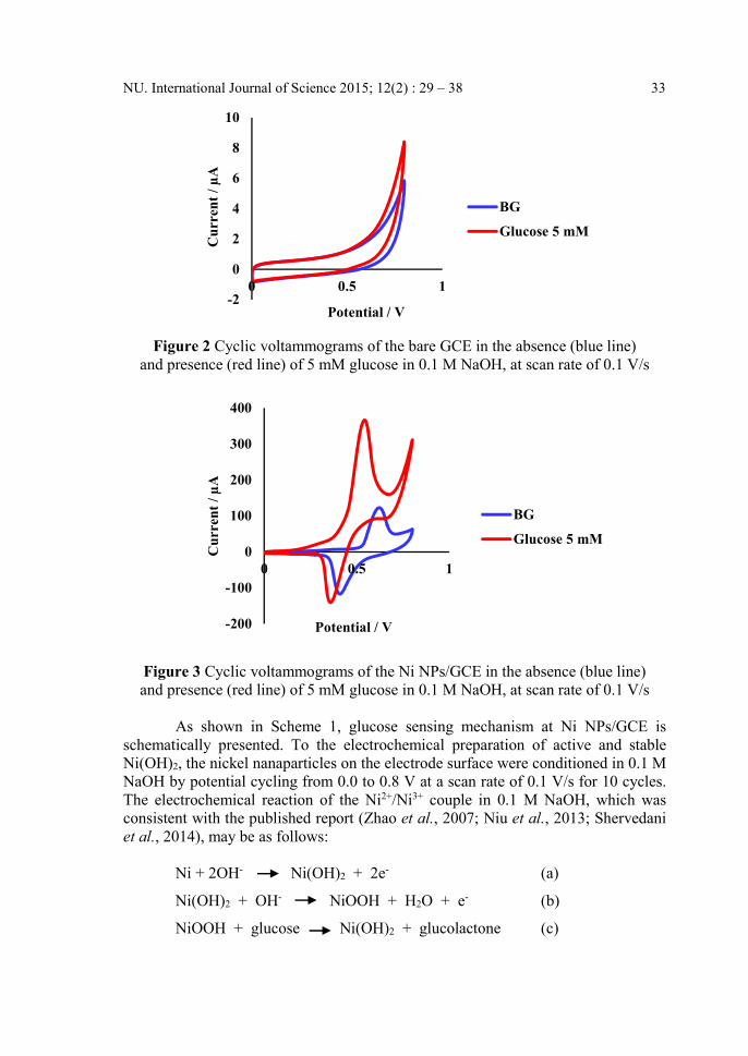
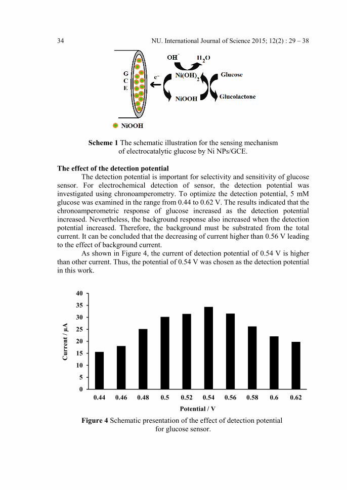
Electrochemical behavior of the Ni NPs/GCE To study the electrochemical behavior, cyclic voltammograms of glucose
using the Ni NPs/GCE were observed, and the bare GCE was also observed for
comparison. Figure 2 shows the cyclic voltammograms of the bare GCE in 0.1 M
NaOH containing 5 mM glucose in the potential window ranging from 0.0 to 0.8 V
with scan rate of 0.1 V/s. It was found that the bare GCE exhibits no electrochemical
response in the present of glucose. As shown in Figure 3, a pair of redox peaks can be
observed with the anodic peak at around 0.5 V and the cathodic peak at around 0.3 V
because the effect of nickel nanoparticles on the GCE exhibits high electrocatalytic
activity towards glucose detection.
B
NU. International Journal of Science 2015; 12(2) : 29 – 38 33
Figure 2 Cyclic voltammograms of the bare GCE in the absence (blue line)
and presence (red line) of 5 mM glucose in 0.1 M NaOH, at scan rate of 0.1 V/s
Figure 3 Cyclic voltammograms of the Ni NPs/GCE in the absence (blue line)
and presence (red line) of 5 mM glucose in 0.1 M NaOH, at scan rate of 0.1 V/s
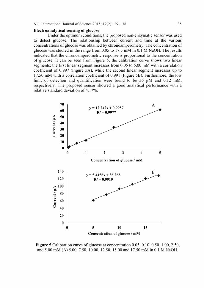
As shown in Scheme 1, glucose sensing mechanism at Ni NPs/GCE is
schematically presented. To the electrochemical preparation of active and stable
Ni(OH)2, the nickel nanaparticles on the electrode surface were conditioned in 0.1 M
NaOH by potential cycling from 0.0 to 0.8 V at a scan rate of 0.1 V/s for 10 cycles.
The electrochemical reaction of the Ni2+/Ni3+ couple in 0.1 M NaOH, which was
consistent with the published report (Zhao et al., 2007; Niu et al., 2013; Shervedani
et al., 2014), may be as follows:
Ni + 2OH- Ni(OH)2 + 2e- (a)
Ni(OH)2 + OH- NiOOH + H2O + e- (b)
NiOOH + glucose Ni(OH)2 + glucolactone (c)
-2
0
2
4
6
8
10
0 0.5 1
Cu
rren
t /
µA
Potential / V
BG
Glucose 5 mM
-200
-100
0
100
200
300
400
0 0.5 1
Cu
rren
t/
µA
Potential / V
BG
Glucose 5 mM
34 NU. International Journal of Science 2015; 12(2) : 29 – 38
Scheme 1 The schematic illustration for the sensing mechanism
of electrocatalytic glucose by Ni NPs/GCE.
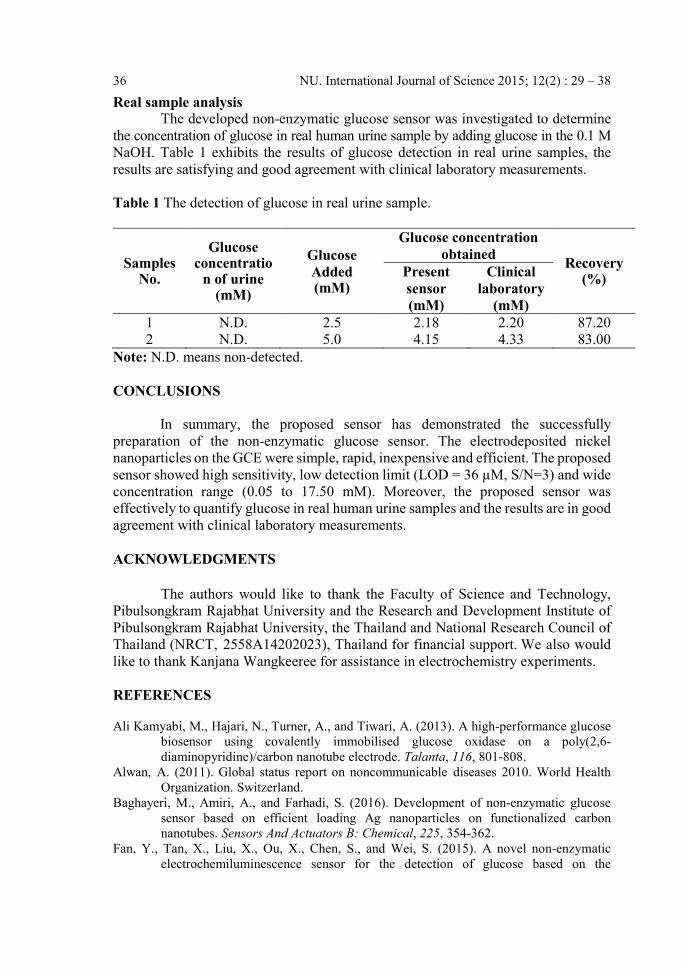
The effect of the detection potential
The detection potential is important for selectivity and sensitivity of glucose
sensor. For electrochemical detection of sensor, the detection potential was
investigated using chronoamperometry. To optimize the detection potential, 5 mM
glucose was examined in the range from 0.44 to 0.62 V. The results indicated that the
chronoamperometric response of glucose increased as the detection potential
increased. Nevertheless, the background response also increased when the detection
potential increased. Therefore, the background must be substrated from the total
current. It can be concluded that the decreasing of current higher than 0.56 V leading
to the effect of background current.
As shown in Figure 4, the current of detection potential of 0.54 V is higher
than other current. Thus, the potential of 0.54 V was chosen as the detection potential
in this work.
Figure 4 Schematic presentation of the effect of detection potential
for glucose sensor.
0
5
10
15
20
25
30
35
40
0.44 0.46 0.48 0.5 0.52 0.54 0.56 0.58 0.6 0.62
Cu
rren
t/
µA
Potential / V
NU. International Journal of Science 2015; 12(2) : 29 – 38 35
Electroanalytical sensing of glucose
Under the optimum conditions, the proposed non-enzymatic sensor was used
to detect glucose. The relationship between current and time at the various
concentrations of glucose was obtained by chronoamperometry. The concentration of
glucose was studied in the range from 0.05 to 17.5 mM in 0.1 M NaOH. The results
indicated that the chronoamperometric response is proportional to the concentration
of glucose. It can be seen from Figure 5, the calibration curve shows two linear
segments: the first linear segment increases from 0.05 to 5.00 mM with a correlation
coefficient of 0.997 (Figure 5A), while the second linear segment increases up to
17.50 mM with a correlation coefficient of 0.991 (Figure 5B). Furthermore, the low
limit of detection and quantification were found to be 36 µM and 0.12 mM,
respectively. The proposed sensor showed a good analytical performance with a
relative standard deviation of 4.17%.
Figure 5 Calibration curve of glucose at concentration 0.05, 0.10, 0.50, 1.00, 2.50,
and 5.00 mM (A) 5.00, 7.50, 10.00, 12.50, 15.00 and 17.50 mM in 0.1 M NaOH.
y = 12.242x + 0.9957
R² = 0.9977
0
10
20
30
40
50
60
70
0 1 2 3 4 5
Cu
rren
t /
µA
Concentration of glucose / mM
y = 5.4456x + 36.268
R² = 0.9919
0
20
40
60
80
100
120
140
0 5 10 15
Cu
rren
t/
µA
Concentration of glucose / mM
A
B
36 NU. International Journal of Science 2015; 12(2) : 29 – 38
Real sample analysis
The developed non-enzymatic glucose sensor was investigated to determine
the concentration of glucose in real human urine sample by adding glucose in the 0.1 M
NaOH. Table 1 exhibits the results of glucose detection in real urine samples, the
results are satisfying and good agreement with clinical laboratory measurements.
Table 1 The detection of glucose in real urine sample.
Samples No.
Glucose concentratio
n of urine (mM)
Glucose
Added (mM)
Glucose concentration
obtained Recovery
(%) Present
sensor
(mM)
Clinical
laboratory
(mM)
1 N.D. 2.5 2.18 2.20 87.20
2 N.D. 5.0 4.15 4.33 83.00
Note: N.D. means non-detected.
CONCLUSIONS
In summary, the proposed sensor has demonstrated the successfully
preparation of the non-enzymatic glucose sensor. The electrodeposited nickel
nanoparticles on the GCE were simple, rapid, inexpensive and efficient. The proposed
sensor showed high sensitivity, low detection limit (LOD = 36 µM, S/N=3) and wide
concentration range (0.05 to 17.50 mM). Moreover, the proposed sensor was
effectively to quantify glucose in real human urine samples and the results are in good
agreement with clinical laboratory measurements.
ACKNOWLEDGMENTS
The authors would like to thank the Faculty of Science and Technology,
Pibulsongkram Rajabhat University and the Research and Development Institute of
Pibulsongkram Rajabhat University, the Thailand and National Research Council of
Thailand (NRCT, 2558A14202023), Thailand for financial support. We also would
like to thank Kanjana Wangkeeree for assistance in electrochemistry experiments.
REFERENCES
Ali Kamyabi, M., Hajari, N., Turner, A., and Tiwari, A. (2013). A high-performance glucose
biosensor using covalently immobilised glucose oxidase on a poly(2,6-
diaminopyridine)/carbon nanotube electrode. Talanta, 116, 801-808.
Alwan, A. (2011). Global status report on noncommunicable diseases 2010. World Health
Organization. Switzerland.
Baghayeri, M., Amiri, A., and Farhadi, S. (2016). Development of non-enzymatic glucose
sensor based on efficient loading Ag nanoparticles on functionalized carbon
nanotubes. Sensors And Actuators B: Chemical, 225, 354-362.
Fan, Y., Tan, X., Liu, X., Ou, X., Chen, S., and Wei, S. (2015). A novel non-enzymatic
electrochemiluminescence sensor for the detection of glucose based on the
NU. International Journal of Science 2015; 12(2) : 29 – 38 37
competitive reaction between glucose and phenoxy dextran for concanavalin A
binding sites. Electrochimica Acta, 180, 471-478.
Fang, B., Zhang, C., Wang, G., Wang, M., and Ji, Y. (2011). A glucose oxidase immobilization
platform for glucose biosensor using ZnO hollow nanospheres. Sensors And
Actuators B: Chemical, 155(1), 304-310.
Guo, M., Wang, P., Zhou, C., Xia, Y., Huang, W., and Li, Z. (2014). An ultrasensitive non-
enzymatic amperometric glucose sensor based on a Cu-coated nanoporous gold film
involving co-mediating. Sensors And Actuators B: Chemical, 203, 388-395.
Guo, M.-M., Xia, Y., Huang, W., and Li, Z. (2015). Electrochemical fabrication of stalactite-
like copper micropillar arrays via surface rebuilding for ultrasensitive nonenzymatic
sensing of glucose. Electrochimica Acta, 151, 340–346.
Kim, H., Jang, K., Veerapandian, M., Kim, H., Seo, Y., Lee, K., and Lee, M. (2014). Reusable
urine glucose sensor based on functionalized graphene oxide conjugated Au electrode
with protective layers. Biotechnology Reports, 3, 49-53.
Kong, F., Gu, S., Li, W., Chen, T., Xu, Q., and Wang, W. (2014). A paper disk equipped with
graphene/polyaniline/Au nanoparticles/glucose oxidase biocomposite modified
screen-printed electrode: Toward whole blood glucose determination. Biosensors
And Bioelectronics, 56, 77-82.
Li, M., Bo, X., Mu, Z., Zhang, Y., and Guo, L. (2014). Electrodeposition of nickel oxide and
platinum nanoparticles on electrochemically reduced graphene oxide film as a
nonenzymatic glucose sensor. Sensors And Actuators B: Chemical, 192, 261-268.
Lin, K., Lin, Y., and Chen, S. (2013). A highly sensitive nonenzymatic glucose sensor based
on multi-walled carbon nanotubes decorated with nickel and copper
nanoparticles. Electrochimica Acta, 96, 164-172.
Lorestani, F., Shahnavaz, Z., Mn, P., Alias, Y., and Manan, N. (2015). One-step hydrothermal
green synthesis of silver nanoparticle-carbon nanotube reduced-graphene oxide
composite and its application as hydrogen peroxide sensor. Sensors And Actuators B:
Chemical, 208, 389-398.
Niu, X., Lan, M., Zhao, H., and Chen, C. (2013). Highly Sensitive and Selective Nonenzymatic
Detection of Glucose Using Three-Dimensional Porous Nickel
Nanostructures. Analytical Chemistry, 85(7), 3561-3569.
Rahman, M.M., Ahammad, A.J.S., Jin,J.H., Ahn, S.J., and Lee, J.J. (2010) A comprehensive
review of glucose biosensors based on nanostructured metal-oxides, Sensors, 10,
4855–4866.
Razmi, H., and Mohammad-Rezaei, R. (2013). Graphene quantum dots as a new substrate for
immobilization and direct electrochemistry of glucose oxidase: Application to
sensitive glucose determination. Biosensors And Bioelectronics, 41, 498-504.
Shaw, J.E., Sicree, R.A. and Zimmet., P.Z. (2010). Global estimates of the prevalence of
diabetes for 2010 and 2030. Diabetes Res. Clin. P. R., 87(1), 4-14.
Shervedani, R., Karevan, M., and Amini, A. (2014). Prickly nickel nanowires grown on Cu
substrate as a supersensitive enzyme-free electrochemical glucose sensor. Sensors
And Actuators B: Chemical, 204, 783-790.
Su, S., Sun, H., Xu, F., Yuwen, L., Fan, C., and Wang, L. (2014). Direct electrochemistry of
glucose oxidase and a biosensor for glucose based on a glass carbon electrode
modified with MoS2 nanosheets decorated with gold nanoparticles. Microchimica
Acta, 181(13-14), 1497-1503.
Thanh, T., Balamurugan, J., Hwang, J., Kim, N., and Lee, J. (2016). In situ synthesis of
graphene-encapsulated gold nanoparticle hybrid electrodes for non-enzymatic
glucose sensing. Carbon, 98, 90-98.
38 NU. International Journal of Science 2015; 12(2) : 29 – 38
Wang, J., Gao, H., Sun, F., and Xu, C. (2014). Nanoporous PtAu alloy as an electrochemical
sensor for glucose and hydrogen peroxide. Sensors And Actuators B: Chemical, 191,
612-618.
Wang, Z., Lei, H., and Feng, L. (2013) A facil channel for d-glucose detection in aqueous
solution. Spectrochim. Acta A., 114, 293-297.
Xiao, F., Zhao, F., Mei, D., Mo, Z., and Zeng, B. (2009). Nonenzymatic glucose sensor based
on ultrasonic-electrodeposition of bimetallic PtM (M=Ru, Pd and Au) nanoparticles
on carbon nanotubes–ionic liquid composite film. Biosensors And
Bioelectronics, 24(12), 3481-3486.
Zhao, C., Shao, C., Li, M., and Jiao, K. (2007). Flow-injection analysis of glucose without
enzyme based on electrocatalytic oxidation of glucose at a nickel
electrode. Talanta, 71(4), 1769-1773.
Zhu, Z., Garcia-Gancedo, L. A., Flewitt, J., Xie, H., Moussy, F., and Milne, W. I. (2012) A
critical review of glucose biosensors based on carbon nanomaterials: carbon
nanotubes and grapheme, Sensors, 12, 5996–6022.