a rapid procedure for catalase determination in blood and tissue
TRANSCRIPT

A Rapid Procedure for Catalase Determination
in Blood and Tissue Samples with the
Van Slyke Manometric Apparatus
John Esben Kirk
A precise and rapid gasometric procedure is described for catalase determination inblood and tissue samples. The enzyme activity assay is performed with the Van Slykemanometric apparatus. In the developed technic, a 45-sec. reaction period is used,and a large liquid-gas interphase is provided with the purpose of preventing theoxygen diffusion from becoming a rate-limiting factor. Under the devised methodo-logical conditions, the rate of oxygen evolution closely follows a parabolic curvewhich permits the calculation of the initial activity velocity of the enzyme. Theoxygen evolvement is directly proportional to the quantity of catalase present in theanalyzed sample. Less than 4 mm. are required for each catalase measurement.
SEVERAL GASOMETRIC METHODS have been used for determination ofcatalase activity by direct measurement of the rate of oxygen evolve-ment resulting from the enzymic decomposition of hydrogen peroxide,
but during the last 40 years, no procedure has been described in whichthe Van Slyke manometric apparatus is employed for the assay ofthis enzyme. Because of the explosive character of the catalase reac-tion and the fast inactivation of the enzyme by the hydrogen peroxidesubstrate, a rapid method is required for reliable estimation of theinitial velocity rate.
A suitable fast method for gasometric catalase determination hasrecently been devised by Greenfield and Price (4); in that procedure,
a special apparatus is employed in which the evolved oxygen is meas-ured by a pressure transducer and a continuous recorder system. Inview of the simplicity and general availability of the Van Slyke mano-
From the Washington University School of Medicine, St. Louis, Mo.The investigation was supported by Grant PHS-891 from the National Institutes of Health,
U. S. Public Health Service.
763
764 KIRK Clinical Chemistry
metric apparatus, it was considered advisable to develop a new meth-od for catalase assay with the use of that equipment.
In the technic described in the present paper special consideration
has been given to the basic features of the catalase assay which haveproved advantageous in the Greenfield and Price method. The re-leased oxygen is measured manometrically after a very short reactiontime and conditions have been selected and arranged which provide alarge surface area for the assay sample in the Van Slyke extractionchamber, thus facilitating the diffusion of oxygen across the gas-liquidinterphase during the reaction period.
It was empirically observed by the author that the rate of oxygenevolvement under the conditions of the developed manometric proce-dure very closely followed a parabolic curve and was directly propor-tional to the amount of purified catalase and quantity of blood and tis-sue homogenate present in the sample. Since the findings were con-sistent and the results were highly reproducible, these observations
permitted the development of a fast and reliable method for determi-
nation of the catalase activity in blood and tissue samples.
Method
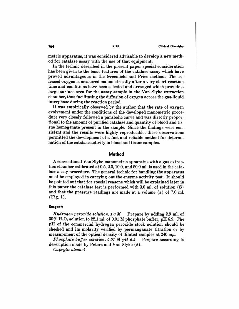
A conventional Van Slyke manometric apparatus with a gas extrac-tion chamber calibrated at 0.5, 2.0, 10.0, and 50.0 ml. is used in the cata-lase assay procedure. The general technic for handling the apparatusmust be employed in carrying out the enzyme activity test. It shouldbe pointed out that for special reasons which will be explained later inthis paper the catalase testisperformed with 3.0ml. of solution (S)
and that the pressure readings are made at a volume (a) of 7.0 ml.(Fig. 1).
Reagents
Hydrogen peroxide solution, 1.0 M Prepare by adding 2.9 ml. of30% H202 solution to 22.1 ml. of 0.01 M phosphate buffer, pH 6.9. ThepH of the commercial hydrogen peroxide stock solution should bechecked and its molarity verified by permanganate titration or bymeasurement of the optical density of diluted samples at 240 m.
Phosphate buffer solution, 0.01 M pH 6.9 Prepare according todescription made by Peters and Van Slyke (8).
Caprylic alcohol

7cc Volume
Vol. 9, No. 6, 1963 CATALASE DETERMINATION 765
Fig. 1. Extraction chamber
of Van Slyke manometric ap-
paratus for use in gasometric
catalase method. Gas is shown
at 7.0-mI. volume for pressurereading.
Surfoc.e Levelof Sompte
Level of -
Mercury Meniscu
Procedure
Transfer of Sample to Extraction Chamber
At the beginning of the test when the wash water is drawn off, thechamber fills with mercury. The chamber stop cock is closed, and thecock to the leveling bulb is left open. With a pipet, 0.9 ml. of phosphatebuffer is measured into the cup of the extraction chamber.
The sample to be analyzed (hemolysate of 1:100 aqueous dilution, ortissue homogenate of suitable concentration) is drawn into a 100-l.Linderstr#{216}m-Lang constriction pipet and is added to the phosphatebuffer in the cup. During the addition, the pipet is held in a verticalposition with its point placed under the surface of the buffer. The
transfer of the sample from the pipet is made slowly, the last part be-ing ejected with the use of a rubber bulb. To insure quantitative trans-fer, the Linderstr#{216}m-Lang pipet is rinsed by drawing phosphatebuffer from the cup to the 100-1.J. constriction mark with subsequentdelivery of the pipet content to the cup sample. The 1.0-ml. volume inthe cup is then introduced into the chamber without admitting any air.
Another 0.9 ml. of phosphate buffer is measured into the cup and
similarly run into the chamber. This second portion of phosphate

768 KIRK Clinical Chemistry
Table 1. FACTORS FOR CALCULATION OF AMOUNT (sL.) OF OXYGEN EVOLVED
Temp.#{176} Factor
15 8.845
16 8.811
17 8.777
18 8.743
19 8.710
20 8.677
21 8.644
22 8.611
23 8.57924 8.54725 8.516
26 8.484
27 8.453
28 8.423
29 8.392
30 8.360
31 8.33132 8.302
33 8.27134 8.242
35 8.213
Factors were calculated for conditions of S = 3.0 ml. and a = 7.0 ml.
gen evolved per second (Equation 1), micromoles of oxygen evolvedper second (Equation 2), or micromoles of hydrogen peroxide decom-posed per second (Equation 3):
(1)
(2)22.4 Vt
k = PXIX2 (
22.4 Vt
where P = observed pressure of oxygen evolved, expressed in milli-meters mercury and f = factor listed in Table 1.
The enzymic velocity values calculated from the above equationsshould subsequently be corrected for the temperature at which the testwas conducted to convert activities to those exhibited by catalase at25#{176}.This correction is made by use of the factors recorded in Table 2.
As an example, a diluted human blood sample equivalent to 1 /Ll. ofwhole undilutel blood after 45 see. of shaking at a temperature of 29#{176}produced an 88.5mm. increase in pressure (cOrrectedfor blank value).

Vol. 9, No. 6, 1963 CATALASE DETERMINATION 765
Fig. 1. Extraction chamber
of Van Slyke manometric ap-paratus for use in gasometric
catalase method. Gas is shown C I
at 7.0-ml. volume for pressure OS 0icc Volume
reading.
Surface Levelof Sample
Level of
Mercury Mmiscus
Procedure
Transfer of Sample to Extraction Chamber
At the beginning of the test when the wash water is drawn off, thechamber fills with mercury. The chamber stop cock is closed, and thecock to the leveling bulb is left open. With a pipet, 0.9 ml. of phosphatebuffer is measured into the cup of the extraction chamber.
The sample to be analyzed (hemolysate of 1:100 aqueous dilution, ortissue homogenate of suitable concentration) is drawn into a 100-.J.Linderstr#{216}m-Lang constriction pipet and is added to the phosphatebuffer in the cup. During the addition, the pipet is held in a verticalposition with its point placed under the surface of the buffer. The
transfer of the sample from the pipet is made slowly, the last part be-ing ejected with the use of a rubber bulb. To insure quantitative trans-fer, the Linderstr#{216}m-Lang pipet is rinsed by drawing phosphatebuffer from the cup to the 100-pJ. constriction mark with subsequentdelivery of the pipet content to the cup sample. The 1.0-ml. volume inthe cup is then introduced into the chamber without admitting any air.
Another 0.9 ml. of phosphate buffer is measured into the cup andsimilarly run into the chamber. This second portion of phosphate
766 KIRK Clinical Chemistry
buffer serves to wash into the chamber residuals of the sample whichmay adhere to the bottom of the cup. With a medicine dropper, fourdrops of caprylic alcohol are then placed in the cup, and a sufficientpart of this reagent is admitted to the chamber to bring the mercurymeniscus to the 2.0-mi. mark. A few drops of mercury are then addedto the small amount of caprylic alcohol remaining in the cup, and withthe chamber stop cock closed, the sample is ready for receipt of thehydrogen peroxide reagent.
Addition of Hydrogen Peroxide Reagent
The addition of the hydrogen peroxide reagent to the sample in theextraction chamber is made with a 1.0-mi. Ostwald-Van Slyke pipetequipped with stop cock and a rubber tip. Between use, this pipet isconveniently kept standing in a tall, narrow beaker placed immediate-ly to the right of the Van Slyke apparatus.
The 1.OM 11202 reagent is drawn into the pipet which is then insertedinto the cup with the rubber tip pressed against the bottom. The pipetcock isopened, and ifheld in correctposition,there willbe no flowfromthe pipet. The admission of the 1.0 ml. of hydrogen peroxide solution
to the chamber is made by means of the chamber stop cock. Tmmediate-ly before the opening of that stop cock, a stop watch is started; if anelectrical stop watch operated by a foot switch is employed, the start-ing of the watch can be made at the same second the chamber stop cockis opened. When the 1.0 ml. of hydrogen peroxide reagent has been
transferred through the mercury seal into the extraction chamber, thechamber stop cock is closed, and the Ostwald-Van Slyke pipet is with-drawn and replaced in the beaker for storage. The bore of the chamberstop cock is then sealed with the mercury remaining in the cup. Be-tween 9 and 11 sec. are required for the delivery of the hydrogen perox-ide reagent to the chamber.
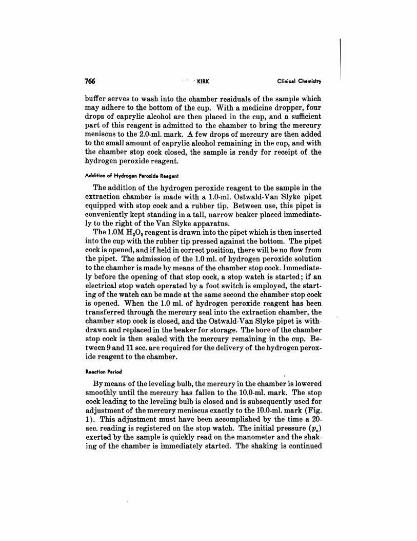
Reaction Period
By means of the leveling bulb, the mercury in the chamber is lowered
smoothly until the mercury has fallen to the 10.0-mi. mark. The stopcock leading to the leveling bulb is closed and is subsequently used for
adjustment of the mercury meniscus exactly to the 10.0-mi. mark (Fig.1). This adjustment must have been accomplished by the time a 20-sec. reading is registered on the stop watch. The initial pressure (p0)exerted by the sample is quickly read on the manometer and the shak-ing of the chamber is immediately started. The shaking is continued
Vol. 9, No. 6, 1963 CATALASE DETERMINATION 767
for 45 sec. at a rate of 250 rpm (the shaking is started when the stopwatch shows a 20-sec. reading and is stopped at 65 sec.).
During the period of shaking, the cock is occasionally opened with asmooth gradual motion to admit mercury from the leveling bulb tokeep the mercury meniscus close to the 10.0-ml. mark. The retainment
of the mercury meniscus in this narrow part of the extraction chamberassures a small area of contact between the hydrogen peroxide-con-taining sample and the mercury, and provides a large surface area be-tween the sample and the gas phase in the chamber.
When the 45-sec. shaking period has been reached (when the watchshows the total time of 65 sec.), the shaking of the chamber is stopped,and the mercury meniscus is exactly adjusted to the 10.0-mi. mark(Fig. 1). The pressure reading (p,) is immediately made on the ma-
nometer and the temperature is recorded.The stop cock leading to the leveling bulb is opened and the sample
is removed from the chamber by suction. The cup and the extractionchamber are then washed twice with 10-15 ml. of distilled water.
A complete catalase test including the measuring of the sample andreagents into the pipets and the washing of the chamber can be made in
less than 4 mm.
Blank Value
A blank value (c) isdetermined by performing a testwith 1.9ml. of
phosphate buffer, 4 drops of caprylic alcohol, and 1.0 ml. of 1.0 lvi H202reagent. The blank value recorded for the 45-sec. test is usually about
6 mm. The mean time required for a blank determination is 31/4 mm.
Calculationof CatalaseActivity
The pressure of oxygen evolved in the 45-sec. reaction period is P =
p1 - p0 - c, where c is the pressure exerted by the blank.The P value is converted to microliters of oxygen (at 0#{176}and 760 mm.
Hg) by multiplication with the appropriate factor listed in Table 1.
Since the catalase reaction rate as recorded in the present manometricprocedure strictly follows a parabolic curve, the initial velocity con-
stant (k) is calculated on the basis of the following formula:
Vt
where q = quantity of oxygen evolved and t = time in seconds.
The initial reaction values may be expressed as microliters of oxy-

768 KIRK Clinical Chemistry
Table 1. FACTORS FOR CALCULATION OF AMOUNT (ILL.) OF OXYor..i EVOLVED
Temp.#{176} Factor
15 8.845
16 8.811
17 8.777
18 8.743
19 8.710
20 8.677
21 8.644
22 8.611
23 8.57924 8.54725 8.516
26 8.484
27 8.453
28 8.423
29 8.39230 8.360
31 8.331
32 8.30233 8.271
34 8.242
35 8.213
Factors were calculated for conditions of S = 3.0 ml. and a = 7.0 ml.
gen evolved per second (Equation 1), micromoles of oxygen evolvedper second (Equation 2), or micromoles of hydrogen peroxide decom-posed per second (Equation 3):
(1)Vt
(2)22.4 Vt
k = PXfX2
22.4 Vt
where P = observed pressure of oxygen evolved, expressed in milli-meters mercury and f = factor listed in Table 1.
The enzymic velocity values calculated from the above equationsshould subsequently be corrected for the temperature at which the testwas conducted to convert activities to those exhibited by catalase at25#{176}.This correction is made by use of the factors recorded in Table 2.
As an example, a diluted human blood sample equivalent to 1 l. ofwhole undilutel blood after 45 sec. of shaking at a temperature of 29#{176}
produced an 88.5 mm. increase in pressure (corrected for blank value).

Vol. 9, No. 6, 1963 CATALASE DETERMINATION 769
Table 2. FACTORS FOR CONVERSION OF BLOOD CATALASE ACTIVITIES OBSERVED AT VARIOUS
TEMPEEATURES TO THOSE EXHIBITED AT 25#{176}
Temp.#{176} Factor
15 1.1374
16 1.1292
17 1.1200
18 1.1096
19 1.0980
20 1.083921 1.071122 1.054223 1.038924 1.019825 1.0000
26 0.9773
27 0.9558
28 0.9301
29 0.903530 0.8758
31 0.851332 0.8231
33 0.797134 0.7680
35 0.7404
According to Equation 1, this corresponds to:
88.5 X 8.392 742.7= = 110.7 pi. 02/sec. in initial reaction
v’45 6.708
The values, expressed as micromoles of oxygen evolved per second(Equation 2) and micromoles of hydrogen peroxide decomposed persecond (Equation 3) are, respectively, 4.94 and 9.88. A conversion ofthese figures to enzymic activity at 25#{176}(Table 2) yields values of 92.9l. of oxygen evolved per microliter undiluted blood per second; 4.15M of oxygen evolved per microliter undiluted blood per second; and8.30 p.MI of hydrogen peroxide decomposed per microliter undilutedblood per second.
If desired,the shaking reaction period may be changed from 45 sec.
(V45 = 6.708) to 30 sec. (v’3O 5.477) or 60 sec. (V60 = 7.746).
Cleaning the Chamber with Chromic-Sulfuric Acid Solution
When the manometric apparatus is used for catalase activity deter-
minations, it is occasionally necessary to clean the extraction chamber

770 KIRK Clinical Chemistry
with a chromic-sulfuric acid solution as described by Peters and VanSlyke (8) ; because of the importance of the pH of the reaction mixturein the catalase determination, the acidity of the final wash water shouldbe checked after this cleaning. After removal of the chromic-sulf uricacid solution, a satisfactory pH (between 6.3 and 7.0) was achieved
when the chamber was washed successively with three portions of wa-ter, one portion of 0.5 N NaOH, and three additional portions of water,
the volume of each washing solution being between 25 and 50 ml.
Experimental
Catalase Reaction Rate
For determination of the correlation between time of reaction andquantity of oxygen evolved,experiments were conducted with samplesof human hemolysates and liver homogenates, and with commercial
catalase preparations. The oxygen evolution was recorded after vari-
ous times of shaking of the chamber; the reaction periods chosen were
15,30, 45, 60, 90, and 120 sec. of shaking. Since it is not possible to makeaccurate readings on the manometer during the shaking of the extrac-tion chamber, the tests were made successively on equal aliquots of thesame catalase-containing samples. Duplicate analyses were per-formed in all the studies and usually showed very close agreement.
The constructed graphs regularly displayed a shape indicating a
parabolic curve. In order to prove the parabolic reaction rate, thequantities of oxygen evolved were plotted against /t. In all the ex-periments this plotting resulted in a straight line passing through theorigin (the origin representing the time at which the shaking was
started). These findings verify the parabolic nature of the catalasereaction rate under the present conditions of assay (3). At the varioustimes of reaction, a close correlation was found between the amount of
enzyme present and the recorded activity values; as an example, thevalues observed for a blood sample are presented in Fig. 2. Similarproportionality was encountered in assays of freshly prepared solu-tions of purified catalase samples (Fig. 3).
Relationship between Shaking Rate of Chamber and Catalase Reaction
A shaking rate of 250 rpm of the driving wheel was chosen for the
manometric catalase procedure because in the determinations of bloodand tissue samples, nearly identical activities were observed with 240,250, and 260 shakes per minute. Slightly lower values were recorded at210 rpm, and a markedly reduced rate of oxygen evolution was found

P (Pienure)
in mm.Hg. 200 Cu. mm.
Vol. 9, No. 6, 1963 CATALASE DETERMINATION 771
without shaking of the chamber (15% of that observed with 250 shakes
per minute).At the selected 250 rpm, approximately similar catalase activity
values were noted for the 45-sec. shaking period when the shaking of
Fig. 2. Correlation betweentime of reaction (shaking pe-riod), volume of blood used, andquantity of oxygen evolved. The
manometric readings are ex-
pressed in mm. Hg corrected for
blank values. A 1:100 dilutedhuman blood sample was used in
test.
Fig. 3. Catalase activities ex
hibited by purified preparation
recorded at Various enzyme con-centrations under standard con-
ditions of manometric method
(100 ul. of solution employed Is
equal to 5.7 tug. of eatalase). Re-action time (shaking period), 45
see.
P1 Pressure)in mm. Hg.
100
0 00 200 300 400 500Cu. mm. Cotolose Solution
the chamber was started either 20 or 25 sec. after the initiation of thehydrogen peroxide addition to the sample. The choice of a 45-sec. shak-ing period, from 25 to 70 sec. instead of the described period startingat 20 sec. and discontinued at 65 sec., may be preferable to analysts whoare not routinely trained in the handling of the Van Slyke apparatus.
Influence of Hydrogen Peroxide Concentration on Catalase Reaction Rate
The catalase activities of blood and tissue samples were found to benearly independent of the 11202 concentration within the range of 0.625

772 KIRK Clinical Chemistry
to 1.25 M (final concentrations 0.208 to 0.417 lvi). Somewhat lowercatalase values were noted with the use of 0.3125 and 2.50 lvi 11202 re-
agent solutions.Under the technical conditions described in the present paper most
of the hydrogen peroxide substrate remains undecomposed during thetest even at the highest catalase reaction rates measurable by themanometric procedure. The evolution of oxygen exerting a 400-mm.Hg pressure is equivalent to a decomposition of 304 /.LM of hydrogenperoxide which represents only 30.4% of the 1000’M of 11202 (0.1 ml.
of 1.0 M 11200) added to the catalase-containing sample in the extrac-tion chamber.
Effect of Temperature on Catalase Activity Rate
A thorough investigation was made of the effect of temperature onhuman blood catalase activity as determined by the present mano-metric method. Five human blood samples were included in the study.The catalase activities of these samples were measured over the rangeof 15-35#{176}.The control of the reaction temperature was accomplishedthrough continuous circulation of water with a special constant tem-perature through the chamber jacket. Large needles were inserted
through the upper and lower rubber stoppers of the jacket, and rubbertubing was attached to the heads of the needles. The circulating waterwas admitted to the jacket through the lower needle.
When a change in the temperature was made, the extraction cham-ber was left filled with mercury for 20 mm. to assure a more uniformtemperature level in the apparatus. After each test, the sample tem-perature was immediately measured.
The phosphate buffer solution employed in the tests was maintainedin a water bath at the same temperature; hemolysate samples werekept in a refrigerater at 4#{176}between assays. The experiments werestarted at 35#{176},and the temperature was then gradually lowered to 15#{176}.In order to verify that no measurable degradation of the blood cata-lase had occurred during the long experimental period, the tempera-ture was finally raised to the original level of 35#{176}.Essentially similarvalues were recorded in the initial and final experiments conducted atthe same temperature.
The effect of the temperature on the blood catalase activity of thefive samples studied was found to be quite similar. Since the tempera-ture curve is not linear, the observed graph is presented in Fig. 4 andthe mean Q,and Q10values are listed in Table 3.

15 7 9 21 23 25 27 29Temperature(‘C)
Vol. 9, No. 6, 1963 CATALASE DETERMINATION 773
% of Activityat 25C115
Ito
Fig. 4. Percentage values ofoxygen evolved (expressed in ml.at 0#{176},760 mm. Hg) by catalase 00
in human blood samples at vari-
ous temperatures, as compared 95
with activities recorded at 25#{176}.90
85
Table 3. Qo AND Qio VALUES FOR BLOOD CATALASE REACTION AS DETERMINED BY MANOMETRIC
PROCEDURE
Temp. rang #{128}0 SE. (.10 SE.
15-20 1.048 0.010
20-25 1.086 0.01025-30 1.143 0.01230-35 1.192 0.01115-25 1.139 0.025
20-30 1.240 0.021
25-35 . 1.345 0.022
Velocity Values Observed for Commercial Catalase Preparations
On the basis of the initial velocity rate determined with the mano-metric method, the turnover number for the enzyme in a purified cata-lase powder* prepared from beef liver was found to be 5,120,000. Thequantity of enzyme present in the catalase solution used in the test was
confirmed through measurement of the optical density of tile stock so-lution at 280 m in a spectrophotometert and by assay according to theprocedure of Beers and Sizer (1); the observed enzyme content of thebeef liver catalase powder agreed closely with that reported by theSigma Chemical Co. (Lot C41B-085). An essentially similar turnover
number of 5,180,000 was found for a catalase preparation obtainedfrom Worthington Biochemical Corp., Freehold, N. J. (Lot 5553-78).The high turnover number for purified catalase recorded with thepresent manometric technic indicates the validity of that procedure.
*Sigma Chemical Company, St. Louis, Mo.
I Model DU, Beckman Instruments Inc., Palo Alto, Calif.

774 KIRK Clinical Chemistry
Catalase Activities of Human Blood Specimens and Liver Tissue Homogenates
Catalase activity measurements were performed on 100 humanblood samples obtained from patients at St. Louis Chronic Hospital(average age of subjects, 75 years). Hematocrit and hemoglobin de-terminations were made by conventional methods. Hemolysates of thespecimens were prepared by adding 100 l. of each blood sample to 9.9in!. of redistilled water. The average initial velocity rate calculatedfrom the manometric catalase assays was 115 l. of oxygen evolved permicroliter undiluted blood per second (S.D. of dist., 35); this activity
rate is somewhat higher than those previously reported with gaso-metric technical procedures (2). The average difference between du-plicate measurements of the samples was 2.6%. When expressed permicroliters of red blood cells, the mean oxygen evolution was 286J./sec. (S.D. of dist., 89) ; the corresponding value per milligrams ofhemoglobin was 842l. (S.D. of dist., 265).
For determination of the catalase activity of liver tissue, 10 speci-mens were obtained fresh at autopsy, and 1.0% aqueous homogenateswere prepared with a Kontes Duall type tissue grinder. The mean
catalatic value observed for human liver tissue was 93 jJ. of oxygenevolved per milligram tissue per second. This activity rate is of thesame order of magnitude as that reported by Greenstein et al. (3) for
normal mouse liver.
Discussion
The factors responsible for the oxygen evolution at a rate corre-sponding to a parabolic curve are not known, but the experimentallyestablished consistency of this type of catalase reaction under the em-ployed technical conditions permits an accurate calculation of the ini-
tial activity velocity. Attempts will not be made to correlate this em-pirical finding with the available general information concerning theaspects of catalase kinetics.
It is of interest to note that a calculation of gasometric catalase datareported by Morgulis (7) and Maisin and Pourbaix (6) indicates a
tendency toward a parabolic curve; this was noted by plotting the pub-lished values of oxygen evolvement against the square root of reactiontimes. However, the graphs constructed on the basis of these previ-ously reported catalase activity measurements do not pass directly
through the zero point, possibly due to the fact that the technics usedby those authors differ in many respects from the procedure describedin thispaper.
Vol. 9, No. 6, 1963 CATALASE DETERMINATION 775
In the present method, the use of a 3.0 ml. sample volume and themaintenance of the mercury meniscus at the 10.0-ml. mark were chosenwith the purpose of establishing a large liquid-gas interphase area
(Fig. 1). The surface of the sample in the Van Slyke extraction cham-ber is approximately 1.3 sq. cm./ml. of sample volume; this is an evenhigher ratio than that of 0.5 sq.cm./ml. of solutioncalculated on the
basis of the size and shape of the reaction flask used in the method byGreenfield and Price (4). The conditions are very satisfactory for
swirling the sample over the inside of the glass surface in both these
rapid gasometric technics.
References1. Beers, R. F., and Sizer, I. W., J. Biol. Chens. 195, 133 (1952).2. Deutsch, L., and Frank!, J., Ztschr. f. Krebsforsch. 40, 98 (1934).3. Feldman, W. M., Biom4jtheonatics, Griffin, London, 1923, p. 299.4. Greenfleld, H. E., and Price, V. E., J. Biol. Chein.. 209, 355 (1954).5. Greenstein, J. P., Jenrette, W. V., Mider, G. B., and Andervont, H. B., J. Nat. Cancer Inst.
2, 293 (1941-42).6. Maisin, J., and Pourbaix, Y., Compt. Rend. Soc. Biot. 129, 46 (1938).7. Morgu)is, S.,J. Biot. Chem. 47, 341 (1921).8. Peters, J. P., and Van Slyke, D. D., Quantitative CUnical Ohemistry, vol. 2, Williams and
Wilkins, Baltimore, 1932, pp. 236, 816.