a comparative expressional analysis of a family of...
TRANSCRIPT

1
A COMPARATIVE EXPRESSIONAL ANALYSIS OF A FAMILY OF CCA-LIKE MYB TRANSCRIPTION FACTORS IN TWO HIGHER PLANT SPECIES
by
MEREDITH L. SULLIVAN
A THESIS PRESENTED TO THE GRADUATE SCHOOL OF THE UNIVERSITY OF FLORIDA IN PARTIAL FULFILLMENT OF THE REQUIREMENTS FOR THE DEGREE OF
MASTER OF SCIENCE
UNIVERSITY OF FLORIDA
2007

2
© 2007 Meredith L. Sullivan

3
To my parents with love and respect

4
ACKNOWLEDGMENTS
Most of all, I thank my family for their endless love and support. They are the pillars of my
strength. I also thank my advisor, Dr. David Oppenheimer for affording me the opportunity to
attend the University of Florida. I also express gratitude to Dr. Bernard Hauser for his support of
this work. A special thanks to Zhengui Zheng for his time and assistance with this project. Also I
thank Xiaoguo Zhang and Stacey Jeffries for the invaluable advice they offered and the support
they provided me throughout my time at UF. Finally I extend special appreciation to the love of
my life, Brad for his continued encouragement.

5
TABLE OF CONTENTS
page
ACKNOWLEDGMENTS ...............................................................................................................4
LIST OF FIGURES .........................................................................................................................6
ABSTRACT.....................................................................................................................................7
CHAPTER
1 LITERATURE REVIEW .........................................................................................................9
Biological Rhythms ..................................................................................................................9 The Circadian Clock ...............................................................................................................10 Clock- Controlled Genes ........................................................................................................13 The Arabidopsis Central Oscillator ........................................................................................18 Evolution of Core Clock Components....................................................................................21
2 TWO CCA-LIKE MYB TRANSCRIPTION FACTORS ARE PRESENT IN CALIFORNIA POPPY...........................................................................................................27
Summary.................................................................................................................................27 Introduction.............................................................................................................................28 Materials and Methods ...........................................................................................................28
Plant Growth Conditions .................................................................................................28 In situ Hybridizations ......................................................................................................30
Results.....................................................................................................................................32 RISE and SHINE Encode CCA1-like Myb Transcription Factors .................................32 RISE and SHINE Expression is Under Circadian Control..............................................32
Discussion...............................................................................................................................34 Feedback Loop Mechanism as the Basis of the Circadian Oscillator .............................34 The RISE and SHINE Genes Encode MYB Transcription Factors That Are Similar
to LHY and CCA1 .......................................................................................................34 The Biological Importance of Circadian Clock Genes....................................................38
3 CONCLUSION.......................................................................................................................49
LIST OF REFERENCES...............................................................................................................50
BIOGRAPHICAL SKETCH .........................................................................................................57

6
LIST OF FIGURES
Figure page 1-1 The three basic components of the circadian clock. ..........................................................25
1-2 Positive and negative factors act upon the circadian clock................................................25
1-3 Comparisons of three conserved regions of LHY and CCA1............................................26
1-4 The Arabidopsis circadian oscillator. ................................................................................26
2-1 Alignment of the LHY/CCA1 genes in Arabidopsis thaliana and Eschscholzia californica. .........................................................................................................................41
2-2 A phylogenetic analysis of CCA-like family of genes. .....................................................42
2-3 Alignment of the RISE (eca_4_183384) and SHINE (eca_4_184056) EST sequences using the BLASTN program from the FGP database (Albert et al, 2005; Carlson et al., 2006). ...........................................................................................................................43
2-4 Analysis of RISE and SHINE mRNA expression. .............................................................44
2-5 Analysis of LHY and CCA1 mRNA expression.................................................................45
2-6 Expression of LHY and CCA1 in Arabidopsis tissue. ........................................................46
2-7 RISE and SHINE transcripts are expressed in both young and mature floral tissue of the California poppy plant. ................................................................................................47
2-8 Proposed mechanism of the central oscillator of Eschscholzia californica.......................48

7
Abstract of Thesis Presented to the Graduate School of the University of Florida in Partial Fulfillment of the
Requirements for the Degree of Master of Science
A COMPARATIVE EXPRESSIONAL ANALYSIS OF A FAMILY OF CCA-LIKE MYB TRANSCRIPTION FACTORS IN TWO HIGHER PLANT SPECIES
By
Meredith L. Sullivan
August 2007
Chair: David Oppenheimer Major: Botany
Circadian clocks are ubiquitous among most living species. Since life on Earth originated
in the presence of light/dark cycles, organisms had to evolve mechanisms to cope with such
environmental fluctuations. An accurate timekeeping apparatus affords an organism with
temporal organization of crucial molecular and cellular processes. The components that
constitute the central oscillator of the clock vary greatly among plants and animals but the basic
architecture appears similar. This resemblance serves as a foundation on which evolutionary-
based investigations on the conservation of such machinery can be conducted. In the plant
Arabidopsis thaliana, two genes involved in circadian regulation were identified and
characterized as members of a family of Myb transcription factors characterized by only one
Myb repeat sequence. These genes, CCA1 and LHY, are necessary to maintain rhythmicity in the
plant and have been shown to have a role in floral induction. Using the Floral Genome Project
(FGP) database of known flowering genes, two EST homologs of CCA1 and LHY were
identified in Eschscholzia californica, eca_4_183384 and eca_4_184056 (E1 and E2), based on
sequence similarity. These orthologs demonstrate transcript oscillations over a 24hr period with
peak levels of expression occurring just prior to dawn. In situ analyses revealed similar patterns
of expression in young and older floral tissue of both plant species. In this paper I present

8
molecular evidence that the transcription- translation- based feedback loop mechanism of the
circadian oscillator is conserved in these higher plant species and suggest that this mechanism
can also be observe din other higher plants.

9
CHAPTER 1 LITERATURE REVIEW
Biological Rhythms
The daily rotation of the earth leads to periodic fluctuations in environmental conditions.
Because life on Earth originated in the presence of a cyclical environment, many organisms have
evolved timing mechanisms to organize important events. In order to adapt to the changes
presented by the environment, organisms must modulate their behaviors with the daily cycles of
light and temperature variation, and establish an effective method for tracking time. Since the
environment imposes a period of approximately 24 hours, organisms with rhythmic behavior that
matches these oscillations have higher fitness than those that do not.
The first noted rhythmic behavior occurred in the fourth century BC when the sleep
movements of the tamarind tree were noted by Androsthenes, a Greek philosopher
(Chandrashekaran, 1998). In the mid-1700s, a French astronomer named Jean- Jacques d’Ortous
de Mairan recorded the daily leaf movements of the Mimosa pudica plant and demonstrated that
the rhythms persisted for several days when the plant was subjected to complete darkness
(Golden and Strayer, 2001; Sweeney, 1987). This was the first evidence that the rhythmic
behavior must be endogenous and that the rhythmicity, once established, continues in the
absence of environmental cues. Erwin Bunning’s work during the 1930s, in which he
demonstrated a rhythm in the eclosion behavior of the fruit fly Drosophila, further supported the
idea of an endogenous timekeeping mechanism. Since the rhythms occurred with a 24 hour
period, the term circadian was coined from the Latin words circa and dias, meaning “about a
day” (Chandrashekaran, 1998; Halberg, 1959).
Circadian rhythms are variations in physiological and behavioral activities that occur over
a period of about 24 hours (Hardin, 2000). In the context of a biological process, the time

10
interval between two successive events is described as a biological ‘rhythm’ (Kumar, 2002).
Biological rhythms are found in almost all living organisms. They have been described
extensively in mammals, insects, fungi, plants and bacteria (Dunlap et al., 1999; McClung, 2001;
Ouyang et al., 1998). These rhythms occur over a large range of time scales: from millisecond
oscillations to seasonal changes. The ubiquity of circadian rhythmicity across a broad taxonomic
spectrum suggests that adaptive fitness is enhanced by the synchronization of certain events with
the diurnal cycle imposed by the environment (McClung 2000).
In a number of different species, a plethora of activities are regulated by circadian rhythms.
Cyanobacteria demonstrate daily oscillations in nitrogenase activity, photosynthesis and
metabolic activities (Kondo and Ishiura, 1999). In vertebrates, a number of behavioral and life
processes, ranging from the molecular level to the cellular and systemic levels, are driven by
circadian rhythms including eating, sleeping, seasonal migration and cell proliferation (Gillette
and Sejnowski, 2005). Similarly, a variety of events such as leaf movement, stomatal opening
and regulation of flowering time and fragrance emission are tightly regulated by the plant
timekeeper. In other organisms, such as Neurospora and Drosophila, development, cell
signaling and stress responses with self-sustaining rhythms can be regulated by the circadian
clock. The role that diurnal rhythms play in a number of different activities suggest that an
endogenous timekeeping system provides an adaptive advantage, enabling the anticipation of
environmental change and the coordination of crucial events to occur at specific phase
relationships with the environment (Más, 2005).
The Circadian Clock
Circadian or biological rhythms, although they parallel the environmental cycles of light
and dark, are generated within an organism by a complex timekeeping system (Hardin, 2000).
Many organisms have evolved an endogenous ‘chronometer’, the circadian clock, to temporally

11
coordinate important processes with the daily variations in the environment. Circadian rhythm of
gene expression has been shown to function as the underlying mechanism of the clock (Wang
and Tobin, 1998).
Two important attributes of the circadian clock, entrainment and temperature
compensation, ensure synchrony between important rhythmic activities and the surroundings of
an organism. The first, entrainment, is the manner in which the clocks are set to ‘local’ time by
environmental cues such as the light / dark cycles. The second, temperature compensation,
describes the ability of the clock to run at the same rate independent of temperature changes
(Hardin 2000).
The notion that biological rhythms are created within an organism stimulated interest in the
mechanism that maintains these oscillatory patterns. In theory, numerous regulatory schemes
could achieve such fluctuations. The most common method involves a regulatory circuit with a
positive and negative product (Fig. 1-2). The negative element feeds back to slow down the rate
of the process itself and creates a delay in the execution of the feedback. A positive element is
required to activate the clock and prevent it from winding down (Dunlap et al., 1999). The
generally conserved clock mechanism consists of an autoregulatory feedback loop in which
positive factors act on genes encoding negative factors that in turn feedback to inhibit their own
expression (Strayer et al., 2000; Dunlap, 1999). A circadian system often consists of one or more
interconnected feedback loops.
The circadian system is divided into three main components: input pathways, a central
oscillator and output pathways (Fig. 1-1). Input pathways, also known as entrainment pathways,
transmit environmental signals to the central timekeeping apparatus. In plants, this signal
transduction pathway adjusts the clock in response to external cues most frequently through the

12
action of cryptochromes and phytochromes. The entrainment of circadian clocks to the light/dark
cycles is usually mediated by light-induced changes in the level of a component of the oscillatory
feedback loop. The ability to re-entrain the clock ensures synchrony with the environment and
allows the anticipation of dawn and dusk (Devlin, 2002).
The core timekeeping component of the circadian clock is the central oscillator (Hardin,
2000). It serves as the system’s pacemaker and is responsible for generating circadian rhythms.
In order for the oscillator to function in the absence of environmental cues it must be
synchronized with the external time via the input pathways. Environmental transitions between
dawn and dusk help to adjust the endogenous period created by the oscillator to precisely match
the 24- hour period found in nature (Más, 2005).
Completing the circadian clock model are the output pathways which provide a link
between the central oscillator and the rhythmic physiological responses they control. These
pathways are activated at specific times of the circadian cycle and the outputs are in phase with
the oscillation of light cycles (Dodd et al., 2005). In both animals and plants, there are a
substantial number of physiological and metabolic processes that are regulated by the circadian
outputs. These events include such varied responses such as olfactory responses in Drosophila,
leaf movements and hypocotyl growth in plants, and enzyme activity. The rhythmic behaviors
that are generated in response to day length are known as photoperiodic responses (Schultz and
Kay, 2003).
The discovery of components of the circadian oscillator has enabled scientists to
concentrate on the mechanisms that interconnect the three components to form an effective
timekeeping system. Recent studies of several model organisms including Drosophila,
Neurospora and mice have revealed a common molecular mechanism at the center of the

13
circadian oscillator (Barak et al., 2000). Clock proteins, encoded by clock genes, serve as
negative elements that repress their own expression by blocking their transcriptional activators,
or positive elements (Fig. 1-2). A decrease in clock transcripts and proteins results in the de-
repression of the transcriptional activators and thus reinitiates the cycle.
Clock- Controlled Genes
The identification of genes which function at the center of the clock machinery has rapidly
increased over the last decades. These clock-controlled genes, or ccgs, encode pieces of the
central oscillator. Their products produce and maintain the oscillations that drive other circadian
rhythms. A substantial number of clock-associated genes whose expression relies on the rhythms
generated by the oscillator have been identified in the genomes of many organisms. Recent
microarray analyses of the model plant Arabidopsis indicated that up to 6% of the genes are
rhythmically expressed (Harmer et al., 2000; Schaffer et al., 2001).
Components of the biochemical feedback loop whose rhythmic activity is required for
oscillator function are referred to as state variables. A number of criteria have been defined to
identify and characterize state variables of the circadian system (Aronson et al., 1994; Kay and
Millar, 1995). First, the component itself must demonstrate circadian oscillations in its activity
and expression. The second criterion requires the component to control its own levels by
feedback inhibition of its synthesis. Additionally, clamping of the amount of the putative
oscillator component at any level from null to high stops the clock and thus rhythmicity. The
final criterion states that induced transient perturbations in the abundance of the component
should cause a phase shift in the clock output. These criteria are applied in the investigations of
putative oscillatory components to assign positive and negative roles to the elements. Knowledge
of interconnected loops is obtained from the identification through classical genetics of genes
that are within one of the core loops of the oscillator (Roenneberg and Merrow, 1998).

14
The molecular basis of circadian clocks is best understood in Neurospora and Drosophila.
The genetics of rhythms originated in these two organisms and much of what scientists have
learned about how mammalian clocks operate closely parallels the behavior of one or both of
these species. In each of these model systems, genetic screens for rhythm mutants that affect the
period length of the clock or abolish its activities were found (Hardin and Siwicki, 1995; Dunlap,
1996).
The first clock mutations to be discovered were the period (per) mutant from Drosophila
and the frequency (.frq) mutant from Neurospora (Dunlap, 1993; Konopka and Benzer, 1971;
Feldman and Hoyle, 1973). The PER gene product, as well as its transcript, oscillates with a
circadian rhythm and the PER protein was required for a feedback regulation of its own gene
products (Williams and Sehgal, 2001). Oscillations of PER transcripts and the protein they
encode persisted in continuous dark conditions, suggesting that the gene must be under the
control of the circadian clock. The phases of the per messenger RNA (mRNA) and protein
rhythms were noticed to be quite distinct; PER protein level peaked in abundance approximately
six hours after the peak in PER mRNA levels. This difference accounts for the delay or lag time
in oscillations required by the central timekeeper. In addition, mutant per fruit flies exhibited
altered rhythmic behavior, which suggests a role for this gene in the oscillator. The tim mutant
was identified in a screen for recessive mutants that affected the eclosion behavior of the fly.
Similar to the per mutant, the tim mutants exhibited arrhythmic behaviors, periods that were
shortened or lengthened, and in some mutant alleles, PER expression was dampened (Allada et
al., 2001).
PER contains a protein interaction domain known as a PAS domain, which enables the
protein to interact with the second Drosophila circadian gene identified, timeless (tim). PER and

15
TIM form a heterodimer that serves as part of a feedback loop to inhibit per and tim
transcription. In return, the PER-TIM heterodimer activates a gene called Drosophila Clock
(dClk), a transcription factor that is also rhythmically expressed (Scully and Kay, 2000). dCLK
interacts with another transcription factor, CYCLE (CYC) and together the two form a
heterodimer complex that is required for the activation of PER and TIM transcription. The
dCLK-CYC complex binds to a specific sequence in PER and TIM promoters known as the E-
box motif, a consensus hexanucleotide sequence found in basic helix-loop-helix (bHLH)
transcriptional factors (Fairman et al., 1993). Binding of the complex to the E-box motif allows
transcription of PER and TIM and results in a increase in PER-TIM heterodimers in the
cytoplasm. When expression is high, usually in the evening, these complexes move into the
nucleus where PER binds to the dCLK-CYC complexes. This releases the dCLK and CYC
proteins from the promoters and results in the shutting off of PER and TIM transcription. As the
levels of PER and TIM decline, the dCLK-CYC dimers are released and thus the dCLK-CYC-
dependent repression of dClk expression is lifted. As the levels of dCLK begin to rise, usually in
the morning, an increased number of dCLK-CYC complexes are formed and per and tim
transcription is re-activated (Glossop et al., 1999). Since the products of the per gene and the
dClk gene inhibit their own synthesis it appears that the Drosophila oscillator consists of two
interconnected negative feedback loops: a per-tim loop and a dClk loop (Glossop et al., 1999). In
this model PER and TIM serve as the negative elements while dCLK and CYC serve as the
positive elements (Dunlap et al., 1999).
The second timekeeping component identified through forward genetic screens for clock
mutants was the FREQUENCY (FRQ) gene in the fungus Neurospora crassa. Mutations of this
gene resulted in arrhythmic expressions, altered periodicity and deficiencies in temperature

16
compensation (Aronson et al., 1994). The FRQ RNA and protein levels cycle with a circadian
rhythm and the protein negatively regulates its own transcript resulting in a feedback loop similar
to observed in Drosophila (Dunlap, 1996). The expression of FRQ is activated by two PAS-
domain-containing transcription factors, WHITE COLLAR 1 (WC-1) and WHITE COLLAR-2
(WC-2) that form the white collar complex (WCC), which bind to circadian photoreceptor
connecting light signals and the oscillator (He et al., 2002). After the accumulation of FRQ, the
proteins begin to dimerize, enter the nucleus and interact with the WCC diminishing its activity
and dampening FRQ expression (Froelich et al., 2003). FRQ also promotes the synthesis of WC-
1 increasing the level of WCC. This results in a mass of WCC that is held inactive by FRQ until
the protein is phosphorylated and targeted for ubiquitination, accounting for the delay that is
required by the circadian oscillator. Therefore Neurospora has a transcriptional/translational
negative feedback loop at the core of its oscillator with WC-1 and WC-2 acting as the positive
elements and FRQ as the negative (Dunlap et al., 1999). Recent evidence suggests that this FRQ-
based oscillator might work in cooperation with other oscillators within the organism as well
(Correa et al., 2003).
Over recent decades, numerous advances in understanding the mechanisms underlying the
biological oscillator in mammals have been made. The first cloned mammalian clock component
identified by forward genetics was the CLOCK (CLK) gene of Mus musculus, the mouse (Antoch
et al., 1997). Like transcription factor proteins that are central to the clock in other organisms, the
mouse Clock gene contains 1) a PAS domain, 2) its levels of mRNA and proteins oscillate, and
3) in clk mutant mice, the cyclic expression the mPeriod (mPer) homolog is reduced. These data
suggest that CLOCK, as a member of the oscillator, controls the transcription of circadian genes
(Gekakis et al., 1998). It was shown that CLOCK binds to another transcription factor, BMAL1

17
(Brain and Muscle Aryl Hydrocarbon Receptor Nuclear Translocator (ARNT)-Like1) and this
complex activates the transcription of the PER and CRYPTOCHROME (CRY) genes. The mPER
and CRY proteins form heterodimers and homodimers and upon translocation to the nucleus,
they inhibit the activity of the CLOCK-BMAL1 complex, which in turn suppresses PER and
CRY transcription (Panda et al., 2002). Once the mPER and CRY proteins are phosphorylated,
they are targeted for degradation and transcriptional repression is relieved. Although other
oscillators might be present in the mouse, central to its core oscillator is the negative feedback
loop in which the negative elements, the PER homologs repress the activation of the positive
elements CLOCK and BMAL1 (Dunlap, 1999).
Circadian rhythms, once only thought to be a feature of eukaryotic organisms, have
recently been identified in some prokaryotes. The cyanobacteria Synechococcus elongatus serves
as the model system for molecular investigations of this group. Approximately one hundred
clock mutants identified from an ethylmethansulfonate (EMS) mutagenesis screen that were
characterized by arrhythmia, atypical periods and some mutants that could be rescued by the
introduction of wild-type DNA from a Synechococcus genomic library (Lorne et al., 2000). One
cluster of DNA fragments that could be rescued represented the kai cluster of genes: kaiA, kaiB
and kaiC. Transcribed from two different promoters, PkaiA and PkaiBC, a monocistronic kaiA
mRNA and a dicistronic kaiBC mRNA are produced and both transcripts cycle in abundance.
Overexpression studies revealed that KaiC represses the activation of the PkaiBC promoter while
KaiA enhances PKaiBC transcription (Ishiura et al., 1998). Since KaiC represses its own
transcription it functions as the negative element of the negative feedback loop at the core of the
Synechococcus elongatus oscillator while KaiA, which helps drive expression from PkaiBC,
functions as the positive element. A role for KaiB has yet to be determined.
18
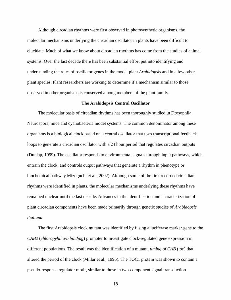
Although circadian rhythms were first observed in photosynthetic organisms, the
molecular mechanisms underlying the circadian oscillator in plants have been difficult to
elucidate. Much of what we know about circadian rhythms has come from the studies of animal
systems. Over the last decade there has been substantial effort put into identifying and
understanding the roles of oscillator genes in the model plant Arabidopsis and in a few other
plant species. Plant researchers are working to determine if a mechanism similar to those
observed in other organisms is conserved among members of the plant family.
The Arabidopsis Central Oscillator
The molecular basis of circadian rhythms has been thoroughly studied in Drosophila,
Neurospora, mice and cyanobacteria model systems. The common denominator among these
organisms is a biological clock based on a central oscillator that uses transcriptional feedback
loops to generate a circadian oscillator with a 24 hour period that regulates circadian outputs
(Dunlap, 1999). The oscillator responds to environmental signals through input pathways, which
entrain the clock, and controls output pathways that generate a rhythm in phenotype or
biochemical pathway Mizoguchi et al., 2002). Although some of the first recorded circadian
rhythms were identified in plants, the molecular mechanisms underlying these rhythms have
remained unclear until the last decade. Advances in the identification and characterization of
plant circadian components have been made primarily through genetic studies of Arabidopsis
thaliana.
The first Arabidopsis clock mutant was identified by fusing a luciferase marker gene to the
CAB2 (chloropyhll a/b binding) promoter to investigate clock-regulated gene expression in
different populations. The result was the identification of a mutant, timing of CAB (toc) that
altered the period of the clock (Millar et al., 1995). The TOC1 protein was shown to contain a
pseudo-response regulator motif, similar to those in two-component signal transduction

19
pathways, at its amino terminus and a CONSTANS (CO)-like motif at its carboxyl terminus
(Strayer et al., 2000). Interestingly the CO family represents a group of plant transcription factors
that are involved in flowering response (Putterill et al., 1995). Since the TOC1 protein shows
similarity to the CO family, TOC1 might play a role in flowering as well. The levels of TOC1
mRNA cycled in light-dark conditions and peak levels of transcript were observed late in the day
while minimal levels were observed at dawn. In addition, since the toc1-1 mutant was
characterized by a circadian oscillator with a shortened period which demonstrated that TOC1
products reduce their own expression (Strayer et al., 2000). Based on the observed data, TOC1
appears to be a component of the central circadian oscillator of Arabidopsis.
A second potential Arabidopsis clock component was identified as a result of the
identification of a day length-insensitive flowering mutant. The mutant late elongated hypocotyl
(lhy) caused an elongated hypocotyl and reduced chlorophyll, as well as an altered flowering
phenotype (Schaffer, 1997). lhy mutants were also arrhythmic for leaf movements and for the
expression of several other clock-regulated genes. Rhythmic expression of LHY was observed
with levels of the transcript peaking at dawn.The sequence of the LHY protein was used to
screen the GenBank database using the TBLASTN program to identify any potential homologs
in the plant. The Arabidopsis DNA-binding protein CIRCADIAN CLOCK ASSOCIATED-1
(CCA1) was most closely related to LHY (Schaffer et al., 1998) CCA1 was first identified as a
factor that binds to the promoter of the Chlorophyll a/b-binding light-harvesting complex
(LHCB) gene in Arabidopsis and functions in the phytochrome signaling pathway to induce the
transcription of LHCB (Wang et al., 1997). Later studies revealed that cca1 mutants display a
shorter period of circadian rhythms. Overexpression of this protein disrupted rhythmicity in
several clock outputs including hypocotyl elongation, leaf movements and circadian gene

20
expression (Green and Tobin, 1999; Wang and Tobin, 1998). CCA1 transcripts also oscillate
with peak levels of expression early in the morning and in constant conditions the rhythms
persist suggesting this gene is under circadian control (Wang and Tobin, 1998). In lhy cca1
double mutants circadian rhythms were observed with an abnormal phase and oscillations of
transcripts were dampened. Early expression of LHY and CCA1 (morning genes) and some
evening genes were also observed in the double mutants suggesting that these two genes function
as components of a negative feedback loop (Schaffer et al., 1998).
LHY and CCA1 genes function redundantly and are required for the maintenance of
circadian rhythms in Arabidopsis (Alabadí et al., 2002). Both genes are closely related MYB-like
transcription factors but are unique in that they only possess a single MYB repeat sequence
whereas other myb transcription factors usually contain two to three of the motifs. LHY and
CCA1 are also related outside of the MYB domain sharing other regions that exhibit at least 80%
identity (Fig. 1-3). Overall, the two genes are 46% identical to one another (Schaffer et al.,
1998). This sequence analysis suggests that LHY and CCA1 encode related DNA-binding
proteins with a single MYB repeat that function as transcription factors. This notion is supported
by evidence that shows LHY and CCA1 bind specifically to a sequence known as the ‘evening
element’ (EE) in the promoter of many genes whose expression peaks nears dusk (Alabadí et al.,
2001; Harmer et al., 2000).
In Arabidopsis, the model for the plant clock components is based on the regulation of the
three plant genes described above, CCA1, LHY and TOC1. These components appear to operate
in a transcriptional/translational-based negative feedback loop similar to that observed in other
studied systems. Light activation of LHY and CCA1 expression results in transcript levels that
peak at dawn followed by a peak in proteins approximately two hours later. Both of the proteins
21
bind to the EE motif located in the promoter of the TOC1 gene, a positive element, and repress
TOC1 expression during the day (Alabadí et al., 2001). A drop in TOC1 protein results in the
reduction of LHY and CCA1 transcript and protein levels. The low levels of expression of the
two genes results in the derepression of TOC1 transcription. In return, levels of TOC1 protein
peak during the late evening resulting in the activation of LHY and CCA1 transcription just prior
to dawn (Carré and Kim, 2002). This cross-regulation between LHY, CCA1 and TOC1 is
proposed to function as the central oscillator of the Arabidopsis clockwork where LHY and
CCA1 function as the negative elements and TOC1 serves as the positive element (Fig. 1-4).
This central clockwork regulates numerous genes in Arabidopsis responsible for photosynthesis,
nitrogen assimilation, biosynthesis of photo-protective pigments, lipid modification, hypocotyl
elongation and flowering (Harmer et al., 2000; Schaffer et al., 2001; Más, 2005).
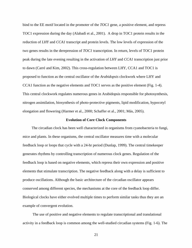
Evolution of Core Clock Components
The circadian clock has been well characterized in organisms from cyanobacteria to fungi,
mice and plants. In these organisms, the central oscillator measures time with a molecular
feedback loop or loops that cycle with a 24-hr period (Dunlap, 1999). The central timekeeper
generates rhythms by controlling transcription of numerous clock genes. Regulation of the
feedback loop is based on negative elements, which repress their own expression and positive
elements that stimulate transcription. The negative feedback along with a delay is sufficient to
produce oscillations. Although the basic architecture of the circadian oscillator appears
conserved among different species, the mechanisms at the core of the feedback loop differ.
Biological clocks have either evolved multiple times to perform similar tasks thus they are an
example of convergent evolution.
The use of positive and negative elements to regulate transcriptional and translational
activity in a feedback loop is common among the well-studied circadian systems (Fig. 1-6). The

22
positive elements serve as the transcriptional activators in the loop and they have been found in
Synecohcoccus (kaiA), Neurospora (WC-1 and WC-2), Drosophila (CLK and CYCLE),
mammals (CLOCK and BMAL1) and Arabidopsis (TOC1). Similarly, negative elements also
compose a portion of the feedback mechanism by inhibiting the action of the positive elements
and these include kaiC, FRQ, PER and TIM, PER and CRY and CCA1 and LHY in
cyanobacteria, fungi, fruit flies, mice and plants, respectively (Dunlap et al., 1999). Yet despite
their similarities, the time at which these elements are expressed, late in the evening or early
morning, differs among the organisms.
Transcription factor proteins serve important roles in the circadian oscillatory system. The
type of transcriptional inducer varies among model systems. In mammals and Drosophila, the
activators are basic helix loop helix (bHLH) proteins which contain a specific region which binds
to DNA (Gekakis et al., 1998; Darlington et al., 1998). The Neurospora positive elements are
similar to the Drosophila complex but they contain an additional zinc finger binding domain thus
they are categorized as zinc finger factors (Loros and Dunlap, 2001). In the plant circadian
oscillator, the activation of transcription is induced by MYB-like transcription factors (Carré and
Kim, 2002). The oscillatory mechanism of cyanobacteria is still under investigation.
Another difference between the circadian machinery of different organisms is the number
and location of oscillators. Mammals have a master circadian pacemaker that is localized to the
suprachiasmatic nucleus (SCN) located within the hypothalamus of the brain. The SCN entrains
multiple clocks that are located in the periphery of the organism (Yamazaki et al., 2000). In
contrast, plants contain at least one oscillator in each cell and these oscillators function
autonomously and independently of any central pacemaker (Thain et al., 2000; Barak et al.,
23
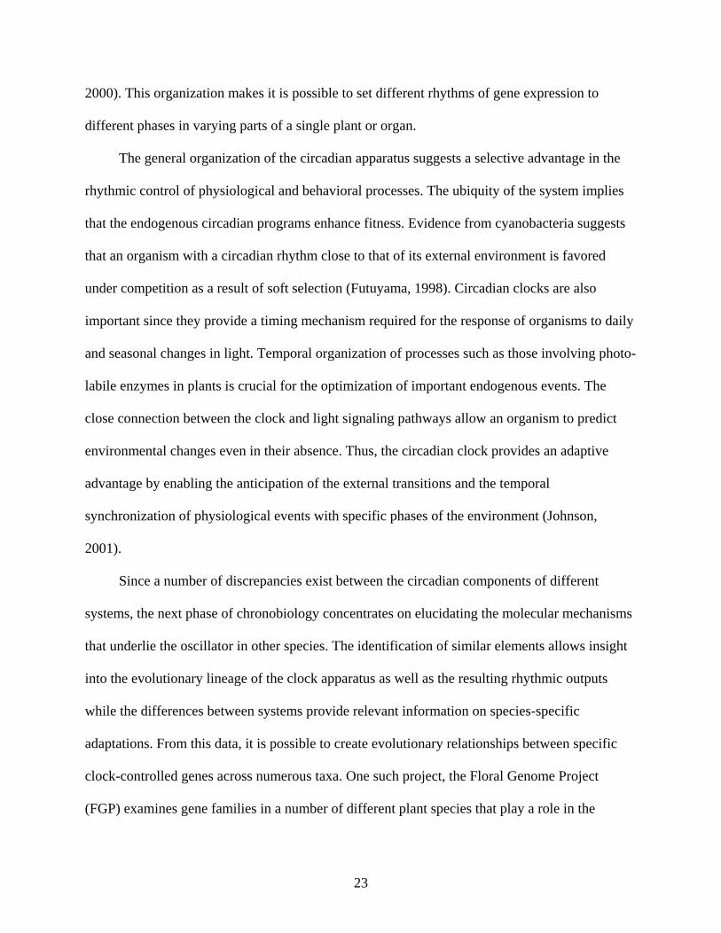
2000). This organization makes it is possible to set different rhythms of gene expression to
different phases in varying parts of a single plant or organ.
The general organization of the circadian apparatus suggests a selective advantage in the
rhythmic control of physiological and behavioral processes. The ubiquity of the system implies
that the endogenous circadian programs enhance fitness. Evidence from cyanobacteria suggests
that an organism with a circadian rhythm close to that of its external environment is favored
under competition as a result of soft selection (Futuyama, 1998). Circadian clocks are also
important since they provide a timing mechanism required for the response of organisms to daily
and seasonal changes in light. Temporal organization of processes such as those involving photo-
labile enzymes in plants is crucial for the optimization of important endogenous events. The
close connection between the clock and light signaling pathways allow an organism to predict
environmental changes even in their absence. Thus, the circadian clock provides an adaptive
advantage by enabling the anticipation of the external transitions and the temporal
synchronization of physiological events with specific phases of the environment (Johnson,
2001).
Since a number of discrepancies exist between the circadian components of different
systems, the next phase of chronobiology concentrates on elucidating the molecular mechanisms
that underlie the oscillator in other species. The identification of similar elements allows insight
into the evolutionary lineage of the clock apparatus as well as the resulting rhythmic outputs
while the differences between systems provide relevant information on species-specific
adaptations. From this data, it is possible to create evolutionary relationships between specific
clock-controlled genes across numerous taxa. One such project, the Floral Genome Project
(FGP) examines gene families in a number of different plant species that play a role in the

24
evolution of flowering (Albert et al., 2005). One identified family of genes involved in floral
initiation was shown to be similar to the LHY and CCA1 transcription factors of the Arabidopsis
central oscillator.
Genetic and molecular analyses have proven valuable tools in the elucidating the central
oscillatory mechanism of circadian clocks. In this study, two EST homologs to LHY and CCA1,
the MYB-like family of transcription factors in Arabidopsis, are identified in Eschscholzia
californica, the California poppy plant. Expressional analyses suggest that these genes are
rhythmic components of the circadian oscillator that participate in the initiation of flowering.
This suggests that the feedback loop mechanism of the plant circadian oscillator is conserved in
these two species.

25
Figure 1-1. The three basic components of the circadian clock. Numerous input and output
pathways function within the system.
Figure 1-2. Positive and negative factors act upon the circadian clock. Negative elements block the activation of the positive elements (blunted arrow) which in turn promotes the expression of the negative element (pointed arrow).
26
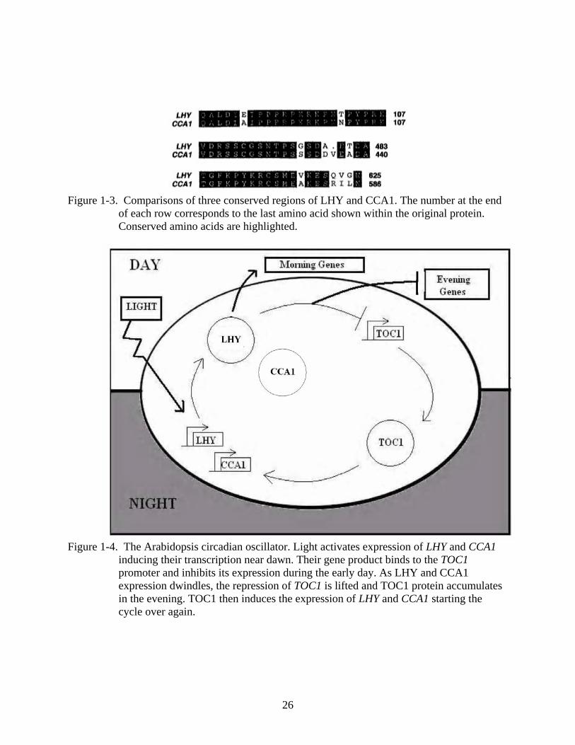
Figure 1-3. Comparisons of three conserved regions of LHY and CCA1. The number at the end
of each row corresponds to the last amino acid shown within the original protein. Conserved amino acids are highlighted.
Figure 1-4. The Arabidopsis circadian oscillator. Light activates expression of LHY and CCA1
inducing their transcription near dawn. Their gene product binds to the TOC1 promoter and inhibits its expression during the early day. As LHY and CCA1 expression dwindles, the repression of TOC1 is lifted and TOC1 protein accumulates in the evening. TOC1 then induces the expression of LHY and CCA1 starting the cycle over again.

27
CHAPTER 2 TWO CCA-LIKE MYB TRANSCRIPTION FACTORS ARE PRESENT IN CALIFORNIA
POPPY
Summary
Biological clocks play an important role in the lives of many organisms. This machinery
allows species to coordinate important activities or behaviors in relation to their environment.
The clock generates rhythms over which these events operate with a period of approximately
twenty- four hours, thus the term circadian rhythms. The clock is primarily composed of three
main components: input pathways, a central oscillator and output pathways.
An analysis was conducted to determine if two circadian clock genes found in the
California poppy plant, RISE and SHINE, are related to two transcription factors involved in
maintaining circadian rhythms in Arabidopsis, LHY and CCA1. In Arabidopsis, these two genes
function in a feedback loop of the central circadian oscillator and are crucial for maintaining
rhythms within the organism. A comparison of the genomic sequences revealed that there was an
acceptable degree of homology between the two sequences. In addition, an expressional analysis
revealed that the levels of messenger RNA (mRNA) of the genes oscillated over a twenty- four
hour period which suggests circadian control. The location of expression was also similar
between the two plant species. In young tissue, the transcripts were localized to the meristematic
regions as well as the premature leaves. In older tissue, expression was highest in the
reproductive organs and pollen grains. Since the circadian clock plays a role in promoting
flowering and the release of pollen, it is no surprise the transcripts were localized to these
various regions.
Due to these similarities this paper proposes that homologs of LHY and CCA1 exist in the
California poppy plant and function as critical components of the negative feedback loop at the
center of the circadian oscillator. Additional potential homologs of LHY and CCA1 have been
28
identified in other species based on sequence similarity. This discovery suggests that the
circadian machinery is conserved among higher plants. In addition, temporal organization of
important events seems to confer a selective advantage for the organisms.
Introduction
Biological clocks are important in maintaining the rhythmicity of crucial events in
different species. In Arabidopsis thaliana, two genes, LHY and CCA1 play important roles in
generating and maintaining the rhythms within the organism (Schaffer, 1997; Schaffer et al.,
1998; Wang et al., 1997). They have been identified as components of the central oscillator of
the clock, one of its three core components. LHY and CCA1 function in a feedback loop, along
with the TOC1 gene, in which they negatively regulate their own expression (Alabadí et al.,
2001; Carré and Kim, 2002). The pauses that occur as a result of this feedback loop are efficient
for generating the observed oscillations. These oscillations, in turn, are conveyed as changes in
the organisms’ physiological or behavioral changes via the output pathway.
Two potential homologs of the LHY and CCA1 genes were identified in Eschscholzia
californica on the FGP database (Albert et al., 2005). These two EST sequences could
potentially serve as components of a central oscillator in California poppy. Designated as RISE
and SHINE, these genes might be the functional equivalents of LHY and CCA1. This paper
reveals that these genes show an acceptable degree of sequence homology and share similar
expressional patterns suggesting they are components of the central oscillator in Eschscholzia
californica.
Materials and Methods
Plant Growth Conditions
Arabidopsis thaliana and Eschscholzia californica plants were grown under ideal
temperate conditions in the University of Florida Department of Botany greenhouse in

29
Gainesville, Florida between March and July 2004 and June through September of 2005.
California poppy was grown on sterile soil under normal light conditions with a period that
matched the exogenous environment. Arabidopsis was sowed on autoclaved soil at an irradiance
of 100 µmol m¯² s¯¹ as recommended by Kranz and Kirchheim (1987), and the day length was set
so that it matched the period of the environment. Tissue for in situ hybridizations was harvested
mid-morning and included floral meristems and floral buds. The collected material was placed in
4% paraformaldehyde to prepare for fixation. For RT-PCR analysis, plant tissue consisting of
small leaves, floral meristems and buds was collected from each species every four hours for
three days. Each sample was placed in liquid nitrogen and stored in a -80°C freezer.
Sequence Analysis and DNA Isolation
The CCA1 and LHY genomic sequences, identified as genes At2g46830 and At1g01060,
were identified in the Floral Genome Project (FGP) database (Albert et al, 2005; Carlson et al.,
2006) and recognized as a distinct family of transcription genes associated with the circadian
clock. The sequences were used to search for homologs in other plant species associated with the
FGP database using the site’s BLAST program and a number of candidate genes were identified.
In Eschscholzia californica (California poppy) two ESTs or expressed sequence tags, which are
small fragments of genes that have been cloned, demonstrated a notable level of similarity to the
Arabidopsis genes. The FGP identification numbers for these two sequences are eca_4_183384
and eca_4_184056 which I will refer to as RISE and SHINE respectively. Alignments of the
cDNA and protein sequences of CCA1, LHY, RISE and SHINE were constructed using the
GenomeNet database program CLUSTALW (Thompson et al., 1994)
Clones of the RISE and SHINE sequences were received from Penn State University, a
participant in the FGP grant. Their preparation has been described previously (Carlson et al.,

30
2006). Luria-Bertani (LB) media was prepared and 2.5 mL cultures were prepared with
ampicillin at a concentration of 50µg/mL. The cultures were grown for 16 hrs in a shaking
incubator at 37°C. It was noted that the cultures grew slowly due to their low turn-over rate. The
alkaline lysis mini-prep protocol (Morelle, 1989) was used to purify plasmid DNA from 1 mL of
culture.
Arabidopsis genetic material was obtained from wild type plants as previously has been
described (Edwards et al., 1991).
In situ Hybridizations
Except for the modifications noted below, previously described methods were used for in
situ hybridization (Jackson, 1991; Drews et al., 1991). To generate templates for probe synthesis,
DNA from plasmids containing the RISE and SHINE EST sequences as well as genomic DNA
isolated from Arabidopsis was PCR amplified. The T7 RNA polymerase promoter sequence
(TAATACGAGTCACTATAGGG) was placed in front of each reverse primer which allowed
direct synthesis of digoxigenin-labeled antisense probes from PCR products. The sense control
probes were designed with the T7 promoter sequence in front of the forward primers. In addition,
probes were designed within the exons of the genomic sequences of each species in order to
hybridize to corresponding messenger RNAs (mRNAs) in situ. Since Myb transcription factors
contain a similar conserved motif in their amino terminus or 5’ region, it was important to design
primers in the carboxyl or 3’ region that would be unique to each sequence. The following
primers were used to amplify RISE templates for probe synthesis:
TCTCTTTCGCCTCTACCGAACA and
TAATACGACTCACTATAGGGAAGCACTCTTCAGGGAACCTCA. The primers used to
amplify SHINE DNA were ACCACCACCAACTGCAACTCCTAT and
TAATACGACTCACTATAGGGTGTACGGCGATTACTGAAGGGT. Amplification of LHY

31
DNA used the following primers: CAGTTCCAACTCCAGCAATGAC and
TAATACGACTCACTATAGGGCTGAAACGCTATACGACCCTCT. The primers for CCA1
(TCTGGTTATTAAGACTCGGAAGCCAT and
TAATACGACTCACTATAGGGTTCATTGGCCATCTCAGGATGC) were used to amplify its
PCR product. RNA probes were synthesized using the Dig-RNA labeling kit (Roche Applied
Science, Indianapolis, IN). The cRNA products, 263bp for RISE, 266bp for SHINE, 476bp for
LHY and 361bp for CCA1, were synthesized and added to the hybridization buffer a so the final
concentration was 500 ng mL¯¹. Slides were hybridized at 45°C overnight and washed at 50°C.
For signal detection, a few grains of tetramisole hyrochloride (Sigma, St. Louis, MO) was added
to the Western Blue substrate (Promega, Madison, WI). Slides were evaluated using a Zeiss
Axiostar Plus Microscope (Carl Zeiss, Inc, Thornwood, NY) and images were photographed
with an Axiocam MRc5 camera (Carl Zeiss, Inc., Thornwood, NY).
Quantitative RT-PCR
For each plant species, fresh tissue including leaves, floral buds and meristematic tissue
was collected every four hours for three days and placed immediately in liquid nitrogen and
stored at -80°C. Total RNA was isolated from the tissues using the RNeasy plant RNA isolation
kit (Qiagen, Valencia, CA). RNA concentration was measured using Ribogreen dye (Molecular
Probes, Eugene, OR) and a TBS-380 Mini Fluorometer (Turner BioSystems, Sunnyvale, CA).
The RNA templates were transcribed using the Reverse Transcriptase product protocol (Roche ,
Applied Science, Indianapolis, IN). PCR was used in order to determine the differences in
transcript expression using the same primers described previously for probe construction.
Differences in the 18S ribosomal RNA (rRNA) positive control transcripts were determined by
using the following primers: TTGTGTTGGCTTCGGGATCGGAGTAAT and

32
TGCACCACCACCCATAGAATCAAGAA (Cho and Cosgrove, 2000). PCR products were
separated by size on agarose gels stained with ethidium bromide and visualized under a UV light.
Images of the gels were captured using a ChemImager 4400 (Alpha Innotech Corp., San
Leandro, CA) and the relative sizes of bands were determined by comparison to a standard 1kb
plus DNA ladder (Invitrogen, Carlsbad, CA).
Results
RISE and SHINE Encode CCA1-like Myb Transcription Factors
Sequence similarity among genes of different species can provide relevant information
about the evolution of particular gene families and the conservation of important mechanisms.
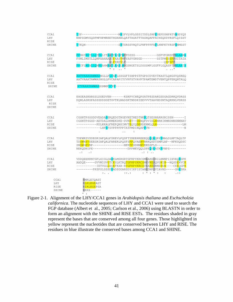
RISE and SHINE share significant sequence identity with the CCA1 and LHY genes of
Arabidopsis. In particular, RISE was shown to be comparable to LHY with over 40% identity in
a region at the C-terminus. SHINE displayed similarity to the CCA1 sequence with 30% identity
in the C-terminal region (Fig. 2-1). These regions located at the carboxyl or 5’ end of the genes
and ESTs corresponds to a DNA-binding domain that is found in plant Myb transcription factors.
This motif is highly conserved among the Myb gene family and provides evidence that RISE and
SHINE are indeed part of the family (Fig. 2-2). Additionally, RISE and SHINE share 40%
identity in the region investigated which demonstrates the redundancy between the two
components (Fig. 2-3).
RISE and SHINE Expression is Under Circadian Control
Circadian clock genes (ccgs) show a rhythmic pattern of transcript and protein expression.
In Eschscholzia californica, this is no exception. The RISE and SHINE transcripts oscillate over
a 24 hr period. Both RISE and SHINE oscillate in a pattern analogous to CCA1 and LHY in
Arabidopsis (Fig. 2-4). The transcripts abundance varies during the day. Peak levels of
transcription occur just prior to dawn and decrease throughout the day. By evening, the levels of

33
RISE and SHINE transcripts are greatly reduced but begin to rise in the early hours of the
morning. This evidence supports the notion that RISE and SHINE are activated by a light signal
similar to the mechanisms of CCA1 and LHY (Fig. 2-5). The transcripts also oscillate with a
period of approximately 24 hrs, which matches the external environment of the organism, further
supporting their role in the circadian clock.
Similar Expression Patterns in a CCA1- like Family of Myb Transcription Factors
In situ hybridizations are ideal for determining the location of transcript expression within
an organism. This method was used to analyze the expression pattern of CCA1 and LHY in
Arabidopsis thaliana and RISE and SHINE in Eschscholzia californica. For both species, two
stages of development were analyzed: a younger stage characterized by premature inflorescence
meristems and a later stage which is exemplified by floral buds.
In the young Arabidopsis tissue, a strong LHY signal is detected in the meristematic region
and in the stamen and carpel primordia (Fig 2-6A). In older tissue, LHY is expressed in the
gynoecium, ovules, and anthers and to a lesser degree in pollen grains (Fig. 2-6B). The
expression of the LHY homolog, CCA1, is similar to its counterpart. High transcript levels are
detected in the young developing floral meristem and include the premature reproductive organs
(Fig. 2-6C). Expression of CCA1 in older tissue is limited to the reproductive tissues (Fig. 2-6D).
The sense probe does not have a signal (Fig. 2-6E).
The expression patterns of the CCA-like Myb transcription factors in Eschscholzia
californica is similar to the patterns observed in Arabidopsis. No signal could be detected on the
sense probe control (Fig. 3-7E). High levels of RISE transcript were detected in the young
developing tissue in the meristematic region, premature leaves and axillary buds (Fig. 2-7A). In
older tissue, expression is highest in the carpel, ovules, anthers and pollen grains but is still

34
detected in the developing petals (Fig. 2-7B). Like its Arabidopsis counterpart, SHINE
transcripts are detected in both the young and older stages of the poppy plant. Transcripts are
detected in the sepal primordia, cauline leaves, and the floral meristem (Fig. 2-7C). SHINE
expression in older tissue is confined to the reproductive organs and petals (Fig. 2-7D).
However, it should be noted that a lower expression level can be detected for both genes
throughout the specimen (Fig. 2-7A-D), showing that RISE and SHINE transcripts are located
within a variety of tissue types..
Discussion
Feedback Loop Mechanism as the Basis of the Circadian Oscillator
An autoregulatory feedback loop involving both positive and negative elements is central
to the circadian oscillator. Circadian systems are often composed of one or more interconnected
loops. Knowledge of these interlocked loops results from the identification of genes that function
within the core loop of the oscillator. In Arabidopsis thaliana, three genes with required roles in
maintaining rhythmicity have been identified: LHY, CCA1 and TOC1 (Schaffer, 1997; Wang et
al., 1997; Millar et al., 1995). The two Myb transcription factors LHY and CCA1 serve as the
negative elements of the core loop and function to block the activation of the positive element
TOC1. The positive regulator, TOC1 activates expression of LHY and CCA1. In this study, we
have identified two potential homologs to LHY and CCA1 in Eschscholzia californica. These
genes are hypothesized to serve similar roles in the core oscillator of the poppy plant, thus
providing a conserved mechanism for maintaining rhythmicity in higher plants.
The RISE and SHINE Genes Encode MYB Transcription Factors That Are Similar to LHY and CCA1
Sequence analyses revealed that two California poppy EST sequences located in the FGP
database (Albert et al, 2005; Carlson et al., 2006) share sequence identity with known

35
components of the central circadian oscillator from Arabidopsis (Fig. 2-1). The Myb domain,
which functions as a DNA-binding domain, shares the most sequence similarity with RISE and
SHINE. Research shows that LHY and CCA1 bind to an evening element (EE) located within
the promoter of TOC1, an evening gene (Alabadí et al., 2001; Harmer et al., 2000). Because of
the similarity between the components of these two circadian systems, I wanted to determine if
RISE and SHINE function in a similar manner to CCA1 and LHY in the Arabidopsis central
oscillator.
A prerequisite for a protein to function as a negative element in the circadian clock is that
its expression and activity must oscillate in synchrony with the environmental oscillations. In
addition, this component regulates its own transcription by negative feedback which creates a
delay in the rhythmic cycle. In the Arabidopsis model plant, LHY and CCA transcripts were
shown to oscillate over a 24 hr period with peak levels accumulating just prior to dawn
(Mizoguchi et al., 2002). Similar results for this species were obtained (Fig. 2-5). The RISE and
SHINE transcripts displayed a similar pattern of expression with minimal levels of mRNA
detected in the evening (Fig.2-4). The accumulation of transcripts just prior to dawn shows that
LHY and CCA1, as well as RISE and SHINE, are regulated by a light signal and are entrained to
anticipate dawn.
The genes at the center of the Arabidopsis circadian oscillator serve as either positive or
negative factors to influence the rate of transcription. The activation and inhibition of ccgs
occurs at particular points within the circadian cycle and when coupled, form a loop in which the
components serve crucial roles in generating and maintaining rhythmicity within an organism.
The LHY and CCA1gene products in Arabidopsis function in a manner that is antagonistic to
TOC1. In Eschscholzia californica, putative homologs for LHY and CCA1 have been identified

36
but other components of the oscillator remain unknown. Based on the previously described
similarities between the two systems, it is reasonable to hypothesize that a TOC1- like gene also
functions in the poppy oscillator (Fig. 2-8.).
The spatial expression patterns of the Myb transcription factors in Arabidopsis and
California poppy provide relative information about their functional similarities. In both species,
the young tissue contained a high level of expression in the meristem and sepal primordia (Fig.
2-6A, C; Fig. 2-7A, C). The older tissues were characterized by high levels of transcript in the
reproductive organs and petals (Fig. 2-6B, D; Fig. 2-7B, D). The similarity in the expression
pattern suggests that these genes might be true orthologs stemming from a common ancestor. In
addition, the location of expression provides relevant information on the processes regulated in
that particular region. The high level of transcript expression in pollen grains in the older tissue
of Arabidopsis and poppy could control the timed release of pollen, a mechanism that evolved
for maximizing reproductive success (Subba et al., 1998). The fact that the circadian clock
regulates expression of floral pathway genes that in turn activate floral meristem identity genes
(Vijayraghavan et al., 2005) seems logical to explain the high level of expression of the Myb
transcription factors in the meristematic regions of the young tissue.
Together these sequence comparison and mRNA expression data suggest that RISE and
SHINE encode Myb transcription factors that could function as the negative elements in the
oscillator of the California poppy plant similar to the manner of LHY and CCA1 in Arabidopsis.
An Evolutionary Conserved Clock Mechanism in Higher Plants
Although the above data suggests similarities exist among the circadian systems of
Arabidopsis thaliana and Eschscholzia californica, little is known about the elements and
mechanisms underlying the clocks of other higher plants. It is possible that the molecular

37
components that form the clock machinery are unique to higher plants. In this case it is
important to determine whether other plant species have homologs for each of the Arabidopsis
clock components and whether they share similar functions. This paper demonstrates that
homologs for two Arabidopsis clock genes exist in the California poppy plant and that they
appear to be expressed in a similar manner to their counterparts. In other plant species,
components of the central oscillator remain unknown however, recent evidence has identified
several clock-associated genes that are involved in the input pathway to the clock. Studies of
Pisum sativum, peas, have revealed circadian clock gene homologs of TOC1, CCA and LHY
referred to as TOC1 and MYB1 (a CCA1/LHY homolog) respectively (Hecht et al., 2007). Two
additional Arabidopsis orthologs, EARLY FLOWERING4 (ELF4) and LATE BLOOMER1
(LATRISE), were characterized in pea plants and their diurnal rhythm expression conformed
closely to those associated with their counterparts, ELF4 and GIGANTEA (GI). In Arabidopsis,
ELF4 promotes clock entrainment and is required for sustained rhythms in the absence of
environmental cues (McWatters et al., 2007). The GI gene regulates flowering in long day (LD)
conditions in a clock-controlled pathway, where it acts as an intermediate between the central
oscillator and the FLOWERING LOCUS T (FLT) gene (Mizoguchi et al., 2005). Investigations in
the clock components in other species continue, including Oryza sativa (rice), Medicago
trunculata( a legume) and Lycopersicon esculentum (tomato). In addition, sequence analysis of
the Myb family of transcription factors using the FGP database (Albert et al, 2005; Carlson et al.,
2006) revealed one CCA1/LHY homolog in Cucumis sativus (cucmber), Asparagus officinalis,
Liriodendron tulipifera (tuliptree) and Saruma henryi (standing ginger) and two homologs in
Acorus americanus (the American Sweet Flag) and Nuphar advena (water lily). The presence of
similar sequences across a wide variety of species suggests that the oscillator mechanism that
38
involves CCA1 and LHY in Arabidopsis is conserved in higher plants. The conservation of this
mechanism and its components implies that such organization is beneficial for the organism.
The ubiquity of the feedback loop mechanism of plant circadian oscillators suggests that an
adaptive advantage results from the spatial and temporal organization of important rhythmic
activities. A recent experiment compared the performance of wild type Arabidopsis plants with
lines having mutations that alter period length in a range of environmental period lengths that
were either matched or mismatched to the endogenous clock. The results showed that a
photosynthetic advantage was conferred by matching the endogenous clock period with the
light/dark period (Dodd et al., 2005). Incorrect matching of the periods resulted in reduced leaf
chlorophyll, reduced assimilation, reduced growth and increased mortality (Dodd et al., 2005).
Optimization of physiological parameters by the circadian clock probably has been selected
during plant evolution. Similar results have been described in the cyanobacteria Synechococcus
as well (Ouyang et al., 1998)
The Biological Importance of Circadian Clock Genes
Although this paper addresses the circadian oscillator and its key mechanisms in higher
plants, the importance of the circadian machinery also resonates throughout the animal kingdom.
In addition, elucidating the components underlying the feedback loops of the oscillator in either
plants or animals provides relative information on the general architecture of the mechanisms.
Both plants and animals use circadian clocks to temporally organize important processes
involving reproduction and development which are crucial in the evolution of every species. In
humans, many behaviors are regulated by the circadian clock including the sleep/wake cycle,
feeding patterns, hormone production and cell regeneration (Edgar et al., 1993; Stokkan et al.,
2001; Czeisler and Klerman, 1999; Shibata, 2004).

39
A number of human illnesses are attributed to a dysrhythmia in a behavioral or
physiological process. Abnormal circadian rhythms have been associated with affective disorders
like A number of human illnesses are attributed to a dysrhythmia in a behavioral or physiological
process. and the existing therapy drugs used to treat these disorders such as lithium act upon the
circadian cycle (Hallonquist et al., 1986). Insomnia and sleep problems also result from
abnormal circadian rhythmicity and usually are characterized by an endogenous clock that runs
faster or slower than the norm (Zisapel, 2001). Individuals that suffer from attention-deficit
hyperactivity disorder (ADHD) are often plagued by sleep disturbances which result from a
dysrhythmic clock (Owens, 2005). In women who suffer from menopause, hot flashes disrupt the
clock’s rhythm resulting in a clock that is misentrained. This abnormal entrainment results in
sudden awakenings during the sleep cycle (Freedman et al., 1995). Recently a role for the
circadian clock has been identified in cancer studies. Research suggests that at least eight central
clock genes coordinate many basic functions, including cell proliferation, tumor growth and
apoptosis in circadian time. This work indicates that circadian clock genes and their products
potentially represent novel targets for the control of cancer growth (Wood et al., 2006).
Elucidating the mechanisms that lie beneath the circadian oscillator has become the
primary focus of chronobiologists. A wealth of knowledge stands to be gained since nearly all
processes crucial for species survival involve rhythmicity of one or more elements. The
availability of technologies to analyze global gene expression should become a powerful tool in
clock research. This advancement should aid in the identification of new genes affected by the
timekeeping apparatus and help characterize the interactions of those clock proteins that have
been previously identified. However, the question of how the genes involved in the clocks are
regulated is just starting to be addressed.

40
In this study, two homologs of the Arabidopsis Myb transcription factors CCA1 and LHY
were identified in Eschscholzia californica. Sequence analyses suggest that these genes are true
orthologs and are similar in their temporal and spatial expression. This information provides
evidence that there is a conserved transcriptional- translational feedback loop at the center of the
circadian oscillator in higher plants. Based on this congruence, other circadian clock genes
involved in the maintenance of rhythms in California poppy should resemble those described in
Arabidopsis (Fig. 2-8).
41
CCA1 TYP--------------------MHIPVLVPLGSSITSSLSHPPSEPDSHPHTVAGDYQS LHY NHPSGMVSQDFMFHPMREETHGHANLQATTASATTTASHQAFPACHSQDDYRSFLQISST RISE ------------------------------------------------------------ SHINE TTEQN------------------SHTSRSSVHQTLPNFPPPFAPLHNPETYRSFANMSST
CCA1 FPNHIMSTLLQTPALYTAATFASSFWPPDSSG------------GSPVPGNSPPNLAAMA LHY FSNLIMSTLLQNPAAHAAATFAASVWPYASVGNSGD--------SSTPMSSSPPSITAIA RISE ------------------ALFRREVLP---------------------QSFSPP------ SHINE FPCFLMSALLQNPAAHMAATLAASLWPGSNGETSLDSSSMPLGGFPLGQASPTPNLAAIA
* : .. * . ..*
CCA1 AATVAAASAWWAANGLLPLCAPLSSGGFTSHPPSTFGPSCDVEYTKASTLQHGSVQSREQ LHY AATVAAATAWWASHGLLPVCAPAPITCVPFSTVAVPTPAMTEMDTVENTQPFEKQNTALQ RISE ------------------------------------------------------------ SHINE ATVAAASAWWAAHGMMPLCPP--------------------------------------
CCA1 EHSEASKARSSLDSEDVEN-------KSKPVCHEQPSATPESDAKGSDGAGDRKQVDRSS LHY DQNLASKSPASSSDDSDETGVTKLNADSKTNDDKIEEVVVTAAVHDSNTAQKKNLVDRSS RISE ---------------------------------------------------------- SHINE ------------------------------------------------------------
CCA1 CGSNTPSSSDDVEADASERQEDGTNGEVKETNEDTNKPQTSESNARRSRISSN------I LHY CGSNTPSGSD-AETDALDKMEKDKE-DVKET--DENQPDVIELNNRKIKMRDNNSNNNAT RISE -----------PDLKKALFREPQNSIMVTEQIQDEKDENMLQLN--------------LM SHINE -------------LHPSFSYPPPPPPTATPMDINQAPPVN-------------------N
. .. :
CCA1 TDPWKSVSDEGRIAFQALFSREVLPQSFTYREEHREEEQQQQEQRYPMALDLNFTAQLTP LHY TDSWKEVSEEGRIAFQALFARERLPQSFSPPQVAENVNRKQSDTSMPLAP--NFKSQDSC RISE SNSWGEVNP-----------------NPPPPSDNNNVEKDSFLS---------------- SHINE NEKQDNIPE-------------------DPPWEVQQLDPEQSEATKPPNPS---------
.: .: .: : ..
CCA1 VDDQEEKRNTGFLGIGLDASKLMSRGRTGFKPYKRCSMEAKESRILNNNPIIHVEQKDPK LHY AADQE-----GVVMIGVGTCKSLKTRQTGFKPYKRCSMEVKESQVGNIN--NQSDEKVCK RISE -----------IETVGLGSGKFKAR-RTGFKPYKRCSVEAKESRMSNGN----CEEQGPK SHINE ---------PKSPSLSSSDSADSGGARSDYIKPISTANEDNPSVIAVHD-----SNKSKA
:. . ::.: : * : * : : .::
CCA1 RMRLETQAST LHY RLRLEGEAST RISE RIRLEGEPSA SHINE RKKX------ * :
Figure 2-1. Alignment of the LHY/CCA1 genes in Arabidopsis thaliana and Eschscholzia californica. The nucleotide sequences of LHY and CCA1 were used to search the FGP database (Albert et al., 2005; Carlson et al., 2006) using BLASTN in order to form an alignment with the SHINE and RISE ESTs. The residues shaded in gray represent the bases that are conserved among all four genes. Those highlighted in yellow represent the nucleotides that are conserved between LHY and RISE. The residues in blue illustrate the conserved bases among CCA1 and SHINE.

42
Figure 2-2. A phylogenetic analysis of CCA-like family of genes. This tree illustrates the
relationships among the CCA-like family of Myb transcription factors in Arabidopsis thaliana (CCA, LHY), Eschscholzia californica (SHINE, RISE) and Nuphar advena (NAD), a basal angiosperm. Here, the NAD gene was identified in the FGP EST database and serves as the outgroup for this analysis. Multiple sequence alignment and tree construction were produced using the MAFFT program (Katoh et al., 2002; Katoh et al., 2005).

43
1 60 RISE --------------------GCTCTCTTCAGGAGAGAA-GTTCTG---CCACAAAGTTTT SHINE GCAGAAACTACTGACGGGGAAAATTGTTCAGAAGTCATTACCCTGATTCGAGAAACTTCT * ***** ** * *** * * *** ** * 61 120 RISE T----CCCCACCACCTGATCTGAAAAAAGCACTCTTCAGGGAACCTCAAAAC---AGTAT SHINE TGTGTTTCTATCCCTTCTGCAAAGAAAAGTTTGATTTCAACATCTGCTGTACTCAAAAGT * * * * * * * * ***** ** * * * ** * * 121 160 RISE TATGGTAACAGAACAGATTCAAGA----TGAGAAAGATGAAAATATGTTGCAATTAAACC SHINE TCCTGCACCTTTATGGAGTTTGTGCCATTGAGAAAGGAACCAACTCAAAATAGTAAACAG * * * * * ** * ******** ** * * ** 161 180 RISE TTATGAGTAATTCATGGGGAGAAGT-AAATCCTAATCCCCCTCCTCCGTCCGACAACAAT SHINE GTCGAAGTTGATGATTCACAGAAATTGGACAAAAATGACCCCAGATTTTCTGATACCGAG * *** * ** **** * * *** *** ** ** * * * 181 240 RISE AACGTAGAGAAGGATAGTTTTTTGTCTAT-AGAAACTGTAGGGCTTGGATCAGGAAAGTT SHINE AATATAAGCTTGGGTGAAGCTCTGAAGTTGAAAAACAGCAAGATTAACTCTAATGAGAAA ** ** ** * * ** * * **** * * * * * * 241 300 RISE CAAGGCCCGTCGAACAGGCTTTAAACCGTATAAGAGATGTTCGGTAGAGGCGAAAGAGA- SHINE CTAGTCCTGGAGAACAATACCGAAA----AACAAAAGCAACCAGAAAAACCGAGTAACTT * ** ** * ***** *** * * * * * * * *** * 301 360 RISE GCAGAATGAGTAA-CGGAAACTGTGAGGAGCAAGGTCCGAAGAGGATACGTTTAGAAGGT SHINE ACATACTGAGGAAGTACAAGGTAACCAGAGCTACCCACGGCATGTTCCTGTCCATATTGT ** * **** ** ** * **** * ** * ** * * ** 361 420 RISE GAACCTTCAGCTTGA--------------------------------------------- SHINE GGATATGAGCTCTGGCCCAGTTGATATTTGCCCTTCATCCAAAATGTACCATTCAGGAGC * * * **
Figure 2-3. Alignment of the RISE (eca_4_183384) and SHINE (eca_4_184056) EST sequences using the BLASTN program from the FGP database (Albert et al, 2005; Carlson et al., 2006). Conserved residues are shaded in gray.

44
Hours 0 4 8 12 16 20 24 28 32 36 40 44 48 52 56 60
RISE
SHINE
18S
Figure 2-4. Analysis of RISE and SHINE mRNA expression. Peak RISE and SHINE transcript
levels occur just prior dawn and dwindle throughout the day. The 18S r RNA transcripts demonstrate a constant expression level throughout the day. The bar above reflects the light/dark cycles to which the plants were exposed for this experiment. The black boxes correspond to periods of darkness.

45
LHY
CCA1
18S
Hours 0 4 8 12 16 20 24 28 32 36 40 44 48 52 56 60 64 68 72
Figure 2-5. Analysis of LHY and CCA1 mRNA expression. Peak transcript levels of the
transcription factors occur just prior dawn and become greatly reduced by evening. The 18S rRNA transcripts demonstrate a constant expression level throughout the day. The bar above reflects the light/dark cycles to which the plants were exposed for this experiment. The black portions represent the evening hours.

46
Figure 2-6. Expression of LHY and CCA1 in Arabidopsis tissue. LHY and CCA1 transcripts were
located in both young and mature Arabidopsis floral tissue. The localization of the transcripts was determined by in situ hybridizations. LHY transcripts were present in A) the floral meristem (fm) and the floral primordia (fp) of the young tissue and in B) the gynoecium (g), ovules (o), anthers (a) and pollen grains (pg) of the mature floral tissue. CCA1 transcripts were detected in C) younger tissue specifically in the fm and fp and in D) the older tissue in the same location as observed with LHY: g, o, a and pg. E) The sense probe control shows no signal.
B
C
E
fp fm
fp fm
o
o
fm
fp
pg
pg
g
g
a
a
A
D
fp

47
Figure 2-7. RISE and SHINE transcripts are expressed in both young and mature floral tissue of
the California poppy plant. The localization of the transcripts was determined by in situ hybridizations. RISE transcripts were present in A) the floral meristematic region (fm), premature leaves (l) and in the axillary buds (ax) of the younger tissue while in B) the older tissue, high expression was detected in the carpel(c), ovules (o), anthers (a) and pollen grains (pg) while lower expression was observed in the floral tube (ft) and developing petals (pe). SHINE transcripts were detected in C) younger tissue in a pattern identical to RISE: fm, l and ax. SHINE transcripts in D) older tissue was restricted to the c, o, a and pg as well as the ft and pe. E) The sense probe control shows no signal.
A B
C
D
E
fm
fm
l
l
ax
ax
c
c
o
o
a
a
pg
pg
ft
ft
ft
pe
pe
pe
ft
o
c
pe
pe
pg
a

48
Figure 2-8. Proposed mechanism of the central oscillator of Eschscholzia californica. RISE and SHINE serve as negative regulators of a gene ‘X’ (designated by a blunted arrow), a TOC1 homolog. That gene in turn should induce expression of RISE and SHINE (pointed arrows). Candidate genes for component ‘X’ have yet to be identified.

49
CHAPTER 3 CONCLUSION
Elucidating the mechanisms that lie beneath the circadian oscillator has become the
primary focus of chronobiologists. A wealth of knowledge stands to be gained since nearly all
processes important for species survival involve rhythmicity of one or more elements. Research
on the crucial components that are required to maintain rhythmicity could provide insight into
possible therapies and treatments for diseases that target the clock system. In addition, by
analyzing the mechanisms and proteins of the central oscillator and comparing them among
different species, the evolutionary history of the clock can be examined.
In this study, it was proposed that a common clock ancestor exists among higher plant
species. In Eschscholzia californica, two homologs of Arabidopsis Myb transcription factors
were identified as potential components of the biological clock. Other probable homologs to
components of the Arabidopsis central oscillator LHY and CCA1 have been identified in other
species including Cucumis sativus (cucumber), Asparagus officinalis, Liriodendron tulipifera
(tuliptree), Saruma henryi (standing ginger), Acorus americanus (the American Sweet Flag) and
Nuphar advena (water lily). Further investigation of these species should provide relevant
information on the clock mechanism and its conservation. Genetic screens for clock mutants aids
in the assignment of potential components to roles within the circadian clockwork. Altered
expression, either increased or greatly reduced, of these constituents should result in aberrant
clock phenotypes. Although it is proposed that the RISE and SHINE genes of Eschscholzia
californica play a role in the central oscillator of the plant’s clock, further studies similar to those
mentioned above are required for verification.
50
LIST OF REFERENCES
Alabadí, D., Oyama, T., Yanovsky, M.J., Harmon, F.G., Más, P. and Kay, S.A. (2001) reciprocal regulation between TOC1 and LHY/CCA1 within the Arabidopsis circadian clock. Science 293, 880-883.
Alabadí, D, Yanovsky, M.J., Más, P., Harmer, S.L. and Kay, S.A. (2002) Critical role of CCA1 and LHY in maintaining circadian rhythmicity in Arabidopsis. Curr. Biol 12, 757-761.
Albert, V.A., Soltis, D.E., Carlson, J.E., Farmerie, W.G., Wall, P.K., Ilut, D.C., Solow, T.M., Mueller, L.A., Landherr, L.L., Hu, Y., Buzgo, M., Kim, S., Yoo, M.J., Frohlich, M.W., Perl-Treves, R., Schlarbaum, S.E., Bliss, B.J., Zhang, X., Tanksley, S.D., Oppenheimer, D.G., Soltis, P.S., Ma, H., dePamphilis, C.W., and Leebens-Mack, J.H. (2005) Floral gene resources from basal angiosperms for comparative genomics research. BMC Plant Bio. 5, 5-20.
Allada, R., Emery, P., Takahashi, J.S., and Rosbash, M. (2001) Stopping time: The genetics of fly and mouse circadian clocks. Annu. Rev. Neurosci. 24, 1091-1119.
Antoch, M.P., Song, E.J., Chang, A.M., Vitaterna, M.H., Zhao, Y., Wilsbacher, L.D., Sangoram, A.M., King, D.P., Pinto, L.H., and Takahashi, J.S. (1997) Functional identification of the mouse circadian Clock gene by transgenic BAC rescue. Cell 89, 655-657.
Aronson, B.D., Johnson, K.A., Loros, J.J., and Dunlap, J.C. (1994) Negative feedback defining a circadian clock: autoregulation of the clock gene frequency. Science 263, 1578-1584.
Barak, S., Tobin, E.M., Andronis, C., Sugano, S. and Green, R.M. (2000) All in good time: the Arabidopsis circadian clock. Trends Plant Sci. 5, 517-522.
Carlson, J.E., Leebens-Mack, J.H., Wall, P.K., Zahn, L.M., Mueller, L.A., Landherr, L.L., Hu, Y., Ilut, D.C., Arrington, J.M., Choirean, S., Becker, A., Field, D., Tanksley, S.D., Ma, H. and dePamphilis, C.W. (2006) EST databse of early flower development in California poppy (Eschscholzia califonrica Cham., Papaveraceae) tags over 6000 genes from a basal eudicot. Plant Mol. Biol. 62, 351-369.
Carré, I.A. and Kim, J.Y. (2002) MYB transcription factors in the Arabidopsis circadian clock. J. Exper. Bot. 53, 1551-1557.
Chandrashekaran, M.K. (1998). Biological rhythms research: A personal account. J. Biosci. 23, 545-555.
Cho, H.Y. and Cosgrove, D.J. (2000) Altered expression of expansin modulates leaf growth and pedicel abscission in Arabidopsis thliana. Proc. Natl. Acad. Sci. 97, 9783-9788.
Correa, A., Lewis, Z.A., Greene, A.V., March, I.J., Gomer, R.H., and Bell-Pedersen, D. (2003) Multiple oscillators regulate circadian gene expression in Neurospora. Proc. Natl. Acad. Sci. USA 100, 13597-13602.

51
Czeisler, C.A. and Klerman, F.B.(1999) Circadian and sleep-dependent regulation of hormone release in humans. Recent Prog. Horm. Res. 54, 97-130.
Darlington, T.K., Wager-Smith, K., Ceriani, M.F., Staknis, D., Gekakis, N., Steeves, T.D.L., Weitz, C.J., Takahashi, J.S. and Kay, S.A. (1998) Closing the circadian loop: CLOCK-induced transcription of its own inhibitors per and tim. Science 280, 1599-1603.
Devlin, P.F. (2002) Signs of the time: environmental input into the circadian clock. J. Exper. Bot. 53, 1535-1550.
Dodd, A.N., Salathia, N., Hall, A., Kévei, E., Tóth, R., Nagy, F., Hibberd, J.M., Millar, A.J., and Webb, A.R.R. (2005) Plant Circadian Clocks Increase Photosynthesis, Growth, Survival, and Competitive Advantage. Science 309, 630-633.
Drews, G.N., Bowman, J.L. and Meyerowitz, E.M. (1991) Negative regulation of the Arabidopsis homeotic gene AGAMOUS by the APETELA2 PRODUCT. Cell 65, 991-1002.
Dunlap, J.C. (1993) Genetic analysis of circadian clocks. Annu. Rev. Physiol. 55, 683-728.
Dunlap, J.C. (1996) Genetic and molecular analysis of circadian rhythms. Annu. Rev. Genet. 30, 579-601.
Dunlap, J.C. (1999) Molecular bases for circadian clocks. Cell 96, 271-290.
Dunlap, J.C., Loros, J.J. Liu, Y., and Crosthwaite, S.K. (1999) Eukaryotic circadian systems: cycles in common. Genes to Cells 4, 1-10.
Edgar, D.M., Dement, W.C. and Fuller, C.A. (1993) Effect of SCN lesions on sleep in squirrel monkeys: evidence for opponent processes in sleep-wake regulation. J.of Neurosci. 13, 1065-1079.
Edwards, K., Johnstone, C. and Thompson, C.(1991)A simple and rapid method for the preparation of plant genomic DNA for PCR analysis. Nucl. Acids Res. 19, 1349.
Fairman, R., Beran-Steed, R.K., Anthony-Cahill, S.J., Lea,r J.D., Stafford, W.F., Degrado, W.F., Benfield, P.A., and Brenner, S.L. Multiple oligomeric states regulate the DNA-binding of helix-loop-helix peptides. Proc. Natl. Acad. Sci. USA. 1993, 10429–10433.
Feldman, J.F. and Hoyle, M. (1973) Isolation of circadian clock mutants of Neurospora crassa. Genetics 75, 605-613.
Freedman, R.R., Norton, D., Woodward, S. and Cornelissen, G. (1995) Core body temperature and circadian rhythm of hot flashes in menopausal women. J. Clin. Endo. Met. 80, 2354-2359.
52
Froelich, A.C., Loros, J.J., and Dunlap, J.C. (2003) Rhythmic binding of a WHITE COLLAR containing complex to the frequency promoter is inhibited by FREQUENCY. Proc. Natl. Acad. Sci. USA 100, 5914-5919.
Futuyama, D.J. (1998) Evolutionary Biology (Sinauer, Sunderland, MA), 3rd Ed., p.386.
Gekakis, N., Staknis, D., Nguyen, H.B., Davis, F.C., Wilsbacher, L.D., King, D.P., Takahashi, J.S., and Weitz, C.J. (1998) Role of the CLOCK protein in the mammalian circadian mechanism. Science 280, 1564-1569.
Gillette, M., and Sejnowski, T. (2005) Biological clocks coordinately keep life on time. Science 309, 1196-1198.
Glossop, N.R.J., Lyons, L.C., and Hardin, P.E. (1999) Interlocked feedback loops within the Drosophila circadian oscillator. Science 286, 766-768.
Golden, S.S., and Strayer, C. (2001) Time for plants. Progress in Plant Chronobiology. Plant Physiol. 125, 98-101.
Green, R.M. and Tobin, E.M. (1999) Loss of the CIRCADIAN CLOCK-ASSOCIATED PROTEIN 1 in Arabidopsis results in altered clock-regulated gene expression. Proc. Natl. Acad. Sci. USA 96, 4176-4179.
Halberg, F. (1959) Physiologic 24 hour periodicty: general and procedural considerations with reference to the adrenal cycle. Z. Vitam. Hom. Fermentforsch. 10, 225-296.
Hardin, P. (2000) From biological clock to biological rhythms. Genome Biology 4, 2-6
Hardin, P. and Siwicki, K. (1995). The multiple roles of per in the Drosophila circadian clock. Trends Neurosci. 7, 15-25.
Harmer, S.L., Hogenesch, J.B., Straume, M., Chang H.S., Han, B., Zhu, T., Wang, X., Kreps, J.A., and Kay, S.A. (2000) Orchestrated transcriptionof key pathways in Arabidopsis by the circadian clock. Science 290, 2110-2113.
Hallonquist, J.D., Goldberg, M.A. and Brandes, J.S. (1986) Affective disorders and circadian rhythms. Can. J. Psych. 31, 259-272.
He, Q., Cheng, P., Yang, Y., Wang, L., Gardner, K.H., and Liu, Y. (2002) White Collar-1, a DNA binding transcription factor and a light sensor. Science 297, 840-843.
Hecht, V., Knowles, C.L., Vander Schoor, J.K., Liew, L.C., Jones, S.E., Lambert, M.J.M. and Weller, J.L. (2007) Pea LATE BLOOMER1 is a GIGANTEA ortholog with roles in photoperiodic flowering, deetiolation, and transcriptional regulation of circadian clock gene homologs. Plant Physiology 144, 648-661.

53
Ishiura, M., Kutsuna, S., Aoki, S., Iwasaki, H., Andresson, C.R., Tanabe, A., Golden, S.S., Johnson, C.H., and Kondo, T. (1998) Expression of a gene cluster kaiABC as a circadian feedback process in cyanobacteria. Science 281, 1519-1523.
Jackson, D. (1991) In situ hybridization in plants In Molecular Plant Pathology: A Practical Approach, D.J. Bowles, S.J. Gurr and M. McPherson, eds (Oxford: Oxford University Press), pp.163-174.
Johnson, C.H. (2001) Endogenous timekeepers in photosynthetic organisms. Annu. Rev. Physiol. 63, 695-728.
Katoh, K., Misawa, K., Kuma, K., and Miyata, T. (2002) MAFFT: a novel method for rapid multiple sequence alignment based on fast Fourier transform. Nucleic Acids Res. 30, 3059-3066.
Katoh, K., Kuma, K., Toh, H., and Miyata, T. (2005) MAFFT version 5: improvement in accuracy of multiple sequence alignment. Nucleic Acids Res. 33, 511-518.
Kay, S.A., and Millar, A.J. (1995) New models in vogue for circadian clocks. Cell 83, 361-364.
Kondo, T. and Ishiura, M. (1999) The circadian clocks of plants and cyanobacteria. Trends in Plants Sci. 4, 171-176.
Konopka, R.J. and Benzer, S. (1971) Clock mutants of Drosophila melanogaster. Proc. Natl. Acad. Sci. USA 68, 2112-2116.
Kranz, A.R. and Kirchhiem, B. (1987).Genetic resources in Arabidopsis.. Arabidopsis Inf. Serv. 24:2-4.
Kumar, V. (2002) Biological Rhythms. Alpha Science International, Ltd., Lucknow, India, pp. 1-6.
Lorne, J., Scheffer, J., Lee, A., Painter, M., and Miao, V.P. (2000)Genes controlling circadian rhythms are widely distributed in cyanobacteria FEMS. Microbiol. Lett. 189, 129-133.
Loros, J.J. and Dunlap, J.C. (2001) Genetic and molecular analysis of circadian rhythms in Neurospora. Annu. Rev. Physiol. 63, 757-794.
Más, P. (2005). Circadian clock signaling in Arabidopsis thaliana: from gene expression to physiology and development. Int. J. Dev. Biol. 49, 491-500.
McClung, C. (2000) Circadian rhythms in plants: a millennial view. Physiologia Plantarum 109, 359- 371.
McClung, C. (2001) Circadian rhythms in plants. Ann. Rev. of Plant Phys. and Plant Mol. Biol. 52, 139- 162.

54
McWatters, H.G., Kolmos, E., Hall, A., Doyle, M.R., Amasino, R.M., Gyula, P., Nagy, F., Millar, A.J. and Davis, S.J. (2007) ELF4 is required for oscillatory properties of the circadian clock. Plant Phys. 144, 391-401.
Millar,A.J., Carré, I.A., Strayer, C.A., Chua, N.H. and Kay, S.A. (1995) Circadian clock mutants in Arabidopsis identified by luciferas imaging. Science 267, 1161-1163.
Mizoguchi, T., Wheatley, K., Hanzawa, Y., Wright, L., Mizoguchi, M., Song, H.R., Carré, I.A. and Coupland, G. (2002) LHY and CCA1 are partially redundant genes required to maintain circadian rhythms in Arabidopsis. Developmental Cell 2, 629-641.
Mizoguchi, T., Wright, L., Fujiwara, S., Cremer, F., Lee, K., Onouchi, H., Mouradov, A., Fowler, S., Kamada, H., putterill, J. and Coupland, G. (2005) Distinct roles of GIGANTEA in promoting flowering and regulating circadian rhythms in Arabidopsis. The Plant Cell 17, 2225-2270.
Morelle, G.(1989) A plasmid extraction procedure on a miniprep scale. Focus 11, 7-8.
Ouyang, Y., Andersson, C.R., Kondo, T., Golden, S.S., and Johnson, C.H. (1998) Resonating circadian clocks enhance fitness in cyanobacteria. Proc. Natl. Acad .Sci. U.S.A. 95, 8660-8664.
Owens, J.A. (2005) The ADHD and sleep conundrum: a review. J. Dev. Behav. Ped. 26, 312-322.
Panda, S., Antoch, M.P., Miller, B.H., Su, A.I., Schook, A.B., Straume, M. Schultz, P.G., Kay, S.A., Takahashi, J.S., and Hogenesch, J.B. (2002) Coordinated transcription of key pathways in the mouse by the circadian clock. Cell 109, 307-320.
Putterill, J., Robson, F., Lee, K., Simon, R. and Coupland, G. (1995) The CONSTANS gene of Arabidopsis promotes flowering and encodes a protein showing similarities to zinc finger transcription factors. Cell 80, 847-857.
Roenneberg, Y and Merrow, M. (1998) Molecular circadian oscillations: an alternative hypothesis. J. Biol. Rhythms 13, 167-179.
Schaffer, R. (1997) LHY, a gene that regulates flowering and hypocotyl elongation of Arabidopsis. PhD thesis, University of East Anglia, Norwich, United Kingdom.
Schaffer, R., Landgraf, J., Monica, A., Simon, B., Larson, M. Wisman, E. (2001) Microarray analysis of diurnal and circadian-regulated genes in Arabidopsis. Plant Cell 13, 113-123.
Schaffer, R., Landgraf, J., Accerbi, M., Simon, V., Larson, M. and Wisman, E. (2001) Microarray analysis of diurnal and circadain-regulated genes in Arabidopsis. Plant Cell 13, 113-123.

55
Schaffer, R., Ramsay, N., Samach, A., Corden, S., Putterill, J., Carré, I.A. and Coupland, G. (1998) The late elongated hypocotyl mutation of Arabidopsis disrupts circadian rhythms and the periodic control of flowering. Cell 93, 1219-1229.
Schultz, T.F. and Kay, S.A. (2003) Circadian clocks in daily and seasonal control of development. Science 301, 326-328.
Scully, A.L. and Kay, S.A. (2000) Time flies for Drosophila. Cell 100, 297-300.
Shibata, S. (2004)) Neural regulation of the hepatic circadian rhythm. Anat. Record 280, 901-909.
Stokkan, K.A., Yamazaki, S., Tei, H., Sakaki, Y. and Menaker, M. (2001) Entrainment of the circadian clock in the liver by feeding. Science 19, 490-493.
Strayer, C., Oyama, T., Schultz, T.F., Raman, R., Somers, D.E., Más, P., Panda, S., Kreps, J.A. and Kay, S.A. (2000) Cloning of the Arabidopsis Clock gene TOC1, an autoregulatory response regulator homolog. Science 289, 768-770.
Subba, R.C., Reddi, N.S. and Atluri, J.K. (1998) Circadian patterns of pollen release in some species of Poaceae. Rev. Palaeobot. Palynol. 54, 11-42.
Sweeney, B.M. (1987) Rhythmic Phenomena in Plants. Academic Press, San Diego, CA, pp. 14-26.
Thain, S.C., Hall, A. and Millar, A.J. (2000) Functional independence of circadian clocks that regulate plant gene expression. Curr. Biol. 10, 951-956.
Thompson, J.D., Higgins, D.G. and Gibson, T.J. (1994) CLUSTAL W: improving the sensitivity of progressive multiple sequence alignment through sequence weighting, positions-specific gap penalties and weight matrix choice. Nucleic Acids Research, 22, :4673-4680.
Vijayraghavan, U., Prasad, K. and Meyerowitz, E. (2005) Specification and maintenance of the floral meristem: interactions between positively-acting promoters of flowering and negative regulators. Current Science 89, 1835-1844.
Wang, Z.Y., Kenigsbuch, D., Sun, L,. Harel, E., Ong, M.S. and Tobin, E.M. (1997) A myb-related transcription factor is involved in the phytochrome regulation of the Arabidosps LHCB gene. Plant Cell 9, 491-507.
Wang, Z.Y., and Tobin, E.M. (1998) Constitutive expression of the CIRCADIAN CLOCK ASSOCIATED 1 (CCA1) gene disrupts circadian rhythms and suppresses its own expression. Cell 93, 1207-1217.
Williams, J.A., and Sehgal, A. (2001) Molecular components of the circadian system in Drosophila. Ann. Rev. Physiol. 63, 729-755.

56
Wood, P.A., Du-Quiton, J., You, S. and Hrushesky, W.J.M. (2006) Circadian clock coordinates cancer cell cycle progression, thymidylate synthase, and 5-fluorouracil therapeutic index. Mol. Cancer Ther. 5, 2023-2033.
Yamazaki, S., Numano, R., Michikazu, A., Hida, A., Takahashi, R., Ueda, M., Block, G.D., Sakaki, Y., Menaker, M. and Tei, H.(2000) Resetting central and peripheral circadian oscillators in transgenic rats. Science 288, 682-685.
Zisapel, N. (2001) Circadian rhythm sleep disorders: pathophysiology and potential approaches to management. CNS Drugs 15, 311-328.

57
BIOGRAPHICAL SKETCH
Meredith Lynn Sullivan was born on January 13, 1979 in Tuscaloosa, Alabama. The
younger of two children, she graduated from Central High School in 1997. Following graduation,
Meredith enrolled at Shelton State Community College where she was a member of the women’s
varsity soccer team. She subsequently enrolled at the University of Alabama (UA) where she
earned her B.S. in Biology in 2002.
Upon receiving her B.S. degree, Meredith enrolled in graduate school at the University of
Florida (UF) in 2003. As a graduate student in the Department of Botany, she pursued molecular
biology- based studies to elucidate the components and mechanisms involved in flower
regulation. This information allowed her to analyze the evolutionary significance of circadian
clocks in development.
Upon completion of her M.S. degree, Meredith will pursue a career in medical research.
She plans to utilize the knowledge she has obtained throughout her education to aid in the
identification of therapeutic drugs for certain illnesses.