6.. instrumentation for brain function
DESCRIPTION
bio medical engineerungTRANSCRIPT

Instrumentation for brain function X Ray equipment transmits high energy
electromagnetic waves and indicates relative tissue density on a photographic plate Eg Cerebral angiography, Cranial XRays, Brain
Scans, CT Scans Ultrasonic equipment transmits high
frequency sound waves and indicate tissue location by reflecting waves
Electrophysiological equipment detects low voltage, low freq bioelectric signals that are picked up by electrodes

Cerebral angiography X-Ray technique to display brain structure
and detailed images of blood vessels with the aid of a contrast medium
Radio opaque dyes that block X-Rays are injected in an artery and dispersed throughout the cerebrovascular tree
X-Ray images are taken at 1 sec intervals and can reveal blockages and tumors


Nuclear medicine Small amounts of short lived
radioactive isotopes are introduced into the cardiovascular system
The concentrated radioactivity is measured with a scintillation counter
Counter responds to impinging alpha, beta or gamma rays given off by the radioactive material

Cranial X-Rays 2 dimensional X-Ray exposures
taken of the cranium Used to indicate fractures in
cranial bones and blood clots or tumors
Positive diagnosis can be made only when the contrast is high

Brain scans Radiographs that are taken through
successive scanning Highly collimated X-Ray beams are
used Small contrast differences can be
seen Provide considerably more info than
simple cranial X-Ray exposures
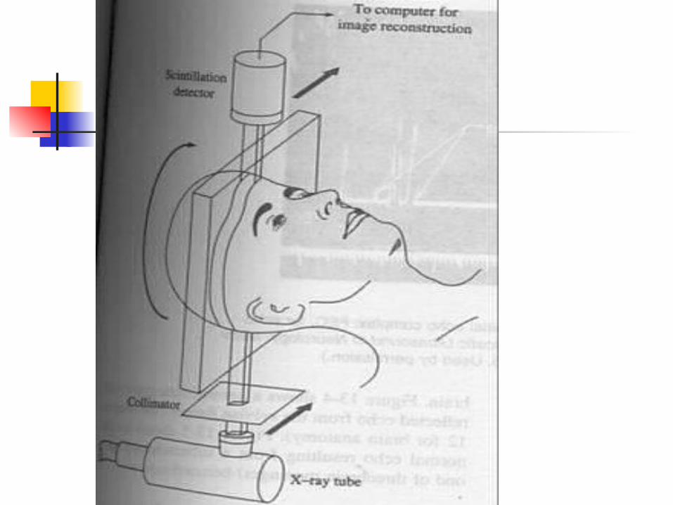
Computed Tomography CT scanning – technique of
recording and processing a set of image projections that represent a reconstruction of the object scanned Scanning system Processing unit Viewing part Storage unit

Computed Tomography To identify structural abnormalities (such
as abscesses, tumors, and hydrocephalus) in the brain
To identify bleeding or evidence of strokes in the brain
To identify ruptured or herniated disks in the spine
To identify spinal fractures To monitor the effects of radiation
therapy on brain cancer or of antibiotics on a brain abscess

EEG The electroencephalogram (EEG) is
a recording of the electrical activity of the brain from the scalp.
The first recordings were made by Hans Berger in 1929 although similar studies had been carried out in animals as early as 1870.

Neuron Membrane potentials

Electroencephalography The EEG is recorded between electrodes
placed in standard positions on the scalp and has a typical amplitude of 2-100 microvolts and a frequency spectrum from 0.1 to 60 Hz.
Most activity occurs within the following frequency bands; delta (0.5 - 4 Hz), theta (4-8 Hz), alpha (8-13 Hz), beta (13-22 Hz) and gamma (30-40 Hz).

EEG Frequency band <100uV <10uV <20uV
<2UuV


EEG EEG activity in particular frequency
bands is often correlated with particular cognitive states.
Signals in the alpha band, for example, are associated with relaxation. Thus, an electrode placed over the visual cortex that detects alpha band signals is detecting visual relaxation.
An electrode over the motor cortex picking up alpha band signals is detecting motor relaxation

EEG electrodes EEG electrodes transform ionic
currents from cerebral tissue into electrical currents. Electrodes used are Scalp Sphenoidal Nasopharyngeal Electrocorticographic Intracerebral


10-20 EEG electrode placement system

EEG Machine (8 channel)

EEG Telemetry system