2 signalling via pi3k/foxo1a pathway modulates … · signalling via pi3k/foxo1a pathway modulates...
TRANSCRIPT

Signalling via PI3K/FOXO1A Pathway Modulates Formation and Survival of Human Embryonic
Stem Cell-Derived Endothelial Cells
Béla Merkely1*, Edit Gara1*, Zsuzsanna Lendvai1, Judit Skopál1, Thomas Leja2, Wenhua Zhou2,
Annamária Kosztin1, György Várady3, Maxime Mioulane2, Zsolt Bagyura1, Tamás Németh1,
Sian E. Harding2, Gábor Földes1,2 1Heart and Vascular Center, Semmelweis University Budapest, Hungary
2National Heart and Lung Institute, Imperial College London, United Kingdom
3Membrane Research Group, Hungarian Academy of Sciences, Budapest, Hungary
*authors equally contributed to the paper
Running title: PI3K/FOXO1A in Human ESC;Derived Endothelial Cells
Béla Merkely email: [email protected] tel:+3614586810 fax:+3614586842
Edit Gara email: [email protected] tel:+3614586810 fax:+3614586842
Zsuzsanna Lendvai email: [email protected] tel:+3614586810 fax:+3614586842
Judit Skopál email: [email protected] tel:+3614586810 fax:+3614586842
Thomas Leja email: [email protected] tel:+447894337400
Wenhua Zhou email: [email protected] tel:+442075943132
Annamária Kosztin email: [email protected] tel:+3614586810 fax:+3614586842
György Várady email: [email protected] tel:+3613724300
Maxime Mioulane email: [email protected] tel:+491778574720
Zsolt Bagyura email: [email protected] tel:+3614586810 fax:+3614586842
Tamás Németh email: [email protected] tel:+3614586810 fax:+3614586842
Sian E. Harding email: [email protected] tel: 442075943009
Corresponding author:
Gábor Földes MD PhD
National Heart and Lung Institute
Imperial College London
Imperial Centre for Experimental and Translational Medicine
Hammersmith Campus
Du Cane Road
London W12 0NN
Tel: +44 7772920771
Page 5 of 36
Mary Ann Liebert Inc., 140 Huguenot Street, New Rochelle, NY 10801
Stem Cells and Development
123456789101112131415161718192021222324252627282930313233343536373839404142434445464748495051525354555657585960

2
Nonstandard Abbreviations and Acronyms
EB embryoid bodies
FOXO1A Forkhead box O transcription factor 1A
hESC;EC human embryonic stem cell;derived endothelial cells
PI3K phosphatidylinositol 3;kinase
VEGF vascular endothelial growth factor
Abstract
Vascular derivatives of human embryonic stem cells (hESC) are being developed as sources of tissue;
specific cells for organ regeneration. However, identity of developmental pathways that modulate the
specification of endothelial cells is not known yet. We studied PI3K;FOXO1A pathways during
differentiation of hESC towards endothelial lineage and on proliferation, maturation and cell death of
hESC;derived endothelial cells (hESC;EC). During differentiation of hESC, expression of FOXO1A
transcription factor was linked to the expression of a cluster of angiogenesis; and vascular remodelling;
related genes. PI3K inhibitor LY294002 activated FOXO1A and induced formation of CD31+ hESC;EC.
In contrast, differentiating hESC with silenced FOXO1A by siRNA showed lower mRNA levels of CD31
and angiopoietin2. LY294002 decreased proliferative activity of purified hESC;EC, whilst FOXO1A
siRNA increased their proliferation. LY294002 inhibits migration and tube formation of hESC;EC; in
contrast, FOXO1A siRNA increased in vitro tube formation activity of hESC;EC. After in vivo
conditioning of cells in athymic nude rats, cells retain their low FOXO1A expression levels.
PI3K/FOXO1A pathway is important for function and survival of hESC;EC as well as in the regulation of
endothelial cell fate. Understanding these properties of hESC;EC may help in future applications for
treatment of injured organs.
Page 6 of 36
Mary Ann Liebert Inc., 140 Huguenot Street, New Rochelle, NY 10801
Stem Cells and Development
123456789101112131415161718192021222324252627282930313233343536373839404142434445464748495051525354555657585960

3
Introduction
Vascularisation of ischemic or engineered tissues is indispensable for cell viability, survival and good
functional integration of cells following transplantation therapies[1]. Therapeutic angiogenesis or
vasculogenesis with autologous cell transplantation using bone marrow;derived or circulating blood;
derived progenitor cells was shown to have beneficial effects in clinical trials [2]. However, the number
and function of these cells might limit the efficiency of autologous stem cell therapy. Human pluripotent
stem cells such as embryonic stem cells (hESC) could serve as future therapeutic source for vascular cell
therapies replacing damaged tissues because of their major proliferative and differentiation potential.
These cells are able to give rise to several cell types of the cardiovascular system such as cardiomyocytes,
smooth muscle cells or endothelial cells [3,4]. Since the first generation of hESC;EC, several studies
demonstrated the ability of hESC lines to differentiate towards endothelial lineage [5,6]. However, key
signals regulating differentiation, function and survival of hESC;derived cells still need to be explored.
Phosphatidylinositol 3;kinase (PI3K), a lipid kinase, has been shown to play an important role in
regulating cell proliferation, adhesion, DNA repair, senescence and stemness [7]. PI3K is highly suited for
pharmacologic intervention, which makes PI3K pathway one of the most attractive therapeutic targets in
cancer and diabetes [8,9]. The effect of PI3K in the therapy of cardiovascular diseases has not been tested
yet. The members of the Forkhead box O (FOXO) transcription factor family are one of the main
downstream components of PI3K pathway [10]. FOXO transcription factors are critical proteins in the
regulation of pluripotency of hESC and in modulation of cardiovascular development [11–13]. FOXO
transcription factors are involved in response to oxidative stress, in cell survival, cell cycle and
angiogenesis [14–16]. FOXO1A;deficient embryos die around embryonic day 11 because of their
inefficient vascular and cardiac systems [17]. FOXO1A is also important in regulation of endothelial cell
fate. Indeed, it has been found that mouse embryonic stem cell;derived endothelial cells lacking FOXO1A
are unable to respond to vascular endothelial growth factor (VEGF), an important angiogenic growth
factor for normal vascular development [16].
Page 7 of 36
Mary Ann Liebert Inc., 140 Huguenot Street, New Rochelle, NY 10801
Stem Cells and Development
123456789101112131415161718192021222324252627282930313233343536373839404142434445464748495051525354555657585960

4
The aim of our study was to examine the changes of genes related to PI3K;FOXO1A pathways,
investigate the role of FOXO1A during differentiation of hESC towards endothelial lineage and describe
the impact of modulation of FOXO1A on proliferation, maturation and cell death of hESC;EC. A better
understanding of PI3K;FOXO1A;related signalling control of specification of human endothelial cells can
provide a therapeutic advantage in cell transplantation and tissue engineering.
Materials and Methods
Human embryonic stem cell culture and differentiation towards endothelial cells
Human embryonic stem cells (H7, Wicell Bank, Madison, WI) were maintained in feeder;free
conditions as described previously [18]. Undifferentiated cells were plated onto Matrigel (BD Sciences);
coated 6;well plates and fed with mouse embryonic fibroblast;conditioned medium supplemented with 8
ng/ml of recombinant human basic fibroblast growth factor (Invitrogen). Before induction of
differentiation, spontaneously differentiated cells were removed by treatment with dispase at 37 °C for up
to 7 min. Differentiation of hESCs towards endothelial;like cells (hESC;EC) were carried out as described
previously [19]. Human ESC colonies were mechanically broken with a 5 ml pipette tip and were placed
into low;attachment 6;well plates in medium containing 2% foetal calf serum (Endothelial Growth
Medium;2, EGM2, Lonza) for 4 days to form embryoid bodies (EB). EGM2 media was prepared by
addition of SingleQuot supplements and growth factors to EBM2 basal medium. Supplements included
human epidermal growth factor (hEGF), gentamicin;amphotericin;B 100, R3; insulin growth factor (IGF);
1, ascorbic acid, vascular endothelial cell growth factor (VEGF), human fibroblast growth factor (hFGF);
B, heparin, and hydrocortisone. EBs were seeded on gelatine coated flasks. CD31+ cells were sorted by a
cell sorter (BD FACSAria, BD Biosciences) from cultures 13 days after differentiation [20] and expanded
in EGM2. For antibody titration, IgG1K;APC isotype control (R&D Systems IC002a) was used. The
medium was changed in every 2 days. Passages between 4 and 9 were used for experiments. Human
umbilical cord vein cells (HUVEC), used as control endothelial cells, were a gift from Caroline Wheeler;
Page 8 of 36
Mary Ann Liebert Inc., 140 Huguenot Street, New Rochelle, NY 10801
Stem Cells and Development
123456789101112131415161718192021222324252627282930313233343536373839404142434445464748495051525354555657585960

5
Jones (Royal Veterinary College, London) and were maintained in EGM2 and used between passages 3
and 11. Human coronary arterial endothelial cells (HCAEC) were purchased from Lonza.
Cell treatments with PI3K inhibitor, FOXO1A siRNA and FOXO1A plasmid
Human ESC were treated with LY294002 (10 µM, Sigma, medium changed every 24 hours)
during endothelial differentiation. Dimethyl sulfoxide was used as control in the same concentration.
Human ESC;EC were plated onto 0.5% gelatinised 96;well plates at a density of 10000 cells/well and
grown to confluence. The CD31+ cells were treated with LY294002 (10 µM) for 24 hours. H2O2 was used
as a stable reactive oxygen species for induction of cell death in three different concentrations (high dose,
900 µM; medium dose, 300 µM; low dose, 100 µM) for 12 hours. For FOXO1A small interfering RNA
(siRNA) knockdown on differentiating hESC as well as purified hESC;EC cultures, FOXO1A Flexitube
siRNA Premix (10 nM, 6h and 24h, Qiagen) was performed as per manufacturer’s instruction. Scrambled,
non;targeting (NT) siRNA (10 nM, Qiagen) was used as negative control. Differentiating hESC cultures
were re;transfected every 48 hours to maintain the effects of siRNA.
Transfection of hESC;EC with plasmids encoding FOXO1A;eGFP [21] or pmaxGFP (Lonza)
used as DNA control was carried out by electroporation. Briefly, 106 hESC;EC were resuspended in 400
Vl of EGM2 medium (Lonza) containing 5µg plasmid DNA, electroporated on a Gene Pulser Xcell Total
modular electroporation apparatus (BioRad) using time constant program (200v, 25ms). Assessment of
overexpression efficiency by TaqMan real time PCR and experiments were performed 48 hours later.
Matrigel tube-formation assay
Matrigel (BD Biosciences) was thawed on ice overnight and 24;well plates were coated with 200
µl Matrigel per well and then allowed to solidify at 37°C for 30 minutes. Control, LY294002, NT or
FOXO siRNA;pretreated hESC;EC (50000 cells/well) were plated onto Matrigel and cultures were
photographed and quantitated after 24 hours of incubation.
Page 9 of 36
Mary Ann Liebert Inc., 140 Huguenot Street, New Rochelle, NY 10801
Stem Cells and Development
123456789101112131415161718192021222324252627282930313233343536373839404142434445464748495051525354555657585960
6
Angiogenesis and soluble receptor proteome profiling
Angiogenesis proteome profiling was carried out with Proteome Profiler Human Angiogenesis
Array Kit (RnD Systems, ARY007). The human soluble receptor proteome profiling array was performed
with Human Soluble Receptor Array Kit Hematopoietic Panel (RnD Systems, ARY011). The sample
preparation and experimental setup followed the product catalogue guide. Proteome profile of hESC;ECs
was compared with control HCAEC. Pixel densities of the x;ray films were analysed by ImageJ software.
Colony formation assay
Cultures of hESC;EC were dissected into single cells, filtered through a 40;Vm cell strainer (BD Falcon),
and 5,000 isolated cells were plated on gelatin into each well of 96;well plates. Cells were treated with
LY294002 or FOXO1A siRNA or overexpressed with FOXO1A;eGFP. Plates were stained for Hoechst at
day 0, 1, 2, and 3. The colony formation activity and the number of nuclei per colony were assessed by
using a Colony Formation BioApplication on Cellomics platform.
In vivo transplantation by Matrigel plug assay
Human ESC;EC and HUVEC were cultured in vitro and 106 cells/plug were injected
subcutaneously into 3;month;old athymic nude rats (Crl:NIH;Foxn1rnu, Charles River) in a suspension of
Matrigel (250µl), heparin (64U/ml), recombinant murine basic FGF (80ng/ml, R&D Systems) and EGM2
(70µl). Matrigel suspension without cells was used as control. The injection sites were four parts of the
abdominal subcutaneous region. After 3 weeks, rats were sacrificed; plugs were harvested and stored at ;
80°C for immunohistochemistry or RNA isolation. The experimental protocols were approved by The
Animal Use and Care Committee of Semmelweis University Budapest. The investigation conforms to the
Guide for the Care and Use of Laboratory Animals published by the US National Institute of Health.
Page 10 of 36
Mary Ann Liebert Inc., 140 Huguenot Street, New Rochelle, NY 10801
Stem Cells and Development
123456789101112131415161718192021222324252627282930313233343536373839404142434445464748495051525354555657585960
7
Immunocytochemistry and ac-LDL-uptake
Endothelial cells were plated into 96;well plates and after treatments cells were fixed with 4%
paraformaldehyde for 10 minutes at room temperature (RT), permeabilized with 0.2% Triton X;100 for 10
minutes and blocked with 4% FBS in PBS for 1 hour at RT. Cells were stained for immunocytochemistry
for CD31 (Santa Cruz, 1:100), FOXO1A (Millipore, 1:200), ve;cadherin (Abcam, 1:100), CD45 (Abcam,
1:100) and Ki67 (Abcam, 1:100) using rabbit anti;human polyclonal antibodies for 1 hour at RT. Primary
antibodies were detected with Alexa 488; and Alexa 546;conjugated secondary antibodies (Life
Technologies, 1:400) for 45 minutes at RT. Hoechst 33342 (Life Technologies, 1:1000) was used for
staining nucleus. For confirmation of ve;cadherin, cells were transfected with ve;cadherin;driven eGFP
construct [22] and visualised confocal microscopy after 2 days.
For further identifying endothelial phenotype, cells were incubated with 10 µg/ml DiI;labelled acetylated
low;density lipoprotein (DiI;Ac;LDL, Invitrogen) contained EGM;2 medium for 4 hours at 37°C, then
washed twice with EGM;2 medium and observed by fluorescent microscope (Zeiss Axio Observer Z1).
For the measurement of cytoplasm to nuclear translocation of FOXO1A [23], hESC were stained with
anti;FOXO1A antibody and plates were scanned using automated high content screening reader Cellomics
VTi HCS ArrayScanner (Thermo Scientific, Pittsburgh, USA). The arrayscanner is an automated
fluorescence microscope that provides comprehensive data on the spatial and temporal distribution of
fluorescence intensities in culture plates. Hoechst staining was used to define the nucleus [24]. Binary
image masks were created also for FOXO1A staining to define regions of interest for analysis. Mean
nuclear;cytosolic intensities of FOXO1A were measured and calculated using Compartmental Analysis
Bioapplication in at least 1000 cells/well across up to 49 fields/well of a 96 well plate. Hoechst and
FOXO1A fluorescence were captured using sequential acquisition to give separate image files for each.
Some wells were treated with secondary antibody to establish background autofluorescence.
Representative images are shown in Supplementary Fig. S1.
Page 11 of 36
Mary Ann Liebert Inc., 140 Huguenot Street, New Rochelle, NY 10801
Stem Cells and Development
123456789101112131415161718192021222324252627282930313233343536373839404142434445464748495051525354555657585960
8
For live imaging for measuring parameters of cell death, supernatant was removed, and hESC;EC and
HUVEC were stained with TOTO;1 or TO;PRO3 (Life Technologies, 1:1000) and Hoechst 33342 (Life
Technologies, 1:1000) for 10 minutes in EGM2 medium. Fresh serum;free medium was added after 1
hour. Change in nuclear morphology is a hallmark of the final stage in cell death. Nuclear events were
recorded as additional toxicity readouts regardless of the cell death pathway involved.
Real time PCR
Cells were lysed in TriReagent (Invitrogen) and total RNA was isolated using the RNeasy Mini
kit (Qiagen). To generate single;stranded cDNA, High Capacity cDNA Reverse Transcription Kit
(Applied Biosystems) was used and for quantifying Oct4, FOXO1A, CD31, Tie2, and angiopoietin2
(Ang2) real;time PCR analyses were performed with TaqMan Gene Expression Assays (Oct4:
Hs00999634_gH; FOXO1A: Hs00231106_m1, CD31: Hs00169777_m1; Ang2: Hs01048043_m1, Tie2:
Tie2: Hs00945142_m1, Applied Biosystems). Human GAPDH Endogenous Control (FAM/MGB probe,
Applied Biosystems) was used as a housekeeping control. The PCR was performed with Rotor;Gene 3000
(Corbett Research) and StepOnePlus (Applied Biosystems) real;time PCR instruments and the relative
expression was determined by XXCt method in which fold increase = 2−XXCt.
For the PCR array (Human PI3K;AKT Signalling RT2 Profiler PCR Array Kit, PAHS;058, SA
Biosciences), cDNA was synthesised from 1 µg of total RNA then hybridized in a 96;well format. PCR
array data intensity values were normalised by gene;centred z score transformation. A modified z;score
threshold of 3.5 was used as criteria defining outliers, these samples were excluded from a rerun of the z;
statistics. By calculating the gene expression stability measure M, which is the mean pair;wise variation
for a gene from all other tested genes, ACTB and RPL13A were accepted as being the most stable
housekeeping ones. Normalized XCT values were derived as XCT(gene); XCT (averaged geometric mean
between above housekeeping controls). Correlation plots and heatmaps were plotted for all samples and
technical replicates were averaged. To reveal biologically relevant interactions for activated kinases,
pathway analysis was performed by Ingenuity Pathways Analysis software
Page 12 of 36
Mary Ann Liebert Inc., 140 Huguenot Street, New Rochelle, NY 10801
Stem Cells and Development
123456789101112131415161718192021222324252627282930313233343536373839404142434445464748495051525354555657585960

9
(https://analysis.ingenuity.com). Ingenuity Pathways are based on a curated database of published
literature on gene functions and interactions. As one of the outputs Ingenuity identifies hub genes based on
high degree of links to other genes in known pathways.
Statistical analysis
Data are expressed as mean ± sem. Experiments were carried out in triplicates. Statistical analyses
were performed by Student’s t;test or ANOVA with Tukey post hoc test. P values less than 0.05 were
considered statistically significant.
Results
PI3K/Akt/FOXO1A pathway during endothelial differentiation of human embryonic stem cells
We differentiated hESC towards endothelial lineage and used FACS to isolate CD31+ cells at day
13 after initiation of differentiation. We found that mRNA levels of OCT4, an embryonic stem cell
marker, were gradually decreased in differentiating hESC and in differentiated hESC;derived CD31+ cells
(Fig. 1A). Expression of CD31 and key angiogenic factor angiopoietin2 showed marked up;regulation
during differentiation of hESC toward endothelial cells; however, it did not bring expression levels to
those of HUVEC used as control endothelial cell (Fig. 1A). Levels of the angiopoietin1 receptor Tie2
showed only modest increase during the differentiation (Fig. 1A). To investigate the changes of genes
related to PI3K pathway during endothelial differentiation of hESC, we analysed the expressions of main
PI3K genes using a quantitative PCR array. Undifferentiated and differentiating hESC (embryoid bodies,
EB, at day 4), and sorted CD31+ hESC;EC showed different expression levels of most genes tested in this
pathway (Fig. 1B). Undifferentiated hESC expressed high mRNA levels of FOXO1A transcription factor;
this critical downstream element of PI3K pathway was strongly down;regulated during differentiation.
When compared to HUVEC, we found matching expression levels of most of the PI3K pathway elements
in hESC;EC (Fig. 1B, bar graph). We also found that mRNA levels of PI3K;partners such as BTK and
CD14 were higher, whereas PIK3R1 and PDGFRA were lower in hESC;EC than those in HUVEC (Fig.
Page 13 of 36
Mary Ann Liebert Inc., 140 Huguenot Street, New Rochelle, NY 10801
Stem Cells and Development
123456789101112131415161718192021222324252627282930313233343536373839404142434445464748495051525354555657585960

10
1BC). To infer gene networks from expression profile of hESC;EC, Ingenuity pathway network analysis
was performed. It suggested that FOXO1A expression is specifically linked to an indispensable cluster of
angiogenesis; and vascular remodelling;related genes, including VEGF2 as well as PDK1, cdc42, and
PRKCB1 (Fig. 1D). Furthermore, analysis suggests that VEGF may signal through a PTEN/PDGFRA
pathway and modulate FOXO1A (Fig. 1D).
PI3K/FOXO1A pathway modulates endothelial differentiation of human embryonic stem cells
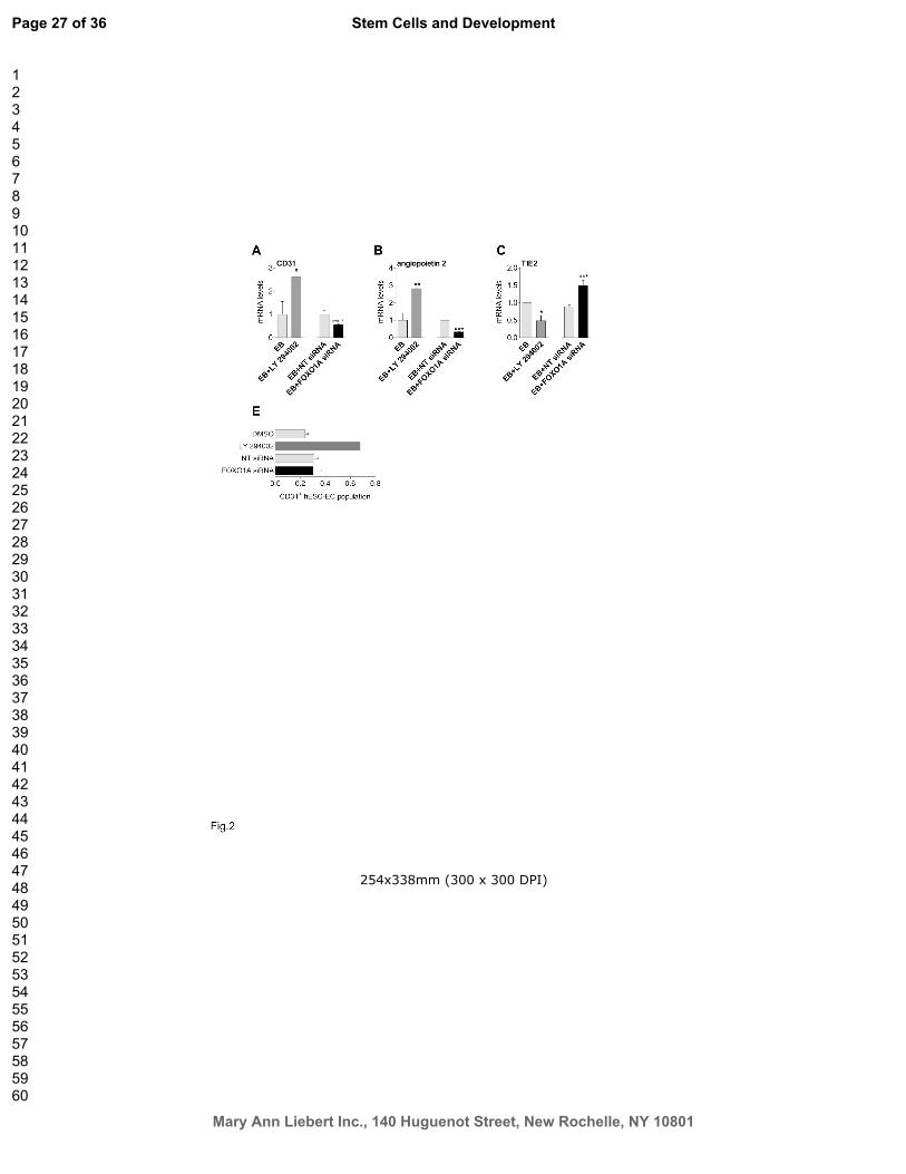
Next we targeted PI3K/FOXO1A pathway to directly assess its role in endothelial differentiation.
Administration of LY294002 during differentiation increased mRNA levels of CD31 and angiopoietin2 in
differentiating hESC (Fig. 2AB). Tie2 mRNA levels were downregulated in response to LY294002 (Fig.
2C). Cells treated with FOXO siRNA showed however lower mRNA levels of CD31 (p=0.07) and
angiopoietin2 (p<0.001) as compared with non;targeting siRNA group (Fig. 2AB). Treatment with FOXO
siRNA resulted in increased mRNA levels of Tie2 (Fig. 2C). FOXO1A mRNA levels were lower after
siRNA treatment on hESC (Supplementary Fig. S2B). Use of PI3K inhibitor/FOXO1A activator
LY294002 tended to increase the percentage of newly formed CD31+ hESC;EC in the presence of EGM2
medium, containing endothelial growth factors (LY294002: 0.62±0.04% vs. 0.24±0.1% of control
differentiating hESC culture; Fig. 2D). LY294002 resulted in higher FOXO1A mRNA levels of
differentiating hESC (Supplementary Fig. S2A). By contrast, silencing of FOXO1A by siRNA during
differentiation had no influence on the percentage of CD31+ cells (Fig. 2D).
Modulation of PI3K/FOXO1A signalling pathway in sorted hESC-EC
Cultures of differentiated CD31+ hESC;EC were highly proliferative and showed specific
endothelial characteristics such as cobblestone morphology, evidence of DiI;labelled acetylated LDL
uptake, and positive expression ve;cadherin;eGFP and staining for ve;cadherin (Supplementary Fig.
S3ABCD). As assessed by proteome profiler, protein levels of angiogenic and endothelial factors showed
similarities in hESC;EC and control adult HCAEC (Supplementary Fig. S4E). Human ESC;EC had low or
Page 14 of 36
Mary Ann Liebert Inc., 140 Huguenot Street, New Rochelle, NY 10801
Stem Cells and Development
123456789101112131415161718192021222324252627282930313233343536373839404142434445464748495051525354555657585960

11
not detectable levels of haematopoietic markers (Supplementary Fig. S4F). As shown by high content
microscopy, hESC;EC did not express the hematopoietic progenitor cell marker CD45 (Supplementary
Fig. S4G).
To test the effects of PI3K/FOXO1A modulation in hESC;EC, we treated cells with LY294002 or
FOXO1A siRNA. Nuclear translocation and mRNA levels of FOXO1A were markedly increased in
hESC;EC in response to LY294002 (Supplementary Fig. S2CD). In contrast, silencing of FOXO1A with
siRNA decreased both mRNA and nuclear translocation levels of FOXO1A as early as after 6 hours
treatment in hESC;EC (Supplementary Fig. S2EF). Similarly to differentiating hESC, mRNA levels of
CD31 tended to increase in response to LY294002 and decreased in cells with FOXO siRNA (Fig. 3AD).
Angiopoietin2 mRNA were also increased in response to LY294002 in hESC;EC; on the other hand,
FOXO siRNA resulted in lower mRNA levels of angiopoietin2 (Fig. 3BE). The mRNA levels of the Tie2
were inversely regulated as compared to angiopoietin2 (Fig.3CF).
Proliferative activity of sorted hESC;EC cultures were assessed by Ki67 marker and colony
formation. Percentage of Ki67+ cells and colony formation activity after 3 days were significantly
decreased in response to LY294002 (Fig. 3GHI); in contrast, further downregulation of FOXO1A
expression by siRNA treatment resulted in a modest increase in the ratio of Ki67+ cells (p<0.05) with no
effect on cell number (Fig. 3GHJ). Overexpression of FOXO1A alone did not change the ratio of Ki67;
positive cells and colony formation in hESC;EC population (Supplementary Fig. S4AB). This may
suggest distinct role of PI3K/FOXO1A pathway in cell generation during hESC differentiation as
compared to those in a later stage in sorted hESC;EC.
Targeting PI3K/FOXO1A pathway alters hESC-EC viability
Next we tested the role of PI3K/FOXO1A pathway on viability and cell survival of hESC;EC. As
assessed by high content microscopy, oxidative stress induced by H2O2 activated FOXO1A as shown by
its increased nuclear density (Supplementary Fig. S5AB). Using H2O2 as a danger signal caused necrosis
and nuclear remodelling (expressed as decreased nuclear size) in a dose;dependent manner in hESC;EC
Page 15 of 36
Mary Ann Liebert Inc., 140 Huguenot Street, New Rochelle, NY 10801
Stem Cells and Development
123456789101112131415161718192021222324252627282930313233343536373839404142434445464748495051525354555657585960

12
(Supplementary Fig. S5CD). Silencing of FOXO1A by siRNA had no effect on levels of necrosis marker
TOTO1 or nuclear remodelling in hESC;EC (p<0.05, Supplementary Fig. S5CD). Pre;treatment with
LY294002 increased the pro;necrotic effects of H2O2 (at a dose of 600 µM, P<0.001) in hESC;EC.
Overexpression of FOXO1A resulted in higher levels of necrosis compared to those in control plasmid;
transfected hESC;EC (Supplementary Fig. S4CDE). The level of necrosis in response to H2O2 was not
modulated by FOXO1A overexpression. Using LY294002 without external danger signal (such as H2O2)
had no effect on viability of hESC;EC (Supplementary Fig. S5CD).
PI3K/FOXO1A pathway modulates angiogenic activity of hESC-EC
In vitro tube formation assay revealed that LY294002 inhibits hESC;EC migration and formation
of tubes (Fig. 4ABCD). On the other hand, silencing of FOXO1A by siRNA markedly increased in vitro
tube formation activity of hESC;EC as shown by total tube length and node count (p<0.001, Fig. 4AEFG).
Overexpression of FOXO1A did not alter tube formation activity of hESC;EC (Supplementary Fig.
S4FGH). To test the viability and maturation of hESC;EC with low FOXO1A levels also in vivo, cell
were transplanted into athymic nude rats. Human ESC;EC showed engraftment (Fig. 4H); after 21 days
conditioning of cells, angiopoietin2 and tie2 mRNA levels were markedly increased in hESC;EC similarly
to those in control HUVEC (Fig. 4JK). However, hESC;EC retain their low FOXO1A expression levels
during the incubation period (Fig. 4I) whereas FOXO1A mRNA levels are further increased in HUVEC.
Discussion
The underlying intracellular signalling mechanisms in the generation and function of human
pluripotent stem cell;derived endothelial cells have not been described in details. In this study we showed
that PI3K pathway is one of the key intracellular signalling mechanisms which have wide;ranging effects
on endothelial differentiation of hESC as well as cell death, proliferation and angiogenic activity of
generated hESC;EC partially through FOXO1A transcription factor (Fig. 5).
Page 16 of 36
Mary Ann Liebert Inc., 140 Huguenot Street, New Rochelle, NY 10801
Stem Cells and Development
123456789101112131415161718192021222324252627282930313233343536373839404142434445464748495051525354555657585960

13
The role of PI3K in generating new endothelial cells was supported by our observation that hESC
differentiation towards endothelial lineage is accompanied with marked changes in expression of most of
the genes related to PI3K/FOXO1A pathways. FOXO1A was shown to be the most abundant FOXO
member at mRNA levels in undifferentiated hESC colonies and alteration of FOXO1A causes different
expression of pluripotency genes in hESC indicating FOXO1A has a critical role in the regulation of
hESC fate [11]. Indeed, here we showed that high FOXO1A expression was significantly downregulated
during embryoid body formation and further differentiation into endothelial cells. Changes in expression
also correlated with loss of pluripotency markers. By using a gene expression array of PI3K pathway
elements, Ingenuity pathway analysis with a curated database and subsequent clustering as an exploratory
tool, we identified connections between FOXO1A and VEGF as well as various further angiogenic
factors. Time development during differentiation modulated the expression of angiogenic factors clustered
with FOXO1A such as PDK1 which play a role in vascular remodelling and endothelial differentiation
[25]; cdc42, known to mediate tubulogenesis [26]; protein kinase C beta, being a stimulus for endothelial
proliferation [27]; and activation of Rheb/mTOR for endothelial cell transformation [28]. As a potential
result of these interactions, we found that inhibition of PI3K and consequent re;activation of FOXO1A by
LY294002 resulted in an increased number of newly formed endothelial cells. Higher FOXO1A levels
were accompanied with increasing CD31 and angiopoietin2 expression levels as well as an inverse
downregulation in tie2 levels in the differentiating culture. This suggests an indirect link between
FOXO1A levels and the propensity of differentiating hESC towards endothelial lineage. The fact however
that the control endothelial HUVEC expressed FOXO1A at a higher level than hESC;EC suggests that
post;transcriptional control of FOXO1A is more important in HUVEC than those in hESC;EC.
The role of FOXO1A in endothelial development was further evidenced by siRNA silencing
experiments. Directly targeting FOXO1A factor decreased expression of endothelial cell markers and
angiogenesis genes. This also suggests that culture medium containing VEGF and other endothelial
cytokines may be potent stimulus for endothelial differentiation where PI3K/FOXO1A pathway, at least in
part, mediates these signals. This is similar to other studies, where VEGF was shown to increase the yield
Page 17 of 36
Mary Ann Liebert Inc., 140 Huguenot Street, New Rochelle, NY 10801
Stem Cells and Development
123456789101112131415161718192021222324252627282930313233343536373839404142434445464748495051525354555657585960

14
of adult endothelial cells in differentiated hESC culture [29] and can also favour endothelial cell survival,
proliferation and cell cycle progression via the PI3K/Akt pathway during tissue regeneration and disease
[30,31].
Intact PI3K signalling as well as low but detectable FOXO1A levels may be prerequisite for
endothelial differentiation. However, our data suggest a different regulatory role for PI3K/FOXO1A in
differentiating ESC and ESC;derived endothelial cells where FOXO1A has a mainly inhibitory feedback
signal. We characterized the temporal expression of FOXO1A and angiopoietin2, one of the FOXO1A
target genes [32–34] and related to angiogenesis and vascular remodelling, during in vivo differentiation
and maturation of hESC;EC. Three weeks after transplantation of hESC;EC into athymic nude rats, cells
showed engraftment and were detectable with histology. As opposed to HUVEC, we detected high
angiopoietin2 and tie2 expressions, with no significant change in FOXO1A levels in hESC;EC. In vitro
differentiation of hESC generated a unique endothelial cell type, where FOXO1A levels are significantly
lower than those in control HUVEC, endothelial cells derived from human umbilical cord vein. This may
suggest that these cells retain low FOXO1A levels and a controllable angiogenic activity even after in
vivo conditioning. On contrary, we found that activation of FOXO1A by LY294002 blocked tube
formation in culture. This activity may be further modulated by the component that inhibition of PI3K and
consequent activation of FOXO1A by LY294002 was also found to block cell proliferation of hESC;EC.
FOXO proteins were shown to be involved in response to oxidative stress, in regulation of
apoptosis and cell survival [14–16]. In line with these reports, here we showed that FOXO1A mediates
danger signals such as oxidative stress by H2O2 in hESC;EC. Indeed, activation of FOXO1A nuclear
translocation by H2O2 was accompanied with cell loss, necrosis and nuclear remodelling in a dose;
dependent manner. Furthermore, overexpression of FOXO1A construct showed direct pro;necrotic effects
and cell loss in hESC;EC cultures. This may suggest that FOXO1A can act as one of the downstream
pathways for oxidative stress in hESC;EC. The fact that FOXO1A expression levels stayed low during
cell transplantation may suggest that these cells have lower responsiveness to in vivo danger signals. On
the other hand, we found that silencing of FOXO1A had no protective effect on stress responsiveness of
Page 18 of 36
Mary Ann Liebert Inc., 140 Huguenot Street, New Rochelle, NY 10801
Stem Cells and Development
123456789101112131415161718192021222324252627282930313233343536373839404142434445464748495051525354555657585960

15
hESC;EC in vitro, which altogether suggests the presence of a broader regulation involving other,
FOXO1A;independent pathways in stress;responsiveness in hESC;EC. LY294002 is one of the potent and
specific cell;permeable inhibitor of PI3K [35]. We have found that inhibition of PI3K pathway by
LY294002 further increased loss of hESC;EC in oxidative stress. This observation may be in line with
earlier animal studies where development of dermal toxicity was an in vivo side effect of LY294002 in
murine model [36]; together with low solubility and bioavailability prevented its use in clinical trials.
However, several further compounds are being developed to inhibit different nodes of the PI3K pathway.
These mainly PI3K/AKT/mTOR inhibitors showed no unexpected toxic effects [8]. However, no clear
correlation between endothelial function and vascular damage, and responsiveness to PI3K/AKT/mTOR
inhibitors has emerged from early clinical trials. It is also still unclear whether downregulation of PI3K
signalling will be sufficient to produce a clinical response.
In conclusion, human embryonic stem cell;derived endothelial cells represent a unique endothelial
population, with controllable proliferative and angiogenic activities. PI3K/FOXO1A pathway is one of the
particular key signalling elements for function and survival of hESC;EC but also in regulation of
endothelial cell fate. We found that activated FOXO1A has various effects in hESC;EC: it mediates
endothelial generation; on the other hand, FOXO1A, at least partly, inhibits proliferation and angiogenic
activity, and plays a role in response to oxidative stress. To test whether using of cells along with direct
pharmacological inhibition of FOXO to release negative feedback may be advantageous in cell therapy
still need to be determined.
Sources of Founding
Project was supported by British Heart Foundation, Hungarian Scientific Research Fund (MB08A 81237,
105555) and National Development Agency (TÁMOP;4.2.2/B;10/1;2010;0013).
Author Disclosure Statement
No competing financial interests exist.
Page 19 of 36
Mary Ann Liebert Inc., 140 Huguenot Street, New Rochelle, NY 10801
Stem Cells and Development
123456789101112131415161718192021222324252627282930313233343536373839404142434445464748495051525354555657585960

16
Reference List
1. Levenberg S. (2005). Engineering blood vessels from stem cells: recent advances and applications.
Curr Opin Biotechnol 16:516;523.
2. Leeper NJ, Hunter AL and Cooke JP. (2010). Stem cell therapy for vascular regeneration: adult,
embryonic, and induced pluripotent stem cells. Circulation 122:517;526.
3. Kehat I, Kenyagin;Karsenti D, Snir M, Segev H, Amit M, Gepstein A, Livne E, Binah O, Itskovitz;
Eldor J and Gepstein L. (2001). Human embryonic stem cells can differentiate into myocytes with
structural and functional properties of cardiomyocytes. J Clin Invest 108:407;414.
4. Ferreira LS, Gerecht S, Shieh HF, Watson N, Rupnick MA, Dallabrida SM, Vunjak;Novakovic G
and Langer R. (2007). Vascular progenitor cells isolated from human embryonic stem cells give rise to
endothelial and smooth muscle like cells and form vascular networks in vivo. Circ Res 101:286;294.
5. Levenberg S, Zoldan J, Basevitch Y and Langer R. (2007). Endothelial potential of human
embryonic stem cells. Blood 110:806;814.
6. Howard L, Mackenzie RM, Pchelintsev NA, McBryan T, McClure JD, McBride MW, Kane NM,
Adams PD, Milligan G and Baker AH. (2013). Profiling of transcriptional and epigenetic changes during
directed endothelial differentiation of human embryonic stem cells identifies FOXA2 as a marker of early
mesoderm commitment. Stem Cell Res Ther 4:36.
7. Engelman JA, Luo J and Cantley LC. (2006). The evolution of phosphatidylinositol 3;kinases as
regulators of growth and metabolism. Nat Rev Genet 7:606;619.
8. Rodon J, Dienstmann R, Serra V and Tabernero J. (2013). Development of PI3K inhibitors: lessons
learned from early clinical trials. Nat Rev Clin Oncol 10:143;153.
9. Furman RR, Sharman JP, Coutre SE, Cheson BD, Pagel JM, Hillmen P, Barrientos JC, Zelenetz
AD, Kipps TJ, Flinn I, Ghia P, Eradat H, Ervin T, Lamanna N, Coiffier B, Pettitt AR, Ma S, Stilgenbauer
S, Cramer P, Aiello M, Johnson DM, Miller LL, Li D, Jahn TM, Dansey RD, Hallek M and O'Brien SM.
(2014). Idelalisib and rituximab in relapsed chronic lymphocytic leukemia. N Engl J Med 370:997;1007.
Page 20 of 36
Mary Ann Liebert Inc., 140 Huguenot Street, New Rochelle, NY 10801
Stem Cells and Development
123456789101112131415161718192021222324252627282930313233343536373839404142434445464748495051525354555657585960

17
10. Brunet A, Bonni A, Zigmond MJ, Lin MZ, Juo P, Hu LS, Anderson MJ, Arden KC, Blenis J and
Greenberg ME. (1999). Akt promotes cell survival by phosphorylating and inhibiting a Forkhead
transcription factor. Cell 96:857;868.
11. Zhang X, Yalcin S, Lee DF, Yeh TY, Lee SM, Su J, Mungamuri SK, Rimmele P, Kennedy M,
Sellers R, Landthaler M, Tuschl T, Chi NW, Lemischka I, Keller G and Ghaffari S. (2011). FOXO1 is an
essential regulator of pluripotency in human embryonic stem cells. Nat Cell Biol 13:1092;1099.
12. Ronnebaum SM, Patterson C. (2010). The FoxO family in cardiac function and dysfunction. Annu
Rev Physiol 72:81;94.
13. Maiese K, Chong ZZ, Shang YC and Hou J. (2009). FoxO proteins: cunning concepts and
considerations for the cardiovascular system. Clin Sci (Lond) 116:191;203.
14. Kops GJ, Dansen TB, Polderman PE, Saarloos I, Wirtz KW, Coffer PJ, Huang TT, Bos JL, Medema
RH and Burgering BM. (2002). Forkhead transcription factor FOXO3a protects quiescent cells from
oxidative stress. Nature 419:316;321.
15. Nakamura T, Sakamoto K. (2008). Forkhead transcription factor FOXO subfamily is essential for
reactive oxygen species;induced apoptosis. Mol Cell Endocrinol 281:47;55.
16. Potente M, Urbich C, Sasaki K, Hofmann WK, Heeschen C, Aicher A, Kollipara R, DePinho RA,
Zeiher AM and Dimmeler S. (2005). Involvement of Foxo transcription factors in angiogenesis and
postnatal neovascularization. J Clin Invest 115:2382;2392.
17. Furuyama T, Kitayama K, Shimoda Y, Ogawa M, Sone K, Yoshida;Araki K, Hisatsune H,
Nishikawa S, Nakayama K, Nakayama K, Ikeda K, Motoyama N and Mori N. (2004). Abnormal
angiogenesis in Foxo1 (Fkhr);deficient mice. J Biol Chem 279:34741;34749.
18. Brito;Martins M, Harding SE and Ali NN. (2008). beta(1); and beta(2);adrenoceptor responses in
cardiomyocytes derived from human embryonic stem cells: comparison with failing and non;failing adult
human heart. Br J Pharmacol 153:751;759.
19. Foldes G, Liu A, Badiger R, Paul;Clark M, Moreno L, Lendvai Z, Wright JS, Ali NN, Harding SE
and Mitchell JA. (2010). Innate immunity in human embryonic stem cells: comparison with adult human
endothelial cells. PLoS One 5:e10501.
Page 21 of 36
Mary Ann Liebert Inc., 140 Huguenot Street, New Rochelle, NY 10801
Stem Cells and Development
123456789101112131415161718192021222324252627282930313233343536373839404142434445464748495051525354555657585960

18
20. Levenberg S, Golub JS, Amit M, Itskovitz;Eldor J and Langer R. (2002). Endothelial cells derived
from human embryonic stem cells. Proc Natl Acad Sci U S A 99:4391;4396.
21. Lara;Pezzi E, Winn N, Paul A, McCullagh K, Slominsky E, Santini MP, Mourkioti F,
Sarathchandra P, Fukushima S, Suzuki K and Rosenthal N. (2007). A naturally occurring calcineurin
variant inhibits FoxO activity and enhances skeletal muscle regeneration. J Cell Biol 179:1205;1218.
22. James D, Nam HS, Seandel M, Nolan D, Janovitz T, Tomishima M, Studer L, Lee G, Lyden D,
Benezra R, Zaninovic N, Rosenwaks Z, Rabbany SY and Rafii S. (2010). Expansion and maintenance of
human embryonic stem cell;derived endothelial cells by TGFbeta inhibition is Id1 dependent. Nat
Biotechnol 28:161;166.
23. Van Der Heide LP, Hoekman MF and Smidt MP. (2004). The ins and outs of FoxO shuttling:
mechanisms of FoxO translocation and transcriptional regulation. Biochem J 380:297;309.
24. Ding GJ, Fischer PA, Boltz RC, Schmidt JA, Colaianne JJ, Gough A, Rubin RA and Miller DK.
(1998). Characterization and quantitation of NF;kappaB nuclear translocation induced by interleukin;1
and tumor necrosis factor;alpha. Development and use of a high capacity fluorescence cytometric system.
J Biol Chem 273:28897;28905.
25. Feng Q, Di R, Tao F, Chang Z, Lu S, Fan W, Shan C, Li X and Yang Z. (2010). PDK1 regulates
vascular remodeling and promotes epithelial;mesenchymal transition in cardiac development. Mol Cell
Biol 30:3711;3721.
26. Kesavan G, Sand FW, Greiner TU, Johansson JK, Kobberup S, Wu X, Brakebusch C and Semb H.
(2009). Cdc42;mediated tubulogenesis controls cell specification. Cell 139:791;801.
27. Meier R, Alessi DR, Cron P, Andjelkovic M and Hemmings BA. (1997). Mitogenic activation,
phosphorylation, and nuclear translocation of protein kinase Bbeta. J Biol Chem 272:30491;30497.
28. Sodhi A, Chaisuparat R, Hu J, Ramsdell AK, Manning BD, Sausville EA, Sawai ET, Molinolo A,
Gutkind JS and Montaner S. (2006). The TSC2/mTOR pathway drives endothelial cell transformation
induced by the Kaposi's sarcoma;associated herpesvirus G protein;coupled receptor. Cancer Cell 10:133;
143.
Page 22 of 36
Mary Ann Liebert Inc., 140 Huguenot Street, New Rochelle, NY 10801
Stem Cells and Development
123456789101112131415161718192021222324252627282930313233343536373839404142434445464748495051525354555657585960

19
29. Nourse MB, Halpin DE, Scatena M, Mortisen DJ, Tulloch NL, Hauch KD, Torok;Storb B, Ratner
BD, Pabon L and Murry CE. (2010). VEGF induces differentiation of functional endothelium from human
embryonic stem cells: implications for tissue engineering. Arterioscler Thromb Vasc Biol 30:80;89.
30. Abid MR, Guo S, Minami T, Spokes KC, Ueki K, Skurk C, Walsh K and Aird WC. (2004).
Vascular endothelial growth factor activates PI3K/Akt/forkhead signaling in endothelial cells. Arterioscler
Thromb Vasc Biol 24:294;300.
31. Ball SG, Shuttleworth CA and Kielty CM. (2007). Vascular endothelial growth factor can signal
through platelet;derived growth factor receptors. J Cell Biol 177:489;500.
32. Daly C, Pasnikowski E, Burova E, Wong V, Aldrich TH, Griffiths J, Ioffe E, Daly TJ, Fandl JP,
Papadopoulos N, McDonald DM, Thurston G, Yancopoulos GD and Rudge JS. (2006). Angiopoietin;2
functions as an autocrine protective factor in stressed endothelial cells. Proc Natl Acad Sci U S A
103:15491;15496.
33. van der Vos KE, Coffer PJ. (2011). The extending network of FOXO transcriptional target genes.
Antioxid Redox Signal 14:579;592.
34. Hurley D, Araki H, Tamada Y, Dunmore B, Sanders D, Humphreys S, Affara M, Imoto S, Yasuda
K, Tomiyasu Y, Tashiro K, Savoie C, Cho V, Smith S, Kuhara S, Miyano S, Charnock;Jones DS,
Crampin EJ and Print CG. (2012). Gene network inference and visualization tools for biologists:
application to new human transcriptome datasets. Nucleic Acids Res 40:2377;2398.
35. Arcaro A, Wymann MP. (1993). Wortmannin is a potent phosphatidylinositol 3;kinase inhibitor: the
role of phosphatidylinositol 3,4,5;trisphosphate in neutrophil responses. Biochem J 296 ( Pt 2):297;301.
36. Hu L, Zaloudek C, Mills GB, Gray J and Jaffe RB. (2000). In vivo and in vitro ovarian carcinoma
growth inhibition by a phosphatidylinositol 3;kinase inhibitor (LY294002). Clin Cancer Res 6:880;886.
Page 23 of 36
Mary Ann Liebert Inc., 140 Huguenot Street, New Rochelle, NY 10801
Stem Cells and Development
123456789101112131415161718192021222324252627282930313233343536373839404142434445464748495051525354555657585960

20
Figure Legends
Fig 1. PI3K/FOXO1A signalling;related gene expression during hESC differentiation. Human embryonic
stem cells (hESC) were differentiated via embryoid body formation (EB) towards human embryonic stem
cell;derived endothelial cells (hESC;EC). HUVEC were used as control endothelial cells. (A) Changes in
mRNA levels of embryonic stem cell marker OCT4, CD31, angiogenesis markers angiopoietin2 and tie2
at different steps of endothelial differentiation. Heat map with agglomerative clustering (B) and bar graphs
(C) showing changes in mRNA levels of PI3K/FOXO elements during different steps of endothelial
differentiation. Bar graphs in panel B showing differences in gene expression in HUVEC vs. those in
hESC;EC as a baseline. Data are expressed as fold changes vs. mRNA levels in undifferentiated hESC;
mean±SEM. Statistical significance was determined by one;way ANOVA. *p<0.05, **p<0.01,
***p<0.01, from 3 biological replicates, one;way ANOVA with Tukey post hoc test. (D) Schematic
diagram of Ingenuity Pathway Analysis;mapped mechanistic interactions of VEGF, PTEN, FOXO1A and
various angiogenic modulator elements. Arrows between nodes represent direct (solid lines) and indirect
(dashed lines) interactions between molecules as supported by information in the Ingenuity pathway
knowledge base. ITGB1, Integrin beta;1; PRKCA, Protein kinase C alpha; PRKCZ, Protein kinase C zeta;
PDGFRA, platelet;derived growth factor receptor alpha; PTEN, Phosphatase and tensin homolog;
PRKCB, Protein kinase C beta; PIK3R2, phosphoinositide;3;kinase, regulatory subunit 2 beta.
Fig 2. PI3K/FOXO1A signalling modulates endothelial differentiation of hESC. Bar graphs showing
mRNA levels of CD31 (A), angiopoietin2 (B), and Tie2 (C) in differentiating hESC (EB) with LY294002
and FOXO1A siRNA. (D) Bar graph showing CD31+ hESC;EC population in the presence of FOXO1A
siRNA or LY294002 (10VM). mean±SEM, *p<0.05, ***p<0.001 vs. control group, 3 biological
replicates, Student’s t;test.
FIG 3. PI3K/FOXO1A signalling modulates proliferative activity of hESC;derived endothelial cells. Bar
graphs showing changes in mRNA levels of CD31, angiopoietin2, and Tie2 in differentiated hESC;EC in
Page 24 of 36
Mary Ann Liebert Inc., 140 Huguenot Street, New Rochelle, NY 10801
Stem Cells and Development
123456789101112131415161718192021222324252627282930313233343536373839404142434445464748495051525354555657585960

21
response to 24 hours treatment with LY294002 (ABC) or FOXO1A siRNA (DEF). (G) Representative
picture of Ki67+ cells in hESC;EC population (magnification: x20). Changes in proliferation rate of the
hESC;EC population (Ki67+ cells) after 24 hours (H) and in colony formation activity (IJ) in response to
LY294002 or FOXO1A siRNA treatments for 3 days. Data are expressed as fold changes vs. percentage
of Ki67+ cells in control hESC;EC group, mean±SEM. Fold changes in mRNA levels are shown vs.
hESC. *p<0.05, ***p<0.001 vs. control, n=3 biological replicates.
FIG 4. In vitro angiogenesis and in vivo conditioning of hESC;EC. (A) Representative Matrigel tube
formation images of hESC;EC pre;treated with LY294002 or FOXO1A siRNA. Cells were stained with
vital dye TMRM (red). Bar graph showing average and total tube lengths and number of formed nodes
after LY294002 (BCD) 24 hours FOXO1A or non;targeting scrambled (NT) siRNA (FGI).***p<0.001
vs. control, n=3 biological replicates. (E) Representative image of histological section stained with
haematoxylin;eosin showing subcutaneously engrafted hESC;EC. (IJK) Bar graphs showing FOXO1A
angiopoietin2 and Tie2 mRNA levels before and after in vivo conditioning of hESC;EC in Matrigel plugs
transplanted into athymic nude rats. Data are mean±SEM (n=3;10 per group from 2 independent
experiments (12 rats per group)).
FIG 5. Mechanism of action and effects of modulation of PI3K;FOXO1A signalling pathway. Schematic
summary shows that activation of FOXO1A with PI3K inhibitor LY294002 resulted in increased yield of
endothelial differentiation (labelled as red panels). Proliferative and tube formation capacity decreased
(labelled as blue panels) in the differentiated endothelial cells. Modulation of PI3K;FOXO1A signalling
also altered angiopoietin2;tie2 system in the differentiating culture. Low FOXO1A levels increased
proliferative activity and tube formation of the developed hESC;EC. FOXO siRNA had no effect (grey
panels) on differentiation and cell death profile.
Page 25 of 36
Mary Ann Liebert Inc., 140 Huguenot Street, New Rochelle, NY 10801
Stem Cells and Development
123456789101112131415161718192021222324252627282930313233343536373839404142434445464748495051525354555657585960

254x338mm (300 x 300 DPI)
Page 26 of 36
Mary Ann Liebert Inc., 140 Huguenot Street, New Rochelle, NY 10801
Stem Cells and Development
123456789101112131415161718192021222324252627282930313233343536373839404142434445464748495051525354555657585960
254x338mm (300 x 300 DPI)
Page 27 of 36
Mary Ann Liebert Inc., 140 Huguenot Street, New Rochelle, NY 10801
Stem Cells and Development
123456789101112131415161718192021222324252627282930313233343536373839404142434445464748495051525354555657585960

254x338mm (300 x 300 DPI)
Page 28 of 36
Mary Ann Liebert Inc., 140 Huguenot Street, New Rochelle, NY 10801
Stem Cells and Development
123456789101112131415161718192021222324252627282930313233343536373839404142434445464748495051525354555657585960

254x338mm (300 x 300 DPI)
Page 29 of 36
Mary Ann Liebert Inc., 140 Huguenot Street, New Rochelle, NY 10801
Stem Cells and Development
123456789101112131415161718192021222324252627282930313233343536373839404142434445464748495051525354555657585960

254x338mm (300 x 300 DPI)
Page 30 of 36
Mary Ann Liebert Inc., 140 Huguenot Street, New Rochelle, NY 10801
Stem Cells and Development
123456789101112131415161718192021222324252627282930313233343536373839404142434445464748495051525354555657585960

Merkely et al. Signalling via PI3K/FOXO1A Pathway Modulates Formation and Survival of
Human Embryonic Stem Cell-Derived Endothelial Cells
Supplemental Figure Legends
Supplemental Fig. 1. Automated high content imaging�based assessments of proliferation and
nuclear translocation of FOXO1A. The system was used to scan multiple fields from well to well and
to acquire and analyze each of the cells in the images. Representative images show hESC�EC stained
for Hoechst (AD), Ki67 (C) and FOXO1A (F). Inlay panel in (F) shows that prior to stimulation, the
cytoplasm is intensely red while the nucleus without FOXO1A is dark (nuclear minus cytoplasmic
fluorescence is low). In the stimulated cell, the amount of cytoplasmic fluorescence is decreased,
while the nuclear fluorescence markedly increases (nuclear minus cytoplasmic fluorescence is high).
For compartmental analysis, nuclear and cytoplasmic regions of interests in each cell are also shown
(BE). In both the nuclear and cytoplasmic areas, the intensity of fluorescence of a given protein is
averaged throughout the measured area. White scale bars represent 20 µm.
Supplemental Fig. 2. Modulation of FOXO1A expression and nuclear intensities. (A) Changes of
FOXO1A mRNA levels after treatment with the PI3K inhibitor/FOXO1A activator LY294002
(10<M) or FOXO1A using siRNA (B) in differentiating hESC. Changes of FOXO1A nuclear
intensity (C) and mRNA levels (D) in hESC�EC after treatment with the PI3K inhibitor/FOXO1A
activator LY294002 (24h). Silencing of FOXO1A using siRNA caused decreased level of (E) nuclear
intensity or (F) FOXO1A mRNA levels.**p<0.01, ***p<0.001 vs. control, n=4 biological replicates.
Supplemental Fig 3. Endothelial characteristics of hESC�EC. (A) Representative picture of
cobblestone morphology of hESC�EC on gelatinised plates. White scale bar represents 20 µm (B)
CD31+ hESC�EC are able to take up acetylated LDL. (C) hESC�EC is positive for ve�cadherin�driven
GFP construct in vitro and (D) stained positive with for endothelial marker ve�cadherin in vivo. (E)
Bar graph showing angiogenesis proteome profiler analysis of hESC�EC. Cell lysates of hESC�EC
express wide range of angiogenesis�related proteins. (F) Bar graph showing expression of cluster of
Page 31 of 36
Mary Ann Liebert Inc., 140 Huguenot Street, New Rochelle, NY 10801
Stem Cells and Development
123456789101112131415161718192021222324252627282930313233343536373839404142434445464748495051525354555657585960

differentiation (CD) soluble receptors assessed by human soluble receptor proteome profiler array
from cell culture supernatant. Data showing expression levels of hESC�EC in fold changes compared
to those in human coronary artery endothelial cells (HCAEC). Data are from two biological replicates.
(G) Plot graph on high content analysis shows that CD45 haemopoietic marker was not expressed in
hESC�EC (red plot). Negative control is also shown (black plot).
Supplemental Fig 4. Cell proliferation, cell death and in vitro angiogenesis of hESC�EC with
FOXO1A overexpression. (A) Changes in proliferation rate of the hESC�EC population (Ki67+ cells,
expressed as percentage) after 24 hours and in colony formation activity (B) in response to
overexpression for 3 days. (C) Changes in FOXO1A mRNA levels, To�Pro3 necrosis marker (D), and
cell number after (E) transfection (48 h) with FOXO1A�eGFP or control eGFP plasmids in hESC�EC.
Angiogenic activity was assessed by node count (F), total (G) and average tube length (H) in Matrigel
tube formation assays. Data are expressed as mean±SEM. *p<0.05, **p<0.01, ***p<0.001 vs. control
eGFP group, one�way ANOVA with Tukey post hoc test, n=3 biological replicates.
Supplemental Figure 5. Cell death of hESC�derived endothelial cells is modulated by
PI3K/FOXO1A signalling pathways. H2O2 was used in different concentrations (300, 600, 900 <M) to
induce oxidative stress. LY294002 (10<M, 24h) and FOXO1A�targeting siRNA (24h) were used to
modulate FOXO1A activity. (A) Representative immunocytochemistry images of FOXO1A intensity
during cell death experiments of hESC�EC (magnification 20x). Human ESC�EC were stained with
anti�FOXO1A antibody and nuclei stained with Hoechst. (B) Bar graph showing changes in FOXO1A
nuclear translocation in hESC�EC pre�treated with LY294002 and FOXO1A siRNA after challenging
with H2O2. (C) Bar graph showing changes in necrotic rate measured by TOTO�1 intensity of hESC�
EC. (D) Changes in nuclear remodelling measuring Hoechst�positive nuclear size of hESC�EC.
***p<0.001 vs. control, n=3 biological replicates.
Page 32 of 36
Mary Ann Liebert Inc., 140 Huguenot Street, New Rochelle, NY 10801
Stem Cells and Development
123456789101112131415161718192021222324252627282930313233343536373839404142434445464748495051525354555657585960

254x338mm (300 x 300 DPI)
Page 33 of 36
Mary Ann Liebert Inc., 140 Huguenot Street, New Rochelle, NY 10801
Stem Cells and Development
123456789101112131415161718192021222324252627282930313233343536373839404142434445464748495051525354555657585960

254x338mm (300 x 300 DPI)
Page 34 of 36
Mary Ann Liebert Inc., 140 Huguenot Street, New Rochelle, NY 10801
Stem Cells and Development
123456789101112131415161718192021222324252627282930313233343536373839404142434445464748495051525354555657585960

254x338mm (300 x 300 DPI)
Page 35 of 36
Mary Ann Liebert Inc., 140 Huguenot Street, New Rochelle, NY 10801
Stem Cells and Development
123456789101112131415161718192021222324252627282930313233343536373839404142434445464748495051525354555657585960

254x338mm (300 x 300 DPI)
Page 36 of 36
Mary Ann Liebert Inc., 140 Huguenot Street, New Rochelle, NY 10801
Stem Cells and Development
123456789101112131415161718192021222324252627282930313233343536373839404142434445464748495051525354555657585960

254x338mm (300 x 300 DPI)
Page 37 of 36
Mary Ann Liebert Inc., 140 Huguenot Street, New Rochelle, NY 10801
Stem Cells and Development
123456789101112131415161718192021222324252627282930313233343536373839404142434445464748495051525354555657585960