1,3-butadiene1.1.4 technical products and impurities butadiene is available commercially as a...
TRANSCRIPT

1,3-BUTADIENE
This substance (hereinafter referred to as butadiene) was considered by previousWorking Groups, in June 1985 (IARC, 1986; see also correction, IARC, 1987a), March1987 (IARC, 1987b) and October 1991 (IARC, 1992). Since that time, new data havebecome available, and these have been incorporated into the monograph and taken intoconsideration in the present evaluation.
One of the metabolites of butadiene, 1,2:3,4-diepoxybutane (hereinafter referred toas diepoxybutane), also was previously evaluated by an IARC Working Group (IARC,1976), and its reevaluation by the present Working Group is included in this monograph.
1. Exposure Data
1.1 Chemical and physical dataButadiene1.1.1 Nomenclature
Chem. Abstr. Serv. Reg. No.: 106-99-0Chem. Abstr. Name: 1,3-ButadieneIUPAC Systematic Name: 1,3-ButadieneSynonyms: Biethylene; bivinyl; butadiene; buta-1,3-diene; α,γ-butadiene; trans-butadiene; divinyl; erythrene; pyrrolylene; vinylethylene
1.1.2 Structural and molecular formulae and relative molecular mass
C4H6 Relative molecular mass: 54.09
1.1.3 Chemical and physical properties of the pure substance(a) Description: Colourless mildly aromatic gas (Budavari, 1996)(b) Boiling-point: –4.4°C (Lide, 1995)(c) Melting-point: –108.9°C (Lide, 1995)(d) Density: d4
20 0.6149 (Lide, 1995)(e) Spectroscopy data: Ultraviolet (Grasselli & Ritchey, 1975), infrared (Sadtler
Research Laboratories, 1995; prism [893a], grating [36758]), nuclear magneticresonance and mass spectral data (NIH/EPA Chemical Information System,1983) have been reported.
–109–
H2C CH CH CH2

(f) Solubility: Very slightly soluble in water (735 mg/L at 20°C); soluble in ethanol,diethyl ether, benzene and organic solvents; very soluble in acetone (Lide,1995; Budavari, 1996; Verschueren, 1996)
(g) Volatility: Vapour pressure, 120 kPa at 0°C (Lide, 1995); 235 kPa at 20°C(Müller & Löser, 1985); relative vapour density (air = 1), 1.87 (Verschueren,1996)
(h) Stability: Flash-point, –76°C; very reactive; may form explosive peroxides uponexposure to air; polymerizes readily, particularly if oxygen is present (Lewis,1993; Budavari, 1996)
(i) Explosive limits: Lower, 2.0%; upper, 11.5% (Budavari, 1996)(j) Conversion factor: mg/m3 = 2.21 × ppm1
Diepoxybutane1.1.1 Nomenclature
Chem. Abstr. Serv. Reg. No.: 1464-53-5Chem. Abstr. Name: 2,2′-BioxiraneIUPAC Systematic Name: 1,2:3,4-DiepoxybutaneSynonym: Butadiene dioxide
1.1.2 Structural and molecular formulae and relative molecular mass
C4H6O2 Relative molecular mass: 86.10
1.1.3 Chemical and physical properties of the pure substance(a) Description: Colourless liquid (Budavari, 1996)(b) Boiling-point: 138°C (Budavari, 1996)(c) Melting-point: –19°C (Budavari, 1996)(d) Solubility: Miscible with water (hydrolyses) (Budavari, 1996)(e) Vapour pressure: 918 Pa at 25°C (United States National Library of Medicine,
1997)
IARC MONOGRAPHS VOLUME 71110
1 Calculated from: mg/m3 = (relative molecular mass/24.47) × ppm, assuming a temperature of 25°C and apressure of 101 kPa
H2C C C CH2
O
O
H
H

1.1.4 Technical products and impuritiesButadiene is available commercially as a liquefied gas under pressure. The polymeri-
zation grade has a minimum purity of 99%, with acetylene as an impurity in the parts-per-million (ppm) range. Isobutene, 1-butene, butane and cis-2- and trans-2-butene havebeen detected in pure-grade butadiene (Miller, 1978). Typical specifications for buta-diene are: purity, ≥ 99.5%; inhibitor (tert-butylcatechol), 50–150 ppm; impurities (ppmmax.): 1,2-butadiene, 20; propadiene, 10; total acetylenes, 20; dimers, 500; isoprene, 10;other C5 compounds, 500; sulfur, 5; peroxides (as H2O2), 5; ammonia, 5; water, 300;carbonyls, 10; nonvolatile residues, 0.05 wt% max.; and oxygen in the gas phase, 0.10vol% max. (Sun & Wristers, 1992). Butadiene has been stabilized with hydroquinone,catechol and aliphatic mercaptans (IARC, 1986, 1992).
1.1.5 AnalysisSelected methods for the analysis of butadiene in various matrices are listed in
Table 1. Methods of analysis of butadiene in air have recently been evaluated. Thereappears to be no single preferred method, but newer methods give higher performance.Thermal desorption methods provide high levels of accuracy and precision (Bianchi et al.,1997).
The specificity and detection limits of methods for determining simple, small mole-cules present in packaging material which migrate into packaged goods have been dis-cussed (Vogt, 1988). Butadiene can be determined in plastic polymers, foods and foodsimulants by chromatographic methods.
Several gas detector tubes are used in conjunction with common colorimetric reac-tions to detect butadiene. The reactions include the reduction of chromate or dichromateto chromous ion and the reduction of ammonium molybdate and palladium sulfate tomolybdenum blue (Saltzman & Harman, 1989).
1.2 Production and use1.2.1 Production
Butadiene was first produced in the late nineteenth century by pyrolysis of petroleumhydrocarbons (Kirshenbaum, 1978). Commercial production started in the 1930s.
Butadiene is manufactured primarily as a coproduct of steam cracking of hydrocarbonstreams to produce ethylene in the United States, western Europe and Japan. However, incertain parts of the world (e.g., China, India, Poland and Russia) it is still produced fromethanol. The earlier manufacturing processes of dehydrogenation of n-butane and oxy-hydrogenation of n-butenes have significantly declined in importance and output. Effortshave been made to make butadiene from other feedstocks such as other hydrocarbons, coal,shale oil and renewable sources such as animal and vegetable oil, cellulose, hemicelluloseand lignin, but in the United States none of these has moved beyond the research anddevelopment stage (Müller & Löser, 1985; Sun & Wristers, 1992).
Steam cracking is a complex, highly endothermic pyrolysis reaction. During thereaction, a hydrocarbon feedstock is heated to approximately 800°C and 34 kPa for less
BUTADIENE 111

than one second, during which carbon–carbon and carbon–hydrogen bonds are broken.As a result, a mixture of olefins, aromatics, tar and gases is formed. These products arecooled and separated into specific boiling-range cuts of C1, C2, C3 and C4 compounds.The C4 fraction contains butadiene, isobutene, 1-butene, 2-butene and some other minorhydrocarbons. The overall process yields of butadiene depend on both the process para-meters and the composition of feedstocks. Generally, heavier steam-cracking feedstocksproduce greater amounts of butadiene. Separation and purification of butadiene fromother components is carried out mainly by an extractive distillation process. The mostcommonly used solvents are acetonitrile and dimethylformamide; dimethylacetamide,furfural and N-methyl-2-pyrrolidinone also have been used for this separation. Anothercommercial process to separate butadiene from other hydrocarbons uses a solution con-taining cuprous ammonium acetate, which forms a weak copper(I) complex with buta-diene (Müller & Löser, 1985; Sun & Wristers, 1992).
Dehydrogenation of n-butane via the Houdry process is carried out under partialvacuum (35–75 kPa) at about 535–650°C with a fixed-bed catalyst. The catalyst contains
IARC MONOGRAPHS VOLUME 71112
Table 1. Methods for analysis of butadiene
Samplematrix
Sample preparation Assayprocedurea
Limit ofdetection
Reference
Air Adsorb (charcoal); extract(carbon disulfide)
GC/FID 200 μg/m3 United StatesOccupational Safetyand Health Admi-nistration (1990a)
Adsorb (charcoal); extract(dichloromethane)
GC/FID 0.2 μg/sample Eller (1994)
Adsorb on Perkin-ElmerATD 400 packed withpolymeric or syntheticadsorbent material; thermaldesorption
GC/FID 200 μg/m3 United KingdomHealth and SafetyExecutive (1992)
Foods andplastic food-packagingmaterial
Dissolve (N,N-dimethyl-acetamide) or melt; injectheadspace sample
GC/MS-SIM ~1 μg/kg Startin & Gilbert(1984)
Plastics,liquid foods
Dissolve in ortho-dichloro-benzene; inject headspacesample
GC/FID 2–20 μg/kg United States Foodand Drug Admi-nistration (1987)
Solid foods Cut or mash sample; injectheadspace sample
GC/FID 2–20 μg/kg United States Foodand Drug Admi-nistration (1987)
a Abbreviations GC/FID, gas chromatography/flame ionization detection; GC/MS-SIM, gas chromato-graphy/mass spectrometry with single-ion monitoring

aluminium oxide and chromium oxide as the principal components. Normal butenes canalso be oxidatively dehydrogenated to butadiene in the presence of a high concentrationof steam with fairly high selectivity. The reaction temperature is kept below 600°C tominimize over-oxidation, and the reaction pressure is about 34–103 kPa (Müller &Löser, 1985; Sun & Wristers, 1992).
An estimated 3570 thousand tonnes of butadiene were produced worldwide in 1983(Anon., 1984). By 1989, that figure had risen to an estimated 6620 thousand tonnes, withthe following breakdown by global area (thousand tonnes): North America, 1520; SouthAmerica, 260; western Europe, 1870; eastern Europe, 1490, Africa and the Middle East,150; and Asia and the Pacific, 1330 (Sun & Wristers, 1992). Production figures bycountry for the years 1981–96 are presented in Table 2.
Butadiene remains a major industrial commodity in the United States, ranking 36thamong all chemicals produced in 1996 (Anon., 1996a). Seven major producers in theUnited States, with 10 plant locations, had a total annual capacity of 1900 thousandtonnes in 1996 (Anon., 1996b). Available information indicates that butadiene is pro-duced by seven companies each in Japan and Korea; four companies each in France andGermany; three companies in The Netherlands; two companies each in the CzechRepublic and the United Kingdom; one company each in Austria, Canada, Finland, Italy,Mexico, Portugal, Romania, Singapore, Spain and Taiwan; and an undisclosed numberof companies in Argentina, Brazil, Bulgaria, China, the Commonwealth of IndependentStates, India, Poland and Saudi Arabia (Anon., 1996b).
Diepoxybutane is not believed to be produced commercially except in small quan-tities for research purposes (United States National Library of Medicine, 1997).
BUTADIENE 113
Table 2. Butadiene production in selected countries from 1981through 1996 (thousand tonnes)a
Country 1981 1984 1987 1990 1993 1996
Canada 126 127 167 192 174 212China NRb 141 181 258 NR NRChina (Taiwan) NR NR NR NR 90 129France 266 302 307 281 320 344Germany NR 753 700 777 879 673Italy 163 181 NR NR NR NRJapan 518 627 707 827 809 1025Korea (Republic of) NR NR NR 168 486 601United Kingdom 207 258 231 198 NR NRUnited States 1354 1112 1329 1401 1414 1744
a From Anon. (1985, 1988, 1991, 1994, 1997); China National Chemical InformationCentre (1993)b NR, not reported

1.2.2 UseButadiene is used primarily in the production of synthetic rubbers, including styrene–
butadiene rubber (SBR), polybutadiene rubber (BR), styrene–butadiene latex (SBL),chloroprene rubber (CR) and nitrile rubber (NR). Important plastics containing butadieneas a monomeric component are shock-resistant polystyrene, a two-phase system consis-ting of polystyrene and polybutadiene; ABS polymers consisting of acrylonitrile, buta-diene and styrene; and a copolymer of methyl methacrylate, butadiene and styrene(MBS), which is used as a modifier for poly(vinyl chloride). It is also used as an inter-mediate in the production of chloroprene, adiponitrile and other basic petrochemicals.The worldwide use pattern for butadiene in 1981 was as follows (%): SBR + SBL, 56;BR, 22; CR, 6; NR, 4; ABS, 4; hexamethylenediamine, 4; other, 4. The use pattern forbutadiene in the United States in 1995 was (%): SBR, 31; BR, 24; SBL, 13; CR, 4; ABS,5; NR, 2; adiponitrile, 12; and other, 9 (Anon., 1996b).
Diepoxybutane has been proposed for use in curing polymers and cross-linkingtextile fibres (United States National Library of Medicine, 1997).
1.3 Occurrence1.3.1 Natural occurrence
Butadiene is not known to occur as a natural product.
1.3.2 Occupational exposureAccording to the 1990–93 CAREX database for 15 countries of the European Union
(Kauppinen et al., 1998) and the 1981–83 United States National Occupational ExposureSurvey (NOES, 1997), approximately 30 000 workers in Europe and as many as 50 000workers in the United States were potentially exposed to butadiene (see GeneralRemarks).
Potential exposure to butadiene can occur in the following industrial activities:petroleum refining and related operations (production of C4 fractions containing buta-diene, and production and distribution of gasoline), production of purified butadienemonomer, production of various butadiene-based rubber and plastics polymers and otherderivatives, and manufacture of rubber and plastics products (tyres, hoses and a varietyof moulded objects).
In the descriptions below, the accuracy of the levels of exposure to butadiene mayhave been affected by the inability to distinguish between butadiene and other C4 com-pounds, low desorption efficiency at low concentrations, possible sample breakthroughin charcoal tubes and possible loss during storage in methods used until the mid-1980s(Lunsford et al., 1990; Bianchi et al., 1997). No measurement data are available on levelsof exposure to butadiene before the 1970s, when different processes and working condi-tions (e.g., during the Second World War) would have resulted in exposure levelsdifferent from those now prevalent in developed countries.
IARC MONOGRAPHS VOLUME 71114

(a) Petroleum refining and production of crude butadieneExposure data collected in Europe in 1984–85 suggested that gasoline contains a
small percentage of butadiene. Levels of exposure of workers in various job groups inthe production and distribution of gasoline are shown in Table 3 (see IARC, 1989).Table 4 shows the exposures since 1984 of workers in different areas of petroleum refi-neries and petrochemical facilities where crude butadiene is produced (usually a C4stream obtained as a by-product of ethylene production). Table 5 shows more recent datafrom crackers of butadiene production plants for the years 1986–93 (ECETOC, 1997).
(b) Monomer productionDetailed industrial hygiene surveys were conducted in the United States by the
National Institute for Occupational Safety and Health in 1985 in four of 10 facilitieswhere butadiene was produced by solvent extraction of C4 fractions originating as ethy-lene co-product streams (Krishnan et al., 1987). Levels of butadiene to which workers invarious job categories were exposed are summarized in Table 6. Jobs that require wor-kers to handle or transport containers, such as voiding sample cylinders or loading andunloading tank trucks or rail cars, present the greatest potential exposure. Geometricmeans of full-shift exposure levels for other job categories were below 1 ppm[2.2 mg/m3]. Short-term samples showed that such activities as open-loop sampling and
BUTADIENE 115
Table 3. Personal exposures (mg/m3) to butadiene associated withgasoline during 1984–85 in 13 European countries (540 measurements)
Activity Arithmeticmean
Range Exposureduration(TWA)
Production on-site (refining) 0.3 ND–11.4 8 hProduction off-site (refining) 0.1 ND–1.6 8 hLoading ships (closed system) 6.4 ND–21.0 8 hLoading ships (open system) 1.1 ND–4.2 8 hLoading barges 2.6 ND–15.2 8 hJetty man 2.6 ND–15.9 8 hBulk loading road tankers Top loading < 1 h 1.4 ND–32.3 < 1 h Top loading > 1 h 0.4 ND–4.7 8 h Bottom loading < 1 h 0.2 ND–3.0 < 1 h Bottom loading > 1 h 0.4 ND–14.1 8 hRoad tanker delivery (bulk plant to service station) NDRail car top loading 0.6 ND–6.2 8 hDrumming NDService station attendant (dispensing fuel) 0.3 ND–1.1 8 hSelf-service station (filling tank) 1.6 ND–10.6 2 min
From CONCAWE (1987); ND, not detected; TWA, time-weighted average

cylinder voiding were associated with peak exposures of 100 ppm [220 mg/m3]. Full-shift area samples indicated that ambient concentrations of butadiene were greatest in therail car terminals (geometric mean, 1.8 ppm [3.9 mg/m3]) and in the tank storage farm(2.1 ppm [4.7 mg/m3]).
Exposure data from 15 monomer extraction sites for the year 1995 (Table 7) indi-cated that in general personal exposure levels were below 5 ppm [11 mg/m3]. Data fromearlier years (1984–93) showed less than 10% of the measured concentrations exceeding5 ppm [11 mg/m3] (Table 8) (ECETOC, 1997).
A recent study on biological monitoring for mutagenic effects of exposure to buta-diene reported estimated average exposures of 1 ppm [2.2 mg/m3] for workers in a buta-diene monomer plant. Ambient air concentrations in production areas averaged 3.5 ppm[7.7 mg/m3], while average concentrations of 0.03 ppm [0.07 mg/m3] were reported forthe control area (Ward et al., 1996a). Sorsa et al. (1996a) reported that 70% of thesamples contained below 0.2 ppm [0.4 mg/m3] butadiene from two plants in Portugal(personal samples) and Finland (area samples), while 5% and 2% of the samples,respectively, were above 10 ppm [22 mg/m3].
Monitoring in a Finnish plant generally indicated ambient air levels of less than10 ppm [22 mg/m3] at different sites (33 samples; mean sampling time, 5.3 h). In personalsamples for 16 process workers, the concentrations ranged from < 0.1 to 477 ppm[< 0.22–1050 mg/m3] (mean, 11.5 ppm [25 mg/m3]; median, < 0.1 ppm [< 0.22 mg/m3];46 samples; mean sampling time, 2.5 h). The highest concentrations were measuredduring sample collection. Protective clothing and respirators were used during this ope-ration (Arbetsmiljöfonden, 1991).
Potential exposures in the monomer industry other than to butadiene include extrac-tion solvents and components of the C4 feedstock. Extraction solvents differ betweenfacilities; some common ones are dimethylformamide, dimethylacetamide, acetonitrile,
IARC MONOGRAPHS VOLUME 71116
Table 4. Eight-hour time-weighted average concentrationsof butadiene to which workers in different jobs in petro-leum refineries and petrochemical facilities were exposedfrom 1984 to 1987
Job area No. offacilities
Arithmetic meana Range
ppm mg/m3 ppm mg/m3
Production 7 0.24 0.53 0.008–2.0 0.02–4.4Maintenance 6 0.11 0.24 0.02–0.37 0.04–0.82Distribution 1 2.9 6.41Laboratory 4 0.18 0.40 0.07–0.4 0.16–0.88
From Heiden Associates (1987)a Weighted by number of exposed workers

BUTA
DIEN
E117
Table 5. Personal exposures to butadiene in crackers of butadiene production plants in the EuropeanUnion
Job category Personal exposure (ppm)Year ofmeasurement
Numberof people
Numberof samples
< 1 1–2 2–3 3–4 4–5 5–10 10–25 ≥ 25
Unloading, loading, storage
1986–92 210 92 82 3 3 2 0 0 1 0
Distillation (hot) 1986–93 394 392 382 0 3 1 2 0 2 2Laboratory, sampling
1986–93 132 184 178 2 1 2 1 0 0 0
Maintenance 1986–92 282 371 364 5 0 1 0 0 1 0Other 1990–92 467 509 487 18 2 1 1 NDa 0 0Total 1986–93 1485 1548 1493 28 9 8 4 0 4 2
From ECETOC (1997)a ND, not detected (detection limit not stated)

IARC MONOGRAPHS VOLUME 71118
Table 6. Eight-hour time-weighted average exposure levels in personalbreathing zone samples at four butadiene monomer production faci-lities, United States, 1985
Job category No. ofsamples
Exposure level (ppm [mg/m3])
Arithmeticmean
Geometricmean
Range
Process technician Control room 10 0.45 [1.0] 0.09 [0.2] < 0.02–1.87 [ < 0.04–4.1] Process area 28 2.23 [4.9] 0.64 [1.4] < 0.08–34.9 [ < 0.18–77] Loading area Rail car 9 14.6 [32.4] 1.00 [2.2] 0.12–124 [0.27–273] Tank truck 3 2.65 [5.9] 1.02 [2.3] 0.08–5.46 [0.18–12.1] Tank farm 5 0.44 [0.97] 0.20 [0.44] < 0.04–1.53 [ < 0.09–3.4]Laboratory technician Analysis 29 1.06 [2.3] 0.40 [0.88] 0.03–6.31 [0.07–14.0] Cylinder voiding 3 126 [277] 7.46 [16.5] 0.42–374 [0.93–826]
From Krishnan et al. (1987)
Table 7. Personal exposures to butadieneat 15 monomer extraction sites in theEuropean Union in 1995
Job category Concentration (ppm)
Time-weightedaverages
Range ofvalues
Production Extraction Derivationa
< 0.01–21.4–3.4
(0–14)(0.07–60)
Storage and filling < 0.02–5 (0–18.1)Transport < 0.1–0.7 (0.02–1.2)Laboratory 0.03–1 (0–13.1)
From ECETOC (1997)a Integrated monomer extraction and styrene–buta-diene production on same site

BUTA
DIEN
E119
Table 8. Personal exposures to butadiene in extraction unitsa of butadiene production plants in theEuropean Union
Job category Year ofmeasurement
Numberof people
Numberof samples
Personal exposures (ppm)
< 1 1–2 2–3 3–4 4–5 5–10 10–25 ≥ 25
Unloading, loading, storage
1986–93 392 224 178 9 8 7 2 11 22 7
Distillation (hot) 1985–93 256 626 535 20 19 6 11 8 12 15Laboratory, sampling 1985–93 45 48 29 4 2 2 2 3 5 1Maintenance 1986–93 248 127 93 14 3 2 1 3 4 7Other 1984–92 45 10 8 2 0 0 0 0 0 0Total 1984–93 986 1035 843 49 32 17 16 25 23 30
From ECETOC (1997)a Isolation of butadiene from C4 stream

β-methoxypropionitrile (Fajen, 1985a), furfural and aqueous cuprous ammonium acetate(United States Occupational Safety and Health Administration, 1990b). Stabilizers arecommonly used to prevent formation of peroxides in air and polymerization. No infor-mation was available on these other exposures, or on exposures to chemicals other thanbutadiene that are produced in some facilities, such as butylenes, ethylene, propylene,polyethylene and polypropylene resins, methyl-tert-butyl ether and aromatic hydro-carbons (Fajen, 1985b,c).
(c) Production of polymers and derivativesDetailed industrial hygiene surveys were conducted in 1986 in five of 17 facilities in
the United States where butadiene was used to produce SBR, nitrile–butadiene rubber,polybutadiene rubber, neoprene and adiponitrile (Fajen, 1988). Levels of butadiene towhich workers in various job categories were exposed are summarized in Table 9. Pro-cess technicians in unloading, in the tank farm, and in the purification, polymerizationand reaction areas, laboratory technicians and maintenance technicians were exposed tothe highest levels. Short-term sampling showed that activities such as sampling a bargeand laboratory work were associated with peak exposures to more than 100 ppm[220 mg/m3]. Full-shift area sampling indicated that geometric mean ambientconcentrations of butadiene were less than 0.5 ppm [1.1 mg/m3] and usually less than0.1 ppm [0.22 mg/m3] in all locations measured at the five plants.
IARC MONOGRAPHS VOLUME 71120
Table 9. Eight-hour time-weighted average exposure levels in personalbreathing-zone samples at five plants producing butadiene-based polymersand derivatives, United States, 1986
Job category No. ofsamples
Exposure level (ppm [mg/m3])
Arithmeticmean
Geometricmean
Range
Process technician Unloading area 2 14.6 [32.27] 4.69 [10.37] 0.770–28.5 [1.7–63.0] Tank farm 31 2.08 [4.60] 0.270 [0.60] < 0.006–23.7 [< 0.01–2.4] Purification 18 7.80 [17.24] 6.10 [13.48] 1.33–24.1 [3.0–53.3] Polymerization or reaction 81 0.414 [0.92] 0.062 [0.14] < 0.006–11.3 [< 0.01–5.0] Solutions and coagulation 33 0.048 [0.11] 0.029 [0.06] < 0.005–0.169 [< 0.01–4] Crumbing and drying 35 0.033 [0.07] 0.023 [0.05] < 0.005–0.116 [< 0.01–0.26] Packaging 79 0.036 [0.08] 0.022 [0.05] < 0.005–0.154 [< 0.01–0.34] Warehouse 20 0.020 [0.04] 0.010 [0.02] < 0.005–0.068 [< 0.01–0.15] Control room 6 0.030 [0.07] 0.019 [0.04] < 0.012–0.070 [< 0.03–0.16]Laboratory technician 54 2.27 [5.02] 0.213 [0.47] < 0.006–37.4 [< 0.01–82.65]Maintenance technician 72 1.37 [3.02] 0.122 [0.27] < 0.006–43.2 [< 0.01–95.47]Utilities operator 6 0.118 [0.26] 0.054 [0.12] < 0.006–0.304 [< 0.01–0.67]
From Fajen (1988)

More recent data are available from 13 of 27 European sites where synthetic rubberand rubber latex were produced and from on-going exposure surveys in an SBR-producing plant in the Netherlands. Less than 10% of the measured concentrations fromthe European sites exceeded 5 ppm (Table 10). Data from the Netherlands were availablefrom 1976 onwards, although for the earlier surveys the measurement methods usedwere unknown and therefore the overview is limited to the period 1983–97. No cleartrend can be seen for these years, but average exposures were relatively low (arithmeticmean < 3 ppm [6.6 mg/m3]) (Table 11).
Other data on levels of exposure to butadiene have been collected during healthsurveys and epidemiological studies (Table 12). At an SBR manufacturing plant in theUnited States in 1979, the only two departments in which levels were greater than10 ppm [22 mg/m3] were the tank farm (53.4 ppm [118 mg/m3]) and maintenance (20.7ppm [46 mg/m3]) (Checkoway & Williams, 1982). In samples taken at one of two UnitedStates SBR plants in 1976, levels above 100 ppm [220 mg/m3] were encountered bytechnical services personnel (115 ppm [253 mg/m3]) and an instrument man (174 ppm[385 mg/m3]) (Meinhardt et al., 1978). Overall mean 8-h time-weighted average (TWA)exposure levels differed considerably between the two plants, however: 1.24 ppm[2.7 mg/m3] in one plant and 13.5 ppm [30 mg/m3] in the other (Meinhardt et al., 1982).
A study by the University of Alabama at Birmingham retrospectively assessedhistorical exposure to butadiene of SBR workers from eight North American plants usingelaborate methods. Estimates of 8-h TWA exposures to butadiene were made for a totalof 664 plant-specific work area group–year combinations and ranged from 0 to 64 ppm[0–140 mg/m3]. The median TWA among groups with any butadiene exposure wasbelow 2 ppm in all plants (Macaluso et al., 1996). The same authors also performed anin-depth study to assess the feasibility of improving the exposure estimation proceduresin one of the plants (Macaluso et al., 1997). The revised procedures led to exposureestimates that were higher than the original ones, especially during the 1950s and 1960s.Historical exposure profiles of exposed employees in this plant showed averageconcentrations of 12–16 ppm [26–35 mg/m3] in the 1940s, 17–25 ppm [38–55 mg/m3] inthe 1950s and a gradual decline to approximately 2 ppm [4.4 mg/m3 ] in the 1980s.
A recent biological monitoring study reported average exposures using personalsampling of 0.30, 0.21, and 0.12 ppm [0.66, 0.46 and 0.27 mg/m3] for the high, inter-mediate and low exposed groups in an SBR plant in Texas (Ward et al., 1996a). A similarstudy in Europe reported exposure levels of 0.2–2.0 ppm [0.44–4.4 mg/m3] in about 50%of the samples and 10% of the samples exceeded 10 ppm [22 mg/m3] in an SBR plant inPoland (Sorsa et al., 1996b).
The manufacture of butadiene-based polymers and butadiene derivatives impliespotential occupational exposure to a number of other chemical agents, which vary accor-ding to product and process, including other monomers (styrene, acrylonitrile, chloro-prene), solvents, additives (e.g., activators, antioxidants, modifiers), catalysts, mineraloils, carbon black, chlorine, inorganic acids and caustic solutions (Fajen, 1986a,b;Roberts, 1986). Styrene, benzene and toluene were measured in various departments of
BUTADIENE 121

IARC M
ON
OG
RAPH
S VO
LUM
E 71122
Table 10. Eight-hour time-weighted average personal exposures to butadiene in synthetic rubber plants in theEuropean Union (1984–93)
Job category Personal exposures (ppm)No. ofworkers
No. ofsamples
< 0.5 0.51–1 1.01–2 2.01–3 3.01–4 4.01–5 5.01–10 10.01–25 ≥ 25
Unloading,loading andstorage
132 77 47 1 8 6 3 0 5 5 2
Polymerization 324 147 61 23 25 18 6 4 7 3 0Recovery 103 165 113 9 9 14 7 4 5 4 0Finishing 247 120 90 16 3 4 5 1 1 0 0Laboratorysampling
115 113 68 13 12 6 4 2 3 5 0
Maintenance 141 39 28 1 2 1 1 2 1 2 1
Total 1062 661 407 63 59 49 26 13 22 19 3
From ECETOC (1997)

a United States SBR-manufacturing plant in 1979: mean 8-h TWA levels of styrene werebelow 2 ppm [8.4 mg/m3], except for tank-farm workers (13.7 ppm [57.5 mg/m3], 8samples); mean benzene levels did not exceed 0.1 ppm [0.3 mg/m3], and those of toluenedid not exceed 0.9 ppm [3.4 mg/m3] (Checkoway & Williams, 1982). Meinhardt et al.(1982) reported that the mean 8-h TWA levels of styrene were 0.94 ppm [3.9 mg/m3] (55samples) and 1.99 ppm [8.4 mg/m3] (35 samples) in two SBR-manufacturing plants in1977; the average benzene level measured in one of the plants was 0.1 ppm [0.3 mg/m3](3 samples). Average levels of styrene, toluene, benzene, vinylcyclohexene and cyclo-octadiene were reported to be below 1 ppm in another SBR plant in 1977 (Burroughs,1977).
(d) Manufacture of rubber and plastics products Unreacted butadiene was detected as only a trace (0.04–0.2 mg/kg) in 15 of 37 bulk
samples of polymers and other chemicals synthesized from butadiene and analysed in1985–86. Only two samples contained measurable amounts of butadiene: tetra-hydrophthalic anhydride (53 mg/kg) and vinylpyridine latex (16.5 mg/kg) (JACA Corp.,1987). Detailed industrial hygiene surveys were conducted in 1984–87 in the UnitedStates at a rubber tyre plant and an industrial hose plant where SBR, polybutadiene andacrylonitrile–butadiene rubber were processed. No butadiene was detected in any of 124personal full-shift samples from workers in the following job categories identified asinvolving potential exposure to butadiene: Banbury operators, mill operators, extruder
BUTADIENE 123
Table 11. Eight-hour time-weighted average exposure levels of butadiene inpersonal breathing-zone samples at a plant producing styrene–butadiene po-lymer in the Netherlands, 1990–97
Year No. ofsamples
Exposure level (mg/m3 [ppm])
Arithmeticmean
Range Methoda
1990 27 5.45 [2.47] 0.35–69.06 [0.16–31.24] 3M 35001991 19 1.11 [0.50] 0.09–2.88 [0.04–1.30] NIOSH 10241992 23 2.79 [1.26] 0.13–11.78 [0.06–5.33] 3M 35201993 38 2.87 [1.30] 0.15–13.13 [0.07–5.94] 3M 3520/
NIOSH 10241996/97 process operators 20 2.77 [1.25] 0.13–46.62 [0.06–21.10] 3M 35201996/97 maintenance workers 14 0.54 [0.24] 0.12–9.89[0.05–4.48] 3M 3520
From Kwekkeboom (1996) and Dubbeld (1998)a Analytical methods used are described by Bianchi et al. (1997). Methods 3M 3500 and 3M 3520involve absorption onto a butadiene-specific activated charcoal, followed by desorption with carbondisulfide or with dichloromethane, respectively, and analysis by direct-injection gas chromatogra-phy with flame ionization detection.

operators, curing operators, conveyer operators, calendering operators, wire winders,tube machine operators, tyre builders and tyre repair and buffer workers (Fajen et al.,1990).
Personal 8-h TWA measurements taken in 1978 and 1979 in companies where acrylo-nitrile–butadiene–styrene moulding operations were conducted showed levels of < 0.05–1.9 mg/m3 (Burroughs, 1979; Belanger & Elesh, 1980; Ruhe & Jannerfeldt, 1980). In a poly-butadiene rubber warehouse, levels of 0.003 ppm [0.007 mg/m3] were found in area samples;area and personal samples taken in tyre plants found 0.007–0.05 ppm [0.016–0.11 mg/m3](Rubber Manufacturers’ Association, 1984). In a tyre and tube manufacturing plant in theUnited States in 1975, a cutter man/Banbury operator was reported to have been exposed tobutadiene at 2.1 ppm [4.6 mg/m3] (personal 6-h sample) (Ropert, 1976).
Occupational exposures to many other agents in the rubber goods manufacturingindustry were reviewed in a previous monograph (IARC, 1982).
1.3.3 AirAccording to the United States Environmental Protection Agency Toxic Chemical
Release Inventory, industrial releases of butadiene to the atmosphere from manufacturing
IARC MONOGRAPHS VOLUME 71124
Table 12. Eight-hour time-weighted average exposure levels of butadienemeasured in two styrene–butadiene rubber manufacturing plants in theUnited States
Job classification or department No. ofsamples
Exposure level Year ofsampling
Reference
ppm mg/m3
Instrument man 3 58.6 130 1976 MeinhardtTechnical services personnel 12 19.9 43.9 et al. (1978)Head production operator 5 15.5 34.3Carpenter 4 7.80 17.2Production operator 24 3.30 7.29Maintenance mechanic 17 3.15 6.96Common labourer 17 1.52 3.36Production foreman 1 1.16 2.56Operator helper 3 0.79 1.75Pipe fitter 8 0.74 1.64Electrician 5 0.22 0.49
Tank farm 8 20.0 44.3 1979 Checkoway &Maintenance 52 0.97 2.14 Williams (1982)Reactor recovery 28 0.77 1.7Solution 12 0.59 1.3Factory service 56 0.37 0.82Shipping and receiving 2 0.08 0.18Storeroom 1 0.08 0.18

and processing facilities in the United States were 4415 tonnes in 1987, 2344 tonnes in1990 and 1321 tonnes in 1995 (United States National Library of Medicine, 1997).
The United States Environmental Protection Agency (1990) estimated that butadieneis emitted in automobile exhaust at 8.9–9.8 mg/mile [5.6–6.1 mg/km] and comprisesabout 0.35% of total hydrocarbon exhaust emissions.
Sidestream cigarette smoke contains approximately 0.4 mg butadiene per cigarette,and levels of butadiene in smoky indoor environments are typically 10–20 μg/m3 (IARC,1992).
Butadiene is also released to the atmosphere from the smoke of brush fires, thethermal breakdown or burning of plastics and by volatilization from gasoline (Agencyfor Toxic Substances and Disease Registry, 1992; IARC, 1992).
Reported concentrations of butadiene in urban air generally range from less than 1to 10 parts per billion [< 2–22 μg/m3] (IARC, 1992).
1.4 Regulations and guidelinesOccupational exposure limits and guidelines for butadiene in several countries are
given in Table 13.
2. Studies of Cancer in Humans
Several reviews of the epidemiology of butadiene and cancer have been published,the latest available being by Himmelstein et al. (1997). In what follows, ICD codes aregiven for lymphohaematopoietic cancers in view of the shifting classification with sub-sequent editions of the International Classification of Diseases.
2.1 Industry-based studiesThe most informative industry-based studies of human exposure to butadiene are
summarized in Table 14.In a case–control study nested within a cohort of 6678 male rubber workers in the
United States, deaths from cancers at the following sites were compared with a sample ofmembers of the whole cohort (controls): stomach (41 deaths), colorectal (63), respiratorytract (119), prostate (52), urinary bladder (13), lymphatic and haematopoietic (51) and lym-phatic leukaemia (14) (McMichael et al., 1976). A 6.2-fold increase in risk for lymphaticand haematopoietic cancers (99.9% confidence interval (CI), 4.1–12.5) and a 3.9-foldincrease for lymphatic leukaemia (99.9% CI, 2.6–8.0) were found in association with morethan five years’ work in manufacturing units producing mainly styrene–butadiene rubberduring 1940–60. Of the five other cancer sites investigated, only cancer of the stomach wasassociated with a significant, 2.2-fold increase in risk (99.9% CI, 1.4–4.3). [The WorkingGroup noted that there was no attempt in this study to assess exposure to specificsubstances; thus, the relevance of the reported findings to the carcinogenicity of butadieneis unknown. A large number of unusually highly significant associations had been reported
BUTADIENE 125

IARC MONOGRAPHS VOLUME 71126
Table 13. Occupational exposure limits and guidelinesfor butadienea
Country Year Concentration(mg/m3)
Interpretationb
Australia 1991 22 (C2) TWABelgium 1991 22 (C2) TWACzechoslovakia 1991 20 TWA
40 CeilingDenmark 1993 22 (Ca) TWAFinland 1998 2.2 TWAFrance 1993 36 TWAGermany 1998 34 (C1) TRK
11Hungary 1993 10 (Ca) STELThe Netherlands 1996 46 TWAThe Philippines 1993 2200 TWAPoland 1991 100 TWARussia 1991 100 STELSweden 1991 20 (C3) TWA
40 (C3) CeilingSwitzerland 1991 11 (C) TWATurkey 1993 2200 TWAUnited Kingdom 1991 22 TWAUnited States ACGIH (TLV)c 1997 4.4 (A2) TWA NIOSH (REL) 1997 (Ca-lfc) OSHA (PEL) 1996 2.2 TWA
a From International Labour Office (1991); United States OccupationalSafety and Health Administration (1996) (OSHA); American Conferenceof Governmental Industrial Hygienists (1997a,b) (ACGIH); United StatesNational Library of Medicine (1997b); Deutsche Forschungsgemein-schaft (1998); Ministry of Social Affairs and Health (1998)b TWA, time-weighted average; STEL, short-term exposure limit; TRK,technical exposure limit; TLV, threshold limit value; REL, recommendedexposure limit; PEL, permissible exposure limit; A2, suspected humancarcinogen; C, suspected of being a carcinogen; C1, human carcinogen;C2, probable human carcinogen; C3, suspected of having a carcinogenicpotential; Ca, potential occupational carcinogen; lfc, lowest feasible con-centrationc Countries that follow the ACGIH recommendations for threshold limitvalues include: Bulgaria, Colombia, Jordan, Korea (Republic of), NewZealand, Singapore and Viet Nam

BUTA
DIEN
E127
Table 14. Epidemiological results from the most informative occupational cohorts with exposure to butadiene
Reference Country Cohort size/no. of deaths
Cancer site Obs.deaths
SMR 95% CI Comments
Divine &Hartman(1996)
UnitedStates
2795/1222 AllLymphohaematopoieticLeukaemia
282 42 13
0.91.51.1
0.8–1.01.1–2.00.6–1.9
31 lymphohaematopoietic cancersamong those with potentially highestexposure (SMR, 1.7; 95% CI, 1.2–2.4);SMR decreased by duration ofemployment
Ward et al.(1995, 1996b)
UnitedStates
364/185 AllLymphosarcoma and reticulosarcomaStomach cancerLeukaemia
48 4
5 2
1.15.8
2.41.2
0.8–1.41.6–14.8
0.8–5.70.2–4.4
All 4 lympho/reticulosarcomas hademployment ≥ 2 years (SMR, 8.3; 95%CI, 1.6–14.8), as had the stomachcancers (SMR, 6.6; 95% CI, 2.1–15.3),all occurring in the rubber reserve plant
Delzell et al.(1996)
UnitedStatesandCanada
15 649/3976 AllLymphosarcomaOther lymphopoieticLeukaemia
950 11 42 48
0.930.81.01.3
0.87–0.990.4–1.40.7–1.31.0–1.7
Among so-called ‘ever hourly-paid’subjects, there were 45 leukaemiadeaths (SMR, 1.4; 95% CI, 1.0–1.9);SMR for hourly subjects having workedfor > 10 years and hired ≥ 20 years agowas 2.2 (95% CI, 1.5–3.2) based on 28leukaemia deaths
Macalusoet al. (1996)(overlappingwith Delzellet al., 1996)
UnitedStatesandCanada
12 412/3271exposed tobutadienea
Leukaemia deaths bycumulative ppm–years 0 < 1 1–19 20–79 ≥ 80
8 4 12 16 18
0.80.41.31.72.6
[0.3–1.5][0.4–1.1][0.7–2.3][1.0–2.7][1.6–4.1]
Including 7 decedents for whomleukaemia was listed as contributorycause of death, Mantel–Haenszel rateratios adjusted by race and cumulativeexposure to styrene were 1.0, 2.0, 2.1,2.4 and 4.5 for cumulative ppm–years,respectively
a Derived from Table 3 in the publication, 75% of the total cohort of 16 610 being exposed

between employment in different work sectors of this industry and different diseases, bothneoplastic and non-neoplastic. The report did not indicate the numbers of subjects withcancers in different work areas and did not provide sufficient information to assess whetherthe computations of relative risks and confidence intervals were appropriate.]
The mortality in a cohort of workers who manufactured butadiene monomer inTexas, United States (Downs et al., 1987) has been continuously updated and the cohorthas also been extended (Divine, 1990; Divine et al., 1993). The latest available updatewas published in 1996 (Divine & Hartman, 1996). The cohort then included 2795 maleworkers regularly employed for at least six months between 1942 and 1994. Exposureassessment was based on job history and industrial hygiene sampling. The number ofworkers lost to follow-up was 574 (20.5%), all but 28 (1%) of those were known to bealive as of the end of 1993. A total of 1222 deaths were identified through 1994, anddeath certificates were obtained for all but 20 of the deaths (1.6%). The standardizedmortality ratio (SMR) for all causes of death was 0.88 (95% CI, 0.83–0.93) and that forall cancers (282 deaths) was 0.9 (95% CI, 0.8–1.0). There were 42 deaths from lympho-haematopoietic cancers (ICD-8, 200–209; SMR, 1.5; 95% CI, 1.1–2.0), nine observeddeaths from lymphosarcoma and reticulosarcoma (ICD-8, 200; SMR, 1.9; 95% CI, 0.9–3.6), 13 observed deaths from leukaemia (ICD-8, 204–207; SMR, 1.1; 95% CI, 0.6–1.9)and 15 observed from cancer of other lymphatic tissues (ICD-8, 202, 203, 208; SMR,1.5; 95% CI, 0.9–2.5). The SMRs for the lymphohaematopoietic cancers decreased withlength of employment. Subcohort analyses were made for groups with background, lowand varied exposure, based on industrial hygiene sampling. The background-exposuregroup included persons in offices, transportation, utilities and warehouse. The low-exposure group had spent some time in operating units and the varied-exposure groupincluded those with greatest potential exposure in operating units, laboratories and main-tenance. There were 11 deaths from lymphatic and haematopoietic cancers (ICD-8,200–209) in the low-exposure group (SMR, 1.0; 95% CI, 0.5–1.8) and 31 in the varied-exposure group (SMR, 1.7; 95% CI, 1.2–2.4); in both groups, the SMR decreased withduration of employment. For lymphosarcoma and reticulosarcoma, there were two deaths(SMR, 1.1; 95% CI, 0.1–4.0) and seven deaths (SMR, 2.5; 95% CI, 1.0–5.1) in the low-and varied-exposure groups, respectively. For leukaemia, there were three cases (SMR,0.7; 95% CI, 0.1–2.0) in the low-exposure subgroup and 11 cases in the varied-exposuregroup (SMR, 1.5; 95% CI, 0.8–2.8). Somewhat elevated SMRs were obtained in the low-exposure group also for cancer of the lung (46 cases, SMR, 1.2; 95% CI, 0.9–1.6) andkidney (6 cases; SMR, 2.1; 95% CI, 0.8–4.7). In the varied-exposure group, there werenine kidney cancers (SMR, 1.9; 95% CI, 0.9–3.7) and 18 prostate cancers (SMR, 1.2;95% CI, 0.7–1.9), both sites with slightly but insignificantly increasing SMRs withduration of employment (> 10 years). The elevated risk for all the lymphohaematopoieticcancers and their subcategories occurred among persons who were first employed before1950. As an adjunct to the SMR analyses, modelling was done using a qualitative cumu-lative exposure score as a time-dependent explanatory variable for all lymphohaemato-poietic cancers (ICD-8, 200–209), lymphosarcoma (ICD-8, 200) lymphosarcoma and
IARC MONOGRAPHS VOLUME 71128

other lymphoma (ICD-8, 200, 202), multiple myeloma (ICD-8, 203) and leukaemia(ICD-8, 204–207). None of these cancers was significantly associated with the cumu-lative exposure score and all risk estimates were close to unity.
A relatively small cohort mortality study included 364 men who were assigned toany of three butadiene production units located within several chemical plants in theKanawha Valley of West Virginia, United States, including 277 men employed in arubber reserve plant which operated during the Second World War and produced buta-diene from ethanol or from olefin cracking (Ward et al., 1995, 1996b). The butadieneproduction units included in this study were selected from an index developed by theUnion Carbide Corporation. Departments included in the study were those wherebutadiene was a primary product and neither benzene nor ethylene oxide was present.The cohort studied was part of a large cohort (with 29 139 individuals) of chemicalworkers whose mortality experience had been reported earlier, although without regardto particular exposures (Rinsky et al., 1988). Three subjects were lost to follow-up(0.8%). A total of 185 deaths were observed; the SMR for all causes of death was 0.9in comparison with the general population of the United States. There were sevendeaths from lymphatic and haematopoietic cancers (SMR, 1.8; 95% CI, 0.7–3.6),including four cases of lymphosarcoma and reticulosarcoma (SMR, 5.8; 95% CI,1.6–14.8 with the population of the United States as the reference and persisting in ananalysis using county referent rates). The four cases all had duration of employment oftwo or more years (SMR, 8.3; p < 0.05). There were two cases of leukaemia (SMR,1.2; 95% CI, 0.2–4.4). A non-significant excess of stomach cancer was observed in theoverall cohort (5 cases; SMR, 2.4; 95% CI, 0.8–5.7). All five stomach cancer casesoccurred among workers employed in the rubber reserve plant for two or more years(SMR, 6.6; 95% CI, 2.1–15.3).
Another relatively small retrospective mortality study, along with prospective mor-bidity and haematological analyses, was performed for male employees at the ShellDeer Park Manufacturing Complex in the United States (Cowles et al., 1994). Therewere 614 male employees who had worked in jobs with potential exposure to buta-diene from 1948 to 1989. Eligible for the cohort were those who had worked for fiveyears or more with potential exposure before 1948 and those who later had achievedfive years of exposure or half of their employment duration with potential exposure.Follow-up of mortality was almost complete through 31 December 1989. Those lost tofollow-up after 1983 were assumed to be alive. Out of the cohort, 438 were employedin 1982 or later and subject to follow-up also regarding morbidity for the period1982–89. Industrial hygiene data from 1979 to 1992 showed that most butadieneexposures did not exceed 10 ppm [22 mg/m3] as an 8-h time-weighted average (TWA),and most were below 1 ppm [2.2 mg/m3], with an arithmetic mean of 3.5 ppm[7.7 mg/m3]. Twenty-four deaths occurred during the mortality study period, whichprovided 7232 person–years of follow-up (average 15 years; range < 1 year to 42years). For all causes of death, the SMR was 0.5 (95% CI, 0.3–0.7) and for all cancers0.3 (n = 4; 95% CI, 0.1–0.9) by comparison with local (county) rates. Two deaths were
BUTADIENE 129

due to lung cancer (SMR, 0.4; 95% CI, 0.1–1.5) and none due to lympho-haematopoietic cancer (1.2 expected). Morbidity events of six days or more for the 438butadiene employees were compared with the unexposed in the rest of the Shell DeerPark Manufacturing Complex. No cause of morbidity was in excess for this group; theall-cause standardized morbidity ratio was 0.85 (95% CI, 0.77–0.93) and that for allneoplasms was 0.5 (95% CI, 0.2–1.0). [The Working Group noted the relatively scantyinformation on the material and methods and the unusually low SMR for all causes inthis study.]
Bond et al. (1992) reported a mortality study on workers engaged in the deve-lopment and manufacture of styrene-based products, including styrene–butadiene latexproduction. The person-years of follow-up during 1970–86 for workers in this pro-duction were 11 754. By comparison with United States mortality rates, the SMR forall causes of death was 0.9, based on 82 deaths. There were 13 cancers in total (SMR,0.6), with no site having an SMR exceeding unity. There was one death from haemato-lymphatic cancer (ICD-8, 200–209). [The Working Group noted the unusually lowSMR for cancer and the limited information relating to butadiene.]
Delzell et al. (1996) and more recently also Sathiakumar et al. (1998) evaluated themortality experience of 15 649 men employed for at least one year at any of eight styrene–butadiene rubber plants in the United States and Canada. Seven of these plants had pre-viously been studied by Matanoski and Schwartz (1987), Matanoski et al. (1990a, 1993) andSantos-Burgoa et al. (1992), and a two-plant complex studied earlier by Meinhardt et al.(1982) and Lemen et al. (1990) was also included. Complete work histories were availablefor 97% of the subjects. About 75% of the subjects were exposed to butadiene and 83% wereexposed to styrene. During 1943–91, the cohort had a total of 386 172 person–years offollow-up and 734 individuals were lost to follow-up (5%). A total of 3976 deaths wereobserved, compared with 4553 deaths expected on the basis of general population mortalityrates for the United States or Ontario (SMR, 0.87; 95% CI, 0.85–0.90). Cancer mortality wasslightly lower than expected, with 950 deaths (SMR, 0.93; 95% CI, 0.87–0.99). Elevenlymphosarcomas were observed (SMR, 0.8; 95% CI, 0.4–1.4) and 42 other lymphopoieticcancers (SMR, 1.0; 95% CI, 0.7–1.3). These other lymphopoietic cancers included 17 non-Hodgkin lymphomas, 8 Hodgkin’s disease, 14 multiple myelomas, one polycythaemia veraand two myelofibrosis. There were slight increases for lymphosarcoma and these otherlymphopoietic cancers in some cohort subgroups, but mortality by number of years workedand process group did not indicate any significant association with occupational exposures.There were 48 observed leukaemia deaths in the overall cohort (SMR, 1.3; 95% CI, 1.0–1.7)and among ‘ever hourly-paid’ subjects there were 45 deaths (SMR, 1.4; 95% CI, 1.0–1.9).The excess was concentrated among ‘ever hourly-paid’ subjects with 10 or more years ofemployment and 20 or more years since hire (28 deaths; SMR, 2.2; 95% CI, 1.5–3.2) andamong subjects in polymerization (15 deaths; SMR, 2.5; 95% CI, 1.4–4.1), maintenancelabour (13 deaths; SMR, 2.7; 95% CI, 1.4–4.5) and laboratories (10 deaths; SMR, 4.3; 95%CI, 2.1–7.9), which were three areas with potential for relatively high exposure to butadieneor styrene monomers.
IARC MONOGRAPHS VOLUME 71130

Nested case–control studies within the United States and Canadian cohort study havebeen reported on earlier (Matanoski et al., 1990b; Santos-Burgoa et al., 1992). Macalusoet al. (1996) reported an additional analysis of leukaemia mortality among 16 610subjects (12 412 exposed to butadiene) employed at six of the eight North Americanstyrene–butadiene rubber manufacturing plants investigated by Delzell et al. (1996)[14 295 workers were included in the Delzell et al. analysis and another 2350 workersfrom plants other than styrene–butadiene rubber manufacturing were not included inDelzell et al.]. There were 418 846 person–years of follow-up through 1991 and 58leukaemia deaths, seven of which were reported as contributory (‘underlying’) cause ofdeath and included only in analyses using internal comparisons. Retrospectivequantitative estimates of exposure to butadiene, styrene and benzene were developed andthe estimation procedure entailed identifying work areas within each manufacturingprocess, historical changes in exposure potential and specific tasks involving exposure,and using mathematical models to calculate job- and time period-specific averageexposures. The resulting estimates were linked with the subjects’ work histories to obtaincumulative exposure estimates, which were employed in stratified and Poissonregression analyses of mortality rates. Mantel–Haenszel rate ratios adjusted by race, ageand cumulative styrene exposure increased with cumulative butadiene exposure from 1.0in the unexposed category through 2.0, 2.1, 2.4 to 4.5 in the exposure categories < 1,1–19, 20–79 and ≥ 80 ppm–years, respectively (p for trend = 0.01). The trend ofincreasing risk with butadiene exposure was still present after exclusion of theunexposed category (p = 0.03). The risk pattern was less clear and nonsignificant forstyrene exposure (rate ratios, 0.9, 5.4, 3.4 and 2.7 in the exposure categories < 5, 5–9,10–39 and ≥ 40 ppm–years, respectively; p for trend = 0.14) and the association withbenzene was nil after controlling for exposure to butadiene and styrene exposure. Ironsand Pyatt (1998) suggested that dithiocarbamates, which were used between the early1950s and 1965 as stopping agents in the cold polymerization reaction forstyrene–butadiene rubber production, might interact with butadiene in causing leukaemiain exposed workers. [The Working Group noted that there is no evidence that dithio-carbamates cause leukaemia and that such an interaction, if demonstrated, would notexclude a contribution of butadiene to the carcinogenic process.]
3. Studies of Cancer in Experimental Animals
3.1 Inhalation exposure3.1.1 Mouse
Groups of 50 male and 50 female B6C3F1 mice, 8–9 weeks of age, were exposed tobutadiene (minimum purity, > 98.9%) at concentrations of 625 or 1250 ppm [1380 or2760 mg/m3] by whole-body inhalation for 6 h per day on five days per week for60 weeks (males) or 61 weeks (females). Equal numbers of animals were sham-exposedand served as controls. The study was terminated after 61 weeks because of a high
BUTADIENE 131

incidence of lethal neoplasms in the exposed animals. The numbers of survivors at 61weeks were: males—49/50 control, 11/50 low-dose and 7/50 high-dose; females—46/50control, 14/50 low-dose and 30/50 high-dose. As shown in Table 15, butadiene producedhaemangiosarcomas originating in the heart with metastases to various organs. Theincidence of haemangiosarcomas of the heart in historical controls was 1/2372 in malesand 1/2443 in females. Other types of neoplasm for which the incidences were signi-ficantly increased (Fisher’s exact test) in animals of each sex were malignant lym-phomas, alveolar–bronchiolar adenomas or carcinomas of the lung and papillomas orcarcinomas of the forestomach. Tumours that occurred with significantly increasedincidence in females only included hepatocellular adenoma or carcinoma of the liver:0/50 control, 2/47 (p = 0.232) low-dose and 5/49 (p = 0.027) high-dose; acinar-cell carci-noma of the mammary gland: 0/50 control, 2/49 low-dose and 6/49 (p = 0.012) high-dose; and granulosa-cell tumours of the ovary: 0/49 control, 6/45 (p = 0.01) low-dose and12/48 (p < 0.001) high-dose (United States National Toxicology Program, 1984; Huffet al., 1985).
Groups of 60 male B6C3F1 and 60 male NIH Swiss mice, 4–6 weeks of age, wereexposed to 0 or 1250 ppm [2760 mg/m3] butadiene (> 99.5% pure) by whole-bodyinhalation for 6 h per day on five days per week for 52 weeks. An additional group of50 male B6C3F1 mice was exposed similarly to butadiene for 12 weeks and held untiltermination of the experiment at 52 weeks. The incidence of thymic lymphomas inB6C3F1 mice was 1/60 control, 10/48 exposed for 12 weeks and 34/60 exposed for 52weeks and, in NIH Swiss mice, 8/57 exposed for 52 weeks. Haemangiosarcomas of theheart were observed in 5/60 B6C3F1 mice and 1/57 NIH Swiss mice (Irons et al., 1989).[The Working Group noted the absence of reporting on NIH Swiss control mice.]
IARC MONOGRAPHS VOLUME 71132
Table 15. Incidences of tumours in B6C3F1 mice exposed to butadiene byinhalation for 61 weeks
Male Female
0 625 ppm 1250 ppm 0 625 ppm 1250 ppm
Haemangiosarcoma ofheart (with metastases)
0/50 16/49(p < 0.001)
7/49(p = 0.006)
0/50 11/48(p < 0.001)
18/49(p < 0.001)
Malignant lymphoma 0/50 23/50(p < 0.001)
29/50(p < 0.001)
1/50 10/49(p = 0.003)
10/49(p = 0.003)
Lung: alveolar–bron-chiolar adenoma orcarcinoma
2/50 14/49(p < 0.001)
15/49(p < 0.001)
3/49 12/48(p = 0.01)
23/49(p < 0.001)
Forestomach papillomaor carcinoma
0/49 7/40(p = 0.003)
1/44(p = 0.47)
0/49 5/42(p = 0.018)
10/49(p < 0.001)
From United States National Toxicology Program (1984); Huff et al. (1985)

Groups of 70–90 male and 70–90 female B6C3F1 mice, 6.5 weeks of age, wereexposed to butadiene (purity, > 99%) at concentrations of 0, 6.25, 20, 62.5, 200 or625 ppm [0, 14, 44, 138, 440 or 1380 mg/m3] for 6 h per day on five days per week forup to two years. Ten animals per group were killed and evaluated after 40 and 65weeks of exposure. Survival was significantly reduced (p < 0.05) in all groups of miceexposed at 20 ppm or higher; terminal survivors were: males: 35/70 control, 39/70 at6.25 ppm, 24/70 at 20 ppm, 22/70 at 62.5 ppm, 3/70 at 200 ppm and 0/90 at 625 ppm;females: 37/70 controls, 33/70 at 6.25 ppm, 24/70 at 20 ppm; 11/70 at 62.5 ppm; 0/70at 200 ppm and 0/90 at 625 ppm. As shown in Table 16, exposure to butadieneproduced increases in the incidences in both sexes of lymphomas, heart haemangio-sarcomas, lung alveolar/ bronchiolar adenomas and carcinomas, forestomach papillo-mas and carcinomas, Harderian gland adenomas and adenocarcinomas and hepato-cellular adenomas and carcinomas. The incidences of mammary gland adenocarci-nomas and benign and malignant ovarian granulosa-cell tumours were increased infemales (Melnick et al., 1990).
Groups of 50 male B6C3F1 mice, 6.5 weeks of age, were exposed to butadiene (purity,> 99%) by whole-body inhalation for 6 h per day on five days per week at 200 ppm[440 mg/m3] for 40 weeks, 312 ppm [690 mg/m3] for 52 weeks, 625 ppm [1380 mg/m3]for 13 weeks, or 625 ppm [1380 mg/m3] for 26 weeks. After the exposures were termi-nated, the animals were placed in control chambers for up to 104 weeks. A group of 70males served as chamber controls (0 ppm). Survival was reduced in all exposed groups; thenumbers of survivors at the end of the study were 35 controls, nine exposed to 200 ppm,one exposed to 312 ppm, five exposed to 625 ppm for 13 weeks, and none exposed to625 ppm for 26 weeks. As shown in Table 17, exposure to butadiene produced increases inthe incidence of lymphoma, heart haemangiosarcomas, lung alveolar/bronchiolar ade-nomas and carcinomas, forestomach papillomas and carcinomas, Harderian gland ade-nomas and adenocarcinomas, preputial gland carcinomas and kidney tubular adenomas(Melnick et al., 1990). [The Working Group noted that this study has also been reported bythe United States National Toxicology Program (1992) with additional data analyses.]
Groups of 60 male and 60 female B6C3F1 mice, 8–10 weeks old, were exposed tobutadiene [purity unspecified] by whole-body inhalation for a single 2-h period at con-centrations of 0, 1000, 5000 or 10 000 ppm [0, 2200, 11 000 or 22 000 mg/m3]. The micewere then held for two years, at which time all survivors were killed and tissues andorgans examined histopathologically. Survival, weight gains and tumour incidences ofexposed mice were not affected by butadiene exposure (survival: males—28/60 control,34/60 low-dose, 44/60 mid-dose, 34/60 high-dose; females—45/60, 36/60, 38/60, 45/60)(Bucher et al., 1993). [The Working Group noted the single short duration of exposure.]
3.1.2 RatGroups of 100 male and 100 female Sprague-Dawley rats, five weeks of age, were
exposed to butadiene (minimal purity, 99.2%) by whole-body inhalation at concen-trations of 0, 1000 or 8000 ppm [0, 2200 or 17 600 mg/m3] for 6 h per day on five days
BUTADIENE 133

IARC M
ON
OG
RAPH
S VO
LUM
E 71134
Table 16. Tumour incidences (I) and percentage mortality-adjusted tumour rates (R) in mice exposed tobutadiene for up to two years
Tumour Sex Exposure concentration (ppm)
0 6.25 20 62.5 200 625
I R I R I R I R I R I R
Lymphoma MF
4/7010/70
820
3/7014/70
630
8/7018/70
1941a
11/7010/70
25a
26 9/7019/70
27a
58a69/9043/90
97a
89a
Heart, haemangiosarcoma MF
0/70 0/70
0 0
0/70 0/70
0 0
1/70 0/70
2 0
5/70 1/70
13a
320/7020/70
57a
64a 6/9026/90
53a
84a
Lung, alveolar–bronchiolar adenoma and carcinoma
MF
22/70 4/70
46 8
23/7015/70
4832a
20/7019/70
4544a
33/7027/70
72a
61a42/7032/70
87a
81a12/9025/90
73a
83a
Forestomach, papilloma and carcinoma
MF
1/70 2/70
2 4
0/70 2/70
0 4
1/70 3/70
2 8
5/70 4/70
1312
12/70 7/70
36a
31a13/9028/90
75a
85a
Harderian gland, adenoma and adenocarcinoma
MF
6/70 9/70
1318
7/7010/70
1521
11/70 7/70
2517
24/7016/70
53a
40a33/7022/70
77a
67a 7/90 7/90
58a
48Hepatocellular adenoma and carcinoma
MF
31/7017/70
5535
27/7020/70
5441
35/7023/70
6852a
32/7024/70
6960a
40/7020/70
87a
68a12/90 3/90
7528
Mammary gland, adenocarcinoma
F 0/70 0 2/70 4 2/70 5 6/70 16a 13/70 47a 13/90 66a
Ovary, benign and malignant granulosa-cell tumour
F 1/70 2 0/70 0 0/70 0 9/70 24a 11/70 44a 6/90 44
From Melnick et al. (1990)a Increased compared with chamber controls (0 ppm), p < 0.05, based on logistic regression analysis

BUTA
DIEN
E135
Table 17. Tumour incidences (I) and percentage mortality-adjusted tumour rates (R) in male mice exposed to buta-diene in stop-exposure studies. After exposures were terminated, animals were placed in control chambers until theend of the study at 104 weeks.
Tumour Exposure
0 200 ppm, 40 wk 312 ppm, 52 wk 625 ppm, 13 wk 625 ppm, 26 wk
I R I R I R I R I R
Lymphoma 4/70 8 12/50 35a 15/50 55a 24/50 61a 37/50 90a
Heart haemangiosarcoma 0/70 0 15/50 47a 33/50 87a 7/50 31a 13/50 76a
Lung alveolar–bronchiolar adenoma and carcinoma 22/70 46 35/50 88a 32/50 88a 27/50 87a 18/50 89a
Forestomach squamous-cell papilloma and carcinoma 1/70 2 6/50 20a 13/50 52a 8/50 33a 11/50 63a
Harderian gland adenoma and adenocarcinoma 6/70 13 27/50 72a 28/50 86a 23/50 82a 11/50 70a
Preputial gland adenoma and carcinoma 0/70 0 1/50 3 4/50 21a 5/50 21a 3/50 31a
Renal tubular adenoma 0/70 0 5/50 16a 3/50 15a 1/50 5 1/50 11
From Melnick et al. (1990)a Increased compared with chamber controls (0 ppm), p < 0.05, based on logistic regression analysis

per week for 111 weeks (males) or 105 weeks (females). Survival was reduced in low-and high-dose females and in high-dose males; the numbers of survivors were: males—45 control, 50 low-dose and 32 high-dose; females—46 control, 32 low-dose and 24high-dose. Tumours that occurred at significantly increased incidence in males werepancreatic exocrine adenomas and carcinomas (3 control, 1 low-dose, 10 (p < 0.05) high-dose) and interstitial-cell tumours of the testis (0 control, 3 low-dose, 8 (p < 0.01) high-dose). Those that occurred at significantly increased incidence (Fisher’s exact test) infemales were follicular-cell adenomas and carcinomas of the thyroid gland (0 control, 4low-dose, 11 (p < 0.001) high-dose) with a significant, dose-related trend (p < 0.001).Tumours that occurred with positive trends (Cochran–Armitage trend test) only infemales were sarcomas of the uterus (p < 0.05; 1 control, 4 low-dose, 5 high-dose),carcinomas of the Zymbal gland (p < 0.01; 0 control, 0 low-dose, 4 high-dose), andbenign and malignant mammary tumours (p ≤ 0.001; 50 control, 79 low-dose and 81high-dose). Mammary adenocarcinomas were found in 18 control, 15 low-dose and 26high-dose rats (Owen et al., 1987). [The Working Group noted that differences in tumourincidence between groups were not analysed using statistical methods that took intoaccount differences in mortality between control and treated groups.]
3.2 Carcinogenicity of metabolites1,2-Epoxy-3-butene (epoxybutene)
A group of 30 male Swiss mice was treated with undiluted epoxybutene, the initialmonoepoxide metabolite of butadiene, by skin application at a dose of 100 mg threetimes per week for life. The median survival time was 237 days and four skin tumourswere observed (Van Duuren et al., 1963). [The Working Group noted that this incidencewas similar to that in control groups that were either administered solvents or leftuntreated.]
1,2:3,4-Diepoxybutane (diepoxybutane)D,L-Diepoxybutane and meso-diepoxybutane induced skin papillomas and
squamous-cell carcinomas when applied to the skin of female Swiss mice at a dose ofapproximately 3 or 10 mg in 100 mg acetone three times per week for life (Van Duurenet al., 1963, 1965). Subcutaneous injection of 0.1 mg D,L-diepoxybutane in 0.05 mLtricaprylin once per week for more than one year induced local fibrosarcomas in femaleSwiss mice; no tumour was observed in three solvent-treated control groups. Similarfindings were seen in female Sprague-Dawley rats (Van Duuren et al., 1966).
L-Diepoxybutane was administered by intraperitoneal injection (12 injections thriceweekly) to male and female strain A mice at total doses ranging from 1.7 to 192 mg/kgbw in water or tricaprylin. It increased the incidence and multiplicity of lung tumours(Shimkin et al., 1966).
IARC MONOGRAPHS VOLUME 71136

4. Other Data Relevant to an Evaluation of Carcinogenicityand its Mechanisms
The toxicokinetics and toxicology of 1,3-butadiene have been reviewed recently(ECETOC, 1997; Himmelstein et al., 1997).
4.1 Absorption, distribution, metabolism and excretion
4.1.1 HumansNo measured data are available on butadiene in exposed humans.
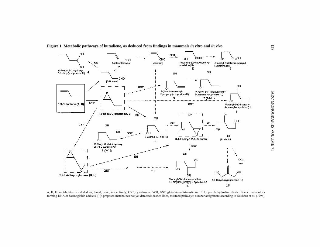
MetabolitesThe currently known metabolic pathways of butadiene in man, cynomolgus monkeys,
rats and mice are presented in Figure 1. In seven employees working in production areas with atmospheric concentrations of
3–4 ppm [6.6–8.8 mg/m3] butadiene over the previous six months, Bechtold et al. (1994)detected urinary excretion of the metabolite N-acetyl-S-(3,4-dihydroxybutyl)-L-cysteine(M-I, no. 3 in Figure 1) (3.2 ± 1.6 μg/mL) but not of N-acetyl-S-(1-hydroxymethyl-2-propenyl)-L-cysteine (M-II, no. 2 in Figure 1). In 10 unexposed employees and nine out-side controls, urinary M-I concentrations were 0.63 ± 0.19 and 0.32 ± 0.07 μg/mL. M-Iwas assumed to result from the conjugation of glutathione (GSH) with 3-butene-1,2-diol(butenediol) and M-II from conjugation of GSH with 1,2-epoxy-3-butene (epoxybutene).From the absence of M-II in human urine, it was concluded that epoxybutene is meta-bolically eliminated in humans predominantly by epoxide hydrolase and not by directGSH conjugation. Hallberg et al. (1997) found the concentration of M-I in urine samplesof 24 workers exposed to 2.4 ± 1.8 ppm [5.3 ± 4.0 mg/m3] butadiene (time-weightedaverage) to be 2.4 ± 1.9 μg/mL. In 19 controls (butadiene exposure below detection limitof 0.3 ppm [0.66 mg/m3]), urinary M-I concentrations of 0.69 ± 0.37 μg/mL were mea-sured. In both groups there was no significant difference between smokers and non-cigarette smokers.
Haemoglobin adducts N-(2-Hydroxy-3-butenyl)valine (HOBVal) as a reaction product of epoxybutene
with N-terminal valine in haemoglobin has been found in workers exposed to butadiene.Osterman-Golkar et al. (1993) recorded adduct levels of 1.1–2.6 pmol HOBVal/g globinin four nonsmoking workers exposed to about 1 ppm [2.2 mg/m3] butadiene as estimatedfrom exposure measurements made three to nine months earlier. A haemoglobin bindingindex of 0.004 pmol HOBVal/(g globin per ppm.h) was estimated from these preliminaryresults. In nonsmoking workers exposed outside the production area to an environmentalbutadiene concentration of about 0.03 ppm [0.07 mg/m3], the adduct levels were belowthe detection limit of 0.5 pmol HOBVal/g globin. Based upon data from a more recent
BUTADIENE 137
IARC M
ON
OG
RAPH
S VO
LUM
E 71138Figure 1. Metabolic pathways of butadiene, as deduced from findings in mammals in vitro and in vivo
A, B, U: metabolites in exhaled air, blood, urine, respectively; CYP, cytochrome P450; GST, glutathione-S-transferase; EH, epoxide hydrolase; dashed frame: metabolitesforming DNA or haemoglobin adducts; [ ]: proposed metabolites not yet detected; dashed lines, assumed pathways; number assignment according to Nauhaus et al. (1996)

study (Osterman-Golkar et al., 1996; Sorsa et al., 1996), an even lower binding index of0.0005 pmol/(g globin per ppm.h) was calculated (Osterman-Golkar & Bond, 1996).
After improving the method to reduce the detection limit to ~0.03–0.05 pmolHOBVal/g globin, Osterman-Golkar et al. (1996) measured adduct levels in controls,either five nonsmokers (≤ 0.05 pmol HOBVal/g globin) or four smokers (0.04–0.13 pmolHOBVal/g globin). Similar values were found in laboratory and maintenance workers(≤ 0.06 and ≤ 0.07 pmol HOBVal/g globin in four nonsmokers and three smokers,respectively) exposed to 0.6 ± 0.9 mg/m3 butadiene. In plant workers exposed to11.2 ± 18.6 mg/m3 butadiene, adduct levels were higher (0.2–0.32 and 0.02–0.24 pmolHOBVal/g in three nonsmokers and seven smokers, respectively) [these values were readfrom a graph]. The mean adduct level given for all 10 workers was 0.16 ± 0.099 pmolHOBVal/g. The authors calculated the amount of butadiene inhaled from the mainstreamsmoke of 30 cigarettes per day to be equal to that inhaled during an 8-h exposure to0.1 ppm [0.22 mg/m3] butadiene. In another plant, measurements of HOBVal were madeat two time points (Sorsa et al., 1996). In the first investigation, butadiene concentrationsat the workplace were > 3 ppm [6.6 mg/m3], and in the second < 3 ppm. The mean adductlevels were 2 ± 3.6 (n = 12) and 0.54 ± 0.33 pmol HOBVal/g globin (n = 4), respectively.In controls, the mean levels were 0.13 ± 0.35 (n =14) and 0.12 ± 0.05 pmol HOBVal/gglobin (n = 8), respectively.
van Sittert and van Vliet (1994) were unable to detect haemoglobin adducts in workersexposed in butadiene manufacture or in smokers.
Pérez et al. (1997) found two stereoisomers of N-(2,3,4-trihydroxybutyl)valine(THBVal) in haemoglobin resulting from the reaction of 3,4-epoxy-1,2-butanediol(epoxybutanediol) with the N-terminal valine. Theoretically, epoxybutanediol can beformed by oxidation of dihydroxybutene and/or hydrolysis of diepoxybutane. THBValcould also form by direct binding of diepoxybutane to haemoglobin with subsequenthydrolysis of the second epoxide ring. In two workers exposed to a median concentrationof 1 ppm butadiene (see Osterman-Golkar et al., 1996), the levels of THBVal for one ofthese isomers were 10 and 14 pmol/g globin, whereas in two control workers thecorresponding adduct levels were 1.8 and 3.3 pmol/g globin. These THBVal values were70-fold higher than corresponding values of HOBVal in the same subjects.
4.1.2 Experimental systemsButadiene
Male Sprague-Dawley rats (Bolt et al., 1984) and B6C3F1 mice (Kreiling et al., 1986a)were exposed in closed chambers to initial butadiene concentrations in the atmosphereranging from about 100 to 12 000 ppm [220–26 500 mg/m3] (rats) or to 5000 ppm[11 000 mg/m3] (mice) or were treated by intraperitoneal injection of about 1 μL butadienegas/g bw (rats). The resulting concentration–time courses in the chamber atmosphererevealed linear kinetics below 1000 ppm [2200 mg/m3] and saturation of metabolism above2000 ppm [4400 mg/m3], with maximum rates (μmol/h/kg bw) of 220 in rats and 400 inmice. In the linear range, rates of metabolism per kg body weight were 1.6-fold higher in
BUTADIENE 139

mice than in rats. The whole body : air concentration ratio of butadiene at steady state was0.5 in rats and 1 in mice. Due to metabolic elimination, these values were below thethermodynamic whole body:air partition coefficient, which was determined to be 2.7 inmice and 2.3 in rats. Following induction of metabolizing enzymes by pretreatment of ratswith Aroclor 1254, no saturation was observed within the exposure range studied. Fromthese data it was concluded that the rate of butadiene metabolism in the linear range waslimited by the uptake from the gas phase into the organism. Metabolism in both species wasinhibited effectively by pretreatment with diethyldithiocarbamate. Medinsky et al. (1994)also exposed male B6C3F1 mice and Sprague-Dawley rats to butadiene in closed chambersat initial concentrations of up to 5000 ppm. In animals pretreated with pyrazole (32 mg/kgbw) and exposed to initial concentrations of 1200 ppm [2650 mg/m3] butadiene, meta-bolism was inhibited completely in rats and the Vmax was reduced by 87% in mice. Using adynamic chamber, Leavens et al. (1996a) determined the rate of butadiene metabolism inmale B6C3F1 mice from the uptake during steady-state exposures (8 h) to 100 or 1000 ppm[220 or 2200 mg/m3] butadiene. Their value of 246 ± 19 μmol/h/kg bw during exposure to1000 ppm was close to that of about 270 μmol/h/kg bw given by Kreiling et al. (1986) forthe same exposure concentration. Bond et al. (1986) determined the retention of [1-14C]-butadiene in male B6C3F1 mice and Sprague-Dawley rats exposed via the nose only for 6 hto various concentrations of butadiene. The percentage of 14C retained decreased from 16–20% at 0.14–13 mg/m3 to 4% at 1800 mg/m3 in mice and from 17% at 0.14 mg/m3 to 2.5%at 1800 mg/m3 in rats, indicating saturation of metabolic elimination. Within the exposurerange up to 1800 mg/m3, the inhaled doses were on average 1.8-fold higher in mice than inrats, when normalized to body surface area. Nose-only exposures (2 h) of male cynomolgusmonkeys to [1-14C]butadiene gave much lower retention of butadiene (2.9% at 10.1 ppm[18 mg/m3], 1.5% at 310 ppm [560 mg/m3] and 1.7% at 7760 ppm [14 000 mg/m3]) thanin mice and rats. As determined by vacuum-line cryogenic distillation of radioactive com-pounds, blood concentrations of butadiene in monkeys reached 0.009 μmol/L at 10.1 ppm,0.6 μmol/L at 310 ppm and 32 μmol/L at 7760 ppm. The resulting blood/air concentrationratios were 0.03, 0.06 and 0.12, respectively, the increase reflecting saturation of butadienemetabolism (Dahl et al., 1991). Using headspace gas chromatography, Himmelstein et al.(1994) measured the blood concentration of butadiene in male B6C3F1 mice and Sprague-Dawley rats during nose-only exposure (6 h) to butadiene. A steady state was reached inblood after 2 h, giving butadiene concentrations (μmol/L) of 2.4, 37, 58 in mice and 1.3,18, 37 in rats at 62.5, 625 and 1250 ppm [138, 1380 and 2760 mg/m3], respectively. Thesevalues indicate nearly linear relationships between butadiene concentrations in blood andair, with blood concentrations in mice being about twice those in rats. Blood concentrationsdeclined within minutes after exposure ceased.
Since GSH conjugation is an important pathway in butadiene metabolism, severallaboratories have investigated the GSH-depleting effect of butadiene.
Deutschmann and Laib (1989) determined the non-protein sulfhydryl (NPSH) contentin lung, liver and heart of male B6C3F1 mice and Sprague-Dawley rats exposed for 7 h toconstant butadiene concentrations between 10 and 2000 ppm [22–4400 mg/m3]. In rats,
IARC MONOGRAPHS VOLUME 71140

hepatic NPSH (% of control) was depleted to 70–80% at 250–1000 ppm and to 40% at2000 ppm. An NPSH reduction in rat lung to about 80% and 70% was observed only at1000 and 2000 ppm, respectively, whereas NPSH in rat heart did not change. In mice,hepatic NPSH content began to decrease significantly (70%) at 250 ppm butadiene, andfell to 40% at 1000 ppm and 20% at 2000 ppm. In mouse lung, marked depletion of NPSH(about 50%) occurred at 500 ppm and reached about 10% at 2000 ppm. NPSH content inthe heart was reduced to about 75% at 1000 ppm and 30% at 2000 ppm. At conditions ofmaximum rate of butadiene metabolism (exposure concentration, > 2000 ppm), Kreilinget al. (1988) and Laib et al. (1990) observed depletion of control levels of hepatic NPSHcontent in male B6C3F1 mice to 20% and 4% after 7 h and 15 h, respectively, of butadieneexposure. In contrast, hepatic NPSH content in male Wistar and Sprague-Dawley ratsdecreased to about 65% and 80%, respectively, after 7 h of exposure; no major changeoccurred after 15 h.
Following 6 h of exposure to 1250 ppm [2760 mg/m3] butadiene, Himmelstein et al.(1995) found hepatic NPSH to decrease to 57 ± 18% and 62 ± 3% in male B6C3F1 miceand Sprague-Dawley rats, respectively. In rats exposed to 8000 ppm [17 700 mg/m3], nofurther depletion occurred. In lungs of mice, 65% depletion of NPSH was alreadyobserved at 62.5 ppm [138 mg/m3] butadiene, the maximum reduction to 26 ± 13% beingreached at 1250 ppm. In rat lung, NPSH was significantly depleted (74 ± 5%) only at1250 ppm, with a similar value at 8000 ppm.
In male Wistar rats exposed for two weeks (6 h per day, five days per week) to buta-diene, urinary mercapturic acids resulting from the conjugation of epoxybutene with GSHwere qualitatively analysed by gas chromatography/mass spectrometry after deacetylationas heptafluorobutanoic anhydride derivatives of the cysteine conjugates. The major pro-duct formed was assumed to be S-(2-hydroxy-3-butenyl)-L-cysteine. Quantitation of thecysteine conjugates as phthaldialdehyde derivatives by high-performance liquid chroma-tography revealed a nearly linear relationsphip between the amount of cysteine conjugatesin afternoon samples [sampling period not given] and the exposure concentration, with amaximum value of about 16 μmol at the highest exposure concentration of 1000 ppmbutadiene [2200 mg/m3] (Osterman-Golkar et al., 1991).
Nose-only exposures of B6C3F1 mice, Sprague-Dawley and Fischer 344/N rats, Syrianhamsters [sexes not specified] to 7600 ppm [14 150 mg/m3] and male cynomolgus monkeysover 2 h to 8000 ppm [17 700 mg/m3] [1-14C]butadiene (Sabourin et al., 1992) and of maleB6C3F1 mice and Fischer 344/NtacfBR rats over 4 h to 11.7 ppm [26 mg/m3] butadiene(Bechtold et al., 1994) resulted in urinary excretion of two major metabolites identified as N-acetyl-S-(3,4-dihydroxybutyl)-L-cysteine (M-I) and N-acetyl-S-(1-hydroxymethyl-2-pro-penyl)-L-cysteine (M-II). The ratio of M-I to M-I + M-II was 0.2 in mice, 0.3–0.5 in rats,about 0.4 in hamsters and about 0.9 in monkeys, compared with a value of nearly 1 in humans(see Section 4.1.1). This ratio was positively correlated with the epoxide hydrolase activity inthe livers of the different species, suggesting that in these species, as in human metabolism(see Section 4.1.1), hydrolysis of epoxybutene to butenediol precedes the formation of M-Iand that M-II is the mercapturate formed from the conjugate of GSH with epoxybutene.
BUTADIENE 141

Nauhaus et al. (1996) analysed butadiene metabolites in urine of male B6C3F1mice and Sprague-Dawley rats exposed via the nose only for up to 5 h to 800 ppm[1770 mg/m3] [1,2,3,4-13C]butadiene. The metabolites identified and their relative quan-tities are listed in Table 18. Metabolites 1, 2 and 9, derived from epoxybutene via theglutathione pathway, amounted to 70% in mice and 61% in rats, whereas the hydrolyticproduct butenediol (5) reached only 2.9% in mice and 5% in rats. Metabolite 3, formedby conjugation of butenediol with glutathione, was found in three- to four-fold higheramounts in rats than in mice. Metabolite 6, assumed to be derived from diepoxybutane,was found in small amounts only in mice, as was metabolite 4, which was attributed tothe hemithioacetal product of 3-butenal. [Metabolite 6 might also be formed via theoxidation of butenediol to epoxybutanediol.] Metabolites 7 and 8, present only in mouseurine, could not be attributed to a single pathway (via metabolite 3), but the involvementof conversion of butadiene to acrolein has been speculated, too. Rats but not miceexcreted 1,3-dihydroxypropanone (10) in urine, probably generated from the postulatederythritol via the pentose phosphate pathway. [The authors assumed erythritol to bederived from diepoxybutane. It might, however, also be formed via the oxidation ofbutenediol to epoxybutanediol.]
Interaction between butadiene, styrene and benzeneLike butadiene, styrene is metabolized in a first step by cytochrome P450-dependent
monooxygenases (Nakajima et al., 1994). Co-exposure could therefore lead to mutualinfluences on the rates of metabolism. Laib et al. (1992) co-exposed male Sprague-Dawley rats to butadiene (20, 100, 500, 1000, 3000, 6000 ppm [44, 220, 1100, 2200, 6600,13 300 mg/m3] and styrene (0, 20, 100, 250, 500 ppm [0, 85, 430, 1070, 2130 mg/m3]).Analysing the measured data by means of a toxicokinetic two-compartment model (Filser,1992), biotransformation rates of both compounds were determined as functions of theexposure concentrations. Whereas butadiene did not affect the metabolic rate of styrene,competitive inhibition of butadiene metabolism by styrene occurred up to a styreneconcentration of 90 ppm [380 mg/m3]. Higher styrene concentrations resulted in only asmall additional inhibition. These findings led to the hypothesis that butadiene is meta-bolized by at least two different cytochrome 450-dependent monooxygenases, only one ofwhich is inhibited by styrene. The presence of several butadiene-metabolizing mono-oxygenases was later verified by studies in vitro (Csanády et al., 1992; Duescher &Elfarra, 1994). The lack of inhibition of styrene metabolism by butadiene was attributedto the higher enrichment of inhaled styrene in the body compared to that of inhaled buta-diene. Using the data of Laib et al. (1992), a physiological toxicokinetic model was deve-loped in order to predict interactions between butadiene and styrene in humans (Filseret al., 1993). For low exposure by inhalation to both compounds, biotransformationappeared to be limited by transport to the metabolizing enzymes. Inhibition of butadienemetabolism by styrene in co-exposed people was predicted to occur.
Bond et al. (1994) simulated interactions of butadiene with styrene or with benzenein rats using their own physiological toxicokinetic model for butadiene and published
IARC MONOGRAPHS VOLUME 71142

models for styrene (Ramsey & Andersen, 1984) and benzene (Medinsky et al., 1989),assuming competitive mutual inhibition of the metabolism of butadiene and styrene andof butadiene and benzene. Whereas the metabolism of butadiene was predicted to bereduced by co-exposure to styrene or benzene, no effect of butadiene on the metabolismof styrene and of benzene was predicted. This was explained by the low solubility ofbutadiene compared with styrene and benzene.
Leavens et al. (1996a, 1997) and Leavens and Bond (1996) further explored themetabolic interactions between butadiene and styrene in male B6C3F1 mice exposed tomixtures of butadiene and styrene by inhalation. At steady state, significant inhibition ofbutadiene metabolism by styrene was observed with mixtures of 1000 ppm butadieneand 250 ppm styrene, but not with 100 ppm butadiene and 250 ppm styrene. Inhibitionby butadiene of styrene metabolism was evidenced by the significant increase in styreneblood concentrations (42% above that in mice exposed to styrene only) in the exposureto 1000 ppm butadiene and 250 ppm styrene. These authors concluded that while expo-sure to mixtures of styrene and butadiene results in inhibition of metabolism of bothstyrene and butadiene, interactive effects are seen only at high concentrations that are oflittle relevance to human exposure.
In order to analyse these observations, Leavens and Bond (1996) developed aphysiological toxicokinetic model based on the model of Medinsky et al. (1994) forbutadiene and the model of Csanády et al. (1994) for styrene. As previously found byLaib et al. (1992), a reasonable model prediction of the reduced butadiene uptake wasobtained only by including two oxidation pathways for both butadiene and styrene, one
BUTADIENE 143
Table 18. Butadiene metabolites in urine of mice and rats exposed to800 ppm [1770 mg/m3] [1,2,3,4-13C]butadiene
Metabolite Percentage of total metabolites
Mouse Rat
1. N-Acetyl-S-(2-hydroxy-3-butenyl)-L-cysteine 43.9 8.0 2. N-Acetyl-S-(1-hydroxymethyl-2-propenyl)-L-cysteine 21.6 52.8 3. N-Acetyl-S-(3,4-dihydroxybutyl)-L-cysteine 7.1 26.4 4. N-Acetyl-S-(1-hydroxy-3-butenyl)-L-cysteine 3.7 Not detected 5. 3-Butene-1,2-diol 2.9 5.0 6. N-Acetyl-S-(1-hydroxymethyl-2,3-dihydroxypropyl)-
L-cysteine 4.6 Not detected
7. N-Acetyl-S-(3-hydroxypropyl)-L-cysteine 5.4 Not detected 8. N-Acetyl-S-(2-carboxyethyl)-L-cysteine 4.8 Not detected 9. S-(1-Hydroxymethyl-2-propenyl)-L-cysteine 4.7 Not detected10. 1,3-Dihydroxypropanone Not detected 5.3
Metabolite numbers correspond to those in Figure 1.

catalysed by the same CYP isoenzyme with competitive interaction and another byseparate CYP isoenzymes without interaction between the two compounds.
Metabolites in vitroAs in humans, two important metabolites of butadiene are epoxybutene and diepoxy-
butane (Figure 1). The half-life of the spontaneous hydration of epoxybutene in water (pH 7) has been
calculated using rate constants given in Ross et al. (1982) to be 13.7 h and that ofdiepoxybutane, using rate constants given in Ehrenberg and Hussain (1981), to be 100 h(Gervasi et al., 1985).
Epoxybutene is the main first product of the NADPH-dependent metabolism ofbutadiene in postmitochondrial liver and lung fractions of mouse, rat, monkey and man(Schmidt & Loeser, 1985) and more specifically in the microsomal fraction of mouseliver (Wistuba et al., 1989; Elfarra et al., 1991; Csanády et al., 1992; Duescher & Elfarra,1992; Recio et al., 1992; Sharer et al., 1992; Maniglier-Poulet et al., 1995), of mouselung (Csanády et al., 1992; Sharer et al., 1992), of mouse kidney and testis (Sharer et al.,1992), of rat liver (Malvoisin et al., 1979; Bolt et al., 1983; Wistuba et al., 1989; Csanádyet al., 1992; Cheng & Ruth, 1993; Maniglier-Poulet et al., 1995), of rat lung (Csanádyet al., 1992; Sharer et al., 1992), of rat kidney and testis (Sharer et al., 1992), of humanliver (Csanády et al., 1992; Duescher & Elfarra, 1994) and of human lung (Csanádyet al., 1992). From the correlations with the activity of human liver microsomes to thespecific substrates chlorzoxazone (Csanády et al., 1992; Duescher & Elfarra, 1994) andcoumarin (Duescher & Elfarra, 1994), CYP2E1 and CYP2A6 were concluded to be themajor isoenzymes catalysing the oxidation of butadiene, CYP2E1 at low and CYP2A6at high butadiene concentrations. This was confirmed using microsomal preparationsfrom six human B-lymphoblastoid cell lines, each expressing a particular human cDNAencoding specific CYP isoenzymes (Duescher & Elfarra, 1994).
The rate of butadiene metabolism in diverse cell fractions has been investigated invarious species and is characterized by the Michaelis–Menten parameters Vmax andapparent Km (Kmapp). Three methods have been used to determine these parameters:
(i) loss of butadiene in the headspace of a closed vial due to metabolism in theincubate; analysis using a two-compartment model (Filser et al., 1992);
(ii) formation of epoxybutene, measured in the headspace of a closed vial; analysisusing a two-compartment model taking into account the further metabolism ofepoxybutene (Csanády et al., 1992; Recio et al., 1992).
(iii) formation of epoxybutene, measured in the incubate without consideration ofhydrolysis or vaporization (Malvoisin et al., 1979; Elfarra et al., 1991; Shareret al., 1992; Cheng & Ruth, 1993; Duescher & Elfarra, 1994; Maniglier-Pouletet al., 1995).
The differences in methodology complicate the direct comparison of the results. Forphysiological toxicokinetic modelling, the data obtained by the first two methods wereused.
IARC MONOGRAPHS VOLUME 71144

Tables 19, 20 and 21 present the Vmax/Kmapp values which were used in physiologicaltoxicokinetic models developed for the formation and degradation of epoxybutene and di-epoxybutane, all of which were obtained from in-vitro measurements. Although theseparameters were obtained in different laboratories, the similarity of the data is striking.Most interestingly, in liver microsomes of NMRI mice, CYP-dependent monooxygenase-mediated oxidation of butadiene was about 10 times lower than in liver microsomes fromB6C3F1 mice. However, oxidative metabolism of inhaled butadiene was accuratelypredicted for conditions in vivo using both values. This can be explained by the fact that,over a broad concentration range, the first step in the metabolism of inhaled butadiene isnot limited by enzymic capacity but by uptake into the blood and transport through themetabolizing organs (Filser et al., 1993). Furthermore, only part of the metabolizedinhaled butadiene is systemically available as epoxybutene (Filser & Bolt, 1984; Johanson& Filser, 1993; Csanády et al., 1996; Sweeney et al., 1997).
Molecular modelling of butadiene oxidation by CYP2E1 has indicated that speciesdifferences in the kinetic parameters might be explained by a non-conservative changefrom Thr-437 to His-437 between rodents and humans and by a conservative changefrom Ile-438 to Val-438 (Lewis et al., 1997).
BUTADIENE 145
Table 19. Vmax/Kmapp values of the NADPH-dependent oxidation of buta-diene and epoxybutene, as determined in cell fractions and used forphysiological toxicokinetic modelling
Vmax/Kmapp(nmol.L/mgprotein/min/mmol)
Reference
Oxidation of butadiene to epoxybuteneNMRI mouse, liver microsomes 134 Filser et al. (1992)Sprague-Dawley rat, liver microsomes 62Human (n = 1), liver microsomes 111B6C3F1 mouse Liver microsomes Lung microsomes
1295461
Csanády et al. (1992)
Sprague-Dawley rat Liver microsomes Lung microsomes
15721
Human Liver microsomes (n = 12) Lung microsomes (n = 5)
23075
Oxidation of epoxybutene to diepoxybutaneB6C3F1 mouse, liver microsomes 12.8 Csanády et al. (1992)B6C3F1 mouse, liver microsomes 9.2 Seaton et al. (1995)Sprague-Dawley rat, liver microsomes 2.8Human (n = 4), liver microsomes 0.15–3.8

Liver microsomes from male Sprague-Dawley rats convert butadiene into theR- and S-enantiomers of epoxybutene (Bolt et al., 1983). The ratios of R- to S-epoxybutene inbutadiene-exposed liver microsomes [concentration not specified] were 1 and 1.6 (pheno-barbital treatment) in mice [strain not specified], 0.33 and 0.43 (phenobarbital treatment) in rats[strain not specified] and 1.08–1.27 (n = 4) in humans (Wistuba et al., 1989). Exposure of livermicrosomes from male Sprague-Dawley rats to 25 000 ppm [55 300 mg/m3] butadiene gaveratios of R- to S-epoxybutene that varied with incubation time from about 0.3 at 5 min to about1 at 30 min (Nieusma et al., 1997). A nearly constant value of 0.75 was determined in livermicrosomes from male B6C3F1 mice.
Crotonaldehyde was formed NADPH-dependently as a minor metabolite of butadiene(partial pressure of 48–52 cm Hg = 660 000 ppm) in microsomes obtained from liver, lungor kidney of male B6C3F1 mice (Sharer et al., 1992) or human liver (Duescher & Elfarra,1994), the formation rate being 20–50 times lower than that of epoxybutene. 3-Butenalwas suggested as an intermediate metabolite. No crotonaldehyde formation was observedwith microsomes from mouse testis or with microsomes of testis, liver, lung or kidney ofmale Sprague-Dawley rats (Sharer et al., 1992).
IARC MONOGRAPHS VOLUME 71146
Table 20. Vmax/Kmapp values of the epoxide hydrolase and glutathioneS-transferase catalysed epoxybutene metabolism, as determined incell fractions
Vmax/Kmapp(nmol.L/mgprotein/min/mmol)
Reference
Epoxide hydrolaseNMRI mouse, liver microsomes 13 Kreuzer et al. (1991)Sprague-Dawley rat, liver microsomes 24Human (n = 1), liver microsomes 28B6C3F1 mouse, liver microsomes 3.6 Csanády et al. (1992)Sprague-Dawley rat, liver microsomes 9.5Human (n = 3), liver microsomes 32–38
Glutathione S-transferaseNMRI mouse, liver cytosol 15 Kreuzer et al. (1991)Sprague-Dawley rat, liver cytosol 11Human (n = 1), liver cytosol 8B6C3F1 mouse Liver cytosol Lung cytosol
147.5
Csanády et al. (1992)
Sprague-Dawley rat Liver cytosol Lung cytosol
172.5
Human (n = 2), Liver cytosol 4.3

Segments from different airway regions or whole airways obtained from male B6C3F1mice and Sprague-Dawley rats were incubated in headspace vials with 10 000 ppm butadienegas (34 μmol/L buffer). Epoxybutene formation in tissues from mice was two-fold higherthan in rats. The quantity of epoxybutene measured was doubled in the presence of theepoxide hydrolase inhibitor trichloropropene oxide, but remained unchanged followingaddition of the GSH depletor diethyl maleate, indicating that epoxide hydrolase contributesmore than glutathione conjugation to epoxybutene detoxification (Seaton et al., 1996).
Bone-marrow cells of B6C3F1 mice do not contain CYP2E1 (Genter & Recio, 1994).Nevertheless, bone-marrow cells of B6C3F1 mice and humans can oxidize butadiene toepoxybutene, the activity being increased two-fold by 1 mmol hydrogen peroxide/L. Themetabolic rate in hydrogen peroxide-fortified lysates of mouse cells (0.0053 nmol/min/mg protein) was two orders of magnitude lower than in mouse and rat liver micro-somes (Maniglier-Poulet et al., 1995).
BUTADIENE 147
Table 21. Kinetic constants of epoxide hydrolase- and gluta-thione S-transferase-catalysed metabolism of diepoxybutane incell fractions
Vmax(nmol/mgprotein/min)
Kmapp(mmol/L)
Vmax/Kmapp(nmol.L/mgprotein/min/mmol)
Epoxide hydrolase(Boogaard & Bond, 1996)B6C3F1 mouse Liver microsomes Lung microsomesSprague-Dawley rat Liver microsomes Lung microsomesHuman (n = 6) Liver microsomes Lung microsomes
32.0 ± 6.049.8 ± 9.7
52.9 ± 3.519.3 ± 7.8
155.8 ± 9.821.7 ± 1.9
8.1 ± 1.87.5 ± 1.7
2.76 ± 0.227.1 ± 3.4
4.8 ± 0.412.83 ± 0.37
3.93 6.65
19.2 2.71
32.5 7.66
Glutathione S-transferase(Boogard et al., 1996)B6C3F1 mouse Liver cytosol Lung cytosol
162 ± 1638.5 ± 2.5
6.4 ± 1.61.70 ± 0.37
25.321.0
Sprague-Dawley rat Liver cytosol Lung cytosol
186 ± 3717.1 ± 3.0
24 ± 64.2 ± 1.7
7.62 4.10
Human (n = 6) Liver cytosol 6.4 ± 1.9 2.1 ± 1.4 3.04

Incubation of butadiene with human myeloperoxidase (from polymorphonuclearleukocytes) in the presence of hydrogen peroxide (1 mmol/L) yielded epoxybutene andsmall amounts of crotonaldehyde by direct oxygen transfer (Duescher & Elfarra, 1992;Maniglier-Poulet et al., 1995). Addition of chloride in the hundred millimolar range ledto the formation of 1-chloro-2-hydroxy-3-butene as the major metabolite (Duescher &Elfarra, 1992).
The kinetics of epoxybutene oxidation to diepoxybutane were investigated byCsanády et al. (1992) in liver microsomes of male B6C3F1 mice and by Seaton et al.(1995) and Krause and Elfarra (1997) in those of male B6C3F1 mice, male Sprague-Dawley rats and human subjects. Similar ratios of Vmax/Kmapp relevant at low epoxybuteneconcentrations were measured by Csanády et al. (1992) and Seaton et al. (1995), whereasKrause and Elfarra (1997) found ratios that were one order of magnitude lower than thoseof Seaton et al. (1995) in all three species. [One possible explanation of this differencecould be that Krause and Elfarra (1997) determined the kinetic parameters at epoxy-butene concentrations that were two to four orders of magnitude higher than those foundin the blood of rodents exposed to butadiene under conditions of metabolic saturation.Thus Krause and Elfarra (1997) may have characterized a low-affinity enzyme that is notrelevant for in-vivo conditions.] The parameters published by Csanády et al. (1992) andSeaton et al. (1995) were used for physiological toxicokinetic modelling (see Table 19).[The data of Seaton et al. (1995) had to be corrected for hydrolytic loss of diepoxybutane(Sweeney et al., 1997).]
Krause and Elfarra (1997) detected NADPH-dependent formation of meso- and (±)-diepoxybutane from racemic epoxybutene in mice, rats and humans.
Whereas liver microsomes from male Sprague-Dawley rats formed nonsignificantlyhigher amounts of diepoxybutane from R- than from S-epoxybutene, in those of maleB6C3F1 mice, the yield was significantly higher from the S-isomer than from the R(Nieusma et al., 1997).
Seaton et al. (1995) and Krause and Elfarra (1997) used human B-lymphoblastoidcell lines from the same source each expressing a cDNA of one of eight different humanCYP isoenzymes. Epoxybutene at 80 μmol/L was oxidized only by CYP2E1, whereasat 5 mmol/L CYP3A4 was similarly active (Seaton et al., 1995). Krause and Elfarra(1997) found CYP2E1 to oxidize epoxybutene at 5 mmol/L nearly four- and six-foldfaster than CYP2C9 and 2A6, respectively, whereas in contrast to the Seaton et al.(1995) study, CYP3A4 was inactive. Diepoxybutane was hydrolysed in human livermicrosomes, the meso form being preferred over the two other stereoisomers (Krause &Elfarra, 1997).
In summary, in-vitro results suggest that the rate of cytochrome P450-mediatedepoxidation of butadiene to epoxybutene and to diepoxybutane is highest in mice com-pared with rats and humans and that the rate in humans varies widely (Seaton et al.,1995; see Table 19).
Investigations in vitro have demonstrated that epoxybutene is eliminated by micro-somal epoxide hydrolase and by cytosolic glutathione S-transferase (GST). Epoxide hydro-
IARC MONOGRAPHS VOLUME 71148

lase activity was determined in liver of mouse, rat and man (Kreuzer et al., 1991, Csanádyet al., 1992; Krause et al., 1997), in lung of mouse, rat and man (Csanády et al., 1992) andin liver of mouse (Recio et al., 1992). GST activity was determined in liver of mouse, ratand man (Kreuzer et al., 1991; Csanády et al., 1992), in lung of mouse, rat and man(Csanády et al., 1992), and in liver, lung, testis and kidney of mouse and rat (Sharer et al.,1992). Sharer et al. (1991) purified π-class GST from human placenta for kinetic studies.The Michaelis–Menten parameters obtained by Kreuzer et al. (1991) and Csanády et al.(1992) have been used for physiological toxicokinetic modelling (see Table 20).
Hydrolysis of R- and S-epoxybutene to the respective enantiomer of 3-butene-1,2-diol(butenediol) is nearly completely stereospecific in liver microsomes from male Sprague-Dawley rats, whereas in liver microsomes from male B6C3F1 mice, an inversion of theconfiguration of 16% (S-epoxybutene) and 24% (R-epoxybutene) was observed (Nieusmaet al., 1997).
Epoxybutene is also metabolized by human θ-class GST purified from placenta.Products formed were S-(1-hydroxy-3-buten-2-yl)glutathione [S-(1-hydroxymethyl-2-propenylglutathione, using the nomenclature of Figure 1] and S-(2-hydroxy-3-buten-1-yl)glutathione. The latter product is in 1:1 equilibrium with the relatively stable sulfuranetautomer formed by intramolecular displacement of the hydroxyl group by the sulfur atom(Sharer et al., 1991).
Diepoxybutane, like epoxybutene, is eliminated by microsomal epoxide hydrolase inliver and lung of mouse, rat and man (Boogard & Bond, 1996) and by cytosolic GST inliver and lung of mouse and rat and in liver of man (Boogard et al., 1996).
In summary, the elimination of epoxybutene and diepoxybutane by GSH conjugationappears to be faster in rodents than in humans. Epoxybutene and diepoxybutane hydro-lysis appears to be fastest in humans (see Tables 20 and 21).
Rydberg et al. (1996) investigated the reaction of diepoxybutane with valinamidein vitro (40°C, pH > 9, 100 h) as a model for the N-terminal valine in haemoglobin. Themain products at the lowest diepoxybutane concentration (1 mmol/L) were N-(2,3,4-trihydroxybutyl)valinamide and erythritol, formed with similar yields. The amount of aring-closed pyrrolidine derivative (2,2-N,N-(2,3-dihydroxybuta-1,4-diyl)valinamide)was three-fold lower. A cross-linked 2,2′-N,N-(2,3-dihydroxybuta-1,4-diyl)bis-valina-mide was detectable at 100 mmol diepoxybutane/L.
Rat θ class GST 5-5 (Thier et al., 1995) and human θ class GSTT1-1 (Thier et al.,1996), both expressed in Salmonella typhimurium TA1535, enhanced the mutagenicityof diepoxybutane but not of epoxybutene. The formation of a reactive glutathione conju-gate of the bifunctional diepoxybutane was assumed, possibly a five-membered thialo-nium ion or a thiiranium (episulfonium) ion. On the other hand, a close correlation wasfound between the diepoxybutane-dependent induction of sister chromatid exchanges(SCE) (Kelsey et al., 1995; Norppa et al., 1995; Wiencke et al., 1995; Landi et al., 1996;Pelin et al., 1996) and of micronuclei (Vlachodimitropoulos et al., 1997) in human peri-pheral blood lymphocytes and the homozygous deletion of GSTT1, suggesting detoxi-fication by GSTT1.
BUTADIENE 149

Butenediol can be oxidized to 3,4-epoxybutanediol (epoxybutanediol), as has beenshown in rat liver microsomes. Incubation for 30 min with butadiene gave concentrationsof butenediol and epoxybutanediol which were nearly three-fold and 10-fold, respec-tively, higher than the corresponding concentration of epoxybutene (Cheng & Ruth,1993). Epoxybutanediol can, however, also be a product of diepoxybutane hydrolysis.
Kemper and Elfarra (1996) demonstrated the oxidation of butenediol by hepaticalcohol dehydrogenase (ADH), yielding 1-hydroxy-2-butanone as a single stable meta-bolite; various intermediates have been proposed. For the ADH-dependent oxidation ofracemic butenediol in liver cytosol of male B6C3F1 mice, male Sprague-Dawley rats andthree humans, saturation kinetics were found. The ratio Vmax/Kmapp was similar in thesespecies. ADH purified from horse liver oxidized butenediol in a stereoselective manner,since Vmax was about seven times higher for the S- than for the R-enantiomer.
The fate of epoxybutanediol has not been studied in vitro.
Metabolites in vivoBolt et al. (1983) exposed male Sprague-Dawley rats in a closed system to initial
butadiene concentrations of 6000–7000 ppm [13 300–15 500 mg/m3] and foundexhaled epoxybutene to accumulate in the atmosphere up to 2–4 ppm within 15 h. Infurther studies, animals were exposed for up to 17 h to butadiene concentrations above2000 ppm [4400 mg/m3] under conditions of maximum metabolism of butadiene(Filser & Bolt, 1984). Exhaled epoxybutene accumulated in the air of the closedsystem, reaching a plateau of about 3.7 ppm. Toxicokinetic analysis with a two-compartment model revealed that only 29% of biotransformed butadiene was systemi-cally available as epoxybutene. From these results, the authors deduced the existenceof an intrahepatic first-pass effect for epoxybutene formed from butadiene. Using thesame experimental design, Kreiling et al. (1987) exposed male B6C3F1 mice tobutadiene at > 2000 ppm; exhaled epoxybutene accumulated in the atmosphere up toabout 10 ppm. From the steady-state concentration of epoxybutene in the atmosphereof the closed chamber containing rats or mice exposed to butadiene under conditionsof metabolic saturation and using thermodynamic body/air partition coefficients of 37for rats (Filser & Bolt, 1984) and 42.5 for mice (Kreiling et al., 1987), the averageconcentration of epoxybutene in the body was calculated to be 5.5 μmol/L in rats and17 μmol/L in mice.
In the experiments of Bond et al. (1986) described on p. 140, at the end of 6-hexposure to butadiene, blood concentrations of epoxybutene reached values of 0.4 and 4μmol/L in rats at 130 and 1800 mg/m3 and 0.7, 0.9 and 15 μmol/L in mice at 13, 130 and1800 mg/m3, respectively. In the cynomolgus monkey, Dahl et al. (1991) found bloodconcentrations of only 1.6, 500 and 1100 nmol epoxybutene/L following 2-h exposuresto 10, 310 and 7760 ppm [18, 560 and 14 000 mg/m3] butadiene, respectively, using thesame method as Bond et al. (1986). [The Working Group noted that due to the unspecificdetermination of radioactivity in cryogenic traps, contamination of epoxybutene withother metabolic products of butadiene cannot be excluded.]
IARC MONOGRAPHS VOLUME 71150

More recently, concentrations of butadiene epoxides were determined by gaschromatography–mass spectrometry in blood and tissues of B6C3F1 mice and Sprague-Dawley rats exposed via the nose only to butadiene. Losses of the volatile epoxybutene thatmay occur in the time between sacrifice and organ dissection have been modelled (Sweeneyet al., 1996). These simulations predicted that epoxybutene concentrations in the liver candecrease by orders of magnitude within minutes. Such losses might differ between large andsmall organs and between those of mouse and rat. However, Himmelstein et al. (1994)removed blood from the animal while it was still breathing the exposure atmosphere, so itis unlikely that epoxybutene was lost during sampling. At exposure concentrations of 62.5,625 and 1250 ppm [138, 1380 and 2760 mg/m3] butadiene, they found steady-stateconcentrations (6 h exposure) in blood of 0.56, 3.7 and 8.6 μmol epoxybutene/L in mice andof only 0.07, 0.94 and 1.3 μmol epoxybutene/L in rats. Diepoxybutane reachedconcentrations of 0.65, 1.9 and 2.5 μmol/L in mice, but was not detected in rats. Bechtoldet al. (1995) measured epoxybutene concentrations in blood of 0.38 and 0.1 μmol/L in miceand rats, respectively, exposed for 4 h to 100 ppm [220 mg/m3] butadiene. Diepoxybutanereached 0.33 μmol/L in mice but was not found in rats. Following 6-h exposures to 625 and1250 ppm butadiene, Himmelstein et al. (1995) found epoxybutene concentrations of 0.58and 0.93 nmol/g (mice) and 0.06 and 0.16 nmol/g (rats) in liver and 2.6 and 3.7 nmol/g(mice) and 0.16 and 0.31 nmol/g (rats) in lung, respectively. Diepoxybutane was detected inmouse lung at concentrations of 0.71 and 1.5 nmol/g tissue at 625 and 1250 ppm butadiene,respectively. Even at 8000 ppm [17 700 mg/m3] butadiene, no diepoxybutane was detectedin rat lung; the detection limit was 0.04 nmol/g.
Thornton-Manning et al. (1995a) exposed male mice and rats for up to 4 h to62.5 ppm [138 mg/m3] butadiene. Using a highly sensitive method, the authors detectedepoxybutene and diepoxybutane in tissues of both species (Table 22). The tissue concen-trations of epoxybutene varied considerably between tissues but in general were 3–10times higher in mice than in rats. With the exception of liver, as the main metabolizingorgan, and bone marrow, diepoxybutane reached similar concentrations in all mousetissues. Corresponding concentrations in rat lung were up to two orders of magnitudelower. The homogeneous distribution of diepoxybutane in the body is also reflected bythe similar tissue:hexane partition coefficients determined experimentally (Table 23;Sweeney et al., 1997). Thornton-Manning et al. (1995b) found tissue concentrations ofepoxybutene to be similar in female and male rats exposed for 6 h to 62.5 ppm(Table 22). However, corresponding concentrations of diepoxybutane were three to fivetimes higher in females than in males.
In a further study, Thornton-Manning et al. (1997) investigated the disposition ofbutadiene epoxides in female B6C3F1 mice and Sprague-Dawley rats following singleand repeated (10 days) nose-only exposures (6 h) to 62.5 ppm [138 mg/m3] butadiene(Table 24). With the exception of lung, tissue and blood concentrations of epoxybutenein rats and mice were higher after repeated exposures. Whereas repeated exposures ofrats did not lead to changes in diepoxybutane concentrations, a reduction of up to 30%was observed in mice.
BUTADIENE 151

Inhalation kinetics of epoxybutene were investigated in Sprague-Dawley rats (Filser &Bolt, 1984; Kreiling et al., 1987) and in male B6C3F1 mice (Kreiling et al., 1987) usingclosed chambers. Animals were exposed to initial concentrations of epoxybutene rangingfrom 10 to 5000 ppm [22–11 000 mg/m3] (rats) and 100 to 2000 ppm [220–4400 mg/m3](mice). The exhalation of intraperitoneally administered epoxybutene (45.6 μL/kg bw) byrats was also determined (Filser & Bolt, 1984). In rats, first-order kinetics were observedover the whole exposure range. In mice, initial enrichment phases were seen. The furthershape of the concentration–time curves was interpreted as showing saturation kinetics. In a
IARC MONOGRAPHS VOLUME 71152
Table 22. Tissue concentrations of epoxybutene and diepoxy-butane in rats and mice after inhalation of butadiene
Tissue Epoxybutene (pmol/g)
4 h exposure (Thornton-Manning et al., 1995a)
6 h exposure (Thornton-Manning et al., 1995b)
Male mice Male rats Male rats Female rats
Blood 295 ± 27 36 ± 7 25.9 ± 2.9 29.4 ± 2Liver 8 ± 4 Not detected n.d. n.d.Lung 33 ± 9 Not detected 12.7 ± 5 2.7 ± 4.3Fat 1302 ± 213 267 ± 14 175 ± 21 203 ± 13Heart 120 ± 15 40 ± 16 n.d. n.d.Spleen 40 ± 19 7 ± 6 n.d. n.d.Thymus 104 ± 55 12.5 ± 3.2 n.d. n.d.Bone marrow 2.3 ± 1.5a 0.2 ± 0.1 9.3; 9.7
(femur)10.4 ± 1(femur)
Mammary n.d. n.d. n.d. 57.4 ± 4
Diepoxybutane (pmol/g)
Blood 204 ± 15 5 ± 1 2.4 ± 0.4 11.4 ± 1.7Liver 20 ± 4 Not detected n.d. –Lung 114 ± 37 0.7 ± 0.2 1.4 ± 0.8 4.8 ± 0.7Fat 98 ± 15 2.6 ± 0.4 1.1 ± 0.1 7.7 ± 1.3Heart 144 ± 16 3 ± 0.4 n.d. –Spleen 95 ± 12 1.7 ± 0.5 n.d. –Thymus 109 ± 19 2.7 ± 0.7 n.d. –Bone marrow 1.4 ± 0.3a Not detected 1.1; 1.8 7.1 ± 1.3Mammary n.d. n.d. n.d. 10.5 ± 2.4
B6C3F1 mice and Sprague-Dawley rats inhaled butadiene via the nose only.Three animals were used for each experiment.n.d., not determineda pmol/mg protein

later publication, however, it was explained by depletion of GSH at high exposure con-centrations, resulting in a loss of GST-mediated detoxification (Johanson & Filser, 1993),on the basis of GSH measurements in tissues of rats and mice exposed to epoxybutene(Deutschmann & Laib, 1989).
Valentine et al. (1997) studied the kinetics of epoxybutene and diepoxybutane inblood following intravenous administration to male Sprague-Dawley rats. The followingtoxicokinetic parameters were obtained for epoxybutene at 71, 143, 286 μmol/kg bw,
BUTADIENE 153
Table 23. Measured partition coefficients of butadiene, epoxybutene and di-epoxybutane
Mouse Rat Rat Man
Butadienetissue:air
(Medinsky et al.,1994)
(Medinsky et al.,1994)
(Johanson & Filser,1993)
(Filser et al.,1993)
BloodFatMuscleLiverLungKidneyBrainSpleen
1.3419.2 4.01 1.35 1.47 n.d. n.d. n.d.
1.49 22.2 1.47 1.19 0.92 n.d. n.d. n.d.
3.03 21.9 0.73 0.94 n.d. 0.92 0.43 0.87
1.00 22.5 0.88 0.68 0.48 0.86 1.05 n.d.
Epoxybutenetissue:air
(Medinsky et al.,1994)
(Medinsky et al.,1994)
(Johanson & Filser,1993)
(Csanády et al.,1996)
BloodFatMuscleLiverLungKidneyBrain
36.691.223.642.156.3n.d.n.d.
50.4138 19.8 72.0 54.7 n.d. n.d.
83.4155 59.9 53.7 n.d. 70.2 51.6
93.3168 45.8 55.3 n.d. n.d. n.d.
Diepoxybutanetissue:hexane
(Sweeney et al., 1997)
BloodFatMuscleLiverKidney
0.4370.9590.7950.6150.672
n.d., not determined

respectively: distribution half-lives of 1.4, 1.8, 1.4 min, terminal half-lives of 5.7, 7.0, 8.5min, systemic clearance of 104, 114, 67 mL/min/kg bw and volume of distribution atsteady state of 0.59, 0.58, 0.53 L/kg bw. The corresponding values for diepoxybutane ata dose of 523 μmol/kg bw were: distribution half-life of 2.7 min, terminal half-life of14 min, systemic clearance of 76 mL/min/kg bw and volume of distribution at steadystate of 0.73 L/kg bw. These values were interpreted as demonstrating the similarity ofdisposition of the two epoxides in rats.
When treated intraperitoneally with epoxybutene (71.3 to 285 μmol/kg bw), maleB6C3F1 mice and Sprague-Dawley rats excreted butenediol in urine, the amount within24 h being less than 1% of the administered dose (Krause et al., 1997).
Conjugation of epoxybutene with GSH in the liver in vivo was demonstrated bySharer and Elfarra (1992) in male Sprague-Dawley rats which excreted S-(2-hydroxy-3-
IARC MONOGRAPHS VOLUME 71154
Table 24. Tissue concentrations of epoxybutene and diepoxybutane infemale mice and rats exposed to butadienea
Tissue Epoxybutene (pmol/g)
Mouse Rat
Single exposure Multiple exposure Single exposure Multiple exposure
Blood 239 ± 24 317 ± 19 44 ± 7 64 ± 8Lung ∼ 25a ∼ 150a ∼ 5a Not detectedMammary ∼ 700a ∼ 1200a,b ∼ 80a ∼ 300a,b
Fat ∼ 1150a ∼ 1650a,b ∼ 200a ∼ 430a,b
Femur ∼ 56 a Not reported ∼ 10a ∼ 15a,b
Diepoxybutane (pmol/g)
Mouse Rat
Single exposure Multiple exposure Single exposure Multiple exposure
Blood 345 ± 33 247 ± 32 14 ± 2 17 ± 2Lung 219 ± 33 144 ± 13 b 5 ± 1 4 ± 0.3Mammary 265 ± 11 191 ± 17 b 11 ± 2 15 ± 1Fat 203 ± 2 173 ± 11 b 8 ± 1 13 ± 0.4 b
Ovary 169 ± 13 152 ± 16 6 ± 2 10 ± 7Femur 214 ± 27 184 ± 19 7 ± 1 9 ± 1
Female B6C3F1 mice and Sprague-Dawley rats were exposed to 62.5 ppm butadiene for 6 hvia the nose only, either on one day only or on 10 successive days. Three or four animalswere used for each experiment (Thornton-Manning et al., 1997).a Read from diagramb Significantly different from single exposure value, p ≤ 0.05

buten-1-yl)glutathione and S-(1-hydroxymethyl-2-propenyl)glutathione in a 3:1 ratio inthe bile within 60 min following intraperitoneal injection of epoxybutene (14.3–286 μmol/kg bw). The total amount of conjugates excreted was linearly related to dose,indicating no saturation, but accounted for only 7.6 ± 4.2% of the dose.
Following single intraperitoneal administrations of epoxybutene (71.5, 143 or285 μmol/kg) to male B6C3F1 mice or Sprague-Dawley rats, diastereomeric pairs of N-acetyl-S-(2-hydroxy-3-buten-1-yl)-L-cysteine (1 in Figure 1) and N-acetyl-S-(1-hydroxy-methyl-2-propenyl)-L-cysteine (2 in Figure 1) were excreted in the urine within 8 h. Inrats, linear dose–response relationships were observed with respect to the excretion ofmetabolites 1 and 2 (mean, 17% of epoxybutene dose), the amount of metabolite 1 beingtwo to three times higher than that of metabolite 2. In mice, an overproportional increasein the excretion of metabolites 1 and 2 occurred at the highest dose (mean 26%, com-pared with 7 and 9% at the lower doses, respectively), the amount of metabolite 1 beingabout one half to one third of that of metabolite 2. The amount per body weight ofmetabolites 1 and 2 in rats was approximately twice as high as in mice at the lower dosesand similar in both species at the high dose (Elfarra et al., 1995).
Haemoglobin adductsUsing haemoglobin and epoxybutene, Osterman-Golkar et al. (1991) observed the
formation of two diastereomeric pairs of adducts to the N-terminal valine of haemo-globin namely N-(2-hydroxy-3-buten-1-yl)valine and N-(1-hydroxy-3-buten-2-yl)valine.These findings were corroborated by Richardson et al. (1996), who incubated ery-throcyte suspensions obtained from mice, rats and humans with epoxybutene. Thesecond-order rate constant of adduct formation for the sum of both adducts (HOBVal)was determined in vitro at 37°C to be 0.29 × 10⁻⁴ L/g globin/h with erythrocytes isolatedfrom mice (Recio et al., 1992; value corrected by the same authors to the one quotedhere, Osterman-Golkar et al., 1993).
In male Wistar rats exposed for two weeks (6 h per day, five days per week) to buta-diene, covalent binding of epoxybutene (mainly at C-1) to the N-terminal valine ofhaemoglobin was observed. Total adduct levels (nmol/g haemoglobin) and the dailyaverage increment (nmol/g haemoglobin) at day 12 were 0.5 and 0.06 at 250 ppm[550 mg/m3], 1.5 and 0.17 at 500 ppm [1100 mg/m3] and 3.0 and 0.33 at 1000 ppm[2200 mg/m3] butadiene. Seventeen days after the end of exposure, the levels haddecreased to nearly two thirds of the original values (Osterman-Golkar et al., 1991).
Osterman-Golkar et al. (1993) observed a linear increase in the HOBVal level up toabout 4 nmol/g globin following exposure of male B6C3F1 mice over four weeks (6 hper day, five days per week) to butadiene (0, 2, 10 and 100 ppm [0, 4, 22 and220 mg/m3]). In Sprague-Dawley rats, the increase of HOBVal was linear up to 10 ppmbutadiene, amounting to about 0.2 nmol/g globin and reached a value of about 1 nmol/gglobin at 100 ppm. The authors also summarized haemoglobin binding indices resultingfrom butadiene exposure (pmol HOBVal/g globin per ppm.h) in different species as ~0.5in B6C3F1 mice, ~0.3 in CD2F1 mice (from Recio et al., 1992), ~0.09 in Wistar rats
BUTADIENE 155

(from Osterman-Golkar et al., 1991), and ~0.3 and ~0.1 in Sprague-Dawley rats at 0–10 ppm and 10–100 ppm butadiene, respectively. For humans, a value of ~0.004 and aneven lower value of 0.0005 have been estimated (see Section 4.1.1).
Albrecht et al. (1993) determined HOBVal in female CB6F1 mice, male and femaleC3H × 101/EL mice and female Wistar rats exposed (6 h per day for five days) tobutadiene concentrations of 0, 50, 200 and 500 ppm [0, 110, 440 and 1100 mg/m3]. Addi-tionally, animals were exposed to 1300 ppm [2870 mg/m3] butadiene, with the exceptionof male C3H × 101/EL mice. In mice, background levels of HOBVal were between 1 and8 nmol/g globin. Up to 200 ppm butadiene, a steep increase of the HOBVal levels wasobserved, reaching values between 10 and 16 nmol/g globin. At higher butadiene con-centrations, the slope of the curve flattened and at 1300 ppm, the HOBVal value reachedabout 25 nmol/g globin. No significant strain or sex difference was observed. In rats,background levels were between 1.3 and 2.2 nmol/g globin. Following exposure, theadduct levels were distinctly lower than in mice. The slope of the dose–response curvebetween 0 and 200 ppm, reaching a level of about 3 nmol/g globin, was somewhatsteeper than between 200 and 1300 ppm, at which the level reached about 5 nmol/gglobin.
Pérez et al. (1997) exposed male Wistar rats for five consecutive days (6 h per day)to constant butadiene concentrations of 0, 50, 200 and 500 ppm [0, 110, 440 and1100 mg/m3]. On day 6, animals were killed; the levels of HOBVal were 0.6, 21, 88 and180 pmol/g.
Osterman-Golkar et al. (1998) investigated the dose–response relationships for adductformation and persistence in rats and mice during long-term low-level exposure tobutadiene by inhalation. Values reported by Osterman-Golkar et al. (1993) were alsorecalculated. HOBVal levels were measured in male B6C3F1 mice and Sprague-Dawleyrats following exposure to 0, 2, 10 or 100 ppm [0, 4, 22 or 220 mg/m3] butadiene for 6 hper day on five days per week for one, two, three or four weeks. The increase anddecrease, respectively, of the adduct levels during and three weeks after the end of thefour-week exposure indicated that adducts are chemically stable in vivo and that elimi-nation follows the turnover of red blood cells. Adduct levels increased linearly with buta-diene concentration in mice, whereas a deviation from linearity between 10 and 100 ppmbutadiene (decrease in slope) was observed in rats. Blood concentrations of epoxybuteneestimated from haemoglobin adduct levels were in general agreement with those reportedin mice and rats exposed to 62.5 ppm butadiene, indicating that HOBVal adduct levels canbe used to predict blood concentrations of epoxybutene in rats and mice.
After intraperitoneal administration of epoxybutene (10, 20, 40 and 60 mg/kg bw) tomale B6C3F1 mice and Sprague-Dawley rats, HOBVal levels increased with doseapproximately linearly in rats and sublinearly in mice. At the highest dose, the bindingefficiency in mice was twice that in rats, HOBVal levels reaching about 950 and460 pmol/g globin in mice and rats, respectively (Richardson et al., 1996).
Tretyakova et al. (1996) exposed female and male B6C3F1/CrlBR mice andCrl:CDBR rats to 1000 ppm [2200 mg/m3] butadiene (6 h per day, 5 days per week, for
IARC MONOGRAPHS VOLUME 71156

13 weeks). Two isomers of HOBVal were found [not further specified], the level ofisomer I being 1.3–1.5-fold that of isomer II. HOBVal levels (means of isomer I up to11 190 and of isomer II up to 8660 pmol/g globin in female mice) were three to fourtimes higher in mice than in rats, the mean levels in females being about twice those inmales.
N-(2,3,4-Trihydroxybutyl)valine (THBVal) in haemoglobin is regarded as a reactionproduct of epoxybutanediol with N-terminal valine. This adduct could, however, form bydirect binding of diepoxybutane to haemoglobin with subsequent hydrolysis of thesecond epoxide (see Section 4.1.1). Two isomers of this adduct were found in maleSprague-Dawley rats 24 or 48 h following intraperitoneal treatment with epoxybutene(78.3 mg/kg bw), epoxybutanediol (30 and 60 mg/kg bw) or diepoxybutane (16.7 and33.4 mg/kg bw) or after exposure to butadiene. Adduct levels were reported only for‘adduct II’. Compared with a control level of about 2 pmol/g globin, THBVal reached amaximum level of 2800 pmol/g globin after 33.4 mg/kg bw diepoxybutane. Ascalculated from THBVal levels, diepoxybutane had higher haemoglobin binding indices(pmol THBVal/g globin per μmol/kg bw) of 9.3 and 7.2 (at 16.7 and 33.4 mg/kg doses,respectively) than epoxybutanediol (3.4 and 4.0 at 30 and 60 mg/kg doses, respectively)and epoxybutene (0.07). In Wistar rats killed one day after exposure (6 h per day, for fivedays) to 0, 50, 200 or 500 ppm [0, 110, 440 or 1100 mg/m3] butadiene, the highestTHBVal levels of 1190 pmol adduct/g globin were found at 200 ppm (controls: 9 pmolTHBVal/g globin). The binding index (pmol THBVal/g globin per ppm × h) decreasedfrom 0.5 at 50 ppm to 0.04 at 500 ppm. Parallel determination of the levels of HOBValdetermined in the same rats were three-fold (500 ppm) to about 32-fold (50 ppm) lowerthan the THBVal levels (Pérez et al., 1997).
Physiological toxicokinetic modelsPhysiological toxicokinetic (or pharmacokinetic) models represent descriptions of
biological systems and can be used to describe the behaviour of chemicals in the intactanimal. Such models have been used to predict the disposition of butadiene and meta-bolites in rats, mice, and humans. For the case of rats and mice, these predictions can becompared with experimental data. In some cases (see below), the models successfullydescribe (and accurately predict) the disposition of butadiene and metabolites. Humanphysiological toxicokinetic model predictions normally cannot be verified due to lack ofexperimental data.
Several models have been developed to simulate the absorption, distribution, meta-bolism and excretion of butadiene, some of its metabolites and its adducts to haemo-globin in mouse, rat and man. Critical aspects are discussed in Csanády et al. (1996) andin Himmelstein et al. (1997). Basically, the models consist of a number of compartmentsrepresenting diverse tissues and organs, several of which are grouped together. Thesecompartments are linked by blood flow. The main differences between models are thenumber of metabolizing and nonmetabolizing compartments, the mechanisms ofmetabolism, the metabolites taken into consideration, and the values of the biochemical,
BUTADIENE 157

physiochemical and physiological parameters. The first group of parameters isrepresented by apparent Michaelis constants, maximum rates of metabolism, tissueconcentrations of GSH and turnover rates. The second group consists of the blood:air,tissue:air and tissue:blood partition coefficients of butadiene and selected metabolites.The structure of the tissue compartments, blood flow rates and alveolar ventilationbelong to the third group.
Physiological toxicokinetic models have been presented describing the behaviour ofinhaled butadiene in the human body. Partition coefficients for tissue:air andtissue:blood, respectively, had been measured directly using human tissue samples orwere calculated based on theoretical considerations. Parameters of butadiene metabolismwere obtained from in-vitro studies in human liver and lung cell constituents and byextrapolation of parameters from experiments with rats and mice in vivo (see above).In these models, metabolism of butadiene is assumed to follow Michaelis–Mentenkinetics.
By means of an apparent Michaelis constant (Kmapp) together with a maximum rate(Vmax) of butadiene metabolism both obtained with human liver microsomes (Filser et al.,1992), Filser et al. (1993) constructed a human model which was later extended byCsanády et al. (1996) for the butadiene metabolites epoxybutene and diepoxybutane. Forbutadiene and epoxybutane, the required human tissue:air partition coefficients weremeasured using autopsy material (Table 23). Filser et al. (1993) investigated theinfluence of styrene co-exposure on butadiene metabolism by assuming competitiveinteraction. Simulations for a 70-kg man exposed over 8 h to 5 or 15 ppm [11 or33 mg/m3] butadiene indicated total amounts of butadiene metabolized of 0.095 and0.285 mmol, respectively, reduced by about 19% and 37% as a result of co-exposure to20 and 50 ppm styrene, respectively. No influence of butadiene on styrene metabolismwas noted.
Kohn and Melnick (1993) and Medinsky et al. (1994) used in their models values ofKmapp and Vmax which had been determined by Csanády et al. (1992) with microsomes fromhuman liver and lung. The tissue:blood partition coefficients used by Kohn and Melnick(1993) were theoretically derived and were 5–10 times higher than those derived from thetissue:air partition coefficients measured by Filser et al. (1992) in human tissues and byJohanson and Filser (1993) and by Medinsky et al. (1994) in rodent tissues. Simulationsof human exposure to butadiene under workplace conditions (8 h per day, five days perweek) indicated that high accumulation in fat would occur, with levels increasing aboutthree-fold during the week. For their model, Medinsky et al. (1994) used either their ownpartition coefficients determined experimentally in mouse tissues or for comparison thosewhich had been published by Kohn and Melnick (1993). Simulations of concentration–time courses in fat tissue resulting from human exposure to 54 ppm [120 mg/m3] buta-diene for 6 h yielded about three-fold lower peak concentrations and an area under theconcentration–time curve (AUC) several times lower when the mouse values were used.With the latter values, which are close to those obtained by Filser et al. (1992) in humantissues, no suggestion of accumulation during the working week was found.
IARC MONOGRAPHS VOLUME 71158

Internal burdens of epoxybutene in humans resulting from exposure to butadienewere predicted from models by Kohn and Melnick (1993), Johanson and Filser (1996)and Csanády et al. (1996) and were compared with simulations for rats and mice. In themodel of Kohn and Melnick (1993), metabolic parameters were incorporated which hadbeen obtained by Csanády et al. (1992) by measuring butadiene and epoxybutene oxi-dation and epoxybutene hydrolysis in human liver and lung microsomes in vitro, andconjugation of epoxybutene with glutathione in human liver and lung cytosol.Tissue:blood partition coefficients were theoretically derived. The body burden ofepoxybutene following exposure to 100 ppm butadiene for 6 h was predicted to be0.056 μmol/kg in humans.
Johanson and Filser (1996) used metabolic parameters which had been obtained forenzymic butadiene oxidation (Filser et al., 1992) and epoxybutene hydrolysis (Kreuzeret al., 1991) in human liver microsomes and for enzymic conjugation of epoxybutenewith glutathione in human liver cytosol (Kreuzer et al., 1991). Tissue:air partitioncoefficients had been determined experimentally for butadiene in human tissues (Filseret al. 1993a) and for epoxybutene in rat tissues (Johanson & Filser, 1993). For an eight-hour exposure to 10 ppm butadiene, the model predicted a blood concentration of epoxy-butene of about 0.08 μmol/L in a man (Johanson & Filser, 1996). Csanády et al. (1996)simulated an exposure to 10 ppm (22 mg/m3) butadiene over 8 h and predicted an AUCof epoxybutene in blood of 0.27 μmol.h/L. Most of the model parameters used by theseauthors were identical to those of Johanson and Filser (1996). Tissue:air partitioncoefficients for epoxybutene in humans used by Csanády et al. (1996) were measuredwith human tissue samples (Table 23). The values suggest an almost homogeneousdistribution of epoxybutene in the body, with about twofold enrichment in fat tissue. Themodels of Johanson and Filser (1996) and Csanády et al. (1996) predict AUCs of epoxy-butene in humans about one order of magnitude higher than those from the model ofKohn and Melnick (1993). The main reason for this difference might lie in the very hightheoretically derived fat:air partition coefficient for butadiene which was used by thelatter authors, leading to prediction of storage of inhaled butadiene in fat tissue, resultingin reduced availability for biotransformation to epoxybutene during the time span of asingle exposure over 6 h.
Physiological toxicokinetic models for experimental systemsModels presented for mice and rats (Evelo et al., 1993; Filser et al., 1993; Johanson
& Filser, 1993; Kohn & Melnick, 1993; Bond et al., 1994; Medinsky et al., 1994;Csanády et al., 1996; Sweeney et al., 1997) predicted, species specifically, similar toxi-cokinetic behaviour of butadiene. The only exception was the first model of Kohn andMelnick (1993), which contained much higher theoretically derived partition coefficientsthan the experimentally determined ones, leading to prediction of butadiene storage infat tissue. In a second, extended version, the authors used average values of the partitioncoefficients determined experimentally by Johanson and Filser (1993) and Medinskyet al. (1994).
BUTADIENE 159

The influence of metabolism in the lung with respect to the toxicokinetics of buta-diene was simulated in the models of Evelo et al. (1993), Kohn and Melnick (1993),Medinsky et al. (1994) and Sweeney et al. (1997). The model of Evelo et al. (1993)yielded the surprising result that the total metabolic activity in lung of mice exposed to1 ppm [2.2 mg/m3] butadiene in air would be nearly equal to that in liver. Experimentaldata confirming this model prediction have not been published. Under similar conditions,the ratios of lung to liver metabolic activity in rats and humans were around 0.2 and 0.08,respectively. This ratio decreased in all species by 30–50% at 1000 ppm [2200 mg/m3]exposure. The simulations indicated a strong first-pass effect of butadiene in the lung atlow concentrations. The model of Kohn and Melnick (1993) predicted that most buta-diene (85–95%) would be metabolized in the liver of the three species, whereas meta-bolism in the lung accounted for only 4% in mice and 1% in rats and humans. Frommodel simulations of their own closed-chamber uptake data, Medinsky et al. (1994)suggested that lung metabolism of butadiene might be important for the total body clea-rance in mice but not in rats. At lower butadiene concentrations, lung metabolism waspredicted to become more important relative to metabolism in the liver, which was attri-buted to the limitation of hepatic metabolism by the blood flow through the liver.
Physiological toxicokinetic models of butadiene metabolite dispositionEpoxybutene was included as the first metabolite of butadiene in several models. The
models of Medinsky et al. (1994) and Kohn and Melnick (1996) overpredicted theburden of epoxybutene in rodents two- to three-fold, since it was assumed that biotrans-formed butadiene would become fully systemically available as epoxybutene. Under thesame assumption, the model of Kohn and Melnick (1993) yielded reasonable simulationsof experimental data, but the predicted rates of butadiene metabolism in this model weremuch lower than those which had been determined experimentally (Bolt et al., 1984;Kreiling et al., 1986; Leavens et al., 1996a). In the model of Sweeney et al. (1997),epoxybutene formation was reasonably simulated by adjusting the systemic availabilityof epoxybutene to measured data. It was assumed that only a fraction of the butadienemetabolized was transformed to epoxybutene. Further intermediates formed within thefirst step of butadiene catabolism not leading to epoxybutene were postulated. Thefraction of butadiene oxidized to epoxybutene was estimated to be 0.19 in mice and 0.24in rats. This fraction is consistent with the ‘extraction factor’ of 0.29 reported by Filserand Bolt (1984). These authors interpreted their findings as indicative of an intrahepaticfirst-pass effect for the epoxybutene formed, since only 29% of this metabolite enteredsystemic circulation. This effect was considered in the models of Johanson and Filser(1993, 1996) and Csanády et al. (1996), in which the liver was modelled as consisting ofcytosol containing GST and endoplasmic reticulum containing a cytochrome P450–epoxide hydrolase complex. Such a complex was proposed to explain the biotrans-formation of a series of olefinic hydrocarbons including naphthalene in vitro and in vivo(Oesch, 1973). Evidence supporting the existence of such a complex comes from thedemonstration that the rat microsomal epoxide hydrolase (mEH) and a CYP2B1–mEH
IARC MONOGRAPHS VOLUME 71160

fusion protein, in which the CYP2B1 membrane anchor signal sequence replaced the N-terminal 20 amino acid residues of mEH, could be co-translationally inserted into dogpancreas microsomes, whereas truncated mEH, in which the N-terminal 20 amino acidswere deleted, was not co-translationally inserted (Friedberg et al., 1994). The bio-chemical parameters of butadiene and epoxybutene metabolism incorporated in themodel of Johanson and Filser (1993, 1996) were derived from in-vitro data for butadiene(Filser et al., 1992) and epoxybutene (Kreuzer et al., 1991). The model overpredictedepoxybutene formation by a factor of about two.
In the model of Csanády et al. (1996), the biochemical parameters for butadiene inrats and mice were obtained by fitting model simulations to in-vivo data of Bolt et al.(1984) and Kreiling et al. (1986). The biochemical parameters for epoxybutene wereidentical to those of Johanson and Filser (1993, 1996). This model accurately predictedexperimental data on epoxybutene. The most advanced models are those of Csanádyet al. (1996) and Sweeney et al. (1997), since they can simulate both epoxybutene anddiepoxybutane as metabolites of butadiene. The tissue:blood partition coefficients fordiepoxybutane were estimated by Csanády et al. (1996) to have a value of 1 for alltissues. Sweeney et al. (1997) obtained tissue:blood partition coefficients from in-vitromeasurements (Table 23). Both models yielded good predictions for mice and rats forboth metabolites. For humans, no measured data have been reported against which thepredictions could be validated. In addition, the model of Csanády et al. (1996) predictedaccurately the measured haemoglobin adduct levels (Osterman-Golkar et al., 1993;Albrecht et al., 1993) of epoxybutene in rodents following exposure to butadiene. Noneof the models published has included the formation and elimination of epoxybutanediol.
4.1.3 Comparison of rodent and human dataBy comparing butadiene metabolites in urine of butadiene-exposed mice and humans,
Bechtold et al. (1994) concluded that in humans epoxybutene was metabolically elimi-nated predominantly by epoxide hydrolase. In rats, GSH conjugation and hydration path-ways were about equal and in mice direct GSH conjugation was more important.
No measured data have been published on the burden of butadiene and its epoxymetabolites in exposed humans that can be used for comparison with the rodent data.However, the models of Kohn and Melnick (1993), Johanson and Filser (1996) andCsanády et al. (1996) predict significantly lower body burdens of epoxybutene, based ondata derived from human tissues (Tables 19, 20 and 21).
The partition coefficients measured in rodent and human tissue samples (Table 23)suggest that there would be no substantial difference between rodents and humans withrespect to distribution of butadiene and its metabolite epoxybutene.
Model predictions have been made of the disposition of butadiene and epoxybutenein rodents and humans. Kohn and Melnick (1993) predicted that the cumulative bodyburden of epoxybutene after a 6-h exposure to 100 ppm butadiene ‘(area under theepoxybutene versus time curve from 0 to 6 h)’ in humans would be 7- and 35-fold lowerthan in rat and mouse, respectively. For a 12-h exposure to 10 ppm butadiene, the model
BUTADIENE 161

of Johanson and Filser (1996) predicted the internal dose of epoxybutene in humans tobe only 3.3- and 5.3-fold lower than in rat and mouse, respectively. Similar results wereobtained with the model of Csanády et al. (1996) for exposure to 10 ppm butadiene over8 h: the AUCs of epoxybutene in blood were 3.7- and 4.8-fold lower in humans than inrat and mouse, whereas the AUCs of butadiene in blood were about three-fold lower inhumans than in both rodent species.
Osterman-Golkar et al. (1993) summarized data on the formation of HOBVal, theadduct of epoxybutene at the N-terminal valine of haemoglobin, in rodents exposedexperimentally and in subjects exposed at the workplace to butadiene. The bindingindices (pmol HOBVal/g globin per ppm.h) were ~0.5 in B6C3F1 mice, ~0.3 in CD2F1mice (Recio, 1992), ~0.09 in Wistar rats (Osterman-Golkar et al., 1991), ~0.3 and ~0.1in Sprague-Dawley rats at 0–10 ppm and 10–100 ppm [0–22 and 22–220 mg/m3]butadiene, respectively, and, as a preliminary value, ~0.004 in humans. [The latter valuewas estimated assuming an average exposure concentration of 1 ppm [2.2 mg/m3], butthe exposure concentrations were mostly below this value (Osterman-Golkar, 1993;Sorsa et al., 1996).] In a later publication, Osterman-Golkar et al. (1996) reported amedian HOBVal level of 0.16 pmol/g globin in 10 workers exposed to a medianbutadiene concentration of 2.1 mg/m3 [0.93 ppm]. Considering a ratio of 3:1 of theC-1:C-2 isomers of the epoxybutene-valine adducts at the N-terminal valine of haemo-globin (Richardson et al., 1996) and assuming a workplace exposure of 8 h per day forfive days, a binding index of (4/3) × 0.16/(0.93 × 8 × 63) ~ 0.0004 pmol HOBVal/(gglobin per ppm.h) can be calculated. An identical low binding index was calculated fromdata given in a review by Osterman-Golkar and Bond (1996).
Taking all these data together, it can be concluded that exposure of humans to buta-diene leads to lower body burdens of the reactive metabolite epoxybutene than in simi-larly exposed rats and mice. No comparative data are available concerning the inter-mediate diepoxybutane. Only limited data have been published on the adducts of epoxy-butanediol at the N-terminal valine of haemoglobin resulting in N-(2′,3′,4′-trihydroxy-butyl)valine (Pérez et al., 1997).
4.2 Toxic effects4.2.1 HumansButadiene
The toxic effects of combined exposures to butadiene and other agents (e.g., styrene,chloroprene, hydrogen sulfide, acrylonitrile) have been reviewed (Parsons & Wilkins,1976). Concentrations of several thousand parts per million of butadiene irritate the skin,eyes, nose and throat (Carpenter et al., 1944; Wilson et al., 1948; Parsons & Wilkins, 1976).
Several studies have been reported on the effects of occupational exposure to butadiene,mainly from the USSR and Bulgaria. Few are substantiated by details on the atmosphericconcentration or duration of exposure, and control data are generally not provided. Theeffects reported include haematological disorders (Batkina, 1966; Volkova & Bagdinov,1969), kidney malfunction, laryngotracheitis, irritation of the upper respiratory tract,
IARC MONOGRAPHS VOLUME 71162
conjunctivitis, gastritis, various skin disorders, a variety of neurasthenic symptoms (Parsons& Wilkins, 1976) and hypertension and neurological disorders (Spasovski et al., 1986).
Checkoway and Williams (1982) reported minimal changes in haematologicalindices among eight workers exposed to about 20 ppm [44 mg/m3] butadiene, 14 ppm[60 mg/m3] styrene and 0.03 ppm [0.1 mg/m3] benzene, compared with those among 145workers exposed to less than 2 ppm [4.4 mg/m3] butadiene, 2 ppm [8.5 mg/m3] styreneand 0.1 ppm [0.3 mg/m3] benzene. Changes included a slight decrease in haemoglobinlevel and a slight increase in red-cell mean corpuscular volume. [The Working Groupconsidered that these changes cannot be interpreted as an effect of butadiene on the bonemarrow, particularly as alcohol intake was not evaluated.]
DiepoxybutaneDiepoxybutane is highly irritant to the eyes and respiratory tract of accidentally
exposed workers (IARC, 1976).
4.2.2 Experimental systemsButadiene
LC50 values for butadiene were reported to be 270 000 mg/m3 in mice exposed for2 h and 285 000 mg/m3 in rats exposed for 4 h; after 1 h of exposure, rats were in a stateof deep narcosis (Shugaev, 1969). Oral LD50 values of 5.5 g/kg bw for rats and 3.2 g/kgbw for mice have been reported (United States National Toxicology Program, 1984).
In female rats exposed to 1–30 mg/m3 butadiene for 81 days, morphological changeswere observed in liver, kidney, spleen, nasopharynx and heart (G.K. Ripp, reported inCrouch et al., 1979). In groups of 24 rats exposed to 600–6700 ppm [1300–14 800 mg/m3] butadiene for 7.5 h per day on six days per week for eight months, noadverse effect was noted, except for a slight retardation in growth at the highest concen-tration (Carpenter et al., 1944). Rats exposed to 2200–17 600 mg/m3 butadiene for 6 hper day on five days per week for three months showed no treatment-related effect otherthan increased salivation in females (Crouch et al., 1979).
Groups of 110 male and 110 female CD Sprague-Dawley rats were exposed to atmo-spheres containing 0, 1000 or 8000 ppm [0, 2200 or 17 600 mg/m3] butadiene for 6 h perday on five days per week. The study was terminated when it was predicted that survivalwould drop to 20–25% (105 weeks for females, 111 weeks for males). Ten animals ofeach sex from each group were killed at 52 weeks. Treatment was associated withchanges in clinical condition and lowering of body weight gain during the first 12 weeks,then nonsignificant changes, reduced survival and increases in certain organ weights andin the incidence of uncommon tumour types (for details, see Section 3.1.2). Increasedmortality in the high-dose males was accompanied by an increase in the severity ofnephropathy (Owen et al., 1987; Owen & Glaister, 1990).
B6C3F1 mice exposed to 0, 625 or 1250 ppm [0, 1380 or 2760 mg/m3] butadiene for6 h per day on five days per week for 60–61 weeks had increased prevalence of atrophyof the ovary and testis, atrophy and metaplasia of the nasal epithelium, hyperplasia of the
BUTADIENE 163

respiratory and forestomach epithelium and liver necrosis (see also Section 3.1.1)(United States National Toxicology Program, 1984).
Haematological changes in male B6C3F1 mice exposed to 62.5, 200 or 625 ppm[138, 440 or 1380 mg/m3] butadiene for 6 h per day on five days per week for 40 weeksincluded decreased red blood cell count, haemoglobin concentration and packed red cellvolume and increased mean corpuscular volume. Similar changes occurred in femalemice exposed to 625 ppm butadiene (for details, see Section 3.1.1) (Melnick et al., 1990).
The role of murine retroviruses in induction of leukaemias and lymphomas followinginhalation of butadiene was evaluated in a series of studies reviewed by Irons (1990).Exposure of groups of male B6C3F1 mice, which have the intact ecotropic murineleukaemia virus, to 1250 ppm [2750 mg/m3] butadiene for 6 h per day on six days perweek for 6–24 weeks resulted in decreases in the number of circulating erythrocytes, intotal haemoglobin and in haematocrit and an increase in mean corpuscular volume.Leukopenia, due primarily to a decrease in the number of segmented neutrophils, and anincrease in the number of circulating micronuclei were observed (Irons et al., 1986a).Persistent immunological defects were not detected after this treatment (Thurmond et al.,1986). Exposure of male NIH Swiss mice, which do not possess intact endogenous eco-tropic murine leukaemia virus, produced similar results (Irons et al., 1986b).
A further study was conducted to examine the expression and behaviour of endo-genous retroviruses in these strains during the preleukaemic phase of butadiene exposure.Chronic exposure of B6C3F1 mice to butadiene (1250 ppm [2760 mg/m3]) for 6 h per dayon five days per week for 3–21 weeks increased markedly the quantity of ecotropicretrovirus recoverable from the bone marrow, thymus and spleen. Expression of otherendogenous retroviruses (xenotropic, MCF-ERV) was not enhanced. No virus of anytype was found in similarly treated NIH Swiss mice (Irons et al., 1987a).
Enhanced susceptibility to butadiene-induced leukaemogenesis as a result of anability to express the retrovirus was suggested by the finding that exposure to 1250 ppmbutadiene for one year resulted in a 57% incidence of thymic lymphoma in B6C3F1 mice(with expression of the virus) and a 14% incidence in NIH Swiss (without viralexpression) (Irons et al., 1989).
Groups of 70 male and 69 female B6C3F1 mice were exposed to 0, 6.25, 20, 62.5,200 or 625 ppm [0, 14, 44, 138, 440 or 1380 mg/m3] butadiene for 6 h per day on fivedays per week for up to 103 weeks. Groups of 10 males and 10 females were killed at 40and 65 weeks. Ovarian atrophy was noted in female mice at 65 weeks of exposure (aftercompletion of the reproductive life of this species) at 20 ppm and higher. Testicularatrophy occurred after 65 weeks in the male mice at 625 ppm (Melnick & Huff, 1992).
The effect of butadiene exposure on arteriosclerotic plaque development was assessedin white leghorn cockerels exposed for 6 h per day on five days per week for 16 weeksto 20 ppm (44 mg/m3) butadiene. Plaque frequency and size in the abdominal aorta wallwere determined for butadiene-exposed animals and controls. Plaques were larger forbutadiene-exposed animals than for corresponding air controls and the authors concludedthat butadiene exposure markedly accelerated arteriosclerotic plaque development. Since
IARC MONOGRAPHS VOLUME 71164

one epidemiological study has suggested a link between death from arteriosclerotic heartdisease and chronic occupational exposure to butadiene, the authors suggested thattheir animal model could be used to further investigate this disease (Penn & Snyder, 1996).
DiepoxybutaneDiepoxybutane is highly toxic to rats (oral LC50 78 mg/kg bw), mice and rabbits.
Among surviving animals, there was eye, skin and respiratory tract damage. Intra-muscular injection of rabbits with 25 mg/kg bw produced leukopenia and lymphopenia.However, once weekly gavage dosing of rats for one year with 5 mg D,L-diepoxybutanewas not toxic (IARC, 1976).
4.3 Reproductive and developmental effects4.3.1 Humans
No data were available to the Working Group.
4.3.2 Experimental systemsButadiene
The reproductive and developmental toxicity of butadiene has been reviewed(Melnick & Huff, 1992; Christian, 1996).
Fertility was reported to be unimpaired in mating studies in rats, guinea-pigs andrabbits exposed to 600, 2300 or 6700 ppm [1300, 5000 or 14 800 mg/m3] butadiene byinhalation for 7.5 h per day on six days per week for eight months (Carpenter et al.,1944). [The Working Group noted the incomplete reporting of this study].
Pregnant Sprague-Dawley rats (24–28 per group) and Swiss (CD-1) mice (18–22 pergroup) were exposed to atmospheric concentrations of 0, 40, 200 or 1000 ppm [0, 88, 440or 2200 mg/m3] butadiene for 6 h per day on days 6–15 of gestation and killed on gestationday 18 (mice) or 20 (rats). Subsequently, the uterine contents were evaluated; individualfetal body weights were recorded, and external, visceral and skeletal examinations wereperformed. In rats, maternal toxicity was observed in the 1000-ppm group in the form ofreduced extragestational weight gain and, during the first week of treatment, decreasedbody weight gain. Under these conditions, there was no evidence of developmental toxi-city. Maternal toxicity was observed in mice given 200 and 1000 ppm butadiene, while40 ppm and higher concentrations of butadiene caused significant exposure-related reduc-tion in the mean body weights of male fetuses. Mean body weights of female fetuses werereduced at the 200 and 1000 ppm exposure levels. No increased incidence of malfor-mations was observed in either species. The frequency of fetal variations (supernumeraryribs, reduced sternebral ossification) was significantly increased in mice exposed to 200and 1000 ppm. In a study of sperm-head morphology, groups of 20 male B6C3F1 micewere exposed to atmospheric concentrations of 0, 200, 1000 or 5000 ppm [0, 440, 2200 or11 000 mg/m3] butadiene for 6 h per day for five consecutive days. Small, concentration-related increases in the frequency of abnormal sperm morphology were seen five weeksafter exposure (the only time of examination) (Hackett et al., 1987; Morrissey et al., 1990).
BUTADIENE 165

[The Working Group noted that sequential examinations were not conducted after exposureto determine the effect of butadiene on all stages of gamete development].
Female Sprague-Dawley rats were exposed to 0, 200, 1000 or 8000 ppm [0, 440,2200 or 17 700 mg/m3] butadiene for 6 h per day for 10 days on days 6–15 of gestation.Maternal body weight gain was significantly reduced at all exposure concentrations, withweight loss at 8000 ppm. Uterine implantation was unaffected. At 8000 ppm, there wasa significant reduction in fetal body weights, delay in ossification of the ribs (wavy ribs)and the thoracic centra and incomplete ossification of the sternum. There were noteratogenic effects that were significant or outside the historical control range. The no-observed-effect level (NOEL) was reported as 200 ppm for maternal toxicity and 1000ppm for developmental effects (Christian, 1996).
EpoxybuteneGroups of 10 female B6C3F1 mice and 10 Sprague-Dawley rats were administered
0.005, 0.02, 0.09, 0.36 or 1.43 mmol/kg bw [0.35, 1.4, 6.3, 25 or 100 mg/kg bw] epoxy-butene in sesame oil by intraperitoneal injection daily for 30 days. There was a 10% bodyweight decrement among the highest-dose mice at the end of the experiment, but therewas no body weight effect in rats. Ovarian and uterine weights also were reduced in miceat the highest dose, with an accompanying reduction in the number of developingfollicles and absence of primordial follicles, but there was no effect in rats (Doerr et al.,1996).
DiepoxybutaneThere are two reports of reproductive toxicity of diepoxybutane in experimental
systems. In the first study, groups of 10 female B6C3F1 mice and 10 Sprague-Dawley ratswere administered 0.002, 0.009, 0.036, 0.14 or 0.29 mmol/kg bw [0.17, 0.78, 3.1, 12, 25mg/kg bw] diepoxybutane in sesame oil by intraperitoneal injection daily for 30 days.Diepoxybutane exposure depressed growth at the two highest doses in both rats and mice.Since rats were extremely sensitive to the high-dose diepoxybutane treatment (0.29mmol/kg), the diepoxybutane was administered to rats at this dose for only 25 days. Atday 25, body weights of these rats were 50% of controls and only four of ten rats werealive at day 30. Animals in this group exhibited signs of severe gastrointestinal toxicity asevidenced by diarrhoea. Ovarian toxicity was determined by assessing reproductive organweights and ovarian follicle number. Although diepoxybutane was ovotoxic in bothspecies, it was more potent in mice than rats. At a dose of 0.14 mmol/kg bw, the ovary wasdepleted of 83% of the small follicles and 52% of the growing follicles in mice. Only 31%and 40% of these follicle populations were depleted in rats at that dose. A decrease inovarian and uterine weights with increasing dose was observed in mice at the 0.14 and0.29 mmol/kg bw doses. Similar observations were also seen in rats (Doerr et al., 1996).
The effects of diepoxybutane on male reproductive cells were investigated by flowcytometric and histological description of testicular cell populations and alterations ofchromatin packaging. Male B6C3F1 mice were treated with a single intraperitoneal
IARC MONOGRAPHS VOLUME 71166

injection of diepoxybutane in saline at doses of 8.5, 17, 26, 34, 43 and 52 mg/kg bw.Groups were killed at intervals of 7, 14, 21, 28 and 35 days after treatment. One groupwas injected with 78 mg/kg bw for observation at three weeks. The treated animals didnot show any clinical signs of toxicity on daily observation. Cytotoxic damage to all post-stem cell stages was assessed by alterations in relative ratios of haploid, diploid, and tetra-ploid testicular cells and by the reduction of relative percentages of cell populations.Dose-dependent reductions of tetraploid cells, round spermatids and elongated spermatidswere detected at 7, 21 and 28 days, respectively, reflecting cytotoxic damage on the diffe-rentiating spermatogonia compartment. The dose necessary to reduce the number of diffe-rentiating spermatogonia to half the control value was 55 mg/kg bw. Stem cells were notaffected by this treatment. Depletion of spermatids and reduction of the secondary sper-matocyte layers were noted in the seminiferous tubules (Spano et al., 1996).
4.4 Genetic and related effects The genetic toxicology of butadiene and of its major metabolites, epoxybutene and di-
epoxybutane, has been reviewed (Adler et al., 1995; Jacobson-Kram & Rosenthal, 1995).Additional information is available in a more recent review of the toxicology and epi-demiology of butadiene (Himmelstein et al., 1997) and a compilation of publications (Adler& Pacchierotti, 1998).
Butadiene4.4.1 Humans
In a small pilot study, the hprt locus mutation frequencies in lymphocytes of eightmale workers from a high-exposure area in a butadiene production plant in Texas, USA,were compared with those of five (four male and one female) low-exposure workers andsix male control area personnel (Ward et al., 1994, 1996a). All subjects were non-smokers. Butadiene concentrations were measured in both area and personal samples,which gave values of 3.5 ± 7.25 ppm [7.7 ± 16.6 mg/m3] and 0.03 ± 0.03 ppm[0.07 ± 0.07 mg/m3] for the high-exposure (production) and the control areas, respec-tively. The levels in the majority of the production area samples were below 1 ppm. Uri-nary concentrations (mean ± S.D. in ng/mg creatinine) of the butadiene metabolite, 1,2-dihydroxy-4-(N-acetylcysteinyl)butane (N-acetyl-S-(3,4-dihydroxybutyl)-L-cysteine,using the nomenclature of Figure 1 (Metabolite 3)), were 1690 ± 201.3, 355 ± 250 and580 ± 191 for the high- and low-exposure area and control area personnel, respectively.The hprt locus mutation frequencies (× 10–6 ± standard deviation (SD)), determined byan autoradiographic technique, were 3.99 ± 2.81, 1.20 ± 0.51 and 1.03 ± 0.12, respec-tively, in the three groups. The value for the high-exposure group was significantlyhigher (p < 0.05) than those for the other groups. A second study was conducted at thesame plant eight months later in which exposures were determined from 8-h personalbreathing zone air samplers (Ward et al., 1996a). Three exposure groups were compared(high, intermediate and low, there being no control group), for which the average buta-diene concentrations were 0.30 ± 0.59, 0.21 ± 0.21 and 0.12 ± 0.27 ppm, respectively
BUTADIENE 167

[0.66 ± 1.30, 0.46 ± 0.46 and 0.27 ± 0.60 mg/m3]. The corresponding urinary concen-trations of 1,2-dihydroxy-4-(N-acetylcysteinyl)butane were 761 ± 245, 596 ± 155 and684 ± 176 ng/mg creatinine. The frequencies of hprt locus mutations were 5.33 ± 3.76,2.27 ± 0.99 and 2.14 ± 0.97, respectively, in the three groups. The value for the high-exposure group was significantly higher (p < 0.05) than those for the other groups.
Preliminary data from an on-going population study of rubber plant workers exposedto butadiene and styrene (16 high-exposure (including five smokers) versus nine low-exposure (including three smokers)) are also available (Ward et al., 1996a). Passivebadge dosimeters were used to measure butadiene and styrene concentrations in the air.The butadiene detection limit was 0.25 ppm [0.55 mg/m3] over an 8-h period. Half of the40 samples collected in the high-exposure areas exceeded the detection limit and 11 weregreater than 1 ppm [2.2 mg/m3]. None of the samples collected in low-exposure areasexceeded the detection limit. The styrene concentration averaged 25% of that ofbutadiene and only one sample from the high-exposure area had > 1 ppm styrene. Thefrequencies of hprt locus mutations for the non-smokers were 7.47 ± 5.69 and 1.68 ± 0.85for the high- and low-exposure groups, respectively, and, for the smokers, 6.24 ± 4.37and 3.42 ± 1.57, respectively. The values for the high-exposure groups were significantlyhigher (p < 0.01) than those for the low-exposure groups.
The hprt mutation frequency was also evaluated in two studies using the T-lym-phocyte clonal assay. The mutation frequency for 41 workers (15 male, 26 female)exposed to butadiene (1–3.5 ppm [2.2–7.7 mg/m3]) at a polybutadiene rubber productionfacility in China was not significantly different from that of the 38 (14 male, 24 female)controls. Mutation frequency decreased with cloning efficiency, increased with age andwas moderately higher in women than in men. After adjustment for age, sex and cloningefficiency by multiple regression analysis, the mean mutation frequency was 32% higherin exposed workers than in controls, but this difference was not significant (p = 0.13) andwas due largely to the greater values among exposed women (Hayes et al., 1996). Thehprt locus mutation frequencies were measured in blood samples collected twice (in1993 and 1994) from 19 workers exposed to butadiene and 19 matched controls from abutadiene production plant in the Czech Republic (Tates et al., 1996). Three exposed andthree control subjects were the same in 1993 and 1994. Personal passive dosimetry wasperformed in 1993 and twice in 1994 on the day preceding blood sampling. About halfof the 1993 samples were lost, so that five exposed and 13 control lymphocyte samplesremained for analysis. The mean exposure level in 1994 was 1.76 ± 4.20 ppm (SD)[3.9 ± 9.3 mg/m3] and tabulated individual exposure levels ranged from < 0.024 ppm to10.2 ppm [0.053 and 22.6 mg/m3]. Using the clonal assay (Tates et al., 1994), the geo-metric mean of hprt mutation frequencies (× 10–6 ± SD) adjusted for cloning efficiency,age and smoking were, respectively, 7.85 ± 7.09 and 10.14 ± 9.16 in pooled (1993 plus1994) exposed and control subjects. The difference was not significant. A similar resultwas obtained for the 1994 subjects alone. There was no difference between adjusted geo-metric mean mutation frequencies of exposed and unexposed non-smokers or betweenexposed and unexposed smokers.
IARC MONOGRAPHS VOLUME 71168

Cytogenetic analysis of peripheral blood lymphocytes of butadiene production workersshowed that occupational exposure to butadiene (median concentration, 1–3.5 ppm [2.2–7.7 mg/m3]) did not induce chromosomal aberrations, micronuclei, sister chromatidexchanges, DNA strand breaks or alkali-labile sites (Comet assay). These results wereobtained from workers in three butadiene production facilities in the United States (Legatoret al., 1993; Kelsey et al., 1995; Hallberg et al., 1997), one in Portugal and one in the CzechRepublic (Sorsa et al., 1994). Lymphocyte cultures from control and exposed subjects fromtwo of these study groups were also irradiated with γ-rays in a challenge assay and chromo-somal damage was assessed. The results indicated that butadiene exposure reduced DNArepair competence of the cells (Au et al., 1995; Hallberg et al., 1997).
As part of the same study in the Czech Republic factory described above (Tateset al., 1996), analysis of chromosomal aberrations in lymphocytes from 1994 subjectsindicated that the percentage of aberrant cells was slightly, but significantly, enhanced inexposed subjects compared with the controls (3.11 ± 1.33 and 2.03 ± 1.53, respectively,p < 0.01), these data being very similar to those from the earlier study conducted in thesame factory (Sorsa et al., 1994), which did not provide evidence for a clastogenic effect(2.9 ± 1.5 and 2.1 ± 1.4, respectively). Frequencies of micronuclei in cytochalasin-Bblocked binucleate lymphocytes in 1994 exposed and unexposed workers were not signi-ficantly different and there was no evidence for differences in the levels of DNA damage,as provided by the single-cell gel electrophoresis assay.
4.4.2 Experimental systems (see Table 25 for references)In all of the following tests, exposure was to gaseous butadiene unless otherwise
indicated. Butadiene induced gene mutations in Salmonella typhimurium strains TA100and TA1535 in the presence of phenobarbital- or 5,6-benzoflavone-induced rat liver S9.It was also weakly mutagenic to TA1535 in the presence of Aroclor 1254-induced ratliver S9, uninduced rat S9 or uninduced mouse S9, but was not mutagenic with un-induced human S9. Mutations were induced in strain TA1530 in the presence of pheno-barbital- or Aroclor 1254-induced rat liver S9 but not uninduced S9. Butadiene was notmutagenic to other Salmonella strains or to Escherichia coli.
Butadiene did not induce somatic cell mutation and recombination or sex-linkedrecessive lethal mutation in Drosophila melanogaster.
Butadiene did not cause DNA single-strand breaks in mouse alveolar macrophagecultures, and was not active in the L5178Y mouse lymphoma (tk+/–) assay. A weakpositive response was reported for induction of sister chromatid exchanges in Chinesehamster ovary CHO cells exposed to butadiene dissolved in ethanol in the presence ofAroclor 1254-induced rat liver S9. In the same laboratory, sister chromatid exchangeswere induced weakly in human whole blood lymphocytes after butadiene dissolved inethanol was added to the culture medium in the presence or in the absence of Aroclor-1254-induced rat liver S9. In a second study, in which S9 from a variety of sourcesincluding mouse and human was used, no sister chromatid exchange was induced inhuman lymphocyte cultures after exposure to gaseous butadiene.
BUTADIENE 169

IARC M
ON
OG
RAPH
S VO
LUM
E 71170Table 25. Genetic and related effects of butadiene
Test system Resultsa Doseb
(LED or HID)Reference
Withoutexogenousmetabolicactivation
Withexogenousmetabolicactivation
SA0, Salmonella typhimurium TA100, reverse mutation – – 1300 ppm Arce et al. (1990)SA0, Salmonella typhimurium TA100, reverse mutation – + 1080 ppm Araki et al. (1994)SA3, Salmonella typhimurium TA1530, reverse mutation – + 86 ppm de Meester et al.
(1980)SA5, Salmonella typhimurium TA1535, reverse mutation – (+) 650 ppm Arce et al. (1990)SA5, Salmonella typhimurium TA1535, reverse mutation – + 216 ppm Araki et al. (1994)SA7, Salmonella typhimurium TA1537, reverse mutation – – 1080 ppm Araki et al. (1994)SA9, Salmonella typhimurium TA98, reverse mutation – – 1300 ppm Arce et al. (1990)SA9, Salmonella typhimurium TA98, reverse mutation – – 1080 ppm Araki et al. (1994)SAS, Salmonella typhimurium TA97, reverse mutation – – 1300 ppm Arce et al. (1990)ECW, Escherichia coli WP2 uvrA, reverse mutation – – 1080 ppm Araki et al. (1994)DMM, Drosophila melanogaster, somatic mutation or recombination – 10000 ppm inh Victorin et al. (1990)DMX, Drosophila melanogaster, sex-linked recessive leathal mutations – 500 ppm inh Foureman et al. (1994)DIA, Single-strand breaks, NMRI mouse alveolar macrophages in vitro – NT 40 ppm Walles et al. (1995)G5T, Gene mutation, mouse lymphoma L5178Y cells, tk locus in vitro – – 650 ppm McGregor et al.
(1991)SIC, Sister chromatid exchange, Chinese hamster ovary cells in vitro – (+) 1.35 Sasiadek et al. (1991a)SHL, Sister chromatid exchange, human lymphocytes in vitro – – 2160 ppm Arce et al. (1990)SHL, Sister chromatid exchange, human lymphocytes in vitro + + 108 Sasiadek et al. (1991b)DVA, DNA cross-links, B6C3F1 mouse liver in vivo + 450 ppm inh 7 h Jelitto et al. (1989)DVA, DNA cross-links, B6C3F1 mouse liver in vivo – 2070 ppm inh 8 h/d, 7 d Ristau et al. (1990)DVA, DNA cross-links, B6C3F1 mouse lung, liver in vivo + 250 ppm inh 7 h Vangala et al. (1993)DVA, DNA single-strand breaks, B6C3F1 mouse liver in vivo + 2000 ppm inh 7 h/d, 7 d Vangala et al. (1993)DVA, DNA single-strand breaks, NMRI mouse lung and liver in vivo + 200 ppm inh 16 h Walles et al. (1995)DVA, DNA cross-links, Sprague-Dawley rat liver in vivo – 550 ppm inh 7 h Jelitto et al. (1989)DVA, DNA cross-links, Sprague-Dawley rat liver in vivo – 1240 ppm inh 8 h/d, 7 d Ristau et al. (1990)DVA, DNA cross-links, Sprague-Dawley rat liver, lung in vivo – 2000 ppm inh 7 h Vangala et al. (1993)DVA, DNA single-strand breaks, Sprague-Dawley rat liver in vivo + 2000 ppm inh 7 h/d, 7 d Vangala et al. (1993)

BUTA
DIEN
E171
Table 25 (contd)
Test system Resultsa Doseb
(LED or HID)Reference
Withoutexogenousmetabolicactivation
Withexogenousmetabolicactivation
DVA, DNA strand breaks, CD-1 mouse liver, bone marrow or testis in vivo
– 130 ppm inh 6 h/d, 4 wk Anderson et al. (1997)
DVA, DNA damage, CD-1 mouse testicular cells in vivo +c 125 ppm inh 6 h Brinkworth et al.(1998)
UPR, Unscheduled DNA synthesis, rat hepatocytes in vivo – 4000c inh Arce et al. (1990)UPR, Unscheduled DNA synthesis, rat hepatocytes in vivo – 4000d inh Arce et al. (1990)UVM, Unscheduled DNA synthesis, B6C3F1 mouse hepatocytes in vivo – 11600c inh Arce et al. (1990)UVM, Unscheduled DNA synthesis, B6C3F1 mouse hepatocytes in vivo – 11600d inh Arce et al. (1990)GVA, Gene mutation, lacZ mouse bone marrow in vivo + 625 ppm inh 6 h/d, 5 d/wk,
1 wkRecio et al. (1992)
GVA, Gene mutation, B6C3F1 mouse T-lymphocytes, hprt locus in vivo + 625 ppm inh 6 h/d, 5 d/wk,4 wk
Cochrane & Skopek(1993)
GVA, Gene mutation, B6C3F1 mouse T lymphocytes, hprt locus in vivo + 625 ppm inh 6 h/d, 5 d/wk,2 wk
Cochrane & Skopek(1994)
GVA, Gene mutation, lacI mice in vivo + 62.5 ppm inh 6 h/d, 5 d/wk,4 wk
Sisk et al. (1994)
GVA, Gene mutation, B6C3F1 mouse T-lymphocytes, hprt locus in vivo + 1300 ppm inh 6 h/d, 5 d/wk,1 wk
Tates et al. (1994)
GVA, Gene mutation, lacI mice in vivo + 1250 ppm inh 6 h/d, 5 d/wk,4 wk
Recio & Meyer (1995)
GVA, Gene mutation, (102/E1 × C3H/E1)F1 mouse splenocytes, hprt locus in vivo
+c 500 ppm inh 6 h/d, 5 d Tates et al. (1998)
GVA, Gene mutation, CD-1 mouse splenocytes, hprt locus in vivo – 1300 ppm inh 6 h/d, 5 d/wk,4 wk
Tates et al. (1998)
MST, Mouse spot test, female T-stock mice + 500 ppm inh 6 h/d, 5 d/wk,1 wk
Adler et al. (1994)
SVA, Sister chromatid exchange, B6C3F1 mouse bone marrow in vivo + 116 ppm inh 6 h Cunningham et al.(1986)

IARC M
ON
OG
RAPH
S VO
LUM
E 71172
Table 25 (contd)
Test system Resultsa Doseb
(LED or HID)Reference
Withoutexogenousmetabolicactivation
Withexogenousmetabolicactivation
SVA, Sister chromatid exchange, Sprague-Dawley rat bone marrow in vivo
– 4000 ppm inh 6 h Cunningham et al.(1986)
SVA, Sister chromatid exchange, B6C3F1 mouse bone marrow in vivo + 7 ppm inh 6 h/d, 5 d/wk,2 wk
Tice et al. (1987)
MVM, Micronucleus test, B6C3F1 mouse bone marrow in vivo + 116 ppm inh 6 h Cunningham et al.(1986)
MVM, Micronucleus test, B6C3F1 mouse peripheral blood in vivo + 70 ppm inh 6 h/d, 5 d/wk,2 wk
Tice et al. (1987)
MVM, Micronucleus test, B6C3F1 mouse peripheral blood in vivo + 7 ppm inh 6 h/d, 5 d/wk,13 wk
Jauhar et al. (1988)
MVM, Micronucleus test, NMRI mouse bone marrow in vivo + 35 ppm inh 23 h Victorin et al. (1990)MVM, Micronucleus test, CB6F1 mice in vivo + 50 ppm inh 6 h/d, 5 d/wk Autio et al. (1994)MVM, Micronucleus test, (102/E1 × C3H/E1)F1 mice in vivo + 50 ppm inh 6 h/d, 5 d/wk Adler et al. (1994)MVM, Micronucleus test, (102 × C3H) mice in vivo + 200 ppm inh 6 h/d, 5 d/wk Xiao & Tates (1995)MVM, Micronucleus test, (102/E1 × C3H/E1)F1 mouse splenocytes in vivo
+ 130 ppm inh 6 h/d, 5 d Stephanou et al.(1998)
MVM, Micronucleus test (102/E1 × C3H/E1)F1 mouse spermatids in vivo
+ 250 ppm inh 6 h/d, 5 d Tommasi et al. (1998)
MVR, Micronucleus test, Sprague-Dawley rat bone marrow in vivo – 4000 ppm inh 6 h/d, 2 d Cunningham et al.(1986)
MVR, Micronucleus test, Sprague-Dawley rats in vivo – 500 ppm 6 h/d, 5 d/wk Autio et al. (1994 )CBA, Chromosomal aberrations, B6C3F1 mouse bone marrow in vivo + 1500 ppm inh 6 h Irons et al. (1987b)CBA, Chromosomal aberrations, NIH mouse bone marrow in vivo + 1500 ppm inh 6 h Irons et al. (1987b)CBA, Chromosomal aberrations, B6C3F1 mouse bone marrow in vivo + 700 ppm inh 6 h/d, 5 d/wk,
2 wkTice et al. (1987)
AVA, Aneuploidy, B6C3F1 mouse bone marrow in vivo – 1500 ppm inh 6 h Irons et al. (1987b)AVA, Aneuploidy, NIH mouse bone marrow in vivo – 1500 ppm inh 6 h Irons et al. (1987b)

BUTA
DIEN
E173
Table 25 (contd)
Test system Resultsa Doseb
(LED or HID)Reference
Withoutexogenousmetabolicactivation
Withexogenousmetabolicactivation
COE, Chromosomal aberrations, (102/E1 × C3H/E1)F1 mouse embryo in vivo
+ 130 ppm inh 6 h/d, 5 d Pachierotti et al.(1998)
DLM, Dominant lethal test, male CD-1 mice + 233 ppm inh 6 h/d, 5 d/wk,1 wk
Morrissey et al. (1990)
DLM, Dominant lethal test, CD-1 mice + 1250 ppm inh 6 h/d, 5 d/wk,10 wk
Anderson et al. (1993)
DLM, Dominant lethal test, CD-1 mice – 6250 ppm inh 6 h Anderson et al. (1993)DLM, Dominant lethal test, (102/E1 × C3H/E1)F1 mice + 1300 ppm inh 6 h/d, 5 d/wk,
1 wkAdler et al. (1994)
DLM, Dominant lethal test (102/E1 × C3H/E1)F1 mice + 500 ppm inh 6 h/d, 5 d Adler et al. (1998)DLM, Dominant lethal test, CD-1 mice + 65 ppm inh 6 h/d, 5 d/wk,
4 wkAnderson et al. (1998)
DLM, Dominant lethal test, CD-1 mice + 125 ppm inh 6 h/d, 5 d/wk,10 wk
Brinkworth et al.(1998)
DLR, Dominant lethal test, Sprague-Dawley rats – 1250 ppm inh 6 h/d, 5 d/wk,10 wk
Anderson et al. (1998)
MHT, Mouse (C3H/E1) heritable translocation test + 1300 ppm inh 6 h/d, 5 d/wk,1 wk
Adler et al. (1995)
MHT, Mouse (102/E1 × C3H/E1)F1 heritable translocation test + 500 ppm inh 6 h/d, 5 d Adler et al. (1998)BVD, Binding to DNA, male B6C3F1 mouse or male Wistar rat liver in vivo
+ 13 ppm inh 4–6.6 h Kreiling et al. (1986b)
BVD, Binding to DNA at N7 of guanine, male B6C3F1 mouse liver in vivo
+ 450 ppm inh 7 h Jelitto et al. (1989)
BVD, Binding to DNA at N7 of guanine, male B6C3F1 mouse liver in vivo
+ NG Bolt & Jelitto (1996)
BVD, Binding to DNA at N6 of adenine, mouse lung in vivo + 200 ppm inh 6 h/d, 5 d/wk,1 wk
Koivisto et al. (1996)
BVD, Binding to DNA at N7 of guanine, male Wistar rat liver in vivo – 550 ppm inh 7 h Jelitto et al. (1989)

IARC M
ON
OG
RAPH
S VO
LUM
E 71174
Table 25 (contd)
Test system Resultsa Doseb
(LED or HID)Reference
Withoutexogenousmetabolicactivation
Withexogenousmetabolicactivation
BVD, Binding to DNA at N7 of guanine, male Wistar rat liver in vivo – NG Bolt & Jelitto (1996)BVD, Binding to DNA at N6 of adenine, rat lung in vivo + 200 ppm inh 6 h/d, 5 d/wk,
1 wkKoivisto et al. (1996)
BVD, Binding to DNA at N7 of guanine, male Sprague-Dawley rat liver in vivo
+ 200 ppm inh 6 h/d, 5 d/wk,1 wk
Koivisto et al. (1997)
BVD, Binding to DNA at N7 of guanine, mouse testis and lung in vivo + 200 ppm 6 h/d, 5 d Koivisto et al. (1998)BVP, Binding to protein, male B6C3F1 mouse or male Wistar rat liver in vivo
+ 13 ppm inh 4–6.6 h Kreiling et al. (1986b)
SPM, Sperm morphology, CD-1 mice in vivo + 1165 ppm inh 6 h/d, 5 d/wk,1 wk
Morrissey et al. (1990)
a +, positive; (+), weakly positive; –, negative; NT, not testedb LED, lowest effective dose; HID, highest ineffective dose; in-vitro tests, μg/mL; in-vivo tests, mg/kg bw /day, inh, inhalation exposure; NG, not givenc Exposed 6 h on day 1, 3 h on day 2, livers sampled 2 h laterd Exposed 6 h on day 1 and 2, livers sampled 18 h later

DNA–DNA and DNA–protein cross-links were formed in the livers and lungs ofmice exposed to butadiene at 250, 500 or 1000 ppm [550, 1100 or 2200 mg/m3] for 7 h.Exposures of up to 2000 ppm [4400 mg/m3] for 8 h per day for seven days did not inducecross-links in the liver or lung DNA of rats. Single-strand breaks were induced in mouseand rat liver DNA following exposure to 2000 ppm for 7 h per day for seven days and inmouse lung and liver following a 16-h exposure to 200 ppm [440 mg/m3] of butadiene.
Unscheduled DNA synthesis was not evident in the livers of Sprague-Dawley rats orB6C3F1 mice after exposure to 10 000 ppm [22 000 mg/m3] butadiene for 6 h per dayfor two days.
Mutations were induced at the hprt locus in mice exposed to butadiene for 6 h perday on five days per week at 625 ppm [1380 mg/m3] for two weeks or at 1300 ppm [2760mg/m3] for one week. Butadiene was mutagenic in the mouse spot test (500 ppm [1100mg/m3] 6 h per day for five days) and in two transgenic mouse models. Exposure to 62.5or 1250 ppm [138 or 2760 mg/m3] butadiene for 6 h per day on five days per week forfour weeks increased the frequency of mutations induced at A:T base pairs in bonemarrow of lacI mice, while exposure to 625 ppm for 6 h per day for five days increasedthe lacZ mutation frequency in lung but not liver or bone marrow of the MutaMouse®.
Butadiene increased the frequency of sister chromatid exchanges and micronuclei inmouse but not rat bone marrow. Micronucleus frequency also increased in peripheralerythrocytes and splenocytes. Butadiene also induced chromosomal aberrations inmouse bone marrow, and dominant lethal mutations, heritable translocations and sperm-head abnormalities in mice. It did not induce aneuploidy in bone marrow cells in vivo.
In a study by Sisk et al. (1994), male B6C3F1 lacI transgenic mice were exposed byinhalation to 0, 62.5, 625 or 1250 ppm [0, 138, 1380 or 2760 mg/m3] butadiene for fourweeks (6 h per day, five days per week). Animals were killed 14 days after the lastexposure and lacI mutants were recovered from the DNA according to establishedprotocols. A 2.5- and 3-fold increase in the lacI mutant frequency was observed in thebone marrow of mice exposed to 625 or 1250 ppm butadiene, respectively, compared withair-exposed control mice. DNA sequence analysis of lacI mutants recovered from thebone marrow of mice exposed to 625 ppm butadiene demonstrated that there was a shiftin the spectrum of base substitution mutations at A:T base pairs in butadiene-exposedmice (6/26, 23%), compared to air control mice (2/45, 4%). Recio and Meyer (1995)examined the lacI mutational spectrum in the bone marrow of mice exposed to 1250 ppmbutadiene in the above study. DNA sequence analysis of lacI mutants revealed an increasein mutations at A:T base pairs (9/49, 20%) similar to that observed by Sisk et al. (1994).
Recio et al. (1998) also examined the lacI mutagenicity and mutational spectrum inthe spleen of mice exposed to butadiene in the above study. The authors reported three-and four-fold increases in the lacI mutant frequency in mice exposed to 625 or 1250 ppmbutadiene, respectively, compared with air control mice. DNA sequence analysis of lacImutants recovered from the spleen of mice exposed to 1250 ppm butadiene once againrevealed an increase in mutations at A:T base pairs (10/57, 18%) in butadiene-exposedmice compared with air control mice (3/41, 7%). In addition, an increased frequency of
BUTADIENE 175

G:C→A:T transitions occurred at non-5′CpG-3′ sites in butadiene-exposed mice. Theincreased frequency of specific mutations at G:C base pairs was not observed in bonemarrow from the same animals; there seem therefore to be tissue-specific differences inthe butadiene mutational spectrum.
To examine the effect of exposure time on the lacI mutant frequency in butadiene-exposed mice, Recio et al. (1996) exposed male B6C3F1 lacI transgenic mice by inha-lation to 625 or 1250 ppm butadiene for 6 h per day for five days. Mice were killed 14days following the last exposure and mutant frequency in the bone marrow was deter-mined. The authors reported a five-fold increase in the lacI mutant frequency in miceexposed to 625 ppm butadiene compared with air control mice. These results demons-trated that there was little difference in the bone marrow lacI mutant frequency betweena short-term exposure and the long-term exposure used in the previous study.
Butadiene metabolitesEpoxybutene4.4.1 Humans
No data were available to the Working Group.
4.4.2 Experimental systems (see Table 26 for references)Epoxybutene was mutagenic to bacteria in the absence of an exogenous metabolic
activation system. It did not induce DNA strand-breaks in mouse splenocytes norunscheduled DNA synthesis in mouse or rat hepatocytes. In one study, it did increase thefrequency of sister chromatid exchanges in Chinese hamster ovary CHO cells in vitro.However, it had no effect on sister chromatid exchanges or chromosomal aberrations inrat or mouse splenocytes, nor did it induce micronuclei in rat spermatids. Gene mutationsat the tk and hprt loci were observed in human TK6 cells treated with epoxybutene andsister chromatid exchanges were induced in human lymphocyte cultures. In singlestudies, treatment with epoxybutene in vivo induced hprt mutations in mouse splenic Tcells, and sister chromatid exchanges and chromosomal aberrations in mouse bonemarrow. Micronucleus frequencies were also elevated in splenocytes and spermatids ofmice and rats following in-vivo exposure to epoxybutene.
Epoxybutanediol4.4.1 Humans
No data were available to the Working Group.
4.4.2 Experimental data (see Table 27 for references)In a single study, epoxybutanediol induced gene mutations in Salmonella typhi-
murium strain TA100 in the presence or absence of an exogenous metabolic activationsystem. It did not induce micronuclei in Sprague-Dawley rat spermatids in vitro.
A marginal response was reported for induction of micronuclei in the bone marrowof rats exposed for 48 h to epoxybutanediol. A positive response was observed in the
IARC MONOGRAPHS VOLUME 71176

BUTA
DIEN
E177
Table 26. Genetic and related effects of epoxybutene
Test system Resultsa Doseb
(LED or HID)Reference
Withoutexogenousmetabolicactivation
Withexogenousmetabolicactivation
SA0, Salmonella typhimurium TA100, reverse mutation + NT 350 de Meester et al.(1978)
SA0, Salmonella typhimurium TA100, reverse mutation + NT 26 Gervasi et al. (1985)SA0, Salmonella typhimurium TA100, reverse mutation + (+) 175 Adler et al. (1997)SA3, Salmonella typhimurium TA1530, reverse mutation + NT 175 de Meester et al.
(1978)SA5, Salmonella typhimurium TA1535, reverse mutation + NT 1750 de Meester et al.
(1978)SA5, Salmonella typhimurium TA1535, reverse mutation + NT 140 Thier et al. (1995)SA7, Salmonella typhimurium TA1537, reverse mutation – NT 8750 de Meester et al.
(1978)SA8, Salmonella typhimurium TA1538, reverse mutation – NT 8750 de Meester et al.
(1978)SA9, Salmonella typhimurium TA98, reverse mutation – NT 8750 de Meester et al.
(1978)SA9, Salmonella typhimurium TA98, reverse mutation – NT 105 Gervasi et al. (1985)ECW, Escherichia coli WP2 uvrA, reverse mutation + NT NG Hemminki et al.
(1980)KPF, Klebsiella pneumonia, forward mutation + NT 70 Voogd et al. (1981)DIA, DNA single-strand breaks, CD-1 mouse splenocytes in vitro – NT 65 Kligerman et al.
(1996)DIA, DNA single-strand breaks, CD rat splenocytes in vitro – NT 65 Kligerman et al.
(1996)URP, Unscheduled DNA synthesis, Sprague-Dawley rat hepatocytes in vitro
– NT 1000 Arce et al. (1990)

IARC M
ON
OG
RAPH
S VO
LUM
E 71178Table 26 (contd)
Test system Resultsa Doseb
(LED or HID)Reference
Withoutexogenousmetabolicactivation
Withexogenousmetabolicactivation
UIA, Unscheduled DNA synthesis, B6C3F1 mouse hepatocytes in vitro – NT 1000 Arce et al. (1990)SIC, Sister chromatid exchange, Chinese hamster cells in vitro + + 0.07 Sasiadek et al.
(1991a)SIM, Sister chromatid exchange, CD-1 mouse splenocytes in vitro – NT 65 Kligerman et al.
(1996)SIR, Sister chromatid exchange, CD rat splenocytes in vitro – NT 65 Kligerman et al.
(1996)MIA, Micronucleus test, Sprague-Dawley rat spermatids in vitro – NT 70 Sjoblom & Lahdetie
(1996)CIM, Chromosomal aberrations, CD-1 mouse splenocytes in vitro – NT 65 Kligerman et al.
(1996)CIR, Chromosomal aberrations, CD rat splenocytes in vitro – NT 65 Kligerman et al.
(1996)GIH, Gene mutation, human TK6 cells, tk locus in vitro + NT 17.5 Cochrane & Skopek
(1993)GIH, Gene mutation, human TK6 cells, hprt locus in vitro + NT 10.5 Cochrane & Skopek
(1993)GIH, Gene mutation, human TK6 cells, hprt locus in vitro + NT 28 Steen et al. (1997b)SHL, Sister chromatid exchange, human lymphocytes in vitro + NT 1.75 Sasiadek et al.
(1991b)SHL, Sister chromatid exchange, human lymphocytes in vitro + NT 35 Wiencke & Kelsey
(1993)SHL, Sister chromatid exchange, human lymphocytes in vitro + NT 3.5 Uuskula et al. (1995)DVA, DNA strand breaks, CD-1 mouse testis in vivo (+) 120 ip × 1 Anderson et al.
(1997)

BUTA
DIEN
E179
Table 26 (contd)
Test system Resultsa Doseb
(LED or HID)Reference
Withoutexogenousmetabolicactivation
Withexogenousmetabolicactivation
DVA, DNA strand breaks, Sprague-Dawley rat bone marrow in vivo + 80 ip × 1 Anderson et al.(1997)
GVA, Gene mutation, B6C3F1 mouse splenic T cells, hprt locus in vivo + 100 ip × 3 Cochrane & Skopek(1993)
GVA, Gene mutation, B6C3F1 mouse splenic T cells, hprt locus in vivo + 60 ip × 3 Cochrane & Skopek(1994)
GVA, Gene mutation, (102/El × C3H/El)F1 mouse splenocytes, hprt locus in vivo
+c 100 ip × 3 Tates et al. (1998)
GVA, Gene mutation, (102/El × C3H/El)F1 mouse splenocytes, hprt locus in vivo
– 100 ip × 1 Tates et al. (1998)
GVA, Gene mutation, Lewis rat splenocytes, hprt locus in vivo – 100 ip × 1 Tates et al. (1998)SVA, Sister chromatid exchange, C57BL/6 mouse bone marrow in vivo + 25 ip × 1 Sharief et al. (1986)MVM, Micronucleus test, (102 × C3H)F1 mouse splenocytes in vivo + 40 ip × 1 Xiao & Tates (1995)MVM, Micronucleus test, (102 × C3H)F1 mouse spermatids in vivo + 40 ip × 1 Xiao & Tates (1995)MVM, Micronucleus test, (102/El × C3H/El)F1 mouse bone marrow in vivo
+ 20 ip × 1 Adler et al. (1997)
MVM, Micronucleus test, CD-1 mouse bone-marrow in vivo + 40 ip × 1 Anderson et al.(1997)
MVM, Micronucleus test, BALB/c mouse lymphocytes in vivo + 25 ip × 1 Russo et al. (1997)MVM, Micronucleus test, BALB/c mouse spermatids in vivo (+) 73 ip × 1 Russo et al. (1997)MVR, Micronucleus test, Lewis rat spermatids in vivo + 40 ip × 1 Xiao & Tates (1995)MVR, Micronucleus test, Lewis rat splenocytes in vivo + 80 ip × 1 Xiao & Tates (1995)MVR, Micronucleus test, Sprague-Dawley rat bone-marrow in vivo (+) 120 ip × 1 Anderson et al.
(1997)

IARC M
ON
OG
RAPH
S VO
LUM
E 71180
Table 26 (contd)
Test system Resultsa Doseb
(LED or HID)Reference
Withoutexogenousmetabolicactivation
Withexogenousmetabolicactivation
MVR, Micronucleus test, Sprague-Dawley rat spermatids in vivo + 13 ip × 1 Lahdetie et al. (1997)MVR, Micronucleus test, Sprague-Dawley rat bone marrow in vivo – 78 ip × 1 Lahdetie & Grawe
(1997)CBA, Chromosomal aberrations, C57BL/6 mouse bone marrow in vivo + 25 ip × 1 Sharief et al. (1986)DLM, Dominant lethal test, (102/El × C3H/El)F1 mice in vivo – 120 ip × 1 Adler et al., 1997BID, Binding (covalent) to DNA, salmon testis in vitro + NT NG Citti et al. (1984)BID, Binding (covalent) to DNA, calf thymus in vitro + NT 21 700 Tretyakova et al.
(1997)
a +, positive; (+), weakly positive; –, negative; NT, not testedb LED, lowest effective dose; HID, highest ineffective dose; in-vitro tests, μg/mL; in-vivo tests, mg/kg bw /day; ; ip, intraperitoneal; NG, notgiven

BUTA
DIEN
E181
Table 27. Genetic and related effects of epoxybutanediol
Test system Resultsa Doseb
(LED or HID)Reference
Withoutexogenousmetabolicactivation
Withexogenousmetabolicactivation
SA0, Salmonella typhimurium TA100, reverse mutation + + 100 Adler et al. (1997)MIA, Micronucleus test, Sprague-Dawley rat spermatocytes treated, spermatids scored in vitro
– NT 10 Sjoblom &Lahdetie (1996)
MVM, Micronucleus test, (102/El × C3H/El)F1 mouse bone marrow in vivo
+ 120 ip × 1 Adler et al. (1997)
MVR, Micronucleus test, Sprague-Dawley rat bone marrow in vivo (+) 30 ip × 1 Lahdetie & Grawe(1997)
MVR, Micronucleus test, Sprague-Dawley rat spermatogonia treated in vivo, spermatids scored
– 30 ip × 1 Lahdetie et al.(1997)
MVR, Micronucleus test, Sprague-Dawley rat spermatocytes treated in vivo, spermatids scored
+ 30 ip × 1 Lahdetie et al.(1997)
DLM, Dominant lethal test, (102/El × C3H/El)F1 mice in vivo – 240 ip × 1 Adler et al. (1997)
a +, positive; (+), weakly positive; –, negative; NT, not testedb LED, lowest effective dose; HID, highest ineffective dose; in-vitro tests, μg/mL; in-vivo tests, mg/kg bw /day; ip, intraperitoneal dose

spermatids after treatment of rat spermatocytes, but not of spermatogonia. Epoxybutane-diol induced micronuclei in (102/E1 × C3H/E1)F1 mouse bone marrow samples 24 hafter intraperitoneal injection, but no dominant lethal effects were induced in mice.
Diepoxybutane4.4.1 Humans
No data were available to the Working Group.
4.4.2 Experimental systems (see Table 28 for references)Diepoxybutane was genotoxic in vitro without the addition of exogenous metabolic
activation. In bacteria, it induced prophage, DNA repair and gene mutations (positive inSalmonella strains TA100 and TA1535 but not TA1537, TA1538 or TA98). The insertionof a rat GST 5-5+ or human GSTT1 plasmid vector in TA1535 increased the activity ofdiepoxybutane as much as 10-fold. It also induced mutation, gene conversion and mitoticrecombination and crossing-over in yeast and reverse mutation in fungi. Diepoxybutanecaused both somatic and sex-linked recessive lethal mutations as well as small chromo-somal deletions and heritable translocations in Drosophila melanogaster.
Costa et al. (1997) reported that DNA–protein cross-links were produced by di-epoxybutane in cultured human lymphoma cells. DNA cross-links were induced inmouse hepatocytes, but DNA strand breaks and/or alkali-labile sites were not detectedin mouse or rat splenocytes in vitro. Unscheduled DNA synthesis was induced in Syrianhamster but not rat primary hepatocytes. Diepoxybutane enhanced gene mutations inChinese hamster ovary CHO and lung V79 cells (hprt locus) and in mouse lymphomaL5187Y cells at the tk locus. It induced dose-related increases in the frequency of sisterchromatid exchanges in CHO cells and in mouse and rat splenocyte cultures and, in asingle study, it induced micronuclei in rat spermatids in vitro. It also induced chromo-somal aberrations in rat and mouse splenocytes and in rat liver epithelial cell cultures.Gene mutations at the tk and hprt loci were induced in human TK6 cell cultures anddose-related increases were induced by diepoxybutane in sister chromatid exchanges incultures of human lymphocytes from healthy donors and from patients with a variety ofsolid tumours, but not from Fanconi’s anaemia homozygotes or heterozygotes. A bi-modal distribution of sensitivity to induction of sister chromatid exchanges by diepoxy-butane was observed in lymphocytes from healthy donors: lymphocyte populationsfrom donors with GSTT1 null genotype showed greater sensitivity to diepoxybutanethan those from donors with the GSTT1 gene. No correlation was seen between GSTM1genotype and sister chromatid exchange induction by diepoxybutane. Chromosomalaberrations were induced in cultures of skin fibroblasts from Fanconi’s anaemia hete-rozygotes, in primary lymphocytes from Fanconi’s anaemia homo- and heterozygotes,and in lymphoblastoid cell lines from normal donors, Fanconi’s anaemia homo- andheterozygotes and patients with xeroderma pigmentosum and ataxia telangiectasia. Po-sitive results were also reported in one of two studies using lymphocytes from healthydonors. Diepoxybutane caused a weak increase in the frequencies of chromosomal
IARC MONOGRAPHS VOLUME 71182

BUTA
DIEN
E183
Table 28. Genetic and related effects of diepoxybutane
Test system Resultsa Reference
Withoutexogenousmetabolicactivation
Withexogenousmetabolicactivation
Doseb
(LED or HID)
PRB, Prophage, induction, Bacillus megaterium + NT NG Lwoff (1953)PRB, Prophage, induction, Pseudomonas pyocyanea + NT NG Lwoff (1953)PRB, Prophage induction, Escherichia coli K-12 + NT 7.5 Heinemann & Howard
(1964)ECB, Escherichia coli H540, DNA repair induction + NT 2500 Thielmann & Gersbach
(1978)SA0, Salmonella typhimurium TA100, reverse mutation (+) (+) 50 Dunkel et al. (1984)SA0, Salmonella typhimurium TA100, reverse mutation + NT 20 Gervasi et al. (1985)SA0, Salmonella typhimurium TA100, reverse mutation (+) – 38 Zeiger & Pagano (1989)SA0, Salmonella typhimurium, TA100, reverse mutation + + 26 Adler et al. (1997)SA5, Salmonella typhimurium TA1535, reverse mutation + NT 25 McCann et al. (1975)SA5, Salmonella typhimurium TA1535, reverse mutation + + 5 Rosenkranz & Poirier
(1979)SA5, Salmonella typhimurium TA1535, reverse mutation + + 5 Dunkel et al. (1984)SA5, Salmonella typhimurium TA1535, reverse mutation + + 2.5 Zeiger & Pagano (1989)SA5, Salmonella typhimurium TA1535, reverse mutation + NT 4.3 Thier et al. (1995)SA5, Salmonella typhimurium TA1535, reverse mutation + NT 8.6 Thier et al. (1996)SA7, Salmonella typhimurium TA1537, reverse mutation – – 167 Dunkel et al. (1984)SA8, Salmonella typhimurium TA1538, reverse mutation – – 50 Rosenkranz & Poirier
(1979)SA8, Salmonella typhimurium TA1538, reverse mutation – – 167 Dunkel et al. (1984)SA9, Salmonella typhimurium TA98, reverse mutation – – 167 Dunkel et al. (1984)

IARC M
ON
OG
RAPH
S VO
LUM
E 71184Table 28 (contd)
Test system Resultsa Reference
Withoutexogenousmetabolicactivation
Withexogenousmetabolicactivation
Doseb
(LED or HID)
SA9, Salmonella typhimurium TA98, reverse mutation – NT 60 Gervasi et al. (1985)ECW, Escherichia coli WP2 uvrA, reverse mutation (+) (+) 167 Dunkel et al. (1984)ECR, Escherichia coli B, reverse mutation + NT 1720 Glover (1956)ECR, Escherichia coli B/r, reverse mutation + NT 860 Glover (1956)KPF, Klebsiella pneumonia, fluctuation test + NT 4 Voogd et al. (1981)SCH, Saccharomyces cerevisiae D4, mitotic gene conversion + NT 430 Zimmermann (1971)SCH, Saccharomyces cerevisiae D81, mitotic crossing-over + NT 2000 Zimmermann & Vig
(1975)SCH, Saccharomyces cerevisiae D3, mitotic recombination + + 400 Simmon (1979)SCH, Saccharomyces cerevisiae D7, gene conversion + + 130 Sandhu et al. (1984)SCH, Saccharomyces cerevisiae D7, mitotic crossing-over + + 130 Sandhu et al. (1984)SCF, Saccharomyces cerevisiae, mitochondrial mutation + NT 4000 Polakowska &
Putrament (1979)SCF, Saccharomyces cerevisiae, cytoplasmic petite mutation – NT 4000 Polakowska &
Putrament (1979)SCR, Saccharomyces cerevisiae, reverse mutation + NT 4000 Polakowska &
Putrament (1979)SCR, Saccharomyces cerevisiae D7, reverse mutation + + 130 Sandhu et al. (1984)NCR, Neurospora crassa, reverse mutation + NT 1720 Pope et al. (1984)NCR, Neurospora crassa, reverse mutation + NT 4300 Kolmark &
Westergaard (1953)DMM, Drosophila melanogaster, somatic mutation + 430 feed Olsen & Green (1982)DMM, Drosophila melanogaster, somatic mutation + 1000 feed Graf et al. (1983)

BUTA
DIEN
E185
Table 28 (contd)
Test system Resultsa Reference
Withoutexogenousmetabolicactivation
Withexogenousmetabolicactivation
Doseb
(LED or HID)
DMX, Drosophila melanogaster, sex-linked recessive lethal mutation + 100 inj Bird & Fahmy (1953)DMX, Drosophila melanogaster, sex-linked recessive lethal mutation + 1000 inj Fahmy & Fahmy (1970)DMX, Drosophila melanogaster, sex-linked recessive lethal mutation + 175 feed Sankaranarayanan
et al. (1983)DMC, Drosophila melanogaster, chromosome aberrations + 1000 inj Fahmy & Fahmy (1970)DMH, Drosophila melanogaster, heritable translocations + 1000 inj Denell et al. (1978)DIA, DNA–DNA cross-links, B6C3F1 mouse liver DNA in vitro + NT 4 Ristau et al. (1990)DIA, DNA single-strand breaks, male CD rat and male CD-1 mouse splenocytes in vitro
– NT 13.7 Kligerman et al. (1996)
URP, Unscheduled DNA synthesis, male Sprague-Dawley rat primary hepatocytes in vitro
– NT 8.6 Kornbrust & Barfknecht(1984)
UIA, Unscheduled DNA synthesis, Syrian hamster hepatocytes in vitro + NT 0.86 Kornbrust & Barfknecht(1984)
GCO, Gene mutation, Chinese hamster ovary CHO cells, hprt locus in vitro
+ NT 2.15 Zhu & Zeiger (1993)
G9H, Gene mutation, Chinese hamster V79 cells, hprt locus in vitro (+) NT 2 Nishi et al. (1984)G5T, Gene mutation, mouse lymphoma L5178Y cells, tk locus in vitro + NT 0.3 McGregor et al. (1988)SIC, Sister chromatid exchange, Chinese hamster ovary CHO cells in vitro
+ NT 0.025 Perry & Evans (1975)
SIC, Sister chromatid exchange, Chinese hamster V79 cells in vitro + NT 0.1 Nishi et al. (1984)

IARC M
ON
OG
RAPH
S VO
LUM
E 71186
Table 28 (contd)
Test system Resultsa Reference
Withoutexogenousmetabolicactivation
Withexogenousmetabolicactivation
Doseb
(LED or HID)
SIC, Sister chromatid exchange, Chinese hamster ovary CHO cells in vitro
+ + 0.01 Sasiadek et al. (1991a)
SIM, Sister chromatid exchange, CD-1 mouse splenocytes in vitro + NT 0.43 Kligerman et al. (1996)SIR, Sister chromatid exchange, CD rat splenocytes in vitro + NT 0.86 Kligerman et al. (1996)MIA, Micronucleus test, rat spermatids (spermatocytes treated) in vitro + NT 0.43 Sjoblom & Lahdetie
(1996)CIM, Chromosomal aberrations, CD-1 mouse splenocytes in vitro + NT 3.44 Kligerman et al. (1996)CIR, Chromosomal aberrations, Carworth Farm E rat liver epithelial (RL1) cells in vitro
+ NT 0.1 Dean & Hodson-Walker(1979)
CIR, Chromosomal aberrations, CD rat splenocytes in vitro + NT 6.88 Kligerman et al. (1996)TCM, Cell transformation, C3H 10T1/2 mouse cells in vitro + NT 0.0001 Nelson & Garry (1983)TCL, Cell transformation, Syrian hamster lung epithelial M3E3/C3 cells in vitro
+ NT 0.009 Lichtenberg et al.(1995)
GIH, Gene mutation, human TK6 cells, tk locus in vitro + NT 0.2 Cochrane & Skopek(1993)
GIH, Gene mutation, human TK6 cells, hprt locus in vitro + NT 0.3 Cochrane & Skopek(1993)
GIH, Gene mutation, human TK6 cells, hprt locus in vitro + NT 0.34 Steen et al. (1997a)

BUTA
DIEN
E187
Table 28 (contd)
Test system Resultsa Reference
Withoutexogenousmetabolicactivation
Withexogenousmetabolicactivation
Doseb
(LED or HID)
SHL, Sister chromatid exchange, human lymphocytes in vitro + NT 0.125 Wiencke et al. (1982)SHL, Sister chromatid exchange, human lymphocytes in vitro + NT 0.01 Porfirio et al. (1983)SHL, Sister chromatid exchange, human lymphocytesc in vitro – NT 0.01 Porfirio et al. (1983)SHL, Sister chromatid exchange, human lymphocytes in vitro + NT 0.04 Sasiadek et al.
(1991b)SHL, Sister chromatid exchange, human lymphocytes in vitro +d NT 0.13 Wiencke et al. (1991)SHL, Sister chromatid exchange, human lymphocytes in vitro +e NT 0.17 Landi et al. (1995)SHL, Sister chromatid exchange, human lymphocytes in vitro +e NT 0.17 Norppa et al. (1995)SHL, Sister chromatid exchange, human lymphocytes in vitro +e NT 0.5 Wiencke et al. (1995)SHL, Sister chromatid exchange, human lymphocytes in vitro + NT 0.17 Landi et al. (1996a,b)SHL, Sister chromatid exchange, human lymphocytes in vitro + NT 0.172 Pelin et al. (1996)MIH, Micronucleus test, human blood lymphotyctes in vitro + NT 172 Vlachadimitropoulos
et al. (1997)CHF, Chromosome aberrations, human skin fibroblastsf in vitro + NT 0.01 Auerbach & Wolman
(1976)CHF, Chromosome aberrations, human skin fibroblasts in vitro – NT 0.01 Auerbach & Wolman
(1976)CHL, Chromosome aberrations, human lymphocytesg in vitro + NT 0.01 Cohen et al. (1982)CHL, Chromosome aberrations, human lymphocytes in vitro (+) NT 0.1 Marx et al. (1983)

IARC M
ON
OG
RAPH
S VO
LUM
E 71188
Table 28 (contd)
Test system Resultsa Reference
Withoutexogenousmetabolicactivation
Withexogenousmetabolicactivation
Doseb
(LED or HID)
CHL, Chromosome aberrations, human lymphocytesc in vitro + NT 0.1 Marx et al. (1983)CHL, Chromosome aberrations, human lymphocytes in vitro – NT 0.01 Porfirio et al. (1983)CHL, Chromosome aberrations, human lymphocytesc in vitro + NT 0.01 Porfirio et al. (1983)CHL, Chromosome aberrations, human lymphocytes in vitro + NT 0.5 Wiencke et al. (1991)CIH, Chromosome aberrations, human bone-marrow cells in vitro (+) NT 0.1 Marx et al. (1983)CIH, Chromosome aberrations, human bone-marrow cellsc in vitro (+) NT 0.1 Marx et al. (1983)HMM, Host-mediated assay, reverse mutation in Salmonella typhimurium TA1530 in Swiss-Webster mice
+ 444 im Simmon et al. (1979)
HMM, Host-mediated assay, mitotic recombination in Saccharomyces cerevisiae D3 in Swiss-Webster mice
– 56 po Simmon et al. (1979)
DVA, DNA single-strand breaks, male CD-1 mouse bone marrow and estis in vivo
+ 15 ip × 1 Anderson et al.(1997)
DVA, DNA single-strand breaks, male Sprague-Dawley rat bone marrow in vivo
(+) 50 ip × 1 Anderson et al.(1997)
GVA, Gene mutation, B6C3F1 mice, splenic T cells, hprt locus in vivo + 21 ip × 3 Cochrane & Skopek(1993)
GVA, Gene mutation, B6C3F1 mice, splenic T cells, hprt locus in vivo + 7 ip × 3 Cochrane & Skopek(1994)
GVA, Gene mutation, male Lewis rats in vivo (hprt locus) – 40 ip × 1 Tates et al. (1998)GVA, Gene mutation, male (102/El × C3H/El)F1 mice in vivo (hprt locus)
– 40 ip × 1 Tates et al. (1998)
GVA, Gene mutation, C57BL mice in vivo (hprt locus) – 14 ip × 3 Tates et al. (1998)

BUTA
DIEN
E189
Table 28 (contd)
Test system Resultsa Reference
Withoutexogenousmetabolicactivation
Withexogenousmetabolicactivation
Doseb
(LED or HID)
SVA, Sister chromatid exchange, Swiss-Webster mouse bone marrow in vivo
+ 1 ip × 1 Conner et al. (1983)
SVA, Sister chromatid exchange, Swiss-Webster mouse alveolar macrophages in vivo
+ 1 ip × 1 Conner et al. (1983)
SVA, Sister chromatid exchange, Swiss-Webster mouse regenerating liver cells in vivo
+ 1 ip × 1 Conner et al. (1983)
SVA, Sister chromatid exchange, NMRI mouse bone-marrow cells in vivo
+ 22 inh 2 h Walk et al. (1987)
SVA, Sister chromatid exchange, NMRI mouse bone-marrow cells in vivo
+ 29 ip × 1 Walk et al. (1987)
SVA, Sister chromatid exchange, Chinese hamster bone-marrow cells in vivo
+ 34 inh 2 h Walk et al. (1987)
SVA, Sister chromatid exchange, Chinese hamster bone-marrow cells in vivo
+ 32 ip × 1 Walk et al. (1987)
MVM, Micronucleus test, (102 × C3H)F1 mouse splenocytes in vivo + 15 ip × 1 Xiao & Tates (1995)MVM, Micronucleus test, (102 × C3H)F1 mouse spermatids in vivo (+) 30 ip × 1 Xiao & Tates (1995)MVM, Micronucleus test, (102/El × C3H/El)F1 mouse bone marrow in vivo
+ 9 ip × 1 Adler et al. (1995b)
MVM, Micronucleus test, male CD-1 mouse bone marrow in vivo + 30 ip × 1 Anderson et al.(1997)
MVM, Micronucleus test, mouse spermatids and peripheral blood lymphocytes in vivo
+ 15 ip × 1 Russo et al. (1997)
MVM, Micronucleus test, male (102/El × C3H/El)F1 mice in vivo + 30 ip × 1 Tates et al. (1998)

IARC M
ON
OG
RAPH
S VO
LUM
E 71190
Table 28 (contd)
Test system Resultsa Reference
Withoutexogenousmetabolicactivation
Withexogenousmetabolicactivation
Doseb
(LED or HID)
MVR, Micronucleus test, Lewis rat spermatids in vivo + 20 ip × 1 Xiao & Tates (1995)MVR, Micronucleus test, Lewis rat splenocytes in vivo + 40 ip × 1 Xiao & Tates (1995)MVR, Micronucleus test, male Sprague-Dawley rat bone marrow in vivo
+ 25 ip × 1 Anderson et al.(1997)
MVR, Micronucleus test, Sprague-Dawley rat bone-marrow in vivo + 17 ip × 1 Lahdetie & Grawe(1997)
MVR, Micronucleus test, Sprague-Dawley rat spermatids in vivo + 16.7 ip × 1 Lahdetie et al. (1997)CBA, Chromosomal aberrations, NMRI mouse bone marrow in vivo + 22 inh 2 h Walk et al. (1987)CBA, Chromosomal aberrations, NMRI mouse bone marrow in vivo + 29 ip × 1 Walk et al. (1987)CBA, Chromosomal aberrations, Chinese hamster bone marrow in vivo
+ 34 inh 2 h Walk et al. (1987)
CBA, Chromosomal aberrations, Chinese hamster bone marrow in vivo
+ 32 ip × 1 Walk et al. (1987)
COE, Chromosomal aberrations, zygotes of (102/El × C3H/El)F1 mice in vivo
+ 17 ip × 1 Adler et al. (1995)
DLM, Dominant lethal test, (102/El × C3H/El)F1 mice in vivo + 18 ip × 1 Adler et al. (1995b)BID, Binding (covalent) to DNA, Chinese hamster ovary AA8 cells in vitro
+ NT 43 Leuratti et al. (1993)
BID, Binding (covalent) to DNA, CHO AA8 cells in vitro (adenine adduct N6)
+ NT 43 Leuratti et al. (1994)

BUTA
DIEN
E191
Table 28 (contd)
Test system Resultsa Reference
Withoutexogenousmetabolicactivation
Withexogenousmetabolicactivation
Doseb
(LED or HID)
BID, Binding (covalent) to calf thymus DNA in vitro + NT 3400 Mabon et al. (1996)BVD, Binding (covalent) to DNA, female ICR mouse skin in vivo + 6.5 skin paint Mabon et al. (1996)BVD, Binding (covalent) to DNA, female ICR mouse skin in vivo + 60 skin paint Mabon & Randerath
(1996)
a +, positive; (+), weakly positive; –, negative; NT, not testedb LED, lowest effective dose; HID, highest ineffective dose; in-vitro tests, μg/mL; in-vivo tests, mg/kg bw /day; inj, injection; im, intramuscular;po, oral; ip, intraperitoneal; inh, inhalationc Fanconi’s anaemia (homozygotes and heterozygotes)d Bimodal response, 24% positive, 76% negative; no correlation to GSTM1 deficiencye Positive response correlates with GSTT1 deficiencyf Fanconi’s anaemia (heterozygotes)g Fanconi’s anaemia (homozygotes and heterozygotes), ataxia telangiectasia, xeroderma pigmentosum, normal

aberrations in bone-marrow cultures from Fanconi’s anaemia patients and normalindividuals.
In the mouse host-mediated assay, diepoxybutane induced mutation in S. typhimuriumTA1530 but did not induce mitotic recombination in Saccharomyces cerevisiae D3.
In a single study, gene mutations were induced at the hprt locus in splenic T cells ofmice following intraperitoneal injection with diepoxybutane. Micronucleus frequencieswere increased in splenocytes as well as spermatids of both mice and rats treated with asingle intraperitoneal injection of diepoxybutane. Dose-related increases in the frequencyof sister chromatid exchanges were observed in bone marrow and in alveolar macro-phages from both intact and partially hepatectomized mice and in the regenerating liversof hepatectomized mice. Diepoxybutane also induced sister chromatid exchanges andchromosomal aberrations in Chinese hamster and NMRI mouse bone-marrow followingexposure by inhalation or intraperitoneal injection. In one study, chromosomal aberra-tions were observed in zygotes from matings of untreated female mice with male miceexposed to diepoxybutane seven days earlier. Negative results were reported for the14–28-day mating periods, indicating that only early spermatozoa were affected. Domi-nant lethal mutations were also induced in this study. The dominant lethal effect wasrestricted to spermatozoa for the two lower doses and only late spermatids could beevaluated at the highest dose because the number of pregnancies during the first eightmating days following treatment was greatly reduced.
Mechanism of mutation inductionThe mechanisms by which epoxybutene and diepoxybutane induce mutagenicity
have been examined using human and lacI transgenic cells in culture. By determining thespectra of mutations induced, the contribution of each metabolite to the genotoxic pro-perties of the parent butadiene can be assessed.
Steen et al. (1997a,b) assessed the mutagenicity of epoxybutene and diepoxybutane athprt in human TK6 lymphoblastoid cells exposed to 400 μM (epoxybutene) or 4 μM(diepoxybutane) for 24 h. These concentrations of epoxybutene and diepoxybutane resultedin approximately 10% survival relative to media controls and induced a five-fold increasein hprt mutant frequency. Molecular analysis of epoxybutene-induced hprt mutantsrevealed an increase (39/50, 78%) in single-base substitution mutations compared withmedia controls (22/43, 51%), and a shift in the spectrum of base substitution mutations atA:T base pairs (21/50, 42%) compared with media controls (8/43, 18%). The most signi-ficant change was a five-fold increase in A:T→T:A transversions among the epoxybutene-induced mutants. The DNA sequence context of the mutations at A:T base pairs among theepoxybutene-induced mutants showed a strand bias; in 19/21 (90%) of these mutants, theA was located in the non-transcribed DNA strand. All of the epoxybutene-inducedA:T→T:A transversions displayed this strand bias. Molecular analysis of diepoxybutane-induced hprt mutants revealed an increase in 5′ partial deletion of the hprt gene (7/51, 14%)compared with media controls (1/43, 2%). Diepoxybutane-induced mutants also had anincreased frequency of A:T→T:A transversions (9/51, 18%) compared with media controls
IARC MONOGRAPHS VOLUME 71192

(2/43, 5%), but with the opposite strand bias compared with epoxybutene; in 8/11 (78%)of these mutants the T was located in the non-transcribed strand.
Saranko and Recio (1998) examined the mutagenicity of diepoxybutane at the lacIgene in Rat2 lacI transgenic fibroblasts exposed to 0, 2, 5 or 10 μM for 24 h. Theseconcentrations of diepoxybutane resulted in approximately 100, 30 and 10% survivalrespectively, compared with media controls. There was no significant increase in lacImutant frequency following exposure to 2, 5 or 10 μM diepoxybutane. However, allthree of these exposure concentrations resulted in significant increases in the formationof micronuclei (2-, 2.5-, and 3.5-fold, respectively) in Rat2 cells. These experimentsdemonstrated the insensitivity of the lambda shuttle vector-based lacI transgenic systemto the clastogenic effects of diepoxybutane. The inability of diepoxybutane to induce amutational response in the Rat 2 lacI transgenic fibroblasts is probably due to the poorrecovery of deletions by the lacI transgenic assay.
The results of these in-vitro studies demonstrate that epoxybutene and diepoxy-butane differ in their mutagenic potency and mechanism of action. Epoxybutene is effec-tive at producing base substitutions as well as large deletions and micronuclei.
DNA adductsButadiene
After exposure of B6C3F1 mice and Wistar rats for up to 6.6 h to [1,4-14C]butadiene(uptake of 0.24 mmol/kg), Laib et al. (1990) found alkylation of DNA to be similar inboth species and a level of alkylation of nuclear proteins which was about twice as highin mice as in rats.
Acid hydrolysis of hepatic DNA isolated from mice exposed to [14C]butadieneyielded two alkylation products: 7-N-(hydroxy-3-buten-2-yl)guanine and 7-N-(2,3,4-tri-hydroxybutyl)guanine. These were not found in similarly exposed rats (Jelitto et al., 1989;Bolt & Jelitto, 1996). DNA adducts at the N7 position of guanine were also detected using32P-postlabelling in B6C3F1 mouse lung following inhalation exposure to 200 ppm[440 mg/m3] butadiene for 6 h per day for five days and in lung and liver of Sprague-Dawley rats treated under the same exposure conditions (Koivisto et al., 1996, 1997).
CB6 F1 mice [sex not indicated] were exposed (6 h per day for five days) to 0, 50,200, 500 and 1300 ppm [0, 110, 440, 1100 and 2870 mg/m3] butadiene. In addition,Wistar rats [sex not indicated] were exposed up to 500 ppm butadiene. Using a post-labelling assay, dose-dependent formation of epoxybutene adducts at N6 of adenine wasfound in lung DNA of both species at the higher concentrations. The mean adduct levels(fmol adducts/100 nmol 3′-dAMP) were similar in mouse lung (up to about 2.6 at 500ppm) and rat lung (up to about 2.3 at 500 ppm), mean background levels being 0.5 inmice and 0.7 in rats (Koivisto et al., 1996; Sorsa et al., 1996). Corresponding meanadduct levels in the liver DNA of the rats exposed to 500 ppm were about 30, whereasbackground levels were about 2 (Sorsa et al., 1996).
Enantio- and regioisomeric formation of the epoxybutene adduct at guanine N7 ofliver DNA (7.2 fmol/10 μg DNA = 2.4 adducts/107 nucleotides) was determined in male
BUTADIENE 193

Sprague-Dawley rats exposed for five days (6 h per day) to 200 ppm [440 mg/m3] buta-diene. The relative formation of the different isomers were 47, 22, 18 and 14%, corres-ponding to the adducts derived from R-epoxybutene (C-2″, C-1″) and from S-epoxy-butene (C-2″, C-1″), respectively (Koivisto et al., 1997).
After single exposures (7 h) of male B6C3F1 mice to butadiene (100–2000 ppm[220–4400 mg/m3]), dose-dependent DNA–DNA and DNA–protein cross-link formationwas suggested from alkaline-elution profiles, the effect being stronger in lung DNA thanin liver DNA. No cross-linking activity was found in Sprague-Dawley rats similarlyexposed to butadiene (Vangala et al., 1993).
MetabolitesEpoxybutene reacts with free DNA bases, nucleosides and DNA to form covalent
adducts. Citti et al. (1984) characterized adducts formed in vitro between epoxybuteneand deoxyguanosine or DNA (pH 7.2). They found N7-(2′-hydroxy-3′-buten-1′-yl)-guanine and N7-(1′-hydroxy-3′-buten-2′-yl)guanine in ratios of 59:4 using the nucleo-sides and of 54:46 using DNA. These results together with those of later investigationsare summarized in Table 29. Selzer and Elfarra (1996a,b, 1997a,b) determined thepseudo-first-order rate constants from the in-vitro reactions between epoxybutene andguanosine, adenosine, deoxycytidine and thymidine at pH 7.4 and 37°C; they rangedfrom 2.67 × 10⁻⁴ to 2.63 × 10⁻² per hour. Comparison of these rate constants indicatesthat the order of adduct formation at the various sites on the bases is likely to be asfollows: α and β N7-guanosine > β N6-adenosine, β N3-deoxyuridine, β N3-de-oxycytidine > α N1- and α N2-guanosine, α N6-adenosine, α N1-inosine > β N3-thymi-dine, β O2-deoxycytidine, α N3-deoxyuridine > α N3-thymidine. Thus, the pseudo-first-order constants suggest that the N3-thymidine adducts are among the least abundantunder these in-vitro conditions. This order of formation may or may not be replicated inreactions of epoxybutene with DNA, where the molecular structure and hydrogenbonding at various sites may modify reactivity. The finding that thymidine adducts arelikely to be less abundant than other adducts does not necessarily exclude them as muta-genic precursors, since analysis of lacI mutants from the bone marrow of B6C3F1 miceexposed to butadiene showed an increase in mutations at A:T base pairs, with A:T→T:Atransversions apparently occurring only in exposed mice (Sisk et al., 1994).
Diepoxybutane also reacts with nucleosides, nucleotides and DNA. Adducts at N6 ofadenine were identified in incubations (pH 7) containing deoxyadenosine, deoxy-adenosine monophosphate or poly(dA-dT)(dA-dT), as determined by mass spectrometry,or calf thymus DNA as determined by a high-performance liquid chromatography/32P-postlabelling method (Leuratti et al., 1994). By the latter method, the authors demons-trated adduct formation to N6 of adenine in DNA from Chinese hamster ovary cellsincubated with diepoxybutane at 37°C.
In calf thymus DNA incubated with diepoxybutane, N7-(2′-hydroxy-3′,4′-epoxybut-1′-yl)guanine (Tretyakova et al., 1997b) and N7-(2′,3′,4′-trihydroxybut-1′-yl)guanine(Tretyakova et al., 1996, 1997b) [enantiomers not resolved] were formed, as characterized
IARC MONOGRAPHS VOLUME 71194

BUTA
DIEN
E195
Table 29. Reactivity of epoxybutene with DNA bases in vitro
Targets Adducts formed Kinetics Comments(References)
Deoxyguanosine;Salmon testisDNA type III
N7-(2-Hydroxy-3-buten-1-yl)guanosine (I)N7-(1-Hydroxy-3-buten-2-yl)guanosine (II)
Half-lives of spontaneousdepurination of I and II in DNA 50 h(pH 7.2; 37 °C).
NMR, MSHPLC, UV(Citti et al., 1984)
Guanosine;Deoxyguanosine;Calf thymus DNA
Diastereomeric pairs ofN7-(2-Hydroxy-3-buten-1-yl)guanosine (I)N7-(1-Hydroxy-3-buten-2-yl)guanosine (II)
Half-lives of spontaneousdepurination of I and II in DNA 48 h(pH 7.4).
HPLC, UV, ECD,NMR, MS(Neagu et al., 1995)
Deoxyadenosine;Deoxyadenosinemonophosphate;Calf thymus DNA
Diastereomeric pairs ofN6-(1-Hydroxy-3-buten-2-yl)adenosine (III)N6-(2-Hydroxy-3-buten-1-yl)adenosine (IV)
HPLC/32P-post-labelling, MS/MS,CD, NMR(Koivisto et al., 1995,1996)
Guanosine Diastereomeric pairs ofN7-(2-Hydroxy-3-buten-1-yl)guanosine (I)N7-(1-Hydroxy-3-buten-2-yl)guanosine (II)N2-(1-Hydroxy-3-buten-2-yl)guanosine (III)N1-(1-Hydroxy-3-buten-2-yl)guanosine (IV)
Pseudo-first-order formation rateconstant (pH 7.4; 37°C) at N7 about10-fold higher than at N2 or N1.Half-lives of decomposition (pH 7.4;37°C) of I 50 h, of II 90 h.III and IV stable up to 192 h (pH 7.4;37°C).
HPLC, UV, NMR,FAB-MS(Selzer & Elfarra,1996a)
Deoxyguanosinemonophosphate;Salmon testis DNA
Diastereomeric pairs ofN7-(2-Hydroxy-3-buten-1-yl)dGMP (I)N7-(1-Hydroxy-3-buten-2-yl)dGMP (II)
Half-lives of decomposition (pH 9.6;37°C) of I 4.5 h, of II 5 h.
HPLC/32P-postlabelling(Kumar et al., 1996)
Adenine;Adenosine;Calf thymus DNA
N1-(2-Hydroxy-3-buten-1-yl)adenine (I)N1-(1-Hydroxy-3-buten-2-yl)adenine (II)N3-(2-Hydroxy-3-buten-1-yl)adenine (III)N3-(1-Hydroxy-3-buten-2-yl)adenine (IV)
Formation in DNA (pH 7.2; 37°C);V and VI 8-fold > IV; IV 2-fold > III;III 3-fold > I and II.
HPLC, UV, NMR,ESI+-MS(Tretyakova et al.,1997a)
Guanosine;Calf thymus DNA
N7-(2-Hydroxy-3-buten-1-yl)guanine (V)N7-(1-Hydroxy-3-buten-2-yl)guanine (VI)

IARC M
ON
OG
RAPH
S VO
LUM
E 71196Table 29 (contd)
Targets Adducts formed Kinetics Comments(References)
Adenosine Diastereomeric pairs ofN1-(1-Hydroxy-3-buten-2-yl)adenosine (I)N1-(2-Hydroxy-3-buten-1-yl)adenosine (II)N6-(1-Hydroxy-3-buten-2-yl)adenosine (III)N6-(2-Hydroxy-3-buten-1-yl)adenosine (IV)N1-(1-Hydroxy-3-buten-2-yl)inosine (V)
Pseudo-first-order formation rateconstants (pH 7.4; 37°C) of the sumof III and IV about 3-fold higher thanof V.Half-lives of decomposition (pH 7.4;37°C) of I 7 h, of II 9.5 h.III, IV, V stable up to 7 days (pH 7.4;37°C).Dimroth rearrangement of I and II toIII and IV (pH 7.4; 37°C).Deamination of I and II to V (pH 7.4;80°C).
HPLC, UV, NMR,FAB-MS(Selzer & Elfarra,1996b)
Thymidine Diastereomeric pairs ofN3-(2-Hydroxy-3-buten-1-yl)thymidine (I)N3-(1-Hydroxy-3-buten-2-yl)thymidine (II)
Pseudo-first-order formation rateconstant (pH 7.4; 37°C) of I about 5-to 6-fold higher than of II.I and II stable up to 7 days (pH 7.4;37°C).
HPLC, UV, NMR,FAB-MS(Selzer & Elfarra,1997a)
Deoxycytidine Diastereomeric pairs ofN3-(2-Hydroxy-3-buten-1-yl)deoxycytidine (I)N3-(2-Hydroxy-3-buten-1-yl)deoxyuridine (II)N3-(1-Hydroxy-3-buten-2-yl)deoxyuridine (III)O2-(2-Hydroxy-3-buten-1-yl)deoxycytidine (IV)
Pseudo-first-order formation rateconstant (pH 7.4; 37°C) of I about 5-to 6-fold higher than of III and IV.Deamination of I to II (pH 7.4; 37°C).Half-lives of decomposition (pH 7.4;37°C) of I c. 2.4 h, of IV 11 h; II andIII stable up to 168 h (pH 7.4; 37°C).
HPLC, UV, 1H-NMR,FAB-MS(Selzer & Elfarra,1997b)
dGMP, deoxyguanosine monophosphate; NMR, nuclear magnetic resonance; MS, mass spectrometry; HPLC, high-performance liquidchromatography; ECD, electrochemical detection; FAB, fast atom bombardment; ESI+, electron spray ionization; CD, circular dichroism

by UV spectrophotometry, electron spray ionization mass spectrometry and nuclearmagnetic resonance. Incubation of diepoxybutane (methanol/Tris-HCl buffer 1:1; pH 7.2)with adenine yielded N3-, N7- and N9-(2′-hydroxy-3′,4′-epoxybut-1′-yl)adenine, whichhydrolysed to the corresponding trihydroxybutyl adducts [enantiomers not resolved]. 2′-Deoxyadenosine reacted in aqueous solution with diepoxybutane, probably forming anN1 adduct, which after acid hydrolysis and heating yielded trihydroxybutyl adducts at N6
through Dimroth rearrangement. Trihydroxybutyl adducts were also found at N3- and N6
of adenine in calf thymus DNA following acidic hydrolysis (Tretyakova et al., 1997c).The molar ratios of adduct formation at N7 of guanine to N3 of adenine in calf thymusDNA were similar for epoxybutene (Tretyakova et al., 1997a) and diepoxybutane(Tretyakova et al., 1997b,c).
Skin application of diepoxybutane for three days to female ICR mice with a dailydose of 1.9–153 μmol per mouse led to the formation of three adenine adducts in skinDNA, as determined by 32P-postlabelling. The relative adduct labelling values correlatedlinearly with dose, reaching a mean maximum value of 185.6 total adducts per 108 DNAnucleotides after application of 153 μmol [13–17 mg] diepoxybutane per mouse per day(Mabon et al., 1996; Mabon & Randerath, 1996).
Alterations of oncogenes and suppressor genes in tumoursMouse tumours from the study of Melnick et al. (1990) were evaluated for the
presence of oncogenes. Activated K-ras oncogenes were detected in 6/9 lung adeno-carcinomas, 3/12 hepatocellular carcinomas and 2/11 lymphomas obtained from B6C3F1mice exposed to butadiene. A specific codon 13 mutation (guanine to cytosine trans-version) was found in most of the activated K-ras genes (Goodrow et al., 1994). Acti-vated K-ras genes have not been found in spontaneously occurring liver tumours orlymphomas from B6C3F1 mice (Reynolds et al., 1987; Goodrow et al., 1994) and wereobserved in only 1/10 spontaneous lung tumours in this strain of mice (Goodrow et al.,1994).
Mutations of the p53 and ras genes were also detected in lymphomas from buta-diene-treated mice by Zhuang et al. (1997). Most of the lymphomas with ras mutationsat codon 13 (CGC) were from the low-dose group (< 200 ppm [440 mg/m3]) or from thehigh-dose group with shortened treatment time (26 weeks), while those with p53 muta-tions were from the high-dose (625 ppm [1380 mg/m3]) continuous-exposure group.These results suggest that the ras genes may be involved in the early stages of butadiene-induced lymphomagenesis, while the p53 gene appears to be more involved with thelate-stage progression of these tumours.
4.5 Mechanistic considerationsMechanistic studies conducted in whole animals and in rodent and human tissues
using biochemical and molecular biological approaches have provided importantinsights into the likely critical steps in the initiation of butadiene carcinogenicity and theidentity of the most likely chemical species responsible for the development of tumours.
BUTADIENE 197

The initial step is metabolic activation of butadiene to its reactive epoxide metabolites bymultiple cytochrome P450 enzymes, including cytochrome P450 2E1 (CYP2E1). Buta-diene is bioactivated to at least two genotoxic metabolites, epoxybutene and diepoxy-butane. These two metabolites have been studied in detail by numerous laboratories. Athird genotoxic epoxide metabolite of butadiene, epoxybutanediol, has not been quan-tified in animals but adducts to haemoglobin that are presumed to be derived from thisepoxide have been detected in rats and humans exposed to butadiene.
Following inhalation exposure to butadiene, blood concentrations of epoxybutenewere up to eight-fold higher in mice than in rats and blood concentrations of diepoxy-butane were 40-fold higher in mice than in rats. Further, tissue concentrations of epoxy-butene were 3–10 times higher in mice than in rats and tissue concentrations of diepoxy-butane were up to 100 times higher in mice than in rats. Mice are much more susceptibleto the carcinogenic effects of butadiene than are rats, with female B6C3F1 mice deve-loping tumours at butadiene concentrations as low as 6.25 ppm [13.8 mg/m3]. Rats, incontrast, developed tumours after exposure to butadiene at concentrations of 1000 and8000 ppm [2200 and 17 700 mg/m3]. Considering the higher mutagenic potency ofdiepoxybutane as compared with epoxybutene and epoxybutanediol, the correlationbetween the measured circulating blood and tissue levels of the epoxides, especiallydiepoxybutane, and the observed development of tumours is suggestive of the role ofdiepoxybutane in the initiation of cancers in rodents exposed to butadiene.
Data on the metabolism of butadiene in vitro, including activation and detoxication,indicate significant species differences, and suggest that levels of epoxides in vivo shouldbe higher in mice than in rats. The data on metabolism and tissue concentrations ofepoxybutene and diepoxybutane in mice and rats in vivo following inhalation exposuresto butadiene are consistent with results in vitro. The substantial variation in enzymaticactivity between tissues from humans for the conversion of epoxybutene to diepoxy-butane suggests the potential for large interindividual variation among humans in suscep-tibility to the potential genotoxic effects of butadiene. Bioactivation of butadiene at lowconcentrations to epoxybutene and diepoxybutane is mediated primarily by CYP2E1, sothat this isoenzyme may play a key role in mediating differences between species inresponse to butadiene.
Studies on the induction of mutations by epoxybutene and diepoxybutane and theresulting mutational spectra have demonstrated clear mechanistic differences betweenepoxybutene- and diepoxybutane-induced mutational events. The concentrations of di-epoxybutane that are genotoxic in vitro are within the range of concentrations measuredin the blood and tissues of mice exposed to butadiene by inhalation, while the concen-trations of epoxybutene that are genotoxic in vitro are 10- to 100-fold greater than con-centrations observed in blood of mice exposed to butadiene. The characterization ofmolecular events induced by epoxybutene and diepoxybutane indicates that epoxy-butene-induced genotoxicity is primarily due to point mutations and small deletionevents. Diepoxybutane induces not only point mutations and small deletions, but alsolarge-scale deletions involving hundreds or thousands of base pairs at an equal frequency.
IARC MONOGRAPHS VOLUME 71198

The molecular biology data suggest involvement of at least diepoxybutane in the deve-lopment of cancer in rodents following butadiene exposure. However, the additive orpossible synergistic involvement of one or both of the other butadiene epoxides cannotbe discounted.
Haemoglobin binding indices of epoxides which are formed as metabolic inter-mediates in the butadiene pathway can be regarded as dose surrogates of the internal bodyburden of these compounds. The haemoglobin binding index of N-(2-hydroxy-3-butenyl)-valine, the adduct with epoxybutene, was about 1.5–5 times higher in butadiene-exposedmice than in rats. In exposed humans, the corresponding binding index was between 25and 250 times lower than in rats. There are only two preliminary reports on the formationin butadiene-exposed rats and humans of haemoglobin adduct of epoxybutanediol whichcan arise from the oxidation of dihydroxybutene and/or the hydrolysis of diepoxybutane.Based on these data, binding indices can be estimated to be more than one order of magni-tude lower in exposed humans than in exposed rats. Together with model predictionswhich are based on in-vitro data obtained with tissues of mouse, rat and human, theavailable in-vivo data indicate a considerably lower body burden of butadiene-derivedepoxides in butadiene-exposed humans than in rats and mice.
5. Summary of Data Reported and Evaluation
5.1 Exposure data1,3-Butadiene is a monomer used in high volume in the manufacture of a wide range
of polymers, including styrene–butadiene rubber, polybutadiene, nitrile rubber, acrylo-nitrile–butadiene–styrene resins and styrene–butadiene latexes. It is also an intermediatein the production of various other chemicals.
Occupational exposure to 1,3-butadiene occurs in the production of monomeric 1,3-butadiene and of 1,3-butadiene-based polymers and 1,3-butadiene-derived products. Themean full-shift, time-weighted average exposure levels measured for workers in theseindustries have usually been below 10 ppm [22 mg/m3], although that level may beexceeded during some short-term activities. Recent data from monomer extraction andstyrene–butadiene rubber plants showed lower average concentrations (< 5 ppm[< 11 mg/m3]). 1,3-Butadiene is not usually found at detectable levels in workplace airduring manufacture of finished rubber and plastic products.
The general population may be exposed to very low levels of 1,3-butadiene due toits occurrence in engine exhausts and cigarette smoke.
5.2 Human carcinogenicity dataOne cohort study of workers in the United States who manufactured 1,3-butadiene
monomer showed a moderate and significant excess of lymphohaematopoietic cancersbased on 42 deaths. Persons employed before 1950 were especially at increased risk, butthere was no convincing association with a cumulative exposure score. A total of
BUTADIENE 199

13 leukaemia cases only slightly and insignificantly contributed to the excess of the lym-phohaematopoietic cancers.
A small cohort study of 1,3-butadiene production workers showed a significantexcess of lymphosarcoma and reticulosarcoma, based on four cases. There was also anexcess of stomach cancer, although represented by only five cases. Two leukaemia caseswere found: this was slightly more than expected.
Several reports have been published on follow-up of styrene–butadiene rubber workersat eight plants in the United States and Canada. The most recent follow-up showed aconsistent excess of leukaemia and a significant dose–response relationship with cumulativeexposure to 1,3-butadiene, which remained after adjustment for exposure to styrene.
Evaluation of the human carcinogenicity of 1,3-butadiene hinges on evidence regar-ding leukaemia risks from one large and well conducted study and two smaller studies.The smaller studies neither support nor contradict the evidence from the larger study. Thelarger, United States–Canada study shows that workers in the styrene–butadiene rubberindustry experienced an excess of leukaemia and that those with apparently high 1,3-butadiene exposure had higher risk than those with lower exposure. The evidence fromthis study strongly suggests a hazard, but the body of evidence does not provide anopportunity to assess the consistency of results among two or more studies of adequatestatistical power. Further, while 1,3-butadiene was a major exposure in this cohort, therewere others, and it remains possible that even if there is an increased risk of cancer in thestyrene–butadiene rubber industry, it may be due to occupational exposures other than1,3-butadiene.
5.3 Animal carcinogenicity data1,3-Butadiene was tested for carcinogenicity by inhalation exposure in four experi-
ments in mice and one experiment in rats. In the studies in mice, tumours were induced in multiple organs at all exposure
concentrations studied, ranging from 6.25 to 1250 ppm [13.8–2760 mg/m3]. The tumoursinduced included malignant lymphomas and heart haemangiosarcomas. Neoplasms atmultiple organ sites were induced in mice after as little as 13 weeks of exposure at expo-sure levels of 625 ppm.
In one inhalation study in rats, 1,3-butadiene increased the incidence of tumours atseveral sites. The tumour increases were mainly in organs in which tumours developspontaneously. The response was seen mainly at 8000 ppm [17 700 mg/m3].
The initial metabolite of 1,3-butadiene, 1,2-epoxy-3-butene, yielded equivocal resultsin carcinogenicity tests, whereas the subsequent metabolite, 1,2:3,4-diepoxybutane, wascarcinogenic to mice and rats when administered by skin application or by subcutaneousinjection.
5.4 Other relevant data1,3-Butadiene is metabolized in experimental animals and human liver micro-
somes to epoxide metabolites, initially 1,2-epoxy-3-butene and subsequently 1,2:3,4-
IARC MONOGRAPHS VOLUME 71200

diepoxybutane, by cytochrome P450. The epoxides can be inactivated by epoxidehydrolase and glutathione S-transferases. Adducts formed by reaction of 1,2-epoxy-3-butene and 3,4-epoxy-1,2-butanediol with haemoglobin and urinary mercapturic acidsderived from 1,2-epoxy-3-butene have been detected in 1,3-butadiene-exposedworkers. There are significant species differences in the metabolism of 1,3-butadieneboth in vitro and in vivo. The in-vitro data are consistent with modelled and measuredconcentrations of 1,2-epoxy-3-butene and 1,2:3,4-diepoxybutane in 1,3-butadiene-exposed mice and rats. In these animals, blood and tissue levels of 1,2-epoxy-3-buteneare several times higher in mice than in rats and those of 1,2:3,4-diepoxybutane up to100 times higher in mice than in rats. There is considerable interindividual variabilityin the ability of human liver microsomes to metabolize 1,3-butadiene and 1,2-epoxy-3-butene in vitro. Mechanistic data suggest that the much higher carcinogenic potencyof 1,3-butadiene in mice than in rats results predominantly from the high burden of1,2:3,4-diepoxybutane.
The haemoglobin-binding index of 1,2-epoxy-3-butene can be considered as a dosesurrogate for this metabolite; corresponding haemoglobin-binding indices have beenpublished for mouse and rat. Haemoglobin-binding indices in occupationally exposedhumans have also been estimated. In agreement with model predictions, these data de-monstrate binding indices for 1,3-butadiene-exposed humans more than one order ofmagnitude lower than those in exposed rats.
There are conflicting results on whether 1,3-butadiene increases hprt mutations inlymphocytes from 1,3-butadiene-exposed humans compared with non-exposed controls.Sister chromatid exchanges, micronuclei, chromosomal aberrations and DNA strandbreaks were not significantly elevated above control levels in peripheral blood lym-phocytes of occupationally exposed workers. 1,3-Butadiene induced DNA adducts anddamage in both mice and rats in vivo, although the damage was significantly greater inmice than in rats. 1,3-Butadiene is mutagenic in virtually all test systems both in vitroand in vivo. Where a direct comparison between rats and mice could be made for thesame end-point, positive effects were observed primarily in mice.
Activated K-ras oncogenes have been detected in lymphomas and in liver and lungtumours induced in mice by 1,3-butadiene. Mutations in the p53 tumour-suppressor genehave been detected in mouse lymphomas.
1,2-Epoxy-3-butene was directly mutagenic in bacteria and induced gene mutations,chromosomal aberrations and sister chromatid exchanges in vivo in rodents. Micronucleiwere induced in both somatic and germ cells of mice and rats in vivo. It induced genemutations and sister chromatid exchanges in cultured human lymphocytes but did notinduce unscheduled DNA synthesis, micronuclei or chromosomal aberrations in mouseor rat cells in vitro.
1,2:3,4-Diepoxybutane is a potent bifunctional alkylating agent which reacts withDNA in vitro and in vivo. As a result, it is mutagenic in virtually all test systems includingeffects in somatic and germ cells of mammals exposed in vivo. In vivo, it induced DNAadducts, dominant lethal mutations and gene mutations in mice; chromosomal aberrations
BUTADIENE 201

and sister chromatid exchanges in Chinese hamsters and mice; and micronuclei insplenocytes and spermatids of rats and mice. It induced gene mutations, chromosomalaberrations and sister chromatid exchanges in human and mammalian cell cultures. In onestudy, 1,2:3,4-diepoxybutane induced DNA–DNA cross-links in murine hepatocytesin vitro. It induced somatic and sex-linked recessive lethal mutations, chromosomal dele-tions and heritable translocations in Drosophila. Gene mutations were induced in bacteriain the mouse host-mediated assay and in vitro. 1,2:3,4-Diepoxybutane also induced bac-terial prophage and DNA repair.
5.5 EvaluationThere is limited evidence in humans for the carcinogenicity of 1,3-butadiene.There is sufficient evidence in experimental animals for the carcinogenicity of 1,3-
butadiene.There is sufficient evidence in experimental animals for the carcinogenicity of
1,2:3,4-diepoxybutane.
Overall evaluation1,3-Butadiene is probably carcinogenic to humans (Group 2A).
6. References
Adler, I.-D. & Anderson, D. (1994) Dominant lethal effects after inhalation exposure to 1,3-buta-diene. Mutat. Res., 309, 295–297
Adler, I.-D. & Pacchierotti, F., eds (1998) Multi-endpoint analysis of genetic damage induced by1,3-butadiene and its major metabolites. Mutat. Res., 397(1) (special issue)
Adler, I.-D., Cao, J., Filser, J.G., Gassner, P., Kessler, W., Kliesch, U., Neuhäuser-Klaus, A. &Nüsse, M. (1994) Mutagenicity of 1,3-butadiene inhalation in somatic and germinal cells ofmice. Mutat. Res., 309, 307–314
Adler, I.-D., Filser, J.G., Gassner, P., Kessler, W., Schöneich, J. & Schriever-Schwemmer, G.(1995a) Heritable translocations induced by inhalation exposure of male mice to 1,3-buta-diene. Mutat. Res., 347, 121–127
Adler, I.-D., Kliesch, U., Tiveron, C. & Pacchierotti, F. (1995b) Clastogenicity of diepoxybutanein bone marrow cells and male germ cells of mice. Mutagenesis, 10, 535–541
Adler, I.-D., Kliesch, U., Nylund, L. & Peltonen, K. (1997) In vitro and in vivo mutagenicity ofthe butadiene metabolites butadiene diol epoxide, butadiene monoepoxide and diepoxy-butane. Mutagenesis, 12, 339–345
Adler, I.-D., Filser, J., Gonda, H. & Schriever-Schwemmer, G. (1998) Dose–response study for1,3-butadiene-induced dominant lethal mutations and heritable translocations in germ cells ofmale mice. Mutat. Res., 397, 85–92
Agency for Toxic Substances and Disease Registry (1992) Toxicological Profile for 1,3-Butadiene(Report No. TR-91/07), Atlanta, GA
IARC MONOGRAPHS VOLUME 71202
Albrecht, O.E., Filser, J.G. & Neumann, H.-G. (1993) Biological monitoring of 1,3-butadiene:species differences in haemoglobin binding in rat and mouse. In: Sorsa, M., Peltonen, K.,Vainio, H. & Hemminki, K., eds, Butadiene and Styrene: Assessment of Health Hazards(IARC Scientific Publications No. 127), Lyon, IARC, pp. 135–142
American Conference of Governmental Industrial Hygienists (1997a) 1997 TLVs® and BEIs®,Cincinnati, OH, p. 15
American Conference of Governmental Industrial Hygienists (1997b) Guide to OccupationalExposure Values—1997, Cincinnati, OH, p. 2
Anderson, D., Edwards, A.J. & Brinkworth, M.H. (1993) Male-mediated F1 effects in miceexposed to 1,3-butadiene. In: Sorsa, M., Peltonen, K., Vainio, H. & Hemminki, K., eds, Buta-diene and Styrene: Assessment of Health Hazards (IARC Scientific Publications No. 127),Lyon, IARC, pp. 171–181
Anderson, D., Dobrzynka, M.M., Jackson, L.I., Yu, T.W. & Brinkworth, M.H. (1997) Somatic andgerm cell effects in rats and mice after treatment with 1,3-butadiene and its metabolites, 1,2-epoxybutene and 1,2,3,4-diepoxybutane. Mutat. Res., 391, 233–242
Anon. (1984) Butadiene imports weaken price/supply shortfalls in the late 1980’s. Chem. Mark.Rep., 225, 3, 17
Anon. (1985) Facts and figures for the chemical industry. Chem. Eng. News, 63, 22–66Anon. (1988) Facts and figures for the chemical industry. Chem. Eng. News, 66, 34–82Anon. (1991) Facts and figures for the chemical industry. Chem. Eng. News, 69, 28–81Anon. (1994) Facts and figures for the chemical industry. Chem. Eng. News, 72, 28–74Anon. (1996a) Facts and figures for the chemical industry. Chem. Eng. News, 74, 38–79Anon. (1996b) Markets and economics/product focus: butadiene. Chem. Wkly, 30, 32Anon. (1997) Facts and figures for the chemical industry. Chem. Eng. News, 75, 38–79Araki, A., Noguchi, T., Kato, F. & Matsushima, T. (1994) Improved method for mutagenicity tes-
ting of gaseous compounds by using a gas sampling bag. Mutat. Res., 307, 335–344Arbetsmiljöfonden (Work Environment Fund) (1991) Development and Evaluation of Biological
and Chemical Methods for Exposure Assessment of 1,3-Butadiene (Contract No. 88-0147),Helsinki, Institute of Occupational Health
Arce, G.T., Vincent, D.R., Cunningham, M.J., Choy, W.N. & Sarrif, A.M. (1990) In vitro andin vivo genotoxicity of 1,3-butadiene and metabolites. Environ. Health Perspect., 86, 75–78
Au, W.W., Bechtold, W.E., Whorton, E.B., Jr & Legator, M.S. (1995) Chromosome aberrationsand response to γ-ray challenge in lymphocytes of workers exposed to 1,3-butadiene. Mutat.Res., 334, 125–130
Auerbach, A.D. & Wolman, S.R. (1976) Susceptibility of Fanconi’s anaemia fibroblasts tochromosome damage by carcinogens. Nature, 261, 494–496
Autio, K., Renzi, L., Catalan, J., Albrecht, O.E. & Sorsa, M. (1994) Induction of micronuclei inperipheral blood and bone marrow erythrocytes of rats and mice exposed to 1,3-butadiene byinhalation. Mutat. Res., 309, 315–320
Batkina, I.P. (1966) Maximum permissible concentration of divinyl vapor in factory air. Hyg.Sanit., 31, 334–338
BUTADIENE 203

Bechtold, W.E., Strunk, M.R., Chang, I.-Y., Ward, J.B., Jr & Henderson, R.F. (1994) Species diffe-rences in urinary butadiene metabolites: comparisons of metabolite ratios between mice, ratsand humans. Toxicol. appl. Pharmacol., 127, 44–49
Bechtold, W.E., Strunk, M.R., Thornton-Manning, J.R. & Henderson, R.F. (1995) Analysis ofbutadiene, butadiene monoxide, and butadiene diepoxide in blood by gas chromatography/gas chromatography/mass spectroscopy. Chem. Res. Toxicol., 8, 182–187
Belanger, P.L. & Elesh, E. (1980) Health Hazard Evaluation Determination, Bell Helmets Inc.,Norwalk, CA (Report No. 79-36-646), Cincinnati, OH, National Institute for OccupationalSafety and Health
Bianchi, A., Boyle, B., Harrison, P., Lawrie, P., Le Lendu, T., Rocchi, P., Taalman, R. & Wieder,W (1997) A Review of Analytical Methods and their Significance to Subsequent OccupationalExposure Data Evaluation for 1,3-Butadiene (Analytical working report), Brussels, EuropeanChemical Industry Council
Bird, M.J. & Fahmy, O.G. (1953) Cytogenetic analysis of the action of carcinogens and tumourinhibitors in Drosophila melanogaster. I. 1:2,3:4-diepoxybutane. Proc. R. Soc. London Ser. Bbiol. Sci., 140, 556–578
Bitzenhofer, U.N., Kessler, W., Kreuzer, P.E. & Filser, J.G. (1994) Kinetics of styrene and itsmetabolite styrene-7,8-oxide in the isolated perfused rat liver (Abstract No. 390). The Toxi-cologist, 14, 118
Bolt, H.M. (1996) Butadiene and isoprene: future studies and implications. Toxicology, 113,356–360
Bolt, H.M. & Jelitto, B. (1996) Biological formation of the 1,3-butadiene DNA adducts 7-N-(2-hydroxy-3-buten-1-yl)guanine, 7-N-(1-hydroxy-3-buten-2-yl)guanine and 7-N-(2,3,4-tri-hydroxy-butyl)guanine. Toxicology, 113, 328–330
Bolt, H.M., Schmiedel, G., Filser, J.G., Rolzhäuser, H.P., Lieser, K., Wistuba, D. & Schurig, V.(1983) Biological activation of 1,3-butadiene to vinyl oxirane by rat liver microsomes andexpiration of the reactive metabolite by exposed rats. J. Cancer Res. clin. Oncol., 106, 112–116
Bolt, H.M., Filser, J.G. & Störmer, F. (1984) Inhalation pharmacokinetics based on gas uptakestudies. V. Comparative pharmacokinetics of ethylene and 1,3-butadiene in rats. Arch. Toxicol.,55, 213–218
Bond, J.A., Dahl, A.R., Henderson, R.F., Dutcher, J.S., Mauderly, J.L. & Birnbaum, L.S. (1986)Species differences in the disposition of inhaled butadiene. Toxicol. appl. Pharmacol., 84,617–627
Bond, G.G., Bodner, K.M., Olsen, G.W. & Cook, R.R. (1992) Mortality among workers engagedin the development manufacture of styrene-based products: an update. Scand. J. WorkEnviron. Health, 18, 145–54
Bond, J.A., Csanády, G.A., Gargas, M.L., Guengerich, F.P., Leavens, T., Medinsky, M.A. & Recio,L. (1994) 1,3-Butadiene: linking metabolism, dosimetry, and mutation induction. Environ.Health Perspect., 109 (Suppl. 9), 87–94
Boogaard, P.J. & Bond, J.A. (1996) The role of hydrolysis in the detoxification of 1,2:3,4-diepoxybutane by human, rat, and mouse liver and lung in vitro. Toxicol. appl. Pharmacol.,141, 617–627
IARC MONOGRAPHS VOLUME 71204

Boogaard, P.J., Sumner, S.C-J. & Bond, J.A. (1996) Glutathione conjugation of 1,2:3,4-diepoxy-butane in human liver and rat and mouse liver and lung in vitro. Toxicol. appl. Pharmacol.,136, 307–316
Brinkworth, M.H., Anderson, D., Hughes, J.A., Jackson, L.I., Yu, T.W. & Nieschlag, E. (1998)Genetic effects of 1,3-butadiene on the mouse testis. Mutat. Res., 397, 67–75
Bucher, J.R., Melnick, R.L. & Hildebrandt, P.K. (1993) Lack of carcinogenicity in mice exposedonce to high concentrations of 1,3-butadiene. J. natl Cancer Inst., 85, 1866–1867
Budavari, S., ed. (1996) Merck Index, 12th Ed., Whitehouse Station, NJ, Merck, pp. 248–249Burroughs, G.E. (1977) Health Hazard Evaluation Determination, Firestone Synthetic Rubber
Company, Akron, OH (Report No. 77-1-426), Cincinnati, OH, National Institute for Occu-pational Safety and Health
Burroughs, G.E. (1979) Health Hazard Evaluation Determination, Piper Aircraft Corporation,Vero Beach, FL (Report No. 78-110-585), Cincinnati, OH, National Institute for Occupa-tional Safety and Health
Carpenter, C.P., Shaffer, C.B., Weil, C.S. & Smyth, H.F., Jr (1944) Studies on the inhalation of1:3-butadiene with a comparison of its narcotic effects with benzol, toluol, and styrene, anda note on the elimination of styrene by the human. J. ind. Hyg. Toxicol., 26, 69–78
Checkoway, H. & Williams, T.M. (1982) A hematology survey of workers at a styrene–butadienesynthetic rubber manufacturing plant. Am. ind. Hyg. Assoc. J., 43, 164–169
Cheng, X. & Ruth, J.A. (1993) A simplified methodology for quantitation of butadiene meta-bolites. Application to the study of 1,3-butadiene metabolism by rat liver microsomes. DrugMetab. Disp., 21, 121–124
China National Chemical Information Centre (1993) World Chemical Industry Yearbook, ChinaChemical Industry—1993, Beijing, p. 170
Christian, M.S. (1996) Review of reproductive and developmental toxicity of 1,3-butadiene.Toxicology, 113, 137–143
Citti, L., Gervasi, P.G., Turchi, G., Bellucci, G. & Bianchini, R. (1984) The reaction of 3,4-epoxy-1-butene with deoxyguanosine and DNA in vitro: synthesis and characterization of the mainadducts. Carcinogenesis, 5, 47–52
Cochrane, J.E. & Skopek, T.R. (1993) Mutagenicity of 1,3-butadiene and its epoxide metabolitein human TK6 cells and in splenic T cells isolated from exposed B6C3F1 mice. In: Sorsa, M.,Peltonen, K., Vainio, H. & Hemminki, K., eds, Butadiene and Styrene: Assessment of HealthHazards (IARC Scientific Publications No. 127), Lyon, IARC, pp. 195–204
Cochrane, J.E. & Skopek, T.R. (1994) Mutagenicity of butadiene and its epoxide metabolites:II. Mutational spectra of butadiene, 1,2-epoxybutene and diepoxybutane at the hprt locus insplenic T cells from exposed B6C3F1 mice. Carcinogenesis, 15, 719–723
Cohen, M.M., Fruchtman, C.E., Simpson, S.J. & Martin, A.O. (1982) The cytogenetic responseof Fanconi’s anemia lymphoblastoid cell lines to various clastogens. Cytogenet. Cell Genet.,34, 230–240
CONCAWE (1987) A Survey of Exposures to Gasoline Vapour (Report No. 4/87), The Hague,Conservation of Clean Air and Water in Europe
BUTADIENE 205

Conner, M.K., Luo, J.E. & de Gotera, O.G. (1983) Induction and rapid repair of sister-chromatidexchanges in multiple murine tissues in vivo by diepoxybutane. Mutat. Res., 108, 251–263
Costa, M., Zhitkovich, A., Harris, M., Paustenbach, D. & Gargas, M. (1997) DNA-protein cross-links produced by various chemicals in cultured human lymphoma cells. J. Toxicol. environ.Health, 50, 433–449
Cowles, S.R., Tsai, S.P., Snyder, P.J. & Ross, C.E. (1994) Mortality, morbidity, andhaematological results from a cohort of long-term workers involved in 1,3-butadiene mono-mer production. Occup. environ. Med., 51, 323–329
Crouch, C.N., Pullinger, D.H. & Gaunt, I.F. (1979) Inhalation toxicity studies with 1,3-butadiene.2. 3-Month toxicity study in rats. Am. ind. Hyg. Assoc. J., 40, 796–802
Csanády, G.A., Guengerich, F.P. & Bond, J.A. (1992) Comparison of the biotransformation of 1,3-butadiene and its metabolite, butadiene monoepoxide, by hepatic and pulmonary tissues fromhumans, rats and mice. Carcinogenesis, 13, 1143–1153
Csanády, G.A., Mendrala, A.L., Nolan, R.J. & Filser, J.G. (1994) A physiologic pharmacokineticmodel for styrene and styrene-7,8-oxide in mouse, rat and man. Arch. Toxicol., 68, 143–157
Csanády, G.A., Kreuzer, P.E., Baur, C. & Filser, J.G. (1996) A physiological toxicokinetic modelfor 1,3-butadiene in rodents and man: blood concentrations of 1,3-butadiene, its metabolicallyformed epoxides, and of haemoglobin adducts—relevance of glutathione depletion. Toxi-cology, 113, 300–305
Cunningham, M.J., Choy, W.N., Arce, G.T., Rickard, L.B., Vlachos, D.A., Kinney, L.A. & Sarrif,A.M. (1986) In vivo sister chromatid exchange and micronucleus induction studies with 1,3-butadiene in B6C3F1 mice and Sprague-Dawley rats. Mutagenesis, 1, 449–452
Dahl, A.R., Sun, J.D., Birnbaum, L.S., Bond, J.A., Griffith, W.C., Jr, Mauderly, J.L. Muggenburg,B.A., Sabourin, P.J. & Henderson, R.F. (1991) Toxicokinetics of inhaled 1,3-butadiene inmonkeys: comparison to toxicokinetics in rats and mice. Toxicol. appl. Pharmacol., 110, 9–19
Dean, B.J. & Hodson-Walker, G. (1979) An in vitro chromosome assay using cultured rat-livercells. Mutat. Res., 64, 329–337
Delzell, E., Sathiakumar, N., Hovinga, M., Macaluso, M., Julian, J., Larson, R., Cole, P. & Muir,D.C. (1996) A follow-up study of synthetic rubber workers. Toxicology, 113, 182–189
Denell, R.E., Lim, M.-C. & Auerbach, C. (1978) Diepoxybutane-induced male-transmissible X-autosome translocations in Drosphila melanogaster: a test of the supporting evidence for theLifschytz-Lindsley model of spermatogenesis. Mutat. Res., 49, 219–224
Deutsche Forschungsgemeinschaft (1998) List of MAK and BAT Values 1998 (Report No. 34),Weinheim, Wiley-VCH Publishers, pp. 31, 129
Deutschmann, S. & Laib, R.J. (1989) Concentration-dependent depletion of non-protein sulfhy-dryl (NPSH) content in lung, heart and liver tissue of rats and mice after acute inhalationexposure to butadiene. Toxicol. Lett., 45, 175–183
Divine, B.J. (1990) An update on mortality among workers at a 1,3-butadiene facility—preli-minary results. Environ. Health Perspect., 86, 119–128
Divine, B.J. & Hartman, C.M. (1996) Mortality update of butadiene production workers. Toxi-cology, 113, 169–181
IARC MONOGRAPHS VOLUME 71206

Divine, B.J., Wendt, J.K, & Hartman, C.M. (1993) Cancer mortality among workers at a buta-diene production facility. In: Sorsa, M., Peltonen, K., Vainio, H. & Hemminki, K., eds, Buta-diene and Styrene: Assessment of Health Hazards (IARC Scientific Publications No. 127),Lyon, IARC, pp. 345–362
Doerr, J.K., Hollis, E.A. & Sipes, I.G. (1996) Species differences in the ovarian toxicity of 1,3-butadiene epoxides in B6C3F1 mice and Sprague-Dawley rats. Toxicology, 113, 128–136
Downs, T.D., Crane, M.M. & Kim, K.W. (1987) Mortality among workers at a butadiene facility.Am. J. ind. Med., 12, 311–329
Dubbeld, H. (1998) Follow-up Study on a Model for Control of Health Hazards Resulting fromExposure to Toxic Substances (Internal Report 1998-298), Environmental and OccupationalHealth Group, Wageningen Agricultural University, Wageningen, The Netherlands
Duescher, R.J. & Elfarra, A.A. (1992) 1,3-Butadiene oxidation by human myeloperoxidase. Roleof chloride ion catalysis of divergent pathways. J. biol. Chem., 267, 19859–19865
Duescher, R.J. & Elfarra, A.A. (1994) Human liver microsomes are efficient catalysts of 1,3-buta-diene oxidation: evidence for major roles by cytochromes P450 2A6 and 2E1. Arch. Biochem.Biophys., 311, 342–349
Dunkel, V.C., Zeiger, E., Brusick, D., McCoy, E., McGregor, D., Mortelmans, K., Rosenkranz,H.S. & Simmon, V.F. (1984) Reproducibility of microbial mutagenicity assays: I. Tests withSalmonella typhimurium and Escherichia coli using a standardized protocol. Environ.Mutag., 6 (Suppl. 2), 1–254
ECETOC (1997) 1,3-Butadiene OEL Criteria Document (Special Report No. 12), Brussels, Euro-pean Centre of Ecotoxicology and Toxicology of Chemicals
Ehrenberg, L. & Hussain, S. (1981) Genetic toxicity of some important epoxides. Mutat. Res., 86,1–113
Elfarra, A.A., Duescher, R.J. & Pasch, C.M. (1991) Mechanism of 1,3-butadiene oxidations tobutadiene monoxide and crotonaldehyde by mouse liver microsomes and chloroperoxidase.Arch. Biochem. Biophys., 286, 244–251
Elfarra, A.A., Sharer, J.E. & Duescher, R.J. (1995) Synthesis and characterization of N-acetyl-L-cysteine S-conjugates of butadiene monoxide and their detection and quantitation in urine ofrats and mice given butadiene monoxide. Chem. Res. Toxicol., 8, 68–76
Eller, P.M., ed. (1994) NIOSH Manual of Analytical Methods (DHHS (NIOSH) Publ. No. 94-113), 4th Ed., Cincinnati, OH, National Institute for Occupational Safety and Health [Method1024]
Evelo, C.T.A., Oostendorp, J.G.M., Ten Berge, W.F. & Borm, P.J.A. (1993) Physiologically basedtoxicokinetic modeling of 1,3-butadiene lung metabolism in mice becomes more importantat low doses. Environ. Health Perspect., 101, 496–502
Fahmy, O.G. & Fahmy, M.J. (1970) Gene elimination in carcinogenesis: reinterpretation of thesomatic mutation theory. Cancer Res., 30, 195–205
Fajen, J.M. (1985a) Industrial Hygiene Walk-through Survey Report of Texaco Company, PortNeches, TX (Report No. 147.14), Cincinnati, OH, National Institute for Occupational Safetyand Health
BUTADIENE 207

Fajen, J.M. (1985b) Industrial Hygiene Walk-through Survey Report of Mobil Chemical Company,Beaumont, TX (Report No. 147.11), Cincinnati, OH, National Institute for OccupationalSafety and Health
Fajen, J.M. (1985c) Industrial Hygiene Walk-through Survey Report of ARCO Chemical Com-pany, Channelview, TX (Report No. 147.12), Cincinnati, OH, National Institute for Occupa-tional Safety and Health
Fajen, J.M. (1986a) Industrial Hygiene Walk-through Survey Report of E.I. duPont deNemoursand Company, LaPlace, LA (Report No. 147.31), Cincinnati, OH, National Institute forOccupational Safety and Health
Fajen, J.M. (1986b) Industrial Hygiene Walk-through Survey Report of the Goodyear Tire andRubber Company, Houston, TX (Report No. 147.34), Cincinnati, OH, National Institute forOccupational Safety and Health
Fajen, J.M. (1988) Extent of Exposure Study: 1,3-Butadiene Polymer Production Industry, Cin-cinnati, OH, National Institute for Occupational Safety and Health
Fajen, J.M., Roberts, D.R., Ungers, L.J. & Krishnan, E.R. (1990) Occupational exposure ofworkers to 1,3-butadiene. Environ. Health Perspect., 86, 11–18
Filser, J.G. (1992) The closed chamber technique—uptake, endogenous production, excretion,steady-state kinetics and rates of metabolism of gases and vapours. Arch. Toxicol., 66,1–10
Filser, J.G. & Bolt, H.M. (1984) Inhalation pharmacokinetics based on gas uptake studies VI.Comparative evaluation of ethylene oxide and butadiene monoxide as exhaled reactive meta-bolites of ethylene and 1,3-butadiene in rats. Arch. Toxicol., 55, 219–223
Filser, J.G., Altthaler, B., Welter, H.F. & Johanson, G. (1992) Metabolism of 1,3-butadiene inmicrosomes from livers of mouse, rat and man (Abstract No. 124). Naunyn-Schmiedeberg´sArch. Pharmacol. Suppl., 345, R31
Filser, J.G., Johanson, G., Kessler, W., Kreuzer, P.E., Stei, P., Baur, C. & Csanády, G.A. (1993)A pharmacokinetic model to describe toxicokinetic interactions between 1,3-butadiene andstyrene in rats: predictions for human exposure. In: Sorsa, M., Peltonen, K., Vainio, H. &Hemminki, K., eds, Butadiene and Styrene: Assessment of Health Hazards (IARC ScientificPublications No. 127), Lyon, IARC, pp. 65–78
Foureman, P., Mason, J.M., Valencia, R. & Zimmering, S. (1994) Chemical mutagenesis testingin Drosophila. IX. Results of 50 coded compounds tested for the National ToxicologyProgram. Environ. mol. Mutag., 23, 51–63
Friedberg, T., Löllmann, B., Becker, R., Holler, R. & Oesch, F. (1994) The microsomal epoxidehydrolase has a single membrane signal anchor sequence which dispensable for the cataliticactivity of this protein. Biochem. J., 303, 967–972
Genter, M.B. & Recio, L. (1994) Absence of detectable P450 2E1 in bone marrow of B6C3F1mice: relevance to butadiene-induced bone marrow toxicity. Fund. appl. Toxicol., 22,469–473
Gervasi, P.G., Citti, L., Del Monte, M., Longo, V. & Benetti, D. (1985) Mutagenicity and chemicalreactivity of epoxidic intermediates of the isoprene metabolism and other structurally relatedcompounds. Mutat. Res., 156, 77–82
IARC MONOGRAPHS VOLUME 71208

Glover, S.W. (1956) A comparative study of induced reversions in Escherichia coli. In: GeneticStudies with Bacteria (Carnegie Institution of Washington Publication 612), Washington DC,Carnegie Institution, pp. 121–136
Goodrow, T.L., Nichols, W.W., Storer, R.D., Anderson, M.W. & Maronpot, R.R. (1994)Activation of H-ras is prevalent in 1,3-butadiene-induced and spontaneously occurringmurine Harderian gland tumors. Carcinogenesis, 15, 2665–2667
Graf, U., Juon, H., Katz, A.J., Frei, H.J. & Wurgler, F.E. (1983) A pilot study on a new Drosophilaspot test. Mutat. Res., 120, 233–239
Grasselli, J.G. & Ritchey, W.M., eds (1975) CRC Atlas of Spectral Data and Physical Constantsfor Organic Compounds, Vol. 2, Cleveland, OH, CRC Press, p. 565
Hackett, P.L., Sikov, M.R., Mast, T.J., Brown, M.G., Buschblom, R.L., Clark, M.L., Decker, J.R.,Evanoff, J.J., Rommereim, R.L., Rowe, S.E. & Westerberg, R.B. (1987) Inhalation Develop-mental Toxicology Studies of 1,3-Butadiene in the Rat (Final Report No. NIH-401-ES-4013),Richland, WA, Pacific Northwest Laboratory
Hallberg, L.M., Bechtold, W.E., Grady, J., Legator, M.S. & Au, W.W. (1997) Abnormal DNArepair activities in lymphocytes of workers exposed to 1,3-butadiene. Mutat. Res., 383,213–221
Hayes, R.B., Xi, L., Bechtold, W.E., Rothman, N., Yao, M., Henderson, R., Zhang, L., Smith,M.T., Zhang, D., Wiemels, J., Dosemeci, M., Yin, S. & O’Neill, J.P. (1996) hprt-Mutationfrequency among workers exposed to 1,3-butadiene in China. Toxicology, 113, 100–105
Heiden Associates (1987) Additional Industry Profile Data for Evaluating Compliance with ThreeButadiene Workplace PEL Scenarios, Washington, DC
Heinemann, B. & Howard, A.J. (1964) Induction of lambda-bacteriophage in Escherichia coli asa screening test for potential antitumor agents. Appl. Microbiol., 12, 234–239
Hemminki, K., Falck, K. & Vainio, H. (1980) Comparison of alkylation rates and mutagenicity ofdirectly acting industrial and laboratory chemicals. Epoxides, glycidyl ethers, methylatingand ethylating agents, halogenated hydrocarbons, hydrazine derivatives, aldehydes, thiuramand dithiocarbamate derivatives. Arch. Toxicol., 46, 277–285
Himmelstein, M.W., Turner, M.J., Asgharian, B. & Bond, J.A. (1994) Comparison of bloodconcentrations of 1,3-butadiene and butadiene epoxides in mice and rats exposed to 1,3-buta-diene by inhalation. Carcinogenesis, 15, 1479–1486
Himmelstein, M.W., Asgharian, B. & Bond, J.A. (1995) High concentrations of butadiene epoxidesin livers and lungs of mice compared to rats exposed to 1,3-butadiene. Toxicol. Appl. Phar-macol., 132, 281–288
Himmelstein, M.W., Acquavella, J.F., Recio, L., Medinsky, M.A. & Bond, J.A. (1997) Toxicologyand epidemiology of 1,3-butadiene. CRC crit. Rev. Toxicol., 27, 1–108
Huff, J.E., Melnick, R.L., Solleveld, H.A., Haseman, J.K., Powers, M. & Miller, R.A. (1985)Multiple organ carcinogenicity of 1,3-butadiene in B6C3F1 mice after 60 weeks of inhalationexposure. Science, 227, 548–549
IARC (1976) IARC Monographs on the Evaluation of Carcinogenic Risk of Chemicals to Man,Vol. 11, Cadmium, Nickel, some Epoxides, Miscellaneous Industrial Chemicals and GeneralConsiderations on Volatile Anaesthetics, Lyon, pp. 115–123
BUTADIENE 209

IARC (1982) IARC Monographs on the Evaluation of the Carcinogenic Risk of Chemicals toHumans, Vol. 28, The Rubber Industry, Lyon
IARC (1986) IARC Monographs on the Evaluation of the Carcinogenic Risk of Chemicals toHumans, Vol. 39, Some Chemicals Used in Plastics and Elastomers, Lyon, pp. 155–179
IARC (1987a) IARC Monographs on the Evaluation of the Carcinogenic Risk of Chemicals toHumans, Vol. 42, Silica and Some Silicates, Lyon, p. 264
IARC (1987b) IARC Monographs on the Evaluation of Carcinogenic Risks to Humans, Suppl. 7,Overall Evaluations of Carcinogenicity: An Updating of IARC Monographs Volumes 1 to 42,Lyon, pp. 136–137
IARC (1989) IARC Monographs on the Evaluation of Carcinogenic Risks to Humans, Vol. 45,Occupational Exposures in Petroleum Refining; Crude Oil and Major Petroleum Fuels,Lyon, pp. 169–174
IARC (1992) IARC Monographs on the Evaluation of Carcinogenic Risks to Humans, Vol. 54,Occupational Exposures to Mists and Vapours from Strong Inorganic Acids; and other Indus-trial Chemicals, Lyon, pp. 237–285
International Labour Office (1991) Occupational Exposure Limits for Airborne Toxic Substances,3rd Ed. (Occupational Safety and Health Series No. 37), Geneva, pp. 58–59
Irons, R.D. (1990) Studies on the mechanism of 1,3-butadiene-induced leukemogenesis: thepotential role for endogenous murine leukemia virus. Environ. Health Perspect., 86, 49–55
Irons, R.D. & Pyatt, D.W. (1998) Dithiocarbamates as potential confounders in butadiene epi-demiology. Carcinogenesis, 19, 539–542
Irons, R.D., Smith, C.N., Stillman, W.S., Shah, R.S., Steinhagen, W.H. & Leiderman, L.J. (1986a)Macrocytic-megaloblastic anemia in male B6C3F1 mice following chronic exposure to 1,3-butadiene. Toxicol. appl. Pharmacol., 83, 95–100
Irons, R.D., Smith, C.N., Stillman, W.S., Shah, R.S., Steinhagen, W.H. & Leiderman, L.J. (1986b)Macrocytic-megaloblastic anemia in male HIN Swiss mice following repeated exposure to1,3-butadiene. Toxicol. appl. Pharmacol., 85, 450–455
Irons, R.D., Stillman, W.S. & Cloyd, M.W. (1987a) Selective activation of endogenous ecotropicretrovirus in hematopoietic tissues of B6C3F1 mice during the preleukemic phase of 1,3-butadiene exposure. Virology, 161, 457–462
Irons, R.D., Oshimura, M. & Barrett, J.C. (1987b) Chromosome aberrations in mouse bone marrowcells following in vivo exposure to 1,3-butadiene. Carcinogenesis, 8, 1711–1714
Irons, R.D., Cathro, H.P., Stillman, W.S., Steinhagen, W.H. & Shah, R.S. (1989) Susceptibility to1,3-butadiene-induced leukemogenesis correlated with endogenous ecotropic retroviral back-ground in the mouse. Toxicol. appl. Pharmacol., 101, 170–176
JACA Corp. (1987) Draft Final Report. Preliminary Economic Analysis of the Proposed Revisionto the Standard for 1,3-Butadiene: Phase II, Fort Washington, PA
Jauhar, P.P., Henika, P.R., MacGregor, J.T., Wehr, C.M., Shelby, M.D., Murphy, S.A. & Margolin,B.H. (1988) 1,3-Butadiene: Induction of micronucleated erythrocytes in the peripheral bloodof B6C3F1 mice exposed by inhalation for 13 weeks. Mutat. Res., 209, 171–176
IARC MONOGRAPHS VOLUME 71210

Jelitto, B., Vangala, R.R. & Laib, R.J. (1989) Species differences in DNA damage by butadiene:role of diepoxybutane. Arch. Toxicol., 13 (Suppl.), 246–249
Johanson, G. & Filser, J.G. (1993) A physiologically based pharmacokinetic model for butadieneand its metabolite butadiene monoxide in rat and mouse and its significance for risk extrapo-lation. Arch. Toxicol., 67, 151–163
Johanson, G. & Filser, J.G. (1996) PBPK model for butadiene metabolism to epoxides: quanti-tative species differences in metabolism. Toxicology, 113, 40–47
Kauppinen, T., Toikkanen, J., Pedersen, D., Young, R., Kogevinas, M., Ahrens, W., Boffetta, P.,Hansen, J., Kromhout, H., Maqueda Blasco, J., Mirabelli, D., de la Orden-Rivera, V., Plato,N., Pannett, B., Savela, A., Veulemans, H. & Vincent, R. (1998) Occupational Exposure toCarcinogens in the European Union in 1990–93, Carex (International Information System onOccupational Exposure to Carcinogens), Helsinki, Finnish Institute of Occupational Health
Kelsey, K.T., Wiencke, J.K., Ward, J., Bechtold, W. & Fajen, J. (1995) Sister-chromatidexchanges, glutathione S-transferase theta deletion and cytogenetic sensitivity to diepoxy-butane in lymphocytes from butadiene monomer production workers. Mutat. Res., 335,267–273
Kemper, R.A. & Elfarra, A.A. (1996) Oxidation of 3-butene-1,2-diol by alcohol dehydrogenase.Chem. Res. Toxicol., 9, 1127–1134
Kirshenbaum, I. (1978) Butadiene. In: Mark, H.F., Othmer, D.F., Overberger, C.G. & Seaborg,G.T., eds, Kirk-Othmer Encyclopedia of Chemical Technology, 3rd Ed., Vol. 4, New York,John Wiley, pp. 313–337
Kligerman, A.D., Doerr, C.L., Milhollad, V.S. & Tennant, A.H. (1996) Cytogenetic effects ofbutadiene metabolites in rat and mouse splenocytes following in vitro exposures. Toxicology,113, 336–340
Kohn, M.C. & Melnick, R.L. (1993) Species differences in the production and clearance of 1,3-butadiene metabolites: a mechanistic model indicates predominantly physiological not bio-chemical control. Carcinogenesis, 14, 619–628
Kohn, M.C. & Melnick, R.L. (1996) Effects of the structure of a toxicokinetic model of butadieneinhalation exposure on computed production of carcinogenic intermediates. Toxicology, 113,31–39
Koivisto, P., Kostiainen, R., Kilpeläinen I., Steinby, K. & Peltonen, K. (1995) Preparation, charac-terization and 32P-postlabeling of butadiene monoepoxide N6-adenine adducts. Carcino-genesis, 16, 2999–3007
Koivisto, P., Adler, I.-D., Sorsa, M. & Peltonen, K. (1996) Inhalation exposure of rats and miceto 1,3-butadiene induces N6-adenine adducts of epoxybutene detected by 32P-postlabelingand HPLC. Environ. Health Perspect., 104 (Suppl. 3), 655–657
Koivisto, P., Sorsa, M., Pacchierotti, F. & Peltonen, K. (1997) 32P-Postlabelling/HPLC assayreveals an enantioselective adduct formation in N7 guanine residues in vivo after 1,3-buta-diene inhalation exposure. Carcinogenesis, 18, 439–443
Koivisto, P., Adler, I.-D., Pacchierotti, F. & Peltonen, K. (1998) DNA adducts in mouse testis andlung after inhalation exposure to 1,3-butadiene. Mutat. Res., 397, 3–10
BUTADIENE 211

Kolmark, G. & Westergaard, M. (1953) Further studies on chemically induced reversions at theadenine locus of Neurospora. Hereditas, 39, 209–224
Kornbrust, D.J. & Barfknecht, T.R. (1984) Comparison of rat and hamster hepatocyte primaryculture/DNA repair assays. Environ. Mutag., 6, 1–11
Krause, R.J. & Elfarra, A.A. (1997) Oxidation of butadiene monoxide to meso- and (+–)-diepoxy-butane by cDNA-expressed human cytochrome P450s and by mouse, rat, and human livermicrosomes: evidence for preferential hydration of meso-diepoxybutane in rat and humanliver microsomes. Arch. Biochem. Biophys., 337, 176–184
Krause, R.J., Sharer, J.E. & Elfarra, A.A. (1997) Epoxide hydrolase-dependent metabolism ofbutadiene monoxide to 3-butene-1,2-diol in mouse, rat, and human liver. Drug Metab. Disp.,25, 1013–1015
Kreiling, R., Laib, R.J., Filser, J.G. & Bolt, H.M. (1986a) Species differences in butadiene metabolismbetween mice and rats evaluated by inhalation pharmacokinetics. Arch. Toxicol., 58, 235–238
Kreiling, R., Laib, R.J. & Bolt, H.M. (1986b) Alkylation of nuclear proteins and DNA after expo-sure of rats and mice to [1,4-14C]1,3-butadiene. Toxicol. Lett., 30, 131–136
Kreiling, R., Laib, R.J., Filser, J.G. & Bolt, H.M. (1987) Inhalation pharmacokinetics of 1,2-epoxybutene-3 reveal species differences between rats and mice sensitive to butadiene-induced carcinogenesis. Arch. Toxicol., 61, 7–11
Kreiling, R., Laib, R.J. & Bolt, H.M. (1988) Depletion of hepatic non-protein sulfhydryl contentduring exposure of rats and mice to butadiene. Toxicol. Lett., 41, 209–214
Kreuzer, P.E., Kessler, W., Welter, H.F., Baur, C. & Filser, J.G. (1991) Enzyme specific kinetics of1,2-epoxybutene-3 in microsomes and cytosol from livers of mouse, rat, and man. Arch.Toxicol., 65, 59–67
Krishnan, E.R., Ungers, L.J., Morelli-Schroth, P.A. & Fajen, J.M. (1987) Extent-of-exposureStudy: 1,3-Butadiene Monomer Production Industry, Cincinnati, OH, National Institute forOccupational Safety and Health
Kumar, R., Vodicka, P., Koivisto, P., Peltonen, K. & Hemminki, K. (1996) 32P-Postlabelling of dia-stereomeric 7-alkylguanine adducts of butadiene monoepoxide. Carcinogenesis, 17, 1297–1303
Kwekkeboom, J. (1996) A Model for Control of Health Hazards Resulting from Exposure to ToxicSubstances (Report V-415), Wageningen, Wageningen Agricultural University, Department ofAir Quality (in Dutch)
Lahdetie, J. & Grawe, J. (1997) Flow cytometric analysis of micronucleus induction in rat bonemarrow polychromatic erythrocytes by 1,2;3,4-diepoxybutane, 3,4-epoxy-1-butene, and 1,2-epoxybutane-3,4-diol. Cytometry, 28, 228–235
Lahdetie, J., Peltonen, K. & Sjoblom, T. (1997) Germ cell mutagenicity of three metabolites of 1,3-butadiene in the rat: induction of spermatid micronuclei by butadiene mono-, di-, and diol-epoxides in vivo. Environ. mol. Mutag., 29, 230–239
Laib, R.J., Filser, J.G., Kreiling, R., Vangala, R.R. & Bolt, H.M. (1990) Inhalation pharmaco-kinetics of 1,3-butadiene and 1,2-epoxybutene-3 in rats and mice. Environ. Health Perspect.,86, 57–63
Laib, R.J., Tucholski, M., Filser, J.G. & Csanády, G.A. (1992) Pharmacokinetic interaction between1,3-butadiene and styrene in Sprague-Dawley rats. Arch. Toxicol., 66, 310–314
IARC MONOGRAPHS VOLUME 71212

Landi, S., Ponzanelli, I. & Barale, R. (1995) Effect of red cells and plasma blood in determiningindividual lymphocytes sensitivity to diepoxybutene assessed by in vitro induced sisterchromatid exchanges. Mutat. Res., 348, 117–123
Landi, S., Frenzilli, G., Sbrana, I. & Barale, R. (1996a) Modulating factors of individual sensi-tivity to diepoxybutane: sister chromatid exchanges induced in vitro in human lymphocytes.Mutat. Res., 357, 75–82
Landi, S., Ponzanelli, I., Hirvonen, A., Norppa, H. & Barale, R. (1996b) Repeated analysis ofsister chromatid exchange induction by diepoxybutane in cultured human lymphocytes:effect of glutathione S-transferase T1 and M1 genotype. Mutat. Res., 351, 79–85
Leavens, T.L. & Bond, J.A. (1996) Pharmacokinetic model describing the disposition ofbutadiene and styrene in mice. Toxicology, 113, 310–313
Leavens, T.L., Moss, O.R. & Bond, J.A. (1996a) Dynamic inhalation system for individualwhole-body exposure of mice to volatile organic chemicals. Inhal. Toxicol., 8, 655–677
Leavens, T.L., Moss, O.R., Turner, M.J., Janszen, D.B. & Bond, J.A. (1996b) Metabolic inter-actions of 1,3-butadiene and styrene in male B6C3F1 mice. Toxicol. appl. Pharmacol., 141,628–636
Leavens, T.L., Farris, G.M., James, R.A., Shah, R., Wong, V.A., Marshall, M.W. & Bond, J.A.(1997) Genotoxicity and cytotoxicity in male B6C3F1 mice following exposure to mixturesof 1,3-butadiene and styrene. Environ. mol. Mutag., 29, 335–345
Legator, M.S., Au, W.W., Ammenheuser, M. & Ward, J.B., Jr (1993) Elevated somatic cell mutantfrequencies and altered DNA repair responses in nonsmoking workers exposed to 1,3-buta-diene. In: Sorsa, M., Peltonen, K., Vainio, H. & Hemminki, K., eds, Butadiene and Styrene:Assessment of Health Hazards (IARC Scientific Publications No. 127), Lyon, IARC,pp. 253–263
Lemen, R.A., Meinhardt, T.J., Crandall, M.S., Fajen, J.M. & Brown, D.P. (1990) Environmentalepidemiological investigations in the styrene-butadiene rubber production industry. Environ.Health Perspect., 86, 103–106
Leuratti C., Jones, N.J., Marafante, E., Peltonen, K., Kostianinen, R. & Waters, R. (1993) Bio-monitoring of exposure to 1,3-butadiene: detection by high-performance liquid chromato-graphy and 32P-postlabelling of an adenine adduct formed by diepoxybutane. In: Sorsa, M.,Peltonen, K., Vainio, H. & Hemminki, K., eds, Butadiene and Styrene: Assessment of HealthHazards (IARC Scientific Publications No. 127), Lyon, IARC, pp. 143–150
Leuratti, C., Jones, N.J., Marafante, E., Kostiainen, R., Peltonen, K. & Waters, R. (1994) DNAdamage induced by the environmental carcinogen butadiene: identification of a diepoxy-butane-adenine adduct and its detection by 32P-postlabeling. Carcinogenesis, 15, 1903–1910
Lewis, R.J., Jr (1993) Hawley’s Condensed Chemical Dictionary, 12th Ed., New York, VanNostrand Reinhold, p. 177
Lewis, D.F.V., Bird, M.G. & Parke, D.V. (1997) Molecular modelling of CYP2E1 enzymes fromrat, mouse and man: an explanation for species differences in butadiene metabolism and poten-tial carcinogenicity, and rationalization of CYP2E substrate specificity. Toxicology, 118, 93–113
BUTADIENE 213

Lichtenberg, G., Nowak, C., Gleier, K., Meckert, C. & Richter-Reichhelm, H.-B. (1995) Ancho-rage independent colony growth of fetal hamster lung epithelial cells after treatment withdiepoxybutane. Toxicol. Lett., 75, 193–199
Lide, D.R., ed. (1995) CRC Handbook of Chemistry and Physics, 76th Ed., Boca Raton, FL, CRCPress, p. 3–88
Lunsford, R.A., Gagnon, Y.T., Palassis, J., Fajen, J.M., Roberts, D.R. & Eller, P.M. (1990) Deter-mination of 1,3-butadiene down to sub-part-per-million levels in air by collection on charcoaland high resolution gas chromatography. Appl. occup. environ. Hyg., 5, 310–320
Lwoff, A. (1953) Lysogeny. Bacter. Rev., 17, 269–337Mabon, N. & Randerath, K. (1996) 32P-Postlabeling of 1,3-butadiene and 4-vinyl-1-cyclohexene
metabolite-DNA adducts: in vitro and in vivo applications. Toxicology, 113, 341–344Mabon, N., Moorthy, B., Randerath, E. & Randerath, K. (1996) Monophosphate 32P-postlabeling
assay of DNA adducts from 1,2:3,4-diepoxybutane, the most genotoxic metabolite of 1,3-butadiene: in vitro methodological studies and in vivo dosimetry. Mutat. Res., 371, 87–104
Macaluso, M., Larson, R., Delzell, E., Sathiakumar, N., Hovinga, M., Julian, J., Muir, D. & Cole,P. (1996) Leukemia and cumulative exposure to butadiene, styrene and benzene amongworkers in the synthetic rubber industry. Toxicology, 113, 190–202
Macaluso, M., Delzell, E., Sanders, M. & Larson, R. (1997) Historical Estimation of Exposure toButadiene and Styrene among Synthetic Rubber Workers (Progress Report, 10/196–8/18/97),University of Alabama at Birmingham; report submitted to the International Institute ofSynthetic Rubber Producers
Malvoisin, E., Lhoest, G., Poncelet, F., Roberfroid, M. & Mercier, M. (1979) Identification andquantitation of 1,2-epoxybutene-3 as the primary metabolite of 1,3-butadiene. J. Chromatog.,178, 419–425
Maniglier-Poulet, C., Cheng, X., Ruth, J.A. & Ross, D. (1995) Metabolism of 1,3-butadiene to buta-diene monoxide in mouse and human bone marrow cells. Chem.-biol. Interact., 97, 119–129
Marx, M.P., Smith, S., Heyns, A.P. & van Tonder, I.Z. (1983) Fanconi’s anemia: a cytogeneticstudy on lymphocyte and bone marrow cultures utilizing 1,2:3,4-diepoxybutane. CancerGenet. Cytogenet., 9, 51–60
Matanoski, G.M. & Schwartz, L. (1987) Mortality of workers in styrene-butadiene polymer pro-duction. J. occup. Med., 29, 675–680
Matanoski, G.M., Santos-Burgoa, C. & Schwartz, L. (1990a) Mortality of a cohort of workers inthe styrene-butadiene polymer manufacturing industry (1943–1982). Environ. HealthPerspect., 86, 107–117
Matanoski, G.M., Santos-Burgoa, C., Zeger, S.L. & Schwartz, L. (1990b) Epidemiological datarelated to health effects of 1,3-butadiene. In: Mohr, U., Bates, D.V., Dungworth, D.L., Lee,P.N., McClellan, R.O. & Roe, F.J.C., eds, Assessment of Inhalation Hazards (ILSI Mono-graphs), New York, Springer-Verlag, pp. 201–214
Matanoski, G., Francis, M., Correa-Villasenor, A., Elliott, E., Santos-Burgoa, C. & Schwartz, L.(1993) Cancer epidemiology among styrene-butadiene rubber workers. In: Sorsa, M.,Peltonen, K., Vainio, H. & Hemminki, K., eds, Butadiene and Styrene: Assessment of HealthHazards (IARC Scientific Publications No. 127), Lyon, IARC, pp. 363–374
IARC MONOGRAPHS VOLUME 71214

McCann, J., Choi, E., Yamasaki, E. & Ames, B.N. (1975) Detection of carcinogens as mutagensin the Salmonella/microsome test: assay of 300 chemicals. Proc. natl Acad. Sci. USA, 72,5135–5139
McGregor, D., Brown, A.G., Cattanach, P., Edwards, I., McBride, D., Riach, C., Shepherd, W. &Caspary, W.J. (1991) Responses of the L5178Y mouse lymphoma forward mutation assay: V.Gases and vapors. Environ. mol. Mutag., 17, 122–129
McGregor, D.B., Brown, A., Cattanach, P., Edwards, I., McBride, D. & Caspary, W.J. (1988) Res-ponses of the L5178Y tk+/tk– mouse lymphoma cell forward mutation assay II: 18 codedchemicals. Environ. mol. Mutag., 11, 91–118
McMichael, A.J., Spirtas, R., Gamble, J.F. & Tousey, P.M (1976) Mortality among rubberworkers: relationship to specific jobs. J. occup. Medicine, 18, 178–185
Medinsky, M.A., Sabourin, P.J., Lucier, G., Birnbaum, L.S. & Henderson, R.F. (1989) A physio-logical model for simulation of benzene metabolism by rats and mice. Toxicol. appl. Phar-macol., 99, 193–206
Medinsky, M.A., Leavens, T.L., Csanády, G.A., Gargas, M.L. & Bond, J.A. (1994) In vivo meta-bolism of butadiene by mice and rats: a comparison of physiological model predictions andexperimental data. Carcinogenesis, 15, 1329–1340
de Meester, C., Poncelet, F., Roberfroid, M. & Mercier, M. (1978) Mutagenicity of butadiene andbutadiene monoxide. Biochem. biophys. Res. Comm., 80, 298–305
de Meester, C., Poncelet, F., Roberfroid, M. & Mercier, M. (1980) The mutagenicity of butadienetowards Salmonella typhimurium. Toxicol. Lett., 6, 125–130
Meinhardt, T.J., Young, R.J. & Hartle, R.W. (1978) Epidemiologic investigations of styrene–butadiene rubber production and reinforced plastics. Scand. J. Work Environ. Health, 4(Suppl. 2), 240–246
Meinhardt, T.J., Lemen, R.A., Crandall, M.S. & Young, R.J. (1982) Environmental epidemiologicinvestigation of the styrene–butadiene rubber industry. Mortality patterns with discussion ofthe hematopoietic and lymphatic malignancies. Scand. J. Work Environ. Health, 8, 250–259
Melnick, R.L. & Huff, J. (1992) 1,3-Butadiene: toxicity and carcinogenicity in laboratory animalsand in humans. Rev. environ. Contam. Toxicol., 124, 111–145
Melnick, R.L., Huff, J., Chou, B.J. & Miller, R.A. (1990) Carcinogenicity of 1,3-butadiene inC57BL/6 × C3HF1 mice at low exposure concentrations. Cancer Res., 50, 6592–6599
Ministry of Social Affairs and Health (1998) Finnish Occupational Exposure Limits 1998, HelsinkiMorrissey, R.E., Schwetz, B.A., Hackett, P.L., Sikov, M.R., Hardin, B.D., McClanahan, B.J.,
Decker, J.R. & Mast, T.J. (1990) Overview of reproductive and developmental toxicitystudies of 1,3-butadiene in rodents. Environ. Health Perspect., 86, 79–84
Müller, H.J. & Löser, E. (1985) Butadiene. In: Gerhartz, W. & Yamamoto, Y.S., eds, Ullmann’sEncyclopedia of Industrial Chemistry, 5th rev. Ed., Vol. A4, New York, VCH Publishers,pp. 431–446
Nakajima, T., Wang, R.S., Elovaara, E., Gonzalez, F.J., Gelboin, H.V., Vainio, H. & Aoyama, T.(1994) Cyp2C11 and Cyp2B1 are major cytochrome P459 forms involved in styrene oxi-dation in liver and lung microsomes from untreated rats, respectively. Biochem. Pharmacol.,48, 637–642
BUTADIENE 215

Nauhaus, S.K., Fennell, T.R., Asgharian, B., Bond, J.A. & Sumner, S.C.J. (1996) Characterizationof urinary metabolites from Sprague-Dawley rats and B6C3F1 mice exposed to [1,2,3,4-13C]butadiene. Chem. Res. Toxicol., 9, 764–773
Neagu, I., Koivisto, P., Neagu, C., Kostiainen, R., Stenby, K. & Peltonen, K. (1995) Butadienemonoxide and deoxyguanosine alkylation products at the N7-position. Carcinogenesis, 16,1809–1813
Nelson, R.L. & Garry, V.F. (1983) Colony transformation in C3H 10T1/2 cells: stepwisedevelopment of neoplasic change in vitro. In Vitro, 19, 551–558
Nieusma, J.L., Claffey D.J., Maniglier-Poulet, C., Imiolczyk, T., Ross, D. & Ruth, J.A. (1997)Stereochemical aspects of 1,3-butadiene metabolism and toxicity in rat and mouse livermicrosomes and freshly isolated rat hepatocytes. Chem. Res. Toxicol., 10, 450–456
NIH/EPA Chemical Information System (1983) Carbon-13 NMR Spectral Search System, MassSpectral Search System, and Infrared Spectral Search System, Arlington, VA, InformationConsultants
Nishi, Y., Hasegawa, M.M., Taketomi, M., Ohkawa, Y. & Inui, N. (1984) Comparison of 6-thio-guanine-resistant mutation and sister chromatid exchanges in Chinese hamster V79 cells withforty chemical and physical agents. Cancer Res., 44, 3270–3279
NOES (1997) National Occupational Exposure Survey 1981–83, Unpublished data as of No-vember 1997, Cincinnati, OH, US Department of Health and Human Services, Public HealthService, National Institute for Occupational Safety and Health
Norppa, H., Hirvonen, A., Järventaus, H., Uusküla, M., Tasa, G., Ojajärvi, A. & Sorsa, M. (1995)Role of GSTT1 and GSTM1 genotypes in determining individual sensitivity to sister chromatidexchange induction by diepoxybutane in cultured human lymphocytes. Carcinogenesis, 16,1261–1264
Oesch, F. (1973) Mammalian epoxide hydrases: Inducible enzymes catalysing the inactivation ofcarcinogenic and cytotoxic metabolites derived from aromatic and olefinic compounds. Xeno-biotica, 3, 305–340
Olsen, O.-A. & Green, M.M. (1982) The mutagenic effects of diepoxybutane in wild-type andmutagen-sensitive mutants of Drosophila melanogaster. Mutat. Res., 92, 107–115
Osterman-Golkar, S.M. & Bond, J.A. (1996) Biomonitoring of 1,3-butadiene and related com-pounds. Environ. Health Perspect., 104 (Suppl. 5), 907–915
Osterman-Golkar, S., Kautiainen, A., Bergmark, E., Hakansson, K. & Mäki-Paakkanen, J. (1991)Hemoglobin adducts and urinary mercapturic acids in rats as biological indicators of buta-diene exposure. Chem. biol. Interact., 80, 291–302
Osterman-Golkar, S.M., Bond, J.A., Ward, J.B., Jr & Legator, M.S. (1993) Use of haemoglobinadducts for biomonitoring exposure to 1,3-butadiene. In: Sorsa, M., Peltonen, K., Vainio, H.& Hemminki, K., eds, Butadiene and Styrene: Assessment of Health Hazards (IARC Scien-tific Publications No. 127), Lyon, IARC, pp. 127–134
Osterman-Golkar, S., Peltonen, K., Anttinen-Klemetti, T., Landin, H. H., Zorcec, V. & Sorsa, M.(1996) Haemoglobin adducts as biomarkers of occupational exposure to 1,3-butadiene. Muta-genesis, 11, 145–149
IARC MONOGRAPHS VOLUME 71216
Osterman-Golkar, S.M., Moss, O., James, A., Bryant, M.S., Turner, M. & Bond, J.A. (1998)Epoxybutene-hemoglobin adducts in rats and mice: dose response for formation and persis-tence during and following long-term low-level exposure to butadiene. Toxicol. appl. Phar-macol., 150, 166–173
Owen, P.E. & Glaister, J.R. (1990) Inhalation toxicity and carcinogenicity of 1,3-butadiene inSprague-Dawley rats. Environ. Health Perspect., 86, 19–25
Owen, P.E., Glaister, J.R., Gaunt, I.F. & Pullinger, D.H. (1987) Inhalation toxicity studies with1,3-butadiene. 3. Two year toxicity/carcinogenicity study in rats. Am. ind. Hyg. Assoc. J., 48,407–413
Pacchierotti, F., Adler, I.-D., Anderson, D., Brinkworth, M., Demopoulos, N.A., Lahdetie, J.,Osterman-Golkar, S., Peltonen, K., Russo, A., Tates, A. & Waters, R. (1998) Genetic effectsof 1,3-butadiene and associated risk for heritable damage. Mutat. Res., 397, 93–115
Parsons, T.B. & Wilkins, G.E. (1976) Biological Effects and Environmental Aspects of 1,3-Butadiene (Summary of the Published Literature) (EPA-560/2-76-004), Washington DC, USEnvironmental Protection Agency
Pelin, K., Hirvonen, A. & Norppa, H. (1996) Influence of erythrocyte glutathione S-transferaseT1 on sister chromatid exchanges induced by diepoxybutane in cultured human lymphocytes.Mutagenesis, 11, 213–215
Penn, A. & Snyder, C.A. (1996) 1,3-Butadiene, a vapor phase component of environmentaltobacco smoke, accelerates arteriosclerotic plaque development. Circulation, 93, 552–557
Pérez, H.L., Lähdetie, J., Landin, H.H., Kilpeläinen, I., Koivisto, P., Peltonen, K. & Osterman-Golkar, S. (1997) Haemoglobin adducts of epoxybutanediol from exposure to 1,3-butadieneor butadiene epoxides. Chem.-biol. Interact., 105, 181–198
Perry, P. & Evans, H.J. (1975) Cytological detection of mutagen-carcinogen exposure by sisterchromatid exchange. Nature, 258, 121–125
Polakowska, R. & Putrament, A. (1979) Mitochondrial mutagenesis in Saccharomyces cerevisiaeII. Methyl methanesulphonate and diepoxybutane. Mutat. Res., 61, 207–213
Pope, S., Baker, J.M. & Parish, J.H. (1984) Assay of cytotoxicity and mutagenicity of alkylatingagents by using Neurospora spheroplasts. Mutat. Res., 125, 43–53
Porfirio, B., Dallapiccola, B., Mokini, V., Alimena, G. & Gandini, E. (1983) Failure of diepoxy-butane to enhance sister chromatid exchange levels in Fanconi’s anemia patients and hetero-zygotes. Hum. Genet., 63, 117–120
Ramsey, J.C. & Andersen, M.E. (1984) A physiologically based description of the inhalationpharmacokinetics of styrene in rats and humans. Toxicol. appl. Pharmacol., 73, 159–175
Rappaport, S.M., Ting, D., Jin, Z., Yeowell-O’Connell, K., Waidyanatha, S. & McDonald, T.(1993) Application of Raney Nickel to measure adducts of styrene oxide with hemoglobineand albumin. Chem. Res. Toxicol., 6, 238–244
Recio, L. & Meyer, K.G. (1995) Increased frequency of mutations at A:T base pairs in the bone marrowof B6C3F1 lacI transgenic mice exposed to 1,3-butadiene. Environ. mol. Mutag., 26, 1–8
Recio, L., Osterman-Golkar, S., Csanady, G.A., Turner, M.J., Myhr, B., Moss, O. & Bond, J.A.(1992) Determination of mutagenicity in tissues of transgenic mice following exposure to1,3-butadiene and N-ethyl-N-nitrosourea. Toxicol. appl. Pharmacol., 117, 58–64
BUTADIENE 217

Recio, L., Meyer, K.G., Pluta, L.J., Moss, O.R. & Saronko, C. (1996) Assessment of 1,3-butadienemutagenicity in the bone marrow of B6C3F1 lacI transgenic mice (big blueR): A review ofmutational spectrum and lacI mutant frequency after a 5-day 625 ppm 1,3-butadiene expo-sure. Environ. mol. Mutag., 28, 424–429
Recio, L., Pluta, L.J. & Meyer, K.G. (1998) The in vivo mutagenicity and mutational spectrum atthe lacI transgene recovered from the spleens of B6C3F1 lacI transgenic mice following a 4-week inhalation exposure to 1,3-butadiene. Mutat. Res., 401, 99–110
Richardson, K.A., Megens, H.J.J.J., Webb, J.D. & Van Sittert, N.J. (1996) Biological monitoringof butadiene exposure by measurement of haemoglobin adducts. Toxicology, 113, 112–118
Rinsky, R.A., Ott, G., Ward, E., Greenberg, H., Halperin, W. & Leet, T. (1988) Study of mortalityamong chemical workers in the Kanawha Valley of West Virginia. Am. J. ind. Med., 13,429–438
Ristau, C., Deutschmann, S., Laib, R.J. & Ottenwalder, H. (1990) Detection of diepoxybutane-induced DNA-DNA crosslinks by cesium trifluoracetate (CsTFA) density-gradient centri-fugation. Arch. Toxicol., 64, 343–344
Roberts, D.R. (1986) Industrial Hygiene Walk-through Survey Report of Copolymer Rubber andChemical Corporation, Baton Rouge, LA (Rep. No. 147.22), Cincinnati, OH, National Insti-tute for Occupational Safety and Health
Ropert, C.P., Jr (1976) Health Hazard Evaluation Determination, Goodyear Tire and RubberCompany, Gadsden, AL (Report No. 74-120-260), Cincinnati, OH, National Institute forOccupational Safety and Health
Rosenkranz, H.S. & Poirier, L.A. (1979) Evaluation of the mutagenicity and DNA-modifying activityof carcinogens and noncarcinogens in microbial systems. J. natl Cancer Inst., 62, 873–892
Ross, A.M., Pohl, T.M., Piazza K., Thomas, M., Fox, B. & Whalen, D.L. (1982) Vinyl epoxidehydrolysis reactions. J. Am. chem. Soc., 104, 1658–1665
Rubber Manufacturers’Association (1984) Requests for Information Regarding 1,3-Butadiene, 49Fed. Reg. 844 and 845 (Jan. 5 1984), Washington, DC
Ruhe, R.L. & Jannerfeldt, E.R. (1980) Health Hazard Evaluation, Metamora Products Corpo-ration, Elkland, PA (Report No. HE-80-188-797), Cincinnati, OH, National Institute forOccupational Safety and Health
Russo, A., Nogara, C., Renzi, L. & Tommasi, A.M. (1997) Micronucleus induction in germ andsomatic cells of the mouse after exposure to the butadiene metabolites diepoxybutane andepoxybutene. Mutat. Res., 390, 129–139
Rydberg, P., Magnusson, A.-L., Zorcec, V., Granath, F. & Törnqvist, M. (1996) Adducts to N-ter-minal valines in hemoglobin from butadiene metabolites. Chem.-biol. Interact., 101, 193–205
Sabourin, P.J., Burka, L.T., Bechtold, W.E., Dahl, A.R., Hoover, M.D., Chang I.Y. & Henderson,R.F. (1992) Species differences in urinary butadiene metabolites; identification of 1,2-di-hydroxy-4-(N-acetylcysteinyl)butane, a novel metabolite of butadiene. Carcinogenesis, 13,1633–1638
Sadtler Research Laboratories (1995) The Sadtler Standard Spectra, Cumulative Index,Philadelphia, PA
IARC MONOGRAPHS VOLUME 71218

Saltzman, B.E. & Harman, J.N. (1989) Direct reading colorimetric indicators. In: Lodge, J.P., Jr,ed., Methods of Air Sampling and Analysis, Chelsea, MI, Lewis Publishers, pp. 171–187
Sandhu, S.S., Waters, M.D., Mortelmans, K.E., Evans, E.L., Jotz, M.M., Mitchell, A.D. & Kasica,V. (1984) Evaluation of diallate and triallate herbicides for genotoxic effects in a battery ofin vitro and short-term in vivo tests. Mutat. Res., 136, 173–183
Sankaranarayanan, K., Ferro, W. & Zijlstra, J.A. (1983) Studies on mutagen-sensitive strains ofDrosophila melanogaster III. A comparison of the mutagenic sensitivities of the ebony (UVand X-ray sensitive) and Canton-S (wild-type) strains to MMS, ENU, DEB, DEN and 2,4,6-Cl3-PDMT. Mutat. Res., 110, 59–70
Santos-Burgoa, C., Matanoski, G.M., Zeger, S. & Schwartz, L. (1992) Lymphohematopoieticcancer in styrene-butadiene polymerization workers. Am. J. Epidemiol., 136, 843–854
Saranko, C.J. & Recio, L. (1998) The butadiene metabolite, 1,2:3,4-diepoxybutane, inducesmicronuclei but is only weakly mutagenic at lacl in the big blueR Rat2 lacI transgenic cellline. Environ. mol. Mutag., 31, 32–40
Sasiadek, M., Jarventaus, H. & Sorsa, M. (1991a) Sister-chromatid exchanges induced by 1,3-butadiene and its epoxides in CHO cells. Mutat. Res., 263, 47–50
Sasiadek, M., Norppa, H. & Sorsa, M. (1991b) 1,3-Butadiene and its epoxides induce sister-chro-matid exchanges in human lymphocytes in vitro. Mutat. Res., 261, 117–121
Sathiakumar, N., Delzell, E., Hovinga, M., Macaluso, M., Julian, J.A., Larson, R., Cole, P. &Muir, D.C.F. (1998) Mortality from cancer and other causes of death among synthetic rubberworkers. Occup. environ. Med., 55, 230–235
Schmidt, L. & Loeser, E. (1985) Species differences in the formation of butadiene monoepoxidefrom 1,3-butadiene. Arch. Toxicol., 57, 222–225
Seaton, M.J., Follansbee, M.H. & Bond, J.A. (1995) Oxidation of 1,2-epoxy-3-butene to 1,2:3,4-diepoxybutane by cDNA-expressed human cytochromes P450 2E1 and 3A4 and human,mouse and rat liver microsomes. Carcinogenesis, 16, 2287–2293
Seaton, M.J., Plopper, C.G. & Bond, J.A. (1996) 1,3-Butadiene metabolism by lung airways iso-lated from mice and rats. Toxicology, 113, 314–317
Selzer, R.R. & Elfarra, A.A. (1996a) Synthesis and biochemical characterization of N1-, N2-, andN7-guanosine adducts of butadiene monoxide. Chem. Res. Toxicol., 9, 126–132
Selzer, R.R. & Elfarra, A.A. (1996b) Characterization of N1- and N6-adenosine adducts and N1-inosine adducts formed by the reaction of butadiene monoxide with adenosine: evidence forthe N1- adenosine adducts as major initial products. Chem. Res. Toxicol., 9, 875–881
Selzer, R.R. & Elfarra, A.A. (1997a) Characterization of four N-3-thymidine adducts formedin vitro by the reaction of thymidine and butadiene monoxide. Carcinogenesis, 18, 1993–1998
Selzer, R.R. & Elfarra, A.A. (1997b) Chemical modification of deoxycytidine at different sitesyields adducts of different stabilities: characterization of N3-and O2-deoxycytidine and N3-deoxyuridine adducts of butadiene monoxide. Arch. Biochem. Biophys., 343, 63–72
Sharer, J.E. & Elfarra, A.A. (1992) S-(2-Hydroxy-3-buten-1-yl)glutathione and S-(1-hydroxy-3-buten-2-yl)glutathione are in vivo metabolites of butadiene monoxide: detection and quanti-tation in bile. Chem. Res. Toxicol., 5, 787–790
BUTADIENE 219

Sharer, J.E., Duescher, R.J. & Elfarra, A.A. (1991) Formation, stability and rearrangements of theglutathione conjugates of butadiene monoepoxide: evidence for the formation of stable sulfu-rane intermediates. Chem. Res. Toxicol., 4, 430–436
Sharer, J.E., Duescher, R.J. & Elfarra, A.A. (1992) Species and tissue differences in the micro-somal oxidation of 1,3-butadiene and glutathione conjugation of butadiene monoxide in miceand rats. Drug Metab. Disp., 20, 658–664
Sharief, Y., Brown, A.M., Backer, L.C., Campbell, J.A., Westbrook-Collins, B., Stead, A.G. &Allen J.W. (1986) Sister chromatid exchange and chromosome aberration analyses in miceafter in vivo exposure to acrylonitrile, styrene, or butadiene monoxide. Environ. Mutag., 8,439–448
Shimkin, M.B., Weisburger, J.H., Weisburger, E.K., Gubareff, N. & Suntzeff, V. (1966) Bioassayof 29 alkylating chemicals by the pulmonary-tumor response in strain A mice. J. natl CancerInst., 36, 915–935
Shugaev, B.B. (1969) Concentrations of hydrocarbons in tissues as a measure of toxicity. Arch.environ. Health, 18, 878–882
Siematycki, J., ed. (1991) Risk Factors for Cancer in the Workplace, Boca Raton, FL, CRC PressSimmon, V.F. (1979) In vitro assays for recombinogenic activity of chemical carcinogens
and related compounds with Saccharomyces cerevisiae D3. J. natl Cancer Inst., 62,901–909
Simmon, V.F., Rosenkranz, H.S., Zeiger, E. & Poirier, L.A. (1979) Mutagenic activity of chemicalcarcinogens and related compounds in the intraperitoneal host-mediated assay. J. natl CancerInst., 62, 911–918
Sisk, S.C., Pluta, L.J., Bond, J.A. & Recio, L. (1994) Molecular analysis of lacI mutants frombone marrow of B6C3F1 transgenic mice following inhalation exposure to 1,3-butadiene.Carcinogenesis, 15, 471–477
van Sittert, N.J. & van Vliet, E.W.N. (1994) Occupational exposure to some industrial chemicalsby determining hemoglobin adducts. Clin. Chem., 40, 1472–1475
Sjoblom, T. & Lahdetie, J. (1996) Micronuclei are induced in rat spermatids in vitro by 1,2,3,4-diepoxybutane but not by 1,2-epoxy-3-butene and 1,2-dihydroxy-3,4-epoxybutane. Muta-genesis, 11, 525–528
Sorsa, M., Autio, K., Demopoulos, N.A., Jarventaus, H., Rossner, P., Sram, R.J., Stephanou, G. &Vlachodimitropoulos, D. (1994) Human cytogenetic biomonitoring of occupational exposureto 1,3-butadiene. Mutat. Res., 309, 321–326
Sorsa, M., Osterman-Golkar, S., Peltonen, K., Saarikoski, S.T. & Šram, R. (1996a) Assessment ofexposure to butadiene in the process industry. Toxicology, 113, 77–83
Sorsa, M., Peltonen, K., Anderson, D., Demopoulos, N.A. & Neumann, H.-G. & Osterman-Golkar, S. (1996b) Assessment of environmental and occupational exposures to butadiene asa model for risk estimation of petrochemical emissions. Mutagenesis, 11, 9–17
Spano, M., Bartoleschi, C., Cordelli, E., Leter, G. & Segre, L. (1996) Flow cytometric and histo-logical assessment of 1,2:3,4-diepoxybutane toxicity on mouse spermatogenesis. J. Toxicol.environ. Health, 47, 423–441
IARC MONOGRAPHS VOLUME 71220

Spasovski, M., Dimitrova, M., Gincreva, N., Hristeva, V., Muhtarova, M., Bencrev, I., Bajnova, A.,Hinkova, L., Halkova, Z., Nosko, M., Hand�ieva, M., Pernov, K., Nikolov, C. & Katjtaska,M. (1986) New data on the epidemiological study in divinyl production. Probl. Khig., 11,81–89 (in Bulgarian)
Startin, J.R. & Gilbert, J. (1984) Single ion monitoring of butadiene in plastics and foods bycoupled mass-spectrometry-automatic headspace gas chromatography. J. Chromatogr., 294,427–430
Steen, A.-M., Meyer, K.G. & Recio, L. (1997a) Analysis of hprt mutations occurring in humanTK6 lymphoblastoid cells following exposure to 1,2,3,4-diepoxybutane. Mutagenesis, 12,61–67
Steen, A.M., Meyer, K.G. & Recio, L. (1997b) Characterization of hprt mutations following 1,2-epoxy-3-butene exposure of human TK6 cells. Mutagenesis, 12, 359–364
Stephanou, G., Russo, A., Vlastos, D., Andrianopoulos, C. & Demopoulos, N.A. (1998) Micro-nucleus induction in somatic cells of mice as evaluated after 1,3-butadiene inhalation. Mutat.Res., 397, 11–20
Sun, H.N. & Wristers, J.P. (1992) Butadiene. In: Kroschwitz, J.I. & Howe-Grant, M., eds, Kirk-Othmer Encyclopedia of Chemical Technology, 4th. Ed., Vol. 4, New York, John Wiley, pp.663–689
Sweeney, L.M., Himmelstein, M.W., Schlosser, P.M. & Medinsky, M.A. (1996) Physiologicallybased pharmacokinetic modeling of blood and tissue epoxide measurements for butadiene.Toxicology, 113, 318–321
Sweeney, L.M., Schlosser, P.M., Medinsky, M.A. & Bond, J.A. (1997) Physiologically basedpharmacokinetic modeling of 1,3-butadiene, 1,2-epoxybutene, and 1,2,3,4-diepoxybutanetoxicokinetics in mice and rats. Carcinogenesis, 18, 611–625
Tates, A.D., van Dam, F.J., de Zwart, F.A., van Teylingen, C.M.M. & Natarajan, A.T. (1994)Development of a cloning assay with high cloning efficiency to detect induction of 6-thio-guanine-resistant lymphocytes in spleen of adult mice following in vivo inhalation exposureto 1,3-butadiene. Mutat. Res., 309, 299–306
Tates, A.D., van Dam, F.J., de Zwart, F.A., Darroudi, F., Natarajan, A.T., Rossner, P., Peterkova,K., Peltonen, K., Demopoulos, N.A., Stephanou, G., Vlachodimitropoulos, D. & Šrám, R.J.(1996) Biological effect monitoring in industrial workers from the Czech Republic exposedto low levels of butadiene. Toxicology, 113, 91–99
Tates, A.D., van Dam, F.J., van Teylingen, C.M., de Zwart, F.A. & Zwinderman, A.H. (1998) Com-parison of induction of hprt mutations by 1,3-butadiene and/or its metabolites 1,2-epoxy-butene and 1,2,3,4-diepoxybutane in lymphocytes from spleen of adult male mice and ratsin vivo. Mutat. Res., 397, 21–36
Thielmann, H.W. & Gersbach, H. (1978) Carcinogen-induced DNA repair in nucleotide-permeable Escherichia coli cells. Z. Krebsforsch., 92, 157–176
Thier, R., Muller, M., Taylor, J.B., Pemble, S.E., Ketterer, B. & Guengerich, F.P. (1995) Enhance-ment of bacterial mutagenicity of bifunctional alkylating agents by expression of mammalianglutathione S-transferase. Chem. Res. Toxicol., 8, 465–472
BUTADIENE 221

Thier, R., Pemble, S.E., Kramer, H., Taylor, J.B., Guengerich, F.P. & Ketterer, B. (1996) Humanglutathione S-transferase T1-1 enhances mutagenicity of 1,2-dibromoethane, dibromomethaneand 1,2,3,4-diepoxybutane in Salmonella typhimurium. Carcinogenesis, 17, 163–166
Thornton-Manning, J.R., Dahl, A.R., Bechtold, W.E., Griffith, W.C., Jr & Henderson, R.F.(1995a) Disposition of butadiene monoepoxide and butadiene diepoxide in various tissues ofrats and mice following a low-level inhalation exposure to 1,3-butadiene. Carcinogenesis, 16,1723–1731
Thornton-Manning, J.R., Dahl, A.R., Bechtold, W.E., Griffith, W.C., Jr, Pei, L. & Henderson, R.F.(1995b) Gender differences in the metabolism of 1,3-butadiene in Sprague-Dawley ratsfollowing a low level inhalation exposure. Carcinogenesis, 16, 2875–2878
Thornton-Manning, J.R., Dahl, A.R., Bechtold, W.E., Griffith, W.C., Jr & Henderson R.F. (1997)Comparison of the disposition of butadiene epoxides in Sprague-Dawley rats and B6C3F1mice following a single and repeated exposures to 1,3-butadiene via inhalation. Toxicology,123, 125–134
Thurmond, L.M., Lauer, L.D., House, R.V., Stillman, W.S., Irons, R.D., Steinhagen, W.H. &Dean, J.H. (1986) Effect of short-term inhalation exposure to 1,3-butadiene on murineimmune functions. Toxicol. appl. Pharmacol., 86, 170–179
Tice, R.R., Boucher, R., Luke, C.A. & Shelby, M.D. (1987) Comparative cytogenetic analysis ofbone marrow damage induced in male B6C3F1 mice by multiple exposures to gaseous 1,3-butadiene. Environ. Mutag., 9, 235–250
Tommasi, A.M., de Conti, S., Dobrzynska, M.M. & Russo, A. (1998) Evaluation and charac-terization of micronuclei in early spermatids of mice exposed to 1,3-butadiene. Mutat. Res.,397, 45–54
Tretyakova, N.Y., Lin, Y.P., Upton, P.B., Sangaiah, R. & Swenberg, J.A. (1996) Macromolecularadducts of butadiene. Toxicology, 113, 70–76
Tretyakova, N., Lin, Y.P., Sangaiah, R., Upton, P.B. & Swenberg, J.A. (1997a) Identification andquantitation of DNA adducts from calf thymus DNA exposed to 3,4-epoxy-1-butene.Carcinogenesis, 18, 137–147
Tretyakova, N., Sangaiah, R., Yen, T.Y., Gold, A. & Swenberg, J.A. (1997b) Adenine adducts withdiepoxybutane: isolation and analysis in exposed calf thymus DNA. Chem. Res. Toxicol., 10,1171–1179
Tretyakova, N.Y., Sangaiah, R., Yen, T.Y. & Swenberg, J.A. (1997c) Synthesis, characterization, andin vitro quantitation of N7-guanine adducts of diepoxybutane. Chem. Res. Toxicol., 10, 779–785
United Kingdom Health and Safety Executive (1992) Methods for the determination of hazardoussubstances (MDHS) 53—Pumped, Molecular Sieve, London, Her Majesty’s StationeryOffice
United States Environmental Protection Agency (1990) Cancer Risk from Outdoor Exposure toAir Toxics, Vol. II, Appendices (EPA-450/1-90-004b), Washington, DC
United States Food and Drug Administration (1987) 1,3-Butadiene. In: Fazio, T. & Sherma, J., eds,Food Additives Analytical Manual, Vol. II, A Collection of Analytical Methods for SelectedFood Additives, Arlington, VA, Association of Official Analytical Chemists, pp. 58–68
IARC MONOGRAPHS VOLUME 71222

United States Food and Drug Administration (1997) Food and drugs. US Code Fed. Regul.,Title 21, Parts 175.125, 175.300, 177.2600, pp. 153, 156–172, 321–326
United States National Library of Medicine (1997) Toxic Chemical Release Inventory (TRI87,TRI90, TRI95) Databases, Bethesda, MD
United States National Toxicology Program (1984) Toxicology and Carcinogenesis Studies of 1,3-Butadiene (CAS No. 106-99-0) in B6C3F1 Mice (Inhalation Studies) (Tech. Rep. Ser.No. 288; NIH Publication No. 84-2544), Research Triangle Park, NC
United States National Toxicology Program (1992) Toxicology and Carcinogenesis Studies of 1,3-Butadiene in B6C3F1 Mice (Tech. Rep. Ser. No. 434; NIH Publication 93-3165), ResearchTriangle Park, NC, US Department of Health and Human Services, Public Health Service,National Institutes of Health
United States Occupational Safety and Health Administration (1990a) OSHA Analytical MethodsManual, Part 1: Organic Substances, Volume 3: Methods 55-80, Salt Lake City, UT, [Method56]
United States Occupational Safety and Health Administration (1990b) Occupational exposure to1,3-butadiene. Fed. Regist., 55, 32736–32826
United States Occupational Safety and Health Administration (1996) Labor. US Code Fed.Regul., Title 29, Part 1910.1000
Uuskula, M., Jarventaus, H., Hirvonen, A., Sorsa, M. & Norppa, H. (1995) Influence of GSTM1genotype on sister chromatid exchange induction by styrene-7,8-oxide and 1,2-epoxy-3-butene in cultured human lymphocytes. Carcinogenesis, 16, 947–950
Valentine, J.L., Boogaard, P.J., Sweeney, L.M., Turner, M.J., Bond, J.A. & Medinsky, M.A.(1997) Disposition of butadiene epoxides in Sprague-Dawley rats. Chem.-biol. Interact., 104,103–115
Van Duuren, B.L., Nelson, N., Orris, L., Palmes, E.D. & Schmitt, F.L. (1963) Carcinogenicity ofepoxides, lactones, and peroxy compounds. J. natl Cancer Inst., 31, 41–55
Van Duuren, B.L., Langseth, L., Orris, L., Teebor, G., Nelson, N. & Kuschner, M. (1966) Carcino-genicity of epoxides, lactones, and peroxy compounds. IV. Tumor response in epithelial andconnective tissue in mice and rats. J. natl Cancer Inst., 37, 825–838
Van Duuren, B.L., Orris, L. & Nelson, N. (1965) Carcinogenicity of epoxides, lactones, andperoxy compounds. Part II. J. natl Cancer Inst., 35, 707–717
Vangala, R.R., Laib, R.J. & Bolt, H.M. (1993) Evaluation of DNA damage by alkaline elutiontechnique after inhalation exposure of rats and mice to 1,3-butadiene. Arch. Toxicol., 67, 34–38
Verschueren, K. (1996) Handbook of Environmental Data on Organic Chemicals, 3rd Ed., NewYork, Van Nostrand Reinhold, pp. 347–348
Victorin, K., Busk, L., Cederberg, H. & Magnusson, J. (1990) Genotoxic activity of 1,3-butadieneand nitrogen dioxide and their photochemical reaction products in Drosophila and in mousebone marrow micronucleus assay. Mutat. Res., 228, 203–209
Vlachodimitropoulos, D., Norppa, H., Autio, K., Catalán, J., Hirvonen, A., Tasa, G., Uuskula, M.,Demopoulos, N.A. & Sorsa, M. (1997) GSTTl-dependent induction of centromere-negativeand -positive micronuclei by 1,2:3,4-diepoxybutane in cultured human lymphocytes. Muta-genesis, 12, 397–403
BUTADIENE 223

Vogt, H. (1988) Reliability of analytical methods for verifying migration data. Food Addit.Contam., 5 (Suppl. 1), 455–465
Volkova, Z.A. & Bagdinov, Z.M. (1969) Problems of labor hygiene in rubber vulcanization. Hyg.Sanit., 34, 326–333
Voogd, C.E., van der Stel, J.J. & Jacobs, J.J.A.A. (1981) The mutagenic action of aliphaticepoxides. Mutat. Res., 89, 269–282
Walk, R.A., Jenderny, J., Rohrborn, G. & Hackenberg, U. (1987) Chromosomal abnormalities andsister-chromatid exchange in bone marrow cells of mice and Chinese hamsters after inha-lation and intraperitoneal administration: I. Diepoxybutane. Mutat. Res., 182, 333–342
Walles, S.A.S., Victorin, K. & Lundborg, M. (1995) DNA damage in lung cells in vivo and in vitroby 1,3-butadiene and nitrogen dioxide and their photochemical reaction products. Mutat.Res., 328, 11–19
Ward, J.B., Jr, Ammenheuser, M.M., Bechtold, W.E., Whorton, E.B., Jr & Legator, M.S. (1994)hprt Mutant lymphocyte frequencies in workers at a 1,3-butadiene production plant. Environ.Health Perspect., 102 (Suppl. 9), 79–85
Ward, E.M., Fajen, J.M., Ruder, A.M., Rinsky, R.A., Halperin, W.E. & Fessler-Flesch, C.A.(1995) Mortality study of workers in 1,3-butadiene production units identified from a che-mical workers cohort. Environ. Health Perspect., 103, 598–603
Ward, J.B., Ammenheuser, M.M., Whorton, E.B., Jr, Bechtold, W.E., Kelsey, K.T. & Legator,M.S. (1996a) Biological monitoring for mutagenic effects of occupational exposure to buta-diene. Toxicology, 113, 84–90
Ward, E.M., Fajen, J.M., Ruder, A.M., Rinsky, R.A., Halperin, W.E. & Fessler-Flesch, C.A.(1996b) Mortality study of workers employed in 1,3-butadiene production units identifiedfrom a large chemical workers cohort. Toxicology, 113, 157–168
Wiencke, J.K. & Kelsey, K.T. (1993) Susceptibility to induction of chromosomal damage bymetabolites of 1,3-butadiene and its relationship to ‘spontaneous’ sister chromatid exchangefrequencies in human lymphocytes. In: Sorsa, M., Peltonen, K., Vainio, H. & Hemminki, K.,eds, Butadiene and Styrene: Assessment of Health Hazards (IARC Scientific PublicationNo. 127), Lyon, IARC, pp. 265–273
Wiencke, J.K., Vosika, J., Johnson, P., Wang, N. & Garry, V.F. (1982) Differential induction ofsister chromatid exchange by chemical carcinogens in lymphocytes cultured from patientswith solid tumors. Pharmacology, 24, 67–73
Wiencke, J.K., Christiani, D.C. & Kelsey, K.T. (1991) Bimodal distribution of sensitivity to SCEinduction by diepoxybutane in human lymphocytes. I. Correlation with chromosomal aberra-tions. Mutat. Res., 248, 17–26
Wiencke, J.K., Pemble, S., Ketterer, B. & Kelsey, K.T. (1995) Gene deletion of glutathione S-transferase theta: correlation with induced genetic damage and potential role in endogenousmutagenesis. Cancer Epidem. Biomarkers Prev., 4, 253–259
Wilson, R.H., Hough, G.V. & McCormic, W.E. (1948) Medical problems encountered in themanufacture of American-made rubber. Ind. Med., 17, 199–207
IARC MONOGRAPHS VOLUME 71224

Wistuba, D., Nowotny, H.P., Träger, O. & Schurig, V. (1989) Cytochrome P-450 catalyzed asym-metric epoxidation of simple prochiral and chiral aliphatic alkenes. Species dependence andeffect of enzyme induction on enantioselective oxirane formation. Chirality, 1, 127–136
Xiao, Y. & Tates, A.D. (1995) Clastogenic effects of 1,3-butadiene and its metabolites 1,2-epoxy-butene and 1,2,3,4-diepoxybutane in splenocytes and germ cells of rats and mice in vivo.Environ. Health Perspect., 26, 97–108
Zeiger, E. & Pagano, D.A. (1989) Mutagenicity of the human carcinogen treosulphan in Salmo-nella. Environ. Health Perspect., 13, 343–346
Zhu, S. & Zeiger, E. (1993) Mutagenicity of the human carcinogen treosulphan, and its hydrolysisproduct, dl-1,2:3,4-diepoxybutane in mammalian cells. Environ. Health Perspect., 21, 95–99
Zhuang, S.-M., Cochran, C., Goodrow, T., Wiseman, R.W. & Soderkvist, P. (1997) Genetic altera-tions of p53 and ras genes in 1,3-butadiene- and 2’,3’-dideoxycytidine-induced lymphomas.Cancer Res., 57, 2710–2714
Zimmermann, F.K. (1971) Induction of mitotic gene conversion by mutagens. Mutat. Res., 11,327–337
Zimmermann, F.K. & Vig, B.K. (1975) Mutagen specificity in the induction of mitotic crossing-over in Saccharomyces cerevisiae. Mol. gen. Genet., 139, 255–268
BUTADIENE 225
