z histology of the skin - thesageapprenticesageapprentice.org/onlinelearninglab... · histology of...
TRANSCRIPT
Histology of the Skin
Skin Diseases & Disorders

“Most People Want To Be
Taller, Prettier, & Very Creative”
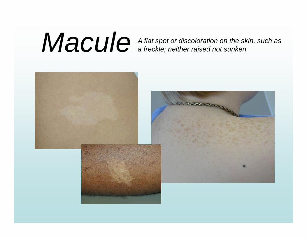
Macule A flat spot or discoloration on the skin, such as a freckle; neither raised not sunken.
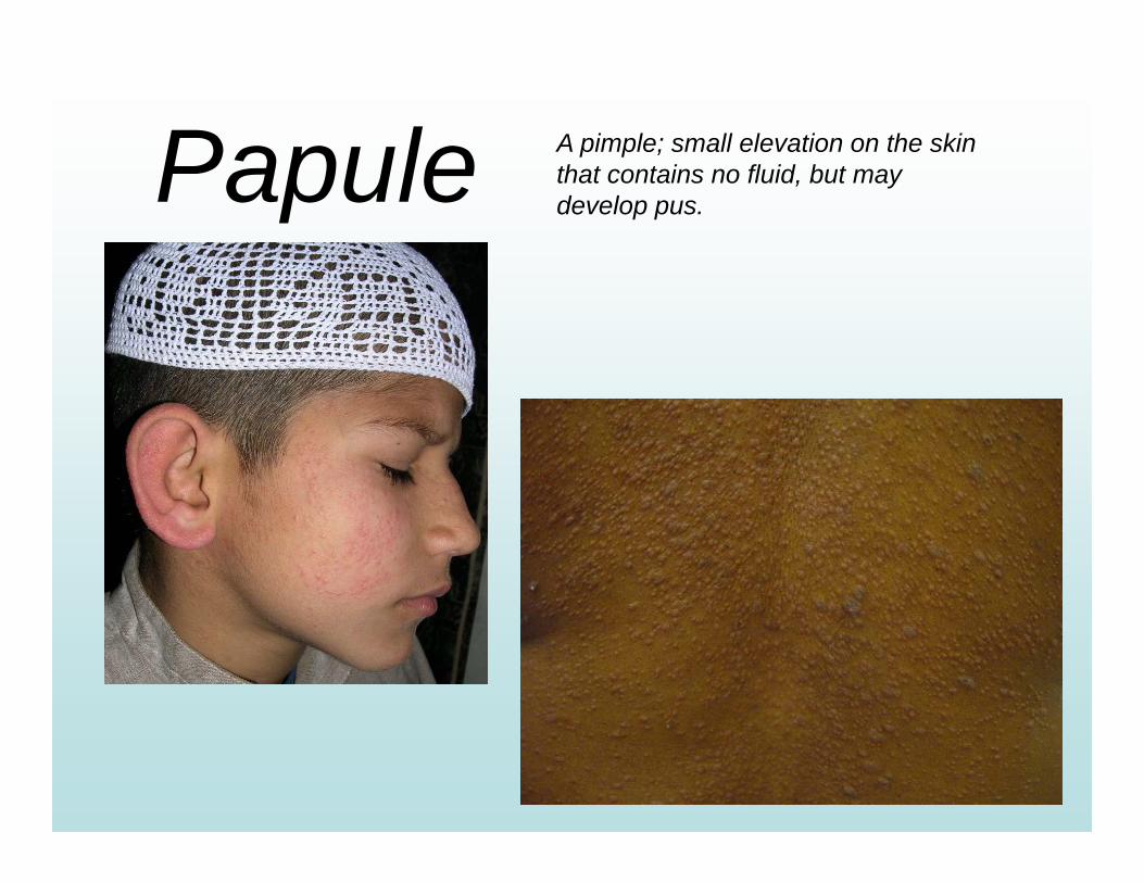
Papule A pimple; small elevation on the skin that contains no fluid, but may develop pus.
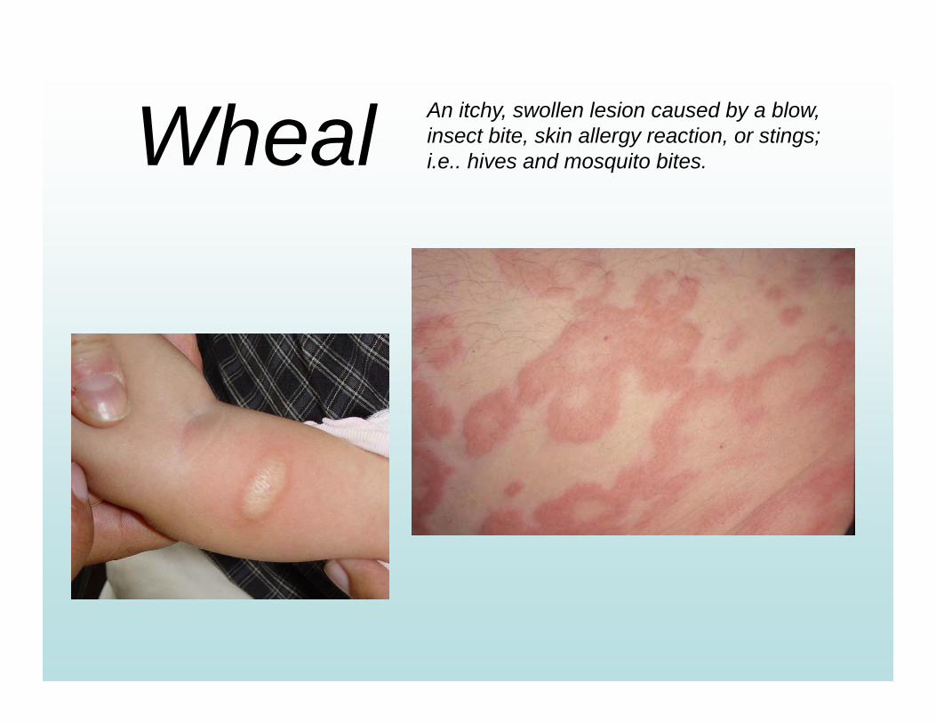
Wheal An itchy, swollen lesion caused by a blow, insect bite, skin allergy reaction, or stings; i.e.. hives and mosquito bites.
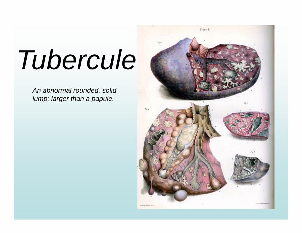
TuberculeAn abnormal rounded, solid lump; larger than a papule.

Bulla A large blister containing watery fluid; similar to a vesicle, but larger.
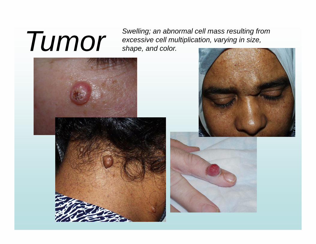
Tumor Swelling; an abnormal cell mass resulting from excessive cell multiplication, varying in size, shape, and color.
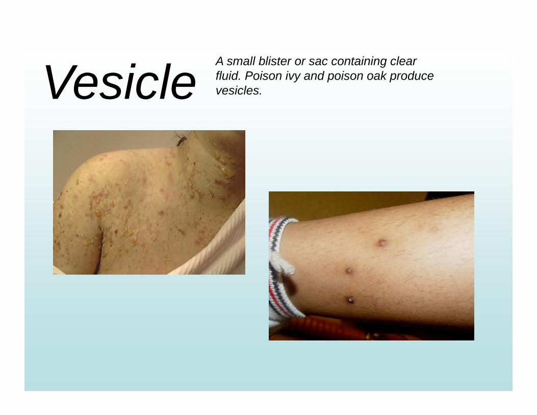
VesicleA small blister or sac containing clear fluid. Poison ivy and poison oak produce vesicles.
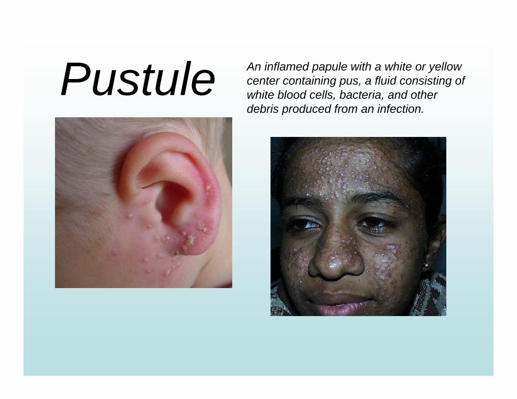
Pustule An inflamed papule with a white or yellow center containing pus, a fluid consisting of white blood cells, bacteria, and other debris produced from an infection.
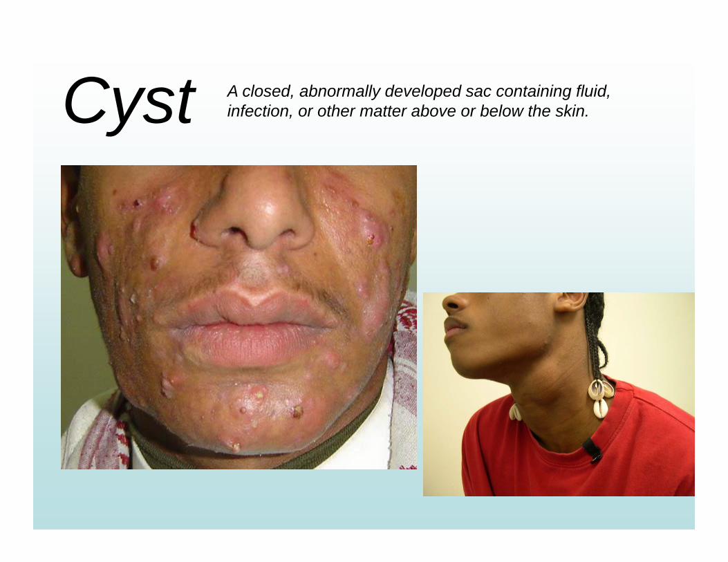
Cyst A closed, abnormally developed sac containing fluid, infection, or other matter above or below the skin.

“Seven Cats Eating Fish Under Starry Skies & Kissing”
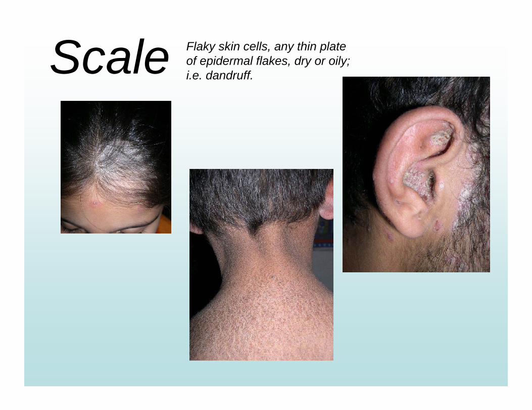
Scale Flaky skin cells, any thin plate of epidermal flakes, dry or oily; i.e. dandruff.
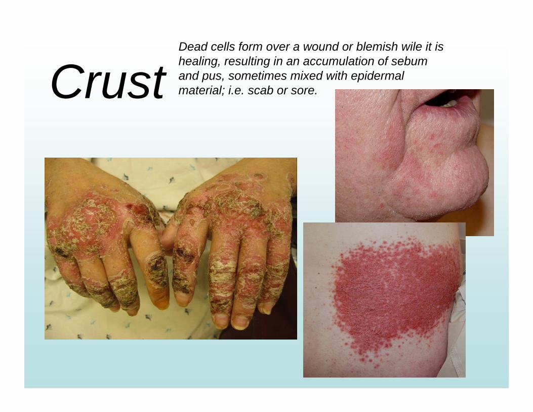
CrustDead cells form over a wound or blemish wile it is healing, resulting in an accumulation of sebum and pus, sometimes mixed with epidermal material; i.e. scab or sore.
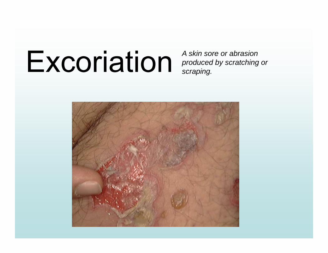
Excoriation A skin sore or abrasion produced by scratching or scraping.
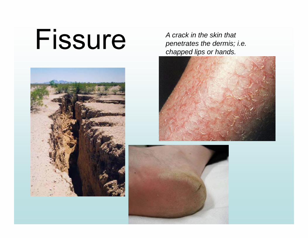
Fissure A crack in the skin that penetrates the dermis; i.e. chapped lips or hands.
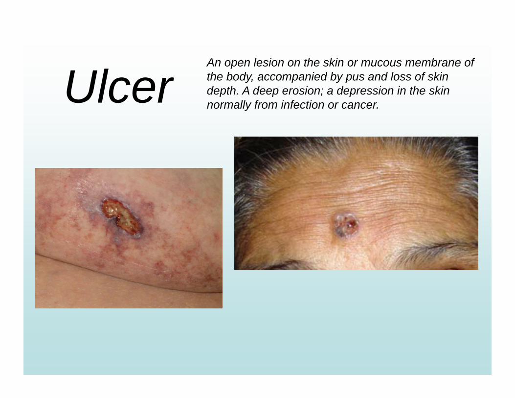
UlcerAn open lesion on the skin or mucous membrane of the body, accompanied by pus and loss of skin depth. A deep erosion; a depression in the skin normally from infection or cancer.
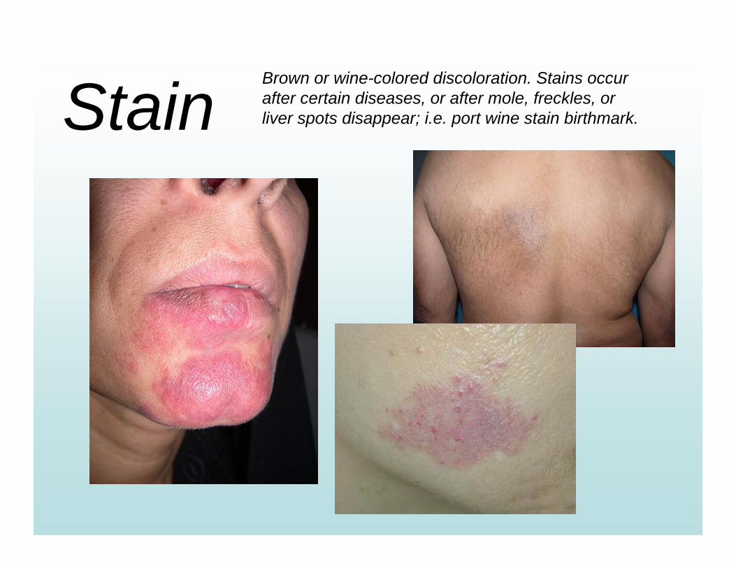
StainBrown or wine-colored discoloration. Stains occur after certain diseases, or after mole, freckles, or liver spots disappear; i.e. port wine stain birthmark.
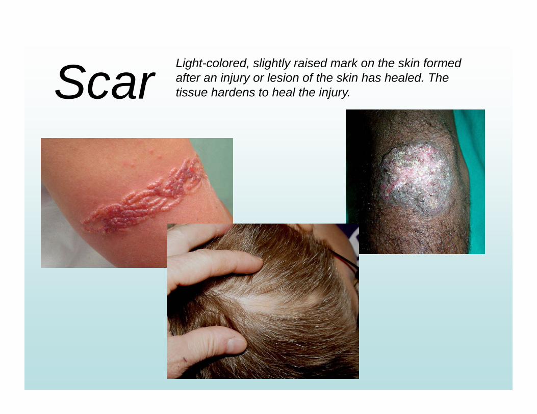
Scar Light-colored, slightly raised mark on the skin formed after an injury or lesion of the skin has healed. The tissue hardens to heal the injury.
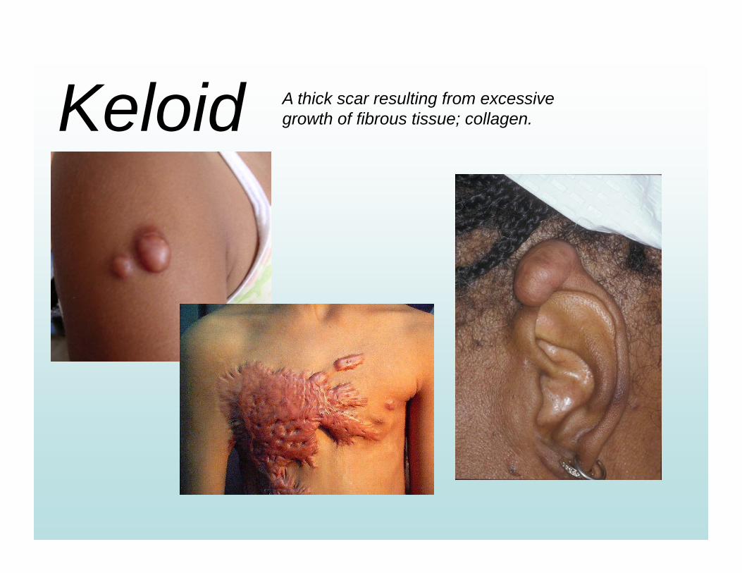
Keloid A thick scar resulting from excessive growth of fibrous tissue; collagen.
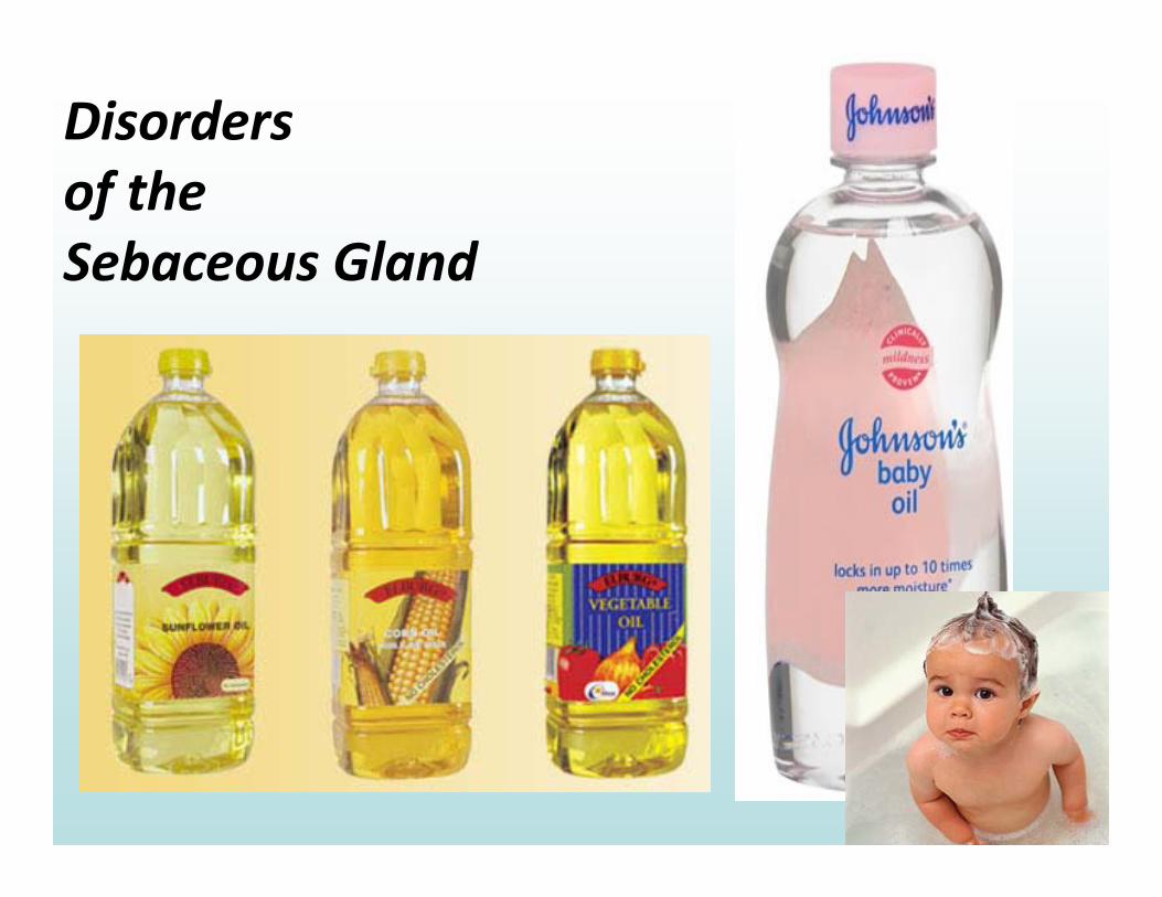
Disordersof theSebaceous Gland
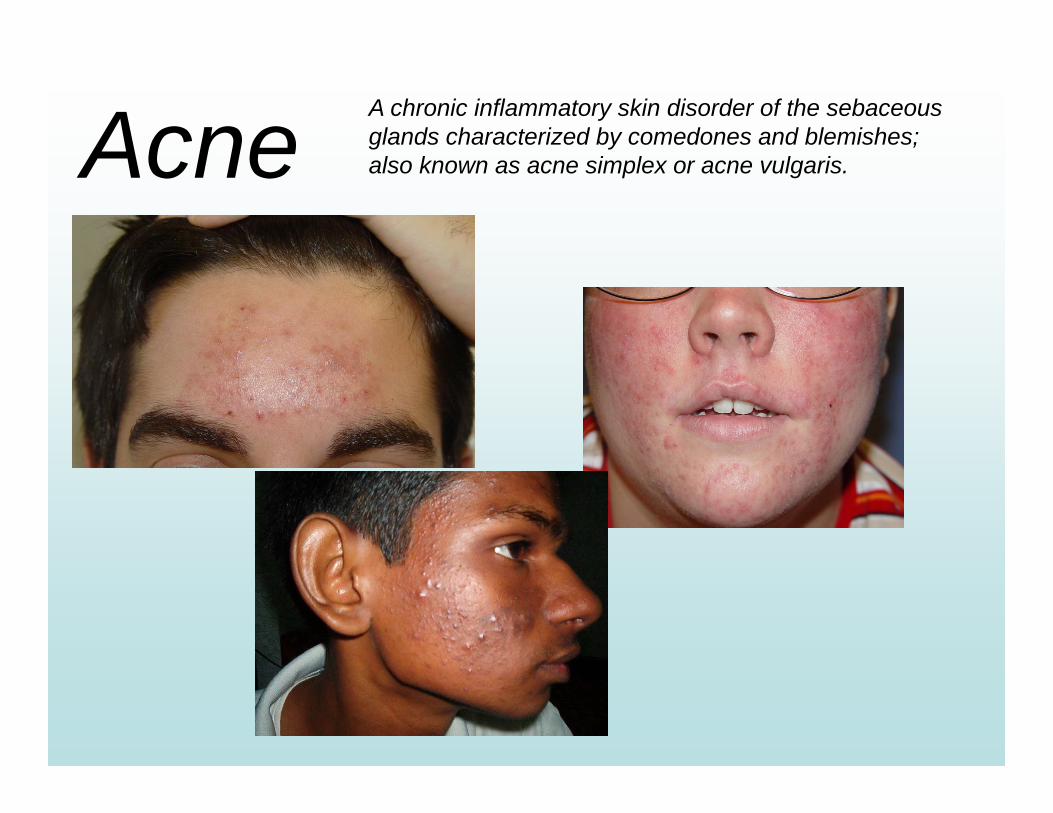
Acne A chronic inflammatory skin disorder of the sebaceous glands characterized by comedones and blemishes; also known as acne simplex or acne vulgaris.
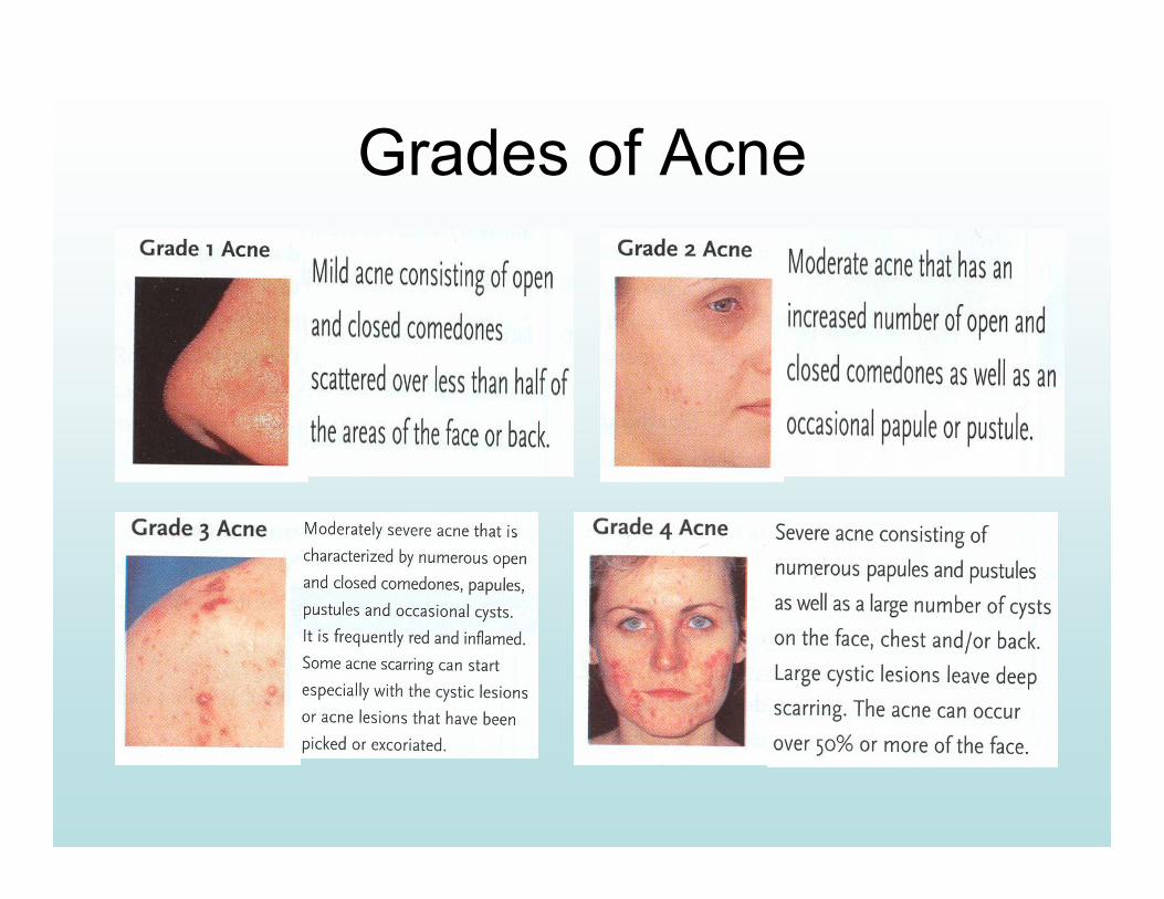
Grades of Acne
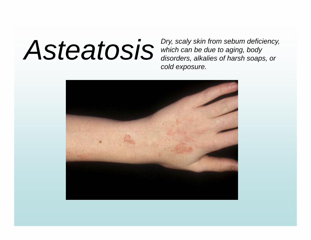
Asteatosis Dry, scaly skin from sebum deficiency, which can be due to aging, body disorders, alkalies of harsh soaps, or cold exposure.
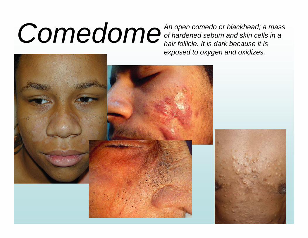
ComedomeAn open comedo or blackhead; a mass of hardened sebum and skin cells in a hair follicle. It is dark because it is exposed to oxygen and oxidizes.
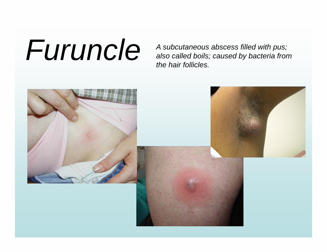
Furuncle A subcutaneous abscess filled with pus; also called boils; caused by bacteria from the hair follicles.
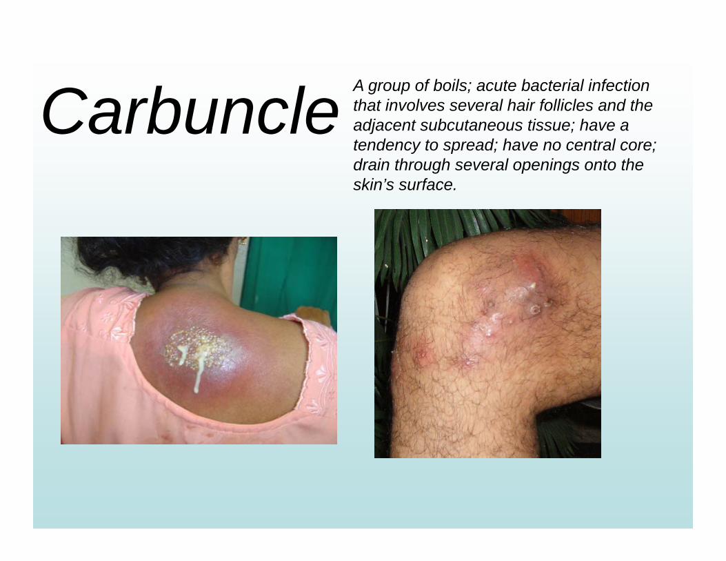
Carbuncle A group of boils; acute bacterial infection that involves several hair follicles and the adjacent subcutaneous tissue; have a tendency to spread; have no central core; drain through several openings onto the skin’s surface.
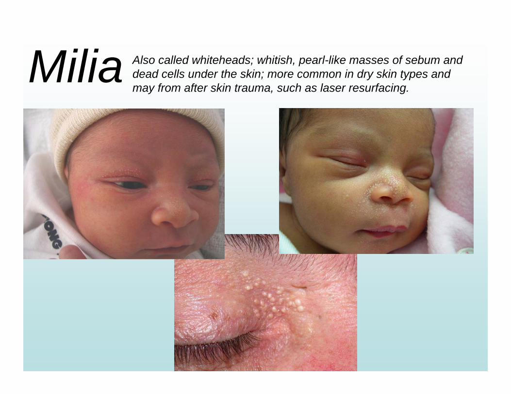
Milia Also called whiteheads; whitish, pearl-like masses of sebum and dead cells under the skin; more common in dry skin types and may from after skin trauma, such as laser resurfacing.
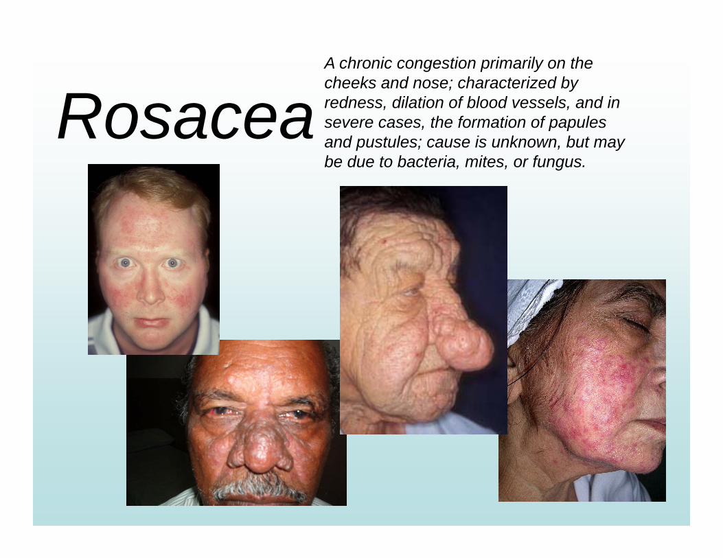
RosaceaA chronic congestion primarily on the cheeks and nose; characterized by redness, dilation of blood vessels, and in severe cases, the formation of papules and pustules; cause is unknown, but may be due to bacteria, mites, or fungus.
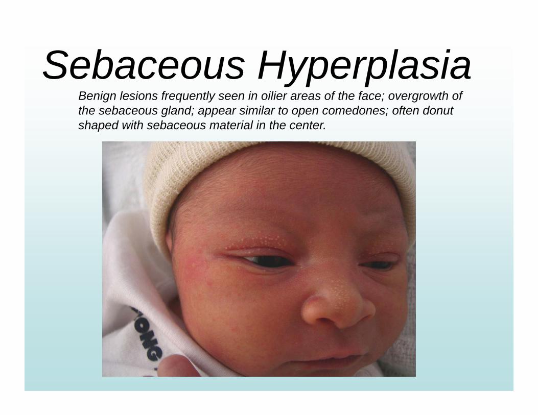
Sebaceous HyperplasiaBenign lesions frequently seen in oilier areas of the face; overgrowth of the sebaceous gland; appear similar to open comedones; often donut shaped with sebaceous material in the center.
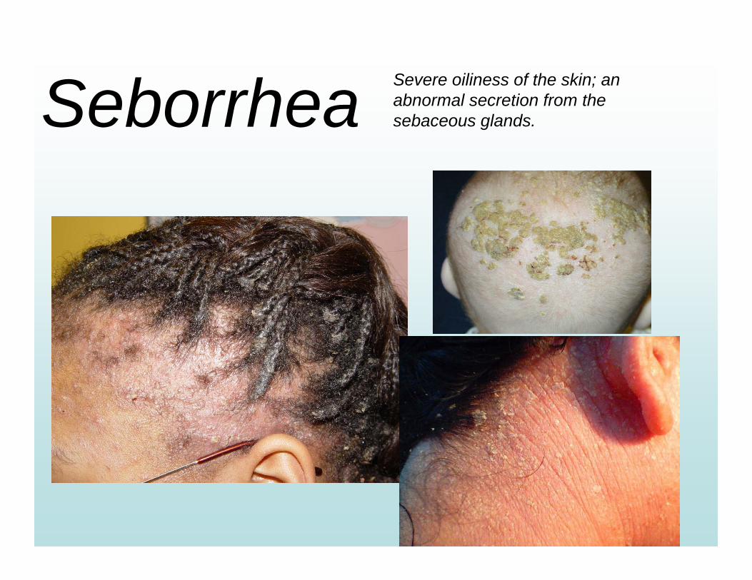
Seborrhea Severe oiliness of the skin; an abnormal secretion from the sebaceous glands.
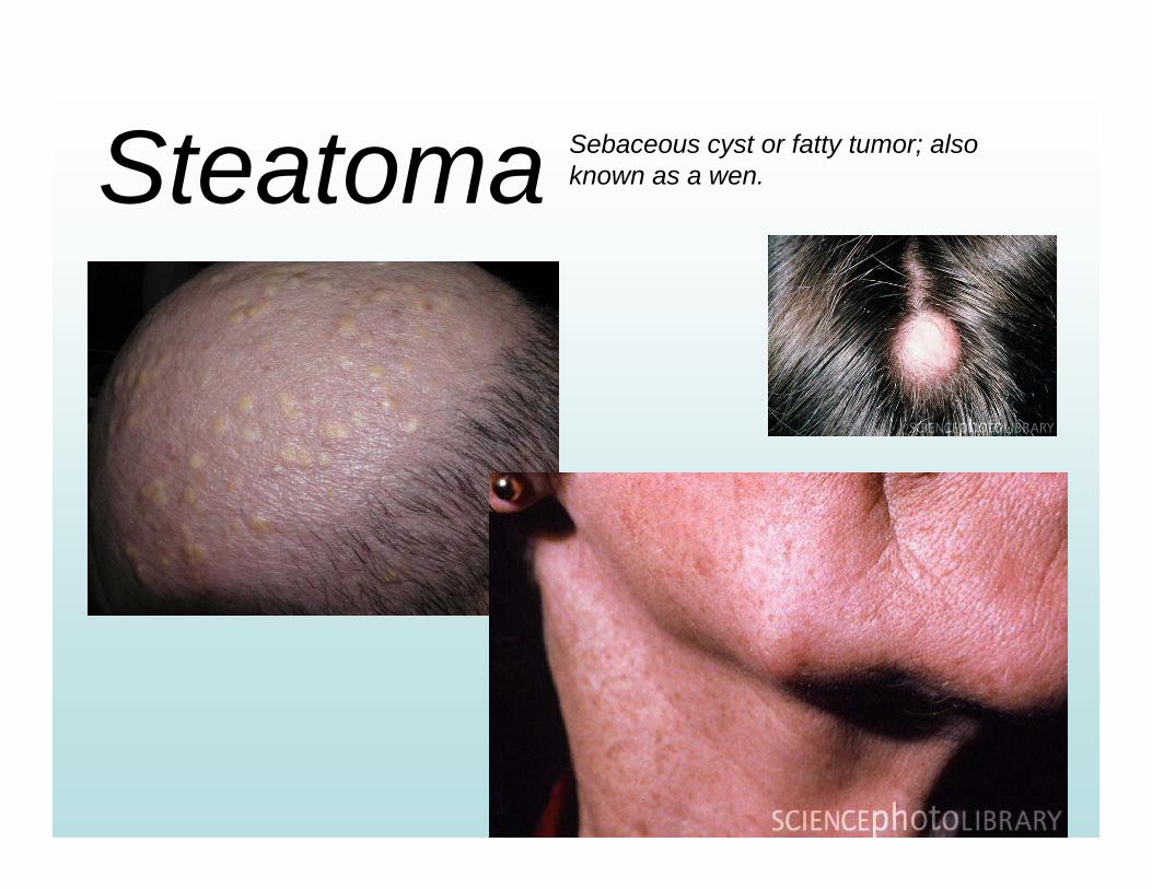
Steatoma Sebaceous cyst or fatty tumor; also known as a wen.
Disordersof the
Suderiferous Glands
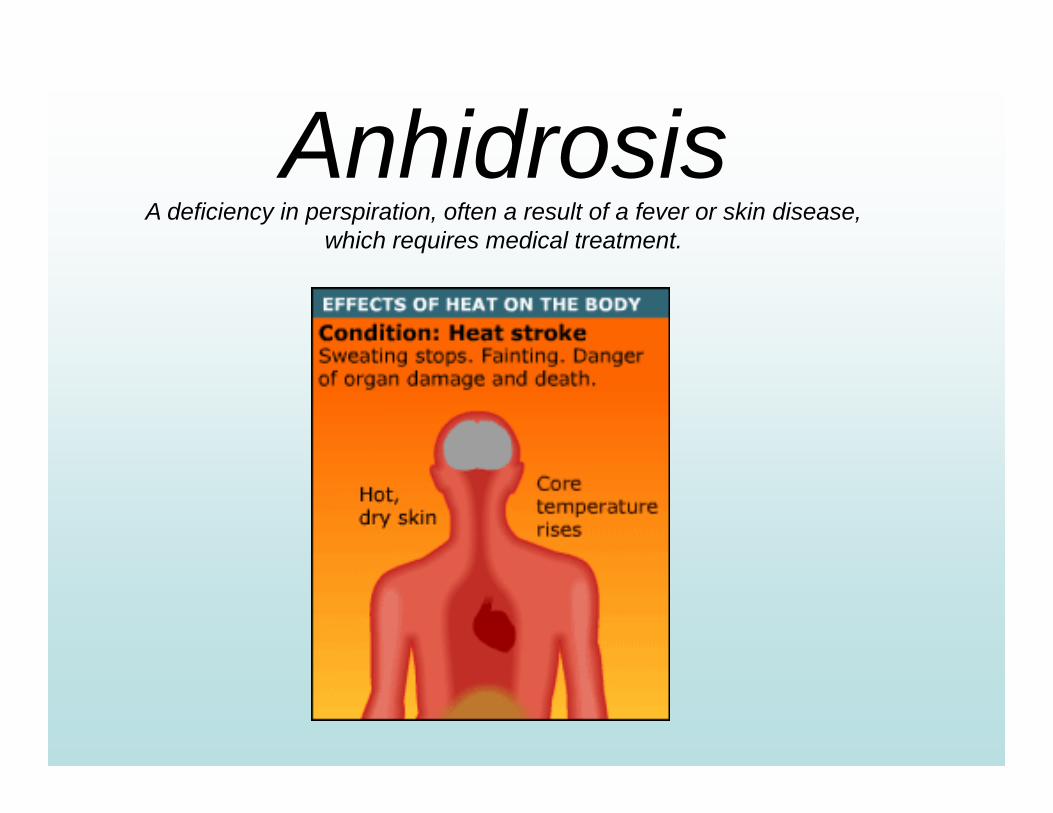
AnhidrosisA deficiency in perspiration, often a result of a fever or skin disease,
which requires medical treatment.
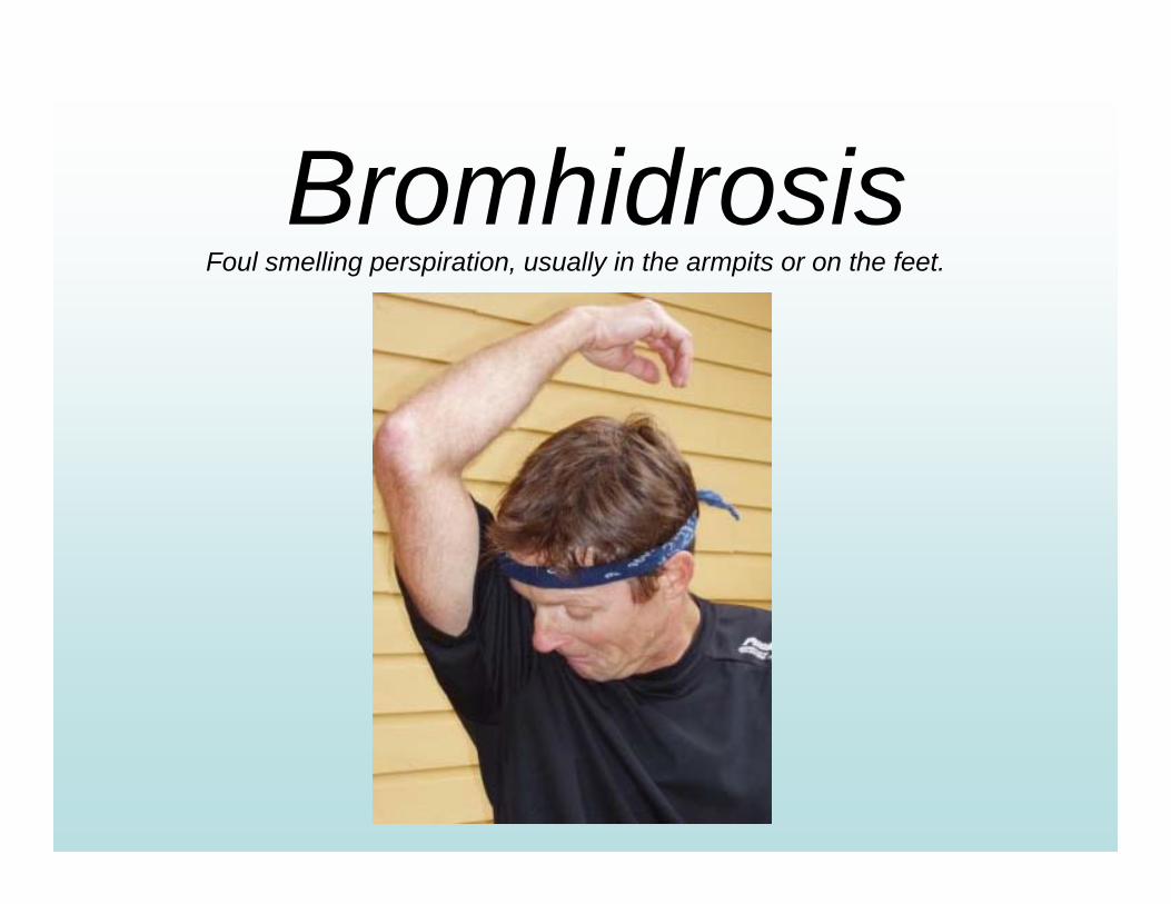
BromhidrosisFoul smelling perspiration, usually in the armpits or on the feet.

HyperhidrosisExcessive perspiration caused by heat or body weakness.
Medical treatment is required.
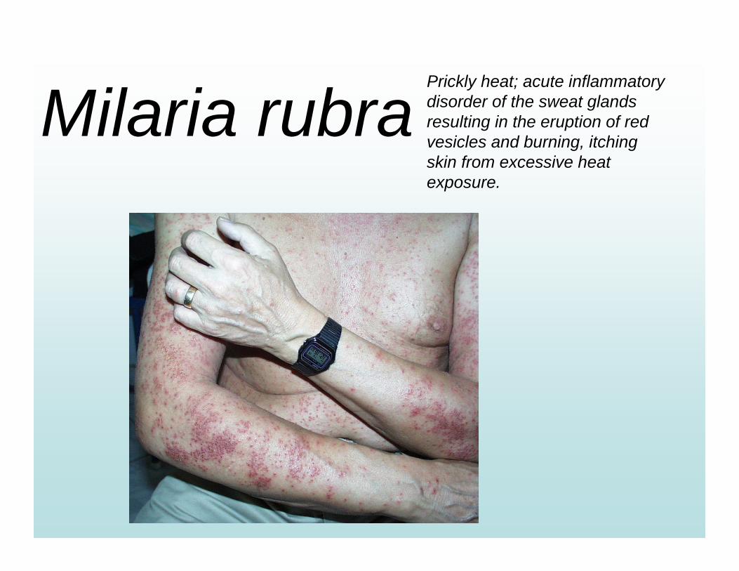
Milaria rubraPrickly heat; acute inflammatory disorder of the sweat glands resulting in the eruption of red vesicles and burning, itching skin from excessive heat exposure.
Inflammations
DermatitisAtopic
Contact
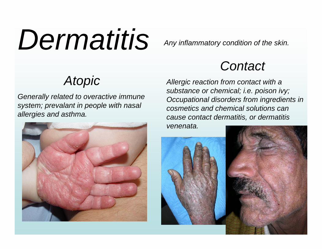
Any inflammatory condition of the skin.
Generally related to overactive immune system; prevalant in people with nasal allergies and asthma.
Allergic reaction from contact with a substance or chemical; i.e. poison ivy; Occupational disorders from ingredients in cosmetics and chemical solutions can cause contact dermatitis, or dermatitis venenata.
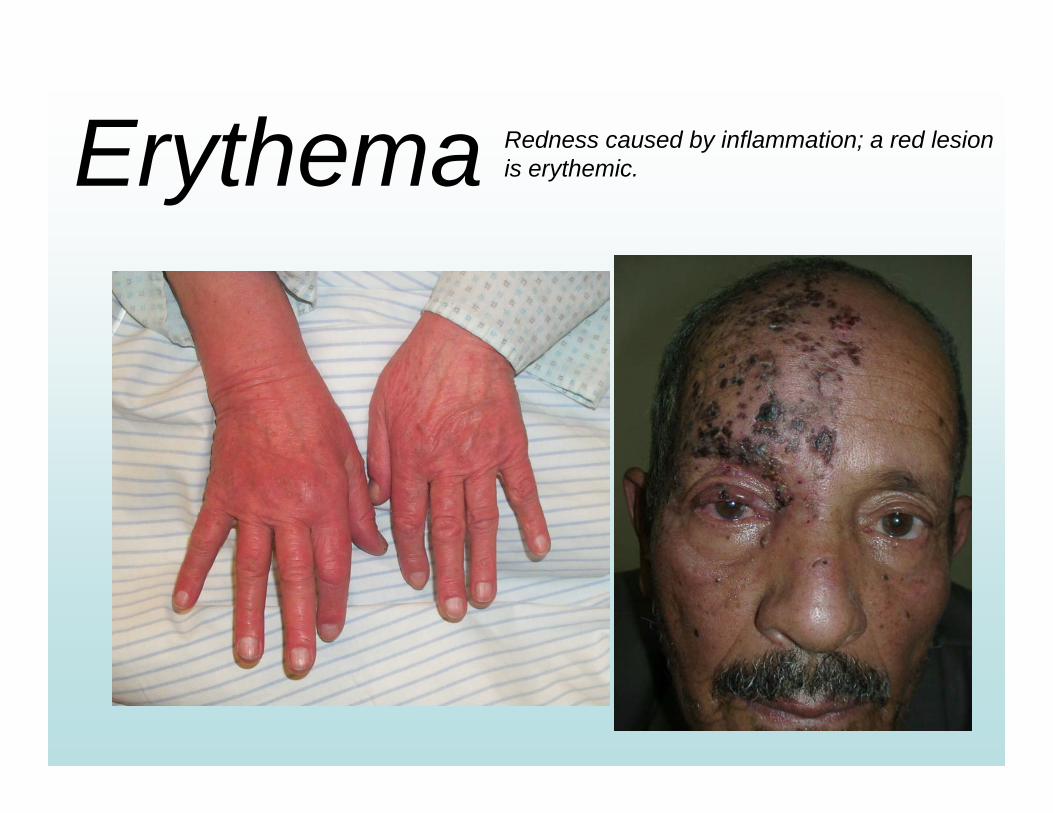
Erythema Redness caused by inflammation; a red lesion is erythemic.
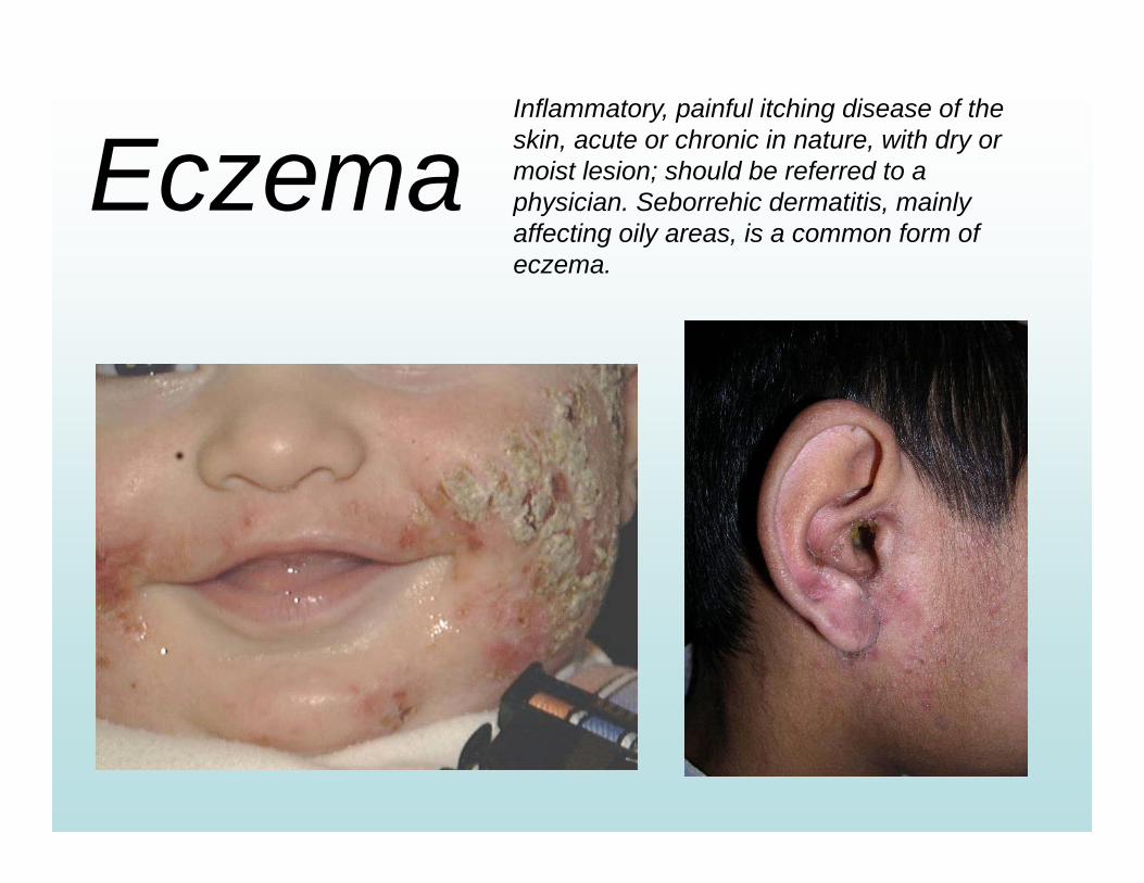
EczemaInflammatory, painful itching disease of the skin, acute or chronic in nature, with dry or moist lesion; should be referred to a physician. Seborrehic dermatitis, mainly affecting oily areas, is a common form of eczema.
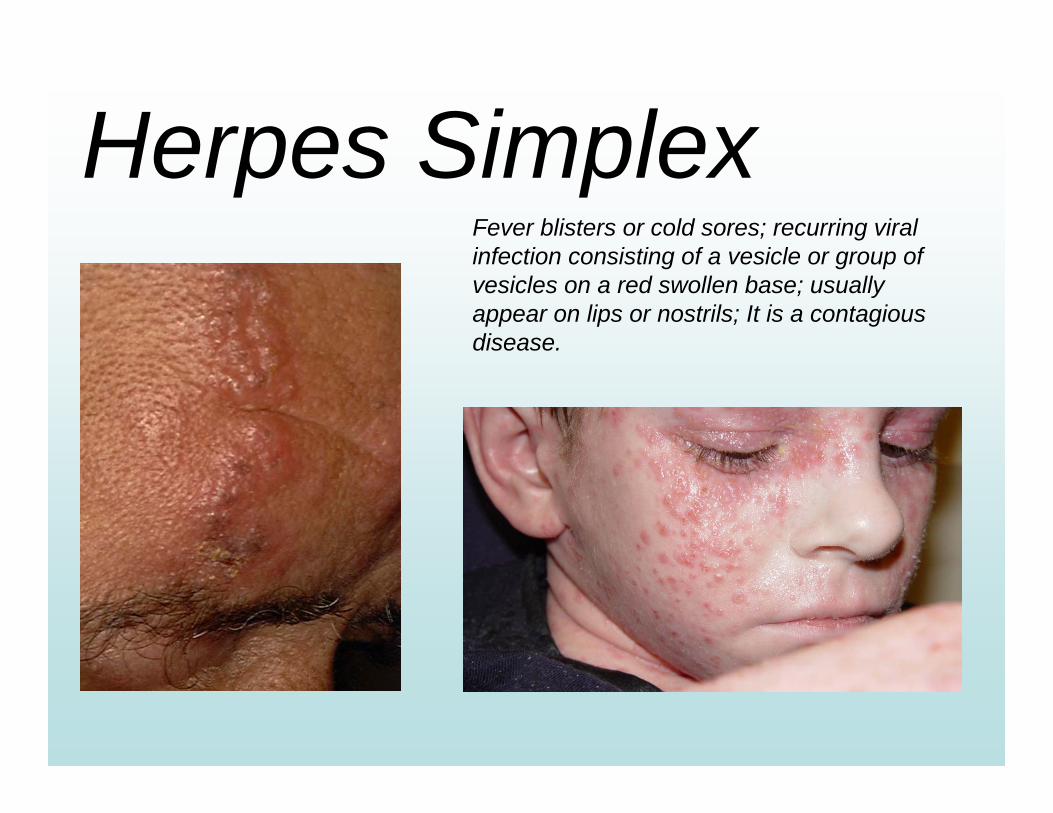
Herpes SimplexFever blisters or cold sores; recurring viral infection consisting of a vesicle or group of vesicles on a red swollen base; usually appear on lips or nostrils; It is a contagious disease.
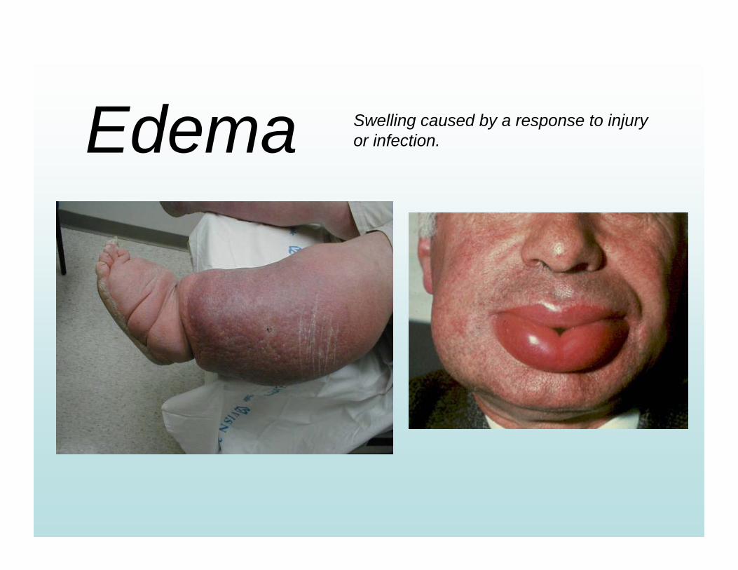
Edema Swelling caused by a response to injury or infection.
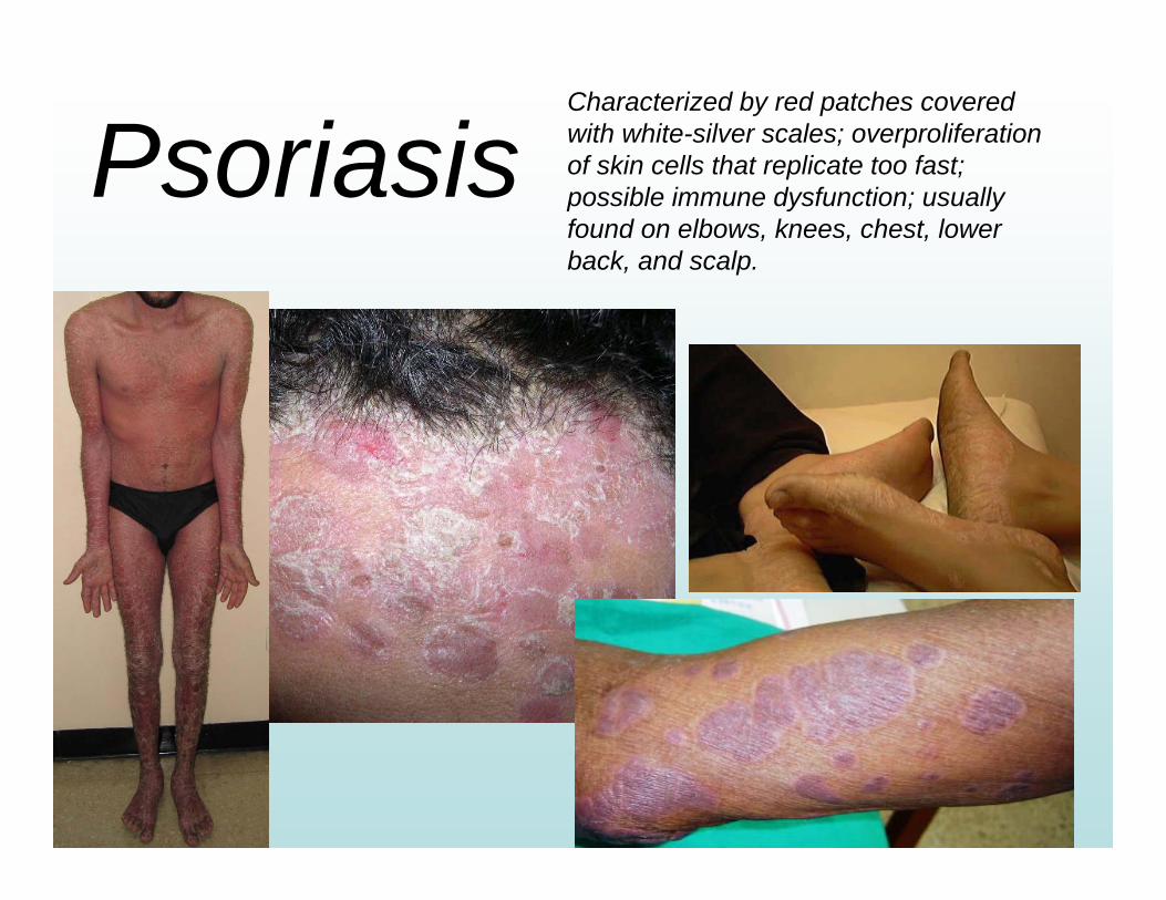
PsoriasisCharacterized by red patches covered with white-silver scales; overproliferation of skin cells that replicate too fast; possible immune dysfunction; usually found on elbows, knees, chest, lower back, and scalp.
PigmentationDisorders
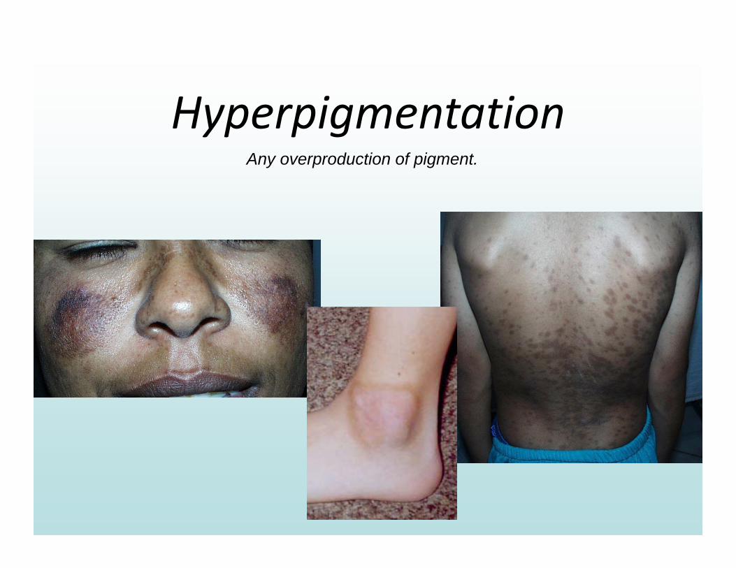
HyperpigmentationAny overproduction of pigment.
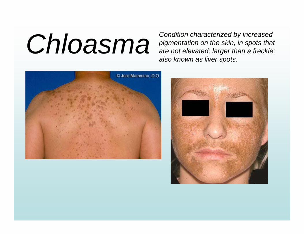
Chloasma Condition characterized by increased pigmentation on the skin, in spots that are not elevated; larger than a freckle; also known as liver spots.
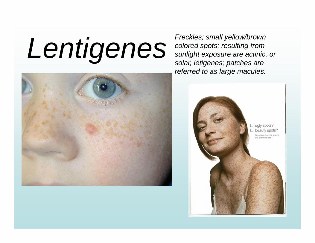
Lentigenes Freckles; small yellow/brown colored spots; resulting from sunlight exposure are actinic, or solar, letigenes; patches are referred to as large macules.
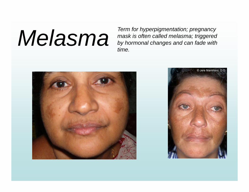
Melasma Term for hyperpigmentation; pregnancy mask is often called melasma; triggered by hormonal changes and can fade with time.
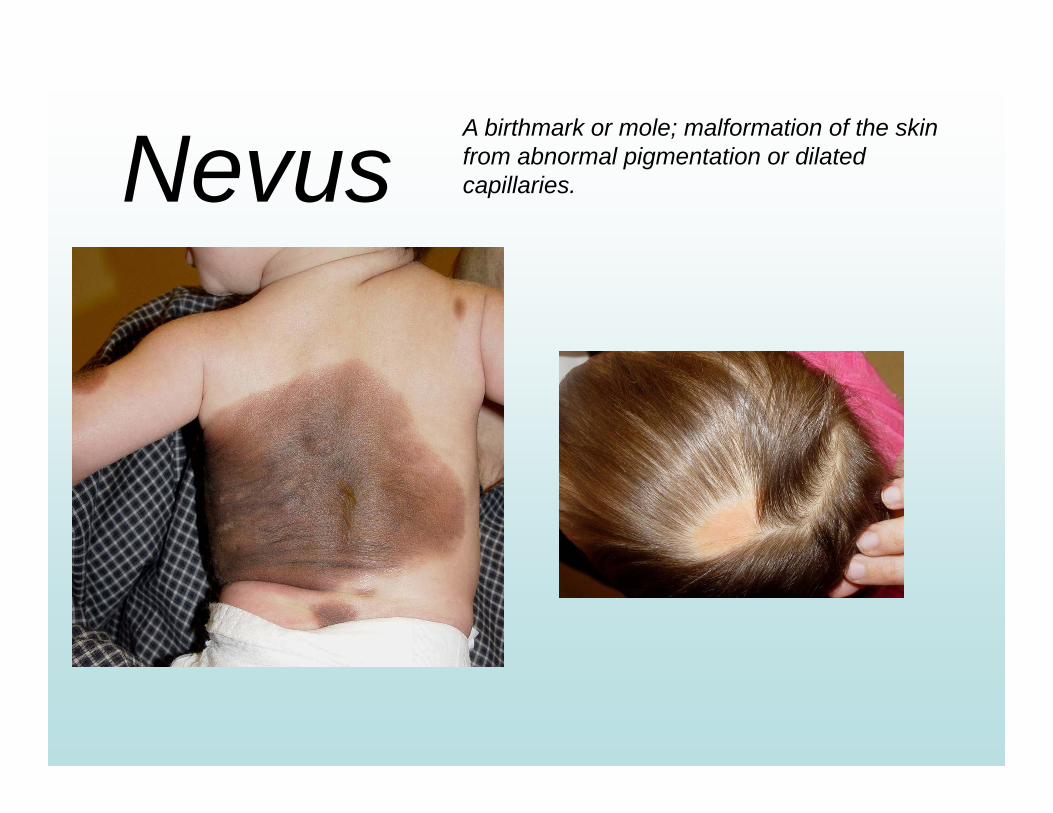
NevusA birthmark or mole; malformation of the skin from abnormal pigmentation or dilated capillaries.
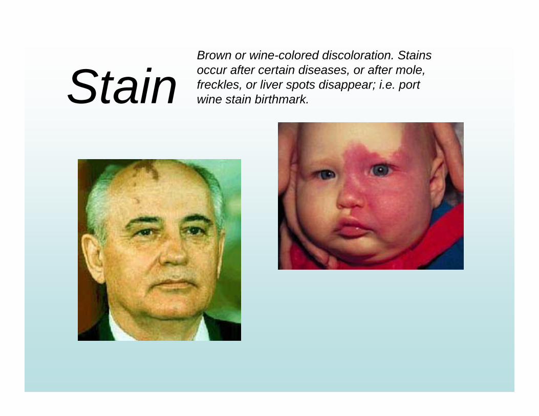
StainBrown or wine-colored discoloration. Stains occur after certain diseases, or after mole, freckles, or liver spots disappear; i.e. port wine stain birthmark.
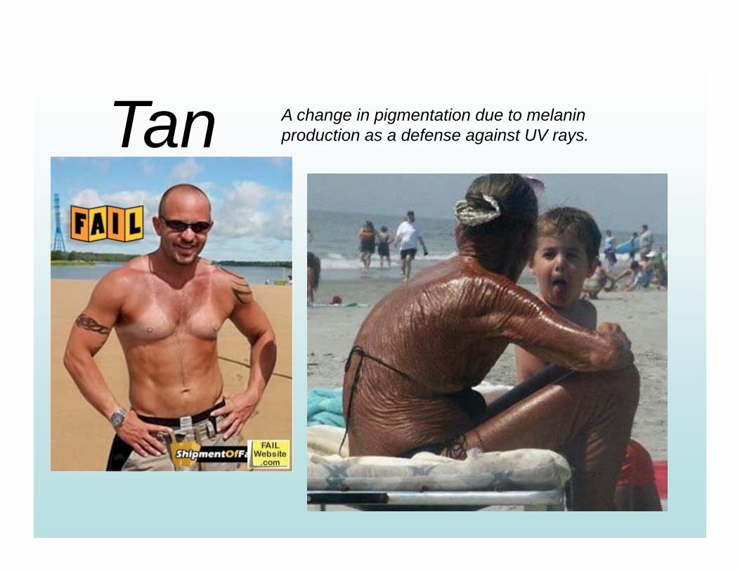
Tan A change in pigmentation due to melanin production as a defense against UV rays.
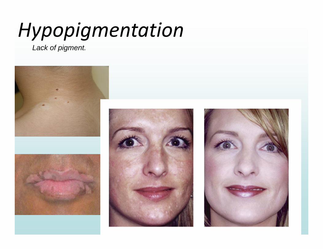
HypopigmentationLack of pigment.
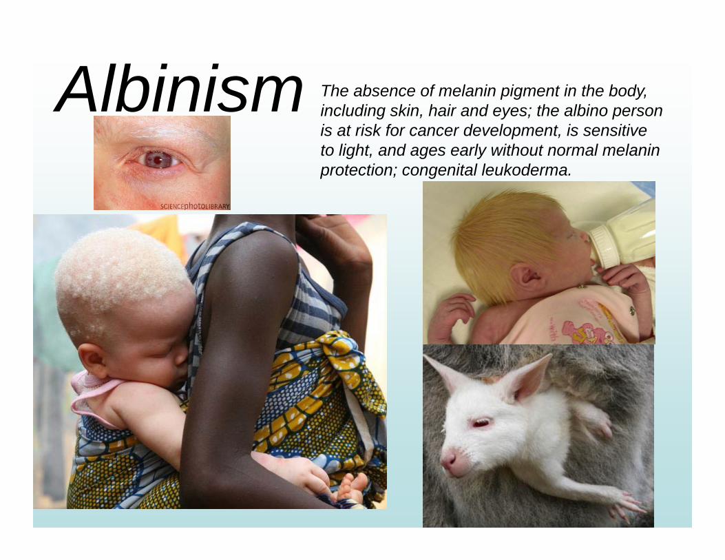
Albinism The absence of melanin pigment in the body, including skin, hair and eyes; the albino person is at risk for cancer development, is sensitive to light, and ages early without normal melanin protection; congenital leukoderma.
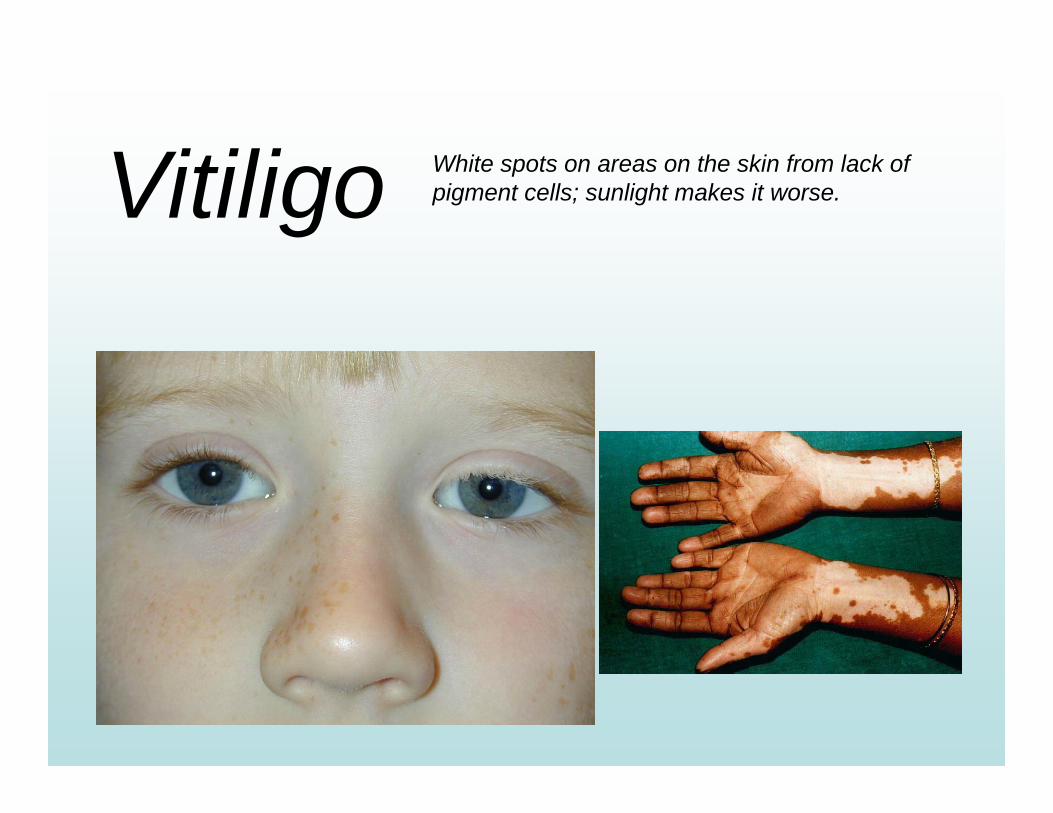
Vitiligo White spots on areas on the skin from lack of pigment cells; sunlight makes it worse.
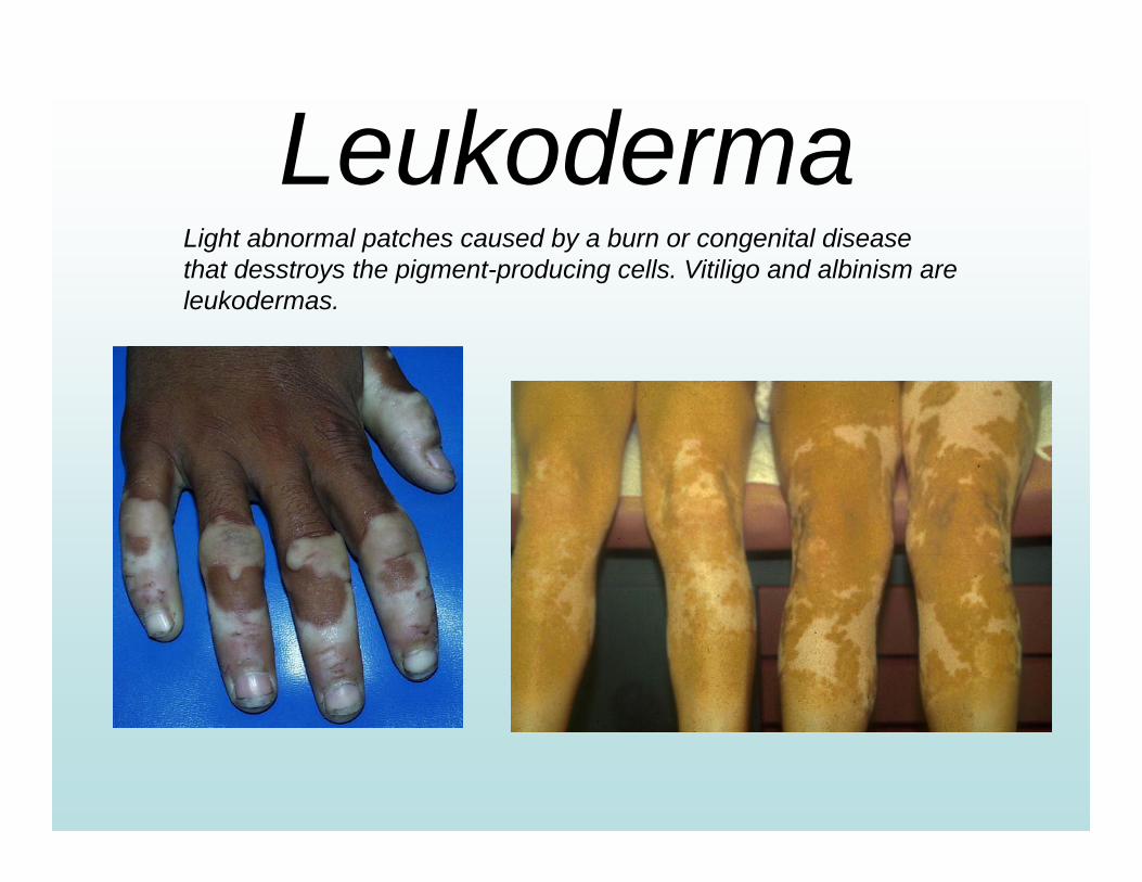
LeukodermaLight abnormal patches caused by a burn or congenital disease that desstroys the pigment-producing cells. Vitiligo and albinism are leukodermas.
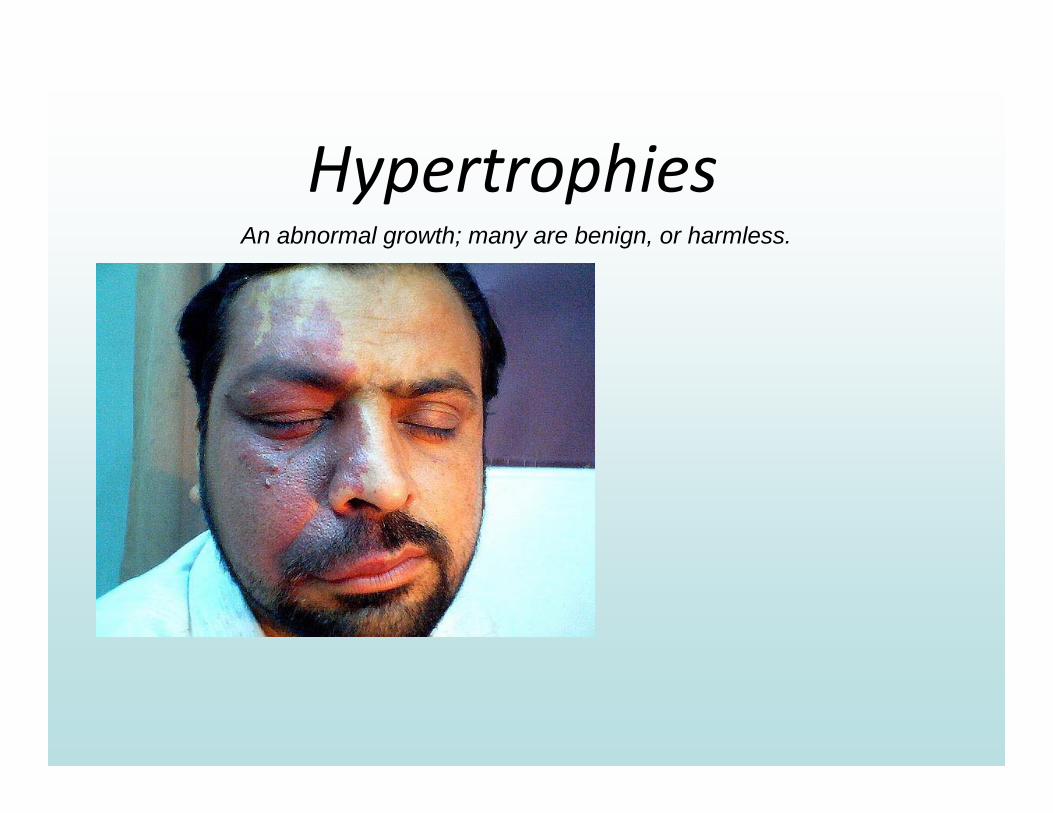
HypertrophiesAn abnormal growth; many are benign, or harmless.
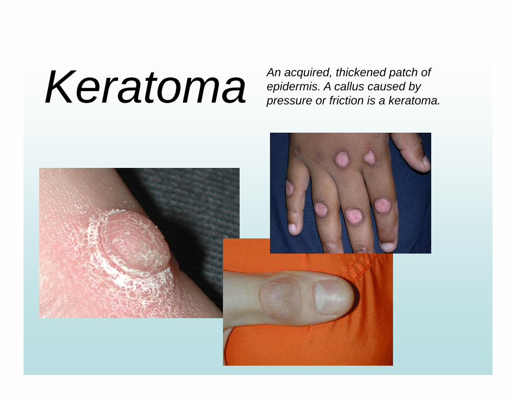
Keratoma An acquired, thickened patch of epidermis. A callus caused by pressure or friction is a keratoma.
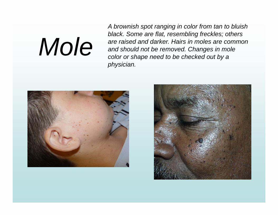
MoleA brownish spot ranging in color from tan to bluish black. Some are flat, resembling freckles; others are raised and darker. Hairs in moles are common and should not be removed. Changes in mole color or shape need to be checked out by a physician.
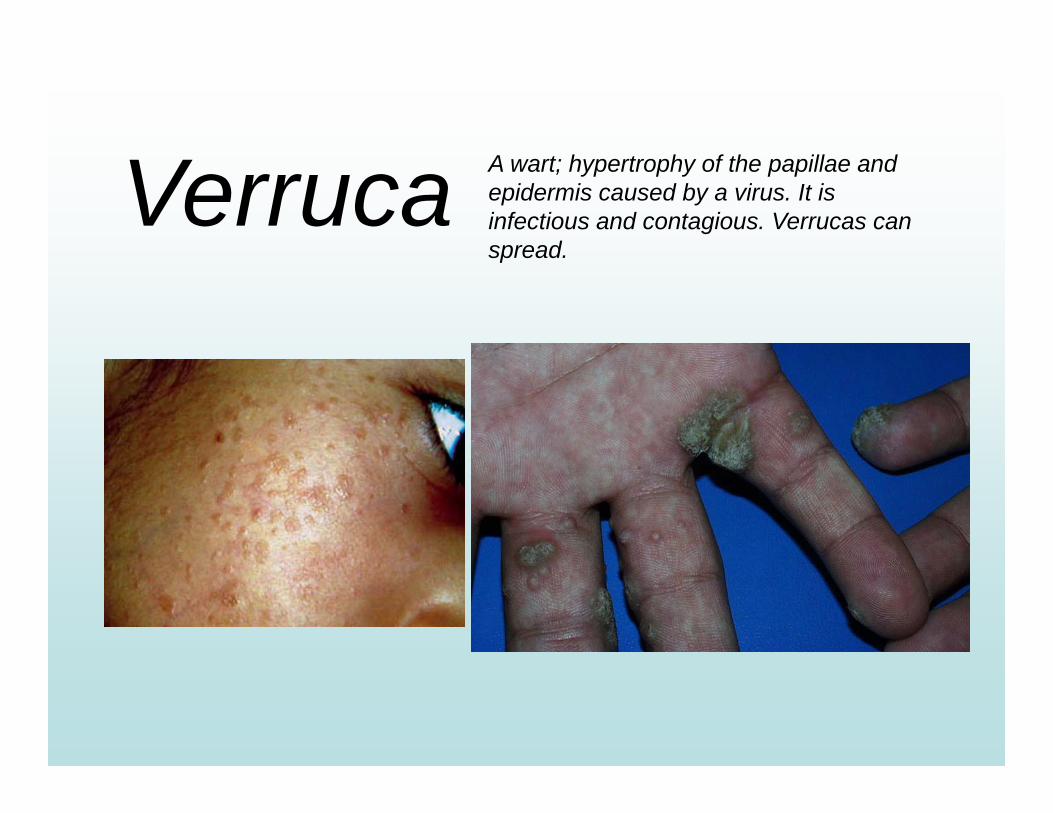
Verruca A wart; hypertrophy of the papillae and epidermis caused by a virus. It is infectious and contagious. Verrucas can spread.
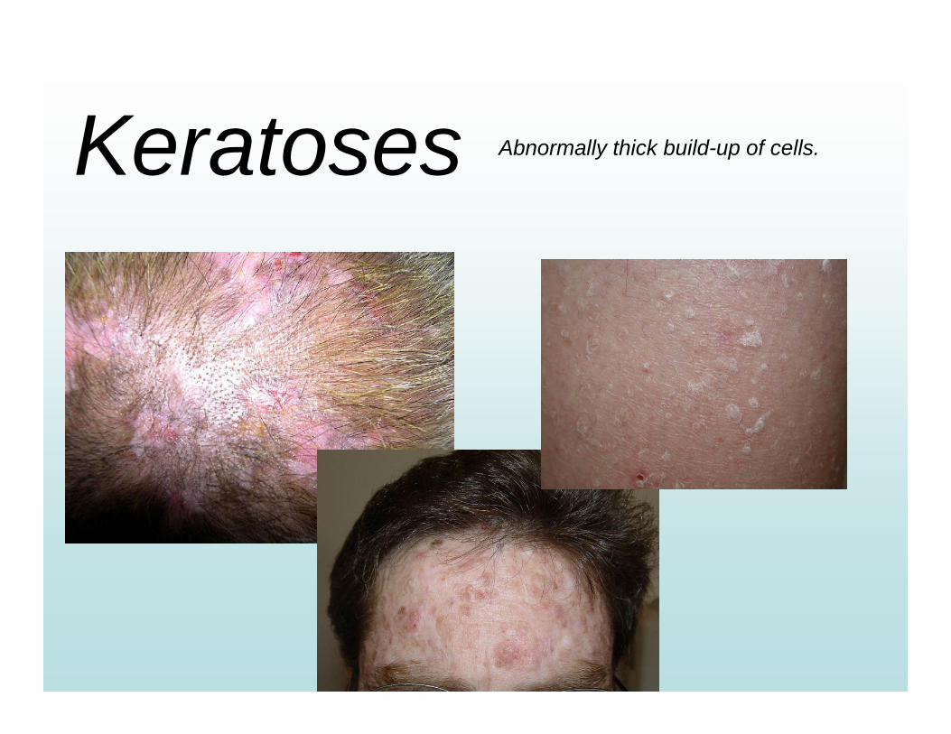
Keratoses Abnormally thick build-up of cells.
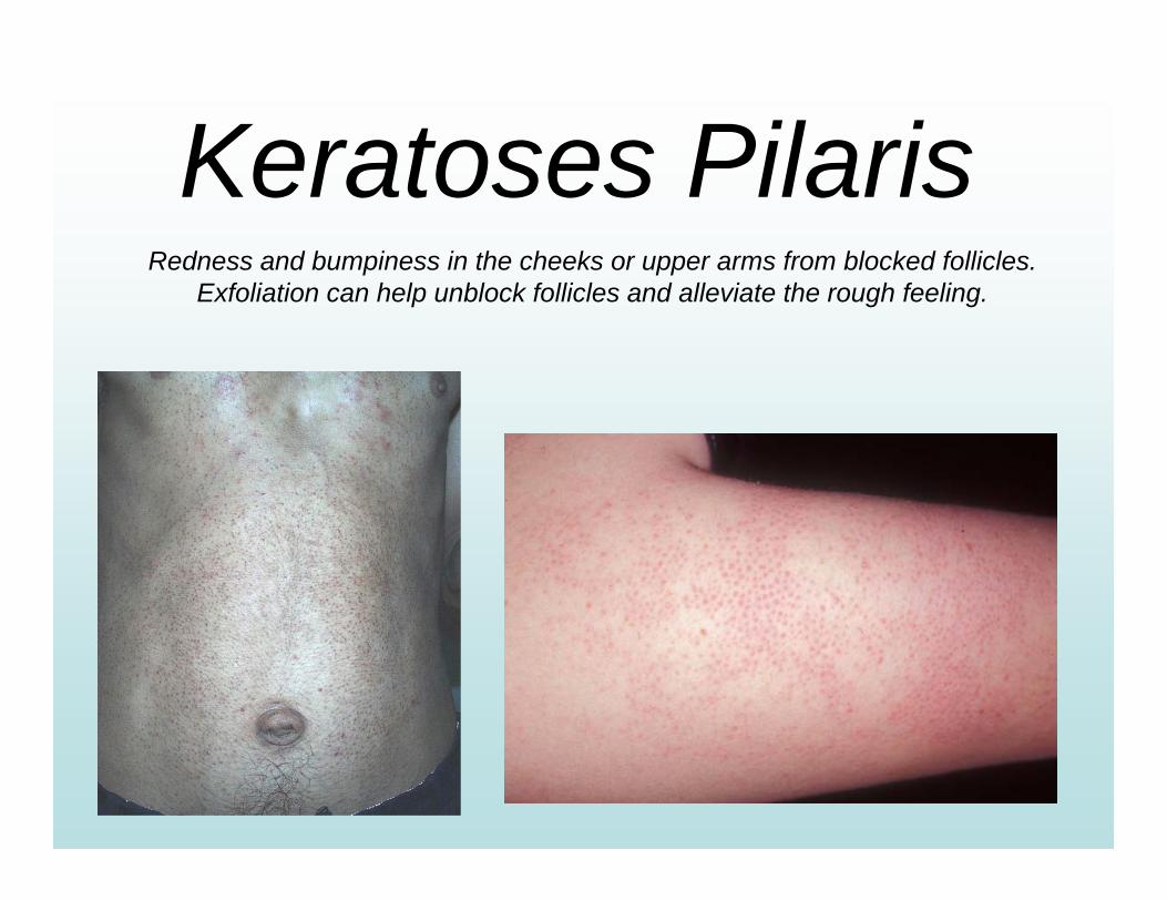
Keratoses PilarisRedness and bumpiness in the cheeks or upper arms from blocked follicles.
Exfoliation can help unblock follicles and alleviate the rough feeling.
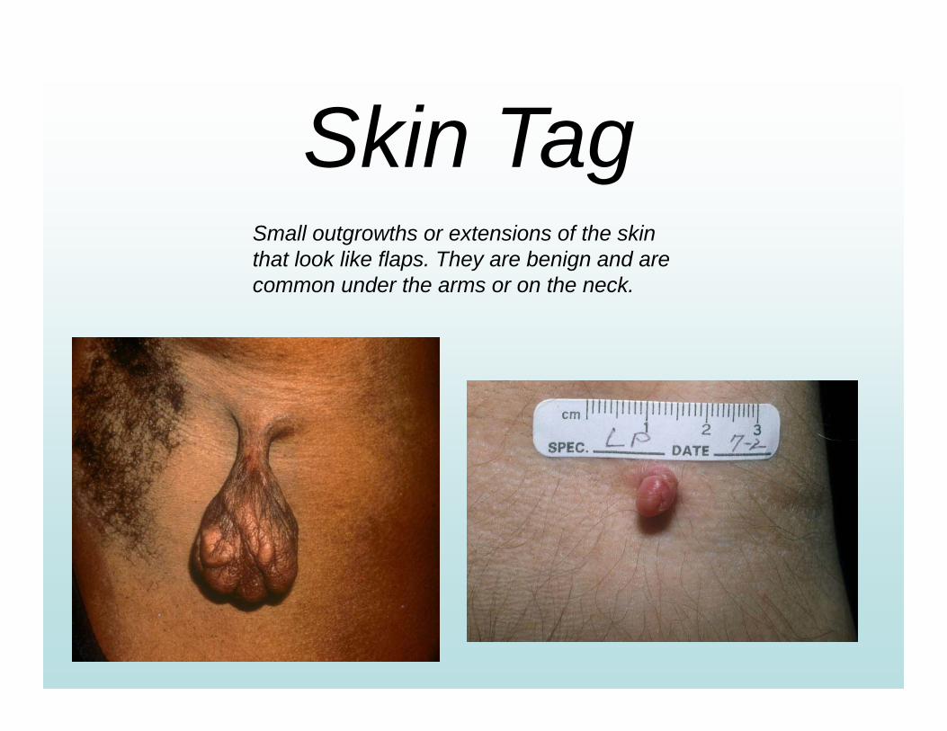
Skin TagSmall outgrowths or extensions of the skin that look like flaps. They are benign and are common under the arms or on the neck.
Skin Cancers
The ABCD’s of Skin Cancer
A - Asymmetry; inconsistent growths should be seen by physician.
B - Border; growth should have a well-defined edge.
C - Color; should be consistent and not vary.
D - Diameter; should be no larger that a pencil eraser.
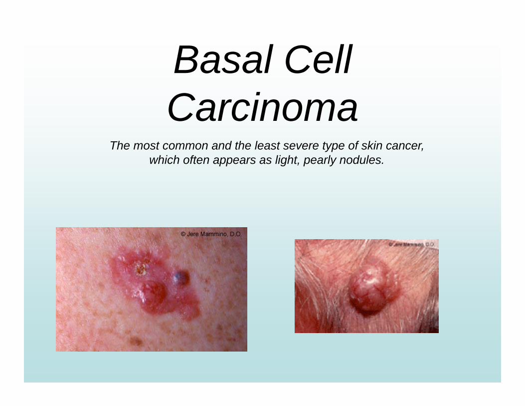
Basal Cell Carcinoma
The most common and the least severe type of skin cancer, which often appears as light, pearly nodules.

Squamous CellCarcinoma
More serious than basal cell carcinoma, it is characterized by scaly red papules or nodules.
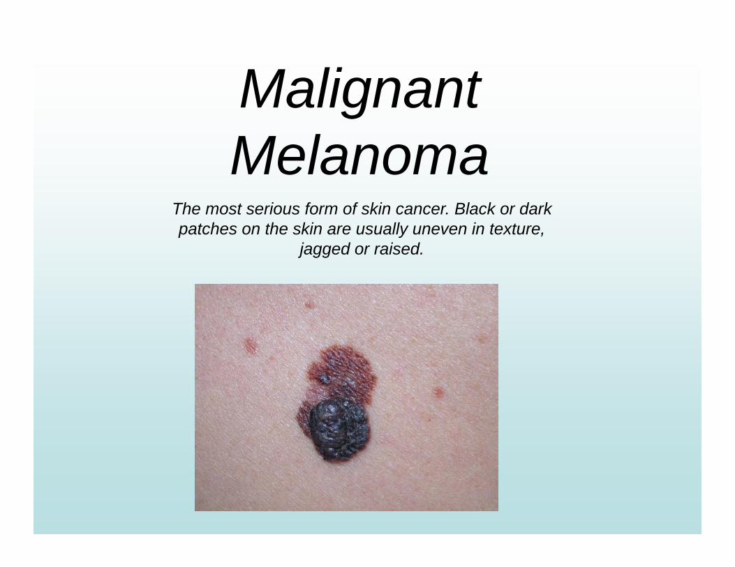
MalignantMelanoma
The most serious form of skin cancer. Black or dark patches on the skin are usually uneven in texture,
jagged or raised.
Contagious Diseases
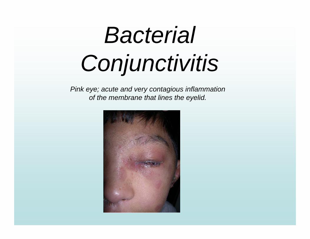
BacterialConjunctivitis
Pink eye; acute and very contagious inflammation of the membrane that lines the eyelid.
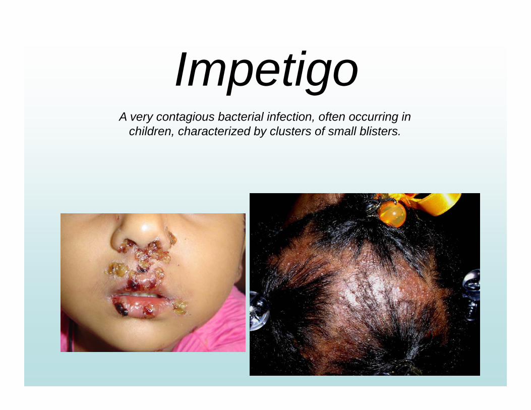
ImpetigoA very contagious bacterial infection, often occurring in
children, characterized by clusters of small blisters.
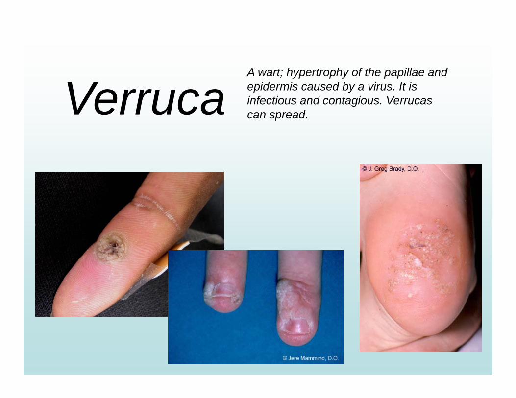
VerrucaA wart; hypertrophy of the papillae and epidermis caused by a virus. It is infectious and contagious. Verrucas can spread.
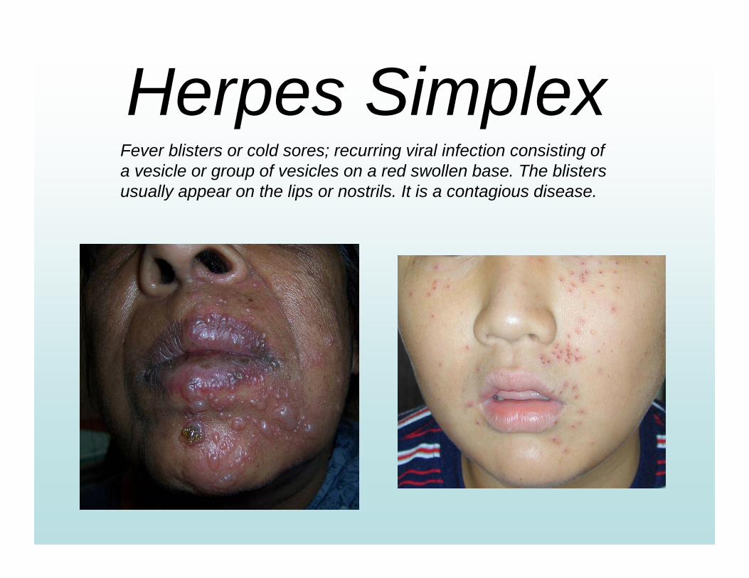
Herpes SimplexFever blisters or cold sores; recurring viral infection consisting of a vesicle or group of vesicles on a red swollen base. The blisters usually appear on the lips or nostrils. It is a contagious disease.
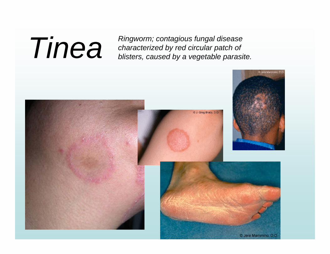
Tinea Ringworm; contagious fungal disease characterized by red circular patch of blisters, caused by a vegetable parasite.
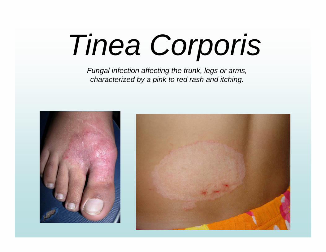
Tinea CorporisFungal infection affecting the trunk, legs or arms, characterized by a pink to red rash and itching.
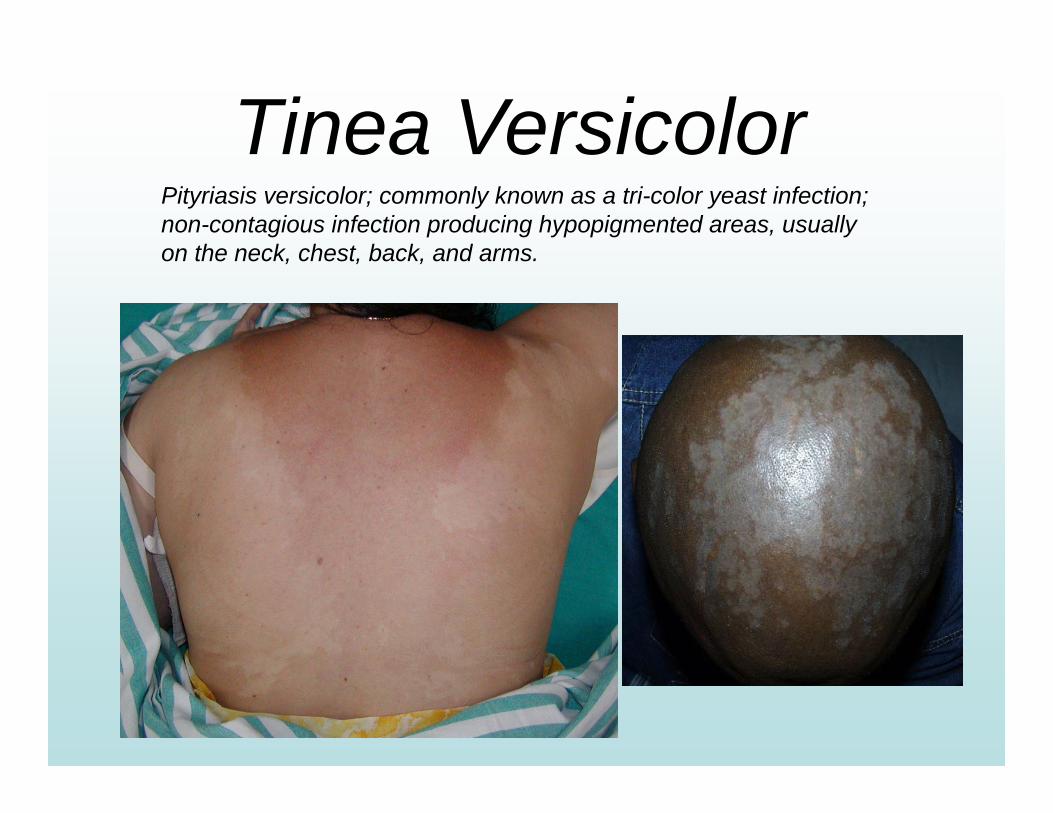
Tinea VersicolorPityriasis versicolor; commonly known as a tri-color yeast infection; non-contagious infection producing hypopigmented areas, usually on the neck, chest, back, and arms.
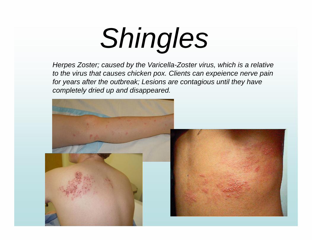
ShinglesHerpes Zoster; caused by the Varicella-Zoster virus, which is a relative to the virus that causes chicken pox. Clients can expeience nerve pain for years after the outbreak; Lesions are contagious until they have completely dried up and disappeared.
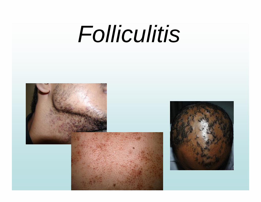
Folliculitis
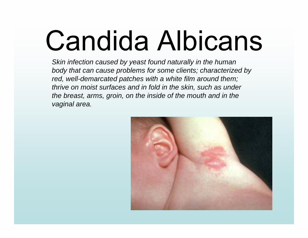
Candida AlbicansSkin infection caused by yeast found naturally in the human body that can cause problems for some clients; characterized by red, well-demarcated patches with a white film around them; thrive on moist surfaces and in fold in the skin, such as under the breast, arms, groin, on the inside of the mouth and in the vaginal area.