integumentary system (skin) - weeblyjumed16.weebly.com/uploads/8/8/5/1/88514776/skin.pdf ·...
TRANSCRIPT
Integumentary system(Skin)
Dr. Heba KalbounehAssistant Professor of Anatomy and Histology
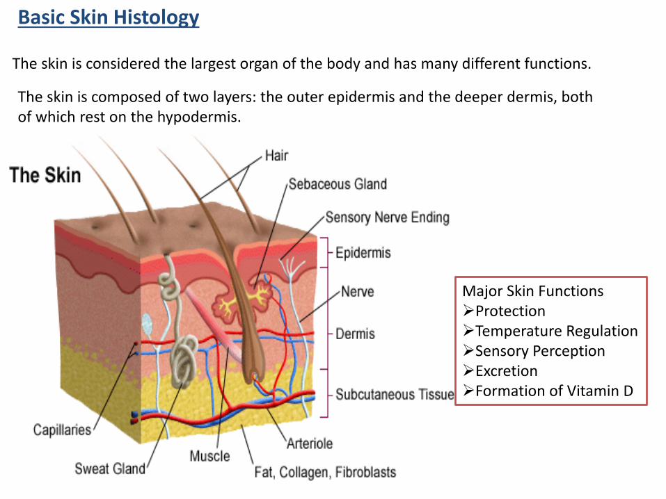
Basic Skin Histology
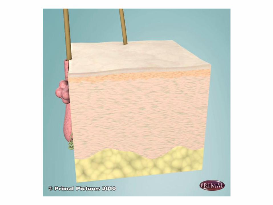
The skin is considered the largest organ of the body and has many different functions.
Major Skin Functions➢Protection➢Temperature Regulation➢Sensory Perception➢Excretion➢Formation of Vitamin D
The skin is composed of two layers: the outer epidermis and the deeper dermis, both of which rest on the hypodermis.
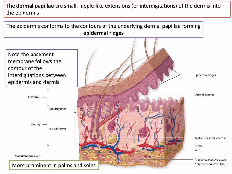
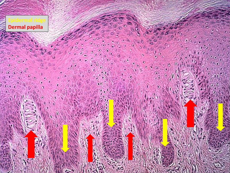
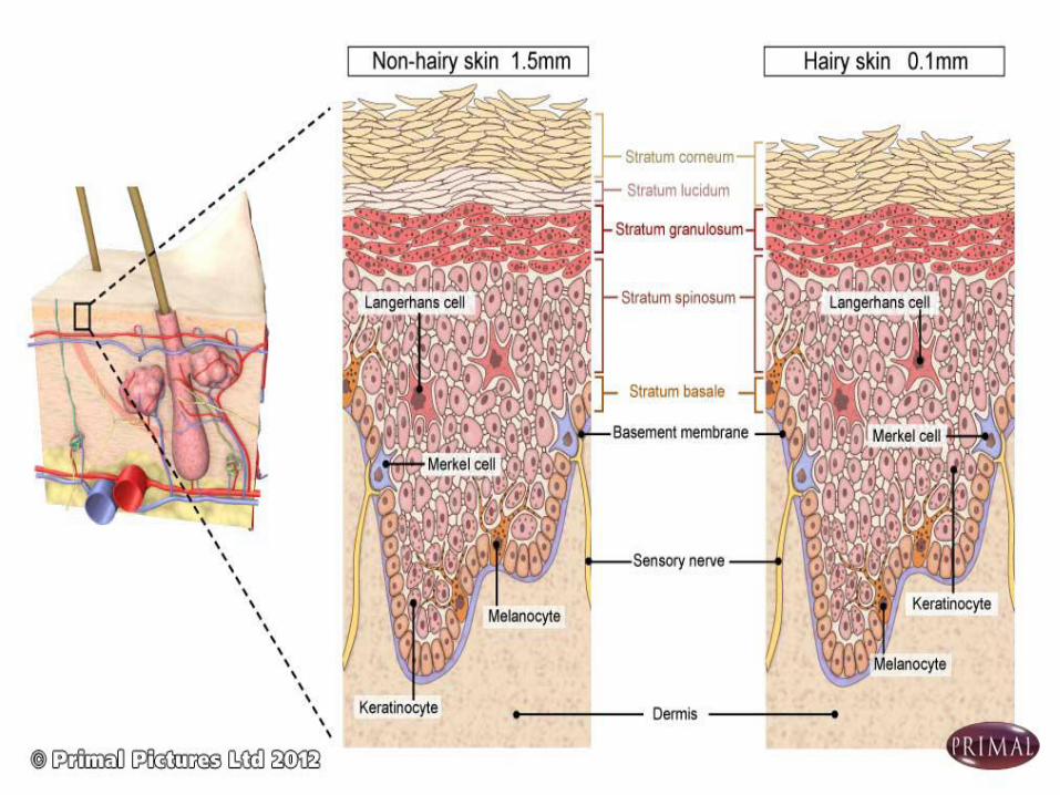
The dermal papillae are small, nipple-like extensions (or interdigitations) of the dermis into the epidermis
More prominent in palms and soles
Note the basement membrane follows the contour of the interdigitations between epidermis and dermis
The epidermis conforms to the contours of the underlying dermal papillae forming epidermal ridges
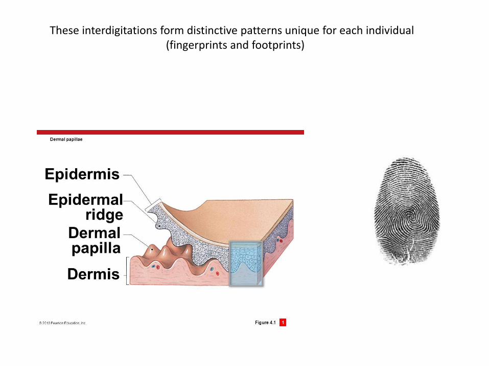
These interdigitations form distinctive patterns unique for each individual(fingerprints and footprints)
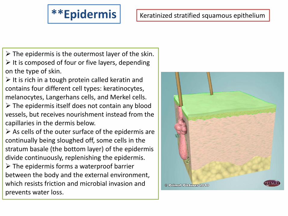
**Epidermis Keratinized stratified squamous epithelium
➢ The epidermis is the outermost layer of the skin.➢ It is composed of four or five layers, depending on the type of skin.➢ It is rich in a tough protein called keratin and contains four different cell types: keratinocytes, melanocytes, Langerhans cells, and Merkel cells.➢ The epidermis itself does not contain any blood vessels, but receives nourishment instead from the capillaries in the dermis below.➢ As cells of the outer surface of the epidermis are continually being sloughed off, some cells in the stratum basale (the bottom layer) of the epidermis divide continuously, replenishing the epidermis.➢ The epidermis forms a waterproof barrier between the body and the external environment, which resists friction and microbial invasion and prevents water loss.
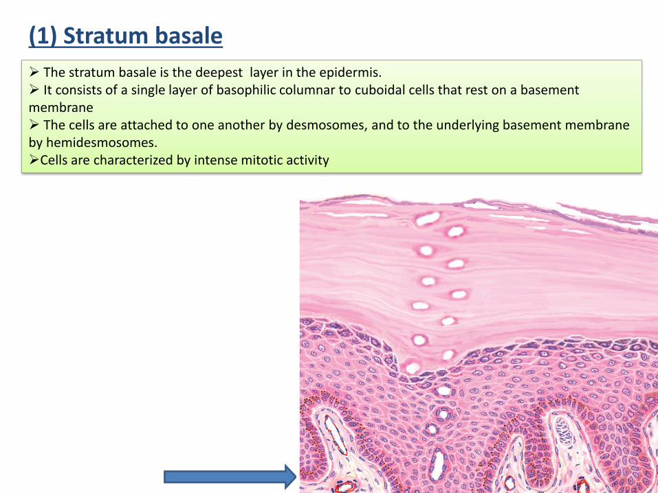
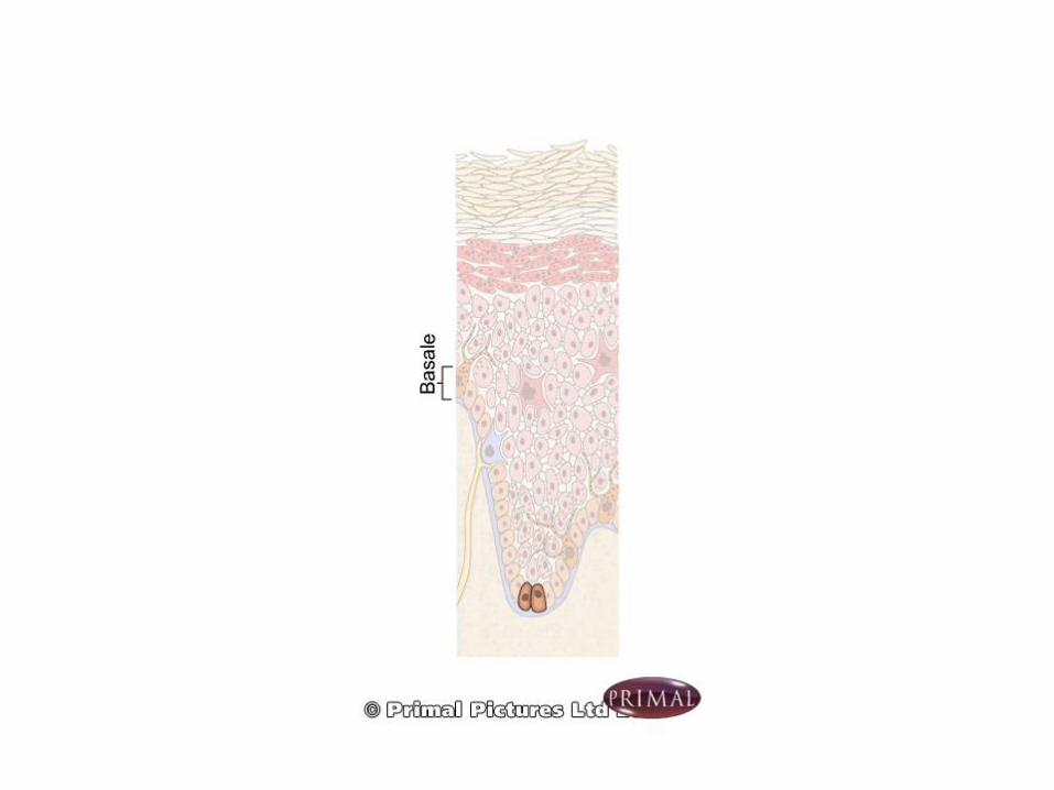
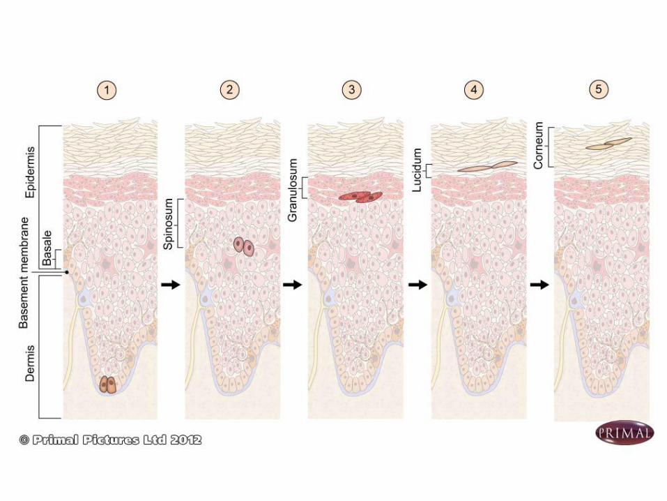
(1) Stratum basale
➢ The stratum basale is the deepest layer in the epidermis.➢ It consists of a single layer of basophilic columnar to cuboidal cells that rest on a basement membrane ➢ The cells are attached to one another by desmosomes, and to the underlying basement membrane by hemidesmosomes. ➢Cells are characterized by intense mitotic activity
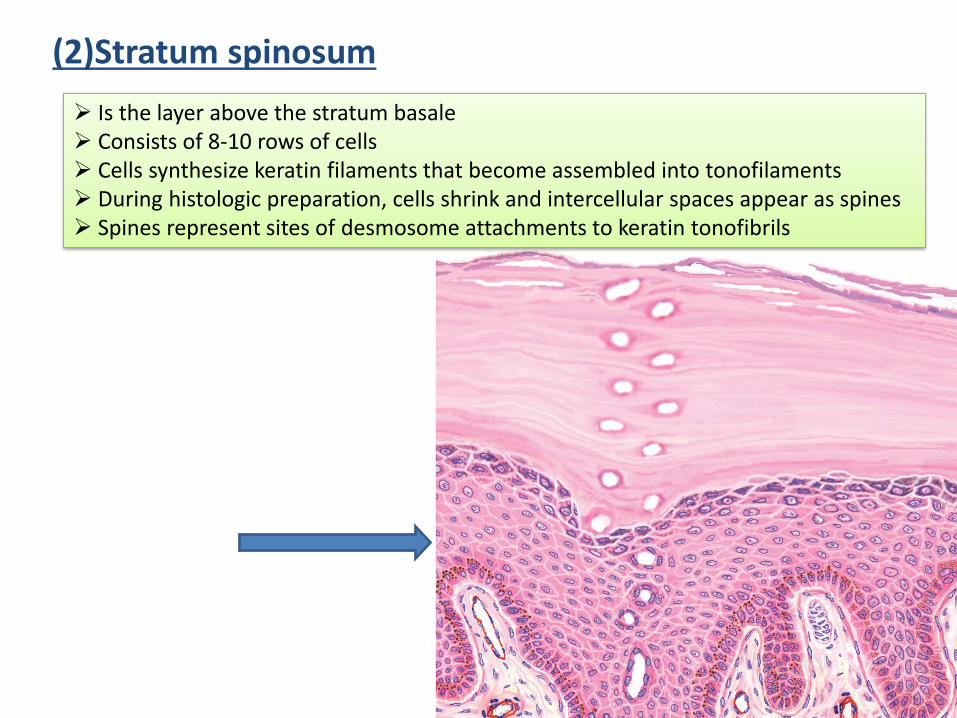
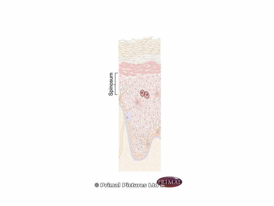
(2)Stratum spinosum
➢ Is the layer above the stratum basale➢ Consists of 8-10 rows of cells➢ Cells synthesize keratin filaments that become assembled into tonofilaments➢ During histologic preparation, cells shrink and intercellular spaces appear as spines➢ Spines represent sites of desmosome attachments to keratin tonofibrils
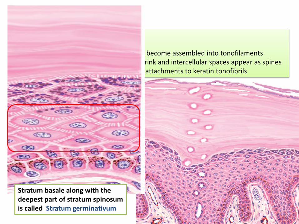
Stratum basale along with the deepest part of stratum spinosumis called Stratum germinativum
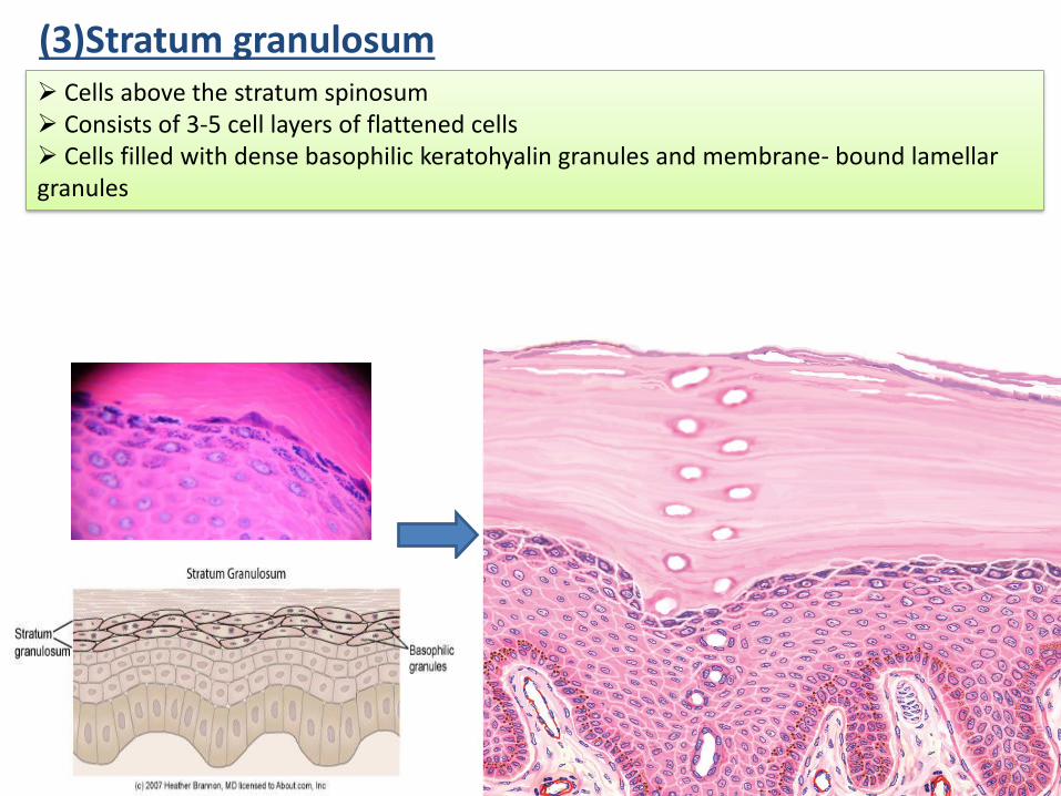
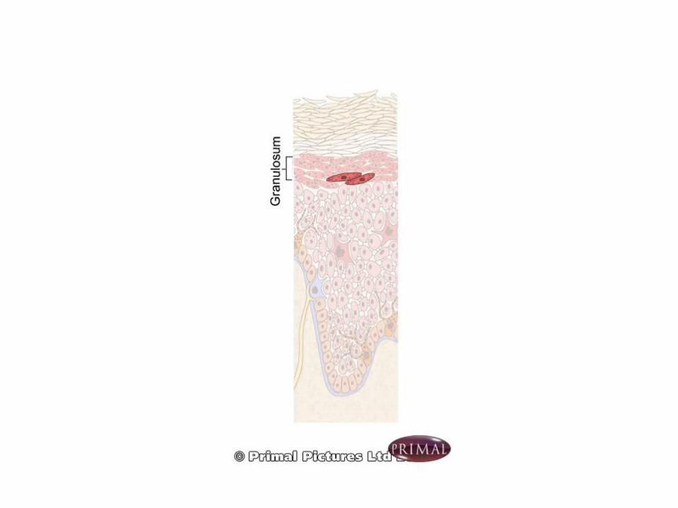
(3)Stratum granulosum➢ Cells above the stratum spinosum➢ Consists of 3-5 cell layers of flattened cells➢ Cells filled with dense basophilic keratohyalin granules and membrane- bound lamellar granules
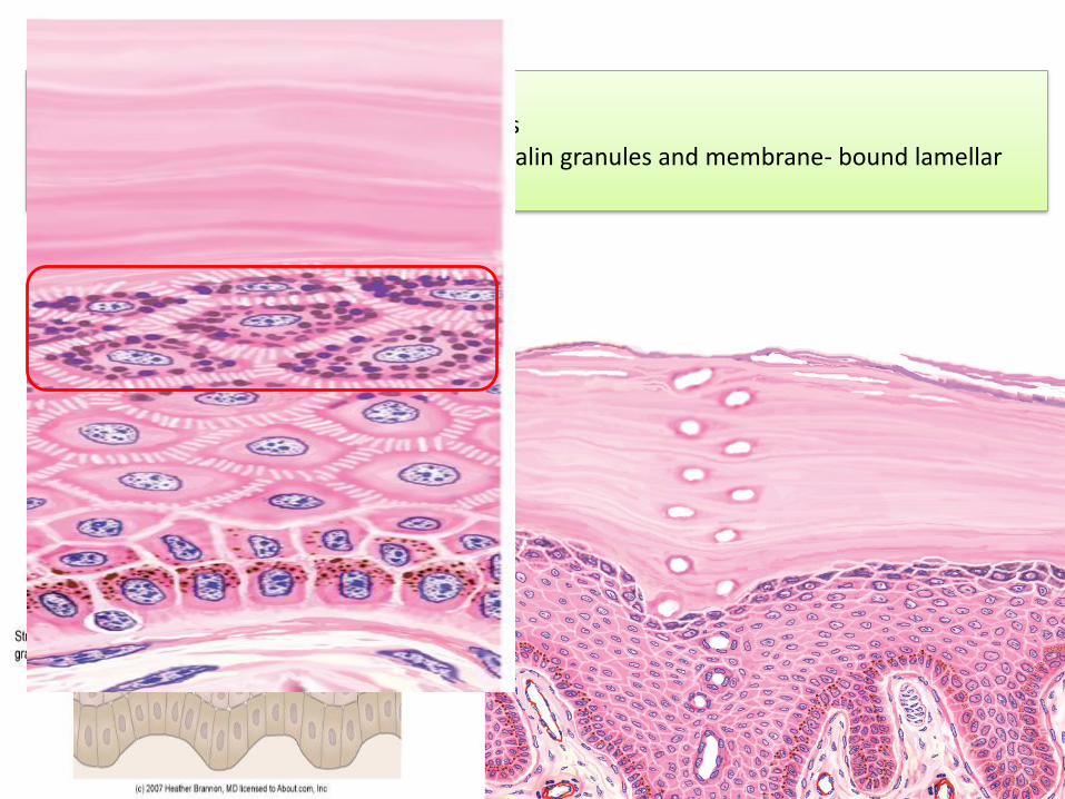
(3)Stratum granulosum➢ Cells above the stratum spinosum➢ Consists of 3-5 cell layers of flattened cells➢ Cells filled with dense basophilic keratohyalin granules and membrane- bound lamellar granules
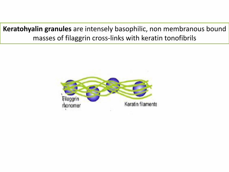
Keratohyalin granules are intensely basophilic, non membranous bound masses of filaggrin cross-links with keratin tonofibrils
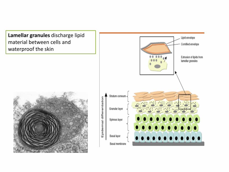
Lamellar granules discharge lipid material between cells and waterproof the skin
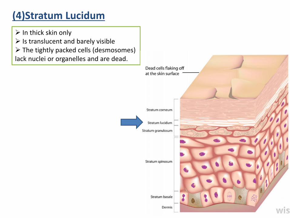
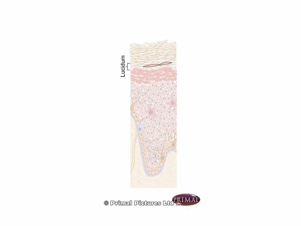
(4)Stratum Lucidum
➢ In thick skin only➢ Is translucent and barely visible➢ The tightly packed cells (desmosomes) lack nuclei or organelles and are dead.
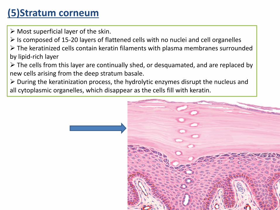
(5)Stratum corneum
➢ Most superficial layer of the skin.➢ Is composed of 15-20 layers of flattened cells with no nuclei and cell organelles➢ The keratinized cells contain keratin filaments with plasma membranes surrounded by lipid-rich layer➢ The cells from this layer are continually shed, or desquamated, and are replaced by new cells arising from the deep stratum basale. ➢ During the keratinization process, the hydrolytic enzymes disrupt the nucleus and all cytoplasmic organelles, which disappear as the cells fill with keratin.
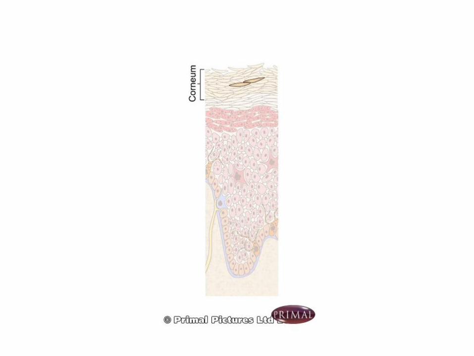
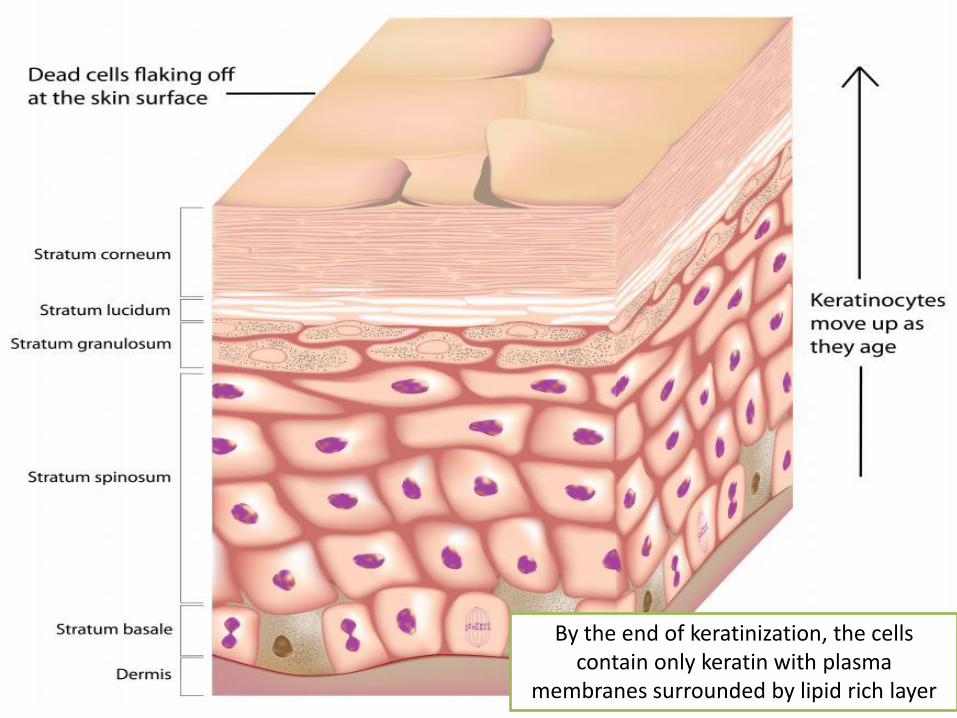
By the end of keratinization, the cells contain only keratin with plasma
membranes surrounded by lipid rich layer
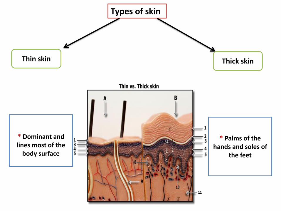
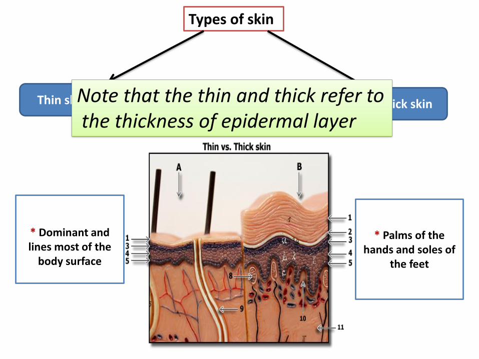
Types of skin
Thick skinThin skin
* Palms of the hands and soles of
the feet
* Dominant and lines most of the
body surface
Types of skin
Thick skinThin skin
* Palms of the hands and soles of
the feet
* Dominant and lines most of the
body surface
Note that the thin and thick refer tothe thickness of epidermal layer
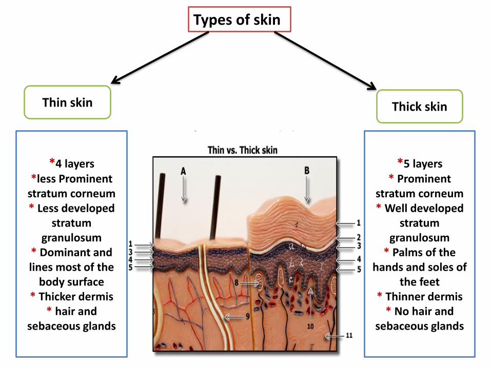
Types of skin
Thin skin Thick skin
*5 layers * Prominent
stratum corneum* Well developed
stratum granulosum
* Palms of the hands and soles of
the feet* Thinner dermis
* No hair and sebaceous glands
*4 layers *less Prominent
stratum corneum* Less developed
stratum granulosum
* Dominant and lines most of the
body surface* Thicker dermis
* hair and sebaceous glands
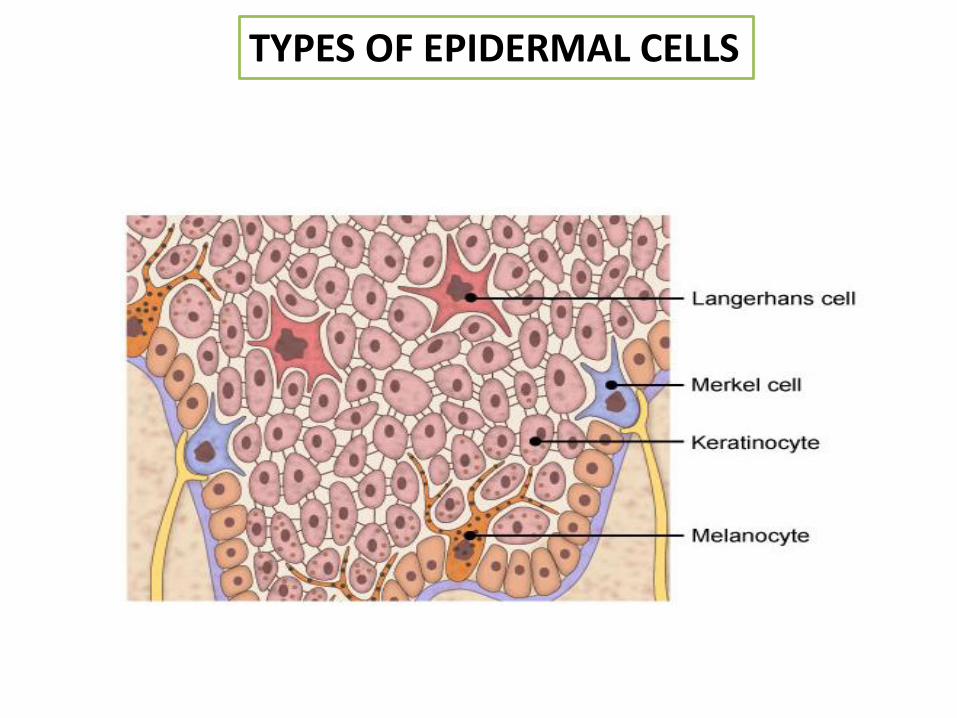
TYPES OF EPIDERMAL CELLS
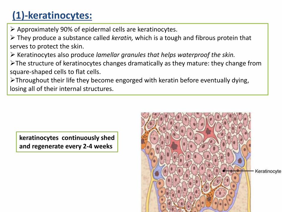
(1)-keratinocytes:
keratinocytes continuously shed and regenerate every 2-4 weeks
➢ Approximately 90% of epidermal cells are keratinocytes.➢ They produce a substance called keratin, which is a tough and fibrous protein that serves to protect the skin.➢ Keratinocytes also produce lamellar granules that helps waterproof the skin.➢The structure of keratinocytes changes dramatically as they mature: they change from square-shaped cells to flat cells. ➢Throughout their life they become engorged with keratin before eventually dying, losing all of their internal structures.
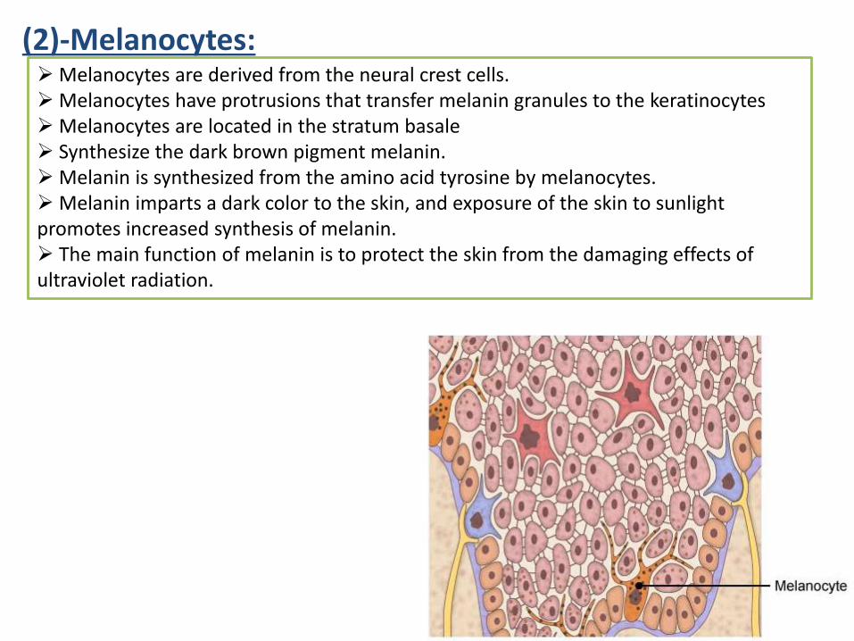
➢ Melanocytes are derived from the neural crest cells. ➢ Melanocytes have protrusions that transfer melanin granules to the keratinocytes➢ Melanocytes are located in the stratum basale➢ Synthesize the dark brown pigment melanin. ➢ Melanin is synthesized from the amino acid tyrosine by melanocytes. ➢ Melanin imparts a dark color to the skin, and exposure of the skin to sunlight promotes increased synthesis of melanin. ➢ The main function of melanin is to protect the skin from the damaging effects of ultraviolet radiation.
(2)-Melanocytes:
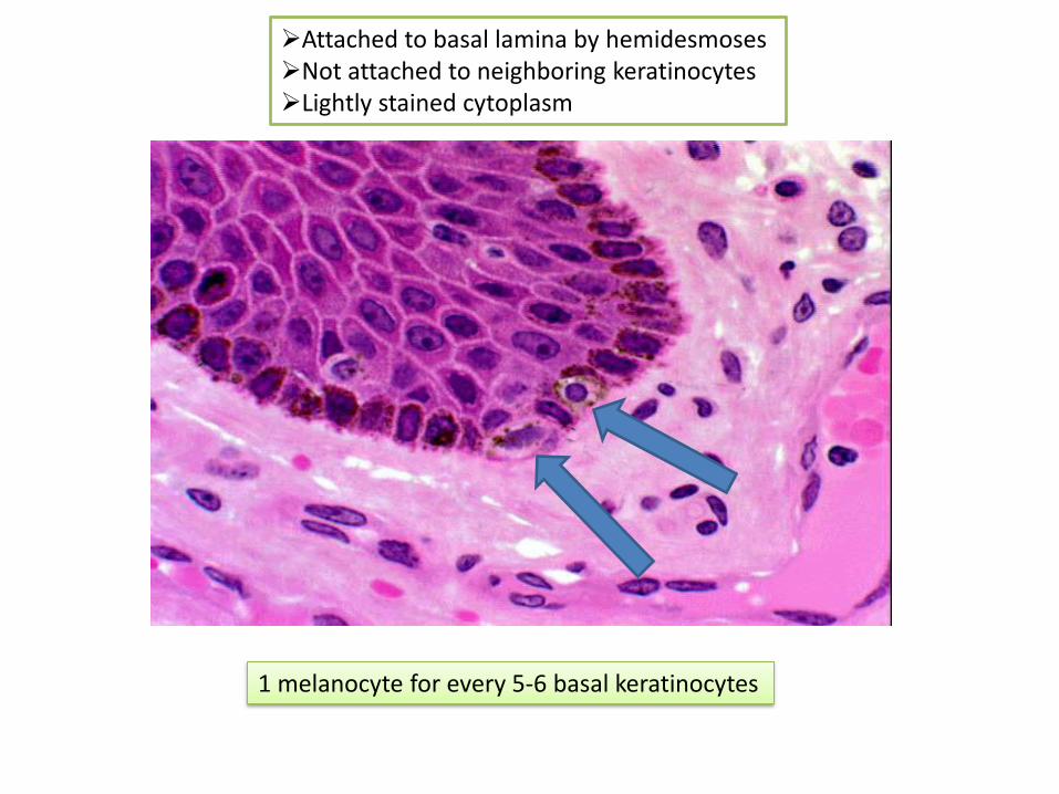
➢Attached to basal lamina by hemidesmoses➢Not attached to neighboring keratinocytes➢Lightly stained cytoplasm
1 melanocyte for every 5-6 basal keratinocytes
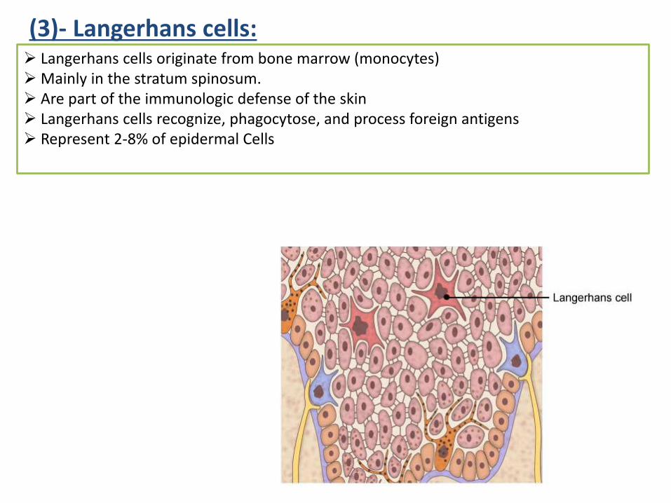
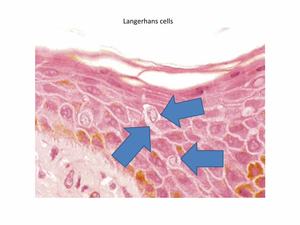
➢ Langerhans cells originate from bone marrow (monocytes)➢ Mainly in the stratum spinosum.➢ Are part of the immunologic defense of the skin➢ Langerhans cells recognize, phagocytose, and process foreign antigens ➢ Represent 2-8% of epidermal Cells
(3)- Langerhans cells:
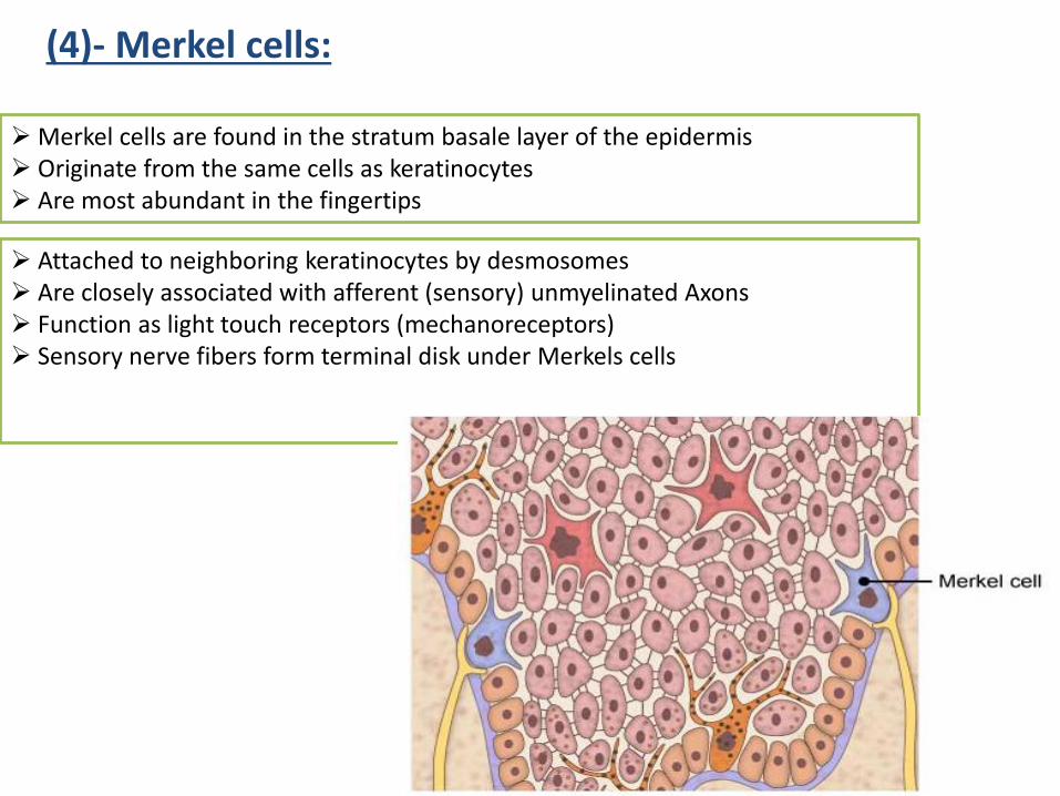
➢ Merkel cells are found in the stratum basale layer of the epidermis➢ Originate from the same cells as keratinocytes➢ Are most abundant in the fingertips
(4)- Merkel cells:
➢ Attached to neighboring keratinocytes by desmosomes➢ Are closely associated with afferent (sensory) unmyelinated Axons➢ Function as light touch receptors (mechanoreceptors)➢ Sensory nerve fibers form terminal disk under Merkels cells
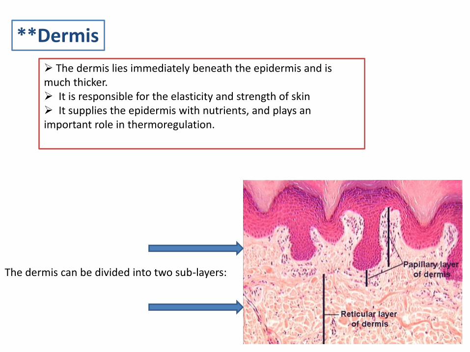
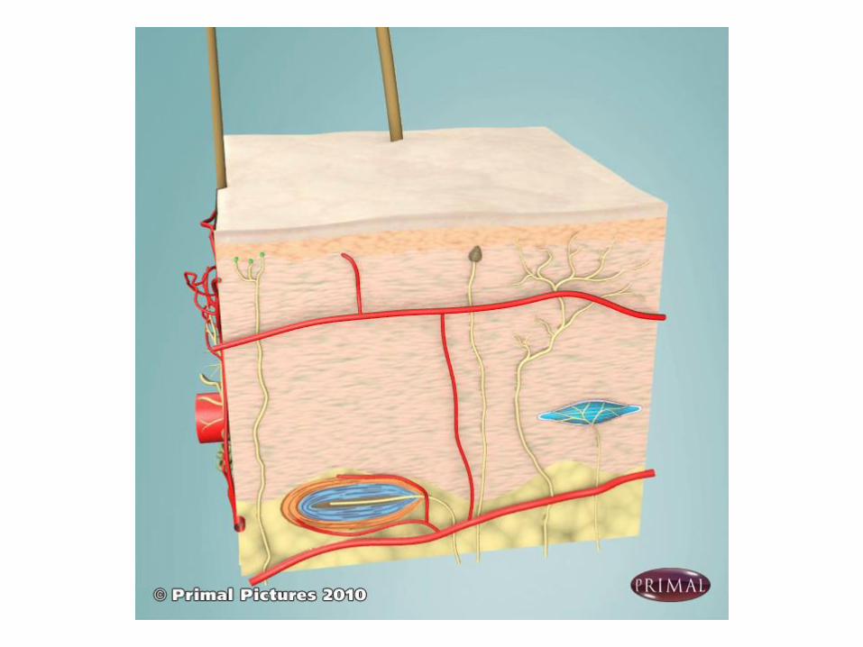
**Dermis
➢ The dermis lies immediately beneath the epidermis and is much thicker.➢ It is responsible for the elasticity and strength of skin➢ It supplies the epidermis with nutrients, and plays an important role in thermoregulation.
The dermis can be divided into two sub-layers:
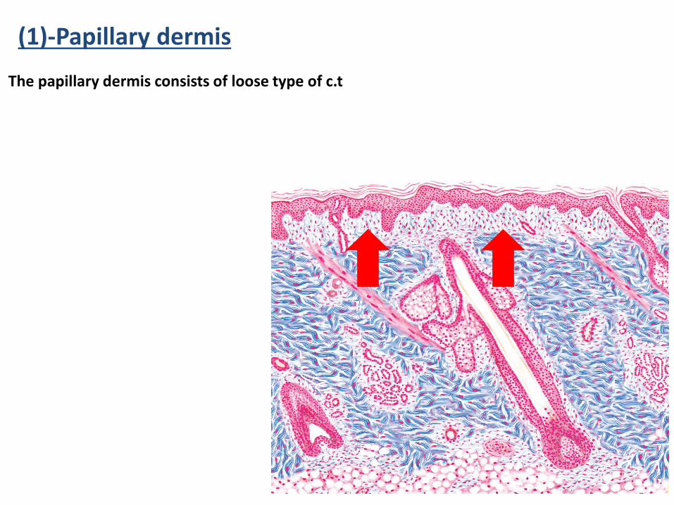
(1)-Papillary dermis
The papillary dermis consists of loose type of c.t
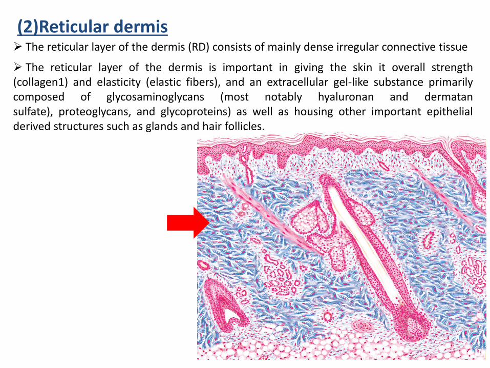
(2)Reticular dermis➢ The reticular layer of the dermis (RD) consists of mainly dense irregular connective tissue
➢ The reticular layer of the dermis is important in giving the skin it overall strength(collagen1) and elasticity (elastic fibers), and an extracellular gel-like substance primarilycomposed of glycosaminoglycans (most notably hyaluronan and dermatansulfate), proteoglycans, and glycoproteins) as well as housing other important epithelialderived structures such as glands and hair follicles.
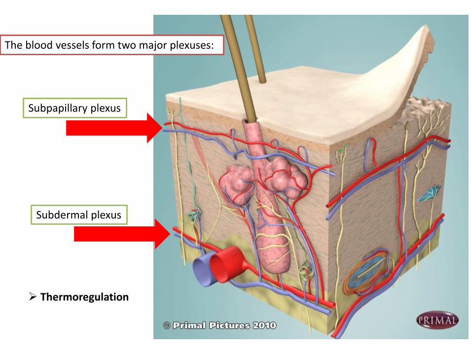
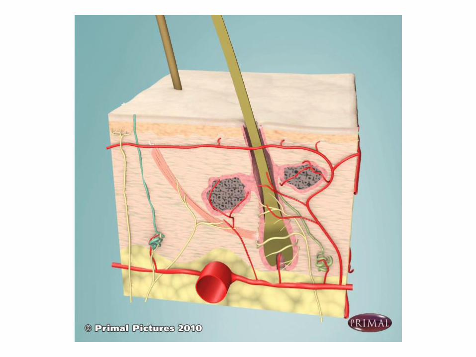
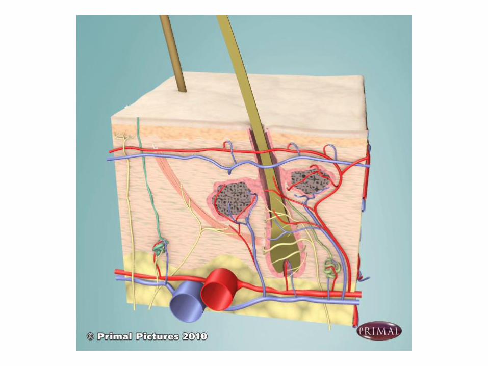
The blood vessels form two major plexuses:
Subpapillary plexus
Subdermal plexus
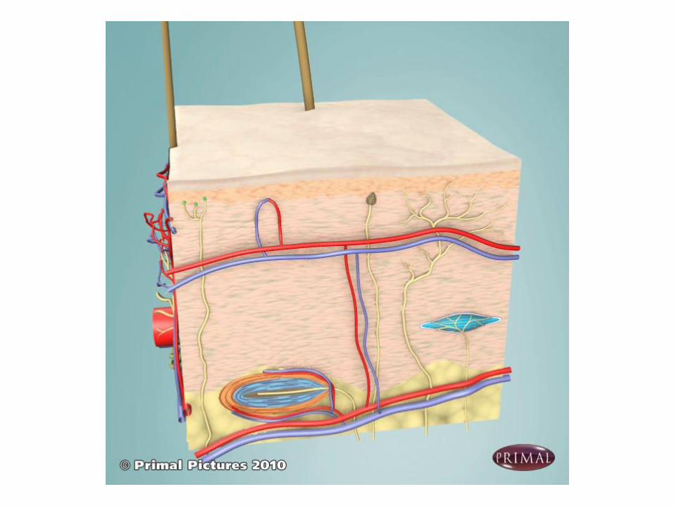
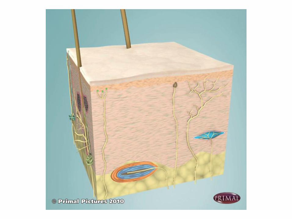
➢ Thermoregulation
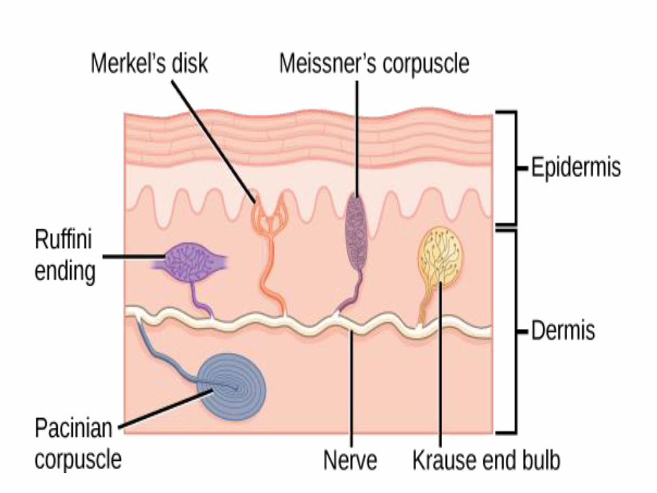
Sensory receptors
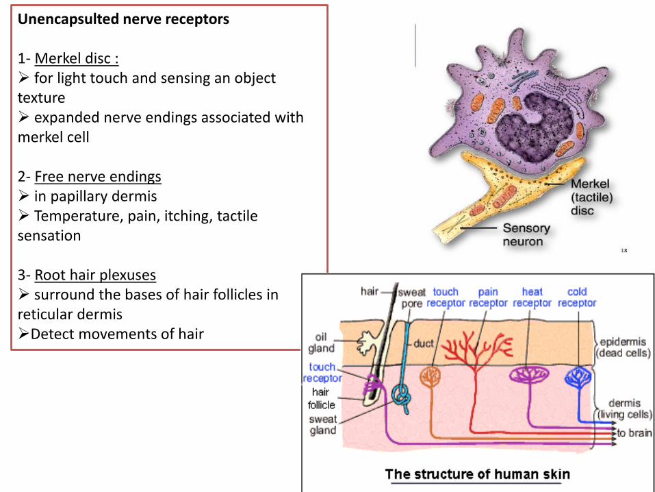
Unencapsulted nerve receptors
1- Merkel disc :➢ for light touch and sensing an object texture➢ expanded nerve endings associated with merkel cell
2- Free nerve endings➢ in papillary dermis➢ Temperature, pain, itching, tactile sensation
3- Root hair plexuses ➢ surround the bases of hair follicles in reticular dermis➢Detect movements of hair
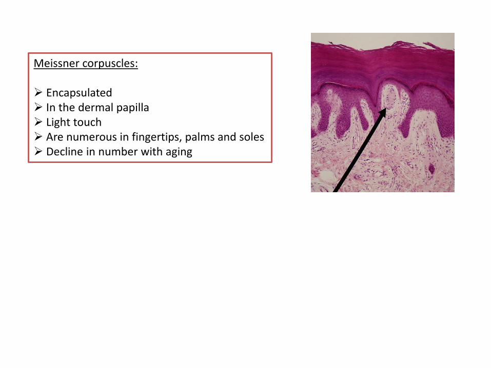
Meissner corpuscles:
➢ Encapsulated➢ In the dermal papilla ➢ Light touch➢ Are numerous in fingertips, palms and soles➢ Decline in number with aging
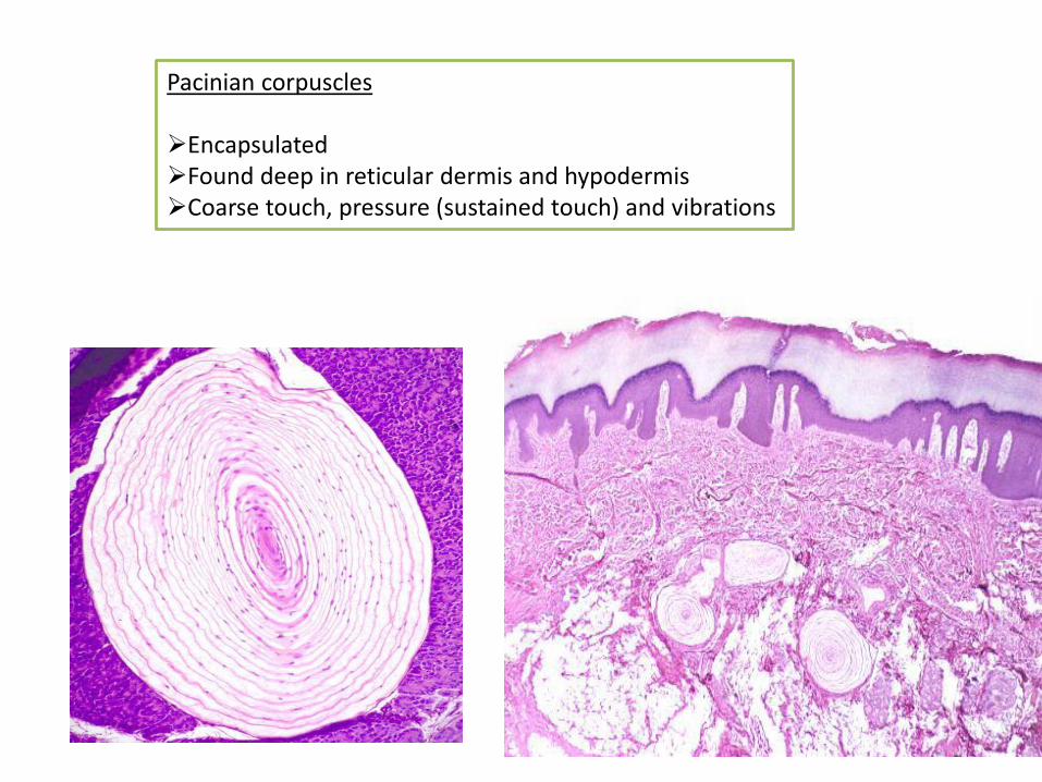
Pacinian corpuscles
➢Encapsulated➢Found deep in reticular dermis and hypodermis➢Coarse touch, pressure (sustained touch) and vibrations
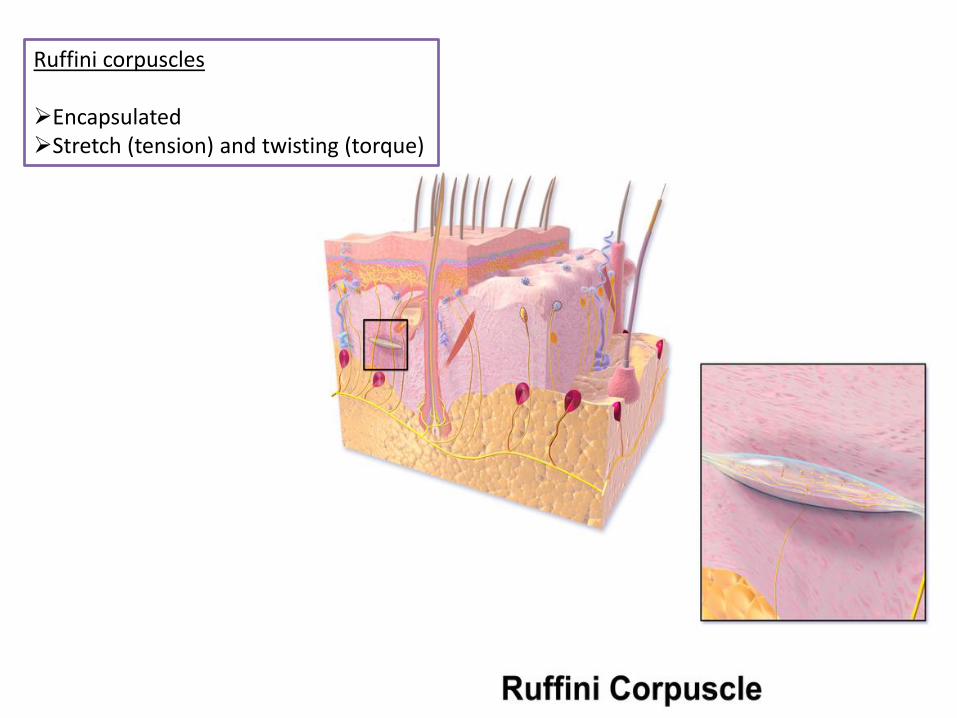
Ruffini corpuscles
➢Encapsulated ➢Stretch (tension) and twisting (torque)
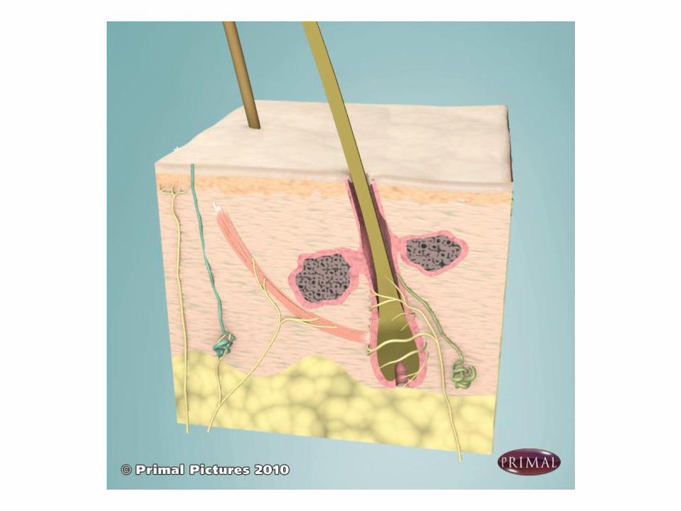
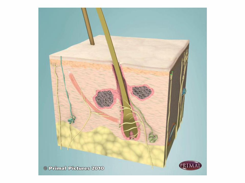
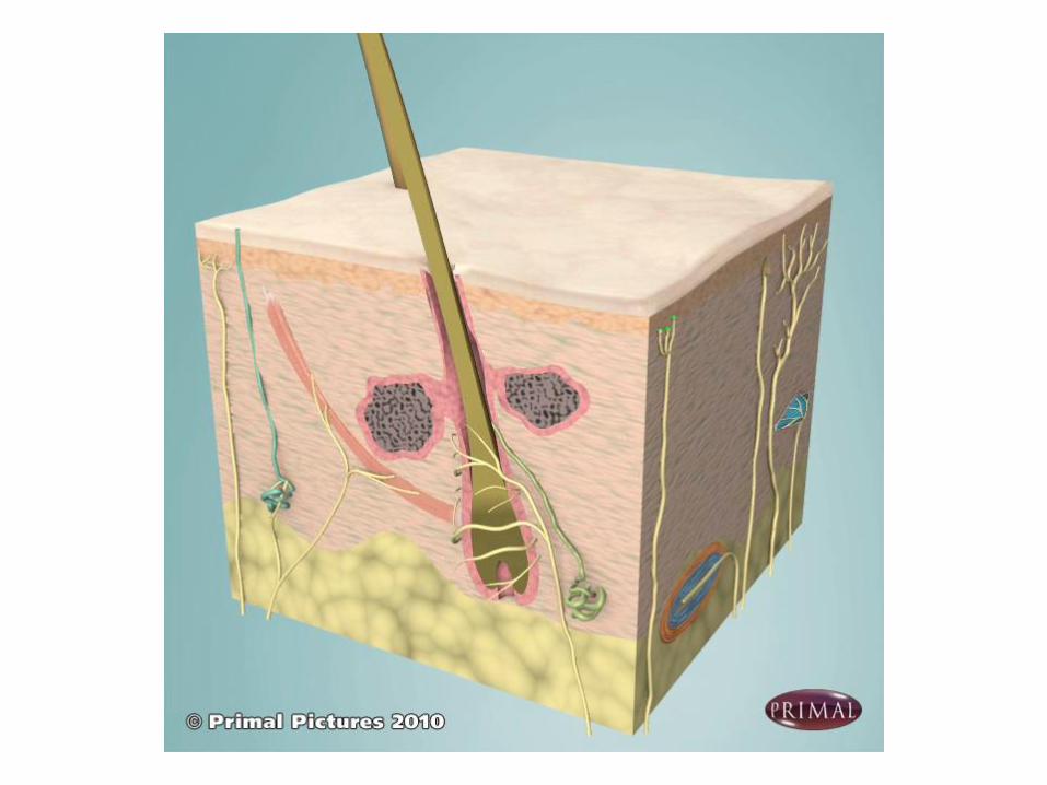
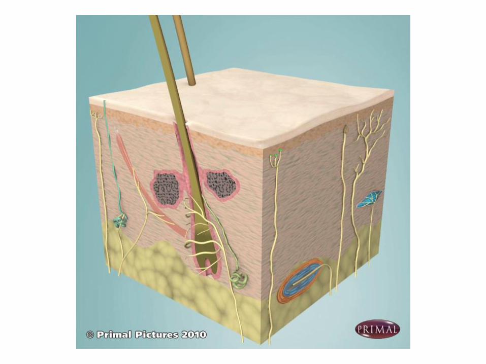
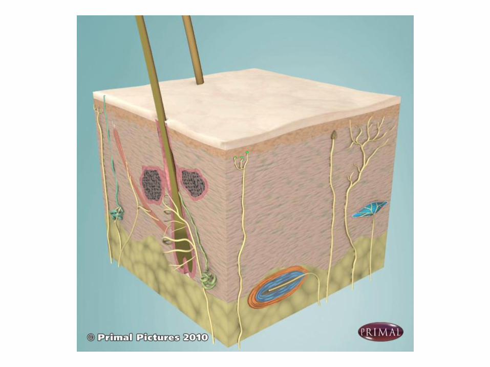
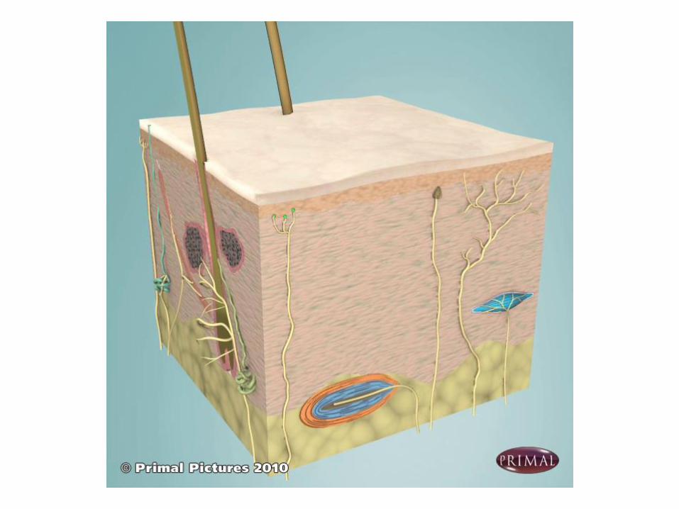

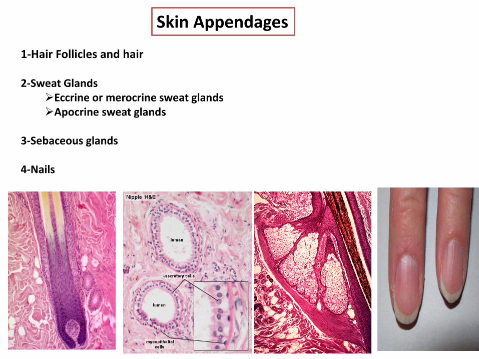
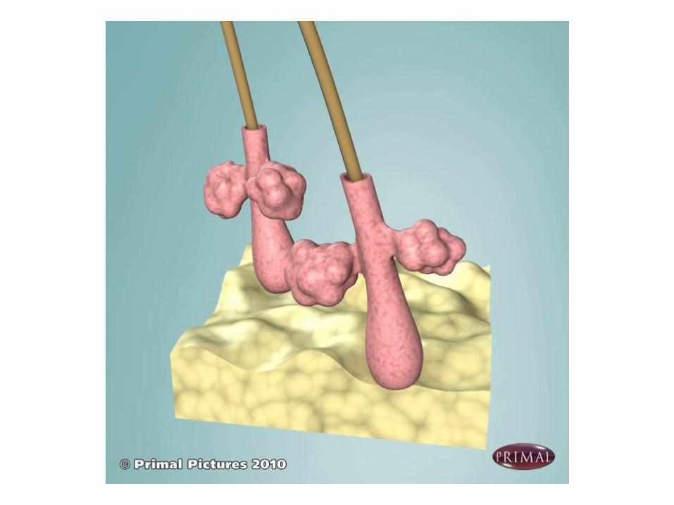
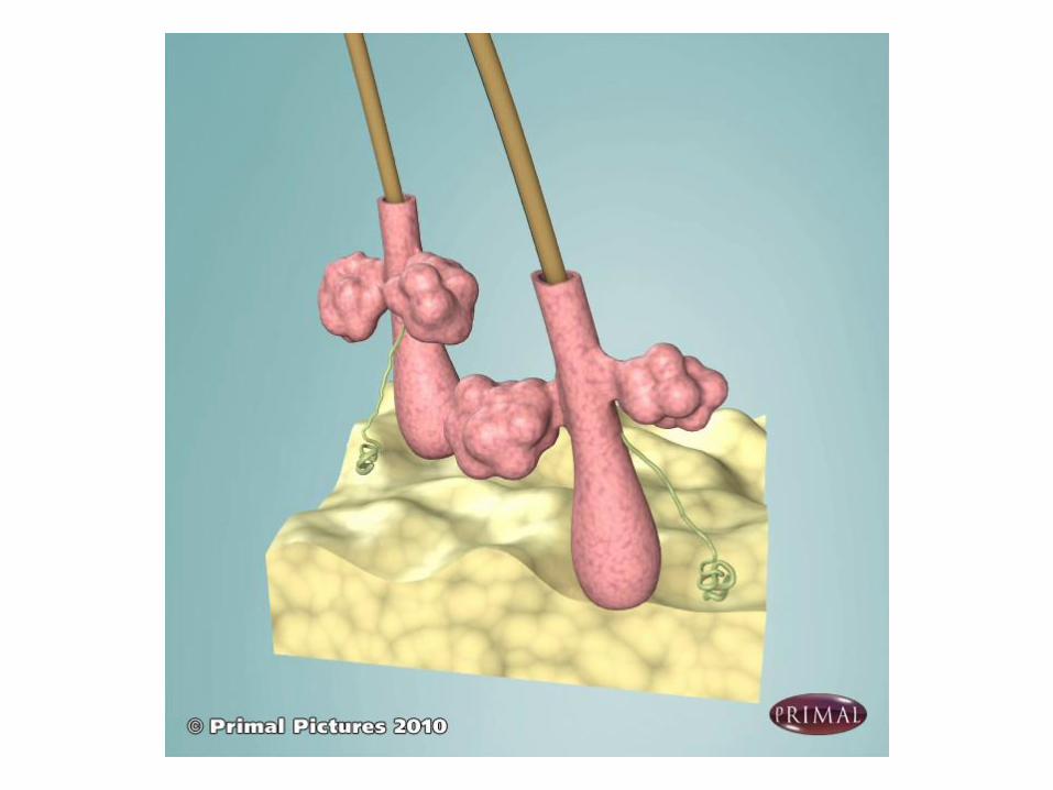
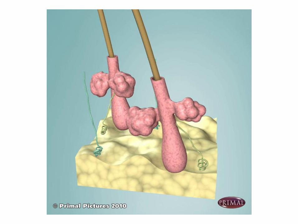
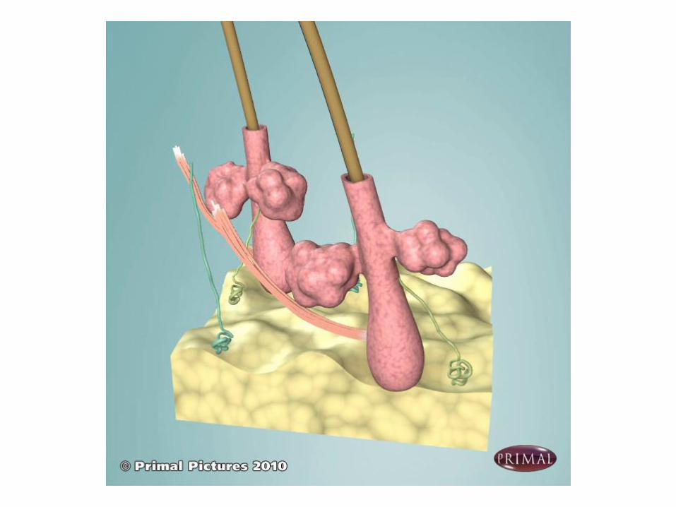
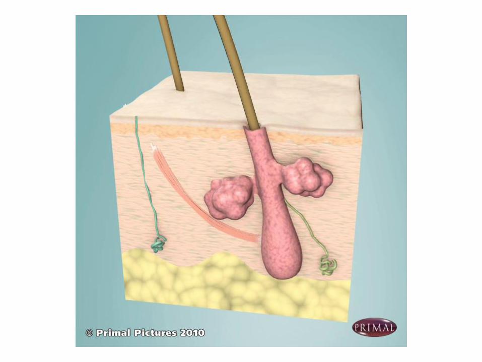
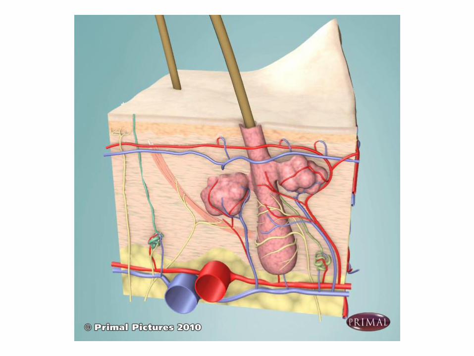
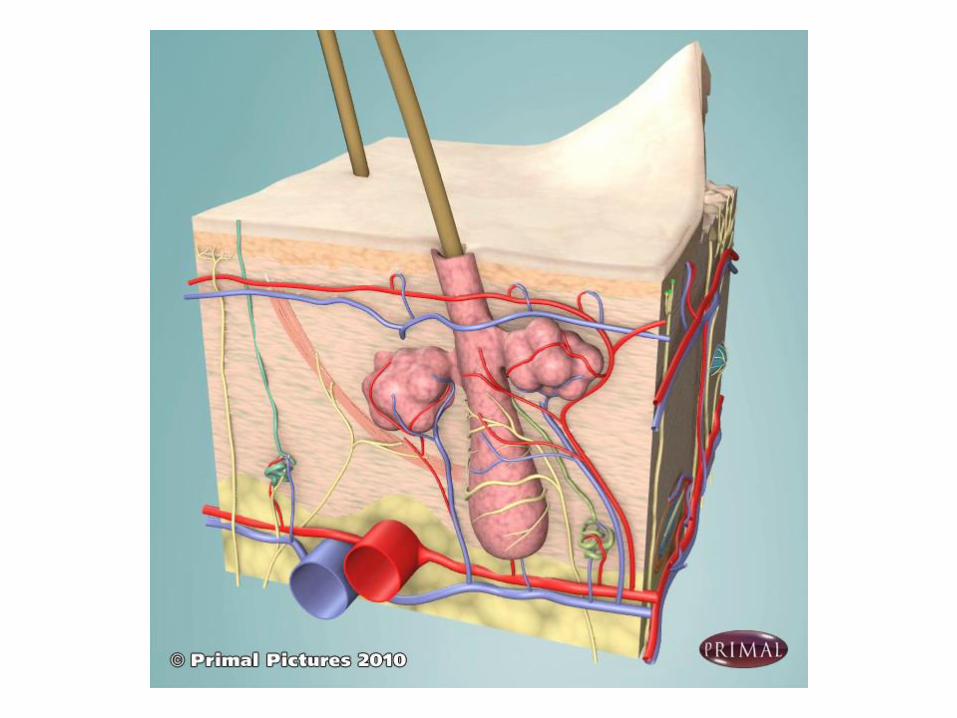
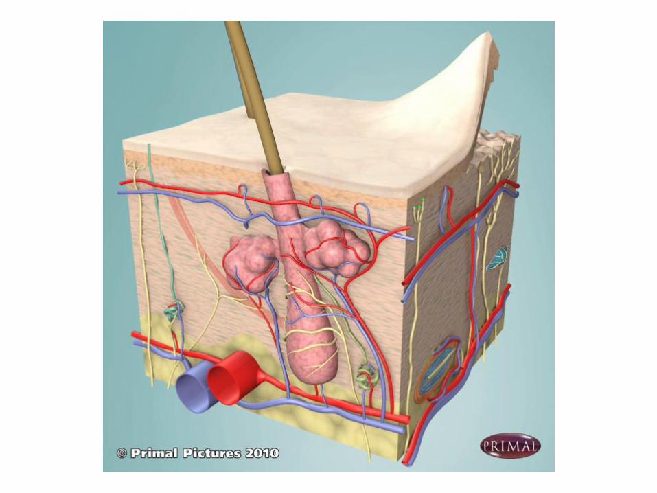
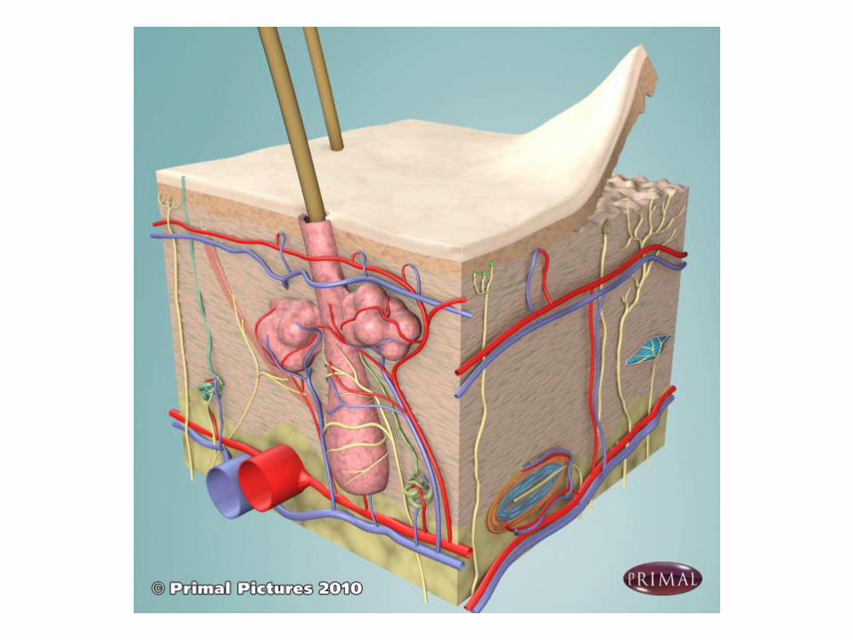
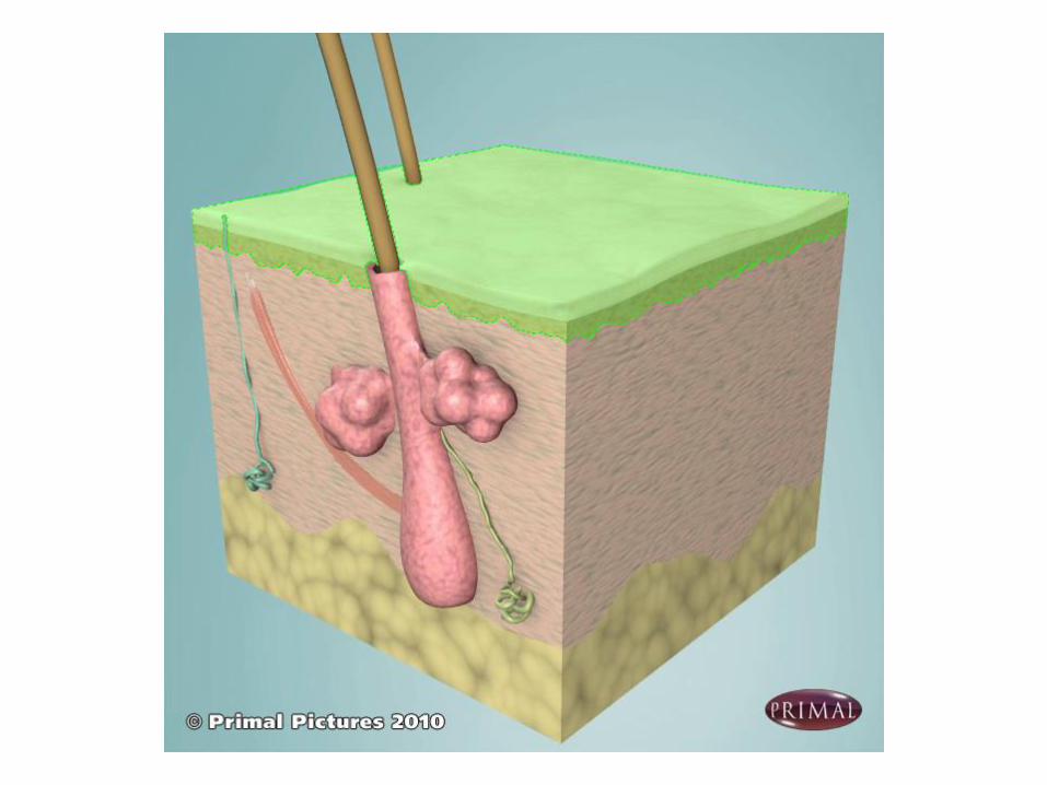
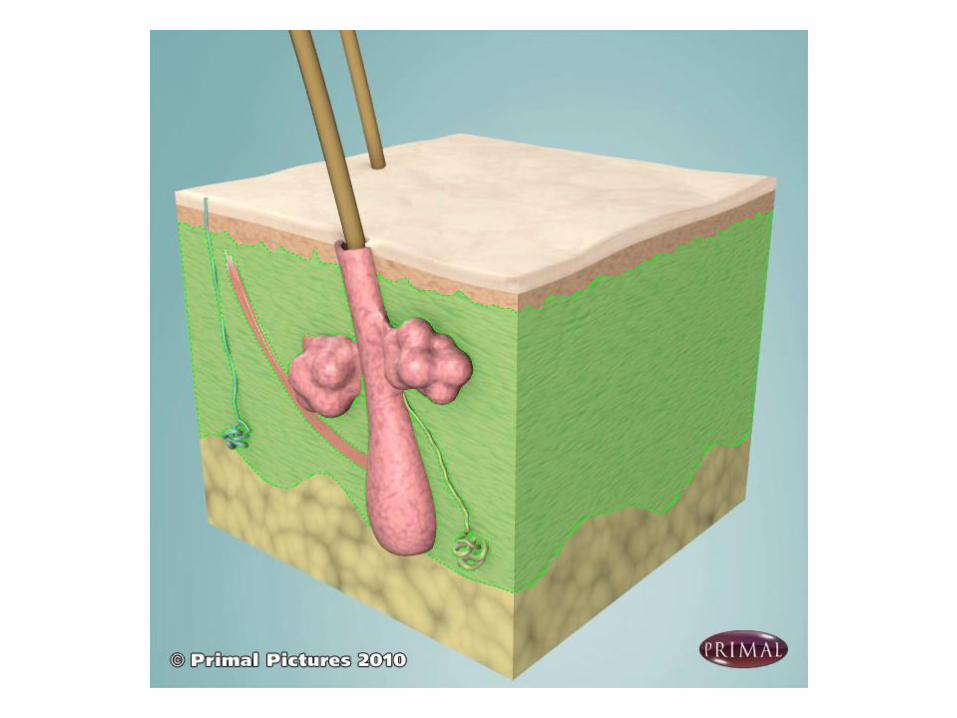
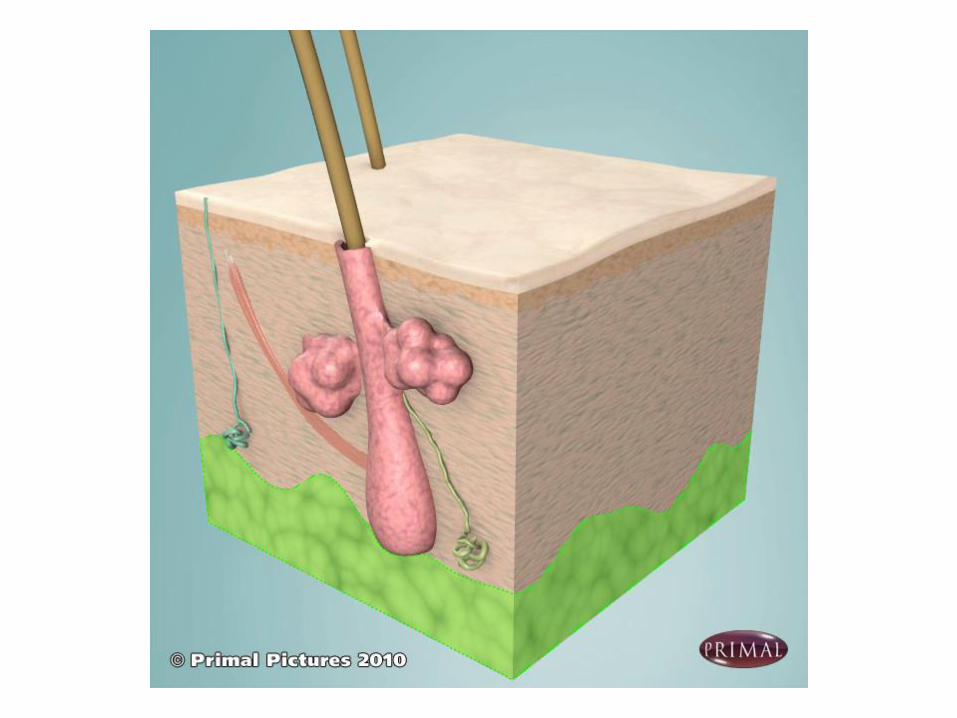
Skin Appendages
1-Hair Follicles and hair
2-Sweat Glands➢Eccrine or merocrine sweat glands➢Apocrine sweat glands
3-Sebaceous glands
4-Nails
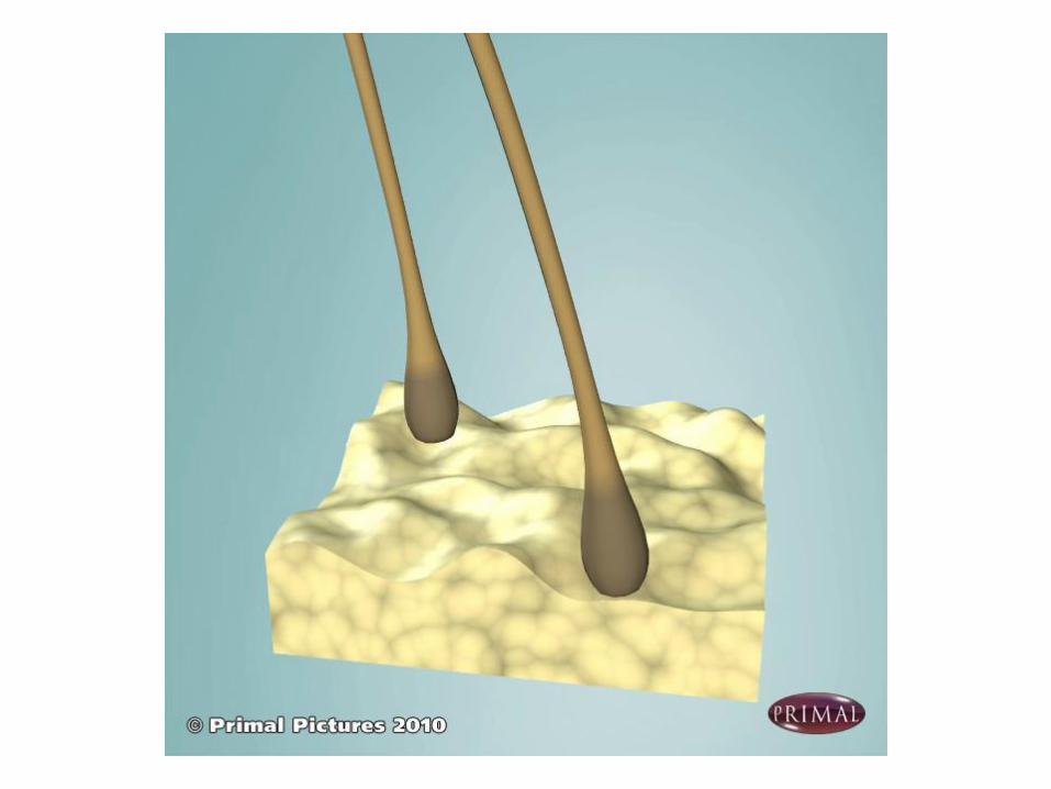
Hair
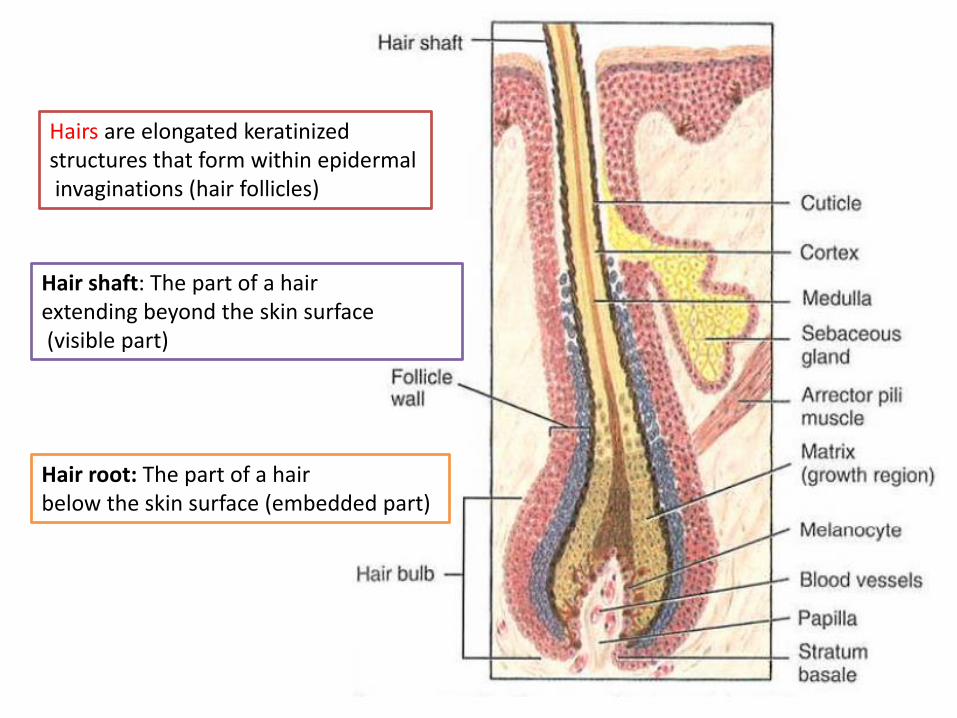
Hairs are elongated keratinizedstructures that form within epidermalinvaginations (hair follicles)
Hair shaft: The part of a hair extending beyond the skin surface(visible part)
Hair root: The part of a hair below the skin surface (embedded part)
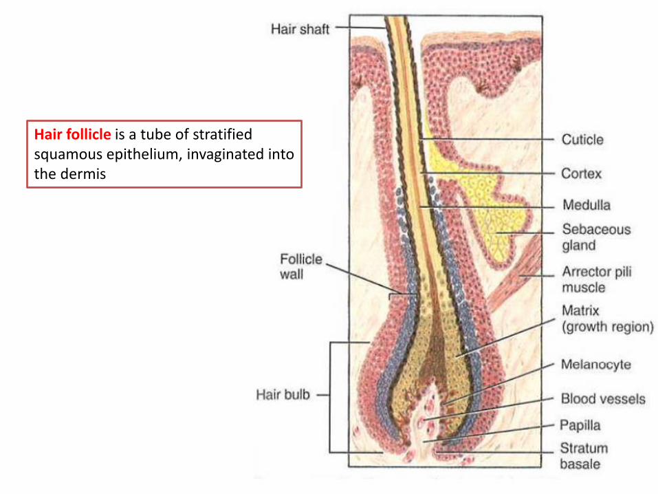
Hair follicle is a tube of stratified squamous epithelium, invaginated into the dermis
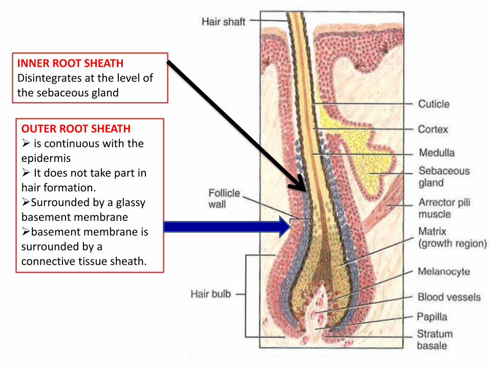
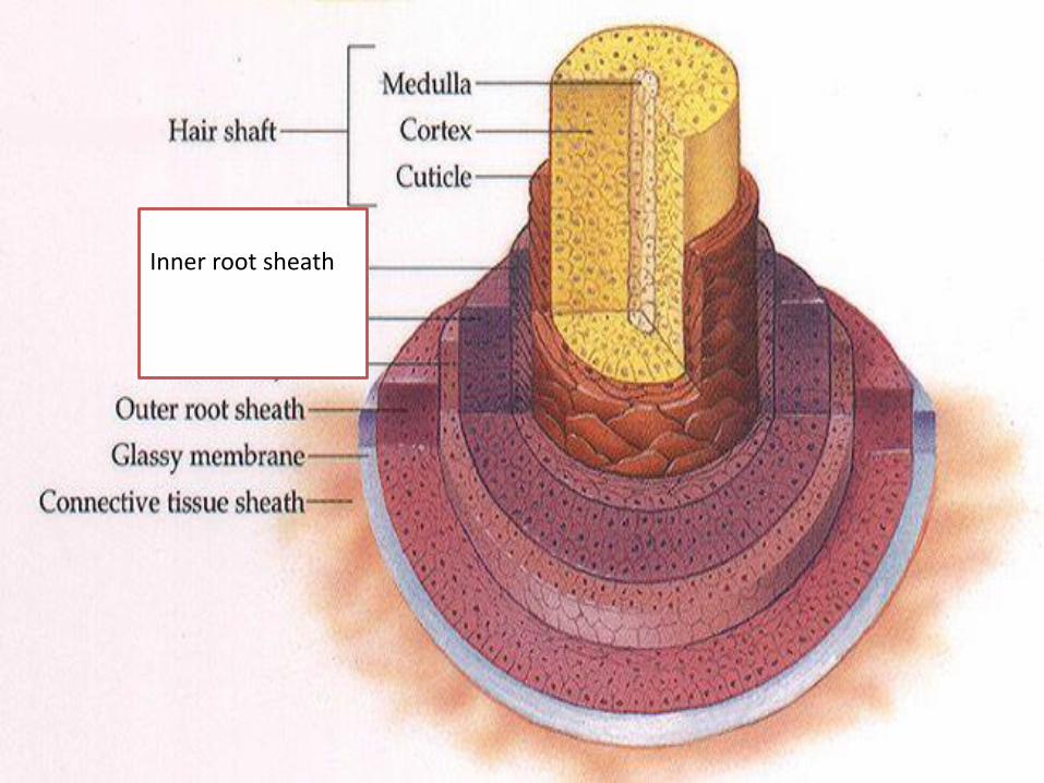
INNER ROOT SHEATH Disintegrates at the level of the sebaceous gland
OUTER ROOT SHEATH➢ is continuous with the epidermis➢ It does not take part in hair formation. ➢Surrounded by a glassy basement membrane➢basement membrane is surrounded by a connective tissue sheath.
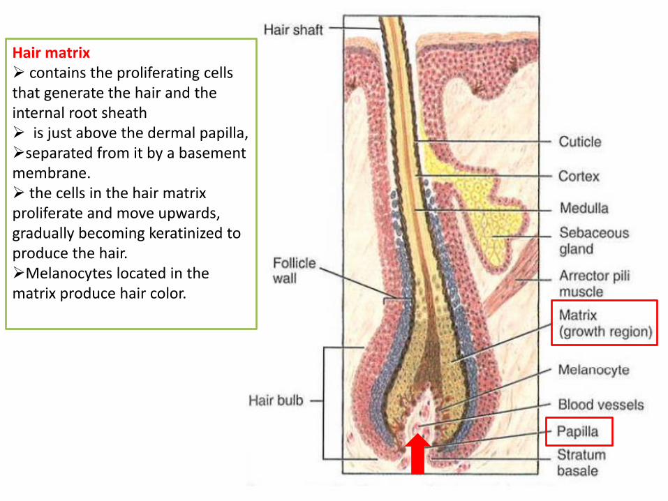
Hair matrix➢ contains the proliferating cells that generate the hair and the internal root sheath➢ is just above the dermal papilla,➢separated from it by a basement membrane. ➢ the cells in the hair matrix proliferate and move upwards, gradually becoming keratinized to produce the hair.➢Melanocytes located in the matrix produce hair color.
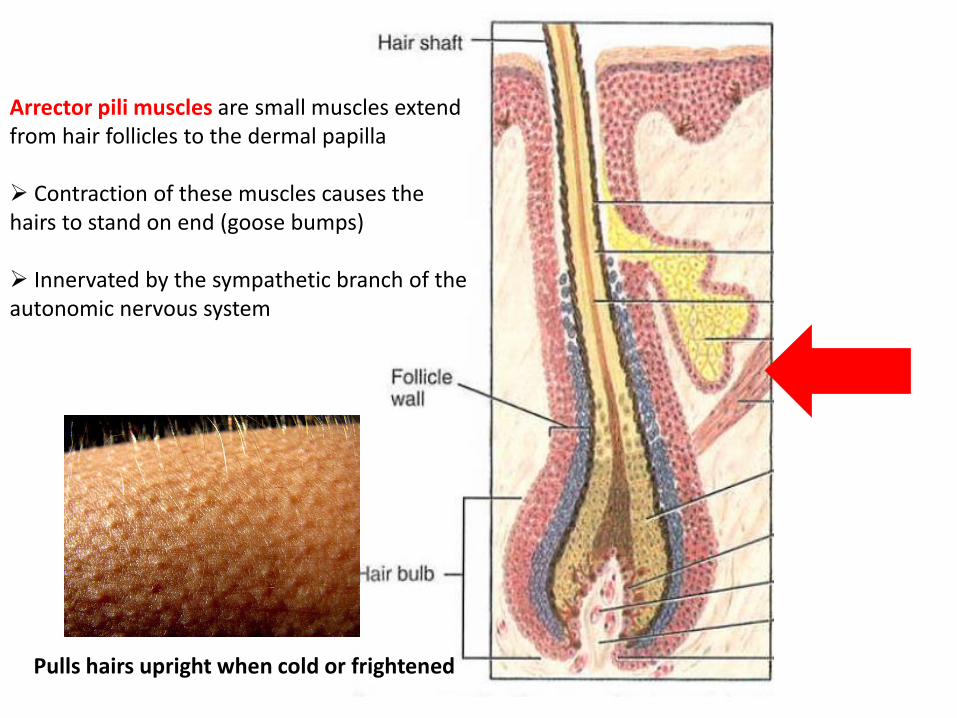
Arrector pili muscles are small muscles extend from hair follicles to the dermal papilla
➢ Contraction of these muscles causes the hairs to stand on end (goose bumps)
➢ Innervated by the sympathetic branch of the autonomic nervous system
Pulls hairs upright when cold or frightened
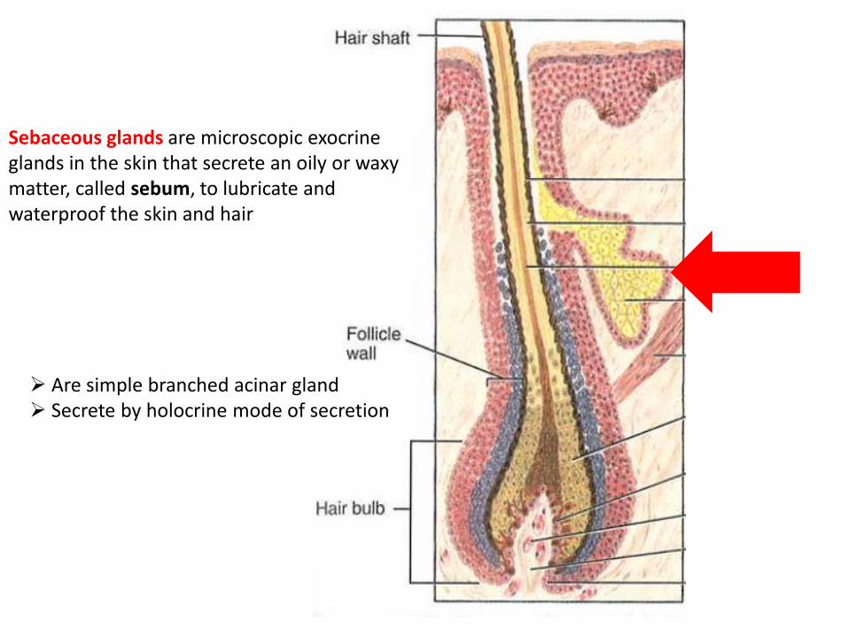
Sebaceous glands are microscopic exocrine glands in the skin that secrete an oily or waxy matter, called sebum, to lubricate and waterproof the skin and hair
➢ Are simple branched acinar gland➢ Secrete by holocrine mode of secretion
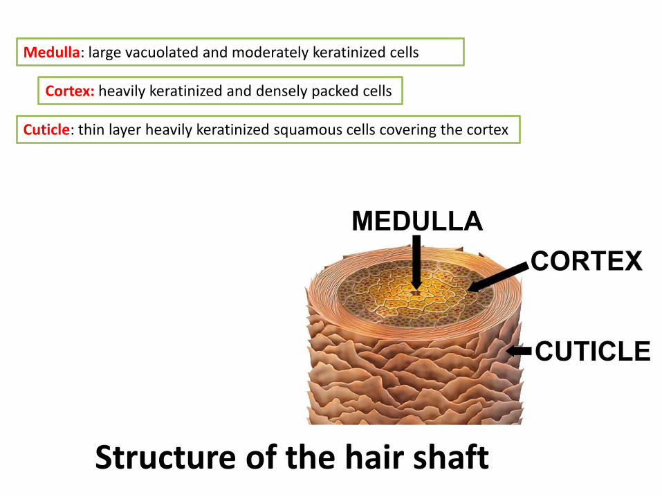
Medulla: large vacuolated and moderately keratinized cells
Cortex: heavily keratinized and densely packed cells
Cuticle: thin layer heavily keratinized squamous cells covering the cortex
Structure of the hair shaft
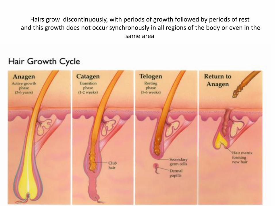
Hairs grow discontinuously, with periods of growth followed by periods of restand this growth does not occur synchronously in all regions of the body or even in the
same area
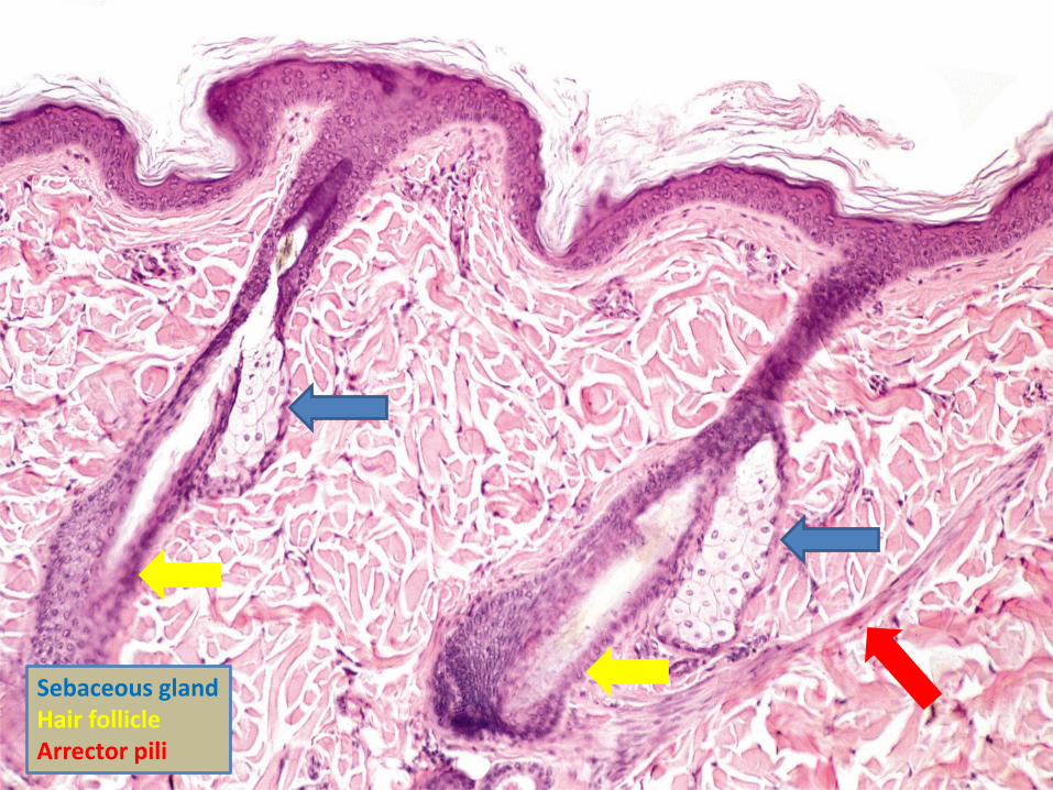
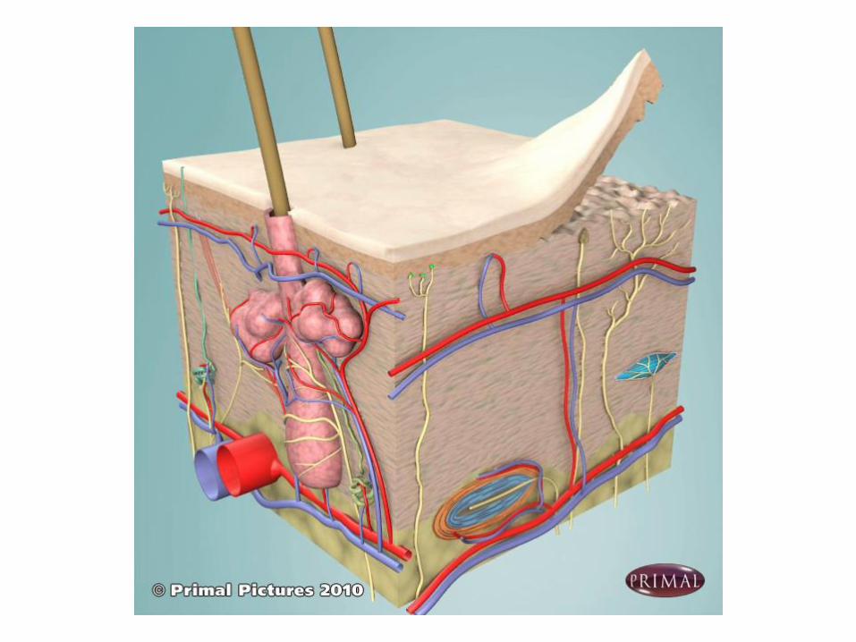
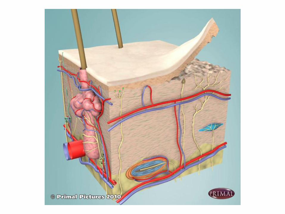
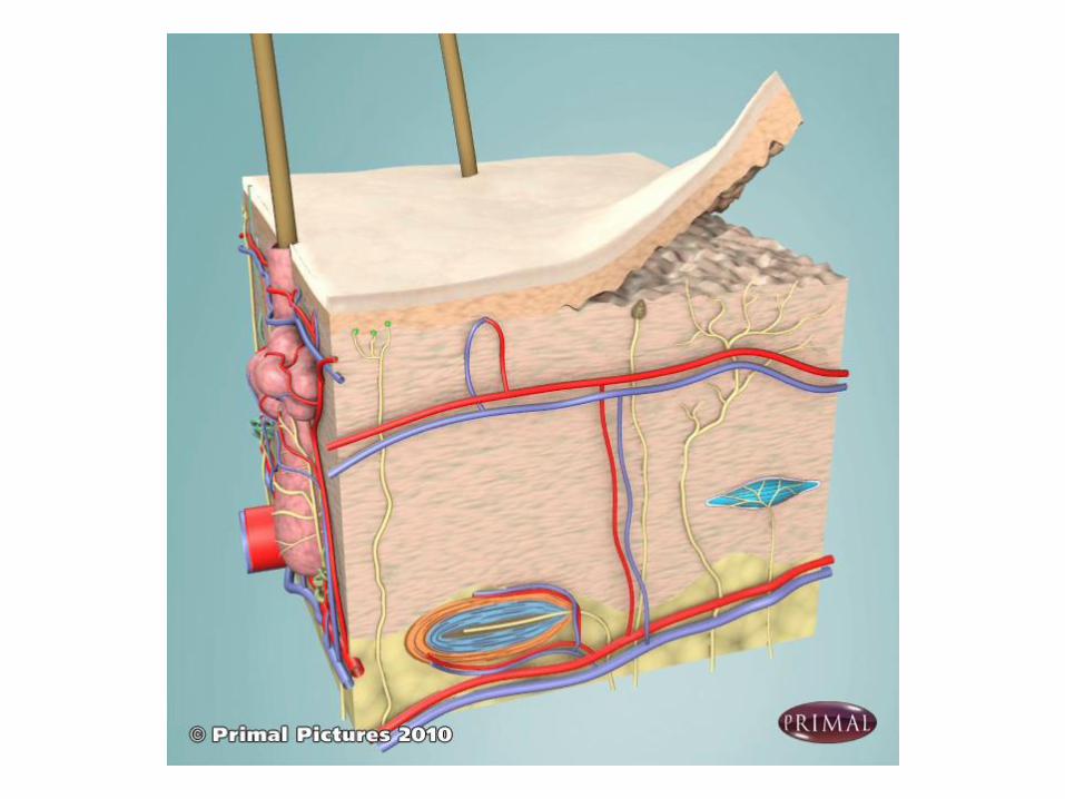
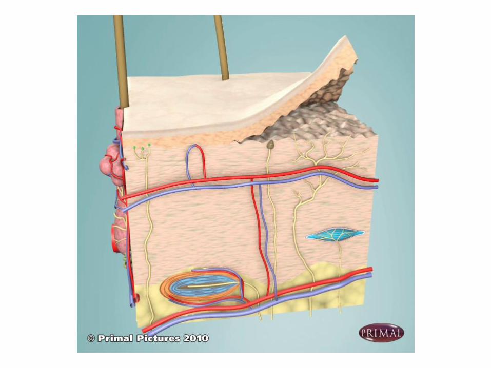
Sebaceous glandHair follicleArrector pili
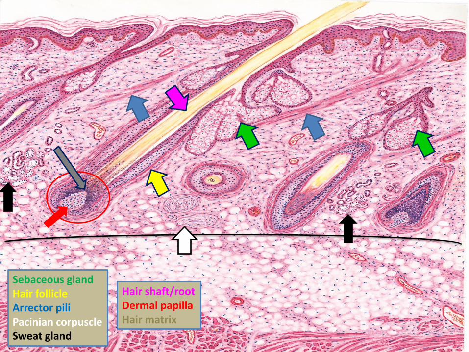
Sebaceous glandHair follicleArrector piliPacinian corpuscleSweat gland
Hair shaft/rootDermal papillaHair matrix
Sweat glands
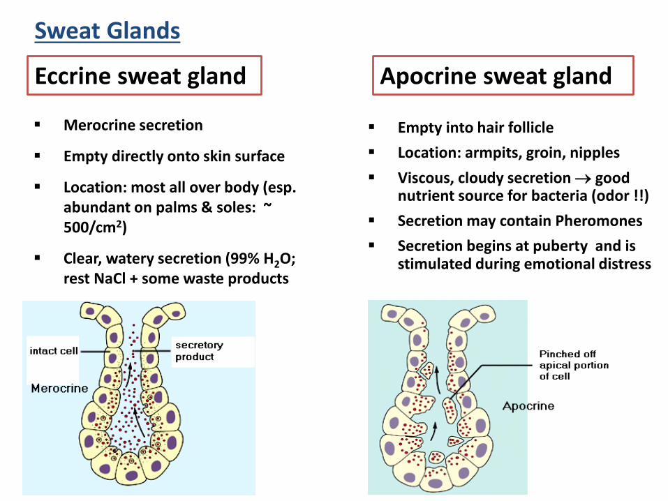
Sweat Glands
▪ Empty into hair follicle
▪ Location: armpits, groin, nipples
▪ Viscous, cloudy secretion good nutrient source for bacteria (odor !!)
▪ Secretion may contain Pheromones
▪ Secretion begins at puberty and is stimulated during emotional distress
Apocrine sweat gland
▪ Merocrine secretion
▪ Empty directly onto skin surface
▪ Location: most all over body (esp. abundant on palms & soles: ~ 500/cm2)
▪ Clear, watery secretion (99% H2O; rest NaCl + some waste products
Eccrine sweat gland
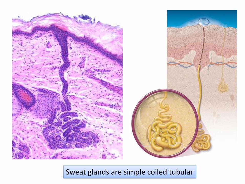
Sweat glands are simple coiled tubular
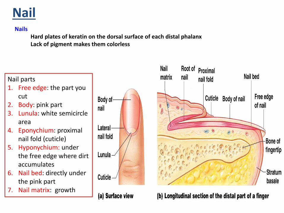
NailNails
Hard plates of keratin on the dorsal surface of each distal phalanxLack of pigment makes them colorless
Nail parts1. Free edge: the part you
cut2. Body: pink part3. Lunula: white semicircle
area4. Eponychium: proximal
nail fold (cuticle)5. Hyponychium: under
the free edge where dirt accumulates
6. Nail bed: directly under the pink part
7. Nail matrix: growth