year 2021 - login
TRANSCRIPT

ANAT 2020
1
UNIVERSITY OF THE WITWATERSRAND, JOHANNESBURG
SCHOOL OF ANATOMICAL SCIENCES
HUMAN ANATOMY FOR
MBBCh II, BHSc II & BSc (Biomed Engineering) III
COURSE CODE: ANAT 2020
YEAR 2021
Professor AO Ihunwo
and
Staff members of the School of Anatomical Sciences
School of Anatomical Sciences
Faculty of Health Sciences
University of the Witwatersrand
Copyright 2021

ANAT 2020
2

ANAT 2020
i
Table of Contents
Page
A. ORGANISATIONAL COMPONENT
1. Course Brief 1
2. Course outline 2
3. Tests and Examinations 3
4. Satisfactory Performance (SP) Requirements 7
5. Textbooks 7
6. Dress Code 8
7. Laboratory Rules 9
8. Important Contact Details 10
9. Guidelines for Students Enquiries and Complaints 10
10. Anatomy School Website Login_______________________________________________10
11. Annexure 1: Answering Questions in Anatomy 11
12. Annexure 2: Rules and Regulations pertaining to the human bones 13
13. Weekly Timetable 15
B. STUDY COMPONENT: HISTOLOGY 36
UNIT THEME 1: PRIMARY TISSUES 36
SUB-UNIT THEME1.1: EPITHELIAL TISSUE
SUB-UNIT THEME1.2 GLANDULAR EPITHELIUM (GLANDS)
SUB-UNIT THEME1.3: CONNECTIVE TISSUE
SUB-UNIT THEME1.4: BONE AND CARTILAGE
SUB-UNIT THEME1.5: MUSCLE TISSUE
SUB-UNIT THEME1.6: NERVOUS TISSUE
UNIT THEME 2: ORGANS AND ORGAN SYSTEMS 41
SUB-UNIT THEME 2.1: SKIN
SUB-UNIT THEME 2.2: CENTRAL NERVOUS SYSTEM
SUB-UNIT THEME 2.3: CARDIOVASCULAR SYSTEM
SUB-UNIT THEME 2.4: LYMPHATIC SYSTEM
SUB-UNIT THEME 2.5: RESPIRATORY SYSTEM
SUB-UNIT THEME 2.6: GASTROINTESTINAL SYSTEM
SUB-UNIT THEME 2.7: GIT GLANDS
SUB-UNIT THEME 2.8: URINARY SYSTEM
SUB-UNIT THEME 2.9: MALE REPRODUCTIVE SYSTEM
SUB-UNIT THEME 2.9: FEMALE REPRODUCTIVE SYSTEM
SUB-UNIT THEME 2.10: ENDOCRINE SYSTEM
C. STUDY COMPONENT: EMBRYOLOGY 45
UNIT THEME 1: EARLY EMBRYOLOGY 45
SUB-UNIT THEME 1.1: THE FIRST WEEK OF DEVELOPMENT
SUB-UNIT THEME 1.2: THE SECOND WEEK OF DEVELOPMENT
SUB-UNIT THEME 1.3: THE THIRD WEEK OF DEVELOPMENT

ANAT 2020
ii
UNIT THEME 2: ORGAN SYSTEMS 47
SUB-UNIT THEME 2.1: DEVELOPMENT OF THE HEART
SUB-UNIT THEME 2.2: DEVELOPMENT OF PHARYNGEAL ARCH ARTERIES & VEINS
SUB-UNIT THEME 2.3: DEVELOPMENT OF FACE AND PALATE
SUB-UNIT THEME 2.4: DEVELOPMENT OF PHARYNGEAL ARCHES, TONGUE &
THYROID
SUB-UNIT THEME 2.5: EMBRYOLOGY OF THE NERVOUS SYSTEM
SUB-UNIT THEME 2.6: EMBRYOLOGY OF THE GIT
SUB-UNIT THEME 2.7: COELOM AND MESENTERIES
SUB-UNIT THEME 2.8: THE DEVELOPMENT OF THE UROGENITAL SYSTEM
D. STUDY COMPONENT: MORPHOLOGICAL ANATOMY 51
BLOCK1:
UNIT THEME 1: UPPER LIMB 51
SUB-UNIT THEME 1.1: THE PECTORAL GIRDLE AND BREAST
SUB-UNIT THEME 1.2: THE AXILLA, BRACHIAL PLEXUS AND ARM
SUB-UNIT THEME 1.3: CUBITAL FOSSA AND FOREARM
SUB-UNIT THEME 1.4: THE HAND
UNIT THEME 2: THORAX 60
SUB-UNIT THEME 2.1: THE THORACIC CAGE, INTERCOSTAL SPACES,
SUB-UNIT THEME 2.2 PLEURA & LUNGS
SUB-UNIT THEME 2.3: THE MEDIASTINUM, PERICARDIUM & HEART
SUB-UNIT THEME 2.4: POSTERIOR MEDIASTINUM
BLOCK 2:
UNIT THEME 3: HEAD & NECK 69
SUB-UNIT THEME 3.1: THE SKULL AND FACE, MASTICATION, FOSSAE AND THE
ORAL CAVITY
SUB-UNIT THEME 3.2: SPECIAL SENSES: THE ORBIT AND EYE, AUDITORY
APPARATUS, NASALCAVITY AND PARANASAL SINUSES &
REVIEW OF LYMPHATICS OF THE HEAD AND NECK
SUB-UNIT THEME 3.3: SUPERFICIAL STRCUTURES AND ANTERIOR TRIANGLES OF
THE NECK
SUB-UNIT THEME 3.4: THE POSTERIOR TRIANGLE OF THE NECK; LARYNX
& PHARYNX
UNIT THEME 4: NEURO ANATOMY 87
SUB-UNIT THEME 4.1: THE SCALP, BASE OF SKULL, CRANIAL FOSSAE, MENINGES
SUB-UNIT THEME 4.2: BLOOD SUPPLY TO THE BRAIN, THE BRAINSTEM &
CRANIAL NERVES

ANAT 2020
iii
SUB-UNIT THEME 4.3: DIENCEPHALON AND CORPUS STRIATUM
SUB-UNIT THEME 4.4: THE CEREBRUM
SUB-UNIT THEME 4.5: CEREBELLUM FOURTH VENTRICLE & SPINAL CORD
BLOCK 3:
UNIT THEME 5: ABDOMEN 100
SUB-UNIT THEME 5.1: ANTERIOR ABDOMINAL WALL AND INGUINAL CANAL
SUB-UNIT THEME 5.2: THE PERITONEUM AND ABDOMINAL ORGANS
SUB-UNIT THEME 5.3: ABDOMINAL ORGANS, POSTERIOR ABDOMINAL WALL,
DIAPHRAGM & LUMBAR PLEXUS
UNIT THEME 6: PELVIS & PERINEUM 111
SUB-UNIT THEME 6.1: THE MALE AND FEMALE PELVES – PELVIC GIRDLE
SUB-UNIT THEME 6.2: PELVIC VISCERA (URINARY BLADDER, MALE & FEMALE
INTERNAL GENITALIA)
SUB-UNIT THEME 6.3: THE PERINEUM THE MALE & FEMALE EXTERNAL GENITALIA
BLOCK 4:
UNIT THEME 7: LOWER LIMB 121
SUB-UNIT THEME 7.1: GLUTEAL REGION, POSTERIOR THIGH, POPLITEAL FOSSA,
POSTERIOR LEG
SUB-UNIT THEME 7.2: THIGH: ANTERIOR & MEDIAL COMPARTMENTS; FEMORAL
TRIANGLE, LEG: ANTERIOR & LATERAL COMPARTMENTS,
FOOT DORSUM
SUB-UNIT THEME 7.3: TARSAL TUNNEL, SOLE OF THE FOOT, JOINTS OF THE
LOWER LIMB
ANAT 2020
1
1. COURSE BRIEF
1.1 PURPOSE OF COURSE
The purpose of this course is to provide knowledge of anatomical sciences and develop
multiple skills as a foundation for prospective health care professionals.
1.1.1 MORPHOLOGICAL (GROSS) ANATOMY This is a course in morphological anatomy, and consists of regional dissection of most of
the human body. Those regions not dissected will be studied by using prosected human
specimens. The basic aspects of the central nervous system, head and neck, thorax,
abdomen, pelvis, perineum, back, upper and lower limbs will be covered.
1.1.2 HISTOLOGY This is a course consisting of lectures, practicals and tutorials dealing with the histology
and, where relevant, the ultrastructure of the primary tissues and organ systems.
1.1.3 EMBRYOLOGY This is a course consisting of a series of lectures in which the development of the embryo
and fetus, and the development of all major systems of the body will be covered.
1.2. COMPETENCIES
This is an outline of the competencies a student should be able to fulfil at the end of
ANAT2020. Specific and comprehensive objectives are provided in this section to direct
student learning.
Primary Competencies:
- Appreciate of general body form and structure.
- Acquire critical knowledge of certain specified anatomical structures / regions.
- Recognise the implications of normal anatomical variations.
- Acquire sufficient knowledge to appreciate the anatomical basis of physical
examination.
Secondary Competencies:
- Develop skills to work in a team within a social context and medical environment.
- Integrate anatomical information with clinical data.
- Develop self-directed learning and problem solving skills.
- Acquire dexterity skills.
Tertiary Competencies:
- Recognize the cadaver as a person with psycho-social, ethical and medical concerns.
- Integrate the core concepts of life and death.
1.3 CRITICAL COURSE OUTCOMES
- Identifying and solving problems in which responses display that responsible
decisions using critical and creative thinking have been made.
- Working effectively with others as a member of a team, group, organisation or
community.
- Organising and managing oneself and one’s activities responsibly and effectively.

ANAT 2020
2
2 COURSE OUTLINE
2.1 MORPHOLOGICAL (GROSS) ANATOMY
The aims and objectives of the morphological anatomy component of this course are to
give the second year medical/health science/biomedical engineering student a basic
understanding of the structure and function of the entire human body. The students will be
introduced to the basic and applied anatomical structure and function of body on
macroscopic level. Reference will be made to clinical application in order to highlight the
importance of structures. As a result, the student should be able to apply this knowledge to
corresponding anatomical structures and provide explanations for relevant clinical
scenarios. In general, students are required to identify, name, describe, discuss and
provide well labelled diagrams. In addition, students will be required to explain the
anatomy underlying specific clinical scenarios. These scenarios are included in the
objectives list.
The course comprises of lectures, tutorials (including Problem Solving Exercises), practical
dissection of most areas of the human body and study by means of prosected specimens
of those areas not dissected. Attendance at practical sessions (histology, dissection or
demonstrations) and tutorials (including problem solving exercises) is COMPULSORY and
failure to attend these sessions without a valid medical certificate or an acceptable written
report will result in the refusal of a Satisfactory Performance (SP) clearance.
ALL OF THE MATERIAL DEALT WITH IN PRACTICAL SESSIONS (HISTOLOGY,
DISSECTION OR DEMONSTRATIONS), TUTORIALS AND LECTURES WILL BE
EXAMINED.
The following is a breakdown of the different regions covered during the course of the year
and the approximate time spent on them:
Upper limb 4 weeks
Thorax 3 weeks
Head and neck 4 weeks
Neuroanatomy 4 weeks
Abdomen, pelvis and perineum 7 weeks
Lower limb 4 weeks
ANAT 2020
3
2.2 HISTOLOGY AND EMBRYOLOGY
The aim of the abbreviated Histology course is to provide the student with the opportunity
to familiarise him/herself with the microscopic appearance of most of the tissues of the
body. This is accomplished by first concentrating on the primary tissues i.e. Epithelium,
Connective Tissue, Muscle and Nerve. Following this, all the major organ systems of the
body are studied and closely integrated with the study of morphological anatomy. The
course comprises lectures, practicals and pre- and post-practical tutorials. Videos, CD-
ROMS, and demonstration slides and posters are available for self-study. A Histology
Practical Manual will be provided.
Embryology is taught by means of lectures only, although a number of videos are available
for self-study. Some tutorial sessions may be arranged. The development and implantation
of the early embryo as well as the development of all major systems will be dealt with.
Note that knowledge of Embryology will be tested in BOTH the Morphological
Anatomy and the Histology components of tests and examinations.
3 TESTS AND EXAMINATIONS
3.1 YEAR’S RECORD AND ALLOCATION OF MARKS
In accordance with Rule G.17 (1) students are hereby informed that tests taken by them
during the course of the academic year will count towards the end-of-year mark. The
year’s record based upon these tests will contribute fifty percent (50%) towards the End-
of-Year total.
“The use of electronic devices including but not limited to smart watches, cell
phones and tablets is strictly prohibited during a Test and Exam and these devices
are banned from Test and Exam venues”.
3.2 TESTS
3.2.1
Three End of Region tests and four End of Block tests will be written during the course of the
year and will include questions from all components of the course i.e. Morphological
Anatomy, Histology and Embryology. Note that the knowledge required, and the testing, will
be cumulative throughout the year.
Two different methods of testing (or any combination of them) will be used:
1. A test consisting of Multiple Choice Questions (MCQs)
2. Two practical (spot) tests where a student has to identify different structures and
answer questions within a given time period using the Riddel programme in the
Computer Lab.

ANAT 2020
4
3.2.2
Absence from any of the tests will require a valid medical certificate, certified by Campus
Health, or a formal document with reason(s) for the absence with the Faculty Office of
Student Success (OSS) properly informed. Whichever document that applies should be
communicated to the course coordinators within 5 days of the test.
Please note that NO DEFERRED TESTS WILL BE GIVEN IN LIEU OF MISSED TESTS.
3.2.3. TEST DATES FOR 2021
Description Type of Assessment Weight (marks) Date
End of Region Test 1 Multiple Choice Questions 50 01 Mar
End of Block 1 Test Multiple Choice Questions 100 23 Mar
End of Region Test 2 Multiple Choice Questions 50 10 May
End of Block 2 Test Practical Test (Spot) 100 10 Jun
End of Region Test 3 Multiple Choice Questions 50 02 Aug
End of Block 3 Test Multiple Choice Questions 100 03 Sept
End of Block 4 Test Practical Test (Spot) 100 20 Oct
TOTAL FOR YEAR MARK = 550 MARKS
3.2.4.
Each End of Region test will cover all the work for that region/period and each End of
Block tests from the beginning of the year, i.e. cumulative. The year mark will contribute
50% to the final Examinations mark.
3.3 SUBMINIMUM REQUIREMENTS
PLEASE NOTE: A 40% subminimum for each of the Morphological (Gross) Anatomy
and Histology/Embryology components of the course will be implemented.
The implications of this are as follows:
3.3.1 EXEMPTION FROM EXAMINATIONS
3.3.1.1
Students who have attained a YEAR MARK of 60% or more after writing ALL End of
Region and Block Tests will be exempted from the final examinations PROVIDED THAT
THEY HAVE ATTAINED A MINIMUM OF 40% IN EACH OF THE TWO COMPONENTS.

ANAT 2020
5
3.3.1.2
Should any student fail to achieve 60% in the year mark and /or fail to attain a minimum of
40% in each of the components they will be required to write the full final examination
comprising of the two components (Morphological anatomy and Histology/Embryology).
3.3.1.3
Failure to take a test, even with a valid reason, will result in disqualification from the
“Exemption” rule. The student will have to write the Final Examination
3.3.1.4
Students who have qualified for “Exemption” may still elect to write the final examination.
However, this may result in either an increase or decrease in the final mark
depending on performance in the End of Year examination. If a student elects to
write the final examination despite qualifying for exemption, the final examination
mark will be included as their final mark.
3.4 FINAL EXAMINATIONS
3.4.1
Students who have attained a FINAL COURSE MARK of 50% and above AFTER THE
EXAMINATION will pass the course PROVIDED THAT THEY HAVE ATTAINED A
MINIMUM OF 40% IN EACH OF THE COMPONENTS IN THE FINAL EXAMINATION.
3.4.2 Failure to meet Subminimum at Final Examination
Students with a pass mark of 50% and above but who have not attained the subminimum
of 40% in any of the two components will have to write the full supplementary examination.
3.5 SUPPLEMENTARY EXAMINATIONS
3.5.1
Students who have attained a SUPPLEMENTARY EXAM MARK of 50% will pass the
course. The subminimum requirement will not apply at the supplementary examination.
3.6 GENERAL INFORMATION ON FINAL EXAMINATIONS
3.6.1
The Continuous Assessments (all End of Regions and End of Block Tests) will contribute
50% to the End of Year final mark while the End of Year examinations proper will
contribute the remaining 50%.

ANAT 2020
6
3.6.2. EXAMINATION DATES FOR 2021
3.6.2.1. END OF YEAR FINAL EXAMINATIONS
Description Type of Assessment Weight (marks) Date
Written Multiple Choice Questions 100 05-Nov.
Practical (Spot) Morphological Anatomy 60 09-Nov.
Histology & Embryology 40 09-Nov.
TOTAL MARKS FOR END OF YEAR EXAMINATION = 200
Students who have attained a FINAL COURSE MARK of 50% AND ABOVE AFTER
THE EXAMINATION will pass the course PROVIDED THAT THEY HAVE
ATTAINED A MINIMUM OF 40% IN EACH OF THE 2 COMPONENTS IN THE FINAL
EXAMINATION.
3.6.2.2. SUPPLIMENTARY EXAMINATIONS
Description Type of Assessment Weight (marks) Date
Written Multiple Choice Questions 100 22-Nov.
Practical (Spot) Morphological Anatomy 60 24-Nov.
Histology & Embryology 40 24-Nov.
TOTAL MARKS FOR THE SUPPLEMENTARY EXAMINATION = 200
Note that in calculating the supplementary examination score, the End of Year
scores will not be included.
THERE IS NO VIVA VOCE (ORAL) EXAMINATIONS
3.6.3.3 RESULTS OF END OF YEAR AND SUPPLEMENTARY EXAMINATIONS
Please note that it is ONLY the Faculty that releases the results of the End of Year and
Supplementary examinations after the Board of Examiners’ Meeting. Students are to
contact the Faculty to confirm if they have been granted supplementary examinations or
not.
ANAT 2020
7
SATISFACTORY PERFORMANCE (SP) REQUIREMENTS
4.1
This School lays down certain minimum requirements in terms of Rule G.9 for satisfactory
participation in course work.
4.2
A student registered for Anatomy in the second year of study leading to the degree
MBBCh, BHSc, or third year in the BSc (Biomedical Engineering) shall attend and actively
participate in ALL of the following:
Morphological (Gross) anatomy practical sessions (dissection and demonstrations).
Histology practical sessions.
Riddel Tutorials in Morphological (Gross) anatomy, histology and embryology.
Any other tutorials, seminars and practicals as may be arranged from time to time.
Surface anatomy practical classes and problem solving exercises.
Writing of all class tests.
4.3
Failure to comply with these regulations WILL disqualify a student from presenting
himself/herself for the final examinations in the course.
4.4
Failure to sit for at least THREE (3) END OF REGION AND THREE (3) END OF BLOCK
TESTS will disqualify a student from presenting himself/herself for the final examinations
in the course.
4 TEXTBOOKS (Prescribed for ANAT 2020)
There are a number of anatomy textbooks that can be used to study anatomy. Due to
learning objectives set for this course, we recommend that you use the textbooks listed
below.
ANATOMY MBBCH II, BHSc II and BSc (Biomed. Eng. III): ANAT2020
Prescribed
Kieser, J & Allan, JC
(Ed. Hutchinson E, Hemingway J & Brits D)
Practical Anatomy Wits University Press
Print ISBN: 978-1-77614-572-0
PDF: 978-177614-573-7
EPUB: 978-1-77614-574-4
Crossman, AR & Neary, D Neuroanatomy; An illustrated colour text. 6th edition
Churchill Livingston (Elsevier)
ISBN 978 0 7020 7462-2

ANAT 2020
8
Kramer, B & Allan, JC Fundamentals of Human Osteology Lexis Nexis, Butterworths
Moore, K, Dalley AF Clinically Oriented Anatomy 8th edition
Williams & Wilkins.
ISBN:13 978 1 4963 5404-4
Ross & Pawlina Histology: A text and atlas Lippincott, Williams & Wilkins
Lee, Agur & Dalley AF
Netter, F
Grant’s Atlas of Anatomy 14th edition (International) OR
Atlas of Human Anatomy (Enhanced International version) 6th edition
Williams & Wilkins
ISBN: 978-1469890685
Elsevier
ISBN: 13-9781455704187
Allan, JC & Kramer B The Fundamentals of Human Embryology - A Student Manual 2nd edition
Wits University Press
Recommended
Burkitt, HG; et al Wheater's Functional Histology Churchill Livingston
Richard Drake The developing human Churchill Livingstone
5 DRESS CODE
Students are advised to consult the Faculty dress code. Copies of this code have been
placed on Notice Boards in the School for student information. These regulations are
strictly enforced in the School at all times.
All students:
Closed shoes and a clean laboratory coat are compulsory. Safety glasses are advisable.
Male students:
Long pants
Shirts, T-shirt or collared shirt - NO VESTS
Female students:
Long pants or suitable dress/skirt
Shirts long enough to reach waistline- no bare midriffs, no strapless or shoestring tops
White coats are to be worn in the Dissection halls and the Ann-Andrew E-
Laboratories during Practical Tests and Examinations (Spot).
Hunterian Museum for Self-Study by Students Opens at 07h45 and closed at 16h45. Not available on dates for Spot Test Covid-19 Protocols to be observed in the museum

ANAT 2020
9
6 LABORATORY RULES
Only signatories of the Anatomy Register are allowed into the dissection halls.
Anyone found taking unauthorized people into the dissection halls will be subjected to
disciplinary action and /or suspension from the course.
NO PHOTOGRAPHY: Anyone who violates this rule will be subjected to disciplinary
action and /or suspension from the Course
Students are required to wear laboratory coats at all times
Identity cards must be visibly displayed at all times
No foods and drinks are allowed
No smoking
No briefcases, suitcases, handbags are allowed. Lockers have been provided by the
Faculty.
Wearing of hats or caps in the labs is forbidden unless it is of a religious nature.
All cadaveric material to be adequately wet with the wetting agent and wrapped at the end of the dissection session.
Ensure that the correct dissection tray is used at all tables. DO NOT MIX CADAVER MATERIALS.
THE ‘C’ CORRIDOR IS COMPLETELY OUT OF BOUNDS FOR ALL STUDENTS.
All students are expected to be seated in the dissection halls by 10h20 and must
have watched the Pre-dissection videos for the week in advance.
No student will be allowed to leave the Dissection Hall before the end of
Dissection except with the permission of a Lecturer
SAFETY GUIDELINES FOR STUDENTS WORKING IN THE DISSECTION HALLS
Embalming solution and Wetting agent contain hazardous chemicals
The embalming solution contains Formaldehyde, Phenol, Methanol, Glycerol and Thymol.
The Wetting agent is 0.355% Phenol. These can cause skin and eye irritations.
Prolonged exposure can also cause headaches and nausea (and may cause fertility
problems). Personal Protective Equipment (PPE) and protective clothing are required
and include:
1. White lab coats and closed shoes.
2. Nitrile gloves recommended, dispose gloves in the Biohazard Bins provided.
3. Safety glasses may be worn if eye irritation occurs
4. Wash your hands after dissection.
Covid-19 Dissection Protocol
Wear a Face mask and the face Shield (provided by the School), Maintain Social
Distancing and Wash your hands after dissection.
Vaccination
All students are advised to get vaccinated against Hepatitis B

ANAT 2020
10
7 IMPORTANT CONTACT DETAILS
Overall Course Coordinator:
Professor AO Ihunwo
School of Anatomical Sciences, 2nd Floor, Room 2B05. Tel: 011 717 2767
Email: [email protected]
Dr Nanette Briers (July to December 2021)
School of Anatomical Sciences, 2nd Floor, Room 2P21. Tel: 011 717 2445
Email: [email protected]
Histology and Embryology Coordinator:
Prof Ejikeme F Mbajiorgu (Overall July to December 2021)
School of Anatomical Sciences, 2nd Floor, Room 2B38 Tel: 011 717 2018
Email: [email protected]
Course Secretary: Ms. Lizzie Marole
School of Anatomical Sciences, 2nd Floor, Room 2B16, Tel: 011 717 2305.
Email: [email protected]
BHSc Admin Course Coordinator: Mrs Rukeya Harris
Bachelor of Health Science Office Tel: 011 717 2920. Email: [email protected]
Head, Morphological Anatomy Division: Professor Amadi O. Ihunwo
Head, Histology & Embryology Division: Professor Ejikeme F. Mbajiorgu 8. GUIDELINES FOR STUDENT ENQUIRIES AND COMPLAINTS
Enquiries and complaints should be directed in the first instance to the Lecturer and then
to the responsible course coordinator, then to the Head of Division and then the Head of
School as appropriate. If not resolved, the matter should be addressed to the Assistant
Dean (Teaching and Learning) in the Faculty, and if unresolved, then the Dean.
The school also has a Staff-Student Liaison Committee which meets once every Block
(four times a year) where all Student Representative(s) for all the Anatomy courses are
members and free to raise issues from the class that have not been resolved as at the
time of the meeting.
9. Anatomy Website Lecture notes, quizzes, tutorials and other learning resources will be posted on the Wits-e / SAKAI page for ANAT2033 as well as the School’s website. The anatomy website address is: http://anatomical-sciences.health.wits.ac.za/ Password: anatomy2021

ANAT 2020
11
ANNEXURE 1: ANSWERING QUESTIONS
Use a systematic approach and supplement your answers with correct and relevant
diagrams wherever possible.
1. Bones
a. position in the body (vertebrae - region)
b. classification (long, flat, irregular, short or sesamoid)
c. articulations
d. side where applicable
e. borders, surfaces, bone markings and formations.
f. special features (tubercles, lines, fossae, foramina, etc.)
g. muscle attachments (humerus, clavicle, scapula, hip bone, femur)
h. attachments of ligaments (especially on vertebrae)
i. attachments of joint capsules
2. Skull
a. mandible (as above)
b. cranium
c. identify separate bones as seen from: norma verticalis (superior view), norma dorsalis
(posterior view), norma lateralis (lateral view), and norma frontalis (anterior view)
d. bones of nasal cavity and paranasal sinuses
e. bones of orbit
f. calotte and sutures
g. some individual bones (e.g., ethmoid, sphenoid, temporal, maxilla)
h. three cranial fossae
i. special features
j. foramina and structures passing through them
3. Joints
a. classification (Fibrous, Cartilaginous and Synovial and subtypes)
b. bones and articular surfaces involved
c. capsule
d. synovial membrane
e. bursae
f. intra-articular disc
g. ligaments
h. blood supply and nerve supply
i. movements and the muscles that cause them k) stability

ANAT 2020
12
4. Muscles
a. proximal and distal attachments of skeletal muscles
b. innervation and action
5. Arteries
a. origin
b. course and relations (limited to adjacent structures)
c. branches (primary) - secondary only in special cases
d. surface markings where possible
e. sheaths (e.g., axillary, carotid, femoral)
6. Veins
a. organ(s) drained
b. course and relations (limited to adjacent structures)
c. tributaries (veins very rarely have branches)
d. end drainage (into which main venous channel or organ does it drain?)
e. surface markings where possible
7. Nerves
a. origin
b. type (cranial nerves - motor, sensory, parasympathetic. mixed)
c. course and relations (limited to adjacent structures)
d. branches and distribution (motor and sensory)
e. surface markings where possible
8. Viscera
a. definition (what is it?) and function (what does it do?)
b. position, shape and size
c. components, borders, surfaces, etc.
d. special features (capsules, ducts, etc.)
e. relations (limited to adjacent structures)
f. arterial supply, venous drainage and lymphatic drainage
g. nerve supply
h. embryology if relevant
i. surface markings if relevant
9. Triangles, fossae, spaces and canals
a. position
b. boundaries I borders including roof and floor (if necessary)
c. recesses where applicable
d. contents

ANAT 2020
13
ANNEXURE 2: RULES AND REGULATIONS PERTAINING TO THE HUMAN BONES
Dear Student,
Please take note of the following rules and regulations pertaining to the human bones you have been entrusted with for the academic year: It is your responsibility to provide a padlock for your bone box before the end of BLOCK 1. Failing to do so will result in the bones being removed from your possession by the School of Anatomical Sciences. You are only allowed to use the bones for study within the boundaries of the Faculty of Health Sciences building and also within the lecture theatres on Wits Education campus. Bones may be used within the School of Anatomical Sciences and in other official venues (i.e. allocated on the timetable) used to teach anatomical sciences in the health sciences or education precincts. The bones are not allowed to be used or exposed in any public setting including but not limited to, for example, the cafeteria, the adjoining hospital or York Road when accessing the education precinct. You are responsible for the bones; any bones that are damaged or lost will either result in disciplinary action being taken against you depending on the extent of the damage or loss of bones. In the event that you lose a bone/s, you will be required to provide an affidavit indicating the details related to the loss of the specimens, and also an inventory indicating which bones are missing. You are not allowed to take any bones home with you. Bones should be returned to their bone boxes housed within the School of Anatomical Sciences before you leave each day. In the event that the School is closed before you return the bones it is your responsibility to ensure that you keep them safe in your locker assigned to you by the Faculty of Health Sciences and return them the next morning when the School opens. By law, you are not allowed to have human remains in your personal possession outside of the institutional domain and as such the School of Anatomical Sciences will not be held responsible should you be apprehended with exposed specimens outside of the above mentioned areas or if legal action is taken against you. Bone handling Don’t place any specimen on an unprotected surface; ensure that there is a foam, bubble-pack or other cushion in place before you set it down. Don’t tip out the contents of the box onto the working surface; unpack it carefully. Don’t pour the bones back into the storage box; re-pack it carefully – biggest bones at the bottom with the smallest bones on top.

ANAT 2020
14
Don’t force bones into the storage box; pack them in with care. Don’t stick your fingers into the orbits of the skull when handling the crania, use both hands and if need be the foramen magnum. Don’t try to store the skull with the mandible articulated; always protect the teeth. Don’t try to fix any damage yourself; report it immediately to the Curator in charge. Don’t mark the bones in any way. Don’t damage the bones by using sharp instruments to point out or probe features (even measuring instruments carelessly used can cause significant damage). Don’t attempt to alter or re-write any number on the specimen, even if it appears smudged or illegible; report it to the Curator. Don’t handle the bones with dirty hands. Don’t attempt to take samples from any of the bones for any purpose, unless you have been specifically authorised to do so by the Head of School or the Head’s appointed nominee. I ___________________________________hereby agree to abide by the rules and regulations pertaining to use of bones as delineated by the School of Anatomical Sciences. I am aware that should I fail to abide by these rules and regulations, the School of Anatomical Sciences may institute disciplinary proceedings against me. I am further aware that should my failure to abide by these rules and regulations result in legal action against me, the School of Anatomical Sciences will not take responsibility. Student Signature Student Number Date Witness Name Witness Signature Date
Curator: Head of School: Dr. Brendon Billings Room 2L02 Email: [email protected] Phone: 011 717-2057

ANAT 2020
15
ANAT 2020 Summary of Weekly Activities 2021 Please note that all our courses are mainly contact based, and conducted on a person to person basis. However, due to the unprecedented global pandemic, some content may be delivered online instead of in person. The modes of delivery may change in response to national and university recommendations, and students are to remain informed by regularly checking their announcement platforms (SAKAI/ Canvas and Anatomy Website: anatomical-sciences.health.wits.ac.za). Similarly, dates for activities may also change in accordance with Covid-19 recommendations or laws, and this will be announced timeously on the teaching platforms. The activities that take place every week are: Lectures (Online), Morphological Anatomy Tutorials on Riddel (Online), Histology Practical (Mostly Online) and Dissections (On campus and Face to Face). Please note that the Dissection Groups, Days, Times and Venues will be Available after Registrations with the school. All students can be placed on one of the Dissection Days except for the BHSc (Exercise Science Track) and BSc (Biomedical Engineering III) students who will dissect on Thursdays.
Week 1: 01 – 05 Feb
Block 1 (7 weeks)
Day Activity Topic Lecturer
Registration Registration (ONLINE) Profs Ihunwo, Mbajiorgu & All Staff
LECTURE Anatomical Terminology and Orientation of human body (Synchronous on MS Teams)
Prof Ihunwo
LECTURE Anatomy Introduction & Rules Profs Ihunwo & Mbajiorgu
LECTURE Anterior Pectoral Region & Breast Prof Ihunwo
Ceremony DEDICATION CEREMONY (VIRTUAL) HoS and Staff
Ceremony Allocation of tables & Signing of Anatomy Register Prof Ihunwo & Staff
LECTURE Shoulder Region, Shoulder Joint and Back Dr Olateju
LECTURE Embryology: Early Embryology I Mr. Tshabalala
LECTURE Embryology: Early Embryology II Mr. Tshabalala
Dissection Anterior Pectoral Region & Breast & Bone distribution All Staff
Due to the surge in the covid-19 and to keep to the regulations of 30% students on
campus and also maximum of 50 people in a gathering, the dissection will only start
in the Week of 08-12 February 2021.

ANAT 2020
16
Week 2: 08 – 12 Feb
Day Activity Topic Lecturer
LECTURE Histology: Epithelium Prof Mbajiorgu
Histology Histology: Intro to Olivia and Epithelium Prof Mbajiorgu
Tutorial Introduction to Riddel & Osteology: Clavicle, Scapula & Humerus
Drs Pillay/Calvey
LECTURE Axilla & Brachial Plexus Prof Ihunwo
Dissection Post. Pec, The Axilla, Brachial Plexus, Ant. & Post. Arm
LECTURE Arm, Cubital Fossa & Elbow Joint Dr Olateju
Week 3: 15-19 Feb
Day Activity Topic Lecturer
LECTURE Histology: Glands Mrs Johnson
Histology Histology: Glands Mrs Johnson
Tutorial ) Ulna, Radius and Bones of the Hand Drs Pillay/Calvey
LECTURE Forearm, Carpal Tunnel & Wrist Joint Dr Olateju
LECTURE Histology: Connective Tissue I Dr Xulu
Dissection Ant. & Post. compartments of forearm & cubital fossa
Week 4: 22 – 26 Feb
Day Activity Topic Lecturer
LECTURE Histology: Connective Tissue II Dr Augustine
Histology Histology: Connective Tissue I & II Dr Augustine
Tutorial Problem Solving Exercise (PSE) Upper Limb Drs Pillay/Calvey
LECTURE The Hand including Joints in Hand Dr Olateju
Dissection Wrist, Hand & Joints of the Upper Limb
LECTURE Histology: Cartilage Dr Ngwenya
Week 5: 01 – 05 Mar
Day Activity Topic Lecturer
TEST END OF REGION TEST (UPPER LIMB) All Staff
Histology Histology: Cartilage Mrs Johnson
Tutorial Sternum, Ribs & Thoracic vertebrae Drs Pillay/Calvey
LECTURE Thoracic cage & intercostal spaces Mediastinum Dr Olateju
LECTURE Pleura and Lungs Dr Olateju
Dissection Thoracic cage, Lungs & Bronchial tree
LECTURE Histology: Bone Dr Augustine
Week 6: 08 - 12 Mar
Day Activity Topic Lecturer
LECTURE Histology: Osteogenesis Dr Augustine
Histology Histology: Bone and Osteogenesis Dr Augustine
Tutorial Diaphragm / Answering SEQs in Upper Limb & Thorax Drs Pillay/Calvey
LECTURE Pericardium & Heart Dr Pillay
LECTURE Innervation & Blood Supply to the Heart Dr Pillay

ANAT 2020
17
LECTURE Histology: Muscle Prof Mbajiorgu
LECTURE Embryology: Early Embryology III Mr. Tshabalala
LECTURE Radiological Anatomy of the Thorax Dr Pillay
` Dissection Mediastinum, Pericardium & Heart
Week 7: 15 – 19 Mar
Day Activity Topic Lecturer
LECTURE Embryology: CVS I Mr. Tshabalala
Histology Histology: Muscle Dr Dlamini
Tutorial PSE Thorax (Radiological Anatomy of Thorax) Drs Pillay/Calvey
LECTURE Embryology: CVS II Mr. Tshabalala
Dissection Posterior mediastinum & Posterior Thoracic Wall
LECTURE Embryology: CVS III Mr. Tshabalala
Wed ETHICS ALIVE
END OF BLOCK 1 TESTS
Week 8: 22 – 26 Mar
Day Activity Topic Lecturer
Mon PUBLIC HOLIDAY FOR HUMAN RIGHTS
Tue TEST (MCQs)
ANATOMY / HEALTH SYSTEM SCIENCES All Staff
Thu MEDICAL THOUGHT & PRACTICE
Week 9: 29 Mar – 02
Apr
Day Activity Topic Lecturer
Mon TEST PHYSIOLOGY / PUBLIC HEALTH All Staff
Thu TEST MOLECULAR MEDICINE/EXERCISE SCIENCE/PHY2006A
Fri PUBLIC HOLIDAY – GOOD FRIDAY
Week 10: 05 – 09 Apr
Mon PUBLIC HOLIDAY – FAMILY DAY
MIDTERM BREAK

ANAT 2020
18
Week 11: 12 – 16 Apr
Block 2 (8 Weeks)
Day Activity Topic Lecturer
LECTURE Histology: Nervous Tissue Dr Ngwenya
Histology Histology: Nervous tissue and test review Dr Ngwenya
Tutorial Bones of the Head and Neck. Face & Scalp Drs Pillay/Calvey
LECTURE Bones of the Head and Neck. Face & Scalp Dr Hutchinson
LECTURE Temporomandibular Joint and Muscle of Mastication Dr Hutchinson
LECTURE Temporal, Infratemporal & Pterygopalatine fossae Dr Hutchinson
Dissection Face, mm of Mastication, Fossae, Oral and Tongue (Specimens)
LECTURE Oral cavity and tongue Dr Hutchinson
Week 12: 19 – 23 Apr
Day Activity Topic Lecturer
LECTURE Histology: Skin Mrs Johnson
Histology Histology Skin Mrs Johnson
Tutorial Special Senses, Nasal cavity & Paranasal Sinuses Drs Pillay/Calvey
LECTURE Eyeball and Accessory structures Dr Hutchinson
Dissection Special Senses: Orbit, Eye, Auditory apparatus, Sinuses
LECTURE Ear (Auditory Apparatus) Dr Hutchinson
Week 13: 26 – 30 Apr
Day Activity Topic Lecturer
LECTURE Cervical Fascia & Anterior Triangle of Neck Dr Hutchinson
Histology Histology: Lymphatic system Mrs Johnson
Tutorial Cervical Vertebrae & Joints, ECA, IJV, Cervical Plexus Drs Pillay/Calvey
LECTURE Embryology: Face and Palate Mr. Tshabalala
LECTURE Histology: Lymphatic System Mrs Johnson
Tue PUBLIC HOLIDAY – FREEDOM DAY LECTURE Embryology: Pharyngeal Derivatives I Mr. Tshabalala
Dissection Ant. Triangles of the neck, Thyroid gland & laryngeal nn
Week 14: 03 – 07 May
Day Activity Topic Lecturer
LECTURE Posterior Triangle of the neck Dr Hutchinson
Histology Histology: Revision
Tutorial Salivary glands, thyroid and parathyroid glands overview of H&N Lymphatics
Drs Pillay/Calvey
LECTURE Pharynx and Cervical esophagus Dr Hutchinson
LECTURE Larynx and Cervical Trachea Dr Hutchinson
LECTURE Embryology: Pharyngeal derivatives II Mr. Tshabalala
Dissection Post. triangle, CN, CCA, Larynx & Pharynx LECTURE Embryology: Pharyngeal derivatives III Mr. Tshabalala
Wed Afternoon (ETHICS ALIVE)

ANAT 2020
19
Week 15: 10 – 14 May
Day Activity Topic
Mon TEST ANATOMY EOR TEST 2: HEAD AND NECK All Staff
Histology Histology: Cardiovascular system (CVS) Mrs Johnson
Tutorial Skull Interior & Dural Venous sinuses Drs Pillay/Calvey
LECTURE Introduction to CNS and Meninges Prof Ihunwo
LECTURE Histology: Cardiovascular system (CVS) Dr Xulu
Dissection Scalp, cranial fossae and general morphology brain
LECTURE Ventricles & CSF circulation Prof Ihunwo
Week 16: 17 – 21 May
Day Activity Topic Lecturer
LECTURE Arterial Supply and Venous Drainage of Brain Prof Ihunwo
Histology Histology: Respiratory system Mrs Johnson
Tutorial Lateral, third & 4th ventricles Drs Pillay/Calvey
LECTURE Histology: Respiratory system I Prof Steyn
LECTURE Brainstem, Cranial Nerves and Lesions Dr Maseko
LECTURE Diencephalon Dr Maseko
LECTURE Corpus striatum Dr Maseko
Dissection Blood supply, brainstem and cranial nerves
LECTURE Histology: Respiratory System II Prof Steyn
Wed Afternoon MSC SPORTS DAY
Week 17: 24 – 28 May
Day Activity Topic Lecturer
LECTURE Histology: Central Nervous System (CNS) Dr Ngwenya
Histology Histology: Central Nervous System (CNS) Dr Ngwenya
Tutorial Autonomic nervous system and Exercise on Spot (Riddel) Drs Pillay/Calvey
LECTURE Cerebrum & Functional Areas of the Brain Dr Maseko
LECTURE Spinal cord and tracts Dr Maseko
LECTURE Cerebellum Dr Maseko
Dissection The cerebrum including cross-sections
LECTURE Optic & Auditory pathways Dr Maseko
Week 18: 31 May – 04 Jun
Day Activity Topic Lecturer
Mon LECTURE Embryology: CNS I Mr. Tshabalala
Histology Revision & Mock spot Prof Mbajiorgu
Tutorial PSE: Neuroanatomy
Tue LECTURE Embryology: CNS II Mr. Tshabalala
Dissection The cerebrum including coronal sections
Wed LECTURE Cross-sectional anatomy of brain and spinal cord Prof Ihunwo

ANAT 2020
20
END OF BLOCK 2 TESTS
Week 19: 07 - 11 June
Day Activity Topic Lecturer
Mon TEST PHYSIOLOGY TEST 2/ PUBLIC HEALTH
Thurs TEST ANATOMY TEST 2 (SPOT TEST) / PUBLIC HEALTH All Staff
Fri
Week 20: 14 – 18 Jun
Day Activity Topic Lecturer
Mon TEST MOLECULAR MEDICINE/ EXERCISE SCIENCE
Tue PHY2006A
Week 21: 21 – 25 Jun
WINTER RESEARCH BREAK TILL 13 JULY
Day Activity Topic Lecturer
Week 22: 28 Jun – 02 Jul
WINTER RESEARCH BREAK TILL 13 JULY
Day Activity Topic Lecturer
Week 23: 05 – 09 Jul
WINTER RESEARCH BREAK TILL 13 JULY

ANAT 2020
21
Week 24: 12 – 16 Jul
Block 3
Day Activity Topic Lecturer
Histology EoB Test 2 Review Mrs Johnson
Tutorial Lumbar vertebrae, sacrum & Coccyx Drs Pillay/Calvey
LECTURE Anterior Abdominal wall Dr Small
LECTURE Inguinal Canal and Hernias Dr Small
LECTURE Embryology: GIT I Mr. Tshabalala
Dissection Anterior Abdominal wall & Inguinal Canal
LECTURE Embryology: GIT II Mr. Tshabalala
LECTURE Peritoneal sacs & Abdominal viscera arrangements Dr Small
LECTURE Embryology: GIT III Mr. Tshabalala
Week 25: 19 – 23 Jul
Day Activity Topic Lecturer
Histology Embryology Revision Self-study
Tutorial Abdominal Aorta and Its branches Drs Pillay/Calvey
LECTURE Histology: GIT I Prof Steyn
LECTURE Histology: GIT II Prof Steyn
LECTURE Abdominal Oesophagus, Stomach & Duodenum New Lecturer
Dissection Peritoneum & Abdominal Organs
LECTURE Liver and Gall bladder New Lecturer
LECTURE Pancreas and Spleen
LECTURE Small & Large Intestines New Lecturer
Week 26: 26 – 30 Jul
Day Activity Topic Lecturer
Histology Histology: GIT I Mrs Johnson
Tutorial Portal vein & portal-systemic anastomoses Drs Pillay/Calvey
LECTURE Histology: GIT glands I Prof Steyn
LECTURE Histology: GIT glands II Prof Steyn
LECTURE Posterior Abdominal Wall and Lumbar plexus Dr Small
Dissection Abdominal Organs: Post. Abd. Wall & Lumbar Plexus
LECTURE Blood Supply to GIT Dr Small
LECTURE Kidneys, Ureters and Suprarenal gland Dr Small
LECTURE Self-study
LECTURE Embryology: Coelom & Mesentery I Mr. Tshabalala
Week 27: 02 – 06 Aug
Day Activity Topic Lecturer
Mon TEST END OF REGION TEST 3: ABDOMEN All Staff
Histology Histology: GIT II Glands Mrs Johnson
Tutorial Hip Bone, Sacrum & Coccyx Drs Pillay/Calvey
LECTURE Male & Female Bony Pelvis New Lecturer
LECTURE Embryology: Coelom & Mesentery II Mr. Tshabalala
Dissection Male & Female Pelvis – Pelvic Girdle
LECTURE Histology: Urinary System I Prof Steyn
LECTURE Urinary Bladder & Male internal genitalia New Lecturer

ANAT 2020
22
LECTURE Histology: Urinary System II Prof Steyn
LECTURE Female Internal Genitalia New Lecturer
Week 28: 09 – 13 Aug
Day Activity Topic Lecturer
Mon PUBLIC HOLIDAY - WOMEN’S DAY
Histology Histology: Urinary System Dr Dlamini
Tutorial Articulated Male and Female Bony Pelves Drs Pillay/Calvey
Dissection Perineum, Male & Female External Genitalia
LECTURE Pelvic Diaphragm New Lecturer
LECTURE Male & Female External genitalia New Lecturer
LECTURE Perineum & Perineal Pouches New Lecturer
LECTURE Rectum & Anal Canal New Lecturer
Week 29: 16 – 20 Aug
Day Activity Topic Lecturer
Histology Histology: Revision Dr Xulu
Tutorial PSE: Pelvis Drs Pillay/Calvey
LECTURE Vessels & Nerves of the Pelvis and Perineum New Lecturer
LECTURE Autonomic Innervation of Abdominopelvic Organs New Lecturer
LECTURE Embryology: Urogenital System I Mr. Tshabalala
Dissection The Perineum
LECTURE Embryology: Urogenital System II Mr. Tshabalala
LECTURE Embryology: Urogenital System III Mr. Tshabalala
Week 30: 23 – 27 Aug
Day Activity Topic Lecturer
Mon Histology Embryology Revision Mr. Tshabalala
Tutorial PSE: Perineum Drs Pillay/Calvey
LECTURE Histology: Male Reproductive System I Dr Ngwenya
LECTURE Histology: Male Reproductive System II Dr Ngwenya
Tue LECTURE Radiological Anatomy: Abdominal & pelvic organs Dr C Hartman/ G.Rubin
Dissection REVISION
Wed LECTURE Interactive Session on Answering Questions Dr Small
LECTURE REVISION of pelvis and perineum – Self-study
Fri LECTURE REVISION of pelvis and perineum – Self-study
END OF BLOCK 3 TESTS
Week 31: 30 Aug-03 Sep
Day Activity Topic Lecturer
Mon
Tue TEST MOLECULAR MEDICINE TEST3 / PHY 2006A
Wed
Thu
Fri TEST ANATOMY TEST 3 WRITTEN / PUBLIC HEALTH

ANAT 2020
23
Week 32: 06 – 10 Sept
Day Activity Topic Lecturer Mon Tue Wed Thu
Fri TEST PHYSIOLOGY TEST 3 / HEALTH SYSTEM SCI
Week 33: 13 – 17 Sept
Day Activity Topic Lecturer
Mon MID TERM BREAK Tue Wed Thu
Fri
Week 34: 20 – 24 Sept
Block 4
Day Activity Topic Lecturer
Histology Histology: Male Reproductive System Dr Ngwenya
Tutorial Femur, Patella, Tibia and Fibula Drs Pillay/Calvey
LECTURE Overview of lower limb & Gluteal Region Dr Briers
LECTURE Posterior thigh and leg & Popliteal fossa Dr Briers
LECTURE Histology: Female reproductive system I Mrs Johnson
Dissection Gluteal Region, posterior thigh and leg, popliteal fossa
LECTURE Anterior & Medial compartments of thigh & Femoral triangle
Dr Briers
LECTURE Histology: Female reproductive system II Mrs Johnson
Thu PUBLIC HOLIDAY: HERITAGE DAY LECTURE Hip & Knee Joints Dr Briers
Week 35: 27 Sep – 01 Oct
Day Activity Topic Lecturer
Histology Histology: Female Reproductive System Mrs Johnson
Tutorial Bones of the Foot Drs Pillay/Calvey
LECTURE Histology: Endocrine system I Dr Xulu
LECTURE Histology: Endocrine system II Dr Xulu
LECTURE Anterior & Lateral aspect of Leg, Dorsum of foot Dr Briers
Dissection Anterior & Medial thigh, anterior & lateral leg Drs Pillay/Calvey
LECTURE Tarsal tunnel, Ankle Joint, Joints of the foot Dr Briers
LECTURE Sole of the foot I Dr Briers
ANAT 2020
24
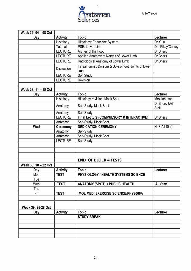
Week 36: 04 – 08 Oct
Day Activity Topic Lecturer
Histology Histology: Endocrine System Dr Xulu
Tutorial PSE: Lower Limb Drs Pillay/Calvey
LECTURE Arches of the Foot Dr Briers
LECTURE Applied Anatomy of Nerves of Lower Limb Dr Briers
LECTURE Radiological Anatomy of Lower Limb Dr Briers
Dissection Tarsal tunnel, Dorsum & Sole of foot, Joints of lower limb
LECTURE Self Study
LECTURE Revision
Week 37: 11 – 15 Oct
Day Activity Topic Lecturer
Histology Histology revision: Mock Spot Mrs Johnson
Anatomy Self-Study/ Mock Spot Dr Briers &All Stall
Anatomy Self-Study
LECTURE Final Lecture (COMPULSORY & INTERACTIVE) Dr Briers
Anatomy Self-Study/ Mock Spot
Wed Ceremony DEDICATION CEREMONY HoS All Staff
Anatomy Self-Study
Anatomy Self-Study/ Mock Spot
LECTURE Self-Study
END OF BLOCK 4 TESTS
Week 38: 18 – 22 Oct
Day Activity Topic Lecturer
Mon TEST PHYSIOLOGY / HEALTH SYSTEMS SCIENCE
Tue
Wed TEST ANATOMY (SPOT) / PUBLIC HEALTH All Staff
Thu
Fri TEST MOL MED/ EXERCISE SCIENCE/PHY2006A
Week 39: 25-28 Oct
Day Activity Topic Lecturer STUDY BREAK
ANAT 2020
25
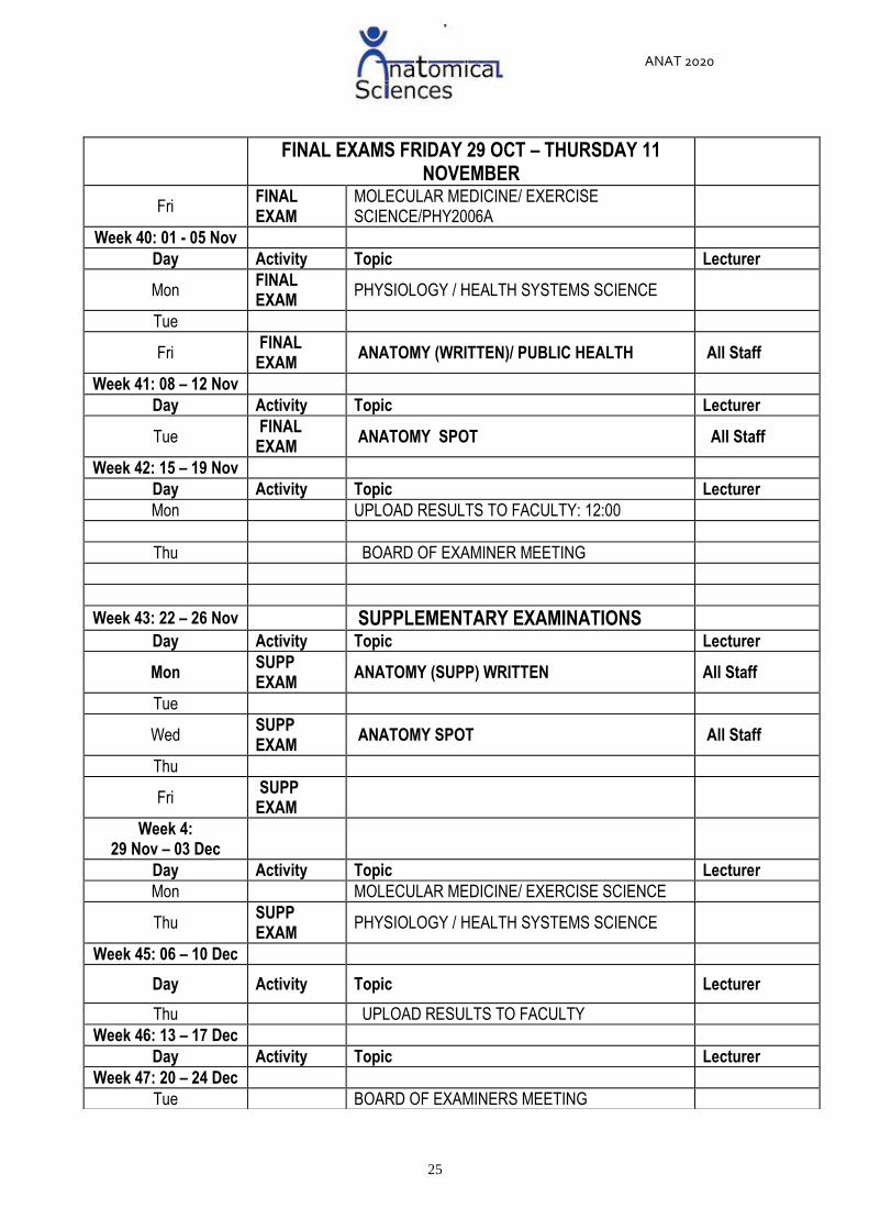
FINAL EXAMS FRIDAY 29 OCT – THURSDAY 11
NOVEMBER
Fri FINAL EXAM
MOLECULAR MEDICINE/ EXERCISE SCIENCE/PHY2006A
Week 40: 01 - 05 Nov
Day Activity Topic Lecturer
Mon FINAL EXAM
PHYSIOLOGY / HEALTH SYSTEMS SCIENCE
Tue
Fri FINAL EXAM
ANATOMY (WRITTEN)/ PUBLIC HEALTH All Staff
Week 41: 08 – 12 Nov
Day Activity Topic Lecturer
Tue FINAL EXAM
ANATOMY SPOT All Staff
Week 42: 15 – 19 Nov
Day Activity Topic Lecturer
Mon UPLOAD RESULTS TO FACULTY: 12:00
Thu BOARD OF EXAMINER MEETING
Week 43: 22 – 26 Nov SUPPLEMENTARY EXAMINATIONS
Day Activity Topic Lecturer
Mon SUPP EXAM
ANATOMY (SUPP) WRITTEN All Staff
Tue
Wed SUPP EXAM
ANATOMY SPOT All Staff
Thu
Fri SUPP EXAM
Week 4: 29 Nov – 03 Dec
Day Activity Topic Lecturer
Mon MOLECULAR MEDICINE/ EXERCISE SCIENCE
Thu SUPP EXAM
PHYSIOLOGY / HEALTH SYSTEMS SCIENCE
Week 45: 06 – 10 Dec
Day Activity Topic Lecturer
Thu UPLOAD RESULTS TO FACULTY
Week 46: 13 – 17 Dec
Day Activity Topic Lecturer
Week 47: 20 – 24 Dec
Tue BOARD OF EXAMINERS MEETING

ANAT 2020
35
ANAT 2020 Lecturers and Dissection Schedule Coordinators 2021
Block Topics Coordinating Staff for Lectures Coordinating Staff for Dissections
Block
1 Upper limb Prof AO Ihunwo and Dr O Olateju
Drs, Olateju, Small, Hutchinson, Briers,
Davimes, New Lecturer
Thorax Drs. O Olateju & D Pillay
Block
2
Head and
Neck Dr. Erin Hutchinson
Neuro Dr. B Maseko and Prof AO Ihunwo
Block
3 Abdomen
Drs D Pillay, B. Maseko & New
Lecturer
Drs Small, Hutchinson, Small, Briers,
Davimes & New Lecturer
Pelvis and
perineum Dr C. Small & New Lecturer
Block
4 Lower limb Dr N Briers
Online
Riddel
Tutorial
Drs D. Pillay and T. Calvey
Responsibilities: The Lecturers in charge of the Block and Dissections will also be responsible for;
a. Attending to students’ enquiries regarding Dissection for that block b. Implementing the Dissection Hall Rules as in the Yellow Book c. Overseeing the Teaching Assistants (TAs) assigned to the Groups d. Signing off the TA hours and the Form that Mrs Cheryl Bove usually provides to the TAs e. Oversee the Prosections in PVT and Vesalian Laboratories
ANAT 2020
36
B. STUDY COMPONENT: HISTOLOGY
ANAT 2020 HISTOLOGY OBJECTIVES
Histology objectives
In this section of the course, the students will be introduced to the basic histological structure
and function of the primary tissues and organ systems at light microscopic level.
Reference will only be made to ultrastructure where deemed relevant and necessary.
The student should be able to apply this knowledge to corresponding anatomical structures
and embryological development. Histology should also be related to the function of the human
body in both normal and pathological conditions.
General Objectives
This is an outline of the objectives a student should be able to fulfil at the end of the
course. Specific and more comprehensive objectives may be obtained from the Histology
Practical Manual.
Students are required to make a diagrammatic representation at different levels of
magnification of all tissues and organs studied (see Histology Practical Manual).
In addition to the Histology Manual, students are required to use the Road maps that
contain the labelled photomicrographs of all primary tissues and organ systems to
complement their study of the specific topic.
After combining the information obtained from the lectures (1), histology practical manual
(2) and road maps (3) the student are required to study the Olyvia slides (4) in order to
identify the specific structure including the related details previously studied using 1, 2
and 3.
Study Resources: Histology Practical Manual Olyvia slides, Road maps, Textbook
UNIT THEME 1: PRIMARY TISSUES
Specific examples of each primary tissue type in sections of many different organs will be
studied.
Capability statement:
After systematic and committed studying of four primary tissues, the student should be
able to fully apply the obtained knowledge in order to understand the histological structure
and function of different organs and organ systems.
Sub-unit themes:
Sub-unit theme 1.1: Epithelial tissue
Sub-unit theme 1.2: Glandular epithelium (glands)
Sub-unit theme 1.3: Connective tissue
Sub-unit theme 1.4: Muscle tissue
Sub-unit theme 1.5: Nervous tissue

ANAT 2020
37
Embedded knowledge (applicable to all primary tissues):
Students must know and understand the following before studying the sub-unit themes:
Cell structure
Definition of tissues
Explain what is understood by the concept of tissue
SUB-UNIT THEME 1.1: EPITHELIAL TISSUE
After studying the histological structure of different types of epithelia as a part of different
organs or organ systems, the student will be able to identify, classify, describe the
structure, give the function and provide location of the following types of epithelia:
Simple squamous, cuboidal and columnar epithelia with various surface modifications
(cilia or microvilli)
Stratified squamous (non-keratinised and keratinised) epithelium
Pseudostratified columnar epithelium with surface modifications (cilia)
Transitional epithelium
Epithelial tissue – Specific outcomes
1. Identify and classify the two types of epithelia found in choroid plexus
2. Provide the function and specific location of the two epithelia found in the choroid
plexus
3. Identify and classify the epithelium found in jejunum, trachea, oesophagus, thick skin
and urinary bladder
4. Provide the function and specific location of the epithelia found in jejunum
5. Identify and classify the two types of epithelia found in trachea, oesophagus, thick skin
and urinary bladder
6. Identify possible junctional complexes present in different epithelia.
7. Provide the diverse functions of epithelia and their specific locations.
SUB-UNIT THEME 1.2: GLANDS
After studying the histological structure of certain types of glands, the student will be able
to identify, classify, describe the structure, function and provide location of the following
types of glands:
Simple tubular with goblet cells (e.g. the colon)
Simple coiled tubular (e.g. eccrine sweat glands)
Simple branched alveolar (e.g. sebaceous glands)
Compound tubulo-alveolar (e.g. the submandibular gland)
Apart from the classification according to the morphology, glands can be classified
according to their mode of secretion (holocrine, merocrine or apocrine) and nature of
secretion (mucous, serous, mixed - mucous-serous, oily/waxy and sudoriferous/sweat
secretion).
ANAT 2020
38
Glands – Specific outcomes
1. Identify and classify the two types of glands found in jejunum
2. Provide the mode of secretion for the glands found in jejunum
3. Identify and classify the gland found in colon, uterus and thin skin
4. Provide the mode of secretion for the glands found in colon, uterus and thin skin
5. Classify the submandibular gland
6. Provide the mode and nature of secretion of submandibular gland
SUB-UNIT THEME 1.3: CONNECTIVE TISSUE
NB: Connective tissue includes:
Connective tissue proper
Connective tissue with a fluid matrix
Connective tissue with a solid matrix
Osteogenesis
After studying the histological structure of different types of connective tissue, the student
will be able to identify, classify, describe the structure, give the function and provide
location of the following types of connective tissues (including cells, fibres and ground
substance as the main structural components):
Connective tissues with a semi-solid matrix
o Loose (areolar) connective tissue
o Dense regular connective tissue
o Dense irregular connective tissue
o Elastic and reticular tissue
Connective tissue with a fluid matrix:
o Blood
Connective tissue with a solid matrix - Cartilage:
o Hyaline
o Fibrocartilage
o Elastic cartilage
Chondrogenesis – Cartilage formation
o Appositional growth
o Interstitial growth
Connective tissue with a solid matrix - Bone:
o Compact bone
o Cancellous bone
Osteogenesis – Bone formation
o Intramembranous ossification
o Endochondral ossification
Connective tissue – Specific outcomes
Identify the three main constituents of all connective tissues (including blood, cartilage
and bone)

ANAT 2020
39
Connective tissue proper
1. Classify connective tissue proper
2. Explain the histology of loose connective tissue
3. Provide the location and function of loose connective tissue
4. Explain the histology of dense irregular connective tissue
5. Provide the location and function of dense irregular connective tissue
6. Explain the histology of dense regular connective tissue
7. Provide the location and function of dense regular connective tissue
8. Explain the histology of elastic tissue
9. Provide the location and function of elastic tissue
10. Explain the histology of reticular tissue
11. Provide the location and function of reticular tissue
Connective tissue with a fluid matrix
1. Classify connective tissue with a fluid matrix (blood)
2. Classify leukocytes
3. Explain the histology and function of erythrocytes, neutrophils, eosinophils, basophils,
lymphocytes, monocytes and thrombocytes
Connective tissue with a solid matrix
1. Identify the three main types of cartilage
2. Explain the histology of hyaline cartilage including perichondrium
3. Provide the location and function of hyaline cartilage
4. Explain the histology of fibrocartilage
5. Provide the location and function of fibrocartilage
6. Explain the histology of elastic cartilage
7. Provide the location and function of fibrocartilage
8. Discuss the main cartilage cell types including their specific location and function
9. Provide detailed explanations of two types of cartilage development
10. Identify the two main types of bone
11. Explain the histology of cancellous (spongy) bone
12. Provide the location and function of cancellous bone
13. Explain the histology of compact bone
14. Provide the location and function of compact bone
Osteogenesis
1. Identify the two types of bone development
2. Discuss the main bone cell types including their specific location and function
3. Provide detailed explanations of intramembranous ossification
4. Provide detailed explanations of endochondral ossification
SUB-UNIT THEME 1.4: MUSCLE TISSUE
After studying the histological structure of muscle, the student will be able to identify,
classify, and describe the structure, function and provide location of the following types of
muscle:
Skeletal (striated) muscle
Cardiac (striated) muscle and Purkinje fibres
Smooth (visceral) muscle

ANAT 2020
40
Muscle tissue - Specific outcomes
1. Identify the three main types of muscle tissue (your identification should be based on
the fibre size, shape and presence/absence of branching, number, shape and position
of nuclei and presence or absence of cross-striations)
2. Provide the histological and ultrastructural organisation of the skeletal muscle
3. Provide the location and function of the skeletal muscle
4. Provide the histological and ultrastructural organisation of the cardiac muscle
5. Provide the location and function of the cardiac muscle
6. Discuss histology of the Purkinje fibres
7. Provide the specific location and function of the Purkinje fibres
8. Provide the histological organisation of the smooth muscle
9. Provide the location and function of the smooth muscle
SUB-UNIT THEME 1.5: NERVOUS TISSUE
After studying the histological structure of nervous tissue, the student will be able to
identify, classify and describe the structure, function and provide location of the following
components of nervous tissue:
Neurons in:
o the CNS (anterior horn cells)
o the PNS (spinal/dorsal root ganglion cells)
Nerve fibres of:
o the PNS (spinal nerves)
o the ANS (autonomic nerves)
o Supporting cells related to these neurons and fibres including Schwann cells and
satellite cells in the PNS and the different types of neuroglia in the CNS
o Neuromuscular junctions
Nervous tissue – Specific outcomes
1. Classify the neurons according to their structure (morphology)
2. Classify the neurons according to their function
3. Discuss the histology of a motor neuron
4. Provide the specific location of the motor neuron
5. Discuss the histology of a sensory neuron
6. Provide the specific location of a sensory neuron
7. Discuss the histology of a peripheral nerve including the three connective tissue sheets
(coverings)
8. Discuss the main parts of a motor neuron

ANAT 2020
41
UNIT THEME 2: ORGANS AND ORGAN SYSTEMS
Specific organs and organ systems will be studied:
Capability statement: After systematic and committed studying of all organs and organ
systems in healthy conditions, the student should be able to apply the obtained knowledge
to the tissues and organs in the pathological settings.
Sub-unit themes: (all organs and organ systems from skin and up to and including the
endocrine system).
Sub-unit theme 2.1: Skin
Sub-unit theme 2.2: Central nervous system (CNS)
Sub-unit theme 2.3: Cardiovascular system (CVS)
Sub-unit theme 2.4: Lymphatic system
Sub-unit theme 2.5: Respiratory system
Sub-unit theme 2.6: Gastrointestinal system (GIT)
Sub-unit theme 2.7: GIT glands
Sub-unit theme 2.8: Urinary system
Sub-unit theme 2.9: Male reproductive system
Sub-unit theme 2.9: Female reproductive system
Sub-unit theme 2.10: Endocrine system
Embedded knowledge (applicable to all organs and organ systems)
Students must know and understand the following before studying the sub-unit themes:
Detailed histology of primary tissue including identification, classification, structure,
location and function
Without the prior detailed knowledge of the primary tissues, understanding of the
organ systems will not be possible!
SUB-UNIT THEME 2.1: SKIN
After studying the histological structure of skin, the student will be able to, identify and
describe the structure and function of the following:
Thin skin (e.g. skin of scalp)
o Epidermis, dermis and hypodermis
o Sebaceous glands → see primary tissues – glands above
o Eccrine sweat glands → see primary tissues – glands above
o Blood vessels and nerves
Thick skin (e.g. skin of palm/fingertip)
o Epidermis, dermis and hypodermis
o Eccrine sweat glands → see primary tissues – glands above
o Blood vessels and nerves
o Encapsulated nerve endings (e.g. Meissner’s and Pacinian corpuscles)

ANAT 2020
42
Skin – Specific outcomes
1. Discuss the histology of the epidermis of thin skin (four distinct layers and the relevant
cell types as well as their importance)
2. Discuss the histology of the dermis of thin skin (two distinct layers, the type of
connective tissue being the main constituent of these layers and associated
appendages
3. Discuss the histology of the epidermis of thick skin (five distinct layers and the relevant
cell types as well as their importance)
4. Discuss the histology of the dermis of thick skin (two distinct layers, the type of
connective tissue being the main constituent of these layers and associated
appendages)
5. Compare and contrast between the epidermis and dermis of thin and thick skin
6. Revise on the basic tissue knowledge involving epithelium and glands associated
with the dermis of thin and thick skin
7. Identify, classify and provide the exact location, structure and function of Meissner’s
and Pacinian corpuscles
SUB-UNIT THEME 2.2: CENTRAL NERVOUS SYSTEM (CNS)
After studying the histological structure of the central nervous system, the student will be
able to identify and describe the structure and function of:
The meninges
Cerebrum
o The white and grey matter and corresponding cells and fibres types
o The layers of the cerebral cortex
o Neuronal types
Cerebellum
o The arbor vitae and folia
o The layers of the cerebellar cortex
o Neuronal types
The choroid plexus
SUB-UNIT THEME 2.3: CARDIOVASCULAR SYSTEM (CVS)
After studying the histological structure of the cardiovascular system, the student will be
able to:
Identify, classify, describe the structure and function of the components of:
o Elastic arteries
o Medium sized arteries and veins
o Arterioles, venules and capillaries
Describe the structure and function of cardiac muscle (including Purkinje fibres → see
primary tissues – muscle above)
SUB-UNIT THEME 2.4: LYMPHATIC SYSTEM
After studying the histological structure of the lymphatic system, the student will be able
to identify, classify, and describe the fibrous framework and cellular components as well
as the function of:
Diffuse and nodular lymphatic tissue
The lymph node (including the filtration of lymph)
ANAT 2020
43
The spleen (including the blood supply)
The thymus
SUB-UNIT THEME 2.5: RESPIRATORY SYSTEM
After studying the histological structure of the respiratory system, the student will be able
to identify and describe the structure and function of:
The conducting passages:
o Nasal cavities and air sinuses
o Trachea
o Intrapulmonary bronchus
o Bronchioles
o Terminal bronchioles
The respiratory units of the lung:
o Respiratory bronchiole
o Alveolar duct
o Alveolar sac
o Alveoli with interalveolar septum (including the blood-air barrier)
SUB-UNIT THEME 2.6: GASTROINTESTINAL SYSTEM (GIT)
Students will study the general four layered histological pattern (mucosa, submucosa,
muscularis externa and serosa/adventitia) of the gastrointestinal tract and will examine
how this pattern changes in the following regions of the digestive tract in accordance to
the function that has to be performed in that particular region.
Oesophagus
Stomach
Small intestine
Large intestine
SUB-UNIT THEME 2.7: GIT GLANDS
After studying the histological structure of selected glands, the student will be able to
identify and describe the structure and function of:
The liver
The pancreas (exocrine component only as the endocrine pancreas will be studied
with the endocrine system)
The salivary glands (→ see primary tissues – submandibular gland above)
The gall bladder
SUB-UNIT THEME 2.8: URINARY SYSTEM
After studying the histological structure of the urinary system, the student will be able to:
Identify and describe the structure and function of:
o The kidney:
The cortex (including the renal corpuscle and associated tubules)
The medulla
o The urinary bladder (epithelium only → see primary tissues – epithelium above)
Describe the blood supply of the kidney
Describe the filtration apparatus and juxtaglomerular apparatus of the kidney

ANAT 2020
44
SUB-UNIT THEME 2.9: MALE REPRODUCTIVE SYSTEM
After studying the histological structure of selected regions of the male reproductive
system, the student will be able to:
Identify and describe the structure and function of the components of the testis
The testes
The seminiferous tubules
Describe spermatogenesis
The epididymis
The tubuli recti
The rete testis
The efferent ductules
The vas deferens
The prostate gland
The penis
The penile urethra
SUB-UNIT THEME 2.9: FEMALE REPRODUCTIVE SYSTEM
After studying the histological structure of selected regions of the female reproductive
system, the student will be able to:
Identify and describe the structure and function of:
o The ovary
o The different stages of the follicular development
o Corpus luteum
o The uterus (preovulatory endometrium)
o The uterus (postovulatory endometrium)
o Non-lactating and lactating mammary gland
Identify and describe the structure and function of the placenta
SUB-UNIT THEME 2.10: ENDOCRINE SYSTEM
After studying the histological structure of selected endocrine glands, the student will be
able to identify and describe the structure and function of:
The hypophysis (pituitary gland)
o Pars tuberalis
o Pars distalis
o Pars intermedia and
o Pars nervosa
The thyroid gland
The suprarenal gland
The endocrine pancreas

ANAT 2020
45
C. STUDY COMPONENT: EMBRYOLOGY
ANAT 2020 EMBRYOLOGY OBJECTIVES
EMBRYOLOGY OBJECTIVES
In this section of the course, the students will be introduced to the development of human
embryo at different stages including gametogenesis, fertilization, implantation, the embryonic
period, the foetal period and development of the pharyngeal arches and their derivatives. The
student should be able to apply this knowledge to corresponding anatomical structures and
embryological development. Also, students will develop the ability to correlate between the
embryological structure and its clinical significance as this course trains the student to
understand any related clinical problems.
Study Resources: The fundamentals of Human Embryology (Allan and Kramer)
UNIT THEME 1: EARLY EMBRYOLOGY
The processes of fertilization, implantation, placentation and gastrulation will be covered.
Capability statement:
Students will be able to describe the factors which are involved in the fusion of the male and
female gametes. In addition students will be able to explain the formation of the placenta and
the notochord which is a critical structure during embryogenesis.
Embedded Knowledge
Students must have the following information/knowledge prior to lectures:
Cell structure
Mitosis and Meiosis
Sperm structure
SUB-UNIT THEMES:
Sub-unit theme 1.1: The first week of development (Fertilization and implantation)
Sub-unit theme 1.2: The second week of development (Placentation)
Sub-unit theme 1.3: The third week of development (Gastrulation and Neurulation)
SUB-UNIT THEME 1.1: THE FIRST WEEK OF DEVELOPMENT
Students will be able to explain the process of fertilization, consequences of fertilization,
cleavage, passageway along uterine tube, implantation and sites of implantation.
Specific outcomes
Students should:
1. Accurately describe the process of fertilization
2. Explain the stages of cell division and the link to formation of zygote
3. Describe the process of implantation
4. Explain the "critical period" in development
5. Define the embryonic stage

ANAT 2020
46
6. Explain the path of an egg from the ovary to implantation.
7. Name the stages of implantation and the structures formed.
SUB-UNIT THEME 1.2: THE SECOND WEEK OF DEVELOPMENT
Students will be able to describe the formation of a bi-laminar embryo and the placenta,
including the extra-embryonic membranes.
Specific outcomes
Students should be able to describe the following:
1. Formation of hypoblast and epiblast layers
2. Formation of syncytiotrphoblast and cytotrophoblast layers
3. Formation of the amniotic cavity and the yolk sac
4. The establishment of the lacunar stage of development
5. Formation of chorionic villi
6. The formation of the extraembryonic mesoderm
7. Early coelom formation
8. Ectopic implantation sites
SUB-UNIT THEME 1.3: THE THIRD WEEK OF DEVELOPMENT
Students will be able to describe the process of gastrulation and the formation of a tri-
laminar embryo, primitive streak formation, folding of “flat” embryo into tubular structure and
neural tube formation.
Specific outcomes
Students should be able to:
1. Name the three germ layers ectoderm, mesoderm and endoderm and list the adult
tissues derived from each of these three layers
2. Describe the formation of the mesenchymal cells from the epiblast layer
3. Describe the differentiation of mesoderm into the paraxial, intermediate and lateral
plate mesoderm
4. Describe the differentiation of somites from paraxial mesoderm
5. Describe neural tube formation and how the neural tube differentiates into specific
components of the nervous system
6. Describe the migration of neural crest cells from the neural tube

ANAT 2020
47
UNIT THEME 2: ORGAN SYSTEMS
Capability statement: Students will be able to describe the fundamental aspects of
embryonic development which are involved in the normal formation of organ systems. In
addition students will be able to explain the factors which may lead to the abnormal
development of certain organs, leading to birth defects.
SUB-UNIT THEMES:
Sub-unit theme 2.1: Development of the heart
Sub-unit theme 2.2: Development of pharyngeal arch arteries and veins
Sub-unit theme 2.3: Development of face and palate
Sub-unit theme 2.4: Development of pharyngeal arches, tongue and thyroid
Sub-unit theme 2.5: Embryology of the Nervous system
Sub-unit theme 2.6: Embryology of the GIT
Sub-unit theme 2.7: Coelom and Mesenteries
Sub-unit theme 2.8: The development of the urogenital system
Embedded Knowledge
Students must have the following information/knowledge prior to lectures:
The process of implantation
Neural tube formation
Folding of the embryo into a tubular structure
Histology of the male and female reproductive systems
The formation of the three germ layers
SUB-UNIT THEME 2.1: THE EMBRYONIC DEVELOPMENT OF THE HEART
After studying the development of the heart students should be able to explain how a single
tubular heart is converted into four chambers to sub-serve the adult circulation.
Students should also be able to explain the development of particular structures e.g. Septum
primum, septum secundum and foramen ovale, which assist in conversion from a fetal to a
neonatal circulation.
Specific outcomes
1. To explain the formation of a single heart tube from cardiogenic mesoderm
2. To explain the formation of the truncus arteriosus, bulbus cordis, ventricle, atrium and the
sinus venosus
3. To explain blood circulation through a primordial heart
4. To describe the sepation of the AV canal
5. Describe the formation of the intermediate bar
6. Describe the sepation of atrium and ventricle
7. Describe the fate of the bulbus cordis and the sinus venosus
8. Describe the formation of the left atrium

ANAT 2020
48
SUB-UNIT THEME 2.2: DEVELOPMENT OF PHARYNGEAL ARCH ARTERIES AND
VEINS
To illustrate the important changes which occur when the fetus foregoes an aquatic
environment and changes to the terrestrial environment of the neonate/adult.
Specific outcomes
Students should be able to explain the following:
1. The steps involved in normal development from the angiogenesis stage to the
completion of the 4-chambered fetal heart.
2. The flow of blood entering and exiting the developing fetal heart and blood flow within the
fetal heart (fetal circulation)- closure or patency of foramen oval (at birth) and changes in
percentage oxygen saturation of the blood at each stage of the fetal circulation.
3. The effects of the following cardiac anomalies to normal cardiac function
4. Atrial Septal Defects of both the primum and secundum type
5. Premature closure of the foramen ovale
6. Persistent atrio-ventricular canal
7. Ventricular Septal Defects of both the membranous and muscular type
8. Ectopia cordis
9. Tricuspid atresia
10. Tetralogy of Fallot
11. Transposition of the great vessels
12. Persistent truncus arteriosus
SUB-UNIT THEME 2.3: DEVELOPMENT OF FACE AND PALATE
To ensure an understanding of the abnormalities which occur in these regions and their
underlying causes.
Specific outcomes
1. Name the prominences that contribute to face formation
2. Frontal prominence, 2 maxillary prominences, 2 mandibular prominences
3. Recognize the contribution of each prominence to the formation of various face parts:
Frontal prominence: Forehead, nasal bridge, lateral nose
Maxillary prominences: Lateral upper lip, Cheeks (above mouth line)
Mandibular prominences: Cheeks (below mouth line), lower lip, chin
4. Understand the formation and position of nasolacrimal duct from the nasolacrimal groove
5. Understand the formation of oblique facial cleft
6. Able to describe the formation of the intermaxillary segment.
7. Recognize the embryological origin of the median cleft lip and be conversant with the
most common associated syndromes.
SUB-UNIT THEME 2.4: PHARYNGEAL ARCHES, TONGUE AND THYROID
To ensure an understanding of the gross anatomy of the head and neck, particularly
innervation.
To enhance the understanding of the major glands/structures of the neck and thorax and
resulting

ANAT 2020
49
To ensure an understanding of the diverse innervation of the tongue; and abnormalities.
To ensure an understanding of the development and position of the thyroid gland and
abnormalities.
Specific Outcomes
1. Understand and describe pharyngeal arches, pharyngeal pouches, pharyngeal grooves,
pharyngeal membranes
2. Understand and list the derivatives of each pharyngeal arch basic structural component
(artery, nerve, muscle, cartilage skeleton).
3. List the derivatives of the first pharyngeal groove and describe the pattern of obliteration
of pharyngeal grooves.
4. List the derivatives of the different pharyngeal pouches.
5. Describe the terms Pharyngeal/Branchial Arch, Pouch & Grooves/Cleft.
6. Understand the abnormalities of the pharyngeal arch apparatus
SUB-UNIT THEME 2.5: DEVELOPMENT OF THE NERVOUS SYSTEM
Formation of neural tube, neural crest, spinal cord and brain, cells layers, alar and basal plates
nuclei, folding of the brain, enormous development of cerebral cortex
Specific outcomes
1. Comprehend neural development and the formation of spinal cord and brain from neural
tube
2. Know the layers of developing spinal cord and its contents
3. Describe the neurogenesis of the neural tube
4. Outline the mantle and marginal zones
5. Differentiate between the grey and white matter from the neural tube.
6. Understand the role of migration of neurons during neural development.
7. Understand the respective adult derivatives of the brain vesicles and their walls.
8. Know the origin of neural crest cells their input in the development some tissues.
9. Appreciate and know the embryological basis of some congenital anomalies of the
nervous system e.g. hydrocephalus, spina bifida, anencephaly and encephalocele
SUB-UNIT THEME 2.6: EMBRYOLOGY OF THE GIT
Students should be able to describe the formation of the following:
Tube, derivatives of the stomodeum, including the development of the hypophysis
cerebri; foregut development
Midgut development and rotation, formation of greater omentum; common abnormalities
Development of hindgut; development of liver including invasion of vessels; common
abnormalities
Development and “rotation” of the pancreas; formation of lesser sac; common
abnormalities
Specific Outcomes
1. Understanding how the germ layer contributes to the primitive gut formation
2. Explain the significance of folding of the embryo during GIT development
3. Describe the three main embryonic divisions of the primitive gut
4. List and describe the derivatives of the foregut, midgut, hindgut and their blood supply

ANAT 2020
50
5. Know the development of other GIT organs (liver, pancreas, spleen)
6. Understanding of the rotation of the stomach and the midgut loop during GIT
development
7. Explain the process of umbilical herniation and the embryonic development of
mesenteries
8. Understand the abnormalities of GIT development
Sub-unit theme 2.7: Coelom and Mesenteries
Students should be able to describe the development of the extra- and intra-embryonic
coelomic cavities; separation of intra-embryonic coelom into four; formation of “folds”;
formation of mesenteries; fixed and free mesenteries; development of diaphragm,
abnormalities
Specific outcomes
1. To understand what a coelomic cavity is and how it comes to protect the major organs of
the body.
2. To ensure an understanding of what a mesentery is and where they are located;
3. To understand the innervation and position of the diaphragm.
SUB-UNIT THEME 2.8: THE DEVELOPMENT OF THE UROGENITAL SYSTEM
Students will be able to explain the early development of the kidney and gonads (migration
of primordial germ cells); ureters and urethra; abnormalities.
In addition students should be able to describe internal genital development and the different
gonads.
Specific outcomes
1. Describe the embryonic development of kidneys, ureters, and urinary bladder.
2. Understand the development of the pronephros, mesonephros, and metanephros
3. Describe the collecting and excretory systems of the kidney
4. Describe the ascent and rotation of the kidneys
5. Understand the congenital anomalies of the kidneys and urinary bladder.
6. Describe the development of testes, ovaries, and their descent.
7. Describe the development of the mesonephric and paramesonephric ducts and genital
ducts.
8. Describe the development of the vagina, uterus and the uterine tubes.
9. List the adult derivatives and vestigial remnants of embryonic urogenital structures.

ANAT 2020
51
D. STUDY COMPONENT: MORPHOLOGICAL ANATOMY
UNIT THEME 1: UPPER LIMB
Capability statement
By completion of this section the student must be able to understand the basic and applied
anatomical structure and function of the upper limb. The student should be able to apply
this knowledge and provide the anatomical basis underlying specific clinical scenarios
pertaining to diseases and injuries to the upper limb.
Embedded knowledge
Students must know and understand the following before studying the sub-unit theme:
Anatomical terminology
Anatomical orientation
SUB-UNIT THEME 1.1: THE PECTORAL GIRDLE AND BREAST
(Practical Anatomy)
WEEK 1: SPECIFIC OUTCOMES
By the end of this week, the student should be able to:
Identify and describe the bones of the pectoral girdle (scapula, clavicle and humerus)
Identify the joints of the pectoral girdle (sternoclavicular, acromioclavicular)
Distinguish between the terms upper limb, arm, forearm and hand
Explain the concept of superficial fascia and list its contents
Describe the anatomy of the female breast, including the surface anatomy, arterial
supply, venous drainage and innervation
Describe position and drainage area of the axillary lymph nodes
Explain the lymphatic drainage of the breast and upper limb and in the spread of tumors
Describe the attachments of the muscles of the anterior aspect of the pectoral girdle,
their actions and nerve supply
Identify the boundaries of the axillary space
Describe the attachments, actions and nerve supply of the muscles around the scapula
Describe the formation of the arterial anastomosis around the scapula
Describe the boundaries and contents of the quadrangular and triangular spaces
Explain how the actions of the muscles would be affected should the nerves be damaged
Identify anatomical features and major relations on diagnostic images, e.g. CT, MRI,
radiographs
WEEK 1: DISSECTION SPECIFIC OUTCOMES
THE PECTORAL GIRDLE
1. Identify the bones of the pectoral girdle on the articulated skeleton: clavicle, scapula, and
humerus.
2. Identify the following joints on an articulated skeleton and a radiograph:
acromioclavicular joint, sternoclavicular joint
ANAT 2020
52
THE BREAST:
1. Examine the female and male breasts, noting the difference in size and extent.
2. Identify the areola and nipple, taking note of the variation in the position of the nipple in
different female cadavers.
3. Examine the cut section of the female breast and identify the duct system.
THE ANTERIOR ASPECT OF THE PECTORAL REGION
1. Identify the 5 major muscles in this region: Pectoralis major, pectoralis minor,
Subclavius, serratus anterior, deltoid.
2. Study the attachments, nerve supply and actions of these muscles.
3. Identify the axillary space and the muscles that form its boundaries.
THE POSTERIOR ASPECT OF THE PECTORAL GIRDLE
1. Identify the 3 most superficial muscles in this region: Trapezius, deltoid, latissimus dorsii
2. Identify the deep muscles, which are in two convenient groups:
Medial group: levator scapulae, rhomboid major, rhomboid minor,
Lateral group: supraspinatus, infraspinatus, teres major, teres minor, subscapularis
3. Identify the arteries in this region and describe the important connections (anastomosis)
that they form around the scapula.
4. Identify the boundaries and contents of the quadrangular and triangular spaces.

ANAT 2020
53
SUB-UNIT THEME 1.2: THE AXILLA, BRACHIAL PLEXUS AND ARM
(Practical Anatomy)
WEEK 2: SPECIFIC OUTCOMES
By the end of this week, the student should be able to:
Describe the contents of the axillary space
Identify the clavipectoral fascia and the structures passing through it
Draw and describe the arrangement of the axillary lymph nodes
Identify and draw the terminal branches of the brachial plexus and follow them out of the
axilla
Describe the consequences of injury to the brachial plexus and its terminal branches
Identify the muscles of the anterior and posterior compartments of the arm
Describe the actions and nerve supply of the muscles of the anterior and posterior
compartments of the arm
Describe the muscle groups of the anterior and posterior compartments of the arm, their
actions and nerve supply
WEEK 2: DISSECTION SPECIFIC OUTCOMES
THE AXILLARY SPACE
1. Review the boundaries of the axilla.
2. What appears to be the most abundant content of the axilla?
3. Identify the following:
Axillary artery and its branches
Axillary vein
Axillary lymph nodes
4. Identify the lateral thoracic artery and long thoracic nerve.
THE BRACHIAL PLEXUS
1. Identify the cords and branches of the brachial plexus. The letter 'M' will be helpful in
this process.
2. Draw the scheme of the brachial plexus on the drawing board or in your book and label
it fully.
3. Think (in general terms) about the likely effect of cutting through (transecting) one of the
branches of the brachial plexus.
THE ANTERIOR COMPARTMENTS OF THE ARM
General:
1. What are the subdivisions of the upper limb?
2. Review the major features of the humerus e.g. head, necks, tuberosities, sulci (grooves),
fossae, capitulum, trochlea, and nutrient foramen.
3. Examine the biceps brachii muscle on your partner and note during which movements of
the upper limb does it become more prominent?

ANAT 2020
54
Muscles of the anterior compartment of the arm
1. Identify and name the 3 muscles in this compartment.
2. What is their nerve supply?
3. Examine the attachments of these muscles. Which joints do these muscles cross?
Examine the orientation of the muscle fibres and try to work out the possible actions of
these muscles.
Neurovascular structures of the arm
Identify the neurovascular structures on the medial aspect of the arm. You should find at
least 5 structures (6 if the vein is present). Trace each of them inferiorly.
POSTERIOR ASPECTS OF ARM
Muscle of the posterior aspect of the arm
This is the triceps muscle.
1. Why is it called “triceps”?
2. The long head contributes to the formation of the boundaries of the quadrangular and
triangular spaces, which you have seen before.
3. Review the boundaries and contents of these spaces now.
Neurovascular structures in the posterior aspect of the arm
These are the radial nerve and profunda brachii artery.
1. What are their origins?
2. Follow the course of the nerve through the arm to the forearm.
3. Note the relationship of the neurovascular structures to each other.

ANAT 2020
55
SUB-UNIT THEME 1.3: CUBITAL FOSSA AND FOREARM
(Practical Anatomy)
WEEK 3: SPECIFIC OUTCOMES
By the end of this week, the student should be able to:
Describe the boundaries and contents of the cubital fossa
Describe the features of the radius and ulna
Identify the bones of the hand
Identify the muscles of the anterior and posterior compartments of the forearm,
Describe the actions and nerve supply of the muscles of the anterior and posterior
compartments of the forearm.
WEEK 3: DISSECTION SPECIFIC OUTCOMES
THE CUBITAL FOSSA
Identify the boundaries of the cubital fossa. These should normally include the roof and floor.
Look for as many of the contents as can be seen, especially the:
1. Biceps tendon
2. Brachial artery and vein (Does the artery divide into radial and ulnar arteries in this
fossa?)
3. Median nerve
4. Radial nerve
Applied anatomy:
What veins lie in the roof of this fossa that may be used for withdrawing blood or
administering substances intravenously?
Attempt to feel your arterial pulse on the medial aspect of the base of the fossa just
medial to the tendon of the biceps brachii muscle. This is easier if you extend the elbow
joint.
THE ANTERIOR ASPECT OF THE FOREARM
Surface anatomy:
1. Palpate (feel with your hand) the head of the radius just distal to the lateral epicondyle of
the humerus as you rotate the forearm.
2. Identify the olecranon process and follow the ulna distally to its styloid process.
Osteology:
1. Examine the radius and ulna of an articulated skeleton or from your bone box. Identify
their major features.
2. Identify the interosseous borders and identify the interosseous membrane in a forearm
specimen. What is the function of this membrane?
3. Identify the bones of the wrist and hand.

ANAT 2020
56
THE ANTERIOR ASPECT OF THE FOREARM
Muscles of the forearm (3 groups)
Identify these muscles and know their actions and innervation.
1. Superficial rotator muscles:
Pronator teres
Brachioradialis
Give the nerve supply of these muscles.
2. Flexor muscles of the forearm:
These may be divided into 3 functional groups:
1. Flexors of the wrist
· flexor carpi radialis; [radial deviation of wrist]
· palmaris longus
· flexor carpi ulnaris; [ulnar deviation of the wrist]
2. Flexors of the fingers
· flexor digitorum superficialis
· flexor digitorum profundus
3. Flexor of the thumb
· flexor pollicis longus
What is the nerve supply of these muscles?
3. Deep rotator muscles of the forearm:
· pronator quadratus (Confirm the quadrangular shape)
· supinator
What is the innervation of these muscles?
Pronate and supinate the forearm bones of an articulated skeleton. At what joints do
these movements take place?
THE POSTERIOR ASPECT OF THE FOREARM
Muscles of the posterior aspect of the forearm (3 functional groups)
Identify these muscles and know their actions and innervation.
1. Extensors of the wrist:
· extensor carpi radialis longus
· extensor carpi radialis brevis
· extensor carpi ulnaris
2. Extensors of the fingers:
· extensor digitorum
· extensor indicis
· extensor digiti minimi
3. Extensors of the thumb:
· extensor pollicis longus
· extensor pollicis brevis

ANAT 2020
57
4. Abductor of the thumb:
· abductor pollicis longus (an additional small abductor will be seen later in the hand)
You can also identify these muscles in two layers.
1. Superficial muscles:
· extensor carpi radialis longus and brevis
· extensor digitorum
· extensor carpi ulnaris
· extensor digiti minimi
2. Deep muscles:
· abductor pollicis longus
· extensor pollicis longus and brevis
· extensor indicis
· Supinator
· anconeus.
Which nerve supplies these muscles?
Most of these muscles originate from a common extensor origin. Where is this common
extensor origin?
Neurovascular components of the forearm
1. Identify the median, ulnar and radial nerves and trace them to the wrist
2. Identify the radial and ulnar arteries just distal to the elbow joint and trace them to the
wrist.
3. There is an arterial anastomosis around the elbow joint. You must know about its
formation. What is the benefit of having such arterial anastomosis? Where did you come
across an anastomosis in your previous dissection?

ANAT 2020
58
SUB-UNIT THEME 1.4: THE HAND AND JOINTS OF THE UPPER LIMB
(Practical Anatomy)
WEEK 4: SPECIFIC OUTCOMES
By the end of this week, the student should be able to:
Relate the gross anatomy of the palmar skin to its function,
Name the structures that pass through the carpal tunnel and, in very general terms, the
effect of damage to them,
Describe the actions and innervation of the muscle groups of the hand,
Explain the basic significance of the palmar spaces
Describe the shoulder joint as a typical example of synovial joints that are designed
more for a wide range of movements,
Describe the movements that take place at the proximal radioulnar joint.
Know the movements taking place at other joints of the upper limb
Identify anatomical features and major relations on diagnostic images, e.g. CT, MRI,
radiographs
WEEK 4: DISSECTION SPECIFIC OUTCOMES
THE HAND
The bones of the wrist and hand
Review the names of the bones of the wrist and hand again. If you look at an articulated
hand, you will see that the carpal bones form a concavity that is directed anteriorly. The
deep fascia of the wrist (flexor retinaculum) converts this into a tunnel.
1. What is the name of the tunnel?
2. Which structures pass deep to the flexor retinaculum?
3. Which structures pass superficial to it?
4. What is the consequence of increased pressure (e.g. from accumulated pus) in this
tunnel?
The palmar skin
1. How does the skin of your palm differ from that of the dorsal surface of the hand?
2. What is the advantage of the skin creases?
Functional groups of muscles of the hand
The muscles are grouped into flexors, extensors, abductors, adductors, and opposition
muscles.
1. What are the names of the muscles that perform these actions in the fingers?
2. What is the innervation of these muscles?
Neurovascular components of the hand
1. Identify the superficial and deep palmar arterial arches?
2. How are they formed?
3. Follow the ulnar and median nerves distal to the wrist.
4. What is the innervation of the skin of the hand (palmar and dorsal surfaces)? Learn to
draw this.

ANAT 2020
59
JOINTS OF THE UPPER LIMB
Compare, in table format, the different joint using the scheme below.
Types of joints
1. Compare, in table format, the structure and mobility of a fibrous, cartilaginous and
synovial joint. Also provide an example of each.
2. When studying joints, the scheme below should be followed:
Name
Classification or Type (e.g. plane, synovial hinge, pivot, condyloid, ball and socket)
Articulating surfaces
Capsule attachment
Synovial membrane
Ligaments (intra and extracapsular)
Nerve supply
Blood supply
Movements and the muscles that produce them
Relations
The shoulder joint
Examine the shoulder joint. Ask the demonstrator for assistance in opening up a shoulder
joint in the cadaver for you.
1. Using the scheme shown above, learn to describe the joint.
2. Which of the above muscles constitute the group of muscles called the 'rotator cuff' and
why are they given this name?
3. Would you say that this is a very stable joint?
4. Classify the features that account for its strength and weakness.
Elbow Joint
Use the scheme above to study the elbow joint.
The superior radio-ulnar joint
1. What ligament keeps the head of the radius in place?
2. What movement of the forearm takes place at this joint?
3. Name the muscles that produce these movements.
Wrist (Radiocarpal), Intercarpal, Carpometacarpal, Metacarpophalangeal &
interphalangeal Joints
Identify these joints on a mounted skeleton and on radiographs.

ANAT 2020
60
UNIT THEME 2: THORAX
Capability statement
By completion of this section the student must be able to understand the basic and applied
anatomical structure and function of structures within the thorax. The student should be
able to apply this knowledge and provide the anatomical basis underlying specific clinical
scenarios pertaining to diseases and injuries to the thorax.
Embedded knowledge
Students must know and understand the following before studying the sub-unit theme:
Anatomical terminology & orientation
Anatomy of the pectoral region, brachial plexus and breast
SUB-UNIT THEME 2.1: THE THORACIC CAGE, INTERCOSTAL SPACES, PLEURA AND
LUNGS
WEEK 5: SPECIFIC OUTCOMES
By the end of this week, the student should be able to:
Describe the bony structure of the thoracic wall,
Describe the arrangement of the intercostal muscles, nerves and blood vessels,
Describe the arterial supply and venous drainage of the anterior walls of the thorax,
Describe the arrangement of the layers of the pleural membrane and the formation of
the pleural cavity and the associated recesses,
Describe the basic structure and major subdivisions of the lungs and respiratory
passages,
Describe lines of pleural reflection and surface markings of lung fissures.
Identify anatomical features and major relations on diagnostic images, e.g. CT, MRI,
radiographs
WEEK 5: DISSECTION SPECIFIC OUTCOMES
THE RELATIONSHIP OF THE THORAX TO THE NECK AND ABDOMEN
The thorax (chest) is located between the root of the neck and the abdomen. It houses the
cardiovascular and respiratory organs and their associated blood vessels and nerves. All
neurovascular and tubular structures that descend or ascend between the neck and the
thoracic cavity have to pass through the superior thoracic inlet.
The posterior part of the thoracic cage (the vertebrae) transmits the weight of the body to the
lumbar vertebrae.
1. Define the boundaries of the cervicothoracic inlet on the articulated skeleton.
2. With the help of a dissected specimen of the root of the neck, identify the main
structures that pass through the cervicothoracic inlet.
The thoracic cavity is bounded inferiorly by the diaphragm (thoraco-abdominal diaphragm).
This has openings in and around it for the passage of nerves and blood vessels between the
thoracic and abdominal cavities, and for the passage of the oesophagus from the thoracic to
the abdominal cavity.

ANAT 2020
61
THE THORACIC CAGE
This is the bony structure that forms the framework of the thoracic wall.
1. Identify the components of the thoracic cage.
2. Explain do ribs articulate with the thoracic vertebrae.
3. What is the single most important feature for differentiating the thoracic vertebrae from
the other vertebrae?
4. Note also the slanting position of the ribs. How does this feature contribute to the
mechanisms that alternately increase and decrease the diameters of the thoracic cavity
during inspiration and expiration?
One of the ways of classifying the 12 pairs of ribs is according to their attachment anteriorly
and posteriorly. Examine the ribs on the articulated skeleton again.
1. How many ribs are attached directly to the sternum?
2. How many ribs are attached to cartilages anteriorly?
3. How many ribs are unattached anteriorly?
4. What names are given to these groups of ribs?
THE INTERCOSTAL SPACES
You have already reflected the skin from the anterior and lateral aspects of the thoracic wall
during your dissection of the pectoral region.
1. Press your fingers against the thoracic wall and identify 2 or 3 ribs.
2. What is the name of the soft area between the two ribs?
3. Dissect carefully through one intercostal space directly inferior to the axilla and identify
the 3 layers of muscle. Name these muscles form superficial to deep.
4. Name and draw a well labelled diagram to demonstrate the blood vessels and the nerve
in the space that you dissected.
5. Between which muscle layers do these nerves and blood vessels lie?
6. How are these structures arranged in the intercostal space?
NOTE:
Although there are 12 pairs of ribs, there are only 11 intercostal nerves, arteries and
veins. The 12th set of nerve and vessels are called subcostal nerve and vessels.
The arteries come from two sources: anteriorly from the internal thoracic artery and
one of its terminal branches (musculophrenic artery), and posteriorly from the
thoracic descending aorta.
The veins drain in two directions: anteriorly to the internal thoracic veins and
posteriorly to the azygos system of veins and the brachiocephalic veins.
You will have the opportunity to identify the azygos veins later when the posterior
thoracic wall will be dissected. The knowledge of the arrangement of these nerve and
blood vessels is important if one is to avoid damage to them when an incision (cut) is
being made in the intercostal space. The nerves and blood vessels supply the
intercostal muscles and the overlying skin, including the breast.

ANAT 2020
62
THE PLEURAL SACS
1. What is the two-layered membrane called that encloses the lung?
2. Name the outer layer of the pleura. What is its position in relation to the thoracic wall?
3. Name the inner layer of the pleura. What is its position in relation to the lungs?
4. What is the potential space between the two pleural layers called?
5. Examine the inside of the anterior thoracic wall.
6. Peel off a part of the parietal pleural membrane from the innermost intercostal muscle so
as to have a feel of this membrane.
7. What are the different parts of the pleural layers?
8. Note that the pleural cavities are larger than the lungs. The extra pleural spaces in the
inferior parts of the thoracic cavity and around the anterior margins of the left and right
lungs are the pleural recesses. They allow for the expansion of the lungs during
inspiration.
9. What is the function of the pleural membrane?
10. What is the nerve supply of the parietal and visceral pleural layers?
NOTE:
The left and right pleura do not communicate and are separated by a movable septum called
the mediastinum (to study at a later stage) but are connected by a tubular fold of pleura that
surrounds the roots of the lungs. The anterior thoracic wall would have been cut for you. Pull
the cut segment of the thoracic cage inferiorly towards the abdomen. The parietal pleural
membrane would most probably have been pulled onto the deep surface of the anterior
thoracic wall.
THE LUNGS Practical Anatomy
Examine the lungs in situ (i.e. in their natural positions). They appear to fill up the entire
thoracic cavity. The two lungs are however, separated by the mediastinum.
1. What are the main structures in the mediastinum? (No details at this stage).
2. Remove the lungs and compare the two lungs and determine their major differences
(especially the size, shape, and weight, number of fissures and lobes and cardiac
impressions).
3. Are the impressions likely to be present in the living state?
4. Make an incision into one of the lungs (Groups X1, X2 and Z1 to cut the left lung and
Groups Y1, Y2 and Z2 the right lung) and examine the cut surface (preferably with the
aid of a hand lens).
NOTE:
Microscopic study of the lungs and bronchi will later help you to understand the structural
arrangement that enables the lungs to perform their gaseous exchange function.
THE BRONCHIAL TREE
Identify a bronchus at the root of the same lung that you are dissecting. Follow it for some
distance into the substance of the lung to confirm that it divides repeatedly into smaller
bronchi. The right lung receives three lobar bronchi while the left lung receives two.
Review the names of the bronchopulmonary segments in each lung.

ANAT 2020
63
NEUROVASCULAR COMPONENTS
Two sets of arteries enter the lung tissue: Pulmonary arteries & Bronchial arteries
1. Identify these blood vessels.
2. What are the differences between these two arteries in terms of the type of blood that
they convey?
Two sets of veins leave the lung tissue: Pulmonary veins & Bronchial veins
1. Identify the pulmonary vein (the bronchial vein may be difficult to identify).
2. What are the differences between these two veins in terms of the type of blood that they
convey?
3. What is the nerve supply of the visceral pleura and the lungs?

ANAT 2020
64
SUB-UNIT THEME 2.2: THE MEDIASTINUM, PERICARDIUM AND HEART
WEEK 6: SPECIFIC OUTCOMES
By the end of this week, the student should be able to:
Define and describe the divisions of the mediastinum,
Identify and describe the arrangements of the main contents of the superior
mediastinum,
Identify the structures that have similar arrangements on both left and right aspects of
the mediastinum,
Identify the structures that differ in their arrangements on both left and right
mediastinum,
Describe the arrangement of the pericardial layers,
Define the position and orientation of the heart in the thoracic cavity,
Draw and demonstrate the surface markings of the heart on the surface of the human
body,
Identify and describe the major differences in the features of the internal surfaces of the
walls of the chambers of the heart,
Describe the path and direction of blood flow through the heart,
Describe the blood and nerve supply of the heart.
Describe the surface markings of the heart valve and their areas of auscultation
Explain the anatomy underlying referred pain from the heart (angina pectoris)
Identify anatomical features and major relations on diagnostic images, e.g. CT, MRI,
radiographs
WEEK 6: DISSECTION SPECIFIC OUTCOMES
THE MEDIASTINUM
This movable septum (partition) consists of all the anatomical structures that lie between the
two pleural cavities.
Divisions
It is divided into Superior and Inferior mediastina.
Superior mediastinum
Above the level of the horizontal line that joins the sternal angle to the lower border of the
body of the 4th thoracic vertebra. Identify these bony points on the articulated skeleton
Inferior Mediastinum
Below the level of the horizontal line that joins the sternal angle to the lower border of the
body of the 4th thoracic vertebra. The inferior mediastinum is subdivided into 3 parts:
Anterior mediastinum – narrow cleft between the pericardium and the sternum
Middle mediastinum – constituted by the pericardium, the heart and the roots of the
great vessels that emerge from or enter it.

ANAT 2020
65
Posterior mediastinum – the interval between the vertebrae (posteriorly) and the
pericardium and diaphragm (anteriorly)
Examine the thoracic cavity and be sure that you can demonstrate the boundaries of these
subdivisions of the mediastinum.
CONTENTS
Superior mediastinum:
Identify, from anterior to posterior, the following main contents:
Remnant of Thymus
Brachiocephalic veins and superior vena cava
Pulmonary trunk and pulmonary arteries
Arch of the aorta and its branches
Trachea, with the vagus nerves in the grooves between it and
Oesophagus. Phrenic nerves, cardiac plexus, Left recurrent laryngeal nerve
Thoracic duct & lymphatics
NOTE:
It is always helpful to learn them from anterior to posterior: thymus. Veins, arteries, airway,
GIT, nerves, lymphatics
Anterior mediastinum:
Thymus gland or its remnants
Middle mediastinum:
Heart
Roots of the great vessels.
Which blood vessels are referred to as “the great vessels”?
Posterior mediastinum:
There is asymmetry in the contents that are seen on the left and right aspects of this
mediastinum.
Right side: Some structures are more prominent on the right surface of the mediastinum.
Examine this side of the mediastinum and list the structures that you can see more
prominently.
Left side:
Examine the left surface of the mediastinum and list the structures that you can see more
prominently.
Other structures have equal presence in both sides of the mediastinum. Identify the
sympathetic chain and take note of its location on the bodies of the vertebrae. It contributes
to the autonomic plexuses that supply the viscera of the thoracic cavity and send splanchnic
nerves through the diaphragm to the abdominal cavity.
ANAT 2020
66
THE PERICARDIUM
1. Examine the pericardium and determine its extent. Note that it is attached inferiorly to the
central tendon of the diaphragm and that a number of large blood vessels emerge from it
superiorly.
2. Identify these blood vessels, noting their relations to one another.
3. Make a circular incision into the pericardium in the coronal plane and remove the anterior
segment. This will expose the glistering serous pericardium, which is in two layers: the
visceral serous layer clothes the heart while the parietal serous layer lines the inside of
the fibrous pericardium. The space between the visceral and parietal layers of serous
pericardium is the pericardial cavity. (Compare this arrangement with that of the visceral
and parietal pleural layers and pleural cavity).
THE HEART
Surfaces and borders of the heart:
1. Observe the position and orientation of the heart in situ.
2. Identify the surfaces and borders of the heart.
Surface markings of the heart:
Place the cut anterior thoracic wall back in place and mark the outline of the heart on it. You
should learn to draw the surface markings of the heart on the living anterior chest wall.
Compare the surface marking of the four heart valves with the area of their
auscultation.
Coronary vessels:
Identify the right and left coronary arteries and their major branches
1. Anterior interventricular artery
2. Posterior interventricular artery
3. Circumflex artery
4. Marginal artery
The interior of the heart: (Incisions shown in Practical Anatomy)
Examine the interior of the heart and identify the following structures:
1. Mitral, tricuspid, pulmonary and aortic valves
2. Interatrial and interventricular septa
3. Pectinate muscles of the atria
4. Trabeculae carneae and papillary muscles of the ventricles

ANAT 2020
67
SUB-UNIT THEME 2.3: POSTERIOR MEDIASTINUM
WEEK 7: SPECIFIC OUTCOMES
By the end of this week, the student should be able to:
Describe the position, division, immediate relations, blood supply and innervation of the
thoracic part of the trachea,
Describe the position, immediate relations, blood supply and innervation of the thoracic
part of the oesophagus,
Describe the origin, course and termination of the thoracic duct,
Define the thoracic vertebral level at which the inferior vena cava, the oesophagus and
the descending aorta penetrate the diaphragm.
Identify anatomical features and major relations on diagnostic images, e.g. CT, MRI,
radiographs
WEEK 7: DISSECTION SPECIFIC OUTCOMES
THE TRACHEA
This is the main tube that conveys air to the lungs. It is continuous with the larynx above at
the level of the 6th cervical vertebra. (The cervical part of the trachea will be seen later). It
terminates inferiorly by dividing into the principal bronchi at the level of the sternomanubrial
joint (disc between thoracic vertebrae 4 and 5).
1. Identify these landmarks on the articulated skeleton.
2. Examine the trachea and ascertain that:
It consists of C-shaped plates of hyaline cartilage, which open posteriorly
The posterior surface, which lies on the oesophagus, is made up of smooth muscle
(trachealis muscle)
It divides into the 2 principal bronchi, which, unlike the trachea, consist of irregularly
arranged C-shaped cartilages.
3. Apart from the smooth muscle fibres, what other structures hold the hyaline cartilages
together?
4. Identify the anterior, left lateral, right lateral and posterior relations of the trachea.
5. What is the arterial supply, venous drainage and nerve supply of the trachea?
6. What is responsible for the “cough reflex”?
Quiz
What is surface marking of trachea bifurcation?
Name other events occurring at the surface marking mentioned above.
THE OESOPHAGUS
This is a fibromuscular tube, which is flattened anteroposteriorly. It consists of 3 parts:
cervical, thoracic and abdominal.
The cervical part is continuous with the pharynx at the level of the 6th cervical vertebra. The
thoracic part lies in the superior and posterior mediastina and becomes continuous with the

ANAT 2020
68
abdominal part by passing through the diaphragm at the level of the 10th thoracic vertebra.
1. Examine the thoracic part of the oesophagus and identify its main relations.
2. Identify the oesophagus as it passes through the diaphragm. What is the position of this
“oesophageal hiatus” in relation to the midline of the vertebral column?
3. Identify the nerves that accompany the oesophagus through the diaphragm.
4. What is the arterial supply, venous drainage and nerve supply of the oesophagus?
THE THORACIC DUCT
This is the main lymphatic duct that receives lymph from the abdomen, pelvis, lower limbs,
left mediastinal nodes, left side of the head, neck and face.
1. Identify the thoracic duct posterior to the right border of the lower part of the oesophagus
and follow its course cranially as far as possible. You should be able to see its terminal
part in the neck at a later stage.
2. Where does it originate?
3. Which lymphatic duct receives lymph from the right side of the head, face, neck and
face?
THE DIAPHRAGM
This fibromuscular septum separates the thoracic from the abdominal cavity. It has 3 main
sites of origin: costal, xiphisternal and vertebral.
1. Identify the costal and xiphisternal origins. The lumbar vertebral origin will be seen later
when the posterior abdominal wall is dissected.
2. At which vertebral levels do the inferior vena cava, oesophagus and descending aorta
pass through the diaphragm?
3. What is the arterial supply, venous drainage and nerve supply of the diaphragm?
4. In which direction (superiorly or inferiorly) does the diaphragm move when it contracts
during inspiration?
NOTE:
Please see later section of this guide for more on the diaphragm.
THE POSTERIOR THORACIC WALL
1. Identify the descending aorta and revise its posterior intercostal branches.
2. Identify and revise the azygos system of veins.
3. Identify and revise the sympathetic trunk. Pay attention to the cardiopulmonary
splanchnic nerves.
ANAT 2020
69
UNIT THEME 3: HEAD AND NECK
Capability statement
By completion of this section the student must be able to understand the basic and applied
anatomical structure and function of structures within the head and neck. The student
should be able to apply this knowledge and provide the anatomical basis underlying
specific clinical scenarios pertaining to diseases and injuries to the head and neck.
Embedded knowledge
Students must know and understand the following before studying the sub-unit theme:
Anatomical terminology & orientation
Anatomy of the pectoral region, brachial plexus, thoracic cage, brachial plexus and
great blood vessels
SUB-UNIT THEME 3.1: THE SKULL AND FACE, MASTICATION, FOSSAE AND THE
ORAL CAVITY
WEEK 11: SPECIFIC OUTCOMES
By the end of this week, the student should be able to:
Identify and name all the bones of the skull and their major landmarks
Identify the landmarks and muscle markings (origins/insertions) on the mandible
Identify and name all the foramina of the skull and the nerves associated with each
Describe the layers of the scalp and its blood supply, innervation and lymphatic drainage
Describe the innervation to the skin of the face
Identify the muscles of facial expression, describe their actions and innervation
Identify the muscles of mastication, describe their origins, insertions, actions and
innervation
Fully classify and describe the temporomandibular joint
Describe the movements possible at the temporomandibular joint and the muscle that
elicit these actions
Describe the trigeminal nerve, it origin and distribution in the face
Describe the borders and contents of the Temporal fossa
Describe the borders and contents of the Infratemporal fossa
Identify the origin of the maxillary artery and identify the major branches from its three (3)
named parts
Describe the mandibular nerve, its origin and its distribution in the face
Draw and describe the borders and contents of the Pterygopalatine fossa
Describe the maxillary nerve, it origin and distribution in the face
Define the boundaries and subdivisions of the oral cavity (mouth)
Describe the basic tissues that form the walls, floor and roof of the oral cavity
Describe the innervation of the mucous lining of the oral cavity
Describe the anatomy of the lip and the tongue, including their blood supply, lymphatic
drainage and innervation

ANAT 2020
70
WEEK 11: DISSECTION SPECIFIC OUTCOMES
DISSECTION OF THE FACE
The structures on the face and temporal fossa to be dissected are:
1. The four groups of muscles of facial expression
2. The facial artery and vein
3. The parotid gland and duct
4. The temporalis, masseter and buccinator muscles. The buccinator muscle is pierced by
the parotid duct and it is a muscle of facial expression
NOTE:
Due to their insertion directly into the skin, these muscles are extremely superficial and should
be dissected with great care.
MUSCLES OF MASTICATION
NOTE: The origins and insertions of the muscles of mastication are examinable
There are 4 muscles of mastication: - temporalis, masseter, medial pterygoid and lateral
pterygoid muscles.
In order to see these muscles, two things have to be done: the skin of the side of the face and
head will have to be reflected, and the muscles deep to the ramus of the mandible will have
to be studied on specimens that will be set out during the session.
1. Identify the four muscles of mastication, noting the orientation of their fibres.
2. With this information, you can reason out their actions on the mandible at the
temporomandibular joint.
3. Define the innervation of these muscles.
4. Describe their origins and insertion points. Identify these landmarks on the skull and
mandible
THE TEMPORAL, INFRATEMPORAL FOSSAE AND PTERYGOPALATINE FOSSAE
Identify and/ or study the following structures, using the specimens provided where
necessary:
Pterygopalatine fossa on a dry skull, noting its borders.
Trace the course and distribution of the maxillary nerve: from entering foramen
rotundum, via the pterygopalatine fossa until it supplies branches to the greater palatine
foramen
Pterygopalatine ganglion that attaches to the maxillary nerve, a cell station where
parasympathetic fibres from the greater petrosal nerve synapse before redistribution to
the lacrimal, nasal and palatine glands
Revise the course and distribution of the facial nerve
Muscles of mastication
Mandibular nerve that innervates them, and
Maxillary artery that supplies them

ANAT 2020
71
The mandibular nerve
The mandibular nerve is a division of the trigeminal nerve (CN V), which is the nerve of the
first pharyngeal arch. The mandibular nerve is a mixed nerve. The sensory components, along
with the other two divisions of the trigeminal nerve that are completely sensory, supply the
skin of the face. The motor component of the mandibular nerve innervates the muscles of
mastication.
1. Look for the mandibular nerve deep to the lateral pterygoid muscle and zygomatic arch.
2. Identify a few of its muscular branches.
3. Identify the lingual and inferior alveolar branches.
4. What is the distribution of the lingual nerve and inferior alveolar nerve?
The maxillary artery
1. What is the origin of the maxillary artery?
2. Identify the artery and observe its relationship to the lateral pterygoid muscle, which is
used to divide this artery into 3 parts (for descriptive purposes).
3. Learn the names of the branches coming from the three parts of the maxillary artery (No
details of the distribution of these branches required at this stage).
4. Through its branches, the artery supplies the principal muscles of mastication, the
buccinator muscle, skin and mucosa of the cheek, the mandible, maxilla, roots of the
teeth and gingivae.
THE ORAL CAVITY (MOUTH)
The mouth is the beginning of the digestive tract. It acts as the initial receptacle for food and
the initiator of the digestive process.
1. Confirm in the mid-sagittal section of the head that the oral cavity opens anteriorly
between the lips and posteriorly into the pharynx. (Refer to Figure in Practical Anatomy)
2. The oral cavity is divided into the vestibule and the oral cavity proper. Identify these two
parts of the oral cavity.
3. Identify each of the teeth in both the maxillary and mandibular arcades
4. Which divisions of the trigeminal nerve supply the mucous lining of the oral cavity, teeth
and gingiva (gums)?
5. Look into the back of the mouth of your partner while he/she says "ah ah!" (This
depresses the tongue).
6. The uvula, palatine tonsils and the two folds between which the tonsils are located
(palatoglossal and palatopharyngeal) are usually visible when the tongue is depressed in
this way. Can you identify them?
7. Into which lymph node does the palatine tonsil drain?
8. Why is this lymph node important?
The walls of the oral cavity
The walls of the oral cavity are made up of the lips and cheeks.
1. What are the layers of the walls of the oral cavity?
2. Examine (by palpation) the walls of your oral cavity and note the difference in the texture
of the inner and outer surfaces
3. What are the small lumps that you can feel through the mucous lining of the wall of the
oral cavity?

ANAT 2020
72
4. What does the palate, which forms the roof of the oral cavity, consist of?
5. Describe the structure and innervation of the palate
LIPS
These are the muscular folds surrounding the oral cavity, which are covered externally by
skin and internally by mucous membrane.
1. What is the musculature of the lips made of?
2. Describe the blood supply, innervation and the lymphatic drainage of the lips.
3. What is the clinical significance of the lymphatic drainage of the lips?
Floor of the oral cavity:
1. What are the main structures that constitute the floor of the oral cavity?
2. Examine the floor of the living mouth and identify, by name, the longitudinal and oblique
ridges that are visible through the mucous layer.
TONGUE
The muscles of the tongue are classified into intrinsic and extrinsic groups.
1. How are the fibres of the intrinsic muscle arranged?
2. Identify the extrinsic muscles of the tongue.
3. What is the main blood supply of the tongue?
4. What is the lymphatic drainage of the tongue?
5. Why is this drainage clinically important?
6. What is the nerve supply of the muscles of the tongue?
7. What is the innervation (common sensation and taste) of the mucous membrane of the
tongue?

ANAT 2020
73
SUB-UNIT THEME 3.2: SPECIAL SENSES: THE ORBIT AND EYE, AUDITORY
APPARATUS, NASALCAVITY AND PARANASAL SINUSES
WEEK12: SPECIFIC OUTCOMES
Describe the major features of the bony walls of the orbit
Describe the features of the eyelids and major landmarks of the eyes of a living subject
Describe the arrangement, innervation and actions of the extra-ocular muscles
Describe the distribution of the optic nerve, ophthalmic nerve and ophthalmic artery in the
orbit
Draw a fully labelled diagram of the horizontal section of the eyeball
Describe the actions and innervation of the constrictor and dilator pupillae muscles
Describe the parts and innervation of the external ear
Describe the basic organization of the external, middle and inner ear and their contents
Draw and describe the middle ear using a fully labelled diagram (figure in Prac Anat)
Draw and describe the features of the tympanic membrane (figure in Prac Anat)
Relate the structure of the middle ear to common pathologies and the spread of infection
Draw and describe the conduction of sound through the ear by relating it to the
anatomical features of the external, middle and inner ear
Describe the bony and membranous labyrinths of the inner ear and relate their structure
to their function of both balance and hearing
Identify and describe the main features of the bony skeleton of the nose
Describe the basic anatomy of the external nose
Describe the walls and septum of the nasal cavities
Describe the blood and nerve supply to the walls of the nasal cavities
Identify the bones of the skull that contain air spaces (sinuses)
Identify the function of the paranasal sinuses and where they open in the lateral wall of
the nasal cavity
Relate the structure of the paranasal sinuses to common pathologies and the spread of
infection
WEEK 12: DISSECTION SPECIFIC OUTCOMES
THE ORBIT
The walls of the orbit
Examine the walls of the orbit on a dry skull and identify the bones that make up:
1. the superior wall (roof)
2. the lateral wall
3. the inferior wall (floor)
4. the medial wall
Note that the ethmoidal air sinuses are inside the bone mass that lies between the medial
walls of the two orbits.

ANAT 2020
74
Identify the following bony features of the orbit the:
1. optic canal
2. superior orbital fissure
3. inferior orbital fissure
4. nasolacrimal groove
5. nasolacrimal canal
THE EYE
1. Examine the eyes of your partner and identify the following:
the eyebrows: folds of skin on the forehead, covered with short stout hairs
the eyelids (palpabrae): upper and lower sheets of skin, lined by mucous membrane,
which cover the eyes
the canthi (singular = canthus): the medial and lateral angles of the eye where the
upper and lower eyelids meet
the palpebral fissure: the space or gap between the upper and lower eyelids
the eyelashes: the curved hairs, which emerge from the eyelids
the lacrimal caruncle: a small triangular patch of skin containing sweat glands, in the
medial palpebral angle
the plica semilunaris: a semilunar fold of conjunctiva immediately lateral to the lacrimal
caruncle
the lacrimal papilla: small elevation on each eyelid, just medial to the plica semilunaris
Examine the eyes of your partner and identify the following:
the eyebrows: folds of skin on the forehead, covered with short stout hairs
the eyelids (palpabrae): upper and lower sheets of skin, lined by mucous membrane,
which cover the eyes
the canthi (singular = canthus): the medial and lateral angles of the eye where the
upper and lower eyelids meet
the palpebral fissure: the space or gap between the upper and lower eyelids
the eyelashes: the curved hairs, which emerge from the eyelids
the lacrimal caruncle: a small triangular patch of skin containing sweat glands, in the
medial palpebral angle
the plica semilunaris: a semilunar fold of conjunctiva immediately lateral to the lacrimal
caruncle
Also, note the lacrimal canaliculi, that drain lacrimal fluid into the lacrimal sac on the
specimens provided
In order to appreciate the functions of the extra-ocular muscles,
1. Make a list of the extra-ocular muscles and indicate the movements that they produce
and the nerves that supply them, and
2. With the aid of “Practical Anatomy” and atlas, identify the extra-ocular muscles.
The nerves of the orbit
The cranial nerves II, III, IV, V and VI and autonomic nerves are the nerves of the orbit.
Cranial nerves III, IV and VI innervate the extra-ocular muscles. The cranial nerves II and V
supply sensory innervation to the coats of the eyeball. The autonomic nerves innervate the
constrictor and dilator muscles of the iris.
ANAT 2020
75
1. What are the names of the cranial nerves mentioned above?
2. Identify the optic nerve.
3. From which layer of the eyeball does the optic nerve originate?
4. Identify the ophthalmic nerve in the orbit.
5. Which main nerve gives rise to the ophthalmic nerve?
6. Through which bony passage does the ophthalmic nerve enter the orbit?
7. Identify the branches of the ophthalmic nerve (nasociliary, lacrimal and frontal).
8. Where, in general terms, are these branches of the ophthalmic nerve distributed?
The blood vessels of the orbit
The ophthalmic artery, the artery of the eye and orbit, enters the orbit with the optic nerve
through the optic canal. It winds round the lateral surface of the optic nerve and then passes
medially in the orbit. It gives rise to several branches by which it supplies the contents of the
orbit.
1. Identify the ophthalmic artery.
2. Which main artery gives rise to the ophthalmic artery?
There are 2 ophthalmic veins (superior and inferior). You may not be able to see these veins
in the specimens provided, but it is important to remember that these veins provide important
communication channels between the cavernous sinus (in the cranial cavity) and the veins of
the face and scalp (scalp = soft tissue covering the skull).
The lacrimal apparatus
The lacrimal apparatus is located in the upper lateral corner of the orbit, within the fossa for
the lacrimal gland of the frontal bone. The gland is responsible for the secretion of lacrimal
fluid via a number of excretory ducts. This fluid is then conveyed over the eye ball by the
eyelids, before accumulating in the lower medial corner of the eye where it either spill onto
the cheeks as tears or is drained into the nasolacrimal sac. Identify the following parts of the
lacrimal apparatus on the specimens provided while tracing the production and flow of
lacrimal fluid:
The lacrimal gland.
The excretory glands of the lacrimal gland
The lacrimal canaliculi
The lacrimal puncta
The lacrimal papillae
The lacrimal lake
The lacrimal sac
The nasolacrimal sac
Where does the nasolacrimal duct drain?
THE EAR
Refer to “Practical anatomy” (Page 311) and identify the parts of the pinna of your partner.
Using a model of the ear note, the following aspects listed below.

ANAT 2020
76
The External ear
1. The tympanic membrane (eardrum) is at the medial end of the external acoustic meatus.
2. The external acoustic meatus is not straight. Therefore, in order to be able to see the
tympanic membrane with an otoscope, the pinna should be pulled up.
The Middle ear (tympanic cavity)
1. This cavity lies between the external ear and the inner ear. Remember that this is the
cavity of the ear that is connected to the nasopharynx by the Eustachian tube. Infection
in the nasopharynx may therefore spread via this tube to the middle ear, resulting in otitis
media (infection of the middle ear cavity). Because of the continuity of the middle ear
cavity with the mastoid antrum, mastoiditis may complicate otitis media.
2. Learn to draw this cavity (Figure in Pract Anat).
3. It contains the three auditory ossicles.
4. Learn the names and arrangement of these ossicles. The joints between them are
synovial joints. Inflammation of the joints (arthritis) will result in impairment of the
affected ear.
5. The stapedius and tensor tympani muscles which protect the tympanic membrane and
cochlea against the effects of excessive vibration of the auditory ossicles in response to
very loud noises. Learn the function and innervation.
The inner ear
1. The bony labyrinth, which is a complex series of passages, lined with endosteum.
2. The membranous labyrinth, a replica of the bony labyrinth, which lies inside the bony
labyrinth.
3. The cochlea which contains the cochlear duct in which the organ of Corti (sense organ of
hearing) resides.
4. The vestibule, which has the 3 semicircular canals projecting from it and housing the
otolith organ (sense organ of balance, which responds to gravity).
5. The facial nerve and vestibulocochlear nerve pass through the internal acoustic meatus
into the inner ear. Otitis media that spreads beyond the confines of the middle ear may
affect the facial nerve and result in facial nerve palsy.
THE NOSE
The nose, which draws air into the respiratory passages, and contains the olfactory
apparatus and the openings of the paranasal air sinuses, may be divided into the:
1. External nose, which is a protuberance on the face and which varies greatly in size and
shape from one individual to the other, and
2. Nasal cavities, which are separated from one another by the nasal septum.
OSTEOLOGY
Examine the following bony landmarks on a dried skull:
the anterior nasal aperture (piriform aperture) that leads into the nasal cavities
the nasal bones, which form the upper boundaries of the anterior nasal aperture
the nasion, a depression that is located at the frontonasal suture
the maxilla, which forms the lateral border of the anterior nasal aperture
the nasal spine where the two maxillary bones meet in the midline

ANAT 2020
77
the perpendicular plate of ethmoid and vomer, which form the bony septum of the
nasal cavities
the choanae, by which the nasal cavities open into the nasopharynx
the superior, middle and inferior nasal conchae, which are bony shelves in the lateral
walls of the nasal cavities
Boundaries of the nasal cavities
Define the following boundaries of the nasal cavity: Floor, roof, lateral wall and medial wall
(the septum).
Blood and nerve supply to the nose
1. Which main arteries supply the walls of the nasal cavities?
2. The veins generally follow the arteries. Which other veins do they communicate with?
(See “Practical Anatomy” ).
3. What is the significance of these communications in spread of infections?
4. Which main nerves carry general and special sensation from the walls of the nasal
cavities?
PARANASAL SINUSES
The paranasal sinuses may be regarded as diverticula of the nasal cavities because they
open into the lateral walls of these cavities. Learn to draw the structures as illustrated in
“Practical anatomy” to illustrate the openings of these sinuses in one of the lateral walls of
the nose.
1. Which bones of the skull have air spaces in them?
2. Do all of these air sinuses qualify, by definition, to be called “paranasal” sinuses? If not,
which one is exempt?
3. What are the functions of the paranasal sinuses?
4. How may infections in the nasal cavities affect these sinuses?
5. Compare, in table format, the location and drainage of the following paranasal sinuses:
Frontal sinus
Maxillary sinus
Ethmoidal sinuses (anterior, middle and posterior groups)
Sphenoidal sinus
LYMPHATICS OF THE HEAD AND NECK (Refer to Clinically Oriented Anatomy for
details)
Describe the lymphatic drainage of the following structures:
1. The face and scalp
2. The lips
3. The tongue
4. The salivary glands
5. The tonsils
6. The neck and structures associated with it

ANAT 2020
78
SUB-UNIT THEME 3.3: SUPERFICIAL STRCUTURES AND
ANTERIOR TRIANGLES OF THE NECK
WEEK 13: SPECIFIC OUTCOMES
By the end of this week, the student should be able to:
Describe the surface anatomy of the neck and identify important surface landmarks
(such as the tip of the chin, the suprasternal notch, the clavicle, the mastoid process of
the temporal bone and the occipital bone) on the articulated skeleton and on a living
subject,
Demonstrate the sternocleidomastoid muscle, the hyoid bone, the thyroid cartilage and
the trachea on a living subject,
Draw and describe the arrangement of the layers of the deep fascia of the neck,
Describe the subdivisions of the triangles of the neck,
Draw and describe the boundaries and contents of the subdivisions of the triangles of
the neck,
Describe the arrangement of the lymph nodes of the neck in the superficial and deep
layers,
Describe the structure, relations, blood supply, lymphatic drainage and innervation of
the thyroid gland,
Describe the origin and distribution of the laryngeal nerves.
Describe the origin and distribution of laryngeal nerves.
WEEK 13: DISSECTION SPECIFIC OUTCOMES
SURFACE ANATOMY
The main landmarks of the neck include the tip of the chin, the suprasternal (jugular) notch,
the clavicle, the mastoid process, the occipital bone and the anterior border of the trapezius
muscle.
1. Identify these points on the articulated skeleton and on yourselves.
2. Put your middle finger on the superior border of the manubrium of the sternum
(suprasternal notch). Push your finger in gently in a posterior direction and feel the
movement of the trachea as you swallow. Tracheostomy (an emergency incision in the
trachea to relieve severe upper airway obstruction) can be performed in this position.
3. Flex your neck and, at the same time, turn your face to one side against resistance and
observe the sternocleidomastoid muscle, which stands out on the opposite side of the
neck. You will soon see how this muscle divides the neck into two main triangular areas.
4. Attempt to identify the laryngeal prominence (Adam’s apple) on your partner. This is the
usually sharp upper anterior border of the thyroid cartilage. It is usually more visible in
males than females.
5. Feel for the hyoid bone in the angle between the anterior aspect of the neck and the jaw.
With your fingers on it, swallow saliva and feel the movement of this bone under your
fingers.

ANAT 2020
79
SUPERFICIAL STRUCTURES OF THE NECK
Superficial structures
Identify and study the positions and important immediate relations of the following structures:
Fascia: investing layer of deep fascia
Muscles: trapezius, sternocleidomastoid, inferior belly of omohyoid and infrahyoid
muscles
Blood vessels: external jugular veins
Nerves: branches of the cervical plexus and spinal accessory
Gland: submandibular gland
Lymph nodes: superficial cervical lymph nodes
Cervical fasciae
Apart from the superficial fascia (tela subcutanea; hypodermis) that is immediately deep to
the skin, there is a deep fascia that consists of 3 layers. The most superficial of these is the
investing layer. It invests the neck like a stocking but splits to enclose the trapezius and
sternocleidomastoid muscles and the inferior belly of omohyoid. Immediately above the
manubrium of the sternum, the investing fascia splits into two to create the suprasternal
space. This space contains the lower ends of the 2 anterior jugular veins, the venous arch
that connects them, a few lymph nodes and the sternal ends of the 2 sternocleidomastoid
muscles.
Look for these structures.
What are the names of the other two layers of the deep fascia of the neck?
Which structures do they enclose?
What is the clinical and structural significance of these 3 layers of the deep cervical
fascia?
Learn to draw the structures as depicted in Figures in “Practical Anatomy”.
Muscles
1. Identify the trapezius, sternocleidomastoid and infrahyoid muscles. The latter lie anterior
to the trachea and cartilages of the larynx.
2. What are the actions and innervation of these muscles?
Blood vessels
1. Identify the anterior and external jugular veins as they descend vertically across the
superficial surfaces of the sternocleidomastoid muscles.
2. How are these veins formed?
3. Where do these veins terminate?
4. Which tributaries do they receive before their termination?
NERVES
Cervical plexus
A. Cutaneous branches: Dissect carefully through the investing layer of deep fascia and
identify the nerves that emerge from the deep surface of the sternocleidomastoid muscle
at about the midpoint of its posterior border. These are the supraclavicular, transverse
cervical, great auricular and lesser occipital nerves. They are the cutaneous branches of
ANAT 2020
80
the cervical plexus derived from the anterior primary rami of C2-C4 segments of the
spinal cord.
Ansa cervicalis: These are motor branches of the cervical plexus. They are derived
from the anterior primary rami of C1-C3 segments of the spinal cord, and innervate the
infrahyoid strap muscles as well as the geniohyoid and thyrohyoid muscles. You will see
these muscles later and you do not have to look for the nerves.
B. Spinal Accessory nerves
1. Look for these nerves as they descend postero-inferiorly on the surface of the levator
scapulae and disappear deep to the anterior border of the trapezius muscle on both
sides.
2. Which muscles do these nerves innervate?
3. Supposing the accessory nerve on one side is cut, what will the individual not be able to
do?
GLANDS
Submandibular gland
Carefully reflect the investing layer of deep cervical fascia from the lower border of the
mandible and identify the submandibular gland immediately deep to this border. Study its
anatomy.
Cervical Lymph nodes
These are disposed in 2 groups:
Superficial group
These are located around the upper part of the external jugular vein.
Deep
These are located deep to the sternocleidomastoid muscle (see Clinically Oriented
Anatomy). The jugulo-omohyoid and jugulo-digastric nodes are of particular clinical
importance.
1. From which structures do they receive lymph?
2. In which clinical conditions may they be implicated?
TRIANGLES OF THE NECK
For ease of description, the neck is divided into 2 main triangular areas, namely the anterior
and posterior triangles. The anterior triangle is further divided into carotid, submandibular,
submental and muscular triangles.
NOTE:
Your prescribed textbook, Clinically Oriented Anatomy by KL Moore, AF Dalley and AMR
Agur, has a slightly different naming convention for triangles of the neck (namely Posterior-
posterior to the anterior border of Trapezius, Lateral- anterior to the anterior border of
Trapezius; and Anterior triangle –as described here). For reference, the Lateral Triangle in
Clin Anat will henceforth be referred to as the Posterior Triangle, both in text and lectures.

ANAT 2020
81
THE ANTERIOR TRIANGLE OF THE NECK
Carotid triangle
1. Define the boundaries of the carotid triangle.
2. Identify the contents of this triangle and take note of their relationships to one another.
3. Identify the two terminal branches of the common carotid artery i.e. internal and external
carotid arteries. At this stage, learn to draw Figure in Pract Anat.
4. What are the branches of the external carotid artery?
5. What are the branches of the vagus/accessory nerves in the neck?
Submandibular triangle
1. Define the boundaries of this triangle.
2. Identify the contents of this triangle.
3. Remove as much of the submandibular gland as is necessary for you to see the
relationship between it and the facial artery and vein.
Submental triangle
1. Define the boundaries of this triangle.
2. Know that the submandibular and submental triangles combine to form the floor of the
mouth. They contain the important submental lymph nodes, which drain the lower lips
ipsilaterally and contralaterally. This implies that cancer on one side of the lower lip
will spread to lymph nodes in the opposite triangle as well as to those on the same
side.
THE THYROID GLAND:
This is an endocrine (ductless) gland that clasps the front and sides of the cervical tubes
(larynx and trachea; pharynx and oesophagus) like a shield at the level of the bodies of the
5th, 6th and 7th cervical vertebrae.
Morphology and relations
1. Identify the thyroid gland and define its morphological parts.
2. Verify the immediate relations of the thyroid gland.
3. On which tracheal rings does the isthmus lie?
4. Does the thyroid gland in the cadaver have a third (pyramidal) lobe?
5. If so, what is its embryonic origin?
Blood vessels
1. Identify the thyroid arteries and veins.
2. How many arteries and veins supply this gland?
3. What are the origins of the thyroid arteries?
4. Where do the thyroid veins drain?
Innervation
The thyroid gland receives its innervation from postganglionic nerves that are derived from
the superior, middle and inferior cervical sympathetic ganglia.

ANAT 2020
82
They are vasomotor nerves, which travel to the gland along the branches of the vagus nerve
and the arteries that supply the gland. What does the term “vasomotor” mean?
Lymphatic drainage
Vessels lie in interlobular fascia near arteries and communicate with capsular network of
lymph vessels. They drain into:
Prelaryngeal lymph nodes (LN) that drain to superior cervical lymph nodes
Pretracheal LN and Paratracheal LN that drain into inferior deep cervical LNs
Laterally, vessels along superior thyroid vein drain via the following nodes:
Inferior deep cervical LNBrachiocephalic LN Thoracic duct
Applied anatomy
What will be the effect of an enlarged thyroid gland (goitre) on the trachea and oesophagus?
THE LARYNGEAL NERVES:
Remove the thyroid gland and look for the recurrent, external and internal laryngeal nerves.
Recurrent laryngeal nerves
1. Look for the left and right recurrent laryngeal nerves in the grooves between the trachea
and the oesophagus.
2. At what level does each of these nerves originate from the corresponding vagus nerve?
3. Which structures do they wind around as they leave the vagus nerves?
Note:
The terminal part of the recurrent laryngeal nerve is also known as the inferior laryngeal
nerve.
Superior laryngeal nerves
1. These are branches (one on each side) of the vagus nerve. Each divides into the
external and internal laryngeal nerves.
2. Identify the superior laryngeal nerve lateral to the hyoid bone.
3. Attempt to locate the external and internal laryngeal nerves. The external laryngeal
nerve crosses the superior thyroid vessels deep to the apex of the lobe of the thyroid
gland and supplies the cricothyroid muscle. The internal laryngeal nerve pierces the
thyrohyoid membrane to supply the laryngeal mucous membrane above the vocal fold.

ANAT 2020
83
SUB-UNIT THEME 3.4: THE POSTERIOR TRIANGLE OF THE NECK; LARYNX,
PHARYNX AND REVIEW OF LYMPHATICS OF THE HEAD AND NECK
WEEK14: SPECIFIC OUTCOMES
By the end of this week, the student should be able to:
Draw and describe the boundaries and contents of the posterior triangle
Identify and describe the three parts into which the subclavian artery is divided
Name the branches of the subclavian artery and the structures that they supply
Identify and observe the continuity of supraclavicular and infraclavicular parts of the
brachial plexus with one another
Identify and name the Scalene muscles and note their relations to the Subclavian artery
and Phrenic nerve
Describe the arrangement and the distribution of the last 4 cranial nerves (namely the
glossopharyngeal, vagus, accessory and hypoglossal) in the neck
Identify the foramina of the skull through which these four (4) cranial nerves pass into the
neck
Describe the course and immediate relations of the common carotid artery
Describe the course, immediate relations, direct and terminal branches of the external
carotid artery (*revise this objective in conjunction with week 13 SPECIFIC OUTCOMES
Describe the course of the internal carotid artery and its exit from the neck
Describe the origin and course of the vertebral artery and its exit from the neck
Describe and identify the structures in the root of the neck
List the important structures at the C6 vertebral level
Identify and name the prevertebral muscles and their relations to the vertebral artery
Draw a well labelled diagram of the structures in the suboccipital triangle
Describe the basic structure, divisions and immediate relations of the pharynx
Describe the blood supply and innervation of the pharynx
Describe the organization of the cartilages, membranes and muscles of the larynx
Define the boundaries of the laryngeal inlet
Describe the blood supply and innervation of the muscles and mucous membrane of the
larynx,
Describe how the rima glottidis is controlled to vary the volume of air that enters the
respiratory passages
Explain how the change in tension of the vocal ligament is produced to vary the pitch of
the sound that is produced in the larynx
Review the lymphatic drainage of various structures of the head and neck and relate this
to the spread of pathologies
Identify anatomical features and major relations on diagnostic images, e.g. CT, MRI,
radiographs

ANAT 2020
84
WEEK 14: DISSECTION SPECIFIC OUTCOMES
THE POSTERIOR TRIANGLE OF THE NECK
1. Define the boundaries, roof and floor of this triangle.
2. The triangle is sometimes subdivided into the upper, larger occipital triangle and the
lower, smaller subclavian triangle.
3. What are the contents of the posterior triangle?
4. Follow some of the contents (blood vessels and nerves) to axilla to verify the continuity
of their cervical and axillary parts with one another.
SUBCLAVIAN ARTERY
1. What is the origin of the subclavian artery?
2. Define the three parts into which the subclavian artery is usually divided when describing
it.
3. What are the branches of the subclavian artery? Learn them in conjunction with Figure
8.23 in “Practical Anatomy”.
4. You may have to detach the sternal and clavicular attachments of the
sternocleidomastoid muscle for you to obtain adequate access to these arteries.
5. Which structures are supplied by each of these branches?
CERVICAL PLEXUS
Please refer to section “superficial structures of the neck” for content. Practice drawing the
plexus.
THE LAST FOUR CRANIAL NERVES:
These are the:
Glossopharyngeal nerve (CN IX)
Vagus nerve (CN X)
Accessory nerve (CN XI)
Hypoglossal nerve (CN XII)
These nerves are closely related to the internal jugular vein, the internal carotid artery and to
one another, at the base of the skull. The first three leave the cranial cavity through the
jugular foramen, while the hypoglossal leaves through the hypoglossal canal.
They all lie between the internal jugular vein and the internal carotid artery.
1. Identify the jugular foramen and hypoglossal canal on the dry skull.
2. Identify the four nerves and follow them inferiorly.
As they descend in the neck, they do so in different directions.
1. Verify the major relations and landmarks along their course that can assist you to identify
them.
2. What structures do these nerves innervate?
3. Learn to draw the structures as illustrated in Figure in “Practical Anatomy”.

ANAT 2020
85
THE COMMON CAROTID ARTERIES:
1. Identify the right and left common carotid arteries.
2. How do the two arteries differ from each other with regard to their origin?
3. Follow one artery cranially until it divides into its 2 terminal branches. At what vertebral
level does this division take place?
4. What are the names of these terminal branches?
5. Look for the dilated area at the beginning of the internal carotid artery.
6. What is the name and physiological importance of this dilated portion?
7. Note the relative positions of the external and internal carotid arteries.
THE EXTERNAL CAROTID ARTERY
1. Define the course of the external carotid artery, its main branches and the structures
supplied by them.
2. Identify the two terminal branches of the external carotid artery.
3. Complete this part of the exercise in conjunction with Figure in Pract Anat.
THE INTERNAL CAROTID ARTERY
The internal carotid artery leaves the neck and passes into the cranial cavity to supply the
brain, by passing through the carotid canal and the upper part of the foramen lacerum in the
petrous part of the temporal bone of the skull.
1. With the aid of your atlas, identify these bony parts on the dry skull.
2. Does the internal carotid artery supply any structure in the neck?
THE VERTEBRAL ARTERIES
You must have seen the left and right vertebral arteries as they originated from the first part
of the corresponding subclavian arteries in the posterior triangles of the neck.
1. Identify one of these arteries again and follow it cranially to the transverse foramen of the
sixth cervical vertebra.
2. Define the three parts of the vertebral artery in the neck. (The last part is in the cranial
cavity where the two arteries unite to form the basilar artery. The third part was
discussed with you in the tutorial/teaching that you had in Week 8 on the suboccipital
triangle).
3. Revise the course of the vertebral artery through and its exit from, the suboccipital
triangle now (follow the course description from Clinical Anatomy textbook).
4. What structure does this artery supply in neck?
PHARYNX (Practical Anatomy:
The pharynx is the muscular cranial end of the digestive tract that lies mainly between the
oral cavity and the oesophagus.
1. Define its three parts on a specimen of the head that has been cut into two along the
mid-sagittal plane.
2. Define the walls of the three parts of the pharynx.
3. Identify the following features in the lateral wall of the nasopharynx - pharyngeal opening
of the auditory tube and the salpingopharyngeal fold, in the depth of which is the
salpingopharyngeus muscle (one of the longitudinal muscles).

ANAT 2020
86
4. What is the importance of the pharyngeal opening of the auditory tube?
5. What is the name of the protuberance in the nasopharynx, formed by the cartilaginous
part of the auditory tube adjacent to its opening?
6. At what vertebral level does the pharynx become continuous with the oesophagus?
7. Identify the muscles of the pharynx, which may be grouped into two: muscles with
circular fibres and muscles with longitudinal fibres.
8. What is the nerve supply of these muscles?
9. There are 4 gaps in the muscular wall of the pharynx. Define each and identify each of
the structures that traverse them.
10. What is the nerve supply of the mucous lining of the pharynx?
THE LARYNX
The membranes convert the larynx into a closed tube that opens into the laryngopharynx
superiorly via the laryngeal inlet and is continuous inferiorly with the trachea at the level of
the 6th cervical vertebra.
The muscles produce appropriate movements at the joints to widen or narrow the rima
glottidis and tighten or slacken the vocal ligament.
1. Identify the 3 parts of the laryngeal cavity.
2. Identify and name the unpaired laryngeal cartilages
3. Identify and name the extrinsic membranes that connect the unpaired cartilages to each
other and to the hyoid bone.
4. Identify and name in the intrinsic ligaments of the larynx that form the true and false
vocal cords.
5. Identify and know the names of the paired cartilages.
6. What are the main morphological features of the thyroid, cricoid and arytenoid cartilages
and the epiglottis?
7. Identify the cricothyroid joint.
8. Identify and know the names of the paired cartilages.
The muscles of the larynx may be classified into intrinsic and extrinsic groups.
1. Identify the muscles of the larynx on appropriate specimens/models available.
2. Which nerves supply these muscles?
3. Classify the muscles of the larynx, on the basis of their actions, into those that widen
(abductors) or narrow (adductors) the rima glottidis and those that tighten or slacken the
vocal ligament or act as a sphincter.
4. What is the effect of tightening or slackening the vocal ligament?
5. What is the blood supply of the larynx?
The interior of the larynx has a number of important features, which you should be able to
identify in mid-sagittal specimens/models, which will be made available to you during this
session. These include the following:
Vocal fold, in the depth of which is the vocal ligament (vocal cord)
Rima glottidis, which is the horizontal gap between the left and right vocal folds
Vestibular fold, in the depth of which is the vestibular ligament
Ventricle and saccule of the larynx

ANAT 2020
87
UNIT THEME 4: NEUROANATOMY
Capability statement
By completion of this section the student must be able to understand the basic and applied
anatomical structure and function of the central nervous system. The student should be
able to apply this knowledge and provide the anatomical basis underlying specific clinical
scenarios pertaining to diseases and injuries to the central nervous system.
Embedded knowledge
Students must know and understand the following before studying the sub-unit theme:
Anatomical terminology & orientation
Anatomy of the head and neck, including osteology
SUB-UNIT THEME 4.1: THE SCALP, BASE OF SKULL, CRANIAL FOSSAE, MENINGES
WEEK 15: SPECIFIC OUTCOMES
By the end of this week, the student should be able to answer the following questions:
List the layers of the scalp, give the arterial supply and nerve supply of the scalp and
discuss the characteristic features of the layers of the scalp that are of importance in the
management of wounds and lacerations of the scalp.
Name the bones that constitute the base of the skull.
List the foramina of the base of the skull in each cranial fossa and name the blood
vessels and nerves that are transmitted by each of them.
Describe the boundaries and main features of the cranial fossae.
Name the parts of the brain that are directly related to the floor of each of the cranial
fossae.
Describe the layers of the meninges and their relationship to the brain and skull bone.
Name and describe the spaces that are associated with the layers of the meninges and
indicate which ones are real spaces and which ones are potential spaces.
Describe the locations of the dural venous sinuses and the circulation of blood through
them.
List the inflow and outflow channels of the cavernous sinuses.
Describe the location of the cavernous sinuses and name the structures that pass
through their medial and lateral walls.
Describe the clinical effects of severe pressure (e.g. a cavernous sinus thrombosis) on
the structures passing through the walls of the cavernous sinus (signs: what does the
doctor observe; symptoms: what does the patient experience).
Which veins of the face and neck communicate with the cavernous sinuses and what
is the implication of these communications?
Draw a coronal section through the body of the sphenoid bone to show the structures in
the walls of the cavernous sinuses and the relation of the cavernous sinuses to the
hypophysis cerebri as in ‘Practical Anatomy’
Describe the general and superficial morphology of the brain.
List the subarachnoid cisterns and give their locations.
List the contents of the cisterna ambiens.
What is the clinical significance of the cerebello-medullary cistern?
Describe the formation, circulation and absorption of cerebrospinal fluid.

ANAT 2020
88
Identify anatomical features and major relations on diagnostic images, e.g. CT, MRI,
radiographs
WEEK 15: DISSECTION SPECIFIC OUTCOMES
REMOVAL OF THE CALOTTE
Make the scalp incisions as shown on page 330 and elevate the quadrants. Use a piece of
chalk to mark out the outline of the calotte to be removed. This line should pass
immediately above the external ear (auricle) and 3 – 5 cm above the external occipital
protuberance. This will provide a wide enough space for the removal of the brain. When this
has been done, the technical staff will cut through the exposed bone for you. Remove the
calotte.
REMOVAL OF THE BRAIN
This will be shown to and done for you by a demonstrator.
THE SCALP
This is the soft tissue covering the skull. It is more than just the skin covering the skull. It is
made up of five layers.
1. Identify these 5 layers of the scalp on your cadaver. Dissect through the anterior, middle
and posterior parts of the scalp that you removed with the calotte and identify the
frontalis and occipitalis muscles, as well as the aponeurosis between them.
2. What are the characteristic features of the layers of the scalp, which are of importance in
the management of wounds and lacerations of the scalp?
NOTE:
You must discuss other features of the scalp according to the first objective.
STRUCTURE OF THE CENTRAL NERVOUS SYSTEM, BASE OF THE SKULL AND
FORAMINA OF THE SKULL
This section gives you an overview and most of its contents will be dealt with in tutorials.
1. With the help of a dry skull, revise the foramina in the base of the skull and the
structures that pass through them. Do this from the internal and external aspects of the
base of the skull.
2. Which bones of the skull contribute to the formation of its base?
3. Can the base of the skull fracture, say in a road traffic accident? What are the likely
complications?
4. The cranial fossae refer to the three hollows on the internal aspect of the base of the
skull in which the brain rests.
5. Examine the cranial fossae on the dry skull and define their boundaries.
6. Identify the parts of the brain that are present in each fossa.
7. Identify the foramina that are present in each cranial fossa.
8. Examine the cranial fossae in the skull from which you have just removed the brain.
Note that the floor of the fossae is lined by dura mater.
9. Identify the features of the floor of the cranial fossae.
10. Identify the nerve stumps (by name and Roman numeral) that you can see passing into
the foramina in the floor of the cranial fossae. You will always be required to give both

ANAT 2020
89
the name and Roman numeral of a cranial nerve. If the two do not correspond, you will
not be given credit for your answer
NOTE: Previous Exam Question
Tabulate the foramina and other apertures of the middle cranial fossa and the structures
traversing them. [10 Marks]
EXAMINATION OF THE EXPOSED DURA MATER
Plastinated specimens will be available, as you will not be able to demonstrate everything
on the cadaver.
1. Name the three layers of meninges.
2. Identify the major folds of the dura mater: falx cerebri, falx cerebelli, tentorium cerebelli
and their attachments, and the tentorial notch.
3. Identify the dural venous sinuses: superior sagittal sinus, inferior sagittal sinus,
confluence of sinuses, straight sinus, transverse sinus, sigmoid sinus, superior and
inferior petrosal sinuses, sphenoparietal sinus and marginal sinus.
4. Into which sinus does the superior sagittal sinus usually drain?
5. Into which sinus does the straight sinus usually drain?
STUDY OF STRUCTURES OF THE INTRACRANIAL BASE
1. Dissect the trigeminal ganglion and find the ophthalmic, maxillary and mandibular
nerves emanating from the ganglion (we often use the singular e.g. trigeminal
ganglion, but most structures are bilateral).
2. What is the trigeminal ganglion?
3. Dissect the cavernous sinus and find the internal carotid artery and abducent nerve.
4. Identify the diaphragma sellae and cut it open to scoop out the hypophysis cerebri. Note
the different embryonic origins of its anterior and posterior lobes.
STUDY OF THE CAVERNOUS SINUSES
1. Note the relations of the cavernous sinuses as indicated by figure in Pract Anat
2. What are the inflow and outflow channels of the cavernous sinuses?
SUMMARY OF THE INTRACRANIAL VENOUS SINUSES
Revise the dural venous sinuses and the direction of the flow of blood in them.
THE BRAIN
Note the instruments that should be at hand when dissecting the brain. Sharp scalpel means
a fixed blade scalpel that is sharpened, not a scalpel with a detachable blade. Only the
demonstrators will use the brain knife.
ORIENTATION OF THE CENTRAL NERVOUS SYSTEM, CISTERNS, CSF AND SPACES
BETWEEN THE MEMBRANES
1. Define the following potential or real spaces, associated with the meninges:
Epidural (extradural) space
Subdural space
Subarachnoid space
2. What are arachnoid villi and arachnoid granulations, and where are they found?

ANAT 2020
90
3. Describe the circulation of CSF.
4. Where is the CSF finally absorbed?
5. What is the effect of obstruction of the normal flow of CSF?
6. Remove the arachnoid mater carefully. Take care not to remove the cranial nerves in the
process.
7. Define hydrocephalus and list three likely causes.
GENERAL AND SUPERFICIAL MORPHOLOGY OF THE BRAIN
For the purposes of this general study, the brain should be kept intact (undivided). Examine
the brain that you removed from your cadaver and demonstrate the following:
1. The cerebrum and cerebral hemispheres
2. The brainstem: midbrain, pons, medulla oblongata
3. The cerebellum
Also demonstrate on the cerebrum:
1. the corpus callosum
2. the lobes of the cerebrum
3. gyri and sulci
central sulcus
lateral sulcus
precentral gyrus
postcentral gyrus
NOTE
Detail of the external features of the lateral surface of the brain will be done in later
dissections. The inferior and medial surfaces will also be studied later.

ANAT 2020
91
SUB-UNIT THEME 4.2: BLOOD SUPPLY TO THE BRAIN, THE BRAINSTEM AND
CRANIAL NERVES
WEEK 16: SPECIFIC OUTCOMES
By the end of this week, the student should be able to answer the following questions:
Draw the arterial blood supply to the brain (figure in Pract Anat).
Draw the cerebral arterial circle (of Willis).
Draw diagrams of the lateral and medial surfaces of the cerebrum to indicate the
distribution of the cerebral arteries (figure in Pract Anat ).
Describe the venous drainage of the brain (internal and external).
Name the parts of the brain that constitute the brainstem.
Describe the external features of the brainstem.
Describe/draw a diagram of the interpeduncular fossa to illustrate its boundaries and
contents (figure in Pract Anat).
List the nerve fibres contained in the crus cerebri.
Draw a diagram of a cross section of the midbrain at the level of the superior colliculus.
Note differences with level of inferior colliculus.
Identify anatomical features and major relations on diagnostic images, e.g. CT, MRI,
radiographs
WEEK 16: DISSECTION SPECIFIC OUTCOMES
THE ARTERIES OF THE BRAIN
The brain is supplied by the:
1. Vertebral arteries
2. Internal carotid arteries
Revise the origin and course of the vertebral arteries and internal carotid arteries in the neck.
1. Identify these arteries and their branches.
2. Identify the cerebral arterial circle (of Willis).
When you have studied the arteries, remove them using fine scissors. Try to keep all of them
connected so that your masterpiece looks like figure in Pract Anat
THE VEINS OF THE BRAIN
The brain is drained by superficial and deep groups of veins.
1. Which are the superficial veins that drain the brain?
2. How is the great cerebral vein formed?
3. Where does this vein terminate?
The essential questions that you should be able to answer about each of the cranial nerves
are:
1. What is the name and Roman numeral of this nerve? (Note: when referring to a cranial
nerve, both the name and Roman numeral must be given. e.g. optic nerve – CN II) Add
cranial nerve names to Figure in Practical Anatomy)
2. What functions do the fibres contained in this nerve serve? (E.g. general sensory,
special sensory, motor, autonomic)
3. Which brainstem nuclei are associated with each functional component of the nerve and
where are these nuclei located?

ANAT 2020
92
4. Where does the nerve leave the surface of the brain?
5. Through which foramen in the skull does the nerve leave or enter the cranial cavity?
6. The course, if you have come across it elsewhere, e.g. nerves passing through the walls
of the cavernous sinus.
7. Main branches, if you have come across them elsewhere (e.g. terminal branches of the
facial nerve on the face)
8. List of the structures innervated (i.e. distribution)
9. Main functions – see Neuroanatomy textbook by Crossman & Neary.
THE BRAINSTEM
The brainstem is the narrow, almost cylindrical part of the brain that connects the spinal
cord to the cerebrum and cerebellum. It consists of three parts, which appear to be
demarcated from each other in their anterior aspects. However, the three parts are
structurally continuous with each other, with no internal demarcation. Internally are found
tracts, cranial nerve nuclei, other nuclei, the reticular formation etc. Look at cross sections of
the different parts of the brainstem to appreciate the complexity of the internal organisation.
1. What are the three parts of the brainstem?
2. Which structures connect the three parts of the brainstem to the cerebellum?
The midbrain
1. Examine the anterior, posterior and lateral aspects of the external surface of the
midbrain and identify their main features.
2. Identify the boundaries and contents of the interpeduncular fossa.
3. Which structures of the interpeduncular fossa (boundaries and contents) are constituents
of the midbrain and which belong to the hypothalamus? Which belong to neither?
4. List the nerve fibres contained in the crus cerebri.
5. With which functions are the superior and inferior colliculi associated?
6. Which cranial nerves have their nuclei in the midbrain?
The pons
1. Examine and identify the main features of the external surface of the pons.
2. Which part of the brain prevents you from seeing the posterior surface of the pons?
3. Which cranial nerves have their nuclei in the pons?
The medulla oblongata
1. Examine the medulla oblongata and identify the main features of its anterior, lateral and
posterior surfaces.
2. Which cranial nerves have their nuclei in the medulla oblongata?
3. What structures are found deep to the pyramids, olives, gracile and cuneate tubercles?

ANAT 2020
93
THE CRANIAL NERVES AND STRUCTURES AT THE POSTERIOR ASPECT OF THE
MIDBRAIN AND THALAMUS
Identify all the cranial nerves and note where CN III to CN XII emerges from the brainstem.
Be specific when you give the point of attachment/emergence, e.g. for CN XII, the
hypoglossal nerve, the answer “medulla oblongata” is not good enough; it should be “from
the posterior surface of the medulla oblongata lateral to the pyramid/between the pyramid
and the olive”.
The superior and inferior colliculi
These are four elevations on the posterior aspect of the midbrain. Collectively they are called
the corpora quadrigemina.
The geniculate bodies
These are four elevations found at the back of the thalami, two on each thalamus. They are
the lateral and medial geniculate bodies. The lateral geniculate body is structurally
associated with the superior colliculus of the midbrain and the optic tract and therefore
functionally with the visual pathway.
Follow the optic tract around the crus cerebri to the lateral geniculate body.
1. Gently separate the cerebellum from the cerebrum to visualise the superior and inferior
colliculi. The membranes should be removed to get a clear picture. The pineal body is
imbedded in the membranes; try to preserve it.
2. Identify the brachium of the superior colliculus.
3. The medial geniculate body is similarly associated with the inferior colliculus and the
auditory pathway.
4. Identify the medial geniculate body inferomedial to the lateral geniculate body.
5. Identify the brachium of the inferior colliculus.
6. Where is the transverse cerebral fissure of the brain and which dural fold occupies most
of it when the brain is still in the cranial cavity?
SECTION OF THE MIDBRAIN AT THE LEVEL OF THE SUPERIOR COLLICULI
1. Cut through the midbrain at the level of both the superior and inferior colliculi.
2. Learn to draw a well labelled diagram of sections at these two levels.
3. Which cranial nerve nuclei are found in the periaqueductal grey matter at the levels of
the superior and inferior colliculi respectively?
ANAT 2020
94
SUB-UNIT THEME 4.3: THE CEREBRUM
WEEK 17: SPECIFIC OUTCOMES
By the end of this week, the student should be able to answer the following questions:
Describe the locations of the functional areas of the cerebral cortex.
Describe the main features of the cerebrum that are visible on the medial aspect of the
cerebral hemisphere.
List the structures related to the floor of the central part of the lateral ventricle from lateral
to medial (figures in Pract Anat).
Draw and label a diagram of a horizontal section through a cerebral hemisphere to
illustrate the deep nuclei and the associated white matter (figure in Pract Anat).
Describe the boundaries/roof/floor/lateral walls of the third ventricle (or any
combination of the parts).
Describe the anatomy of the frontal horn/central part/occipital horn/temporal horn of the
lateral ventricle (or any combination of the four parts).
What are the components of the diencephalon? Relate function to these components.
Identify anatomical features and major relations on diagnostic images, e.g. CT, MRI,
radiographs
WEEK 17: DISSECTION SPECIFIC OUTCOMES
THE EXTERIOR OF THE CEREBRUM
The lateral surface
Revise the lobes of the cerebrum (the insula can also be regarded as a lobe). Use an atlas
to identify the following:
1. Central sulcus
2. Lateral sulcus: anterior (horizontal) ramus, ascending ramus, posterior ramus
3. Precentral and postcentral sulci
4. Superior and inferior frontal sulci; superior, middle and inferior frontal gyri
5. Opercular, triangular and orbital portions of the inferior frontal gyrus (these parts are not
easy to identify accurately, but knowledge of them is required when studying functional
areas)
6. Superior and inferior temporal sulci; superior middle and inferior temporal gyri
7. On the superior surface of the superior temporal gyrus: transverse temporal gyri
8. Intraparietal sulcus; superior and inferior parietal lobules; supramarginal and angular gyri
(those portions of the inferior parietal lobule that surround the upturned ends of the
lateral sulcus and superior temporal sulcus are called the supramarginal and the
angular gyrus, respectively) – these gyri and lobules of the parietal lobe are not easy to
identify accurately, but knowledge of them is required. Use the lateral and superior
temporal sulci as guide.
On the LEFT hemisphere, cut away the frontal, parietal and temporal opercula to expose the
insula. Identify:
1. Circular sulcus and central sulcus of the insula
2. Limen insulae, long and short gyri of the insula

ANAT 2020
95
The inferior surface
Identify the following:
1. Sulcus rectus (also known as the olfactory sulcus because the olfactory bulb and tract
occupy the sulcus) and gyrus rectus
2. Orbital gyri (they are irregular)
3. Collateral sulcus, parahippocampal gyrus and uncus, lingual gyrus
4. Occipitotemporal sulcus, medial and lateral occipitotemporal gyri (the lateral one is
continuous with the inferior temporal gyrus)
5. Olfactory bulb and tract, medial and lateral olfactory striae, anterior perforated substance
Functional areas
Identify and describe the location and function of the following functional areas:
1. Somatomotor area: precentral gyrus
2. Somatosensory area: postcentral gyrus
3. Auditory area: the two most anterior transverse temporal gyri (Heschl’s convolutions;
corresponds to areas 41 and 42 of Brodmann)
4. Olfactory area: uncus
5. Wenicke’s area (= auditory association cortex): posterior to the transverse gyri of Heschl,
extending onto the lateral surface of the superior temporal gyrus
6. Receptive (sensory) language area: auditory association cortex (Wenicke’s area) and
supramarginal and angular gyri of the LEFT hemisphere
7. Expressive speech area (Broca’s area or motor speech area): opercular and triangular
portions of the inferior frontal gyrus of the LEFT hemisphere
8. Taste (gustatory) area: adjacent to the general sensory area for the tongue at the inferior
end of the postcentral gyrus
9. Visual area: surrounds the calcarine sulcus on the medial surface of the occipital lobe
(corresponds to area 17 of Brodmann)
NOTE:
It is interesting to note that both the taste and olfactory areas extend onto the insula, which
suggests that the insula may be a site of integration of these two functionally related special
senses
THE INTERIOR OF THE CEREBRUM
The interior of the cerebrum is best studied by means of a series of horizontal, sagittal and
coronal sections. The demonstrators will make horizontal sections from one cerebral
hemisphere and coronal sections from the other.
The lateral ventricles
These are the largest ventricles in the brain.
1. Define the walls of the lateral ventricle and note the lobes of the cerebrum into which the ventricle extends.
2. What role does this ventricle play in CSF production?
3. Identify the prominent head of the caudate nucleus in the frontal horn.
4. Identify the interventricular foramen and the structures related to the floor of the central
part.
5. Identify the bulb of the occipital horn (a bulge formed by the forceps major) and inferior to
it another bulge, the calcar avis, formed by the calcarine sulcus.

ANAT 2020
96
The deep nuclei of the cerebrum
With the use of these sections, you are expected to study the deep nuclei of the cerebrum.
These include, in each hemisphere the following:
Caudate nucleus
Lentiform nucleus
Thalamus
Claustrum
Internal capsule
External capsule
Extreme capsule 1. Use a flow chart to show the components of the basal ganglia.
2. List the afferents and efferent connections of the striatum.
3. Draw a well labelled 3- D diagram of the corpus striatum.
4. What are the functions of the corpus striatum?
5. Describe the four basal ganglia circuits.
6. List any three disorders of the basal ganglia.
The interior of the temporal lobe
Explore the depths of the temporal lobe of the cerebrum and identify the following:
Crus of the fornix
Fimbria
Hippocampus and pes hippocampus
Dentate gyrus
Collateral eminence (formed by the collateral sulcus)
Collateral trigone (the triangular area where the occipital and temporal horns diverge from
the central part)
Gyrus fasciolarus.
THE MEDIAL SURFACE OF THE CEREBRAL CORTEX
Before this surface of the brain can be studied, the corpus callosum needs to be identified
and divided longitudinally. A demonstrator will cut the brain in half for you.
Examine the medial surface of the cerebral cortex.
1. Identify the following major gyri and sulci:
Medial frontal gyrus
Cingulate sulcus
Cingulate gyrus
Callosal sulcus
Paracentral lobule
Parieto-occipital sulcus
Calcarine sulcus
Cuneus
Precuneus.
2. Identify the corpus callosum and its named parts.
3. Identify the following structures:
Septum pellucidum

ANAT 2020
97
Lamina terminalis
Anterior commissure
Optic chiasma
Mammillary body
Column and body of the fornix
Habenular commissure and trigone
Pineal gland
Posterior commissure 4. To which functional system does the cingulate gyrus belong?
5. The diencephalon lying on the medial aspect of the cerebral hemisphere consists of the
thalamus, hypothalamus and third ventricle.
6. What are the components of the diencephalon?
7. Identify the hypothalamus, hypothalamic sulcus, thalamus, stria medullaris thalami,
interthalamic adhesion and third ventricle and study their relationship to each other.
8. What are the boundaries of the third ventricle?
9. How does the third ventricle communicate with the fourth ventricle?

ANAT 2020
98
SUB-UNIT THEME 4.4: CORONAL SECTIONS OF THE CEREBRUM; CEREBELLUM
AND THE FOURTH VENTRICLE, SPINAL CORD
WEEK 18: SPECIFIC OUTCOMES
By the end of this week, the student should be able to answer the following questions:
Describe the external anatomy of the cerebellum.
Describe the internal structure of the cerebellum.
Describe the location and arrangements of the nuclei of the cerebellum.
Describe the type of nerve fibres that are associated with the nuclei of the cerebellum.
Distinguish between the motor function of the cerebellum and that of the basal nuclei.
Draw and label a diagram of the floor of the 4th ventricle (figure in ‘Practical Anatomy ‘).
Describe the boundaries of the fourth ventricle
Identify anatomical features and major relations on diagnostic images, e.g. CT, MRI,
radiographs
WEEK 18: DISSECTION SPECIFIC OUTCOMES
CORONAL SECTIONS OF THE CEREBRUM
When MRI investigations are done on the brain, coronal, horizontal and sagittal views are
obtained. The student should learn how to interpret them.
Identify the structures in a coronal section
Through the head of the caudate nucleus and anterior commissure (figure in Pract Anat )
Through the mammillary body showing body of the caudate nucleus curving over the
thalamus (figure in Pract Anat)
You may be provided with additional sections. Study these sections.
THE CEREBELLUM
The position of the cerebellum
1. In which cranial fossa is the cerebellum located?
2. Examine it in relation to the fourth ventricle.
3. What is the contribution of the cerebellum to the boundaries of the fourth ventricle?
4. Revise the embryonic development of the cerebellum.
Ask a demonstrator to detach the cerebellum from the brainstem. Do not cut the cerebellum
in half before you have studied the external morphology. Only some of the specimens will be
sectioned, therefore ask a demonstrator if your specimen should be sectioned or kept intact.
The external morphology of the cerebellum
Examine the external anatomy of the cerebellum and identify the following:
1. the cerebellar hemispheres
2. the superior and inferior vermis
3. the vallecula
4. the superior, middle and inferior cerebellar peduncles
5. the floccules,
6. the tonsils
7. the primary and horizontal fissures
8. the lobes of the cerebellum

ANAT 2020
99
The internal morphology of the cerebellum
If the cerebellum at your table is to be kept intact, join another table for the sectioning.
Divide the cerebellum into two halves.
1. Identify the arbor vitae.
2. What does this term mean?
3. Identify the fastigium.
4. Identify the superior medullary velum with the lingula of the vermis lying on it. Identify the
nodule of the vermis (part of the flocculonodular lobe). You do not need to know the
other named parts of the vermis.
Remove as many slices of the cerebellum as are necessary to expose its nuclei.
1. Identify the dentate nucleus (fastigial, globose and emboliform nuclei are not
visible to the naked eye).
2. How are they arranged in relation to each other?
3. What is the function of these nuclei in terms of the types of fibres (afferent or efferent)
that are associated with them?
General
1. What are the functions of the cerebellum?
2. What fibre groups (tracts) enter or leave the cerebellum to enable it to perform these
functions?
3. How does its motor function differ from that of the basal nuclei? (i.e. is its motor function
associated with voluntary or involuntary movements?)
4. Revise the blood supply of the cerebellum.
THE FOURTH VENTRICLE
1. Define the boundaries of the fourth ventricle.
2. Identify the features of the floor (Rhomboid fossa) of the fourth ventricle.
3. Which ventricle does the fourth ventricle communicate with and how does it do this?
4. How does CSF leave the fourth ventricle?
SPINAL CORD
1. At what vertebral level does the spinal cord terminate in the adult and in the neonate?
2. What are the cauda equina, the conus medullaris and the filum terminale?
3. Where would you insert the needle to do a lumbar puncture and how would you locate
the correct level to insert the needle?
4. In which regions are there enlargements of the spinal cord and why do they occur?

ANAT 2020
100
UNIT THEME 5: ABDOMEN
Capability statement
By completion of this section the student must be able to understand the basic and applied
anatomical structure and function structures within the abdomen. The student should be
able to apply this knowledge and provide the anatomical basis underlying specific clinical
scenarios pertaining to diseases and injuries to the abdomen.
Embedded knowledge
Students must know and understand the following before studying the sub-unit theme:
Anatomical terminology & orientation
Anatomy of the thorax
SUB-UNIT THEME 5.1: ANTERIOR ABDOMINAL WALL AND INGUINAL CANAL
WEEK 25: SPECIFIC OUTCOMES
Describe the general arrangement of the dermatomes of the anterior abdominal wall
Describe the general arrangement and attachments of the muscles and aponeuroses
of the anterior abdominal wall
Define the boundaries (wall) and describe the contents of the rectus sheath
Identify the folds on the deep surface of the anterior abdominal wall and state what
they represent
Define the walls and describe the contents of the inguinal canal
Describe and explain the principle of the formation of inguinal hernias
Differentiate between the different types of inguinal hernia
differentiate between an inguinal hernia and a femoral hernia
WEEK 25: DISSECTION SPECIFIC OUTCOMES
DERMATOMES
1. Revise the definition of the word “dermatome”. The skin of the anterior abdominal wall
receives its nerve supply mainly from the intercostal nerves, which are derived from the
anterior primary rami of T7 – T11 and from the subcostal nerve (T12). These nerves
descend obliquely and anteriorly beyond the costal margin to supply the skin of the
anterior abdominal wall. Two dermatomes form important reference points for marking
out the remaining ones.
2. The skin of the epigastrium (area below the xiphoid process of the sternum) is
innervated by T7.
3. The umbilicus and the skin that extends obliquely superiorly and laterally from it, is
innervated by T10.
4. What is the usefulness of the knowledge of dermatomes in clinical practice?
ANAT 2020
101
THE ANTERIOR ABDOMINAL WALL
1. Follow the dissection instruction in the Practical Anatomy textbook.
2. Identify a few of the cutaneous nerves. Remember that the cutaneous nerve that
supplies the skin around the umbilicus is derived from T10 segment of the spinal cord.
3. Note that the deep fascia of the anterior abdominal wall is thinner than the one that
covers limb muscles. What is the advantage of having minimal deep fascia in the
abdominal wall?
Muscles of the anterior abdominal wall:
The knowledge of the attachments of the muscles of the anterior abdominal wall is essential
to the understanding of the inguinal canal, linea alba and the rectus sheath, all of which are
of great clinical importance. Therefore, the detailed attachments of these muscles must be
learned.
The 3 pairs of flat muscles are the:
External oblique muscle
Internal oblique muscle
Transversus abdominis muscle
Note that the three muscles correspond to, and are in the same plane as, the 3 intercostal
muscles of the thoracic wall.
1. Identify these abdominal muscles.
2. Note the orientation of their muscle fibres.
3. What are the attachments and nerve supplies of these muscles?
The two long muscles:
These are the rectus abdominis muscles. They lay vertically, one on either side of the
midline of the anterior abdominal wall.
1. Note that the lateral border of the muscle is gently curved laterally and is called the
semilunar line (linea semilunaris).
2. Identify where this line crosses the 9th costochondral junction. This is an important
landmark as it overlies the fundus of the gallbladder on the right side. The horizontal line
that joins these points on both sides is the transpyloric plane. You will see the
importance of this plane later.
3. The rectus abdominis muscle is enclosed in an aponeurotic sheath that must be cut
open before the muscle can be seen.
4. Study the attachments of the rectus abdominis muscles.
5. What are the blood supply and innervation of these muscles?
6. What are the functions of these muscles?
The rectus sheath:
The aponeuroses of the three flat muscles enclose the intermediate parts of the rectus
abdominis muscle to form the rectus sheath (see Figure in Practical Anatomy). The
proximal and distal parts of these muscles lie on costal cartilages and transversalis fascia
respectively.

ANAT 2020
102
Therefore, the rectus sheath is incomplete in these parts.
1. Identify the 3 flat muscles and their aponeuroses.
2. Follow each aponeurosis to the midline of the anterior abdominal wall where they form
the linea alba and ascertain its contribution to the formation of the rectus sheath at 3
levels: above the costal margin, above the umbilicus and above the pubic symphysis.
3. Is the linea alba very vascular?
4. How does the degree of vascularity of the linea alba influence the choice for making
abdominal incisions and subsequent wound healing in this part of the abdominal wall?
5. What are the contents of the rectus sheath?
6. Learn to draw and label the structures as depicted in Figure in Pract Anat.
DEEP SURFACE OF THE ANTERIOR ABDOMINAL WALL
Make incisions through the whole thickness of the anterior abdominal wall along vertical
lines that begin just below the costal margins and extend inferiorly to the iliac crest in the
mid-axillary lines. Extend the inferior end of the incisions forward to the level of the anterior
superior iliac spine. Join the top ends of the vertical incisions. This sets the anterior
abdominal wall free, which can now be turned inferiorly.
1. Identify the median, medial and lateral umbilical folds on the posterior surface of the
anterior abdominal wall.
2. What structures lie within these folds?
3. Identify the falciform ligament.
4. Why does it persist?
5. To which organ does the falciform ligament attach superiorly?
DIVISIONS OF THE ANTERIOR ABDOMINAL WALL
The anterior abdominal wall is often divided into 9 areas by imaginary lines (Addison’s
planes). The positions of the organs of the abdomen are then described with reference to
these planes and the areas between them.
1. Name the imaginary lines (planes) that are used for this purpose.
2. What are the names of the 9 areas between these lines?
3. List the organs/structures that are found within each region.
THE INGUINAL CANAL
1. On the skeleton, identify the pubic tubercle, pubic crest and the anterior superior iliac
spine.
2. Identify these bony parts on one side of the cadaver.
3. As you dissect the inguinal canal, identify
the spermatic cord in the male
the round ligament of the uterus in the female
the superficial or external inguinal ring
the deep or internal inguinal ring
conjoint tendon
transversalis fascia
inferior epigastric artery
the ilio-inguinal nerve
You should also study the inguinal canal under the following headings:
Definition and position, including size (length)
ANAT 2020
103
Walls: Anterior, Posterior, Floor, Roof
Rings
Contents
Hernia formation
Differentiation between direct and indirect hernias
Congenital/acquired inguinal hernias
Differentiation between inguinal and femoral hernias.

ANAT 2020
104
SUB-UNIT THEME 5.2: THE PERITONEUM AND ABDOMINAL ORGANS
WEEK 26: SPECIFIC OUTCOMES
By the end of this week, the student should be able to:
Describe the general organization of the peritoneal layers and sacs
Describe the general arrangement of the abdominal viscera
Describe the anatomy of the liver and gall bladder
Describe the biliary apparatus
Describe the anatomy of the stomach
Demonstrate the coeliac trunk and its branches
Know the anatomy and relations of the duodenum
Describe the anatomy and relations of the pancreas
Describe the morphological features and blood supply of the spleen
Identify anatomical features and major relations on diagnostic images, e.g. CT, MRI,
radiographs
WEEK 26: DISSECTION SPECIFIC OUTCOMES
THE PERITONEUM
The peritoneum is the smooth membrane that covers completely some of the abdominal
viscera and lines the abdominal cavity. It may be likened to the pleural and pericardial
membranes of the thoracic cavity.
Read carefully through relevant page in “Practical anatomy” (taking note of, and learning to
draw the diagrams) for an understanding of:
1. How peritoneum surrounds or covers abdominal viscera,
2. The term peritoneal cavity,
3. The term abdominal cavity,
4. The term mesentery,
5. The retroperitoneal position of certain abdominal organs.
GENERAL ARRANGEMENT OF ABDOMINAL ORGANS
1. Examine the contents of the abdominal cavity with minimal displacement of these
contents so as to see them in situ (their normal, undisturbed position).
2. Examine particularly the following structures, noting in which part of the abdominal
cavity they are located with reference to the nine areas into which the abdominal cavity
is usually divided in clinical practice:
the greater omentum
the liver
the gall bladder
the small intestine and the mesentery
the large intestine
the hepatic and splenic flexures
the kidney
the lesser omentum
the pancreas (this is not readily visible at this stage)

ANAT 2020
105
THE PERITONEAL SACS
1. What is the difference between the greater and lesser peritoneal sacs?
2. Identify the following:
supracolic and infracolic compartments of the greater peritoneal sac
the left and right subphrenic recesses between the abdominal surface of the diaphragm
and the corresponding lobes of the liver.
the subhepatic (hepatorenal) space between the inferior surface of the right lobe of the
liver and the right kidney
the left and right paracolic quarters or grooves lateral to the ascending and descending
colons
3. Identify and define the boundaries of the omental (epiploic) foramen, which leads into
the lesser sac (omental bursa).
4. What is the relationship of the lesser omentum to the omental foramen?
5. What is the relationship of the lesser sac to the stomach?
THE LIVER AND GALL BLADDER
Take note of, and be able to demonstrate the following features of the liver and gall bladder:
the position of the liver in the abdominal cavity
the shape of the liver
the surfaces of the liver
the borders of the liver
the lobes of the liver
the peritoneal relations of the liver (not in detail)
the grooves for the inferior vena cava, gall bladder, ligamentum teres and
ligamentum venosum
the vena porta hepatis
Blood supply and innervation of the liver
The liver has dual blood supply.
1. Identify the three blood vessels that supply the liver?
2. What is the source of the blood in each of these vessels?
3. Where are these blood vessels found in relation to the lesser omentum?
4. Do somatic or autonomic nerves innervate the liver and gall bladder?
THE STOMACH
1. Examine the stomach of your cadaver in situ and determine in which of the 9 areas of
the abdominal cavity it is located.
2. What is the shape of the stomach in this cadaver?
3. What other possible shapes may it have?
4. Identify the junction of the stomach and the abdominal part of the oesophagus.
5. Follow the oesophagus cranially and confirm that it passes through the diaphragm.
6. Identify the five named parts of the stomach.
7. Draw and label the 5 parts as well as the borders, and notches.
8. Study the peritoneal relations of the stomach.
9. Define the term “mesentery.
10. Which two folds of mesentery are attached to the stomach?

ANAT 2020
106
11. What is the arterial blood supply of the stomach?
12. Locate these arteries and trace them to the coeliac trunk, a branch of the aorta.
13. The lymphatic drainage of the stomach is of importance because of its involvement in the
spread of cancer of the stomach. Figure 5.37 of “Practical anatomy” provides you with a
summary of the lymph nodes. Learn to draw this diagram.
14. What is the nerve supply of the stomach?
15. Examine the mucosal surface of the stomach.
16. What is the name of the folds or ridges of mucosa?
THE DUODENUM
1. Identify and examine the duodenum, and determine its shape.
2. Identify the pyloric antrum of the stomach and feel the thickness of its wall between two
fingers. Move these fingers distally until you are well into the duodenum. How would you
know when your fingers have passed from the stomach to the duodenum?
3. What are the different parts of the duodenum and which parts are retroperitoneal?
4. What are the major relations of the duodenum? (See Figure in Pract Anat)
5. Identify the structures that you referred to in Figure 5.38.
6. Make a full thickness longitudinal incision into the second part of the duodenum, turn the
duodenal wall inside out and look for the duodenal papillae.
THE PANCREAS
This is an abdominal organ that has both exocrine and endocrine functions.
1. What do these two terms mean?
2. Peel off the peritoneum from the posterior abdominal wall, starting from the concavity of
the duodenum and ending at the hilus of the spleen.
3. Identify the pancreas and its 5 named parts.
4. How would you describe the position of the pancreas in relation to the peritoneum?
5. Define the relations of the pancreas.
6. Identify again the splenic artery and vein and note their relation to the pancreas.
7. What effect will the carcinoma of the neck of the pancreas have on the biliary system?
THE SPLEEN
Although the spleen is usually described with the gastrointestinal tract, it is functionally not
related to intestine. The reason is that the spleen develops in close relation to the stomach.
1. Identify and name the function of the spleen.
2. In what part of the abdominal cavity is the spleen located?
3. To which part of the stomach is the spleen closely related?
4. Take out the spleen and study its morphological features.
5. Identify the blood vessels that supply the spleen and note their relationship to the
pancreas and spleen.
6. Can the human being survive after the surgical removal of the spleen (splenectomy)?
ANAT 2020
107
SUB-UNIT THEME 5.3: ABDOMINAL ORGANS, POSTERIOR ABDOMINAL WALL,
DIAPHRAGM & LUMBAR PLEXUS
WEEK 27: SPECIFIC OUTCOMES
By the end of this week, the student should be able to:
Differentiate between the jejunum and the ileum
Describe the arterial blood supply and venous drainage of the small intestine
Know the nerve supply of the small intestine
Differentiate between the different parts of the large intestine
Describe the peritoneal relations of the different parts of the large intestine
Differentiate the large intestine from the small intestine
Draw the surface marking of the base of the appendix on a living subject and define the
different positions in which the apex of the appendix may lie
Describe the blood supply and nerve supply of the large intestine
Describe the anatomy of the kidneys
Identify and distinguish the suprarenal glands from the kidneys
Describe the branches of the abdominal aorta and be able to relate them to the
derivatives of the three parts of the embryonic gut tube
Describe the sites of the porto-systemic venous anastomosis
Describe the muscles, arteries and nerves of the posterior abdominal wall
Define the attachments and openings of the thoraco-abdominal diaphragm
Describe the formation and branches of the lumbar plexus
Explain the anatomy underlying referred pain from the kidney and appendix
Identify anatomical features and major relations on diagnostic images, e.g. CT, MRI,
radiographs
WEEK 27: DISSECTION SPECIFIC OUTCOMES
THE SMALL INTESTINE
The duodenum (See previous sub-unit)
The jejunum and ileum
These are the parts of the small intestine that are immediately distal to the duodenum.
1. Note that the jejunum and ileum occupy the central part of the abdominal cavity.
2. Identify the duodeno-jejunal junction and determine where it lies in relation to the
midline of the abdominal cavity.
3. At this point, the duodenum is supported by a suspensory ligament (of Treitz). Identify
this ligament.
4. Lift the jejunum and ileum up and identify the mesentery that attaches them to the
posterior abdominal wall.
5. Feel for the blood vessels that are present in the mesentery. Mesenteries form the routes
through which blood vessels, lymph vessels and nerves reach or leave the viscera to
which they are connected. The arteries terminate on the intestinal wall by dividing into
straight branches called vasa recta.
6. Identify the terminal part of the ileum and take note of where it is attached to first part
of the large intestine.
ANAT 2020
108
7. What are the differences between the jejunum and the ileum?
8. What are the arterial blood supply, venous drainage and nerve supply of the small
intestine?
THE LARGE INTESTINE
The large intestine occupies peripheral parts of the abdominal cavity and continues into the
pelvis.
1. Examine and identify the different parts of the large intestine. These include the caecum,
appendix, ascending colon, transverse colon, descending colon, sigmoid colon
and rectum
2. Note the peritoneal relations of these parts of the large intestine and identify the parts
that have mesentery.
3. What term will you use to describe the other parts that have no mesentery?
4. The fibres of the outer longitudinal muscle layer of the large intestine are not distributed
uniformly around the circumference of the wall of the gut tube. Rather, they are grouped
into 3 longitudinal bands called taenia coli.
5. Identify the taenia coli of the ascending colon and follow them inferiorly.
6. Where do the 3 taenia coli converge?
7. Are taenia coli present in the wall of the small intestine?
8. What are the differences between the small and large intestine?
9. Identify the appendix and its base. The position of its base is relatively constant but its
apex may lie in different parts of the abdominopelvic cavity.
10. What is the surface marking of the base of the appendix? The surgical incision for the
removal of the appendix (appendectomy) is usually placed at this point.
11. In what positions may the apex of the appendix lie?
12. What are the arterial blood supply and venous drainage of the large intestine?
13. Which main arteries supply the derivatives of the embryonic foregut, midgut and
hindgut?
14. In which parts of the body are porto-systemic anastomoses present?
15. What is the nerve supply of the large intestine?
THE KIDNEYS AND SUPRARENAL GLANDS
The kidneys, which develop in the pelvis, normally ascend to and lie in the upper part of the
paravertebral gutter of the abdominal cavity.
1. Identify the two kidneys and note the difference in the horizontal levels of their upper
poles. Which kidney lies at a lower level and what is the probable reason for this?
2. The kidney is coated by two capsules – an outer, fatty false capsule and an inner firmly
adherent true capsule. Confirm the presence of these two capsules by peeling away the
entire false capsule and peeling away the true capsule over a small area of the kidney.
3. Identify the suprarenal gland, which is related to the superior pole of the kidney.
4. Examine the kidney in situ and identify the structures that form its immediate relations.
(Figures 5.51 & 5.52 of Practical Anatomy are very useful. You should learn to draw
them)
5. What is the position of the kidney in relation to the peritoneum?
6. Identify the arteries and veins of the kidney, taking note of where the arteries arise from
and to where the veins drain.
7. Is there more than one artery on each side?

ANAT 2020
109
8. What is the developmental reason for the possible presence of additional renal
arteries?
9. Divide one kidney longitudinally into anterior and posterior halves and examine its
internal structure. Draw and label what you see.
10. Identify the ureter and follow it distally across the pelvic brim, taking note of the blood
vessels that it crosses and how they are related.
11. What is the position, shape, relations and blood supply of the suprarenal glands
THE POSTERIOR ABDOMINAL WALL
The posterior abdominal wall consists of the lumbar vertebrae, the lower ribs, the posterior
part of the diaphragm and the psoas major, psoas minor (if present) and quadratus
lumborum muscles. Lying on the posterior abdominal wall are the abdominal aorta and its
branches, the inferior vena cava and its tributaries, the sympathetic trunk and nerves,
which are terminal branches of the lumbar plexus.
1. Identify these structures and note their relationship to one another. Practical Anatomy
contains relevant and simple line drawings to illustrate these structures (Figures 5.55 –
5.57).
2. At what vertebral level does the aorta bifurcate?
3. Note the relationship between the inferior vena cava and the right renal artery.
Supposing a distended inferior vena cava compresses the right renal artery, what will be
the pathophysiological effect on the right kidney?
THE DIAPHRAGM
The diaphragm, which you have seen before, is the muscular partition between the
abdominal and thoracic cavities.
1. Examine its posterior attachments to the lumbar vertebrae and 12th rib.
2. Identify the median, medial and lateral arcuate ligaments.
3. What structures pass posterior to these ligaments?
This is an opportunity for you to review the other openings in the diaphragm through which
structures pass between the thoracic and abdominal cavities. On the whole, the openings
may be classified into major and minor ones as follows:
Major openings:
aortic opening
oesophageal opening
inferior vena caval opening
Question
What are the vertebral levels of these openings and the structures that pass through each
opening?

ANAT 2020
110
Minor openings:
behind medial arcuate ligament
behind lateral arcuate ligament
between attachments of diaphragm to the xiphoid process and the 7th costal cartilage
between costal slips of origin of the diaphragm
There are some structures which pass from the thoracic cavity to the abdominal cavity, but
which do not make use of specific openings in the diaphragm. These are the left phrenic
nerve, which pierces the diaphragm to supply its abdominal surface, and the splanchnic
nerves, which pierce the substance of the crura of the diaphragm. (See pages 143-144 of
Practical Anatomy).
LUMBAR PLEXUS
1. Which spinal nerves are involved in the formation of the plexus
2. In which muscle is the plexus formed
3. List the 7 branches of the plexus
REVISE THE ABDOMINOPELVIC SPLANCHNIC NERVES
T5—T9 constitute the Greater splanchnic nerves
T10-T11, the Lesser splanchnic nerves
T12, the Least splanchnic nerve
Question
What are diaphragmatic hernias?

ANAT 2020
111
UNIT THEME 6: PELVIS & PERINEUM
Capability statement
By completion of this section the student must be able to understand the basic and applied
anatomical structure and function of the pelvis. The student should be able to apply this
knowledge and provide the anatomical basis underlying specific clinical scenarios
pertaining to diseases and injuries to the pelvis.
Embedded knowledge
Students must know and understand the following before studying the sub-unit theme:
Anatomical terminology & orientation
Anatomy of the abdomen
SUB-UNIT THEME 6.1: THE MALE AND FEMALE PELVIS – PELVIC GIRDLE &
PERITONEUM
WEEK 28: SPECIFIC OUTCOMES
By the end of this week, the student should be able to:
Describe the components of the pelvic girdle, their main bony features and surface
markings
Differentiate between male and female pelvic bones,
Distinguish between the true pelvis and the false pelvis
Describe the bony components of the pelvic wall
Differentiate between male and female pelvic bones and pelvic cavities
Appreciate the continuity of the pelvic cavity with the abdominal cavity
Describe the reflections of the pelvic peritoneum
Identify anatomical features and major relations on diagnostic images, e.g. CT, MRI,
radiographs
WEEK 28: DISSECTION SPECIFIC OUTCOMES
THE PELVIC GIRDLE:
1. Which bones constitute the pelvic girdle?
2. Identify the following landmarks on the pelvis:
Anterior superior and inferior iliac spines
Iliopubic crest and pubic tubercle
Obturator foramen (and bony groove that is concerted to a canal)
Ischial tuberosity and spine
Posterior superior and inferior iliac spines
Iliac crest and tuberosity
THE PELVIS:
The pelvis consists of a musculoskeletal wall (pelvic wall), which encloses a cavity (pelvic
cavity). The bony structure of the pelvic wall is bound together and to the vertebral column
by strong ligaments. This is necessary as the pelvis transmits the weight of the body to the
bones of the lower limb. The cavity of the pelvis contains the urinary bladder and rectum as
well as the internal organs of reproduction (internal genitalia) in males and females.
ANAT 2020
112
The major differences between male and female pelvis result mainly from the fact that the
female pelvis is designed for childbearing. Compare the male and female pelves in table
format.
Question
What are the major differences between the male and female pelvis?
SUBDIVISIONS OF THE PELVIC CAVITY
As a part of the lower limb, you studied the parts of the pelvic bones and in week 21, you
studied the sacrum and coccyx. The two pelvic bones, the sacrum and coccyx are
articulated to form the bony pelvis.
1. Examine the articulated bony pelvis and identify its major parts.
2. Identify the pelvic brim (pelvic inlet), which demarcates the true pelvic cavity from the
false pelvic cavity.
3. How do these two parts of the pelvic cavity differ from one another?
4. Why are they called true and false parts of the pelvic cavity?
5. What are the main differences between the male and female pelvic cavities?
CONTINUITY OF PELVIC AND ABDOMINAL CAVITIES
Examine the pelvis of the cadaver and:
1. Define the true and false parts of the pelvic cavity.
2. Confirm that there is no muscular partition between the abdominal and pelvic cavities.
Recollect that the coils of small intestine (which are abdominal structures) extended to the
iliac fossae, which are parts of the pelvic cavity. The iliac fossa is therefore common to both
the abdomen and the pelvis. Some pelvic organs sometimes extend to the abdominal cavity,
for example a pregnant uterus. This is possible because of the continuity of the two cavities.
For this reason, the two cavities are sometimes called the abdominopelvic cavity.
PELVIC FASCIA AND PELVIC PERITONEUM
The viscera in the pelvis are surrounded by loose, fatty connective tissue called the pelvic
fascia. For descriptive purposes, this fascia may be divided into the visceral pelvic fascia,
which immediately surrounds the viscera, and the parietal pelvic fascia, which lines the
pelvic wall. The viscera and the surrounding fascia are then covered above (and to some
extent on the sides) by the pelvic part of the peritoneum, which is simply called the pelvic
peritoneum.
Study the reflections (outline) of the pelvic peritoneum from the posterior surface of the
anterior abdominal wall to the rectum and note the pouch between the urinary bladder and
the rectum (in the male) and between the urinary bladder and the uterus (in the female).
1. Describe the peritoneal reflections, endopelvic fasciae and ligaments of the pelvis
1. What are the names of these pouches?
2. Note the presence of an additional, but deeper pouch between the uterus and the rectum
in the female.
3. What is the name of this pouch?
4. Which abdominal structures are normally present in these pouches?
5. Since the pouches between the uterus and the rectum and between the urinary bladder
and the rectum are the most dependant (lowermost) parts of the peritoneal cavity, fluid
(blood, serous fluid or infected fluid) may collect in them.

ANAT 2020
113
6. Peel away the peritoneum from the pelvic viscera. Dissect around these viscera in order
to convince yourself that they are surrounded by fascia
7. Why is this fascia of the loose type?

ANAT 2020
114
SUB-UNIT THEME 6.2: PELVIC VISCERA
(URINARY BLADDER, MALE & FEMALE INTERNAL GENITALIA)
WEEK 28 (cont.): SPECIFIC OUTCOMES
By the end of this week, the student should be able to:
Describe the basic anatomy, including the relations, of the urinary bladder in males and
females
Describe the basic anatomy and relations of the male internal genitalia
Describe the basic anatomy and relations of the female internal genitalia
Describe the peritoneal relations, shape, blood supply and immediate relations of the
rectum in males and females
Identify the internal iliac artery and its major branches
Describe the muscular components of the pelvic wall
Identify muscle groups of the pelvic diaphragm and the perineal body.
Identify anatomical features and major relations on diagnostic images, e.g. CT, MRI,
radiographs
WEEK 28 (cont.): DISSECTION SPECIFIC OUTCOMES
PELVIC VISCERA THAT ARE COMMON TO BOTH SEXES
The urinary bladder and rectum are common to the male and female pelvis.
The Urinary Bladder
This organ may not be readily identifiable in the cadaver because it is usually empty and
pressed down posterior to the body of the pubic bone and the pubic symphysis.
1. Identify the urinary bladder in this location and dissect it clear of the pubic bone.
2. Describe the anatomy of the bladder
3. What are the immediate relations of the urinary bladder?
4. Note that there is a small space between the bladder and the pubic bone. What is the
name of this space?
5. You should study the morphological features of the urinary bladder.
6. Compare, in table format, the structure of the male & female urethra
7. What remnant of an embryonic structure extends from the fundus of the urinary bladder
to the umbilicus?
8. Is a fracture of the pubic bone likely to affect the urinary bladder? If yes, in what
physiological state?
The Rectum
1. Identify the rectum.
2. What is the shape of the rectum in the coronal and median planes?
3. What are the peritoneal relations of the rectum?
4. What are the other immediate relations of the rectum?
5. What are the arterial blood supply and venous drainage of the rectum?
ANAT 2020
115
THE MALE INTERNAL GENITALIA
The internal genitalia of the male are the seminal vesicles, the prostate gland, the ductus
(vas) deferens and the urethra. If the cadaver is a female, team up with the students on a
nearby table with a male cadaver for this segment of the dissection.
1. Identify these structures in the male cadaver.
2. Study the prostatic urethra.
3. The prostate gland enlarges (hypertrophies) frequently in old age. This hypertrophy may
be a benign one (not life threatening) or it may be due to a carcinoma.
4. What is the possible effect of prostatic hypertrophy on the prostatic urethra?
5. Identify the ductus deferens and the ureter at the pelvic brim. Follow the ureter to the
urinary bladder and the ductus deferens to the seminal vesicle. Note the very important
relationship between the ductus deferens and the ureter (See Figure 5.61).
6. Explain the structure & organisation of seminal vesicles, ejaculatory ducts and ductus
deferens.
7. Describe the anatomy of the prostate and bulbo urethral glands.
THE FEMALE INTERNAL GENITALIA
The female internal genitalia are the uterus, the uterine tubes, the ovaries, the vagina and
the urethra. The uterus, uterine tubes, ovaries and vagina are covered to varying extents by
pelvic peritoneum. The uterine tube, in particular, is draped by a double layer of peritoneum,
which is called the broad ligament.
1. Identify these structures in the female cadaver. If the cadaver is a male, you need to
team up with members of a table with a female cadaver.
2. Which of the female internal genitalia may expand into the abdominal cavity and under
what conditions?
3. List the structures of the female internal genitalia
4. Describe the function of the ovaries and the boundaries of the ovarian fossa
5. List the parts of the uterine tubes
6. Describe the uterus, its positions and the relationship between the uterus and other
pelvic structures
7. Describe the functions and relations of the vagina
THE PELVIC WALL
The demonstrator will select a few cadavers in which the pelvic organs will be removed in
order that the pelvic wall may be accessible. The pelvic organs in the other cadavers will be
left intact for future revision exercises. When you have removed these organs, examine the
pelvic wall. It consists of bony, muscular and neurovascular components.
Bony components
1. Study the articulated bony pelvis again to determine the parts that contribute to the wall
of the pelvis.
2. Identify these bony parts as far as possible in the cadaver.
ANAT 2020
116
Muscular components
1. Identify the piriformis and obturator internus muscles.
2. Can you still remember the nerve that passed into the gluteal region, inferior to the
piriformis muscle? What is its name?
Neuromuscular component
This consists of the internal iliac artery and its branches, the accompanying veins,
lymphatics, sympathetic trunk and branches of the sacral plexus.
Identify the following:
Internal iliac artery, which is the main artery of the pelvis
Lumbosacral trunk
Sciatic nerve, the nerve of the back of the thigh and the leg and foot and
Sympathetic trunk
THE PELVIC DIAPHRAGM
This is the muscle of the pelvic floor. It is perforated by the outlets of the gastrointestinal tract
(anal canal), the urinary tract (urethra) and the reproductive tract (vagina). It also forms the
roof of the soft tissue area that surrounds these outlets (the perineum).
1. What are the different muscle components of the pelvic diaphragm?
2. Identify these different components.
3. Are these muscles of skeletal or smooth type?
4. What type of nerves supplies them?
5. What are the other functions of the pelvic diaphragm?
6. Identify the perineal body (central tendon of perineum).
7. What is the function of this body?
8. From what does this body develop?

ANAT 2020
117
SUB-UNIT THEME 6.3: THE PERINEUM THE MALE AND FEMALE EXTERNAL
GENITALIA
WEEK 29: SPECIFIC OUTCOMES
By the end of this week, the student should be able to:
Describe the developmental process that resulted in the formation of the spermatic cord
Describe the layers of the spermatic cord
Describe the coverings and the basic structure of the testis
Describe the origin, course and termination of the ductus deferens
Describe the anatomy of the penis
Describe the component parts of the female external genitalia
Describe the boundaries of the perineum and how it is subdivided into urogenital and
anal triangles
Demonstrate the position and boundaries of the male and female deep perineal pouches
Demonstrate the position and boundaries of the male and female superficial perineal
pouches
Describe the contents of the perineal pouches in both sexes
Describe the position, boundaries and contents of the ischio-anal fossa
Describe the anatomy of the anal canal
Describe the position and arrangement of the internal and external anal sphincters
Identify anatomical features and major relations on diagnostic images, e.g. CT, MRI,
radiographs
WEEK 29: DISSECTION SPECIFIC OUTCOMES
THE SPERMATIC CORD
The testis developed in the posterior abdominal wall at about the level of the first lumbar
vertebra. From this position, it descended through the lower part of the anterior abdominal
wall into the scrotum in the perineum. In its course, it took a fold of the peritoneum and of
every other layer of the anterior abdominal wall around itself and the ductus deferens.
Therefore, the ductus deferens and accompanying neurovascular structures, together with
the tissue layers that surround them form the spermatic cord.
1. Identify the spermatic cord as it descends across the surface of the pubic bone.
2. Follow the spermatic cord proximally and determine the n opening through which it exits
the anterior abdominal wall.
3. Dissect through the spermatic cord and identify the ductus deferens. What makes it
different from the other structures in the spermatic cord?
4. What are the other contents of the spermatic cord?
5. Tabulate the layers of the spermatic cord against the layers of the anterior abdominal
wall from which they are derived.
THE TESTIS
Since the testis descended through the anterior abdominal wall, it necessarily has the same
tissue coverings as the ductus deferens, with the addition of a smooth muscle layer called
the dartos muscle. These coverings constitute the wall of the scrotum. The testis lies in
the scrotal sac.

ANAT 2020
118
1. What is the function of the dartos muscle?
2. Make an incision through the scrotal wall to release the testis.
3. If the outer coat of peritoneum (parietal tunica) has been cut and reflected with the
scrotal wall (this is usually the case in the cadaver), you will see a shining surface to the
testis. This is due to the presence of a visceral tunica on the surface of the testis
4. What is the name given to the two coats (tunica) and the cavity between them?
5. What is the name of the embryonic proximal continuation of this sac that is normally
obliterated as the testis descends?
6. Identify the epididymis and its different parts.
7. Follow the ductus deferens proximally to determine its course through the spermatic
cord, inguinal canal and pelvic cavity.
8. Cut the testis into two and examine its internal structure.
9. Identify the mediastinum testis, the tunica albuginea and the region between these
two where the seminiferous tubules are concentrated.
THE PENIS
Compress the penis between your fingers and notice the firmness of its texture. This is due
to the presence of 3 bundles of erectile tissue called the corpora cavernosa (two) and
corpus spongiosum.
1. Examine the dorsal surface of the penis and identify the dorsal vein of penis.
2. Divide the penis transversely into two and identify the structures seen on the cut surface.
3. Reflect the coverings of the distal stump of the penis and follow the corpora cavernosa
to the glans penis, taking note of their continuation with the glans.
4. Put a probe into the external urethral orifice and confirm the presence of the probe in the
corpus spongiosum.
5. Follow the corpora cavernosa of the proximal stump as far into the perineum as possible
and ascertain their attachment to the ischiopubic rami of the pelvic bones.
6. What are the names of the muscles that you find associated with the erectile tissues?
THE FEMALE EXTERNAL GENITALIA
Examine the anterior part of the perineum where the female external genitalia are located.
1. Identify the mons pubis, the area of skin on the anterior surface of the pubic bone,
which usually bears a lot of hairs.
2. Identify also the labia majora, labia minora and the clitoris.
3. Does the clitoris contain the same three erectile tissues as are in the penis?
4. Does the urethra pass through clitoris?
The space between the labia minora is generally referred to as the vagina. But, in fact, it
leads to the vaginal vestibule. The vestibule contains the vaginal introitus (or vaginal
opening), and the external urethral orifice.
1. Identify the vaginal introitus and the external urethral orifice.
2. Put a finger (respectfully) in the vaginal introitus and assess the capacity and limits of
the vaginal cavity.
3. Put a probe into the urethra and feel for it in the urinary bladder (if the urinary bladder is
still present).
4. Dissect through the skin of the labia minora and identify the bulbs of the vestibule and
the greater vestibular glands. What are the male equivalents of these structures?

ANAT 2020
119
THE PERINEUM
This is the lowermost part of the trunk, which lies between the upper medial surfaces of the
thighs and surrounds the openings of the three tubular systems, namely the anal canal
(gastrointestinal tract), the urethra (urinary tract) and the vagina (genital tract). It is diamond-
shaped, with the angles at the pubic symphysis, coccyx and ischial tuberosities.
1. Define the boundaries of the perineum.
2. Which imaginary line is used to divide the perineum into two triangular areas?
3. What are the names of these triangles?
4. Where is the surface marking of the perineal body?
THE DEEP PERINEAL SPACE (POUCH)
This is the potential space, which lies inferior to the medial free borders of the levator ani
muscle. A fascial layer, which is attached to the perineal body posteriorly and the ischio-
pubic rami anterolaterally enclose the space. It leaves a small gap between its anterior
margin and the pubic symphysis for the passage of the deep dorsal vein of the penis or
clitoris. Inside the space is a triangular sheet of muscle called the deep transverse perineal
muscle. The central fibres of the muscle surround the urethra forming the external urethral
sphincter in the male. Similar muscle fibres surround the vagina in the female forming the
sphincter vaginae. Together with its covering fascia, the muscle is called the urogenital
diaphragm.
1. Which blood vessels and nerves are present in this space?
2. What is the name of the gland that is present in this space in the male?
3. What are the named parts of the fascia covering the muscle?
4. Which of the male internal genitalia rests on the superior surface of the superior fascia
of the urogenital diaphragm?
THE SUPERFICIAL PERINEAL SPACE (POUCH)
This is the fascial space inferior to the urogenital diaphragm, which encloses the external
genitalia in both sexes.
The space is bounded externally by the membranous layer of superficial fascia called Colles’
fascia, which is an extension of the fascia from the anterior abdominal wall to the perineum.
Hence, the superficial perineal space is continuous with the space under the membranous
layer of superficial fascia in the anterior abdominal wall.
1. Define the outline of the perineal fascia (Colles’ fascia).
2. What is the territorial spread of urine following a rupture of the urethra in the superficial
perineal spaces of the male and female?
THE ISCHIO-ANAL FOSSA
The ischio-anal fossa is a wedge-shaped fat-filled space between the anal canal and the
ischium of the pelvic bone. The space is to allow for the expansion of the anal canal when
necessary.
1. What are the boundaries of the ischio-anal fossa?
2. What are the other contents of the ischio-anal fossa?
3. Considering the large amount of fat that is present in the fossa, what do you think is the
disadvantage of an infection of the ischio-anal fossa?

ANAT 2020
120
THE ANAL CANAL
The anal canal begins at the ano-rectal junction and slopes postero-inferiorly to its opening.
The anal canal developed from two sources. This fact explains the dual source of the blood
supply, nerve supply and lymphatic drainage of the anal canal.
1. What are the two sources from which the anal canal developed?
2. Identify the beginning of the anal canal.
3. What are the characteristic features of the mucosa of the anal canal?
4. What are the relations of the anal canal?
5. What are the arterial blood supply and venous drainage of the anal canal?
6. It is a useful exercise at this stage to review the locations of the porto-systemic
anastomoses and the effects of portal hypertension at these sites.
7. What is the nerve supply of the anal canal?
8. What is the lymphatic drainage of the anal canal?
9. Which muscles form the sphincter of the anal canal and how are they arranged?

ANAT 2020
121
UNIT THEME 7: LOWER LIMB
Capability statement
By completion of this section the student must be able to understand the basic and applied
anatomical structure and function of the lower limb. The student should be able to apply
this knowledge and provide the anatomical basis underlying specific clinical scenarios
pertaining to diseases and injuries to the lower limb.
Embedded knowledge
Students must know and understand the following before studying the sub-unit theme:
Anatomical terminology & orientation
Anatomy of the pelvis
SUB-UNIT THEME 7.1: GLUTEAL REGION, POSTERIOR THIGH, POPLITEAL FOSSA,
POSTERIOR LEG,
WEEK 34: SPECIFIC OUTCOMES
By the end of this week, the student should be able to:
Describe the muscles, nerves and blood vessels of the gluteal region,
Describe the arrangement, actions and innervation of the muscles of the posterior
compartment of the thigh
Describe the boundaries (including floor and roof) and contents of the popliteal fossa,
Describe the relationship of the common fibular nerve to the fibula,
Describe the basic osteological features of the tibia and fibula and side both bones,
Identify the muscles of the posterior compartment of the leg, and describe their actions
and nerve supply
Explain the clinical implication of nerve injuries
Identify anatomical features and major relations on diagnostic images, e.g. CT, MRI,
radiographs
WEEK 34: DISSECTION SPECIFIC OUTCOMES
OSTEOLOGY
1. Identify the following bony landmarks on the:
Femur: Fovea capitis femoris, the head and neck, the greater and lesser* trochanters,
the inter-trochanteric crest and line, the quadrate tubercle*, the linea aspera, the
adductor tubercle*, the condyles, and epicondyles
Tibia: Medial and lateral condyle, tibial tuberosity*, intercondylar eminence*, articular facet
for fibula, lateral and medial malleolus, anteromedial subcutaneous surface, soleus line*
Fibula: Head of fibula, neck of fibula, shaft of fibula, lateral malleolus
Foot: Calcaneus, talus, navicular, cuboid, cuneiform boned (medial*, intermediate, lateral),
metatarsals (1st metatarsal*), phalanges
2. Note which muscles and ligaments attach to bony landmarks marked with *.
3. Explain the difference between the angle of inclination and the angle torsion at the
proximal part of the femur.
4. Explain, with special reference to the blood supply to the femur head, how a femur neck
fracture would cause avascular necrosis.

ANAT 2020
122
5. Explain anatomically, why would the foot be rotated laterally and the limb in question be
shorter than the “normal” limb after femur neck fracture.
THE GLUTEAL REGION
Superficial muscles: These are the gluteus maximus and tensor fasciae latae.
1. What are their distal attachments?
2. What are their actions and innervation?
Deep structures: These consist of muscles, nerves and blood vessels:
Muscles:
gluteus medius
gluteus minimus
piriformis
superior gemellus
obturator internus
inferior gemellus
quadratus femoris
These muscles can be classified into either functional or positional groups.
1. What are these groups and the corresponding actions?
2. What is the innervation of these muscles?
3. Explain how injury to the inferior gluteal nerve and superior gluteal nerve will affect the
functioning of these muscles.
4. Explain the anatomical basis of a positive Trendelenburg’s sign.
Neurovascular structures:
The piriformis is a major landmark in the identification of the nerves and blood vessels in this
region. Identify this muscle again.
The neurovascular structures enter the gluteal region in relation to the upper and lower
borders of the muscle.
1. Identify the sciatic nerve.
2. What are the root values of this nerve?
3. Examine the general disposition of the nerves and blood vessels in the gluteal region.
4. In which quadrant of the buttock is it safe to give an intramuscular injection? Motivate
your answer by referring to:
a) the surface anatomy of the sciatic nerve
b) location of other important structures.
5. Explain how injury to the sciatic nerve will affect the patient.

ANAT 2020
123
THE POSTERIOR COMPARTMENT OF THE THIGH
Muscles
There are 3 muscles in this compartment:
biceps femoris
semimembranosus
semitendinosus
1. Identify these muscles and name their innervation.
2. What are their actions?
3. What actions would be difficult if a patient has weakness of the hamstring muscles?
Sciatic nerve
1. Name, identify and schematically illustrate the anatomical landmarks would you use to
make sure you did not damage the sciatic nerve when performing intramuscular
injections into the gluteal region.
2. Where did it divide into its two terminal branches and what are these branches?
3. Is this point of division constant in all human beings?
4. Note the course of the lateral branch (common fibular (peroneal) nerve). It has an
important bony relation at the neck of the fibula.
5. What may happen to this nerve in the event of a fracture of the neck of the fibula?
Differentiate between the motor and cutaneous effects in case injury to the nerve.
THE POPLITEAL FOSSA:
The popliteal fossa is the diamond-shaped depression on the posterior surface of the knee
joint.
1. Identify the boundaries of this fossa.
2. The roof consists of the skin and fascia (popliteal fascia) of the posterior aspect of the
knee. The floor consists of the popliteal surface of the femur, the popliteus muscle and
the capsule of the knee joint.
3. Identify the popliteal artery and vein, and note the change in their positions relative to
each other as they pass through the fossa.
4. Attempt to palpate the popliteal pulse on yourself. Flex your knee as you do this.
5. Did you palpate the artery easily? If not, why not?
6. Note at what level the artery divides into its terminal branches.
7. What are the names of these terminal branches?
8. Describe the trifurcation of the popliteal artery as well as the supply area of each.
9. Identify and describe the origin, course and termination of the lesser (small) saphenous
vein
10. Identify the sciatic nerve and follow its terminal branches out of the popliteal fossa.
11. What are the names and distribution of these branches?
THE POSTERIOR ASPECT OF THE LEG:
Remember that this compartment is bounded posteriorly by skin and anteriorly by the
posterior intermuscular septum, fibula, interosseous membrane and tibia.

ANAT 2020
124
The muscles in this compartment are the principal plantar flexors of the foot. They are
arranged in two layers:
Superficial Muscles:
gastrocnemius
soleus
plantaris (which is not always present)
Explain how the triceps surae is formed.
Deep Muscles:
tibialis posterior
flexor digitorum longus
flexor hallucis longus
popliteus
1. Identify these muscles and note the relationship of their tendons to the ankle joint.
2. What is their innervation?
3. In addition to plantar flexion of the foot, tibialis posterior inverts the foot (i.e. turns the
sole of the foot inward). This movement and its opposite movement (eversion) are
important in walking on uneven roads.
4. What are the names of the artery and nerve in the posterior compartment of the leg?
5. Describe the landmarks and nerve in direct relation to the posterior tibial pulse.
Radiological anatomy:
1. On a normal radiograph of the hip joint and thigh: Ischial tuberosity, ischial spine,
obturator foramen, head of femur, neck of femur, greater and lesser trochanters, medial
and lateral condyles of the femur, shaft of the femur, fovea capitis femoris and adductor
tubercle.
2. On arteriogram: External iliac artery, femoral artery, deep femoral artery.

ANAT 2020
125
SUB-UNIT THEME 7.2: THIGH: ANTERIOR & MEDIAL COMPARTMENTS; FEMORAL
TRIANGLE, LEG: ANTERIOR & LATERAL COMPARTMENTS, FOOT DORSUM
WEEK 35: SPECIFIC OUTCOMES
By the end of this week, the student should be able to:
Describe the muscles of the following regions, their actions and innervation: Thigh:
anterior and medial compartments; and leg: anterior and lateral compartments
Distinguish between extrinsic and intrinsic muscles of the foot and give examples of each
Explain the clinical implication of nerve injuries
Identify anatomical features and major relations on diagnostic images, e.g. CT, MRI,
radiographs
WEEK 35: DISSECTION SPECIFIC OUTCOMES
THE ANTERIOR ASPECT OF THE THIGH:
The great saphenous vein:
1. Identify this vein as it joins the femoral vein in the upper part of the thigh.
2. Follow its course distally to the dorsal surface of the foot.
3. Identify the small saphenous vein posterior to the lateral malleolus, and note how this
vein is connected to the great saphenous vein on the dorsal surface of the foot.
4. Where does the small saphenous vein terminate?
5. What will be the effect of blockage of the saphenous veins proximally, close to their
termination?
6. Identify, schematically illustrate and describe the course of great saphenous vein in the
lower limb with special reference to its relations at the dorsum of the foot, the ankle and
the knee;
7. Describe and identify the relationship of the great saphenous vein to the medial
malleolus and knee joint;
8. Explain the anatomical basis of varicose veins and venous “cut down” in volume
depleted patients;
The anterior compartment of the thigh:
The main contents of this compartment are muscles, the femoral artery, femoral vein,
femoral nerve and lymph nodes.
Muscles of the anterior compartment:
Identify and study the actions and innervation of Iliacus, psoas major, sartorius, and
quadriceps femoris
The deep fascia of the lower limb is very strong and fits the limb like an elastic stoking. It limits
outward expansion of contracting muscles thereby increasing the efficiency of venous return
to the heart.

ANAT 2020
126
Fascia lata
1. Identify and schematically illustrate the fascia lata, lateral and medial intermuscular septa
and iliotibial tract.
2. Explain how the iliotibial tract is formed and its relationship to the lateral epicondyle of the
femur.
3. Explain the role of the tensor fascia lata and m. gluteus maximus on the iliotibial tract.
4. Identify the great saphenous vein as it enters through a hiatus in the fascia lata near the
femoral vein in the femoral triangle. This is the saphenous opening.
Iliotibial tract:
1. Identify and describe the iliotibial tract.
2. Explain the relationship of the tensor fascia lata to the iliotibial tract.
3. Describe the attachment of the iliotibial tract.
4. Identify the anterolateral ligament of the knee joint
NOTE:
The iliotibial tract is formed by a laterally thickened potion of the fascia lata. It also forms the
conjoint tendon of tensor fascia lata and gluteus maximus. It extends from the iliac tubercle
on the os coxa to the anterolateral tibial tubercle where the anterolateral ligament of the
knee also attaches to.
Muscles:
1. Identify the muscles. Quadriceps femoris is composed of the following: rectus femoris,
vastus medialis, vastus lateralis, vastus intermedius
2. What are the actions of these muscles at the hip joint?
3. What is the action of quadriceps femoris at the knee joint?
4. What is the nerve supply of the muscles in this compartment and how would the patient
present should this nerve be impinged or injured?
5. What is the major artery that supplies the muscles in this compartment?
The femoral triangle:
This is the triangular intermuscular space in the anterior aspect of the upper part of the thigh.
1. Identify the boundaries (including floor and roof) of this space.
2. Identify the contents of the space, taking note of their arrangement and relationship to
one another.
3. List the relation of the structures within the femoral triangle from lateral to medial and
vice versa;
4. Name, identify and schematically illustrate the contents of the femoral sheath;
5. Tabulate the differences between a direct hernia, inguinal hernia and femoral hernia in
terms of: Position, structures involved and prevalence.
6. Describe and schematically illustrate the position of the adductor canal.
7. Identify, name and schematically illustrate the borders and contents of the adductor
canal.
8. Differentiate, in table format, between a femoral hernia, direct inguinal hernia and indirect
inguinal hernia in terms of location and relations.

ANAT 2020
127
NOTE:
The femoral canal opens superiorly into the abdominal cavity and may be the route by which
an abdominal structure could descend, under pressure, into the upper part of the thigh. Such
an abnormal protrusion is called a hernia. In this case, it is a femoral hernia.
Nerve of the anterior compartment:
This is the femoral nerve.
1. Identify this nerve just inferior to the inguinal ligament and follow it distally until it breaks
up into its terminal branches.
2. What is the name of the nerve plexus from which this nerve arises?
3. Revise the names of the muscles that this nerve supplies.
Arteries of the anterior compartment:
1. Identify the femoral artery, profunda femoris artery (deep artery of the thigh), medial and
lateral circumflex femoral arteries and perforators.
2. Follow the femoral artery and vein through the apex of the femoral triangle to the
popliteal fossa where you have already seen them. The narrow intermuscular passage,
through which the femoral artery and vein pass from the femoral triangle to the popliteal
fossa, is the adductor (subsartorial) canal.
3. Identify, name, schematically illustrate and describe the course and name the origin of
the femoral artery, deep artery of the thigh, medial and lateral circumflex femoral
arteries, and the obturator artery;
4. Identify, name, schematically illustrate and name the origin of the arteries that form the
cruciate anastomosis on the level of the lesser trochanter;
5. Describe and schematically illustrate the relation of the femoral artery to the hip joint; and
6. Explain, identify and schematically illustrate the surface anatomy relevant to palpating
the femoral pulse.
Veins of the anterior compartment
1. Identify the femoral and great saphenous veins.
2. Illustrate by means of labelled diagrams, the deep and superficial venous drainage of the
lower limb. Also add important landmarks.
3. Why might stripping the lesser saphenous vein for varicose veins leave the patient with a
tingling in her little toe?
4. Why will a patient complain of pain and paraesthesia in the medial side of the lower leg
following stripping of the greater saphenous vein for varicosities?
5. Describe the so-called "venous pump" of the leg.
6. Illustrate by means of labelled diagrams the difference between direct and indirect
venous perforators.
7. A patient admitted to the emergency department requires an instant intravenous line. His
veins are collapsed so the attending physician performs a "cut-down" on the greater
saphenous vein. Where would he find it? Provide an anatomical reason for your answer.
8. Illustrate, by means of a well labelled diagram, the location of the venous perforators of
the lower limb and their connections.

ANAT 2020
128
Lymphatic drainage
1. Explain the lymphatic drainage of the lower limb.
2. Explain how infection can spread from the foot to the groin area via the lymphatic
system.
THE ANTERIOR COMPARTMENT OF THE LEG AND THE DORSUM OF THE FOOT
NOTE:
This compartment lies between the lateral surface of the tibia and the anterior intermuscular
septum. The deep fascia of the lower limb extends from the thigh to the leg where it is known
as the crural fascia. It is continues with the periosteum of the tibia. Around the ankle joint,
the crural fascia forms fibrous bands.
1. Identify the superior and inferior extensor retinacula, superior and inferior peroneal
retinacula and flexor retinaculum
2. Which important artery and nerve passes underneath the extensor retinacula to supply
the dorsum of the foot?
Muscles:
These are the extensors (dorsiflexors) of the foot. They are:
Tibialis anterior
Extensor digitorum longus
Fibularis (peroneus) tertius
Extensor hallucis longus
1. Identify these muscles.
2. Which of these muscles supports the medial longitudinal arch of the foot?
3. Which nerve supply these muscles?
4. Why would damage to the nerve supplying these muscles result in the patient presenting
with a “drop foot”?
Nerves:
1. Identify again the common fibular (peroneal) nerve as it winds around the neck of the
fibula.
2. Trace this nerve through the lateral compartment to the anterior compartment of the leg.
3. What will be the effect of a severe injury to this nerve in a fracture of the neck of the
fibula?
Arteries:
1. Identify again the popliteal artery and its terminal branches (anterior and posterior tibial)
in the popliteal fossa as well as the fibular (peroneal artery).
2. Follow the anterior tibial artery into the anterior compartment of the leg. This artery
crosses the anterior aspect of the ankle joint where it becomes the dorsalis pedis artery.

ANAT 2020
129
3. Identify the dorsalis pedis artery and know where the pulse can be palpated by referring
to its direct relations.
4. Palpate the dorsalis pedis artery on yourself.
5. What landmarks are used to find the following pulses: anterior tibial, posterior tibial and
dorsalis pedis pulses?
6. Name the nerve in direct relation to the anterior tibial artery, posterior tibial artery and
dorsalis pedis artery.
Radiological anatomy:
1. Identify the following on a normal radiograph of the hip joint, knee joint, ankle joint and
foot:
Shadows of quadriceps, gastrocnemius, hamstrings
Femur: Fovea capitis femoris, the head and neck, the greater and lesser trochanters, the adductor tubercle, the condyles, and epicondyles
Tibia: Medial and lateral condyle, tibial tuberosity, intercondylar eminence, lateral and medial malleolus, anteromedial subcutaneous surface, soleus line
Fibula: Head of fibula, neck of fibula, shaft of fibula, lateral malleolus
Foot: Calcaneus, talus, navicular, cuboid, cuneiform boned (medial, intermediate, lateral), metatarsals, phalanges
Tibio-fibular syndesmoses
Mortise of the ankle joint 2. On arteriogram: External iliac artery, femoral artery, deep femoral artery.
3. Identify the trifurcation of the popliteal artery and the anterior tibial artery, posterior tibial
artery and fibular artery on an arteriogram.
THE MEDIAL COMPARTMENT OF THE THIGH
In this compartment are four muscles that adduct the thigh at the hip joint, one that flexes
and laterally rotates the thigh and one that laterally rotates the thigh and steadies the head
of the femur in the socket.
Muscles
Four of them are arranged in three layers.
interior layer – pectineus and adductor longus (same plane)
intermediate layer – adductor brevis
posterior layer – adductor magnus
Medially is gracilis. This extends from the pelvic bone to the tibia.
Deep to the adductor group of muscles is obturator externus. This also extends from the
pelvic bone to the femur.
Nerves:
1. What is the main nerve of this compartment?
2. How would the patient be affected should this nerve be injured?
3. Which other nerves may supply the pectineus and the adductor magnus muscles?

ANAT 2020
130
Blood vessels:
The blood vessels in this compartment are the profunda femoris artery and vein. The
profunda femoris artery has 4 main branches called arterial perforators. These are not to be
confused with “venous perforators”.
1. Distinguish between the term “arterial perforator” and “venous perforator”.
2. What are the anatomical differences between the two types of perforators?
3. Which main artery gives rise to the profunda femoris artery?
4. Names the braches of the profunda femoris artery that participate in the cruciate
anastomosis.
THE LATERAL COMPARTMENT OF THE LEG:
This is also called the fibular (peroneal) compartment. It is bounded by the anterior and
posterior intermuscular septa, fibula and skin. The two muscles in this compartment are the
evertors of the foot. These are fibularis (peroneus) longus and fibularis (peroneus) brevis.
1. Identify these muscles.
2. Note the relationship of the tendons of these muscles to the ankle joint.
3. What is the nerve supply of these muscles?
4. Explain how will injury to the common fibular nerve, deep fibular (peroneal) nerve and
the superficial fibular (peroneal) nerve affect a patient

ANAT 2020
131
SUB-UNIT THEME 7.3:
TARSAL TUNNEL, SOLE OF THE FOOT, JOINTS OF THE LOWER LIMB
WEEK 36: SPECIFIC OUTCOMES
By the end of this week, the student should be able to:
Describe the origin, course and termination of the small and great saphenous veins,
Identify and name the muscles in the anterior compartment of the thigh, and describe
their innervation and actions,
Describe the boundaries (including floor and roof) and contents of the femoral triangle,
Explain the principle of hernia formation,
describe the muscles of the anterior compartment of the leg, their nerve supply and
actions,
Describe the distribution of the blood vessels of the leg,
Describe the muscles of the dorsum of the foot, their actions and innervation,
Describe the structure of the hip joint and compare this with the structure of the shoulder
joint,
Describe the cruciate ligaments and menisci in the knee joint.
Describe the basic arrangement of the ligaments of the ankle joint.
Identify anatomical features and major relations on diagnostic images, e.g. CT, MRI,
radiographs
WEEK 36: DISSECTION SPECIFIC OUTCOMES
TARSAL TUNNEL
1. Describe the boundaries and contents of the tarsal tunnel.
2. Describe the termination of the structures within the tarsal tunnel.
3. What is the function of the flexor retinaculum?
4. Identify and name the positions of the other retinacula around the ankle joint.
THE SOLE (PLANTAR ASPECT) OF THE FOOT:
Skin:
1. How does the skin of the sole of the foot differ from that of the dorsal surface?
2. Compare the skin of the foot with that of the hand.
3. What prevents the skin from slipping?
Muscles:
1. Compare this arrangement with that of the hand.
2. Explain the difference between intrinsic and extrinsic muscles of the foot.
3. List and identify the extrinsic and intrinsic muscles in the different muscle layers of the
foot.
4. The big toe has an adductor hallucis muscle (compare this with adductor pollicis).
5. Lumbricals, plantar and dorsal interosseous muscles are also present in the foot.
Compare similar muscles in the hand (although the movements these muscles produce
in the fingers are much greater than in the toes).

ANAT 2020
132
Tibial nerve and posterior tibial artery:
1. Identify the tibial nerve and posterior tibial artery posterior to the medial malleolus of the
tibia.
2. identify the surface anatomy of the posterior tibial artery behind the medial malleolus and
palpate the pulse on yourself
3. Identify the two branches into which each of these structures divides.
These are the medial and lateral plantar nerves and arteries. The medial and lateral plantar
nerves are equivalent to the median and ulnar nerves respectively. Their distribution to the
muscles and skin in the sole of the foot has some similarities to that of the median and ulnar
nerves in the hand.
The bones of the foot:
1. Identify and examine the bones of the foot.
2. Notice that the articulated bones form longitudinal and transverse arches. These arches
allow the weight of the body to spread to the ground in all directions. They are also
useful in the “springing” of the foot during walking and running. At a later stage, you will
see which nerve damage can result in the collapse of these arches.
3. Using this knowledge, draw a diagram of:
normal footprint made on the floor
a footprint in which the “arch factors” have been damaged.
4. Identify the plantar calcaneonavicular ligament (spring ligament), long plantar ligament
and short plantar ligament and discuss their role in maintaining the foot arches.
5. Explain how extrinsic and intrinsic mm of the foot contribute to maintaining the arches of
the foot.
6. How does the shape of the bones contribute to the stability of the arches?
7. Name and explain the role of dynamic and passive stabilizers of the arches of the foot.
JOINTS OF THE LOWER LIMB
The hip joint:
1. Examine a dissected hip joint.
2. Using the scheme given in week 4, learn to describe the joint
3. Comparison between lower limb and upper limb joints.
4. Classify the joint and name the bones involved.
5. Identify and name the major ligaments.
6. Identify the labrum of the acetabulum and the capsule of the hip joint.
7. Name and demonstrate the movements of the joint and the muscles responsible for
them.
8. Identify the femoral artery anterior to the hip joint.
9. Describe and schematically illustrate the relation of the femoral artery to the hip joint.
10. Explain and schematically illustrate, with special reference to ligaments, bony
components and blood supply, the anatomy underlying the following hip joint injuries:
Femur neck fracture, dislocation of the hip.
11. Compare the structure of this joint with that of the shoulder joint and tabulate the
similarities and differences between them.

ANAT 2020
133
The knee joint:
With the assistance of a demonstrator, open up a knee joint.
1. Examine the cruciate ligaments and menisci.
2. What are the functions of these ligaments?
3. What are the functions of the menisci?
4. Identify and name the anterolateral (ALL), tibial and fibular collateral, and cruciate
ligaments of the knee, patellar ligament, and state the most important relations of each.
5. Describe the attachments and functions of the cruciate ligaments.
6. Identify popliteus and explain its action.
7. Define the term “bursa” and list & identify the bursae around the knee joint.
8. Name and demonstrate the movements of the knee joint; also list the muscles
responsible for each movement.
9. Know the capsule and synovial membrane of the joint.
10. Explain the anatomy underlying the anterior and posterior drawer’s tests.
11. Explain and schematically illustrate, with special reference to ligaments and menisci, the
anatomy underlying knee joint injuries in the following cases:
Anterior cruciate ligament tear
Posterior cruciate ligament tear
Medial meniscus tear.
12. Explain the anatomy underlying the testing of the cruciate ligaments with the anterior and
posterior drawer tests
The ankle joint:
Examine the articular surfaces of the tibia, fibula and talus that are involved in the formation
of the ankle joint.
1. What type of joint is this joint?
2. Which movements are possible at this joint?
3. Do inversion and eversion take place at the ankle joint?
4. The ankle is often involved in injuries (e.g. sprains) that excessively stretch or tear the
ligaments of the joint. These ligaments are grouped into medial and lateral ligaments.
Name the medial (and its 4 different parts) and lateral ligaments of the ankle joint.
5. View the arrangement of these ligaments.
6. Explain and schematically illustrate, with special reference to ligaments and bony
components, the anatomy underlying inversion and eversion ankle injuries.
Tibiofibular joints:
1. Distinguish and tabulate the differences between the superior and inferior tibiofibular
joints with respect to position, bony elements, type of joint and ligaments stabilizing the
joint.
2. Explain the relationship of the common fibular (peroneal) nerve and tendon of popliteus
to the superior tibiofibular joint.
3. Explain how the inferior tibiofibular joint contributes to the formation of the ankle mortise.

ANAT 2020
134
Joints of the foot:
1. Identify and classify the subtalar, transverse tarsal, interatrial, tarsometatarsal,
intermetatarsal, metatarsophalangeal and interphalangeal joints.
2. Identify the plantar calcaneonavicular ligament (spring ligament), long plantar ligament
and short plantar ligament and discuss their role in maintaining the foot arches.
3. Revise the names of the remaining joints of the foot
Radiological anatomy:
Point out anatomical features and major relations using radiographs as set out in previous
sub-units.
nb20Dec2017